University of Groningen
Recurrent respiratory papillomatosisTjon-Pian-Gi, Robin Edward Adrianus
IMPORTANT NOTE: You are advised to consult the publisher's version (publisher's PDF) if you wish to cite fromit. Please check the document version below.
Document VersionPublisher's PDF, also known as Version of record
Publication date:2016
Link to publication in University of Groningen/UMCG research database
Citation for published version (APA):Tjon-Pian-Gi, R. E. A. (2016). Recurrent respiratory papillomatosis: From diagnosis to treatment[Groningen]: Rijksuniversiteit Groningen
CopyrightOther than for strictly personal use, it is not permitted to download or to forward/distribute the text or part of it without the consent of theauthor(s) and/or copyright holder(s), unless the work is under an open content license (like Creative Commons).
Take-down policyIf you believe that this document breaches copyright please contact us providing details, and we will remove access to the work immediatelyand investigate your claim.
Downloaded from the University of Groningen/UMCG research database (Pure): http://www.rug.nl/research/portal. For technical reasons thenumber of authors shown on this cover page is limited to 10 maximum.
Download date: 26-08-2018
Chapter 5
Safety of intralesional cidofovir in patients with recurrent
respiratory papillomatosis: an international retrospective study
on 635 RRP patientsRobin E.A. Tjon Pian Gi1, Taru Ilmarinen2, Edwin R. van den Heuvel3, Leena-Maija Aaltonen2,
Janne Andersen4, Jan Wouter Brunings5, Magdalena Chirila6, Andreas Dietz7, Ferran Ferran Vilà8, Gerhard Friedrich9, Henriëtte H.W de Gier10, Wojciech Golusinski11, Matthias Graupp9,
Anastasios Hantzakos12, Ramon Horcasitas13, Joanna Jackowska14, Jan Constantin Koelmel15, George Lawson16, Franziska Lindner7, Marc Remacle16, Christian Sittel15, Viktor Weichbold17, Malgorzata Wierzbicka14, Frederik G. Dikkers1 on behalf of the RRP study group of the ELS
(European Laryngological Society).
1 Department of Otorhinolaryngology/ Head & Neck Surgery, University of Groningen, University Medical Center Groningen, Groningen, The Netherlands
2 Department of Otorhinolaryngology/ Head & Neck Surgery, Helsinki University Hospital, Helsinki, Finland3 Department of Epidemiology, University of Groningen, University Medical Center Groningen, Groningen,
The Netherlands4 Ear-nose-throat Department, Regionshospitalet Viborg, Viborg, Denmark
5 Department of Otorhinolaryngology/ Head & Neck Surgery, Maastricht University Medical Centre, Maastricht, The Netherlands
6 ENT Department, “Iuliu Hatieganu”, University of Medicine and Pharmacy, Cluj-Napoca Emergency County Hospital, Cluj-Napoca, Romania
7 Department of Otorhinolaryngology, University of Leipzig, Leipzig, Germany8 Department of Otorhinolaryngology, Hospital Gral de Catalunya Sant Cugat del Vallès, Barcelona, Spain
9 Department of Otorhinolaryngology, Medical University of Graz, Graz, Austria10 Department of Otorhinolaryngology/ Head & Neck Surgery, Erasmus University Rotterdam, Rotterdam,
The Netherlands11 Department of Otorhinolaryngology, Greater Poland Cancer Centre, Poznan, Poland
12 1st Department of Otorhinolaryngology, Hippocrateion General Hospital, University of Athens, Athens, Greece
13 ENT Department, Hospital Infantil del Estado de Chihuahua, Chihuahua, México14 Department of Otolaryngology, Poznan University of Medical Sciences, Poznan, Poland
15 Department of otorhinolaryngology/Head and Neck Surgery, Klinikum-Stuttgart, Katharinenhospital, Stuttgart, Germany
16 Department of Otorhinolaryngology, University Hospital of Louvain de Mont-Godinne, Yvoir, Belgium17 Department of Hearing Speech and Voice Disorders, Innsbruck Medical University, Innsbruck, Austria
Eur Arch Otorhinolaryngol. 2013 May;270(5):1679-87
Chapter 5
66
Abstract
Objectives: Intralesional use of cidofovir (Vistide®) has been one of the mainstays of
adjuvant therapy in patients with recurrent respiratory papillomatosis (RRP) since
1998. In 2011, a communication provided by the producer of cidofovir addressed
very serious side effects concerning its off-label use. As this was a general warning,
it was inconclusive whether this would account for its use in RRP. The aim of this
study is to determine whether nephrotoxic, neutropenic, or oncogenic side effects
have occurred after intralesional use of cidofovir in patients with RRP.
Methods: Update of recent developments in RRP, a multicentre questionnaire and
a multicentre retrospective chart review.
Results: Sixteen hospitals from eleven countries worldwide submitted records of
635 RRP patients, of whom 275 were treated with cidofovir. RRP patients received
a median of 3 intralesional injections [interquartile range 2-6]. There were no
statistical differences in occurrence of neutropenia or renal dysfunction before and
after cidofovir. There was no statistical difference in occurrence of upper airway
and tracheal malignancies between the cidofovir and the non cidofovir group.
Conclusions: In this retrospective patient chart review, no clinical evidence was
found for more long-term nephrotoxicity, neutropenia or laryngeal malignancies
after the administration of intralesional cidofovir in RRP patients.
Safety of intralesional cidofovir
67
5
Introduction
Recurrent respiratory papillomatosis (RRP) is a rare, sometimes debilitating disease
compromising voice and airway. It is characterized by a variable course of disease,
potentially leading to frequent surgical procedures, the number of which may
exceed a hundred during a life time.
Epidemiology
The disease has been reported in people aged 1 day through 84 years (1). The
first peak incidence occurs at an age before 5 and the second between 20-
40 years of age (2,3). The incidence of juvenile onset recurrent papillomatosis
(JoRRP) is approximately 0.17- 4.3 per 100.000 and approximately 0.54-3.94 per
100.000 in adult onset recurrent respiratory papillomatosis (AoRRP). The disease is
predominantly seen in male (1,3-5). Most JoRRP patients are firstborns, and have
young prima gravida mothers (6,7). A child has 200 fold increased risk of acquiring
RRP if the mother has condylomata acuminata (6).
Etiology
RRP is caused by the low risk human papilloma virus (HPV) types 6 and 11, where
HPV 11 is associated with a more aggressive course (8). However, other types
of HPV are associated with RRP as well (9). The clinical recurrence is due to the
activation of latent HPV DNA infection in normal appearing mucosa (10,11). There
is no consensus in the literature on HPV typing and the malignant transformation
of RRP. In general, the most frequently reported risk factors for head and neck
squamous cell carcinomas are alcohol and smoking. A substantial portion of
oropharyngeal cancers are associated with oncogenic HPV types 16 and 18 (12).
Histology
The characteristic clinical appearance of papillomata is that of multinodular
growth, each with a core of vascular connective tissue covered by stratified
squamous cell epithelium (13). A papilloma is histopathologically typified by basal
cell hyperplasia, increased mitoses in the basal layers of the epithelium, koilocytotic
changes, nucleomegaly, and dyskeratotic cells (14). The reported incidence of
malignant transformation ranges from 1.6% to 4% (15,16).
Chapter 5
68
Clinic
Dysphonia is the most common presenting symptom of RRP for patients of all
ages. Stridor and obstruction are less common, and are almost exclusively seen in
childhood. Therapy focuses on repeated surgical removal of the mucosal lesions
in order to keep the airway open and the voice satisfactory (17). Up till now, no
curative therapy exists for the virus infection itself. As repeat surgery alone is
insufficient in many cases, adjuvant therapy is increasingly used e.g. interferon,
bevacizumab (Avastin®), human papillomavirus quadrivalent (Types 6, 11, 16, and
18) vaccine (Gardasil®). One of the most commonly used adjuvant therapies is the
administration of intralesional cidofovir (Vistide®).
Cidofovir
Cidofovir is an antiviral agent, registered for intravenous treatment of
cytomegalovirus (CMV) retinitis in patients with human immunodeficiency virus
(HIV), and is approved by the US Food and Drug Administration. Since 1998 the
drug has also been one of the mainstays in the treatment of RRP (18).
Cidofovir {(S)-1-[3-hydroxy-2-(phosphonomethoxy) propyl] cytosine dehydrate,
HPMPC} is a deoxynucleoside triphophate analogue of cytosine and can be regarded
as a prodrug, since it has inactive compounds that require metabolic activation by
cellular enzymes (19). The active diphosphorylated form exerts the antiviral effect
by decreasing the efficiency of DNA transcription following incorporation into the
growing DNA chain (19). Cidofovir is a broad spectrum antiviral medicine against
papilloma-, herpes-, and poxviruses (19) and is contraindicated in patients with
renal impairment. Maximum plasma concentrations after intralesional cidofovir
were 62 % of the levels after intravenous administration (20).
Some reviews conclude that intralesional use of cidofovir is a safe treatment in
patients with RRP lesions (21,22). Animal studies have shown contradictory
results on cidofovir and suggest that it causes nephrotoxic or carcinogenic side
effects (22). An in vitro study by Donne et al. shows that cidofovir can increase
cell survival and induce alterations in gene expression that are associated with
malignant transformation in cells (23). The use of cidofovir has been advocated in
cases of papilloma refractory to repeated surgery, either due to its spread, or to its
recurrence rate. Some case series showed good effect of cidofovir treatment, with
few or no side effects (24-29).
Safety of intralesional cidofovir
69
5
On January 31st, 2011, alarming news by the producer of cidofovir addressed very
serious side effects associated with its off-label use (30). The indications for use
of cidofovir in these cases were not revealed. The warning included reports on
nephrotoxicity, neutropenia, oncogenicity and even some fatalities. Compared
to its use in laryngology, in other medical specialties (e.g. internal medicine or
ophthalmology) the dosage of cidofovir is much higher (5 mg/kg once a week), or
has another route of administration (intravenously or intra ocular). The manufacturer
did not specify the severity of the reported complications, the off-label indication
of the drug, or its way of administration or concentration dosage. This warning has
made many otorhinolaryngologists reduce or stop the administration of cidofovir
and has caused much concern among RRP patients.
Until now, previous human studies have not shown significantly more side effects
in RRP patients who received intralesional cidofovir compared to RRP patients
treated without cidofovir. The purpose of this study is to determine whether
these side effects are present in RRP patients who were treated with intralesional
cidofovir.
Methods
A newsletter and editorial were written to call on otorhinolaryngologists who
treated RRP patients with intralesional cidofovir (31). The newsletter was sent to
all members of the European Laryngological Society (ELS), the main laryngological
organization in Europe, representing laryngologists from more than 50 countries
in all six continents.
Centres were eligible for inclusion if they treated patients for RRP between 1998
and 2012. All patients with histopathologically confirmed RRP were included, no
patients were excluded.
All participating centres received a research design which consisted of two parts:
A) Questionnaire
B) Retrospective case file report

Chapter 5
70
To make the observations of the otorhinolaryngologists as objective as possible,
a manual was attached. This document with definitions was a guideline for filling
out the entry forms of the questionnaire and the retrospective case file report.
A) Information on the use of cidofovir was obtained by a two page questionnaire.
Questions involved surgical technique, concentration of administered intralesional
cidofovir, dose, intervals between the administrations and which considerations
were made to subject the patient to surgical treatment. One question referred
to side effects other than neutropenic, nephrotoxic or oncogenic after the use of
cidofovir.
B) In a retrospective case file review laboratory parameters for kidney function
and neutrocyte levels were compared before and after administration of cidofovir.
Furthermore, the incidence of upper airway and tracheal malignancies were
compared between RRP patients who had undergone cidofovir treatment and
RRP patients who did not receive cidofovir. The retrospective case file review was
divided into two parts: a clinical segment and a laboratory segment.
In the clinical segment, all patient charts were reviewed by the otorhinolaryngologist
of the participating centre. The patient characteristics existed of: sex, age, onset
of RRP (before or after 18th birthday), HPV typing, use of cidofovir and number
of cidofovir gifts. Charts were reviewed for follow-up after diagnosis (date of
diagnosis of RRP until the last clinical outpatient visit), and follow-up after cidofovir
(first gift of cidofovir until the last clinical outpatient visit) and malignancy of the
upper airway and trachea. Clinical signs of renal toxicity were defined as: anuria,
dialysis, admission on intensive care because of renal failure, mortality from renal
failure. Carcinoma in situ was defined as malignancy. Malignancies were counted
if the malignancy occurred after the diagnosis RRP.
In the laboratory segment test values collected before and after the first cidofovir
injection were retrospectively reviewed and listed by the participating centres.
These included blood neutrocytes (normal range 1.8-7.7 x10⁹ g/l), serum creatinine
(normal range 0-110 mmol/l) and eGFR (normal range 52-max ml/min/1.73 m2).
Serum creatinine levels documented in mg/dl were converted by factor 88.4 to
mmol/l (32). Proteinuria was defined as > 20 mg/l.
Safety of intralesional cidofovir
71
5
Statistical methods
To compare the demographic characteristics, the chi square test was used. The
laboratory values before and after cidofovir were compared by paired sample T
tests. Box plots were used to visualize the abnormalities in laboratory values. For
the comparison of development of a malignancy in RRP patients between the
cidofovir group and non cidofovir group, a survival analysis was performed using
the method of Kaplan and Meier. Occurrence of malignancies in the upper airway
or the trachea between the cidofovir and non cidofovir group was compared by
the log rank test. To show the differences between the malignancies in the upper
airway / trachea and non malignancies chi square test and the Fisher Exact test
were used. Non-parametric variables are given as medians [interquartile range].
An increase of 1% occurrence of malignancies after intralesional cidofovir was
considered as clinically relevant. P-values of <0.05 were considered significant.
All data was collected and entered into a database (Microsoft Excel 2003). Statistical
analyses were executed using SPSS 20.0. Medical ethics committee approval is not
required in The Netherlands for a retrospective case file study.
Results
Sixteen hospitals from 11 countries worldwide submitted 635 RRP patients, of
whom 275 were treated with cidofovir (table 1). In total, RRP patients received
intralesional cidofovir in 1323 procedures. The mean follow-up after the diagnosis
of the whole RRP group was 7.7 years with a median of 4.3 years [1.6 – 9.1 years].
During follow-up 25 (3.94%) patients developed an upper airway or tracheal
malignancy. 71.2% of all RRP patients were male. When RRP patients are divided
into JoRRP and AoRRP, the ratio m:f is approximately 1:1 (53.4% : 46.6%) for JoRRP,
and 3:1 (75.2% : 24.8%) for AoRRP. This is a statistically significant difference in ratio
m:f between JoRRP and AoRRP (p<0.001). Further baseline characteristics of the
cidofovir and non cidofovir group are listed in table 1. There were statistically more
women and more patients with JoRRP in the cidofovir study group. HPV typing
was performed in 198 patients (31%).
Chapter 5
72
n (%)Cidofovir 275
Non cidofovir 360
Total 635
p
Country
Finland 32 (12) 212 (59) 244
Netherlands UMCG 32 (12) 36 (10) 68
Austria MUG 34 (12) 13 (4) 47
Germany Stuttgart 23 (8) 22 (6) 45
Poland PUMS 37 (13) 7 (2) 44
Denmark 1 (0) 29 (8) 30
Netherlands EUR 27 (10) 0 (0) 27
Belgium 26 (9) 0 (0) 26
Netherlands MUMC 11 (4) 14 (4) 25
Romania 9 (3) 7 (2) 16
Mexico 14 (5) 0(0) 14
Austria IMU 9 (3) 1 (0) 10
Greece 0 (0) 10 (3) 10
Poland GPCC 1 (0) 9 (2) 10
Spain 10 (4) 0 (0) 10
Germany UL 9 (3) 0 (0) 9
Sex
Male 184 (67) 268 (74) 452 0,038
FemaleVariety
91 (33) 92 (26) 183
JoRRP 70 (25) 48 (13) 118 <0,001
AoRRP 205 (75) 312 (87) 517
HPV (n 198)
HPV neg 2 (1) 8 (15) 10 0,001
Low risk 125 (86) 40 (77) 165
High risk 8 (6) 2 (4) 10
Low and high 5 (3) 2 (4) 7
Hpv pos* 6 (4) 0 (0) 6
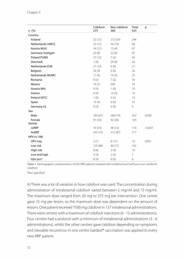
Table 1. Demographic characteristics of 635 RRP patients treated with (cidofovir) and without (non cidofovir) cidofovir.
*Not specified
A) There was a lot of variation in how cidofovir was used. The concentration during
administration of intralesional cidofovir varied between 5 mg/ml and 75 mg/ml.
The maximum dose ranged from 20 mg to 375 mg per intervention. One centre
gave 25 mg per lesion, so the maximum dose was dependent on the amount of
lesions. One patient received 7500 mg cidofovir in 137 intralesional administrations.
There were centres with a maximum of cidofovir injections (6 - 15 administrations).
Four centres had a protocol with a minimum of intralesional administrations (3 - 6
administrations), whilst the other centres gave cidofovir depending on symptoms
and viewable recurrence. In one centre Gardasil® vaccination was applied to every
new RRP patient.

Safety of intralesional cidofovir
73
5
Five centres routinely checked renal function pre and post cidofovir admission.
Two centres hydrated the patients before giving cidofovir. Two patients received
cidofovir intravenously beside intralesional administrations; they both developed
no side effects.
Side effects other than neutropenia, nephrotoxicity or oncogenesis after the use of
intralesional cidofovir were: diarrhoea in two patients, nausea in one patient and
chronic uveitis in one patient. Elevation of liver enzymes ALT and AST occurred in
two patients (in the first patient ALT and AST increased from respectively 14 and 12
U/L before, to 189 and 102 U/L after intralesional cidofovir; in the second patient
from 18 and 13 U/L before, to 92 and 47 U/L after intralesional cidofovir).
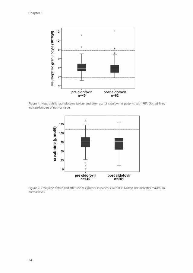
B) When laboratory values tested before and after administration of cidofovir were
compared, no statistically significant differences emerged in blood neutrocyte
count, eGFR or proteinuria (table 2). Creatinine concentrations controlled after
cidofovir treatment were significantly higher compared to those tested before
cidofovir injections. However, there was no significant difference in number of
patients with results exceeding the normal values. Figures 1, 2 and 3 show the
unpaired boxplots with concentrations of neutrophilic granulocytes, creatinine
and eGFR before and after use of cidofovir. None of the patients developed clinical
nephrotoxicity after receiving intralesional cidofovir. One patient had proteinuria
both before and after cidofovir application (30 and 100 mg/l respectively).
Pre cidofovir Post cidofovir
Patients Mean Mean p
Neutrocytes 35 4.1 4.1 0.748
Creatinine 93 68.5 71.6 0.029
eGFR 22 101.0 103.4 0.457
Proteinuria 20 1.55 5.0 0.337
Table 2. Individual dependent laboratory values before and after intralesional cidofovir.
Chapter 5
74
Figure 1. Neutrophilic granulocytes before and after use of cidofovir in patients with RRP. Dotted lines indicate borders of normal value.
Figure 2. Creatinine before and after use of cidofovir in patients with RRP. Dotted line indicates maximum normal level.
Safety of intralesional cidofovir
75
5
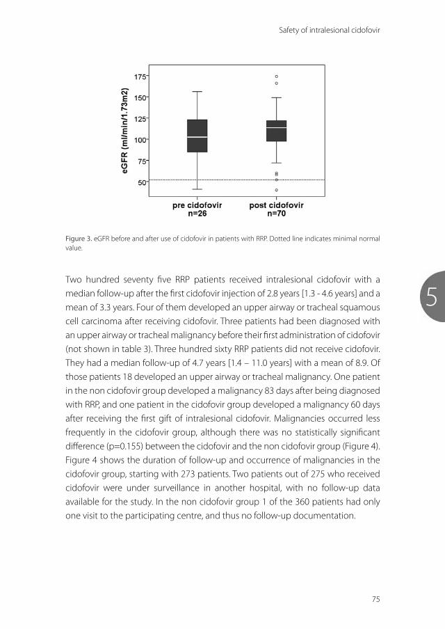
Figure 3. eGFR before and after use of cidofovir in patients with RRP. Dotted line indicates minimal normal value.
Two hundred seventy five RRP patients received intralesional cidofovir with a
median follow-up after the first cidofovir injection of 2.8 years [1.3 - 4.6 years] and a
mean of 3.3 years. Four of them developed an upper airway or tracheal squamous
cell carcinoma after receiving cidofovir. Three patients had been diagnosed with
an upper airway or tracheal malignancy before their first administration of cidofovir
(not shown in table 3). Three hundred sixty RRP patients did not receive cidofovir.
They had a median follow-up of 4.7 years [1.4 – 11.0 years] with a mean of 8.9. Of
those patients 18 developed an upper airway or tracheal malignancy. One patient
in the non cidofovir group developed a malignancy 83 days after being diagnosed
with RRP, and one patient in the cidofovir group developed a malignancy 60 days
after receiving the first gift of intralesional cidofovir. Malignancies occurred less
frequently in the cidofovir group, although there was no statistically significant
difference (p=0.155) between the cidofovir and the non cidofovir group (Figure 4).
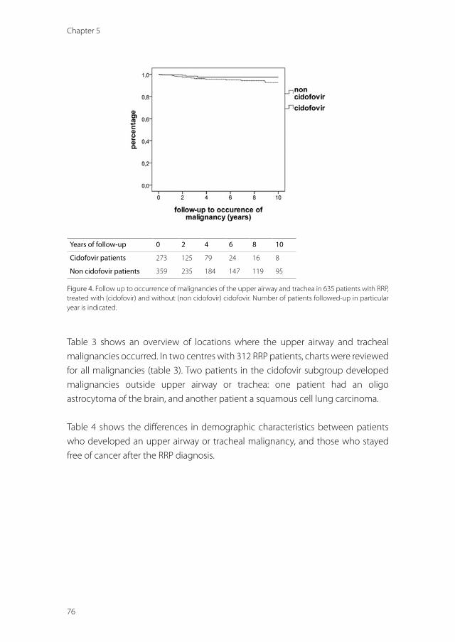
Figure 4 shows the duration of follow-up and occurrence of malignancies in the
cidofovir group, starting with 273 patients. Two patients out of 275 who received
cidofovir were under surveillance in another hospital, with no follow-up data
available for the study. In the non cidofovir group 1 of the 360 patients had only
one visit to the participating centre, and thus no follow-up documentation.
Chapter 5
76
Years of follow-up 0 2 4 6 8 10
Cidofovir patients 273 125 79 24 16 8
Non cidofovir patients 359 235 184 147 119 95
Figure 4. Follow up to occurrence of malignancies of the upper airway and trachea in 635 patients with RRP, treated with (cidofovir) and without (non cidofovir) cidofovir. Number of patients followed-up in particular year is indicated.
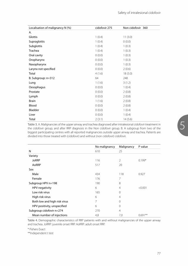
Table 3 shows an overview of locations where the upper airway and tracheal
malignancies occurred. In two centres with 312 RRP patients, charts were reviewed
for all malignancies (table 3). Two patients in the cidofovir subgroup developed
malignancies outside upper airway or trachea: one patient had an oligo
astrocytoma of the brain, and another patient a squamous cell lung carcinoma.
Table 4 shows the differences in demographic characteristics between patients
who developed an upper airway or tracheal malignancy, and those who stayed
free of cancer after the RRP diagnosis.
Safety of intralesional cidofovir
77
5
Localisation of malignancy N (%) cidofovir 275 Non cidofovir 360
A.
Glottis 1 (0.4) 11 (3.0)
Supraglottis 1 (0.4) 0 (0.0)
Subglottis 1 (0.4) 1 (0.3)
Trachea 1 (0.4) 1 (0.3)
Oral cavity 0 (0.0) 1 (0.3)
Oropharynx 0 (0.0) 1 (0.3)
Nasopharynx 0 (0.0) 1 (0.3)
Larynx not specified 0 (0.0) 2 (0.6)
Total 4 (1.6) 18 (5.0)
B. Subgroup: n=312 64 248
Lung 1 (1.6) 3 (1.2)
Oesophagus 0 (0.0) 1 (0.4)
Prostate 0 (0.0) 2 (0.8)
Lymph 0 (0.0) 2 (0.8)
Brain 1 (1.6) 2 (0.8)
Blood 0 (0.0) 2 (0.8)
Bladder 0 (0.0) 1 (0.4)
Liver 0 (0.0) 1 (0.4)
Total 2 (3.1) 14 (5.6)
Table 3. A. Malignancies of the upper airway and trachea diagnosed after intralesional cidofovir treatment in the cidofovir group, and after RRP diagnosis in the Non cidofovir group. B. A subgroup from two of the biggest participating centres with all reported malignancies outside upper airway and trachea. Patients are divided into those treated with (cidofovir) and without (non cidofovir) cidofovir.
No malignancy Malignancy P value
N 610 25
Variety
JoRRP 116 2 0.199*
AoRRP 517 20
Sex
Male 434 118 0.927
Female 176 7
Subgroup HPV n=198 190 8
HPV negativity 6 4 <0.001
Low risk virus 165 0
High risk virus 6 4
Both low and high risk virus 7 0
HPV positivity, unspecified 6 0
Subgroup cidofovir n=274 270 4
Mean number of injections 4,8 7,0 0.691**
Table 4. Demographic characteristics of RRP patients with and without malignancies of the upper airway and trachea. JoRRP: juvenile onset RRP. AoRRP: adult onset RRP.
* Fishers Exact**Independent t test

Chapter 5
78
Discussion
To the best of our knowledge, this is the largest retrospective case study describing
the side effects of intralesional cidofovir in RRP patients. This study shows that
the use of cidofovir varies between different countries. Due to differences in
the treatment of RRP patients the ELS will develop treatment guidelines for RRP.
There are a lot of case series that show significantly better results after cidofovir.
In those studies there is often no control group and no reference to the fact that
the natural course of RRP decreases in severity (33). The only randomized double
blind placebo controlled study showed a significant better improvement in voice
handicap index in the cidofovir group (34). This study did not show any differences
in Derkay severity score or surgical interventions. Placebo controlled RCT with
other commonly used adjuvant medical treatment as interferon or bevacizumab
do not exist.
Gardasil® seems highly effective as prevention against HPV related diseases.
Vaccination was associated with reduced risk of subsequent low risk HPV diseases
by 60.3% (35). Pawlita et al. showed in two patients only a humoral immune
response after vaccination with Gardasil® (36). These results with Gardasil® seems
to be very encouraging. More research is needed to find clinical benefits in RRP
patients. Predisposition of males in RRP patients is a well-known phenomenon
(1,4,5). In this study the predisposition was particularly seen in the AoRRP group,
and to a lesser extent in the JoRRP patient. There is no explanation for the fact that
there are statistically more women in the cidofovir group. An occurrence of more
JoRRP in the cidofovir group can be explained by the fact that JoRRP is associated
with a worse clinical course (37), and therefore JoRRP patients are more likely to
receive cidofovir.
Some side effects not related to neutropenia, nephrotoxicity or malignancies may
have been missed by the questionnaire because it did not specifically ask for every
possible side effect. However, we found no evidence of other encountered side
effects during the use of intralesional cidofovir in RRP patients.
Limitations of this study include those inherent to a retrospective analysis. Because
of the small amount of patients per centers, statistic correction for the multicenter
character was impossible. A concern of this study was that a few patients were
Safety of intralesional cidofovir
79
5
only admitted to the participating centres for cidofovir treatment, and thereafter
were treated in other hospitals. Although severe side effects should normally be
reported to the hospital in which patients received cidofovir, it cannot be assured
that all neutropenia, renal toxicity, and malignancies have come to our attention.
Multicenter studies have the advantage that a large patient population can be
included. However, in this study there are also some disadvantages. In the 16
participating centres, 16 different otorhinolaryngologists reviewed all their RRP
patient charts. Considering the observers bias it would be better for the data
consistency to have one person go through all patient charts. The bias was managed
by attaching a manual to make the observations of the otorhinolaryngologists
as objective as possible. To search for additional adverse events, contacting all
RRP patients personally is possible. However the risk of a selection bias would be
inevitable, since patients with an extensive disease are more likely to reply. The
laboratory tests were done in different laboratories all over the world. Although
this can cause bias, the laboratorial tests were simple tests and no differences were
to be expected on the basis of these findings between the cidofovir group and non
cidofovir group. The serum creatinine levels are time, sex and person dependent.
Their levels alone are difficult to interpret. That is why eGFR and proteinuria are
included to describe the renal function. Beside the increased serum creatinine
levels there were more patients with an increasing than decreasing serum
creatinine level. In this study it seems not clinically relevant because there were no
significant differences in the number of patients with results exceeding the normal
values, and there were no patients with clinical signs of impaired renal function.
However these measurements give enough reason to monitor renal function
during the use of intralesional cidofovir. There were more patients with a decrease
than an increase of neutrocyte count after the use of cidofovir. This has no clinical
significance as the neutrocyte count remains the same within the normal limits.
In this study, a wide range of normal values was used. However, even with smaller
ranges of normal values, there were no differences between laboratory values.
A prospective study with controlled measurements of laboratory values during
intralesional cidofovir is recommended.
Two patients developed an early malignancy (one in the cidofovir group and one
in the non cidofovir group). It might be possible that they were misdiagnosed or
that the malignancy already occurred before cidofovir therapy. Retrospectively the
cause of the malignancy could not be determined.
Chapter 5
80
Table 4 shows a significant difference in the distribution of HPV types between
the malignancy group and the non malignancy group. Malignancy seems to be
more common in HPV negative patients and in patients with high risk HPV. These
results should be interpreted carefully because different HPV typing systems
(ISH, PCR) were used, and the participating centers tested different HPV types.
Skepticism about the correct diagnosis of patients with HPV negative RRP should
be deliberated. Because table 4 raises questions, and there is no consensus about
HPV typing and the malignant transformation, the ELS RRP study group decided
to conduct further research.
Conclusion
Within laryngology, the use and application of cidofovir differs from other medical
specialties. The dosage of cidofovir is much lower and has another route of
administration than that used in other specialties (e.g. ophthalmology, internal
medicine). Our study, concerning the use of cidofovir therapy in RRP, found no
further evidence of side effects comparable to those described in the newsletter
from its manufacturer. However, only long surveillance can determine whether or
not RRP patients with previous cidofovir therapy have increased risk of malignant
transformation compared to RRP patients without cidofovir therapy.
Cidofovir is one of the mainstays in additional treatment against RRP. This study
shows that there is a worldwide variation of intralesional use of cidofovir in RRP
patients. After use of intralesional cidofovir a statistically significant increase in the
creatinine level was found. The change of the values has no clinical significance
as the values remain within normal limits. Although this retrospective patient
chart review found no clinical evidence for more nephrotoxic, neutropenic or
oncogenic side effects after the use of intralesional cidofovir in patients with RRP
caution should be exercised and laboratory values should be monitored before
and after cidofovir. More prospective controlled research is necessary to determine
the safety and clinical impact of intralesional cidofovir.
Safety of intralesional cidofovir
81
5
References
(1) Derkay CS. Task-Force on Recurrent Respiratory Papillomas - A Preliminary-Report. Archives of Otolaryngology-Head & Neck Surgery 1995 12;121(12):1386-1391.
(2) Armstrong LR, Derkay CS, Reeves WC. Initial results from the national registry for juvenile-onset recurrent respiratory papillomatosis. RRP Task Force. Arch Otolaryngol Head Neck Surg 1999 07;125(7):743-748.
(3) Omland T, Akre H, Vardal M, Brondbo K. Epidemiological aspects of recurrent respiratory papillomatosis: A population-based study. Laryngoscope 2012 Jul;122(7):1595-1599.
(4) Armstrong LR, Preston EJ, Reichert M, Phillips DL, Nisenbaum R, Todd NW, et al. Incidence and prevalence of recurrent respiratory papillomatosis among children in Atlanta and Seattle. Clin Infect Dis 2000 Jul;31(1):107-109.
(5) Lindeberg H, Elbrond O. Laryngeal papillomas: the epidemiology in a Danish subpopulation 1965-1984. Clin Otolaryngol Allied Sci 1990 Apr;15(2):125-131.
(6) Shah KV, Stern WF, Shah FK, Bishai D, Kashima HK. Risk factors for juvenile onset recurrent respiratory papillomatosis. Pediatr Infect Dis J 1998 May;17(5):372-376.
(7) Kashima HK, Shah F, Lyles A, Glackin R, Muhammad N, Turner L, et al. A comparison of risk factors in juvenile-onset and adult-onset recurrent respiratory papillomatosis. Laryngoscope 1992 Jan;102(1):9-13.
(8) Wiatrak BJ, Wiatrak DW, Broker TR, Lewis L. Recurrent respiratory papillomatosis: a longitudinal study comparing severity associated with human papilloma viral types 6 and 11 and other risk factors in a large pediatric population. Laryngoscope 2004 Nov;114(11 Pt 2 Suppl 104):1-23.
(9) Penaloza-Plascencia M, Montoya-Fuentes H, Flores-Martinez SE, Fierro-Velasco FJ, Penaloza-Gonzalez JM, Sanchez-Corona J. Molecular identification of 7 human papillomavirus types in recurrent respiratory papillomatosis. Arch Otolaryngol Head Neck Surg 2000 Sep;126(9):1119-1123.
(10) Smith EM, Pignatari SS, Gray SD, Haugen TH, Turek LP. Human papillomavirus infection in papillomas and nondiseased respiratory sites of patients with recurrent respiratory papillomatosis using the polymerase chain reaction. Arch Otolaryngol Head Neck Surg 1993 May;119(5):554-557.
(11) Rihkanen H, Aaltonen LM, Syrjanen SM. Human papillomavirus in laryngeal papillomas and in adjacent normal epithelium. Clin Otolaryngol Allied Sci 1993 Dec;18(6):470-474.
(12) Leemans CR, Braakhuis BJ, Brakenhoff RH. The molecular biology of head and neck cancer. Nat Rev Cancer 2011 01;11(1474-175; 1):9-22.
(13) Brandwein-Gensler MS, Mahadevia P, Gnepp DR. Nonsquamous pathologic diseases of the hypopharynx, larynx, and trachea. In: Gnepp DR, editor. Diagnostic surgical pathology of the head and neck: Saunders Elsevier; 2009.
(14) Sajan JA, Kerschner JE, Merati AL, Osipov V, Szabo S, Blumin JH. Prevalence of dysplasia in juvenile-onset recurrent respiratory papillomatosis. Arch Otolaryngol Head Neck Surg 2010 01;136(1538-361; 1):7-11.
(15) Gerein V, Rastorguev E, Gerein J, Draf W, Schirren J. Incidence, age at onset, and potential reasons of malignant transformation in recurrent respiratory papillomatosis patients: 20 years experience. Otolaryngol Head Neck Surg 2005 Mar;132(3):392-394.
(16) Preuss SF, Klussmann JP, Jungehulsing M, Eckel HE, Guntinas-Lichius O, Damm M. Long-term results of surgical treatment for recurrent respiratory papillomatosis. Acta Otolaryngol 2007 Nov;127(11):1196-1201.
(17) Silver RD, Rimell FL, Adams GL, Derkay CS, Hester R. Diagnosis and management of pulmonary metastasis from recurrent respiratory papillomatosis. Otolaryngol Head Neck Surg 2003 Dec;129(6):622-629.
(18) Snoeck R, Andrei G, De Clercq E. Specific therapies for human papilloma virus infections. Curr Opin Infect Dis 1998 Dec;11(6):733-737.
(19) Cundy KC. Clinical pharmacokinetics of the antiviral nucleotide analogues cidofovir and adefovir. Clin Pharmacokinet 1999 Feb;36(2):127-143.
(20) Naiman AN, Roger G, Gagnieu MC, Bordenave J, Mathaut S, Ayari S, et al. Cidofovir plasma assays after local injection in respiratory papillomatosis. Laryngoscope 2004 Jul;114(7):1151-1156.
(21) Shehab N, Sweet BV, Hogikyan ND. Cidofovir for the treatment of recurrent respiratory papillomatosis: a review of the literature. Pharmacotherapy 2005 Jul;25(7):977-989.
Chapter 5
82
(22) Broekema FI, Dikkers FG. Side-effects of cidofovir in the treatment of recurrent respiratory papillomatosis. Eur Arch Otorhinolaryngol 2008 08;265(8):871-879.
(23) Donne AJ, Hampson L, He XT, Day PJ, Salway F, Rothera MP, et al. Potential risk factors associated with the use of cidofovir to treat benign human papillomavirus-related disease. Antivir Ther 2009;14(7):939-952.
(24) Bielamowicz S, Villagomez V, Stager SV, Wilson WR. Intralesional cidofovir therapy for laryngeal papilloma in an adult cohort. Laryngoscope 2002 Apr;112(4):696-699.
(25) Naiman AN, Ceruse P, Coulombeau B, Froehlich P. Intralesional cidofovir and surgical excision for laryngeal papillomatosis. Laryngoscope 2003 Dec;113(12):2174-2181.
(26) Pransky SM, Albright JT, Magit AE. Long-term follow-up of pediatric recurrent respiratory papillomatosis managed with intralesional cidofovir. Laryngoscope 2003 Sep;113(9):1583-1587.
(27) Dikkers FG. Treatment of recurrent respiratory papillomatosis with microsurgery in combination with intralesional cidofovir - a prospective study. Eur Arch Otorhinolaryngol 2006;263:440-443.
(28) Tanna N, Sidell D, Joshi AS, Bielamowicz SA. Adult intralesional cidofovir therapy for laryngeal papilloma: a 10-year perspective. Arch Otolaryngol Head Neck Surg 2008 May;134(5):497-500.
(29) Wierzbicka M, Jackowska J, Bartochowska A, Jozefiak A, Szyfter W, Kedzia W. Effectiveness of cidofovir intralesional treatment in recurrent respiratory papillomatosis. Eur Arch Otorhinolaryngol 2011 Apr 26.
(30) Gillen D. Direct Healthcare Professional Communication regarding serious adverse reactions following off-label use of Vistide. 2011; Available at: http://www.cbg-meb.nl/NR/rdonlyres/FFB51936-EC22-4180-A213-9E907F06A774/0/VistideDHPCletterJanuary2011.pdf. Accessed 1/12, 2011.
(31) Tjon Pian Gi RE, Dietz A, Djukic V, Eckel HE, Friedrich G, Golusinski W, et al. Treatment of recurrent respiratory papillomatosis and adverse reactions following off-label use of cidofovir (Vistide((R))). Eur Arch Otorhinolaryngol 2012 Feb;269(2):361-362.
(32) McAuley D. Conventional Units - International Units. 04-17-2012; Available at: http://www.globalrph.com/conv_si.htm.
(33) Silverberg MJ, Thorsen P, Lindeberg H, Ahdieh-Grant L, Shah KV. Clinical course of recurrent respiratory papillomatosis in Danish children. Arch Otolaryngol Head Neck Surg 2004 Jun;130(6):711-716.
(34) McMurray JS, Connor N, Ford CN. Cidofovir efficacy in recurrent respiratory papillomatosis: a randomized, double-blind, placebo-controlled study. Ann Otol Rhinol Laryngol 2008 Jul;117(7):477-483.
(35) Joura EA, Garland SM, Paavonen J, Ferris DG, Perez G, Ault KA, et al. Effect of the human papillomavirus (HPV) quadrivalent vaccine in a subgroup of women with cervical and vulvar disease: retrospective pooled analysis of trial data. BMJ 2012 Mar 27;344:e1401.
(36) Pawlita M, Gissmann L. Recurrent respiratory papillomatosis: indication for HPV vaccination? Dtsch Med Wochenschr 2009 Apr;134 Suppl 2:S100-2.
(37) Buchinsky FJ, Donfack J, Derkay CS, Choi SS, Conley SF, Myer CM,III, et al. Age of child, more than HPV type, is associated with clinical course in recurrent respiratory papillomatosis. PLoS One 2008;3(5):e2263.


Recommended