Massachusetts Institute of TechnologyMassachusetts Institute of TechnologyHarvard Medical SchoolHarvard Medical School
Brigham and Women’s HospitalBrigham and Women’s HospitalVA Boston Healthcare SystemVA Boston Healthcare System
2.79J/3.96J/BE.441/HST522J2.79J/3.96J/BE.441/HST522J
TISSUE TYPESTISSUE TYPES
M. Spector, Ph.D. and I.V. Yannas, Ph.D. M. Spector, Ph.D. and I.V. Yannas, Ph.D.
Harvard-MIT Division of Health Sciences and TechnologyHST.523J: Cell-Matrix MechanicsProf. Myron Spector

The Cell and Its Membrane MoleculesThe Cell and Its Membrane Molecules
Figures by MIT OCW. After Darnell et al., Molecular Cell Biology, 1990.

Cryo-electron tomography image of the actin in a cell.
Actin (red), membrane (blue), and ribosomes green.
O Medalia, Sci. 298:1209 (2002)
Photos removed for copyright reasons.
Image showing the trangular structures and nodal points fomred by actin filaments (green). Nucleus (blue).
Sci 292:1047 (2001)
Photo removed for copyright reasons.

Viewing Histological SectionsEffects of the Plane of Sectioning
Figure by MIT OCW. After "Histology." General Biology II Laboratory website, Purchase College, StateUniversity of New York (http://www.ns.purchase.edu/biology/bio1560lab/histology-1.htm)
TISSUE CLASSIFICATIONTISSUE CLASSIFICATION
Connective TissueConnective TissueEpitheliaEpitheliaMuscleMuscleNerveNerve

TISSUE CLASSIFICATIONTISSUE CLASSIFICATION
Connective TissueConnective TissueEpitheliaEpitheliaMuscleMuscleNerveNerve
Matrix and cell continuousMatrix and cell continuous
Cell continuousCell continuousCells surrounded by basal Cells surrounded by basal lamina (basement membrane)lamina (basement membrane)

Connective Tissue
Diagrams removed for copyright reasons.
Sketches from Illustrated Physiology, AB McNaughtand R Callander, Williams and Wilkins, 1967

http://cal.vet.upenn.edu/histo/connective/connective.html
Connective Tissues
Photo removed for copyright reasons.
Loose and dense connective tissue from a cow's planum.

Loose Connective Tissue
Figure removed for copyright reasons.
Ciba Collection, FH Netter, 1987

Dense Connective Tissue
Figure removed for copyright reasons.
Ciba Collection, FH Netter, 1987

Connective Tissue: Adipose Tissue (Fat)
Diagram and photo removed for
copyright reasons.

Connective Tissue: Bone
Figure removed for copyright reasons.
Ciba Collection, FH Netter, 1987

Connective Tissue: Cartilage
Diagram removed for copyright reasons.

http://cal.vet.upenn.edu/histo/cartilage/cartilage.html
Connective Tissue: Cartilage
Hyaline Cartilage: Trachea Elastic Cartilage: Epiglottus
Photo removed for copyright reasons.
Photo removed for copyright reasons.
Fibrocartilage
Photo removed for copyright reasons.

Epithelia
Diagrams removed for copyright reasons.

http://www.uoguelph.ca/zoology/devobio/210labs/epithelial1.html
Simple Squamous Epithelium(chick blastodisc at about 33 hours of incubation )
Top View Cross-Sectional View
Photo removed for copyright reasons.
Photo removed for copyright reasons.
Simple squamous epithelium, which generally occurs as a thin sheet-like layer allowing for minimal resistance to diffusion, is also been called "pavement” epithelium, because it can look like like paving stones as seen from above. Examples include the linings of the peritoneal, pleural and pericardial cavities. Other places simple squamous epithelium can be found include: the glomerulus of the kidney, the walls of capillaries, and the alveoli of the lungs.
http://www.uoguelph.ca/zoology/devobio/210labs/epithelial1.html
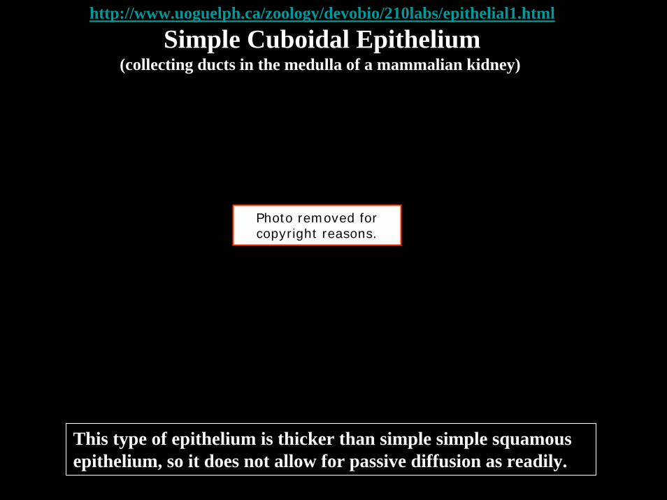
Simple Cuboidal Epithelium(collecting ducts in the medulla of a mammalian kidney)
Photo removed for copyright reasons.
This type of epithelium is thicker than simple simple squamousepithelium, so it does not allow for passive diffusion as readily.

http://www.uoguelph.ca/zoology/devobio/210labs/epithelial1.html
Simple Columnar Epithelium(small intestine)
Photo removed for copyright reasons.
Since columnar cells are quite thick, they do not readily allow passive diffusion. As a result, these cells use active transport to move nutrients through them from the intestine to the blood. This is what we commonly call "absorption." To help with this, they have numerous microvilli on their apical (lumenal) surface, which increases their surface area to allow for greater absorption.

http://cal.vet.upenn.edu/histo/epithelium/epithelium.html
Simple Columnar Epithelium
Photo removed for copyright reasons.
This is a section through the edge of a gallbladder. There is a layer of simple columnar epithelium overlying the connective tissue as indicated by the arrows.

http://cal.vet.upenn.edu/histo/epithelium/epithelium.html
Stratified Squamous Epithelium
Photo removed for copyright reasons.
This is an example of stratified squamous epithelium from the esophagus of a cat. Arrows show nuclei of the outermost layer. This is normal for mucosa. Most stratified squamous cells in other areas, such as skin, lose their nuclei by the time they approach the outermost layers.

http://www.uoguelph.ca/zoology/devobio/210labs/epithelial1.html
Stratified Squamous Epithelium(epidermis)
Photo removed for copyright reasons.
The cells of the basal layer of the epidermis (closest to the dermis) are cuboidal to columnar in shape. These cells are actively mitotic, producing new cells that get pushed upward into the overlying layers. As these cells are pushed up, they become flatter and longer taking on the typical squamous shape. When the cells reach the top, they are sloughed off and replaced by cells from below. The dermis which underlies the epidermis is composed of a dense, irregular connective tissue, which we will see again later.
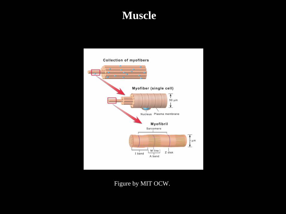
Muscle
Figure by MIT OCW.

http://www.uoguelph.ca/zoology/devobio/210labs/epithelial1.html
MuscleSmooth (Involuntary) Muscle Striated (Skeletal; Vountary) Muscle
Photo removed for copyright reasons.
Photo removed for copyright reasons.
Cardiac Muscle
Photo removed for copyright reasons.

Skeletal Muscle
Diagrams removed for copyright reasons.
Ciba Collection, FH Netter, 1987

Nerve
Figure by MIT OCW. After McNaught and Callander, Illustrated Physiology, Williams and Wilkins, 1967.

http://www.uoguelph.ca/zoology/devobio/210labs/epithelial1.html
Nerve
Photo removed for copyright reasons.
An isolated nerve cell - neuron (large arrow) - from a mammalian spinal cord showing and the nuclei of the surrounding neuroglial cells (small arrows). Note the numerous cytoplasmic extensions emanating from the neuronal cell body and the size of the neuron compared with the neuroglial cells.

Peripheral Nerve: Rat Sciatic
Photo removed for copyright reasons.
Molecular Cell Biology, J Darnell, et al., 1990

http://cal.vet.upenn.edu/histo/nerves/nerves.html
Nerve
Photo removed for copyright reasons.
This is a myelinated nerve from the thoracic wall. A indicates the myelin sheath around the actual nerve fibers (B).

TISSUE CLASSIFICATIONTISSUE CLASSIFICATION
Connective TissueConnective TissueSynthesize and maintain a structurally competent ECM Synthesize and maintain a structurally competent ECM
(including a supporting and connecting framework for (including a supporting and connecting framework for all other tissue types); matrix and cell continuousall other tissue types); matrix and cell continuous
Muscle Cells Muscle Cells Contraction; cell continuous, BMContraction; cell continuous, BM
EpitheliaEpitheliaLining and secretory cells; cell continuous, BMLining and secretory cells; cell continuous, BM
NerveNerveVoltage conduction; cell continuous, BMVoltage conduction; cell continuous, BM
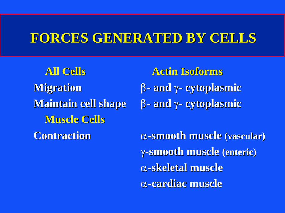
FORCES GENERATED BY CELLSFORCES GENERATED BY CELLS
All CellsAll Cells Actin IsoformsActin IsoformsMigrationMigration ββ-- and and γγ-- cytoplasmiccytoplasmicMaintain cell shapeMaintain cell shape ββ-- and and γγ-- cytoplasmiccytoplasmic
Muscle CellsMuscle CellsContractionContraction αα--smooth muscle smooth muscle (vascular)(vascular)
γγ--smooth muscle smooth muscle (enteric)(enteric)
αα--skeletal muscleskeletal muscleαα--cardiac musclecardiac muscle

TISSUE CLASSIFICATIONTISSUE CLASSIFICATION
Connective Tissue CellsConnective Tissue CellsMuscle Cells (contractile cells)Muscle Cells (contractile cells)
skeletalskeletal αα--skeletal actinskeletal actincardiaccardiac αα--cardiac actincardiac actinsmooth musclesmooth muscle αα-- and and γγ--smooth muscle actinsmooth muscle actin
Epithelial CellsEpithelial CellsNerve CellsNerve Cells
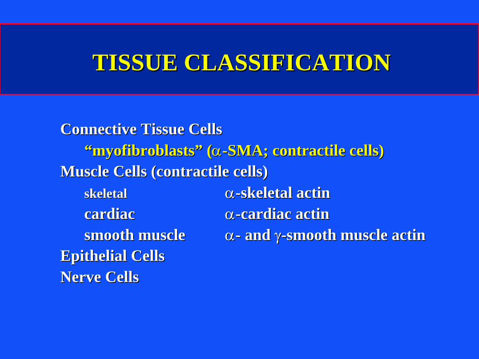
TISSUE CLASSIFICATIONTISSUE CLASSIFICATION
Connective Tissue CellsConnective Tissue Cells““myofibroblastsmyofibroblasts”” ((αα--SMA; contractile cells)SMA; contractile cells)
Muscle Cells (contractile cells)Muscle Cells (contractile cells)skeletalskeletal αα--skeletal actinskeletal actincardiaccardiac αα--cardiac actincardiac actinsmooth musclesmooth muscle αα-- and and γγ--smooth muscle actinsmooth muscle actin
Epithelial CellsEpithelial CellsNerve CellsNerve Cells

CONNECTIVE TISSUE CELLS THAT CAN CONNECTIVE TISSUE CELLS THAT CAN EXPRESS EXPRESS αα--SMOOTH MUSCLE ACTINSMOOTH MUSCLE ACTIN
Articular chondrocyteArticular chondrocyteOsteoblastOsteoblastMeniscus fibroblast and fibrochondrocyteMeniscus fibroblast and fibrochondrocyteIntervertebral disc fibroblast and Intervertebral disc fibroblast and
fibrochondrocytefibrochondrocyteLigament fibroblastLigament fibroblastTendon fibroblastTendon fibroblastSynovial cellSynovial cellMesenchymal stem cellMesenchymal stem cell
M. Spector, M. Spector, Wound Repair Wound Repair RegenRegen. 9:11. 9:11--18(2001)18(2001)
Recommended