1
BNL-112534-2016-JA File # 93466
Three-Phase 3D Reconstruction of a
LiCoO2 Cathode via FIB-SEM Tomography
Zhao Liu, Yu-chen Karen Chen-Wiegart,
Jun Wang, Scott A. Barnett, Katherine T. Faber
Submitted to Microscopy and Microanalysis
February 2016
Photon Sciences Department
Brookhaven National Laboratory
U.S. Department of EnergyUSDOE Office of Science (SC),
Basic Energy Sciences (BES) (SC-22)
Notice: This manuscript has been authored by employees of Brookhaven Science Associates, LLC under Contract No. DE- SC0012704 with the U.S. Department of Energy. The publisher by accepting the manuscript for publication acknowledges that the United States Government retains a non-exclusive, paid-up, irrevocable, world-wide license to publish or reproduce the published form of this manuscript, or allow others to do so, for United States Government purposes.

2
DISCLAIMER
This report was prepared as an account of work sponsored by an agency of the United States Government. Neither the United States Government nor any agency thereof, nor any of their employees, nor any of their contractors, subcontractors, or their employees, makes any warranty, express or implied, or assumes any legal liability or responsibility for the accuracy, completeness, or any third party’s use or the results of such use of any information, apparatus, product, or process disclosed, or represents that its use would not infringe privately owned rights. Reference herein to any specific commercial product, process, or service by trade name, trademark, manufacturer, or otherwise, does not necessarily constitute or imply its endorsement, recommendation, or favoring by the United States Government or any agency thereof or its contractors or subcontractors. The views and opinions of authors expressed herein do not necessarily state or reflect those of the United States Government or any agency thereof.

# Corresponding author current address: California Institute of Technology, MC 138-78, Pasadena, CA, 91125, USA
Three-Phase 3D Reconstruction of a LiCoO2 Cathode via FIB-SEM Tomography
Zhao Liua, Yu-chen Karen Chen-Wiegartb, Jun Wangb, Scott A. Barnetta*, Katherine T. Fabera*#
a Department of Materials Science and Engineering, Northwestern University, Evanston, IL, 60208,
USA
b Photon Science Directorate, Brookhaven National Laboratory, Upton, NY, 11973, USA
*Corresponding authors, Email: [email protected]; [email protected]
Abstract:
Three-phase three-dimensional (3D) microstructural reconstructions of lithium-ion battery electrodes
are critical input for 3D simulations of electrode lithiation/delithiation, which provide a detailed
understanding of battery operation. In this report, 3D images of a LiCoO2 electrode are achieved
using focused ion beam-scanning electron microscopy (FIB-SEM), with clear contrast among the
three phases: LiCoO2 particles, carbonaceous phases (carbon and binder) and the electrolyte space.
The good contrast was achieved by utilizing an improved FIB-SEM sample preparation method that
combined infiltration of the electrolyte space with a low-viscosity silicone resin and triple ion-beam
polishing. Morphological parameters quantified include phase volume fraction, surface area, feature
size distribution, connectivity, and tortuosity. Electrolyte tortuosity was determined using two
different geometric calculations that were in good agreement. The electrolyte tortuosity distribution
versus position within the electrode was found to be highly inhomogeneous; this will lead to
inhomogeneous electrode lithiation/delithiation at high C-rates that could potentially cause battery
degradation.
Keywords: Li-ion batteries, three-phase 3D reconstruction, FIB-SEM tomography, cathode,
microstructure
2
1. Introduction
Lithium-ion battery electrode performance depends not only on chemistry but on microstructure,
such that an improved understanding of the electrode microstructure-performance relationship is
needed (Fergus, 2010; J. Vetter, et al., 2005). Three-dimensional (3D) characterization methods such
as focused ion beam – scanning electron microscopy (FIB-SEM) and X-ray computed
micro/nanotomography (XCT) have gained significant attention in the past few years due to their
advantages in effectively quantifying electrode microstructural parameters including grain size,
phase volume, surface area, phase connectivity, and tortuosity (Babu, et al., 2015; Chen-Wiegart, et
al., 2013; Cooper, et al., 2014; Ebner, et al., 2013b; Ebner, et al., 2013c; Ender, et al., 2012; Ender, et
al., 2011; Hutzenlaub, et al., 2012; Liu, et al., 2013; Shearing, et al., 2010; Wang, et al., 2014;
Wilson, et al., 2011; Wu & Jiang, 2013). Moreover, measured 3D electrode microstructures can be
used as input for 3D model simulations of lithiation/delithiation (A. H. Wiedemann, et al., 2013;
Hutzenlaub, et al., 2014; Malavé, et al., 2014; Yan, et al., 2013), providing more detailed information
and more accurate predictions of electrode electrochemical processes compared with well-known
low-dimensional models (e.g. Newman-type models (M. Doyle & Newman, 1995; M. Doyle, et al.,
1993)) or 3D models based on ideal spherical particles (Goldin, et al., 2012).
Typical Li-ion battery electrodes contain three phases with different functions. The active material
acts as reservoir for lithiation/delithiation; the electrolyte-filled pores serve as Li-ion pathways, while
the carbon and binder (CB) phase provides electronic conductivity. Thus, the ability to resolve all
three phases in 3D microstructure reconstruction is critical for accurately analyzing electrode
processes (Hutzenlaub, et al., 2014; Zielke, et al., 2014a). In initial reports describing 3D battery
electrode microstructure, the active oxide particles were resolved but the electrolyte and
carbon/binder phases could not be separated since the electrolyte space was filled with a carbon-
based epoxy during imaging (Liu, et al., 2013; Wilson, et al., 2011). Recently, a few methods have

3
been proposed for three-phase reconstructions, but each has limitations during data acquisition and
processing (Babu, et al., 2015; Chen-Wiegart, et al., 2013; Ender, et al., 2012; Ender, et al., 2011;
Hutzenlaub, et al., 2012). In one case using X-ray computed tomography, the three phases were
resolved via Zernike phase contrast imaging (Babu, et al., 2015; Chen-Wiegart, et al., 2013).
However, such data are hard to segment due to the low contrast between each phase, thus limiting
quantitative analysis. For FIB-SEM tomography, there are two approaches for three-phase
reconstruction to date. Hutzenlaub et al. performed serial-sectioning directly on the electrode
without using any filling materials (Hutzenlaub, et al., 2012). This technique is an efficient method
in terms of sample preparation and phase contrast is observable between empty pores and
surrounding materials. However, severe electron charging on the FIB-milled surfaces, together with
curtaining effects caused by the rough top surface, can induce image artifacts, and hence, require
significant post-processing effort in manual segmentation. The alternative method is to infiltrate
filling materials into the porous electrode for contrast enhancement. Instead of using carbon-based
epoxy resin, which does not allow differentiation between resin-filled pores and CB phase, silicone
resin is employed to obtain considerable atomic Z-contrast (Ender, et al., 2012; Ender, et al., 2011).
This method effectively avoids the overcharging and curtaining effects. However, attempts to use
this relatively high viscosity silicone resin with the present low-porosity electrodes always led to
incomplete pore infiltration. In addition, the overlap between carbon and resin gray scale frequency
peaks may cause uncertainty during image segmentation. Therefore, a method that incorporates the
advantages of both FIB methods, namely, clear phase contrast with minimal artifacts, is needed for
acquiring high-quality three-phase 3D data sets.
In the present work, an improved method was developed utilizing a low viscosity silicone resin as a
filling material and three-phase 3D reconstruction was demonstrated via FIB-SEM on a commercial
LiCoO2 cathode. The low viscosity resin enables full infiltration of low porosity electrodes and
4
provides good contrast among phases, the latter of which allows for automatic segmentation.
However, it is difficult to mechanically polish samples filled with such a low viscosity material.
Thus, a straightforward sample preparation method using a triple ion-beam cutter was developed to
guarantee a smooth top surface, minimizing image artifacts during data collection. Microstructural
parameters such as phase volume fraction, surface area density, and feature size distribution were
extracted from the as-obtained 3D reconstruction. In addition, electrolyte connectivity, tortuosity,
and tortuosity distribution (tortuosity along the planar section within the electrode) were determined.
The results are quite different than those obtained when the electrolyte and carbon phases are not
resolved, confirming the importance of performing full three-phase imaging of battery electrodes.
2. Materials and Methods
2.1 Sample preparation for FIB-SEM tomography
In present report, a LiCoO2 cathode from a commercial cylindrical 18650 energy cell (Molicel ICR
18650J, Taiwan) was investigated. In order to obtain clear contrast between carbonaceous materials
(including the conducting carbon and binder, CB) and electrolyte space, a commercial two-part
silicone resin (ELASTOSIL RT 604, Wacker, Germany) was used as filling material to infiltrate the
pore regions remaining after the electrolyte was removed from the cathode. It is noteworthy that one
of the biggest advantages of using RT 604 is its low viscosity (800 mPa•s), compared to RT675
viscosity (50,000 mPa•s) used in a previous report (Ender, et al., 2012), which allows the resin to
fully penetrate the entire open pore space. This is critical when infiltrating electrode samples with
small pore sizes. The pristine cell was opened in an argon-filled glove box. After separating the
LiCoO2 cathode from the carbon anode and separator, the cathode was rinsed with dimethyl
carbonate (DMC) to remove the electrolyte as well as any salt residue. The rinsed cathode was then
slowly dried in a glove box for two days and cut into a 1 x 1 cm2 sample with a razor blade for FIB-
SEM tomography. The low viscosity silicone resin was used for filling the porosity in the electrodes.
5
The infiltration process was performed at vacuum levels lower than ~50 mbar to further improve
infiltration. After curing the resin for 24 h at room temperature, samples were roughly cut into cubes
before further polishing. Because the cured silicone resin is too soft for mechanical polishing
(Hardness Shore A 25, ISO 868), the triple ion-beam cutter (Leica EM TIC 3X) was used to obtain a
smooth top surface required for the subsequent FIB milling. During the polishing process via the
triple ion-beam cutter, each ion gun was operated at 4 KV accelerating voltage and 2 mA ion current
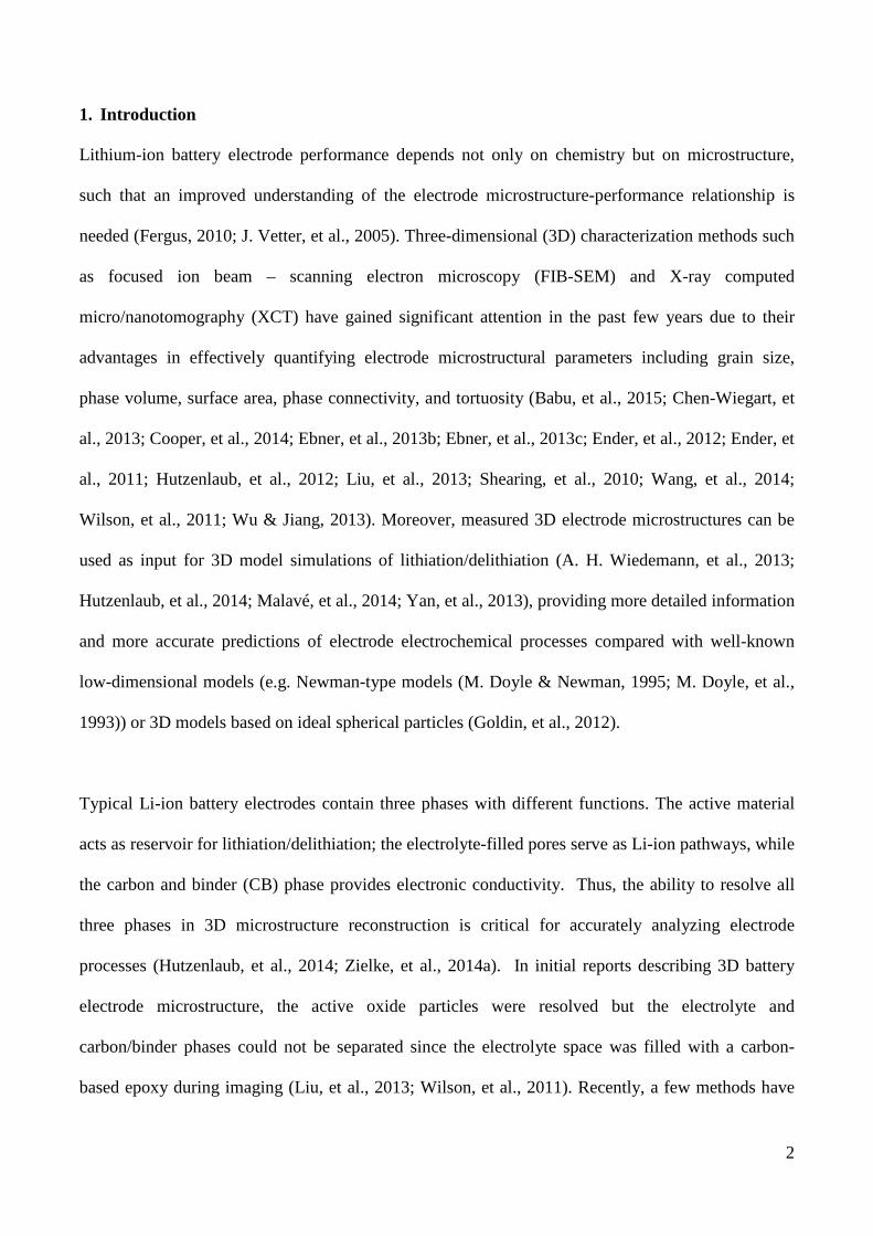
for 8 hours of milling. Figure 1 (a) shows the schematic view of polishing the silicone resin
infiltrated cathode sample via the triple ion-beam cutter. The sample was mounted ~50 µm above the
mask to expose enough space for milling. Triple ion-beams formed a ~100o wide milling section
after cutting the exposed sample and created a smooth region, as shown in Figure 1 (b), magnified
further in Figure 1 (c). Based on these images, the electrode is estimated to be ~ 60 µm thick and the
aluminum current collector is ~20 µm thick. It is clear that the top surface satisfied the requirement
on the surface smoothness, which effectively avoided most of the curtaining effect during the
subsequent FIB milling. After obtaining a smooth top surface with the triple ion-beam cutter, a ~30
nm osmium coating was deposited on the sample to minimize charging during FIB-SEM data
collection.
2.2 FIB-SEM Tomography
An FEI Helios FIB-SEM (FEI Company, OR, USA) was used for serial sectioning and data
collection. A trench (50 x 30 x 15 µm3) was first milled to expose side walls for serial-sectioning
(trench not shown). The pixel size of the SEM images is 31.25 nm, while the slice thickness is 150
nm. A through-the-lens detector (TLD) was used to collect the backscattered electron signal so as to
obtain atomic Z-contrast among phases. As indicated in Figure 1 (c), a typical serial-section area of
50 µm x 45 µm was selected; the slicing direction was parallel to the current collector. Recognizing
the spatial location of the current collector relative to the as-obtained 3D electrode structure is useful

6
in characterizing directional connectivity and tortuosity. In the present case, the current collector is
adjacent to the left YZ plane of the 3D data set. Overall, 268 consecutive images were collected.
After data collection, the 2D image sequences were aligned, cropped and stacked into a 3D
microstructure using a method previously described (J. R. Kremer, et al., 1996; J. R. Wilson, et al.,
2006). A total 3D volume of ~35,000 µm3 was reconstructed for further analysis.
2.3 Image Processing:
In order to quantitatively analyze the 3D reconstructed volume, image segmentation was needed to
attribute different gray scale intensity values to different phases. In the current case, white (gray
scale intensity: 255) is assigned to LiCoO2 particles, with gray (127) to resin-infiltrated porosity and
black (0) to carbon and binder. Firstly, ImageJ software (National Institutes of Health, MD, USA)
was used (Abramoff, et al., 2004) in which a combination of gray scale histogram equalization,
background subtraction, and median filtering was performed to alleviate the shadowing of the FIB-
milled trench wall and any curtaining effects. Then a multi-level image threshold method, Otsu’s
method (Otsu, 1979), was applied to segment a three-phase image. In brief, the as-collected SEM
image was assumed to contain three classes of pixels that follow tri-modal distribution. The
threshold value is then determined by finding maximum inter-class variance between CB/electrolyte
and electrolyte/ LCO phases. Finally, the noise filter built in ImageJ was used on images that needed
further noise reduction. Once segmented, the images were imported into Amira 5.5.0 (FEI
Visualization Sciences Group, MA) for 3D visualization.
2.4 Microstructural Quantification
Microstructural parameters including volume fraction, surface area density, feature size distribution,
connectivity, and tortuosity (τ) were calculated to quantitatively evaluate the electrode microstructure
characteristics (Chen-Wiegart, et al., 2014; J. R. Wilson, et al., 2006; Liu, et al., 2013; Münch &
7
Holzer, 2008; Shanti, et al., 2014). Phase volume fraction (Vf) was calculated by counting voxels of
each phase within the 3D data set. For surface area, both interfacial surface area density (SAI) and
specific surface area density (SAs), also known as the volume specific surface area, were calculated
using a built-in function in IDL software (EXELIS, CO, USA). The SAI is determined by
normalizing the surface area of each phase with total reconstruction volume, while SAs is calculated
by dividing the surface area of each phase by the volume of corresponding phase. Continuous
feature size distributions of all three phases were calculated using the method introduced by Münch
and Holzer (Münch & Holzer, 2008). This algorithm measures the particle size by filling the 3D
volume with a sphere of a given radius. The cumulative size distribution is calculated by
incrementally decreasing the sphere radius, and thus, filling increasingly larger volumes until the
volume of interest is fully occupied. The size distribution of the analyzed volume can also be
calculated by taking the derivative of the cumulative size distribution. This algorithm was
implemented via a lab-made MATLAB code.
Connectivity and tortuosity are two important parameters that represent transport properties of the
electrode, and realistic values of these can only be measured after resolving contrast between
carbonaceous materials and electrolyte regions. The connectivity was determined using the function
“bwlabeln” in MATLAB software (Mathworks, MA, USA), which defines the regions connected to
the opposite face of the current collector (current collector is touching the YZ plane on the left) as
“Percolated”. Those regions which touch other faces but not the percolating volume (not the opposite
face of current collector) are labeled as “Unknown”. Finally, regions that are completely isolated
from the percolating and unknown regions are “Isolated”. By segmenting the electrolyte phase with
this definition, the directional connectivity towards the current collector was calculated via dividing
the segmented phases by total electrolyte volume.
8
For tortuosity calculations, two different methods were used, namely the path length ratio method
(PLR) (Shanti, et al., 2014) and the distance propagation method (DP) (Chen-Wiegart, et al., 2014).
(Schematics of the two algorithms are shown in supplemental information, Figure S1 and S2). The
calculations are limited to the interconnected regions only. Both methods are based on the
geometrical calculations of the effective actual path (Leff) and the Euclidean distance (straight
distance, L) ratio using the simple geometrical definition of tortuosity, τ:
τ = 𝐿𝐿𝑒𝑒𝑒𝑒𝑒𝑒𝐿𝐿
(1)
For the PLR method, the 3D microstructure of the phase of interest was skeletonized into a matrix
containing nodes and pathways that connect neighboring nodes. L is determined by calculating the
straight distance between randomly selected node pairs, and Leff is determined using Dijkstra’s
algorithm (Dijkstra, 1959). In this report, five sets of 1000 nodes per set were selected to measure τ
and its standard deviation. In contrast, using the DP method, a propagation distance map was first
generated within the phase of interest starting from the “seed plane”. After labeling all voxels by
defining specific neighbors (the city-block method was used in current report), the correlations
between Leff and L are then built for extracting τ based on the “average” method (Chen-Wiegart, et
al., 2014). It is noted that both geometrical methods are less computationally intensive than the well-
developed diffusion method (J. R. Wilson, et al., 2006), while still obtaining comparable tortuosity
values.
3. Results and Discussion
3.1 Three-phase 3D reconstruction
Figure 2 summarizes images, chemical identification, and segmentation of the data from the LiCoO2
electrode. A typical 2D cross-section from the 3D data set is shown in Figure 2 (a). With the smooth
top surface created by triple ion-beam cutter, the curtaining effect is much reduced compared to
samples without any filling materials (Hutzenlaub, et al., 2012). The three phases demonstrated clear
9
contrast, namely, white (LiCoO2 active materials), gray (infiltrated silicone resin), and black
(carbonaceous materials including conductive carbon and binder (CB)). Note that the resin-infiltrated
porosity corresponds to the regions that were occupied by the liquid electrolyte, prior to its removal
upon battery disassembly; hence this region will be referred to as either silicone resin or electrolyte
in the following text. No nano-porosity was observed within the CB phase in these images collected
at ~30 nm resolution. Nanoporosity was observed within the CB by utilizing a combination of a
multi-length scale imaging technique and 3D modeling (Stephenson, et al., 2011; Zielke, et al.,
2014a; Zielke, et al., 2014b). However, the emphasis of the present 3D reconstructions was to
measure a large total volume in order to obtain excellent statistics in the larger-scale microstructure
quantification, rather than to resolve nano-scale features.
The energy dispersive X-ray spectroscopy (EDS) mapping (Figure 2 (b)) was performed using the
Integrated Oxford EDS system (equipped on FEI Helios FIB-SEM) to confirm the microstructural
chemical specificity of each phase. The signals from Co, C and Si elements correspond well with
substantiated the above assignments of the LiCoO2 (white), CB (black), and silicone resin (gray),
respectively. A gray scale histogram of the highlighted region in Figure 2(a) is presented as Figure
2(c). The histogram with three well-separated frequency peaks is ideal for using Otsu’s multi-level
method to assign each phase with a specific gray scale intensity value, where white (gray scale
intensity: 255) is assigned to LiCoO2 particles, gray (127) to silicone resin and black (0) to CB.; the
corresponding segmented image is shown in Figure 2 (d). The benefits of applying automatic
segmentation in processing this data set are two-fold. First, it reduces the random errors introduced
by manual segmentation and provides consistency to the entire image processing procedure. Second,
the time required for the automatic segmentation process can be efficiently reduced to a several
hours instead of days for manual segmentation.

10
Figure 3 shows the reconstructed 3D volume after segmenting the whole image volume and stacking
2D image slices. Microstructural parameter results, including volume fraction and surface area of
each phase, are compiled in Table 1. It is apparent that the active LiCoO2 material occupies most of
the reconstructed volume, while the carbon phases have a relatively low volume fraction. In general,
active materials are expected to be more than 70% of the volume in high-energy Li-ion 18650 cells
to achieve high energy density within a single cell (Ender, et al., 2014). In addition, the volume
fractions of CB (10.3%) and electrolyte (12.7%) satisfy the requirements for electron and Li-ion
transport in this product, where the highest C-rate (1 hour for full charge/discharge) recommended is
limited to 1C. The electrolyte volume fraction in such high-energy Li-ion cells is significantly lower
than those of high power Li-ion cells where the electrolyte can occupy up to 38% of the volume to
allow fast Li-ion transport (Ender, et al., 2012; Ender, et al., 2014).
One of the benefits of resolving all three electrode phases is that the interfacial surface areas between
each phase can be accurately determined. If the electrolyte is assumed to only allow for Li+ transport
while the carbon-binder phase only permits electron transport, partial coverage of LiCoO2 by carbon
and binder can effectively enhance the electron transport while blocking the Li+ reaction sites on the
oxide particles, which can cause high transport loss (Hutzenlaub, et al., 2014). In the present case,
the actual available oxide surface area density is 0.36 µm-1, but in cases where carbon was not
differentiated as in prior 3D imaging (Liu, et al., 2013; Wilson, et al., 2011), the apparent oxide
surface area density (the sum of electrolyte/LCO and CB/LCO interfacial areas) for the current data
set, is nearly two times higher, 0.71 µm-1. Consequently, not considering CB coverage on active
materials may lead to significant errors in simulating electrode properties. For example, the CB
coverage presumably impedes the Li+ charge transfer process by reducing the oxide-electrolyte
interfacial area (Malavé, et al., 2014). This may result in an underestimation of the stress and local
Li-ion concentration gradient within particles. In addition, the interface between active materials and

11
CB also plays an important role in generating surface heat during the battery cycling process (Yan, et
al., 2013).
The specific surface area densities (SAS) were also calculated. The largest SAs among three phases is
CB, while LiCoO2 is the smallest, i.e., the CB phase possesses the smallest average feature size and
LiCoO2 the largest, which is also evident in the 3D reconstruction shown in Figure 3. The feature
size distributions, shown in Figure 4, provide a more complete picture of feature sizes. The volume
weighted feature size for LiCoO2, CB and electrolyte are 2.94 µm, 0.64 µm and 0.93 µm,
respectively. That is, the median feature size of LiCoO2 is larger than that of electrolyte and CB,
consistent with the SAs calculations.
2.3 Connectivity and Tortuosity Calculations
Taking advantage of the capability to resolve carbonaceous materials and silicone resin enables the
3D reconstruction of an electrolyte network, important for simulating Li+ transport to oxide particles.
The directional connectivity and tortuosity of the electrolyte phase were determined from the 3D data.
The connectivity of the percolated electrolyte volume toward the current collector is 97.7% with
1.3% unknown and 1% isolated regions (Figure S3, supplemental information). Considering the
errors associated with data processing, the result indicates that there is no isolated electrolyte within
experimental error.
Figure 5 shows electrolyte tortuosity values calculated via the distance propagation (DP) and path
length ratio (PLR) methods (Chen-Wiegart, et al., 2014; Shanti, et al., 2014). Results from the DP
and PLR methods values are in good agreement overall (the former was obtained as the average of
values calculated in three orthogonal directions), yielding a tortuosity of 1.85. The directional
tortuosity values calculated using the DP method suggest tortuosity anisotropy, with tortuosity in the
12
X and Z directions ~18% greater than that in the Y direction. The observation can be explained by
the interplay between anisotropic particle shapes and anisotropic particle alignment during battery
manufacturing (Ebner, et al., 2013a). The calendaring process presses the electrodes towards the
current collector, which causes most of the particles to align along the Y direction, and therefore,
reduce the tortuosity in this direction. It is noted that the absolute value of electrolyte tortuosity in
the current report agrees with experimental or simulated values for similar (not identical) Li-ion
cathodes obtained previously (Hutzenlaub, et al., 2013; Stephenson, et al., 2011; Zielke, et al.,
2014b).
Clearly, If one attempts to calculate the tortuosity directly from FIB-SEM images when the
electrolyte and CB phases are not resolved, as is the case with carbon-based epoxies or unfilled
samples, there is likely to be considerable error. To test this supposition, the tortuosity was calculated
for the electrolyte-plus-CB volume, and was found to be 1.41 (supplemental information, Figure S4
and Table S1), substantially less than 1.85 when the electrolyte and CB phases are resolved. A
similar outcome was noted by Hutzenlaub, et al. (Hutzenlaub, et al., 2013). Such underestimates are
not expected with X-ray tomography or transport measurements.
In addition to the volume-averaged tortuosity, the 3D distance map and tortuosity distribution
provides spatially resolved tortuosity information. Figure 6 (a) shows the 3D distance map of
electrolyte along the X direction, i.e., the direction of Li transport during battery operation. At some
points near the current collector (furthest from the electrolyte), the longest Li diffusion pathway is as
high as 85 µm, significantly larger than the electrode thickness 34. 5 µm. Figure 6 (b) shows the
electrolyte, tortuosity distribution on the YZ plane at X = 34.5 µm (the plane adjacent to the current
collector). Figure 6 (c) plots the corresponding tortuosity histogram. The tortuosity is quite
heterogeneous with values ranging from 2.2 to 1.3. This large heterogeneity may result in

13
inhomogeneous electric-potential and Li content within the electrode, especially during battery
operation at high C-rates. These variations could lead to large local potential deviations from the
average cell potential and consequently large local currents that lead to degradation (A. H.
Wiedemann, et al., 2013).
In order to further illustrate the importance of resolving the carbonaceous phases, we also
determined the tortuosity distribution from the 3D images while intentionally ignoring the contrast
between electrolyte and CB; this yields not only lower propagation distance and tortuosity values,
but also much less heterogeneity, with τ ranging from 1.1 to 1.5 (see Figure S5, supplemental
information). Thus, one cannot obtain meaningful local tortuosity information without resolving
electrolyte and carbonaceous phases.
4. Conclusions
A three-phase 3D reconstruction of a commercial LiCoO2 cathode was demonstrated using FIB-SEM
tomography. Clear contrast between electrolyte and carbonaceous regions was achieved by using
low-viscosity silicone resin to fill the emptied electrolyte regions. Microstructural parameters
including volume fraction, surface area, and feature size distributions of all three phases were
determined. Moreover, the electrolyte connectivity, tortuosity, and tortuosity distributions were
calculated and tortuosity was compared between electrolyte and electrolyte-plus-CB phases to
emphasize the important role of the realistic electrolyte structure in evaluating the electrolyte
transport property. For connectivity calculations, the electrolyte network was found to reach almost
complete percolation (97.4%) towards the current collector. Based on tortuosity calculations, the
electrolyte structure possesses a more tortuous 3D pathway than the electrolyte-plus-CB phases for
Li-ion transport. In addition, the heterogeneous tortuosity distribution observed in the electrolyte

14
structure may result in inhomogeneous charge/discharge states, and consequently cause battery
degradation.
The present study provides a straightforward approach to prepare electrode samples with three-phase
contrast under SEM and can be easily transferred to other battery electrodes. This work demonstrates
the importance of differentiating all three phases in extracting battery microstructure characteristics
and analyzing the microstructure-performance correlations. The as-obtained 3D microstructures can
further be used as exact templates for 3D simulations of battery transport and thermal properties.
Supplemental Information
Supplemental Information is available online or from the author.
Acknowledgements
The authors acknowledge the financial support from the Office of Naval Research Grant #N00014-
12-1-0713 and Northwestern University Cabell Terminal Year Fellowship. We thank Dr. James
W. Fleming for providing the battery samples studied in this report. We also thank Dr. Noah Shanti
and Sarah Miller for help with the tortuosity calculations via the PLR method. The triple-ion cutting
was performed at the Optical Microscopy and Metallography Facility at the Materials Research
Center of Northwestern University. FIB-SEM (FEI) was performed in the EPIC facility of NUANCE
Center at Northwestern University. NUANCE Center is supported by NSF-NSEC, NSF-MRSEC,
Keck Foundation, the State of Illinois, and Northwestern University.
15
References
A. H. WIEDEMANN, G. M. GOLDIN, S. A. BARNETT, H. ZHU & KEE, R.J. (2013). Electrochem Acta Effects of three-dimensional cathode microstructure on the performance of lithium-ion battery cathodes. Electrochemica Acta 88, 580-588.
ABRAMOFF, M.D., MAGALHÃES, P.J. & RAM, S.J. (2004). Image processing with ImageJ. Biophotonics international 11(7), 36-42.
BABU, S.K., MOHAMED, A.I., WHITACRE, J.F. & LITSTER, S. (2015). Multiple Imaging Mode X-ray Computed Tomography for Distinguishing Active and Inactive Phases in Lithium-Ion Battery Cathodes. Journal of Power Sources.
CHEN-WIEGART, Y.-C.K., DEMIKE, R., ERDONMEZ, C., THORNTON, K., BARNETT, S.A. & WANG, J. (2014). Tortuosity characterization of 3D microstructure at nano-scale for energy storage and conversion materials. Journal of Power Sources 249, 349-356.
CHEN-WIEGART, Y.-C.K., LIU, Z., FABER, K.T., BARNETT, S.A. & WANG, J. (2013). 3D analysis of a LiCoO2–Li(Ni1/3Mn1/3Co1/3)O2 Li-ion battery positive electrode using x-ray nano-tomography. Electrochemistry Communications 28, 127-130.
COOPER, S.J., EASTWOOD, D.S., GELB, J., DAMBLANC, G., BRETT, D.J.L., BRADLEY, R.S., WITHERS, P.J., LEE, P.D., MARQUIS, A.J., BRANDON, N.P. & SHEARING, P.R. (2014). Image based modelling of microstructural heterogeneity in LiFePO4 electrodes for Li-ion batteries. Journal of Power Sources 247, 1033-1039.
DIJKSTRA, E.W. (1959). A note on two problems in connexion with graphs. Numerische Mathematik 1, 269-271. EBNER, M., CHUNG, D.-W., GARCÍA, R.E. & WOOD, V. (2013a). Tortuosity Anisotropy in Lithium-Ion Battery
Electrodes. Advanced Energy Materials 4(5), 1301278. EBNER, M., GELDMACHER, F., MARONE, F., STAMPANONI, M. & WOOD, V. (2013b). X-Ray Tomography of Porous,
Transition Metal Oxide Based Lithium Ion Battery Electrodes. Advanced Energy Materials 3(7), 845-850.
EBNER, M., MARONE, F., STAMPANONI, M. & WOOD, V. (2013c). Visualization and quantification of electrochemical and mechanical degradation in Li ion batteries. Science 342(6159), 716-720.
ENDER, M., JOOS, J., CARRARO, T. & IVERS-TIFFEE, E. (2012). Quantitative Characterization of LiFePO4 Cathodes Reconstructed by FIB/SEM Tomography. Journal of the Electrochemical Society 159(7), A972-A980.
ENDER, M., JOOS, J., CARRARO, T. & IVERS-TIFFÉE, E. (2011). Three-dimensional reconstruction of a composite cathode for lithium-ion cells. Electrochemistry Communications 13(2), 166-168.
ENDER, M., JOOS, J., WEBER, A. & IVERS-TIFFÉE, E. (2014). Anode microstructures from high-energy and high-power lithium-ion cylindrical cells obtained by X-ray nano-tomography. Journal of Power Sources 269, 912-919.
FERGUS, J.W. (2010). Recent developments in cathode materials for lithium ion batteries. Journal of Power Sources 195(4), 939-954.
GOLDIN, G.M., COLCLASURE, A.M., WIEDEMANN, A.H. & KEE, R.J. (2012). Three-dimensional particle-resolved models of Li-ion batteries to assist the evaluation of empirical parameters in one-dimensional models. Electrochimica Acta 64, 118-129.
HUTZENLAUB, T., ASTHANA, A., BECKER, J., WHEELER, D.R., ZENGERLE, R. & THIELE, S. (2013). FIB/SEM-based calculation of tortuosity in a porous LiCoO2 cathode for a Li-ion battery. Electrochemistry Communications 27, 77-80.
HUTZENLAUB, T., THIELE, S., PAUST, N., SPOTNITZ, R., ZENGERLE, R. & WALCHSHOFER, C. (2014). Three-dimensional electrochemical Li-ion battery modelling featuring a focused ion-beam/scanning electron microscopy based three-phase reconstruction of a LiCoO2 cathode. Electrochimica Acta 115, 131-139.
HUTZENLAUB, T., THIELE, S., ZENGERLE, R. & ZIEGLER, C. (2012). Three-Dimensional Reconstruction of a LiCoO2 Li-Ion Battery Cathode. Electrochemical and Solid-State Letters 15(3), A33.
J. R. KREMER, D. N. MASTRONARDE & J. R. MCLNTOSH. (1996). Computer Visualization of Three-Dimensional Image Data Using IMOD. Journal of Structural Biology 116, 71-76.
16
J. R. WILSON, W. KOBSIRIPHAT, R. MENDOZA, H.-Y. CHEN, J. M. HILLER, D. J. MILLER, K. THORNTON, P. W. VOORHEES, S. B. ADLER & BARNETT, S.A. (2006). Three-dimensional reconstruction of a solid-oxide fuel-cell anode. Nature Materials 11, 541-544.
J. VETTER, P. NOVAK, M. R. WAGNER, C. VEIT, K. -C. MOLLER, J. O. BESENHARD, M. WINTER, M. WOHLFAHRT-MEHRENS, C. VOGLER & HAMMOUCHE, A. (2005). Ageing mechanisms in Lithiuim ion batteries. Journal of Power Sources 147, 269-281.
LIU, Z., SCOTT CRONIN, J., CHEN-WIEGART, Y.-C.K., WILSON, J.R., YAKAL-KREMSKI, K.J., WANG, J., FABER, K.T. & BARNETT, S.A. (2013). Three-dimensional morphological measurements of LiCoO2 and LiCoO2/Li(Ni1/3Mn1/3Co1/3)O2 lithium-ion battery cathodes. Journal of Power Sources 227, 267-274.
M. DOYLE & NEWMAN, J. (1995). The use of mathematical modeling in the design of lithium/polymer battery systems. Electrochimica Acta 40(13), 2191-2196.
M. DOYLE, T. F. FULLER & NEWMAN, J. (1993). Modeling of Galvanostatic Charge and Discharge of the Lithium/Polymer/Insertion Cell. Journal of the Electrochemical Society 140(6), 1526-1533.
MALAVÉ, V., BERGER, J.R., ZHU, H. & KEE, R.J. (2014). A Computational Model of the Mechanical Behavior within Reconstructed LixCoO2 Li-ion Battery Cathode Particles. Electrochimica Acta 130, 707-717.
MÜNCH, B. & HOLZER, L. (2008). Contradicting Geometrical Concepts in Pore Size Analysis Attained with Electron Microscopy and Mercury Intrusion. Journal of the American Ceramic Society 91(12), 4059-4067.
OTSU, N. (1979). A threshold selection method from gray-level histograms. IEEE Transactions on Systems, Man and Cybernetics 9(1), 62-66.
SHANTI, N.O., CHAN, V.W.L., STOCK, S.R., DE CARLO, F., THORNTON, K. & FABER, K.T. (2014). X-ray micro-computed tomography and tortuosity calculations of percolating pore networks. Acta Materialia 71, 126-135.
SHEARING, P.R., HOWARD, L.E., JØRGENSEN, P.S., BRANDON, N.P. & HARRIS, S.J. (2010). Characterization of the 3-dimensional microstructure of a graphite negative electrode from a Li-ion battery. Electrochemistry Communications 12(3), 374-377.
STEPHENSON, D.E., WALKER, B.C., SKELTON, C.B., GORZKOWSKI, E.P., ROWENHORST, D.J. & WHEELER, D.R. (2011). Modeling 3D Microstructure and Ion Transport in Porous Li-Ion Battery Electrodes. Journal of the Electrochemical Society 158(7), A781.
VIJAYARAGHAVAN, B., ELY, D.R., CHIANG, Y.-M., GARCÍA-GARCÍA, R. & GARCÍA, R.E. (2012). An Analytical Method to Determine Tortuosity in Rechargeable Battery Electrodes. Journal of the Electrochemical Society 159(5), A548.
WANG, J., CHEN-WIEGART, Y.C. & WANG, J. (2014). In Situ Three-Dimensional Synchrotron X-Ray Nanotomography of the (De)lithiation Processes in Tin Anodes. Angewandte Chemie.
WILSON, J.R., CRONIN, J.S., BARNETT, S.A. & HARRIS, S.J. (2011). Measurement of three-dimensional microstructure in a LiCoO2 positive electrode. Journal of Power Sources 196(7), 3443-3447.
WU, W. & JIANG, F. (2013). Simulated annealing reconstruction and characterization of the three-dimensional microstructure of a LiCoO2 Lithium-ion battery cathode. Materials Characterization 80, 62-68.
YAN, B., LIM, C., YIN, L. & ZHU, L. (2013). Simulation of heat generation in a reconstructed LiCoO2 cathode during galvanostatic discharge. Electrochimica Acta 100, 171-179.
ZIELKE, L., HUTZENLAUB, T., WHEELER, D.R., CHAO, C.-W., MANKE, I., HILGER, A., PAUST, N., ZENGERLE, R. & THIELE, S. (2014a). Three-Phase Multiscale Modeling of a LiCoO2Cathode: Combining the Advantages of FIB-SEM Imaging and X-Ray Tomography. Advanced Energy Materials, 1401612.
ZIELKE, L., HUTZENLAUB, T., WHEELER, D.R., MANKE, I., ARLT, T., PAUST, N., ZENGERLE, R. & THIELE, S. (2014b). A Combination of X-Ray Tomography and Carbon Binder Modeling: Reconstructing the Three Phases of LiCoO2Li-Ion Battery Cathodes. Advanced Energy Materials 4(8), 1301617.
17
Figure captions:
Figure 1. (a) Schematic view of the silicone resin-infiltrated LiCoO2 electrode surface milled by a
triple ion-beam cutter. (b) SEM image of the milled top surface. (c) higher magnification SEM image
corresponding to the region indicated by the dashed rectangle in (b), a typical serial-sectioned region
of electrode, where the arrow indicates the FIB slicing direction and the coordinate system is also
shown.
Figure 2. (a) Cross-sectional SEM image of the fresh LiCoO2 cathode sample after FIB milling. For
the region corresponding to the red dashed rectangle region in (a), (b) shows EDS chemical mapping,
(c) shows the gray scale histogram and (d), shows the segmented image
Figure 3. Three-dimensional reconstruction renderings of the electrode showing (a) LiCoO2, (b)
carbon and binder, (c) electrolyte, and (d) all three phases superimposed.
Figure 4. Feature size distributions of (a) LiCoO2, (b) carbon-binder and (c) electrolyte phase; (d)
cumulative feature size distributions of all three phases.
Figure 5. Electrolyte tortuosity values obtained using the path length ratio (PLR) method and the
distance propagation (DP) method, including values in three directions and their average.
Figure 6. (a) Three-dimensional distance map of electrolyte and along direction to the current
collector (along X direction); (b) Tortuosity distribution of electrolyte at X = 34.5 µm; (c) Histogram
of the tortuosity distribution of electrolyte

18
Table 1 Microstructural parameters calculated from LiCoO2 cathode 3D reconstruction
Vf (%) Surface Area Density, SAI (µm-1)
Specific Surface Area Density, SAs (µm-1)
LiCoO2 77.0 0.71 0.92 Carbon and Binder 10.3 0.43 4.17 Electrolyte 12.7 0.42 3.31 LiCoO2/Electrolyte 0.36 LiCoO2/CB 0.35 Electrolyte/CB 0.07
Recommended