Thoracic Outlet Syndrome
Muhammad Ammar Shafique
Resident, Surgical Unit I
SIMS/Services Hospital, Lahore

Case Presentation
• 22 years old male presented in emergency department with
Following Complaints :
Numbness of right hand and forearm from last 2 weeks
Pain right hand and forarm
Discoloration of right hand and forearm
Swelling of right hand and forearm

Non smoker
Family history of IHD and Dibetes Mellitus

Examination
• Radial and brachial pulses were palpable in left arm but absent in right arm
• Capillary refill was more than 2 seconds in right hand.
• Distal pahlanges were cyanosed.
• Hypothermia.
• Sensory and motor system were intact.
• Left hand was normal.

Workup
• Doppler Scan showed thrombosed brachial artery at midarm.
• Normal CBC ,caogulation profile,LFT’s, RFT’s.
• ECG was normal.

• Embolectomy was done in emergency operation theater. Thrombus was removed from brachial artery.
• Patient was admitted in ward.
• Clexane and analgesics were given.
• Despite embolectomy patient arm was still cold ,pale and radial and brachial pulses were absent.


Workup
• CT Angiogram
Showed Partial thrombosis of brachial artery with absent contrast.
Distal subtle opacification of radial artery noted which is because of collateral flow.
Ulnar artery seen opacified in its entire course.No evidence of external compression seen.

• X-RAY Thoracic Inlet
Showed right sided cervical rib
Which was the actual cause of all his symptoms

X-ray Thoracic Inlet

X ray Cervical Spine

Treatment
• Surgery was planned
• Supraclavicular approach
• Cervical rib was identified and divided
• Compression was relieved
• Patient symptoms were settled
• Discharged on anticoagulants and analgesics

Thoracic Outlet Syndrome
• History
• Galen – 2nd century – first description of cervical ribs in medical literature
• Vesalius – 1543 – Belgian anatomist described cervical ribs
• Gruber – 1842 – 4 types of cervical ribs
• Coote - 1861 – first cervical rib resection
• Paget – 1875 – subclavian vein thrombosis

• Peet – 1956 – “thoracic outlet syndrome”
• Clagett – 1962 – posterior approach to first rib resection
• Roos – 1966 – transaxillary first rib resection
• Gol – 1968 – infraclavicular approach

TOS
Combination of anatomic anomalies, physical activities, and life events
• Constellation of upper extremity symptoms
• Compression of neurovascular bundle at thoracic outlet
– Brachial plexus (C5-T1)
– Subclavian vein
– Subclavian artery

Anatomy
• Scalene triangle
• Costoclavicular space
• Pectoralis minor space

Anatomic Variations
• Scalene Muscles– Wide vs narrow triangle– Congenital bands/ligaments
• Cervical ribs– Incidence 0.74%– Female:male ratio 7:3– Complete vs incomplete– More common on left
• Anomalous 1st ribs– Incidence 0.76%– Equal occurrence in men and women

Epidemiology
• 20-50yo
– <5% teenagers
– 10% over 50
– Rarely >65
• 70% female
– 70% cervical ribs occur in females

Types of TOS
• Neurogenic TOS – 95%
– Most difficult to diagnose and treat
• Venous TOS – 2-3%
• Arterial TOS - <1%
Neurogenic TOS
– Etiology
– Hyperextension neck injury (whiplash)
– Repetitive stress injuries (typing, assembly lines)
– Falls on slippery floors/ice

Neurogenic TOS
• Predisposing Factors
– Scalene muscle anomalies
– Narrow scalene triangles
– Congenital ligaments/bands
– High plexus roots
– Cervical ribs

Neurogenic TOS
• Symptoms
– Pain, parasthesias, numbness, weakness
– Throughout affected hand/arm
• Not necessarily localized to peripheral nerve distribution
– Extension to shoulder, neck, upper back not infrequently
– “Upper plexus” disorders – radial and musculocutaneous nerve distributions
– “Lower plexus” disorders – median and ulnar nerve distributions

Neurogenic TOS
• Symptoms
– Occipital headaches
– Perceived muscle weakness
• Actual weakness and atrophy are rare
– Vasomotor symptoms
• Vasospasm, edema, hypersensitivity

Venous TOS
• Etiology
– Developmental anomalies of costoclavicular space
– Repetitive arm activities – throwing, swimming, overhead activities

Venous TOS
• Predisposing Factors
– Relationship of vein to subclavius tendon and costoclavicular ligament
– Dimensions of costoclavicular space
• Repetitive trauma to vein causing fibrosis, stenosis, thrombosis

Venous TOS
• Acute occlusion– Pain
– Tightness
– Discomfort during exercise
– Edema
– Cyanosis
– Increased venous pattern
– Tenderness over the axillary vein
– Gangrene

Venous TOS
• Physical activities
– Lifting or pulling heavy objects, basketball, baseball, painting, tennis, raquet ball, football, golf, wrestling, weightlifting, scrubbing, shoveling snow, swinging rifle
• Up to 40% had residual symptoms after treatment

Arterial TOS
• Etiology
– Cervical or anomalous first rib
– Anomalous anterior scalene insertion

Arterial TOS
• Pathophysiology
– Arterial compression resulting in post-stenoticdilatation or aneurysm
– Distal embolization of thrombus

Arterial TOS
• Symptoms
– Digital or hand ischemia
– Cutaneous ulcerations
– Forearm pain with use
– Pulsatile supraclavicular mass/bruit

Diagnosis
• “the most accurate diagnosis of TOS…must rely on a careful history and thorough, appropriate physical examination”
• No single diagnostic test has sufficient specificity to prove or exclude the diagnosis

Physical Exam
• Pulse exam
• Listen for bruits
• Edema/cyanosis/collateral veins
• Tenderness over scalene muscles (trigger points) or pectoralis minor
• Reduced sensation to very light touch in fingers
• Provocative maneuvers

Adson Test
• With the patient seated, arms at the sides, the radial pulse is palpated and the examiner listens for bruits above the clavicle
• Elevate arm and turn the chin both toward and away from the involved side
• A positive test results in diminished radial pulse, bruit, and numbness and tingling
• Up to 50% of healthy volunteers have a positive test – unreliable for diagnosis of TOS

EAST
• Elevated arm stress test
• Most accurate clinical test (Roos)
• Hold “surrender” position for 3 minutes while opening/closing hands

EAST
• nTOS
– Heaviness, progressive weakness, numbness
– Tingling in fingers, progressing up arm
• vTOS
– Cyanotic arm with distended forearm veins
• aTOS
– Ischemic, cramping pain
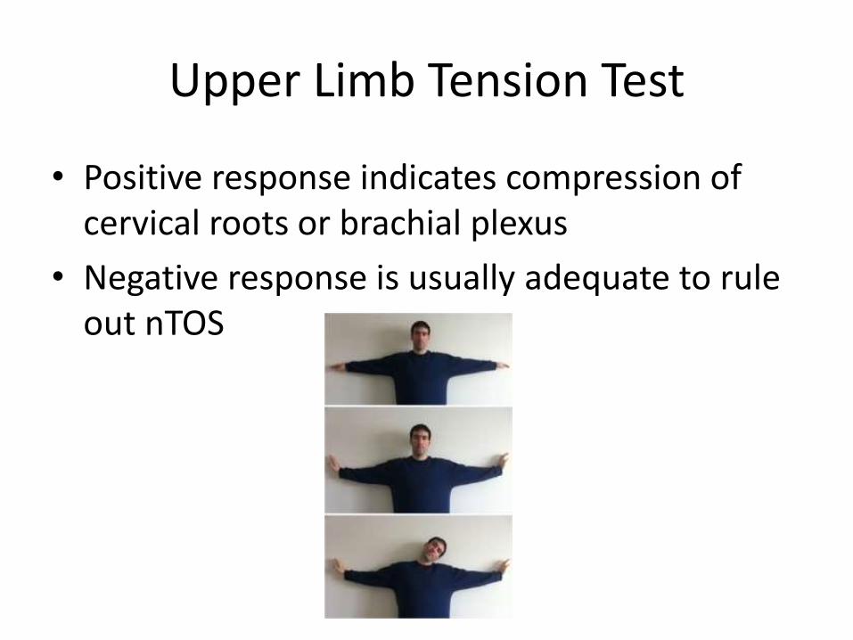
Upper Limb Tension Test
• Positive response indicates compression of cervical roots or brachial plexus
• Negative response is usually adequate to rule out nTOS

Imaging
• Xrays– Cervical rib
– Elongated C7 transverse process
– Hypoplastic 1st rib
– Callous formation from clavicle or 1st rib fracture
– Pseudoarthrosis of 1st rib
• Unable to image soft tissue anomalies and fibromuscular bands – seen only at time of surgery
Imaging
• CT/MRI usually negative but can rule out other pathologies
• MR neurography – newer technology to detect localized nerve function abnormality

Imaging
• aTOS
– Segmental arterial pressures
– Angiography
• vTOS
– Duplex U/S
– Venography
• Use positional maneuvers during the studies
• Consider bilateral studies

EMG/NCS
• Positive results
– Aid in evaluation of other conditions
– Poor prognostic factor if truly nTOS – indicate advanced neural damage
• Negative results
– Exclude other conditions
– May still be nTOS

Scalene muscle block
• Most useful when diagnosis is unclear
• Correlation between relief of symptoms after block and successful outcome after surgical decompression

Treatment nTOS
• Physical therapy
– Therapist must have experience in evaluation and treatment of nTOS
– 20-30% of patients respond, do not require surgical treatment

Treatment nTOS
• Neck stretching
• Posture correction
• Avoid neck traction, weights, resistance exercises, strengthening exercises

Treatment nTOS
• If no improvement after several months
– Live with symptoms
– Surgical decompression

Treatment vTOS
• Catheter-directed thrombolysis
• Anticoagulation
• Surgical decompression with intraoperativevenography and subclavian vein PTA(percutaneous transluminal angioplasty)

Surgical Treatment
• Transaxillary approach– Advantages
• Limited field of operative dissection
• Cosmetically placed incision
• Sufficient exposure (for 1 person)
• Achieve 1st rib resection and anterior scalenectomy
• Removal of anomalous ligaments and fibrous bands
– Disadvantages
• Incomplete exposure of entire scalene triangle
• Difficulty achieving brachial plexus neurolysis
• Limited if vascular reconstruction is needed

Surgical Treatment
• Supraclavicular approach
– Advantages
• Wide exposure of all anatomic structures
• Permits complete resection of anterior and middle scalenes as well as brachial plexus neurolysis
• Allows resection of cervical ribs and anomalous 1st ribs
• Vascular reconstruction is possible

Surgical Treatment
• Adjunctive procedures
– Pectoralis minor tenotomy
– Cervical sympathectomy

Complications
• Injury to– Subclavian artery/vein– Brachial plexus– Phrenic nerve– Long thoracic nerve– Thoracic duct– Sympathetic chain– Intercostal brachial cutaneous nerve (axillary)
• Pneumothorax• Lymph leakage

Outcomes
• No difference in long term results between the 2 approaches
• No difference in outcome based on– Presence of any particular provocative test results
– Experience of operating surgeon
• Predictors of ongoing disability– Amount of work disability preop
– Longer intervals between injury and diagnosis
– Older age at time of surgery

Outcomes
• Associations between preexisting psychological factors and socioeconomic characteristics have been examined
• Independent risk factors associated with persistent disability
– Major depression
– Single

Outcomes
• Results vary by etiology of symptoms
– Failure in 42% with symptoms after a work-related injury or repetitive stress
– Failure in 26% with symptoms after auto accident
– Failure in 18% with nonspecific etiology

Conclusion
• “A surgeon recognizing nTOS should not be dissuaded by the impression that these problems are frequently associated with psychiatric overtones, dependency on pain medications, and ongoing litigation”

THANK YOU
Recommended