The use of bioassays for establishing the biological potency
of biologicals Robin Thorpe PhD.,FRCPath.
Head, Biotherapeutics Group, NIBSC, UK. email: [email protected]

Measuring potency of Biotherapeutics
• Measuring the potency of Biologicals
normally requires the use of a bioassay
• This applies to Biotherapeutics as these
are a sub-group of Biologicals

Bioassay
• A quantitative procedure for measuring
the potency of a biological substance
• Can be carried out in vivo or in vitro
• A functional response is measured and
analysed to provide a statistically
qualified potency estimate

Bioassay
• Bioassays involve the response of a living system to a biological substance
• This distinguishes them in an important way from physico-chemical methods and procedures which simply involve binding, e.g. immunoassays and receptor binding assays
• This affects the results obtained and their significance
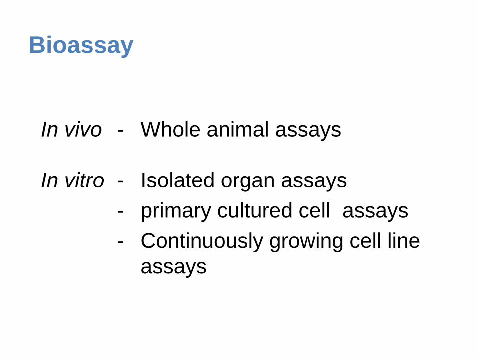
Bioassay
In vivo - Whole animal assays
In vitro - Isolated organ assays
- primary cultured cell assays
- Continuously growing cell line
assays

Bioassay
Appropriately designed bioassays reflect
(assess):
(i) ligand-receptor binding
(ii) signal transduction processes
(iii) final (observed) biological effects

Use of bioassays
Substance Bioassays
Viral Vaccines
Bacterial Vaccines
Neutralization of Infectivity
Tumour “Vaccines” Killing of tumour cells
Coagulation Factors Clotting Assays
Fibrinolytics Clot Lysis
(Enzyme Assays)
Immunoglobulins Neutralisation of relevant micro-organisms.
Binding to appropriate antigens
Endocrine hormones In Vivo & In Vitro assays assessing the biological effect of hormone
Cytokines Bioassays which detect biological effects of cytokines (various)

In vivo and in vitro assays for biological
therapeutics Protein In vivo Assay Biological Response
Qualified
In vitro Assay Biological Response Qualified
GM-CSF Bone marrow stimulation Number of leukocytes Stimulation of cell lines: TF-1, MO7e, AML-193
Cellular proliferation
TNF Necrosis of tumours Number of tumours Cytotoxicity on cell lines: L929, KYM-D4
Cell survival
Erythropoietin Stimulate erythrocytes in hypobaric rats
Number of erythrocytes Stimulation of cell line TF-1 Cellular proliferation
Epidermal Growth Factor
Maturation of mouse newborn
Time to eye opening Stimulation of cell lines: 3T3, Balb/MK, 4MBr-5
Cell proliferation
Interferon-alpha Treatment of virally infected rats
Elevation of IFN inducible enzymes or MHC antigens
Antiviral activity on cell lines, e.g.: HEp2, WISH, MDBK
Cell survival
Interleukin-1 Pyrogenicity in rabbits Temperature Stimulation of cell lines: D10, NOB-1, Monomac
Cellular proliferation Production of cytokines
Bone Morphogenetic Protein-2
Bone callous formation at injection site
Size of callous Stimulation of cell line W20 Production of alkaline phosphatase
Growth Hormone Growth of rats Weight Stimulation of cell lines: NB2, 3T3
Cellular proliferation differentiation
Heparin Antithrombotic activity in rabbits
Size of jugular vein thrombi Anticoagulant activity Inhibition of activated clotting factors
Influenza Vaccine Mouse immunogenicity test Levels of anti-flu antibodies Single radial difussion Area of precipitation
Follicle Stimulating Hormone
Steelman-Pohley test on immature female rats
Ovarian weight Stimulation of sertoli cells or granulosa cells
Aromatisation of androgens, levels of cAMP

Choice of assay
• Assay needs to be selected for each biological
• Assay may use primary cells or cell-lines
• Cell-lines -
– continuously growing
– factor dependent, cloned cell-lines
– transfected with an appropriate receptor
• Select a cell-line which yields a functional end-point
response. These can be –
– Early – phosphorylation of intracellular substrates
– Late – cytokine secretion, proliferation, cytotoxic effects etc
– Early/Late (depending on kinetics) – changes in mRNA expression; PCR readout, reporter gene based assa

When are bioassays useful?
During pre-product development:
Research and R & D activities
During product development:
Characterization of biological activity(s) of product(s)
Stability studies
Dosing
Formulation studies
Post-product development:
Batch-to-batch consistency
Stability

Bioassays
• The responses of samples and standards in
the assay must be parallel
• If not, the assay is responding differently to
the molecular species or forms in the
samples/standards

Dose-response plots

Standardization of bioassays
requires biological potency standards
- calibrated in units (e.g. IU)
Standardization of bioassays

Bioassays - Units
• International Units - relate to
potency of ampoule of I.S. contents
Often arbitrary units

Use of one standard world-wide
facilitates comparability of assay
results
Bioassays - Standards

Biological estimates of IL-4
(nominal ng/ampoule)
Same standard ‘In-house’ standards

Bioassay-derived potency data must
be accompanied by a valid statement
of the uncertainty associated with the
measurement
Bioassays
Bioassays must be carefully designed
to minimize and allow assessment of
errors
Bioassays

Problems with bioassays
1. Can be non-specific - respond to several substances, culture additives, LPS etc.
2. Can be variable, tedious and time-consuming (and difficult)
3. Can require long-term maintenance of (factor-dependant) cell lines
4. Can be ‘inhibited’ by a variety of specific and non-specific molecules
5. Cannot distinguish between molecular ‘forms’ of some molecules, e.g. IL-1α and IL-1ß; TNF-α and TNF-ß

Measuring potency of Biotherapeutics
Need to carry out a validated bioassay
for each batch. This must be analysed
to provide:
(i) A mean potency estimate
(ii) Fiducial limits of error for the batch
(usually equivalent to confidence, i.e.
95% limits)

Measuring potency of Biotherapeutics
The mean potency estimate should lie
within specified limits - these limits
should be derived from results of mean
estimate of potency for different batches
and limit the batch-to-batch variation in
potency

Measuring potency of Biotherapeutics
The fiducial limits of error are derived
from the individual potency estimates
(from which the means are derived) -
they are a limit to the assay variability

Example of how statistical limits are
applied to biological assays
Measuring potency of Biotherapeutics
Stated potency
Potency on label. Backed by mean potency
estimate (i.e. this is within limits) + fiducial
limits of error (these are acceptable)
Usually inappropriate to label vials with
individual batch mean potency estimate value
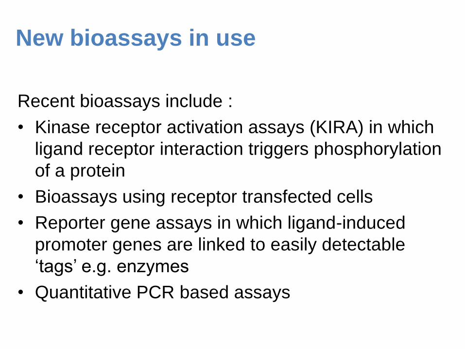
New bioassays in use
Recent bioassays include :
• Kinase receptor activation assays (KIRA) in which
ligand receptor interaction triggers phosphorylation
of a protein
• Bioassays using receptor transfected cells
• Reporter gene assays in which ligand-induced
promoter genes are linked to easily detectable
‘tags’ e.g. enzymes
• Quantitative PCR based assays
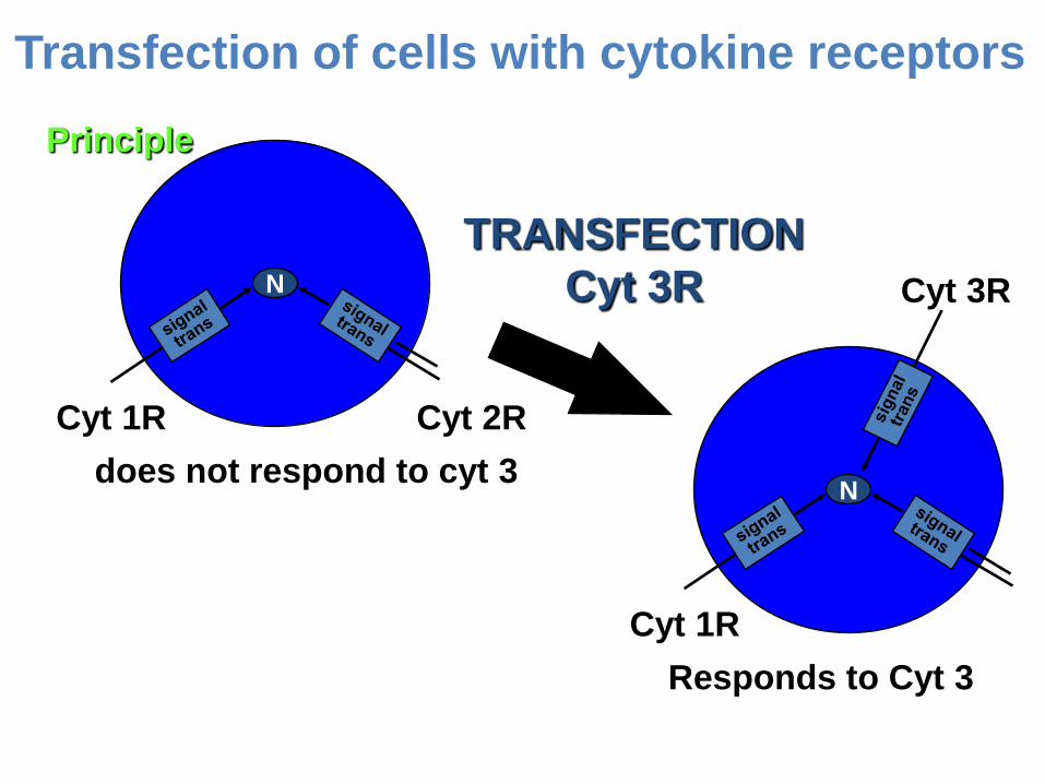
N
Cyt 1R Cyt 2R
does not respond to cyt 3 N
Cyt 1R
Cyt 3R
Responds to Cyt 3
TRANSFECTION
Cyt 3R
Principle
Transfection of cells with cytokine receptors

Transfection of non-responsive cells with
cytokine receptors
CT.h4S
mIL-2R mIL-4R
CTLL-2
mIL-2R mIL-4R
32D/Mpl+ 32D
mIL-3R mIL-3R
responds to huTPO
hu TPO R
Ba8.1cl BAF3
mIL-3R mIL-3R
responds to hu IL-10
hu IL-10R
grows in mIL-3
(a) human IL-4R
Transfection
with hu IL-4R
Transfection
with hu TPO R
Transfection
with hu IL-10R
(b) human TPO
(c) human IL-10
m/hu IL-4R
responds to hu IL-4
grows in mIL-3
grows in hu IL-2

A549 Luciferase Reporter Gene Assay
.
0
1000
2000
3000
4000
5000
6000
7000
0.1 1 10 100 1000 10000
IFN ALPHA (pg/ml)
CP
S
IFN ALPHA PRODUCT
MODIFIED IFN ALPHA PRODUCT
IFN-a2
IL-28
IL-29
Dose responses for IFN-α and IFN-l
preparations ( A) and 2 IFN-α preparations
(B) from a reporter gene assay using a cell
line transfected with the promoter of the
MxA gene fused with luciferase. The MxA
gene codes for a type 1 interferon-inducible
protein involved in antiviral activity.
A
B

qPCR assay protocol
Incubate cells in 96-well plate with doses
of test substance and standard
Lyse cells and isolate good quality RNA
Reverse transcribe RNA into cDNA
Measure mRNA expression using real-time qPCR
Data analysis
20 minutes – 1 hour
Approx 1hr
Approx 1hr
Approx 2hr
Liquid-handling robots
Qiacube RNA isolation

VEGF bioassays – IL-8 mRNA in HUVECs
0.0 1.0 2.0 3.9 7.8 15.6 31.3 62.5 125.0 250.0
VEGF (ng/mL)
0.2
0.4
0.6
0.8
1.0
1.2
1.4
1.6
1.8
2.0
2.2
ala
marB
lue r
eduction (
A570nm
-A600nm
)
62.531.315.67.83.92.01.00.50.20.0
VEGF (ng/mL)
0.02
0.03
0.04
0.05
0.06
0.07
0.08
0.09
0.10
0.11
0.12
0.13
Tis
sue f
acto
r (U
/mL)
0 0.25 0.5 1 2 4 8 16 32 64 1250
3
6
9
12
15
18
VEGF (ng/mL)
rela
tive IL8 e
xpre
ssio
n
IL-8 mRNA - qPCR
0 10 20 40 80 160 320 640 12800.0
2.5
5.0
7.5
10.0
12.5
15.0
VEGF mAb 293(ng/ml)
VEGF (7.8ng/ml)
rela
tive IL8 e
xpre
ssio
n0 7.8 31.2 62.5 125 250 500 1000
0
2
4
6
8
10
12
14
VEGF sR1(ng/ml)
VEGF (7.8ng/ml)
rela
tive IL
8 e
xp
ressio
n
64hr
24hr
4.5hr

Conclusions
• Bioassays provide crucial data at all
stages of product development
• Bioassay data is essential as part of
product characterization and
complements information obtained
using non-bioassay procedures

Acknowledgements
• Meenu Wadhwa
• Chris Burns
• Jane Robinson
• Paula Dilger
• Chris Bird
• Tony Meager
• Guenther Adolf
• Tony Mire-Sluis
Recommended