The Special Senses
• Smell, taste, vision, hearing and equilibrium
• Housed in complex sensory organs
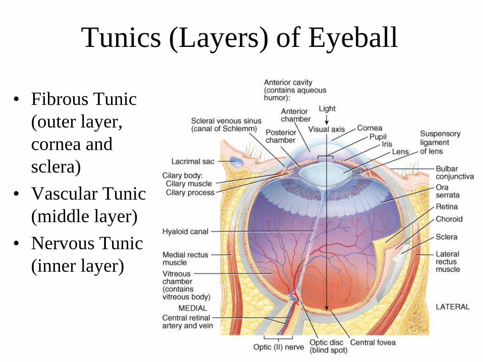
Tunics (Layers) of Eyeball
• Fibrous Tunic
(outer layer,
cornea and
sclera)
• Vascular Tunic
(middle layer)
• Nervous Tunic
(inner layer)
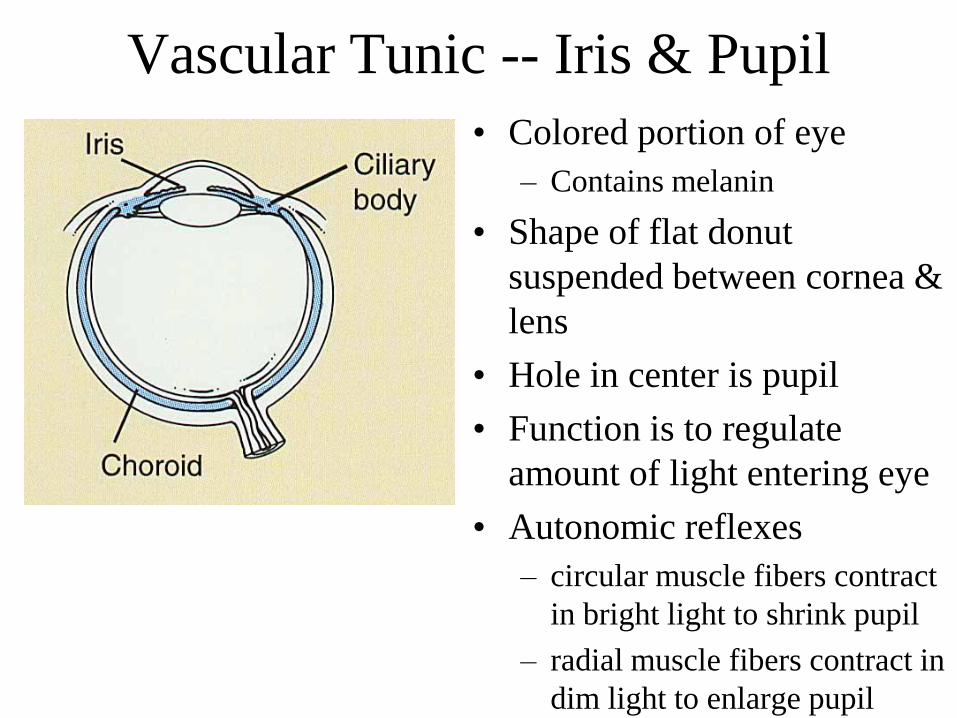
Vascular Tunic -- Iris & Pupil
• Colored portion of eye
– Contains melanin
• Shape of flat donut
suspended between cornea &
lens
• Hole in center is pupil
• Function is to regulate
amount of light entering eye
• Autonomic reflexes
– circular muscle fibers contract
in bright light to shrink pupil
– radial muscle fibers contract in
dim light to enlarge pupil
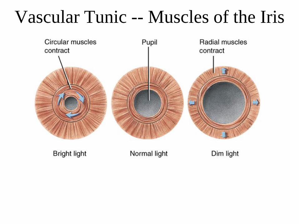
Vascular Tunic -- Muscles of the Iris
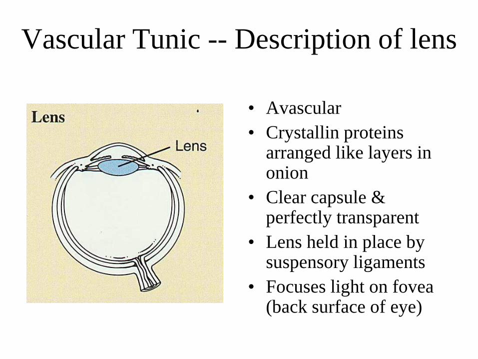
Vascular Tunic -- Description of lens
• Avascular
• Crystallin proteins arranged like layers in onion
• Clear capsule & perfectly transparent
• Lens held in place by suspensory ligaments
• Focuses light on fovea (back surface of eye)
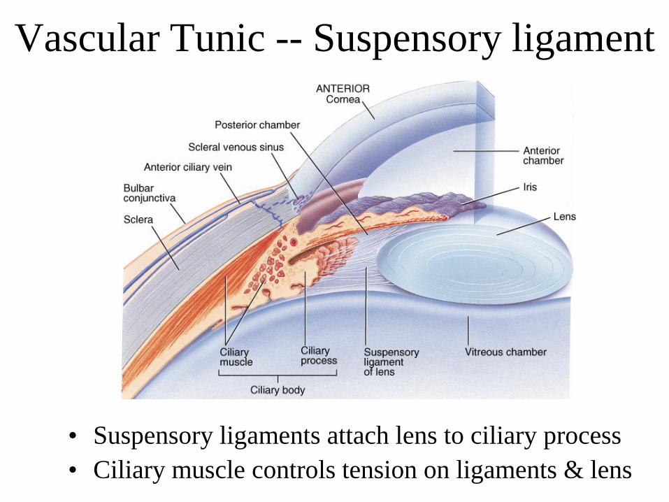
Vascular Tunic -- Suspensory ligament
• Suspensory ligaments attach lens to ciliary process
• Ciliary muscle controls tension on ligaments & lens
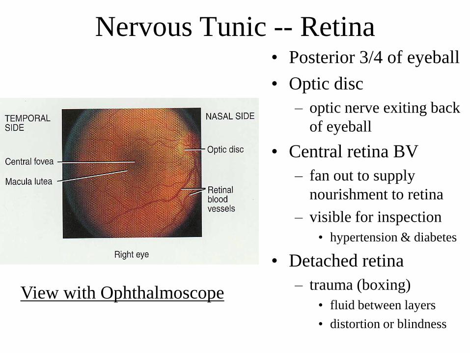
Nervous Tunic -- Retina• Posterior 3/4 of eyeball
• Optic disc
– optic nerve exiting back
of eyeball
• Central retina BV
– fan out to supply
nourishment to retina
– visible for inspection
• hypertension & diabetes
• Detached retina
– trauma (boxing)
• fluid between layers
• distortion or blindness
View with Ophthalmoscope
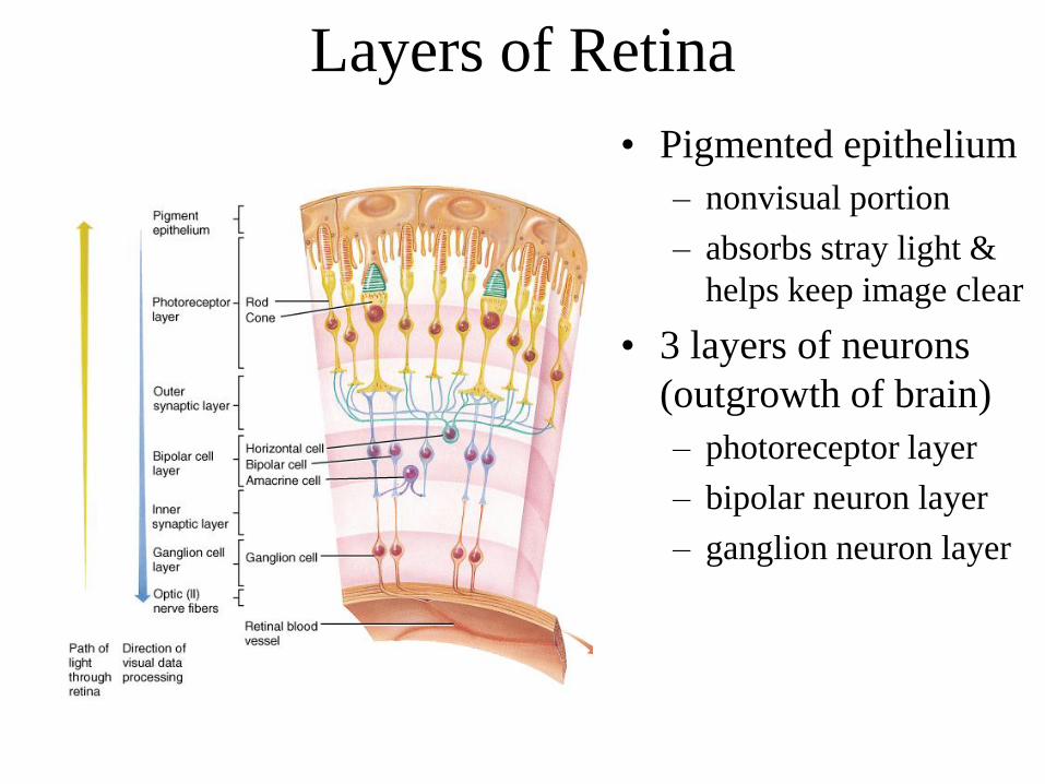
Layers of Retina
• Pigmented epithelium
– nonvisual portion
– absorbs stray light &
helps keep image clear
• 3 layers of neurons
(outgrowth of brain)
– photoreceptor layer
– bipolar neuron layer
– ganglion neuron layer
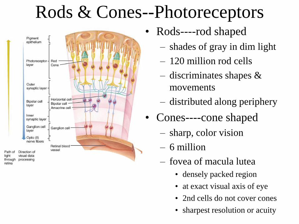
Rods & Cones--Photoreceptors• Rods----rod shaped
– shades of gray in dim light
– 120 million rod cells
– discriminates shapes &
movements
– distributed along periphery
• Cones----cone shaped
– sharp, color vision
– 6 million
– fovea of macula lutea
• densely packed region
• at exact visual axis of eye
• 2nd cells do not cover cones
• sharpest resolution or acuity
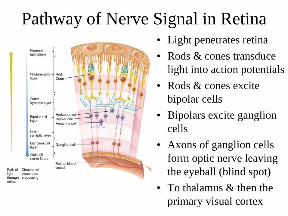
Pathway of Nerve Signal in Retina• Light penetrates retina
• Rods & cones transduce
light into action potentials
• Rods & cones excite
bipolar cells
• Bipolars excite ganglion
cells
• Axons of ganglion cells
form optic nerve leaving
the eyeball (blind spot)
• To thalamus & then the
primary visual cortex
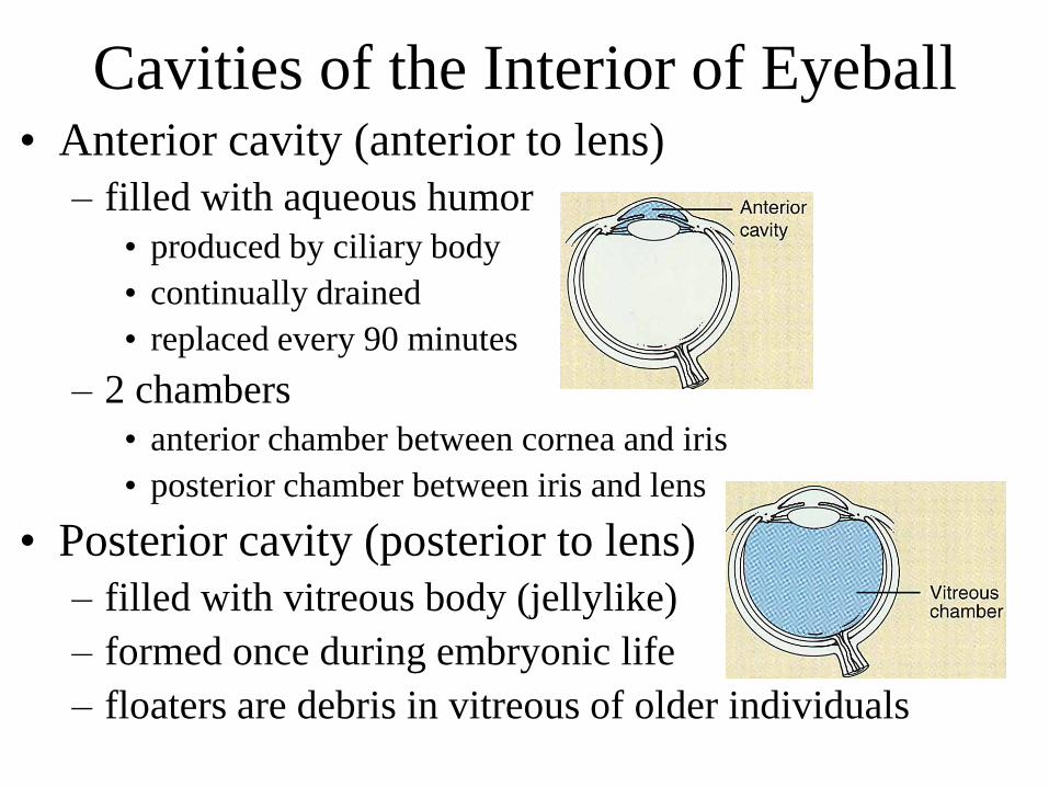
Cavities of the Interior of Eyeball• Anterior cavity (anterior to lens)
– filled with aqueous humor
• produced by ciliary body
• continually drained
• replaced every 90 minutes
– 2 chambers
• anterior chamber between cornea and iris
• posterior chamber between iris and lens
• Posterior cavity (posterior to lens)
– filled with vitreous body (jellylike)
– formed once during embryonic life
– floaters are debris in vitreous of older individuals
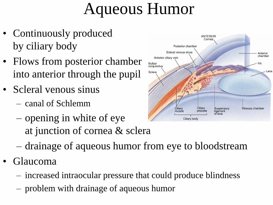
Aqueous Humor
• Continuously produced
by ciliary body
• Flows from posterior chamber
into anterior through the pupil
• Scleral venous sinus
– canal of Schlemm
– opening in white of eye
at junction of cornea & sclera
– drainage of aqueous humor from eye to bloodstream
• Glaucoma
– increased intraocular pressure that could produce blindness
– problem with drainage of aqueous humor
Major Processes of Image Formation
• Refraction of light
– by cornea & lens
– light rays must fall upon the retina
• Accommodation of the lens
– changing shape of lens so that light is focused
• Constriction of the pupil
– less light enters the eye
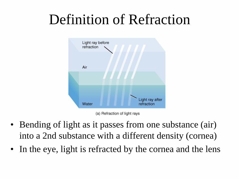
Definition of Refraction
• Bending of light as it passes from one substance (air)
into a 2nd substance with a different density (cornea)
• In the eye, light is refracted by the cornea and the lens
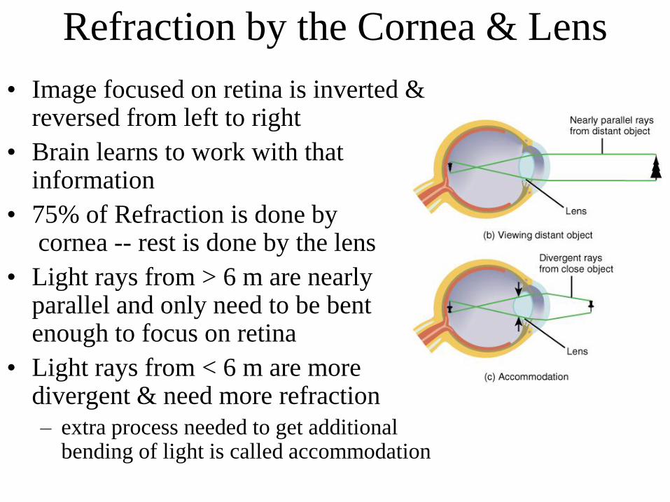
Refraction by the Cornea & Lens
• Image focused on retina is inverted & reversed from left to right
• Brain learns to work with that information
• 75% of Refraction is done bycornea -- rest is done by the lens
• Light rays from > 6 m are nearly parallel and only need to be bent enough to focus on retina
• Light rays from < 6 m are more divergent & need more refraction
– extra process needed to get additional bending of light is called accommodation
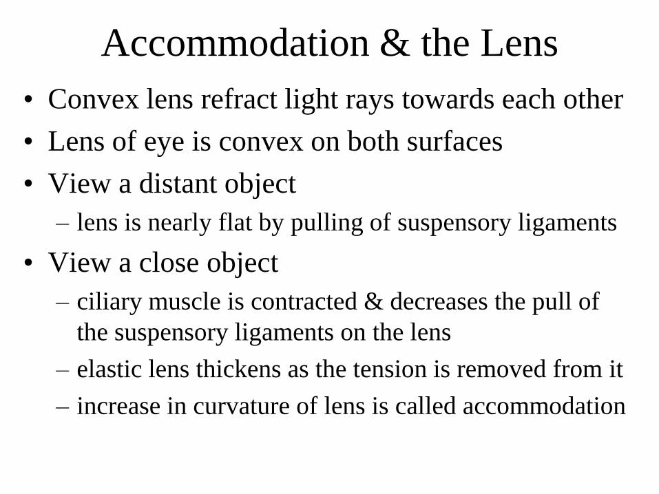
Accommodation & the Lens
• Convex lens refract light rays towards each other
• Lens of eye is convex on both surfaces
• View a distant object
– lens is nearly flat by pulling of suspensory ligaments
• View a close object
– ciliary muscle is contracted & decreases the pull of
the suspensory ligaments on the lens
– elastic lens thickens as the tension is removed from it
– increase in curvature of lens is called accommodation
Near Point of Vision and Presbyopia
• Near point is the closest distance from the eye
an object can be & still be in clear focus
– 4 inches in a young adult
– 8 inches in a 40 year old
• lens has become less elastic
– 31 inches in a 60 to 80 year old
• Reading glasses may be needed by age 40
– presbyopia
– glasses replace refraction previously provided by
increased curvature of the relaxed, youthful lens
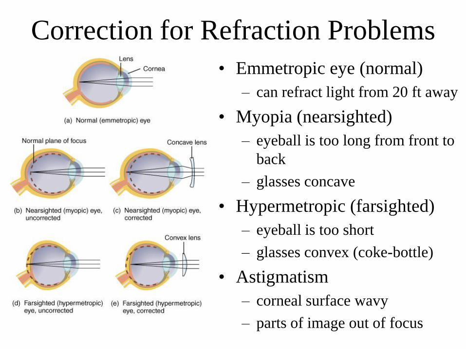
Correction for Refraction Problems
• Emmetropic eye (normal)
– can refract light from 20 ft away
• Myopia (nearsighted)
– eyeball is too long from front to
back
– glasses concave
• Hypermetropic (farsighted)
– eyeball is too short
– glasses convex (coke-bottle)
• Astigmatism
– corneal surface wavy
– parts of image out of focus
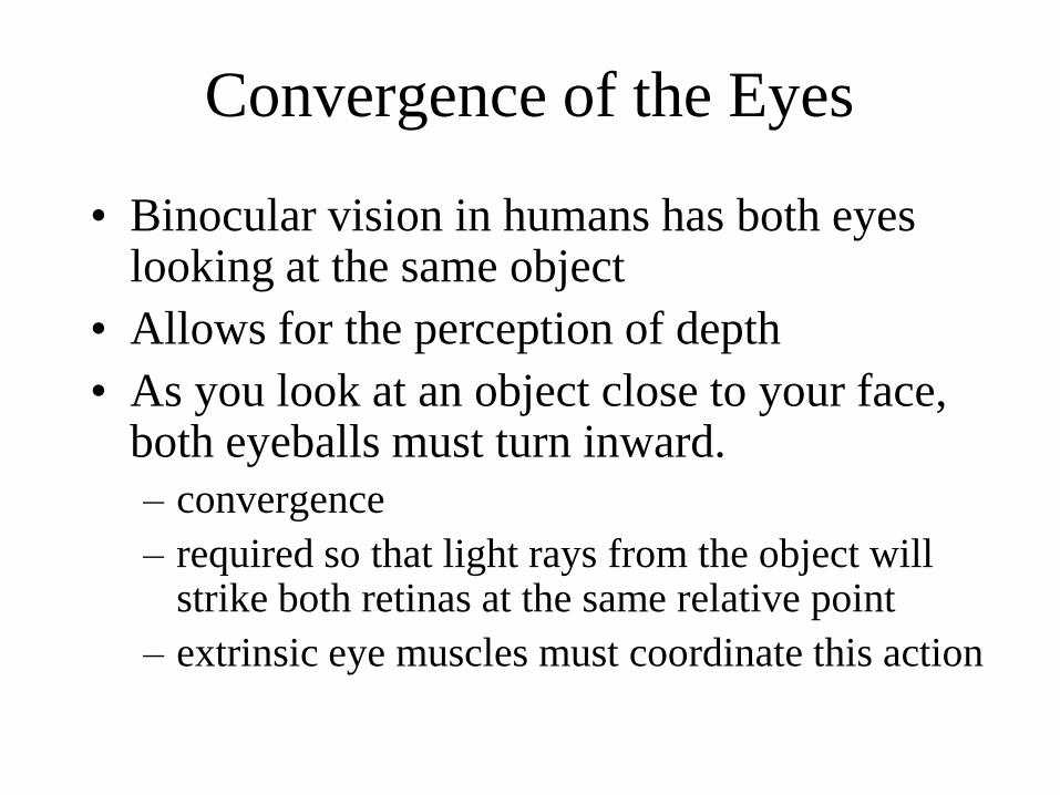
Convergence of the Eyes
• Binocular vision in humans has both eyes looking at the same object
• Allows for the perception of depth
• As you look at an object close to your face, both eyeballs must turn inward.
– convergence
– required so that light rays from the object will strike both retinas at the same relative point
– extrinsic eye muscles must coordinate this action
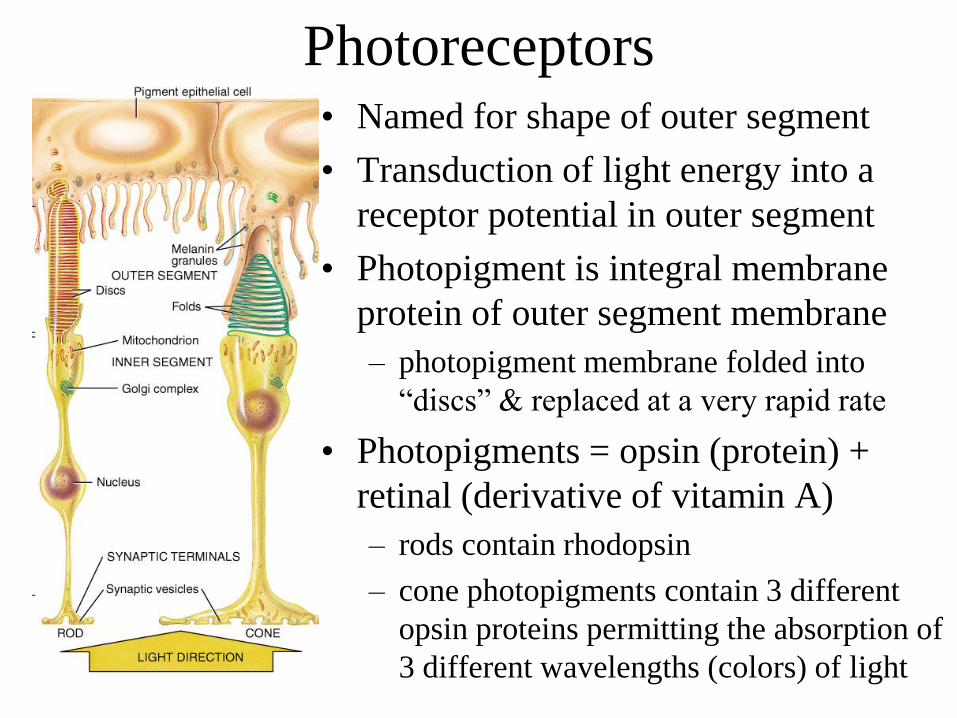
Photoreceptors• Named for shape of outer segment
• Transduction of light energy into a
receptor potential in outer segment
• Photopigment is integral membrane
protein of outer segment membrane
– photopigment membrane folded into
“discs” & replaced at a very rapid rate
• Photopigments = opsin (protein) +
retinal (derivative of vitamin A)
– rods contain rhodopsin
– cone photopigments contain 3 different
opsin proteins permitting the absorption of
3 different wavelengths (colors) of light
Color Blindness & Night Blindness
• Color blindness
– inability to distinguish between certain colors
– absence of certain cone photopigments
– red-green color blind person can not tell red from
green
• Night blindness
– difficulty seeing in low light
– inability to make normal amount of rhodopsin
– possibly due to deficiency of vitamin A
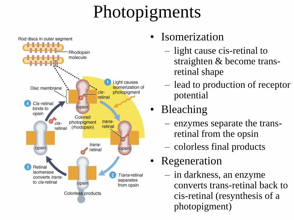
Photopigments
• Isomerization
– light cause cis-retinal to straighten & become trans-retinal shape
– lead to production of receptor potential
• Bleaching
– enzymes separate the trans-retinal from the opsin
– colorless final products
• Regeneration
– in darkness, an enzyme converts trans-retinal back to cis-retinal (resynthesis of a photopigment)
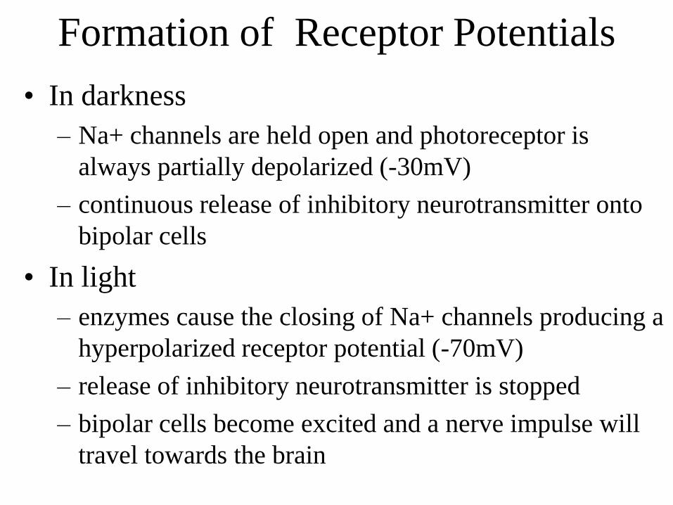
Formation of Receptor Potentials
• In darkness
– Na+ channels are held open and photoreceptor is
always partially depolarized (-30mV)
– continuous release of inhibitory neurotransmitter onto
bipolar cells
• In light
– enzymes cause the closing of Na+ channels producing a
hyperpolarized receptor potential (-70mV)
– release of inhibitory neurotransmitter is stopped
– bipolar cells become excited and a nerve impulse will
travel towards the brain
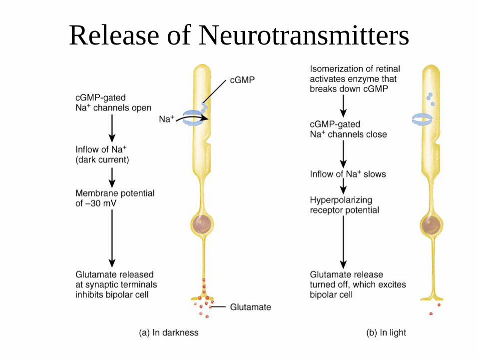
Release of Neurotransmitters
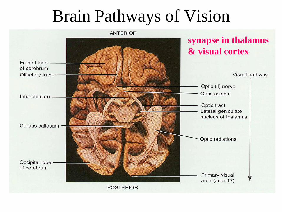
Brain Pathways of Vision
synapse in thalamus
& visual cortex
Processing of Image Data in the Brain
• Visual information in optic nerve travels to
– occipital lobe for vision
– midbrain for controlling pupil size &
coordination of head and eye movements
– hypothalamus to establish sleep patterns based
upon circadian rhythms of light and darkness
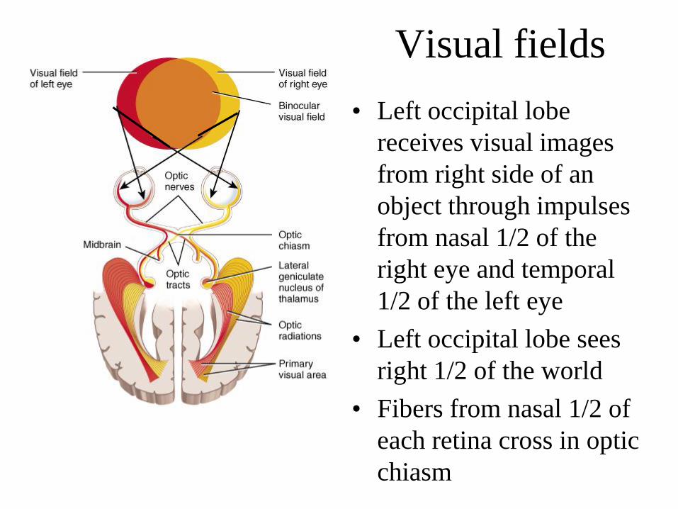
Visual fields
• Left occipital lobe
receives visual images
from right side of an
object through impulses
from nasal 1/2 of the
right eye and temporal
1/2 of the left eye
• Left occipital lobe sees
right 1/2 of the world
• Fibers from nasal 1/2 of
each retina cross in optic
chiasm
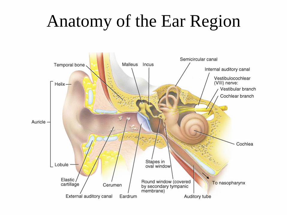
Anatomy of the Ear Region
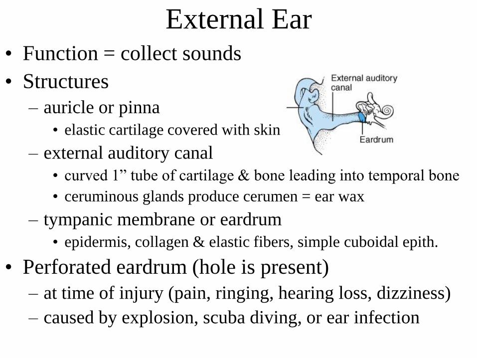
External Ear• Function = collect sounds
• Structures
– auricle or pinna
• elastic cartilage covered with skin
– external auditory canal
• curved 1” tube of cartilage & bone leading into temporal bone
• ceruminous glands produce cerumen = ear wax
– tympanic membrane or eardrum
• epidermis, collagen & elastic fibers, simple cuboidal epith.
• Perforated eardrum (hole is present)
– at time of injury (pain, ringing, hearing loss, dizziness)
– caused by explosion, scuba diving, or ear infection
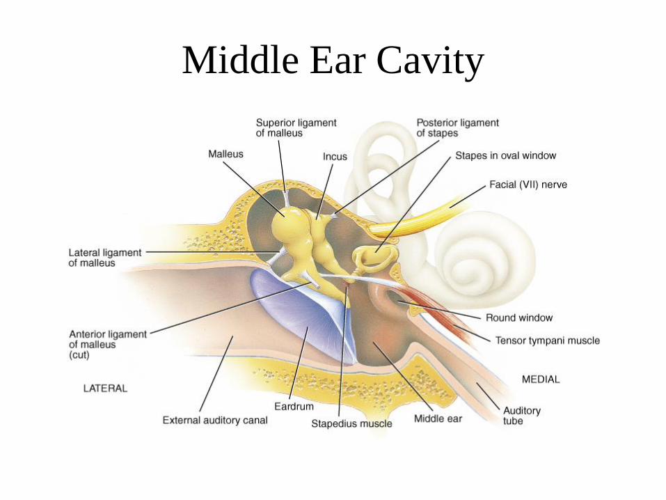
Middle Ear Cavity
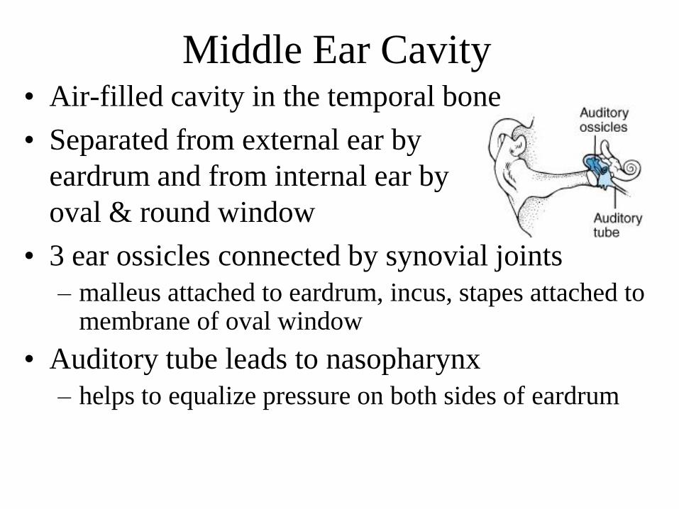
Middle Ear Cavity• Air-filled cavity in the temporal bone
• Separated from external ear by
eardrum and from internal ear by
oval & round window
• 3 ear ossicles connected by synovial joints
– malleus attached to eardrum, incus, stapes attached to membrane of oval window
• Auditory tube leads to nasopharynx
– helps to equalize pressure on both sides of eardrum
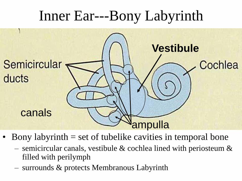
Inner Ear---Bony Labyrinth
ampulla
Vestibule
• Bony labyrinth = set of tubelike cavities in temporal bone
– semicircular canals, vestibule & cochlea lined with periosteum & filled with perilymph
– surrounds & protects Membranous Labyrinth
canals
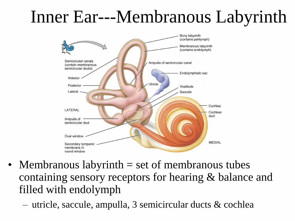
Inner Ear---Membranous Labyrinth
• Membranous labyrinth = set of membranous tubes containing sensory receptors for hearing & balance and filled with endolymph
– utricle, saccule, ampulla, 3 semicircular ducts & cochlea
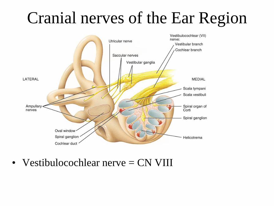
Cranial nerves of the Ear Region
• Vestibulocochlear nerve = CN VIII
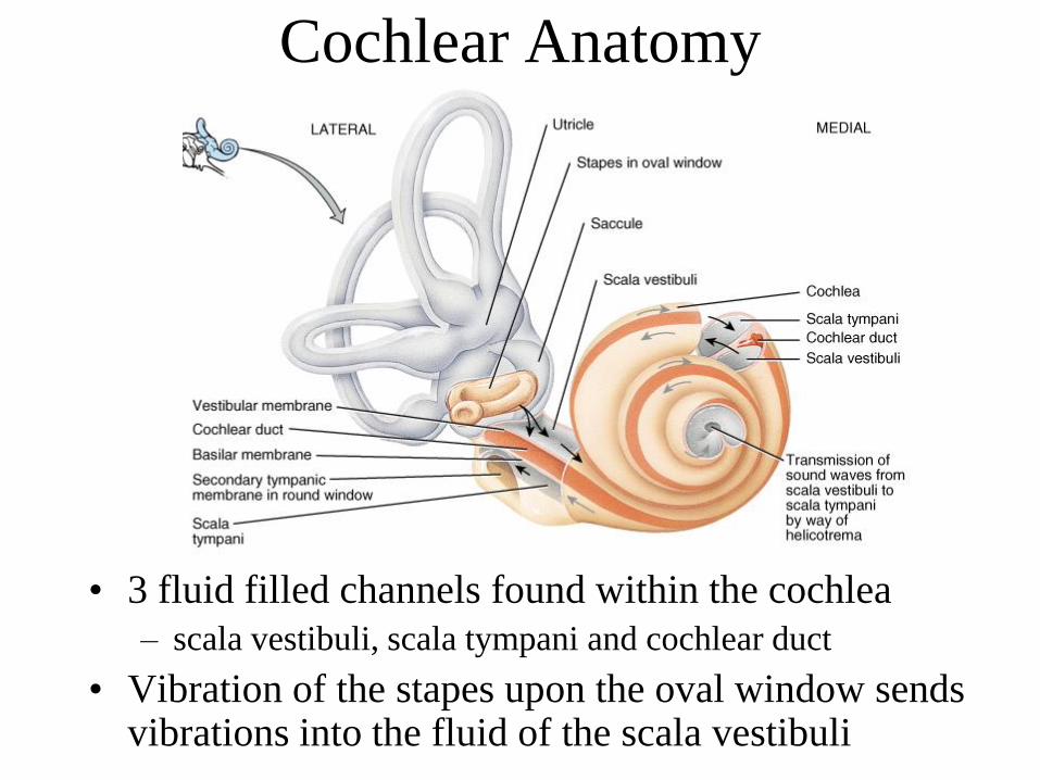
Cochlear Anatomy
• 3 fluid filled channels found within the cochlea
– scala vestibuli, scala tympani and cochlear duct
• Vibration of the stapes upon the oval window sends vibrations into the fluid of the scala vestibuli
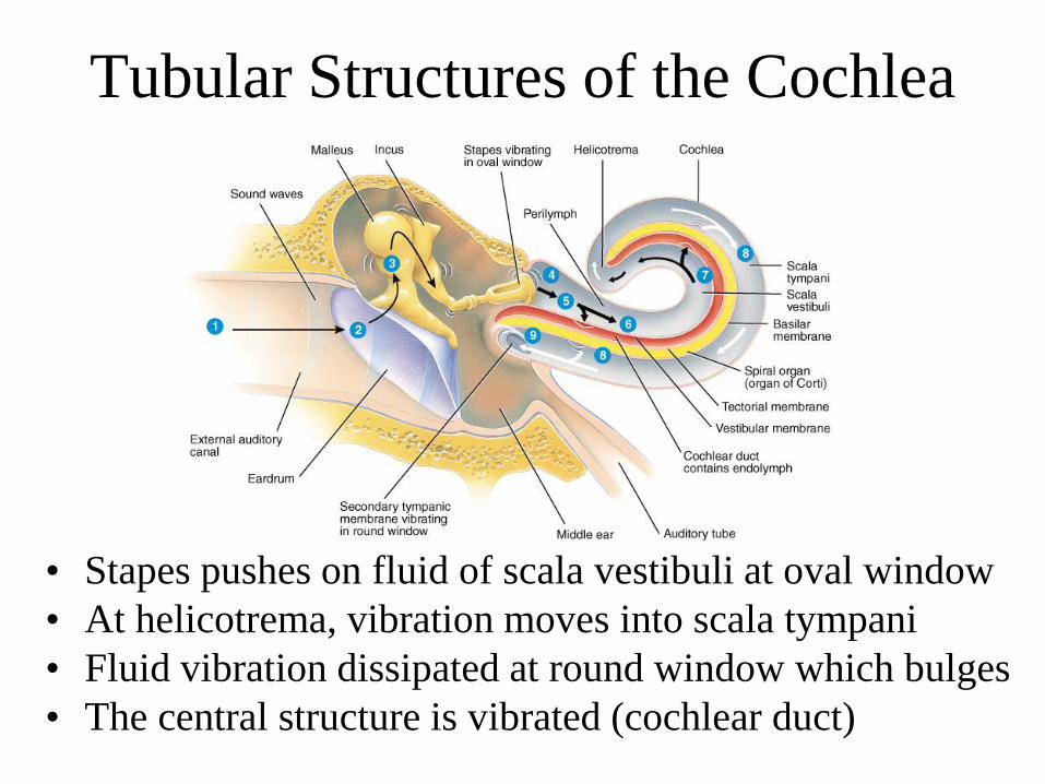
Tubular Structures of the Cochlea
• Stapes pushes on fluid of scala vestibuli at oval window
• At helicotrema, vibration moves into scala tympani
• Fluid vibration dissipated at round window which bulges
• The central structure is vibrated (cochlear duct)
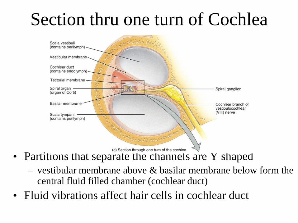
Section thru one turn of Cochlea
• Partitions that separate the channels are Y shaped
– vestibular membrane above & basilar membrane below form the central fluid filled chamber (cochlear duct)
• Fluid vibrations affect hair cells in cochlear duct
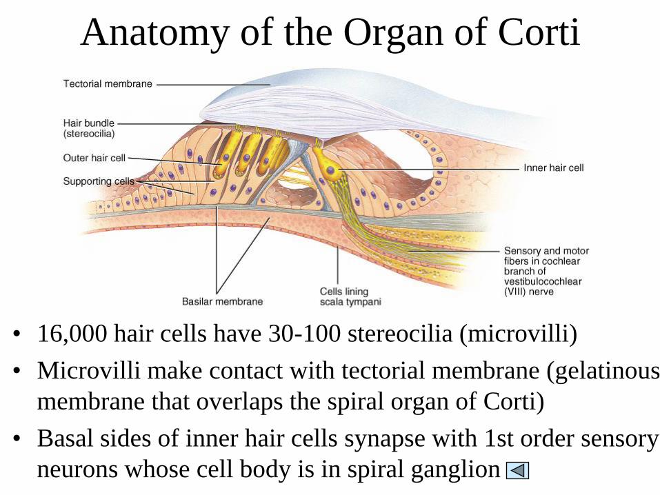
Anatomy of the Organ of Corti
• 16,000 hair cells have 30-100 stereocilia (microvilli)
• Microvilli make contact with tectorial membrane (gelatinous
membrane that overlaps the spiral organ of Corti)
• Basal sides of inner hair cells synapse with 1st order sensory
neurons whose cell body is in spiral ganglion
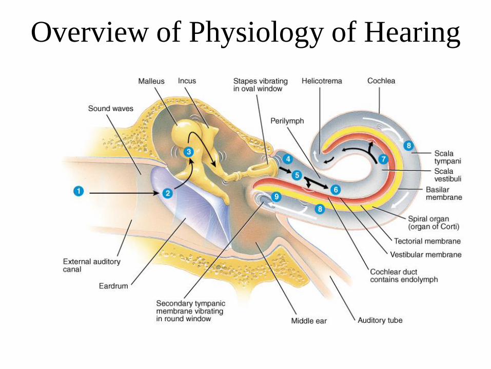
Physiology of Hearing
• Auricle collects sound waves
• Eardrum vibrates
– slow vibration in response to low-pitched sounds
– rapid vibration in response to high-pitched sounds
• Ossicles vibrate since malleus attached to eardrum
• Stapes pushes on oval window producing fluid
pressure waves in scala vestibuli & tympani
– oval window vibration 20X more vigorous than eardrum
• Pressure fluctuations inside cochlear duct move the
hair cells against the tectorial membrane
• Microvilli are bent producing receptor potentials
Overview of Physiology of Hearing
Hair Cell Physiology
• Hair cells convert mechanical deformation into
electrical signals
• As microvilli are bent, mechanically-gated
channels in the membrane let in K+ ions
• This depolarization spreads & causes voltage-
gated Ca+2 channels at the base of the cell to
open
• Triggering the release of neurotransmitter onto
the first order neuron
– more neurotransmitter means more nerve impulses
Cochlear Implants
• If deafness is due to destruction of hair cells
• Microphone, microprocessor & electrodes
translate sounds into electric stimulation of
the vestibulocochlear nerve
– artificially induced nerve signals follow normal
pathways to brain
• Provides only a crude representation of
sounds
Physiology of Equilibrium (Balance)
• Static equilibrium
– maintain the position of the body (head) relative to
the force of gravity
– macula receptors within saccule & utricle
• Dynamic equilibrium
– maintain body position (head) during sudden
movement of any type--rotation, deceleration or
acceleration
– crista receptors within ampulla of semicircular ducts
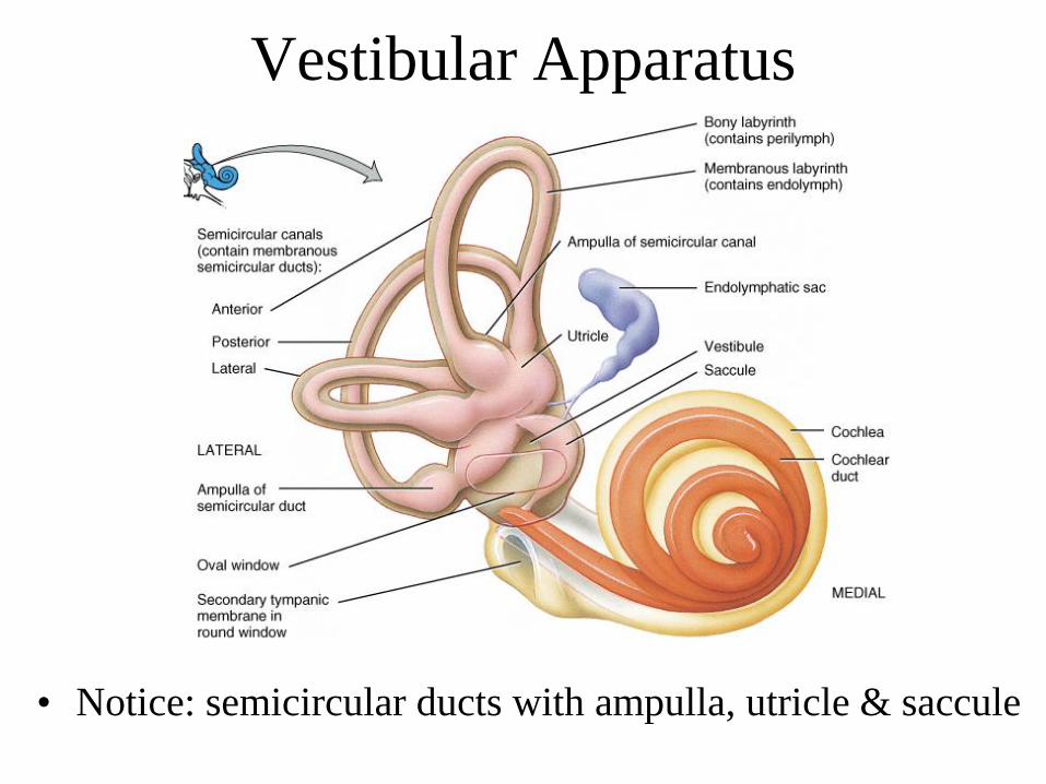
Vestibular Apparatus
• Notice: semicircular ducts with ampulla, utricle & saccule
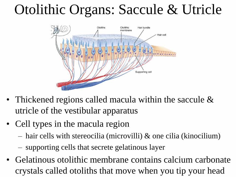
Otolithic Organs: Saccule & Utricle
• Thickened regions called macula within the saccule &
utricle of the vestibular apparatus
• Cell types in the macula region
– hair cells with stereocilia (microvilli) & one cilia (kinocilium)
– supporting cells that secrete gelatinous layer
• Gelatinous otolithic membrane contains calcium carbonate
crystals called otoliths that move when you tip your head
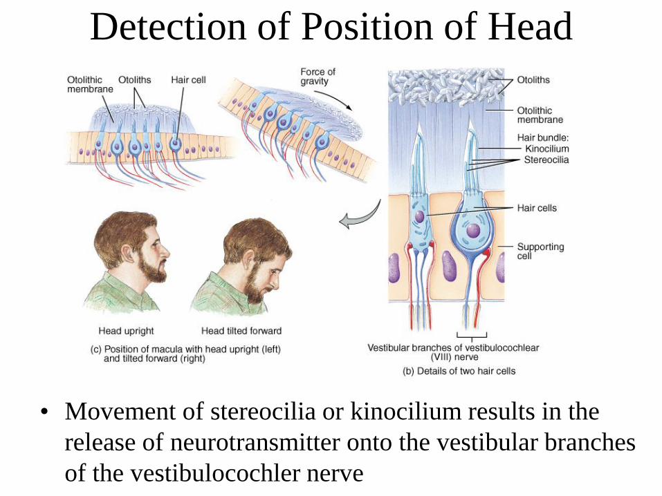
Detection of Position of Head
• Movement of stereocilia or kinocilium results in the
release of neurotransmitter onto the vestibular branches
of the vestibulocochler nerve
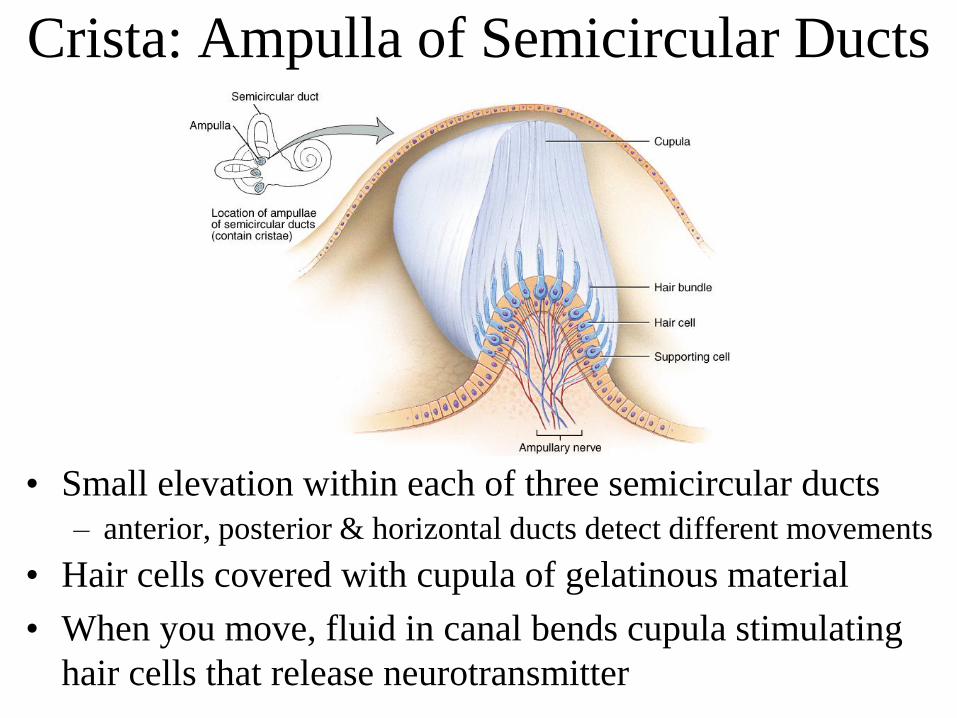
Crista: Ampulla of Semicircular Ducts
• Small elevation within each of three semicircular ducts
– anterior, posterior & horizontal ducts detect different movements
• Hair cells covered with cupula of gelatinous material
• When you move, fluid in canal bends cupula stimulating
hair cells that release neurotransmitter
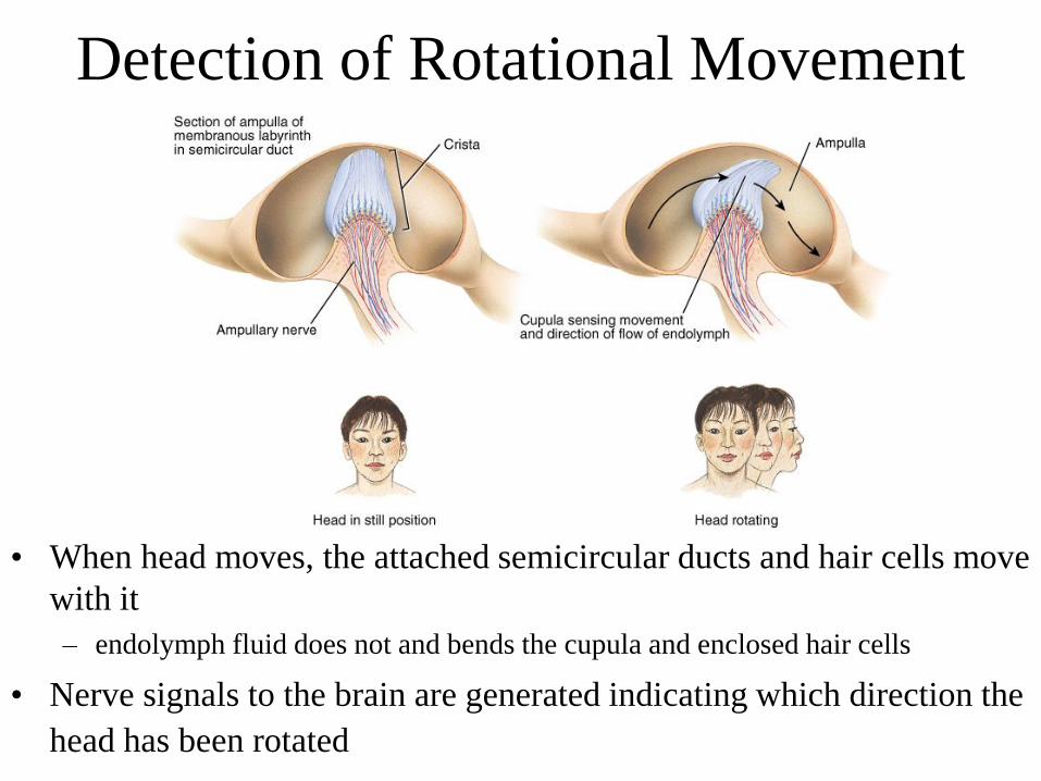
Detection of Rotational Movement
• When head moves, the attached semicircular ducts and hair cells move
with it
– endolymph fluid does not and bends the cupula and enclosed hair cells
• Nerve signals to the brain are generated indicating which direction the
head has been rotated
Recommended