University of Massachusetts BostonScholarWorks at UMass Boston
Honors College Theses
12-2015
The role of valproic acid, a histone deacetylaseinhibitor, in reducing anxiety levels in rats: anepigenetics studyJuliet ButemeUniversity of Massachusetts Boston
Follow this and additional works at: http://scholarworks.umb.edu/honors_theses
Part of the Biological Psychology Commons, and the Biology Commons
This Open Access Honors Thesis is brought to you for free and open access by ScholarWorks at UMass Boston. It has been accepted for inclusion inHonors College Theses by an authorized administrator of ScholarWorks at UMass Boston. For more information, please contact [email protected].
Recommended CitationButeme, Juliet, "The role of valproic acid, a histone deacetylase inhibitor, in reducing anxiety levels in rats: an epigenetics study"(2015). Honors College Theses. 12.http://scholarworks.umb.edu/honors_theses/12
The role of valproic acid, a histone deacetylase inhibitor, in reducing anxiety levels in rats: An epigenetics study
Undergraduate Honors Thesis
Submitted to the Faculty of
the University of Massachusetts Boston
in partial fulfillment of graduation with
Honors in Biology
in the College of Science and Math
by
Juliet Buteme
September 09/08/2015
©Copyright 2015
Juliet Buteme
All Rights Reserved
2
Abstract
Interestingly, rats, like humans, show personality traits: they are born with different anxiety
levels (i.e., high (HAn) or low anxiety (LAn) levels). In this study, we investigated (1) if treatment
with valproic acid (VPA) can lower anxiety level in trait anxiety rats and (2) how VPA may interact
with environment (e.g., enriched (EE), standard (SE) and isolated environments (IE)). VPA is a
deacetylase inhibitor that increases Histone 3 acetylation, a known epigenetic mechanism that
interacts with stress response proteins, and treats epilepsy and mood disorders. Since rats reared
in IE exhibit heightened anxiety levels as compared to those in EE, we suspected that there would
be an additive effect of VPA treatment and EE – that this combined treatment would lower the
anxiety levels of HAn rats. In order to verify that VPA improved performance on anxiety tests by
interacting with the stress response system, Long Evan Rats were perfused with 4%
paraformaldehyde for brain tissue analysis using immunohistochemistry to measure corticotropin
releasing factor (CRF), a protein implicated in stress. We found that Long Evan rats housed in EE
showed less anxiety-like behavior on the EPM compared to other housing settings, and surprising,
this was reversed with VPA. VPA did lower anxiety-like behavior in IE reared animals. There was
a decrease in the CRF levels in LANEE, and an increase CRF in the HANEE housed animals,
suggesting no additive effect of combining VPA and EE treatment for anxiety-like behavior, but a
benefit for reversing anxiety due to isolation rearing.
3
Introduction
Background
Anxiety Disorders are Complex
Behavior is a complex response to stimuli mediated both by genes and by the environment.
For example, pathological anxiety, a complex psychological disorder, involves an exaggerated
response to stimuli that is a consequence of genetic and environmental influences. Many of the
psychological and physiological responses to the class of anxiety disorders (e.g., Panic Disorder,
Agoraphobia, Social Phobia) overlap in that they are characterized by tension, excessive worry,
stress, irritability, restlessness, to mention but a few. These responses are mediated by genes,
neurotransmitters and neurohormones and their receptors as well as by environmental factors
and/or interactions between genes, chemicals and the environment. Phenomenologically, anxiety
disorders seem to have in common an increased reactivity to the environment, driven by
uncertainty and fear of perceived threats (Le-Niculescu et al. 2011). Recent studies of mental
health indicate a high frequency of occurrence for anxiety disorders in the general population. In
fact, anxiety disorders are the most prevalent (28.8%) of all classes of disorders (Kessler et al.
2005). According to Center for Disease Control, the estimated lifetime prevalence of any anxiety
disorder is over 15%, while the 12-month prevalence is 10%. Moreover, the annual cost of anxiety
disorders in the US is estimated to be approximately $42.3 billion in the 1990s, and $300 billion
in 2002. This increase calls for more studies on mental health, particularly anxiety disorders,
focused on understanding the contribution of genes, the impact on different parts of the brain,
neurotransmitters and their receptors with the hope of manufacturing new drug targets and
treatment options for this disorder that has reached epidemic proportions. Investigations of
4
anxiety-like behavior in rodents have revealed significant neural, endocrine and endocrine
responses associated with anxiety levels in animals (Ravenelle et al. 2013).
Neurotransmitters Implicated in Anxiety Disorders
Several brain neurotransmitters including serotonin, gamma-amino butyric acid (GABA),
and glutamate have been implicated in anxiety disorders because of the important role each plays
in the balance of physiological processes such as cognition and mood. For example, GABA in the
dorsal raphe nucleus (DRN) has been implicated in regulating sleep/wake states and influencing
anxiety and aggression. As a primary inhibitory neurotransmitter in the central nervous system,
GABA counterbalances the excitatory activity of the neurotransmitter glutamate, the most
prevalent excitatory neurotransmitter in the central nervous system (CNS). GABA also interacts
with serotonin released from the DRN that modulates forebrain circuits involved in emotional
states, sleep, motivation and aggression (Soiza-Reilly et al. 2013). In addition, GABA plays an
equally critical role in the hippocampus and amygdala again acting as an inhibitory transmitter via
ligand gated GABAA and G protein coupled GABAB receptors where it may facilitate emotional
learning. Each of these receptor types mediates inhibitory postsynaptic transmission in the
hippocampus (Isaacson et al. 1993).
Given its crucial role in inhibition and emotion regulation, GABA-containing neural
pathways are a primary focus for the treatment of anxiety disorders. Moreover, dysregulation of
the DRN has been implicated in the pathophysiology of affective disorders including anxiety and
depression (Soiza-Reilly et al. 2013). As in the hippocampus, GABA and serotonin also target the
excitatory signals in the amygdala hence reducing the physiological and psychological markers of
stress and anxiety. Since the activity of inhibitory neurotransmitters is increased by anti-epileptic
drugs (AEDs) it is believed that valproic acid, an AED will increase the inhibitory work of GABA
5
and serotonin, and decrease the excitatory work of glutamate in the DRN, hippocampus, and
amygdala, regions of the brain highly involved in emotional processing in the CNS.
Adrenocortical Response in Anxiety Disorders
Of note, it is well known that psychological threats initiate dramatic changes in the
endocrine system of the body as another method of cellular communication besides
neurotransmitter stimulation. With the onset of anxiety disorders, aberrant stress responses are led
by a network of neuroendocrine systems that are found both in the CNS and the periphery. The
adrenocortical system originates in the hypothalamic (H) neuroendocrine cells, projecting to the
neurohypophysis in the pituitary (P) and signaling the adrenal (A) cortex to release stress
hormones. This HPA axis appears to perform three major functions in stress resistance. First, along
with other hormones and systems, it participates in the mobilization of energy resources that are
necessary for fight or flight. Second, cortisol serves a homeostatic function in regulating the
activity of other sensitive systems. Third, cortisol, adrenocortical hormone (ACTH) and
corticotrophin-releasing hormone (CRH or factor (CRF)) act in the brain to affect memory,
learning and emotions (Stansbury et al. 2008). Recent research has shown that these hormones are
secreted in increased amounts for individuals presenting with anxiety disorders (Tringali et al.
2004).
CRH is a 41-amino acid protein that is believed to play a crucial role in stress, anxiety,
Alzheimer’s and major depression (Dunn et al.1999, Lowry et al.2006, Stout et al.2002). CRH is
a highly conserved neuropeptide hormone (Huising et al. 2004, Lovejoy et al. 2014) that is secreted
in response to psychological threats. It is a metabotropic neuropeptide (Lovejoy et al. 2014) with
two receptors CRH1 (Gilmor et al. 2003, Künzel et al. 2003, Lovejoy et al. 2014 and Mazon et al.
2006, Varman et al. 2012) and CRH2 (Gilmor et al. 2003, Lovejoy et al. 2014, Mazon et al. 2006,
6
Varman et al. 2012) found in the central nervous system and peripheral tissues (Gilmor et al. 2003,
Lovejoy et al. 2014), with CRH1 having higher selectivity for its ligand (Künzel et al. 2003,
Lovejoy et al. 2014), which explains its coordination of the HPA stress response (Lovejoy et al.
2014, Pariante et al. 2008, Varman et al. 2012).
Anxiety can be a healthy or unhealthy response to stress, and it leads to enhanced CRF
release from the hypothalamus and this, in turn, provokes ACTH release from the anterior
pituitary. Under stressful conditions, the paraventricular nucleus of the hypothalamus secrete
corticotropin-Releasing hormone (CRH), which, in turn, stimulates the efflux of
adrenocorticotropic hormone (ACTH) that controls the production and release of corticosteroid
hormones from the adrenal cortex (Dautzenberg, et al. 2001, Pariante et al. 2008, and Holsboer et
al. 2008). These hormones are secreted in a series of steps with one initiating the secretion of the
other, hence working together to cause numerous neuronal and hormonal changes (Pariante et al.
2008).
CRH is also believed to function as a neurotransmitter as it stimulates the brain and spinal
cord to activate the sympathetic nervous system. It also functions as a mediator of stress-related
behaviors as stress may lead to elevated secretions of CRH operates via a feedback look to alter
future release; as such, altered functioning of this axis in response to stress may underlie anxiety
disorder (Grammatopoulos et al. 2002, Lovejoy et al. 2014, and Pariante et al. 2008). Mutations
(Dautzenberg et al. 2001). Sustained stress may lead to long term abnormal elevation of CRH with
subsequent HPA axis hyperactivity, hence increasing the odds of promoting a heightened stress
and high anxiety-like behavior profile (Grammatopoulos et al. 2002, Lovejoy et al. 2014, Pariante
et al. 2008, Künzel et al 2003 and Dautzenberg et al. 2001).
7
In addition, the elevated secretions of CRH and glucocorticoids such as cortisol, their
feedback and regulation are believed to be a classic test of HPA axis functioning in those
presenting with anxiety disorders. Research has shown that anxiety disorders are associated with
increased CRH (Tringali et al.2004) and decreased glucocorticoid receptors (Keck et al. 2005).
Given that these hormones are secreted in a series of steps, changes along any region can influence
an organism’s capacity for managing future stress-provoking events. Stress leads to enhanced CRH
release from the hypothalamus which provokes ACTH release from the anterior pituitary. The
ACTH then travels through the blood stream to the adrenal cortex where it stimulates the synthesis
and secretion of glucocorticoids. Along these lines, long term elevation of central CRH with
subsequent HPA axis hyperactivity (Keck et al. 2005), increases anxiety susceptibility and
promotes a heightened stress profile and/or instances of high anxiety-like behavior in animals
(Ravenelle et al. 2013). In the current thesis, we investigated how changes in CRH protein levels
may correspond to behavioral responses to anxiogenic stimuli as other research has implicated that
CRH is affected by valproic acid (VPA) in regulating anxiety (Gilmor et al. 2003, Stout et al.2001,
Tringali et al. 2004). For example, VPA alters CRH neuronal systems by increasing the molecular
concentrations in the endocrine system. Furthermore, repeated administration of VPA has been
reported to decrease receptor binding of CRH/CRF1 in the amygdala (Varman et al. 2012), and
CRH/CRF2A mRNA in the paraventricular nucleus of the hypothalamus (Gilmor et al. 2003, Stout
et al. 2001, Tringali et al. 2004).
Valproic Acid as a Novel Treatment for Anxiety
Few studies have examined the combined effects of environmental enrichment and valproic
acid treatment in combating anxiety and drug sensitivity. A variety of drug groups have been
shown to be effective in treating many of the anxiety disorders, with selective serotonin reuptake
8
inhibitors (SSRIs) being considered first-line agents for virtually all anxiety disorders (Ameringen
et al. 2004). Due to a clinical need for alternative drug treatments since not all patients achieve a
complete response with SSRIs (Ameringen et al. 2004), other drugs such as valproic acid have
been suggested. Valproic acid (VPA), the drug of focus for the current study, has been suggested
to affect anxiety through its epigenetic action on histone deacetylase, thereby disrupting the
balance between excitatory and inhibitory neuronal activities (Fukuchi et al. 2009). VPA, a
deacetylase inhibitor, increases Histone 3 and Histone 4 acetylation, a known epigenetic
mechanism that interacts with stress response proteins. Chronic stress affects the brain and
influences the outcome of numerous diseases (i.e., neurodegenerative disorders) and contributes
to pathological anxiety due to its ability to increase the secretion of excitatory neurotransmitters,
but HDAC inhibitors may provide neuroprotection for these anxious animals. Recent studies have
shown that HDAC inhibitors are effective for treating neurodegenerative disorders (Fukuchi et al.
2009, Machado-Vieira et al. 2011) and modulating stress and anxiety as they increase the balance
between excitatory and inhibitory neural pathways in the brain. In addition to inhibiting HDAC,
VPA has been suggested to increase the brain levels of GABA (Ameringen et al. 2004), and/or
inhibit its catabolism in the CNS (Balfour et al. 1994) resulting in anxiolytic effect on behavior.
Therefore, the ability of HDAC inhibitors to reverse dysfunctional excitatory-inhibitory regulation
(Machado-Vieira et al. 2011), implicates VPA as a suitable therapeutic for the treatment of anxiety
disorders. In an effort to explore the therapeutic actions of VPA on anxiety-like behavior, a second
aim in this study is to investigate any cumulative advantage of VPA and environmental enrichment
on the regulation of the expression of CRH protein.
9
The Role of the Environment in Modulating Behavior
Chronic treatment with anxiety drugs produces long lasting changes in the brain and on
behavior. As such, the treatment of anxiety disorders should not only focus on drugs but also
explore the impact of the environment and dynamic changes associated with re-training and other
cognitive-behavioral interventions. A large number of studies have shown significant differences
in animals’ behavior associated with environmental enrichment living conditions. Environmental
enrichment – the addition of tubes, ladders and running wheels in an enlarged home environment
– enhances cognitive, motor and sensory function (Schloesser et al. 2010, Ravenelle et al 2013).
This enrichment leads to better performance in various learning tasks, enhanced social play
behavior (Morley-Fletcher et al. 2003), and lowers anxiety. Moreover, the enriched environment
shows advantages over just social rearing and isolated conditions in reducing anxiety and
psychostimulant locomotor sensitivity (Ravenelle et al. 2013) because animals are given a chance
to interact, play and engage in sensorimotor enrichment activities in a social setting at all times in
EE as opposed to those that are secluded or only have social contact. In addition, a study by
Schneider et al. 2005 shows that the interaction with the toys by the animals reared in EE cages
compared to SE reversed the harmful effects of prenatal VPA exposure (i.e., reversed pain
sensitivity, lowered locomotor activity, enhanced exploratory activity, decreased anxiety and
increased social behavior).
Significance
Anxiety disorders are among the most prevalent class of disorders contributing to loss of
work, emotional challenges and significant comorbidity with substance abuse disorders (Kesler et
al. 2005). Understanding the mechanisms that may successfully contribute to environmental
modulation of anxiety disorders and/or comorbidity with substance abuse is of great importance.
10
While epigenetic mechanisms are indicated in mediating environmental enrichment effects such
as modifying anxiety profiles, and implicated in modifying changes associated with stimulant
drugs of abuse, no research has yet been done to establish the role of histone deacetylase alterations
in the benefits of environmental enrichment in reversing anxiety and drug sensitivity. Since rats
housed in extreme conditions (isolation) exhibit heightened anxiety levels as compared to those in
EE, it was suspected that there would be an additive effect of VPA treatment and EE housing on
reducing anxiety and stimulant drug sensitivity.
Experiments have also shown that improvements in behavioral performances were
accompanied by a significant increase in cell survival in the limbic system (Schloesser et al. 2010).
An investigation of “some brain structures implicated in anxiety such as the hippocampus,
amygdala, and the paraventricular nucleus of the hypothalamus, and how these brain structures
can be altered by different rearing conditions” (Chapillon et al. 1998) was carried out by measuring
the level of stress-related hormone,CRH. It has been shown that stressful stimuli cause a release
of CRH in the paraventricular nucleus of the hypothalamus, hippocampus and amygdala (Gilmor
et al. 2003, Stout et al. 2001, Tringali et al. 2004). Interestingly, in the absence of stressful living
conditioning, such as that created by enriching the environment, adding toys to cages, can similarly
reduce anxiety-like behavior en par with antidepressant treatment. Moreover, both types of
“dosing” reduce levels of CRF mRNA along the same brain regions implicated in stress. In the
present study, we investigated if treatment with valproic acid (VPA) can lower anxiety level in
trait anxiety rats, and how VPA may interact with environment (e.g., enriched (EE), standard (SE)
and isolated environments (IE)). Since rats reared in IE exhibit heightened anxiety levels as
compared to those in EE, we hypothesized that the combined treatment of enriching the
environment (EE) and VPA treatment would lower the anxiety levels of HAn rats.
11
Methods
Animals and Vivarium Care
Long Evans rats from filial 6 (generational line established at UMass Boston from
selectively bred parental lines (F0) purchased from Charles River Breeding Labs, Wilmington,
MA) were mated overnight and the morning when spermatozoa were found was designated as day
one of gestation. All females were allowed to raise their own litters until postnatal day (PND) 21.
The offspring were weaned on PND21, and housed separately according to sex. All of the current
experiments were carried out on male offspring. All animals were housed in the vivarium of the
University of Massachusetts Boston under a controlled temperature environment of 21±1ºC on a
12-h light cycle (on at 07:00h and off at 19:00h). The subjects had free access to food (standard
laboratory pellets) and water, except when experiencing the 2wk valproic acid treatment in
drinking water. All cages were changed weekly with minimum handling. All protocols followed
federal guidelines for the use of live animals in research and were approved by the Institutional
Animal Care and Use Committee.
Selective Housing Conditions
At weaning, a total of 57 male animals were separated into three different environments.
18 rats (9 from High anxiety, and 9 from Low Anxiety – see below for description of phenotyping
protocol) were housed in two separate large cages under Enriched Environment (EE), 20 rats (10
from High anxiety, and 10 from Low Anxiety) were housed under Standard Environment (SE)
with two rats per cage, 19 rats (10 from High anxiety, and 9 from Low Anxiety) were housed
individually per cage under Isolated Environment (IE).
Behavioral Testing Timeline
12
An initial screen was completed using the elevated plus maze (EPM) that was performed
on PND50 from which the animals were grouped into extreme trait anxiety level groups. We used
the percent open arm (%OA) time and %OA entries on the EPM to measure the level of anxiety
in Long Evans rats. We selectively bred non-sibling pairs of low (above median split/lower
quartile, greater %OA time) and high (below median split/lower quartile) then randomly housed
them in different enrichment environments according to anxiety level (as reported in detail in
Ravenelle et al. 2013). Animals showing few %OA time and %OA entries (lower quartile) were
designated as high anxiety (HAn) and those showing more percent open arm time and entries
(upper quartile) were designated as low anxiety (LAn).
We used an AB design, getting pre-housing data on all behavioral assays and again after
the valproic acid treatment (a new round of behavioral testing on the same measures). Each of the
protocols listed below was repeated after the two-week VPA treatment regimen.
Elevated Plus Maze
The EPM apparatus used in this experiment was purchased from MED Associates (St.
Alban, VT). It consisted of two opposite arms open arms and two opposite closed arms with the
middle as the stage. The maze was elevated 70 cm above the floor in a room with 70-lux
illumination. Each rat was placed on the junction of the maze alternating the direction the animal
was facing, and given a chance to move about the maze in a closed room. The experimental
procedure was similar to that described by Ravenelle et al. (2013). Rats were habituated for 15
minutes prior to EPM testing, and the cages were thoroughly cleaned with warm soapy (mild
detergent) water. This testing was repeated after a two-week valproic acid in drinking water
protocol (see below).
Open Field Activity
13
On PND80, a re-test on EPM was completed and PND239 through PND240 the animals
underwent Open Field Testing (OFT) to measure their exploratory behavior. The OFT apparatus
used in this experiment was made of four square boxes, elevated 70cm above the floor. We
collected data on distance traveled (cm), time spent moving and rearing, and change in activity
over 60 minutes were recorded.
Amphetamine-Induced Locomotor Activity
Three weeks later, the rats were tested for locomotor response to low dose-amphetamine
in locomotor activity cages equipped with photocells and purchased from MED Associates (St.
Alban, VT). It was made of four transparent open Plexiglas boxes and automated to a PC to collect
whole-body movement and rearing behavior. On a testing day, four rats were placed in room
adjacent to the LMA apparatus, then after, they were habituation for 30 min on the test day prior
to amphetamine injection (0.5mg/kg, IP). They were then placed back in the LMA boxes for 60
minutes to measure their activity in response to drug treatment
Valproic Acid (VPA) in Drinking Water and Post-VPA Testing
Three weeks after amphetamine-induced locomotor activity testing, subjects were given
valproic acid (VPA). VPA (4mg/ml) was dissolved in water and administered to the test subjects
through drinking water, which was changed on a weekly basis. This treatment went on for 14 days.
After 14 days, post-treatment anxiety-like behavior was measured again on all behavioral tests:
EPM for 5 min, after which animals were tested in the OFA for 5 min. On another test day, animals
received injection of amphetamine (0.5 mg/kg) for locomotor activity testing. Prior to testing, the
rats were given 15 min of habituation in a secluded and quiet environment. After the culmination
of all behavioral tests, the animals were sacrificed using an overdose of Fatal Plus (sodium
14
pentobarbital mixture) and transcardially perfused with isotonic saline (0.9%) followed by 4%
paraformaldehyde.
Immunohistochemistry
Following the perfusions, brains were extracted and post-fixed further in 4%
paraformaldehyde and later cryoprotected in 10% sucrose, 4% paraformaldehyde followed by 20%
sucrose, 4% paraformaldehyde. After blocking, brain tissue was then frozen micro sectioned using
an automated cryostat; tissue was stored until time of immunohistochemical protocol, then it was
incubated in CRH antibody (polyclonal (p)AB) that was purchased from Phoenix Pharmaceuticals.
The dilution factor used in this research was 1:1000 The IHC protocol took about two days with
the first day including thoroughly washing the brain tissue in 6 flushes of 0.05M sodium phosphate
buffered saline (NaPBS) for 10 min each. This was followed by 1 h incubation of the brain tissue
in the primary pAB in NaPBS + 0.4% Triton-X at room temperature. Tissue was then stored in for
48 h under 40C, then the pAB was washed out thoroughly using 0.05 NaPBS in 6 flushes for 10
min each. This was followed by incubation for 1 h in secondary AB, and then flushed five times
in NaPBS for 10 min each. Tissue was then incubated in A/B solution for 1 h, followed by 3 washes
for 5 min in NaPBS first then rinsed 3 times in Tris-buffered saline (TBS) for 5 min each. Brain
tissue was then incubated in diaminobenzidine solution, rinsed three times in TBS for 5 min each,
followed by a 3 rinses in NaPBS for 5 min each. After IHC, the tissue sections were mounted on
gelatin-coated slides and cover-slipped and examined under the microscope for final analysis.
Scion image analysis was used to capture the region of interest and then tissue was analyzed using
NIH ImageJ software.
Data Analysis
15
We analyzed the data using group means ± S.E.M of the average response of subjects
tested. Statistical analyses were carried out with Graph Pad Prism (v. 12.0, San Diego, CA).
Behavioral measures of both trait anxiety levels (Han and LAn) were examined on the EPM on
the basis of the number of entries in the open (OA) and closed arms (CA). From these
measurements, the %time spent on OA was calculated as follows: OA time spent/OA time spent
+ CA time spent. For the LMA and OFA novel tests, the average distance traveled was measured
at two time-points, once active a minimum of 40 days in select housing, designated post-housing,
and again after post-valproic acid treatment. These measurements evaluated the effects of
environmental enrichment alone, valproic acid treatment and the interaction with the environment.
We set the alpha at p<0.05for all analyses throughout the study.
Results
Figure 1A. The graph represents anxiety-like behavior (ALB) of F8 male rats before select housing. The LAn animals consistently showed greater percent time on the open arms (%OA), lower ALB.
46.6
20.4
45.5
25.1
39.834.7
0.0
10.0
20.0
30.0
40.0
50.0
LAn HAn LAn HAn Lan HAn
EE SE IE
MEA
N %
OA
ANXIETY LEVEL
EPM BASELINE OF %OPEN ARM TIME
16
Figure 1B. A bar graph of anxiety-like behavior (ALB) of F8 male rats after housing conditions, illustrating that environmental enrichment (EE)-only decreased ALB, i.e. greater %OA time.
Elevated Plus Maze – Baseline
Before housing conditions (Figure 1A), rats were tested on the EPM to allocate them
according to their anxiety level. High anxiety rats showed the least amount of time and activity
on the open arms. High anxiety rats spent 35% or less in the open arm as shown on the graph,
while Low Anxiety rats spent 35% and more on the open arm. After housing, we examined the
behavior of the same rats on the same EPM apparatus. Bar Graph B shows that post housing, the
anxiety trait was expressed at higher levels in the IE housing as compared to the other housing
conditions. In SE, although not as high as IE, the anxiety level is expressed at a less % time than
in EE. EE rats spent in total ≈ 16% more time on the OA after housing than baseline. IE rats spent
in total spent ≈ 25% less time on the OA after Isolation housing conditions as opposed to baseline.
SE rats spent ≈ 18% less time on the OA after housing conditions.
To evaluate the effects of rearing environment only on trait anxiety, we analyzed activity
on the EPM, and found that Han SE rats spent slightly more time on the OA than LAn IE,
suggesting that social housing improves anxiety levels (Figs. 1A and 1B above). In addition, a
32.6
51.2
18.7
34.4
21.527.9
HAn LAn HAn LAn HAn LAn
EE SE IE
%O
A TI
ME
ANXIETY LEVEL
EPM POST HOUSING %OA TIME
17
significant difference in the Trait effect was observed with LAn IE spending more %time on the
OA than HAn IE. High anxiety rats reared in SE and EE were not very different in the %time spent
on the OA. As expected, LAn EE subjects spent significantly more % time on the OA than the rest
of the test subjects under different housing conditions. Overall, EE reared rats spent more %time
on the OA than rats from other housing environments (Fig 1B, 2B).
A.
B.
Fig. 2A-B Scatterplot of anxiety-like behavior (ALB) of F8 male rats following select housing conditions,
pre-VPA. LAn SE (A) rats spent ~42%, LAn IE (B) rats spent ~25%, and LAn EE (B) rats spent ~50% of
their time on the OA. HAn EE (B) rats spent ~36%, HAn IE spent ~20%, and HAn SE spent ~35% of
their time on the OA.
18
A.
B.
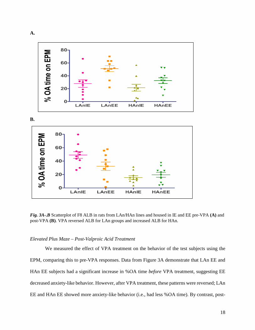
Fig. 3A-.B Scatterplot of F8 ALB in rats from LAn/HAn lines and housed in IE and EE pre-VPA (A) and post-VPA (B). VPA reversed ALB for LAn groups and increased ALB for HAn.
Elevated Plus Maze – Post-Valproic Acid Treatment
We measured the effect of VPA treatment on the behavior of the test subjects using the
EPM, comparing this to pre-VPA responses. Data from Figure 3A demonstrate that LAn EE and
HAn EE subjects had a significant increase in %OA time before VPA treatment, suggesting EE
decreased anxiety-like behavior. However, after VPA treatment, these patterns were reversed; LAn
EE and HAn EE showed more anxiety-like behavior (i.e., had less %OA time). By contrast, post-

19
VPA, a substantial increase in %OA time was observed in the LAn IE (Fig. 3B). Although there
was a decreasing trend in %OA time of the HAn rats, HAn EE rats explored the OA more than the
HAn IE post-VPA. Figure 3A-B below shows the bar graphs comparing average %OA time for
animals pre (salmon-colored bar) and post (orange) VPA in animals reared in IE (Fig. 3A) and EE
(Fig. 3B). Rats in IE spent less % time on the OA before treatment, as opposed to rats in EE. LAn
animals of both housing conditions had extreme differences in their behavior on the OA before
and after treatment. Before exposure to VPA, animals in the EE (Fig. 4B) spent more time on the
open arms, after exposure, the same animals (both LAn and Han) become less active on the OA.
IE animals (Fig. 4A), however, spent more time on the OA after valproic acid treatment in drinking
water.
A.
B.
20
Fig. 4A-B Bar graph of %OA in LAn/HAn lines for pre- and post-VPA after IE-housing (A) and after EE-housing (B). We found a significant effect of housing and treatment (*p<0.05, compared to pre-VPA, same trait; #p<0.05, LAn v. HAn).
A.
B.
21
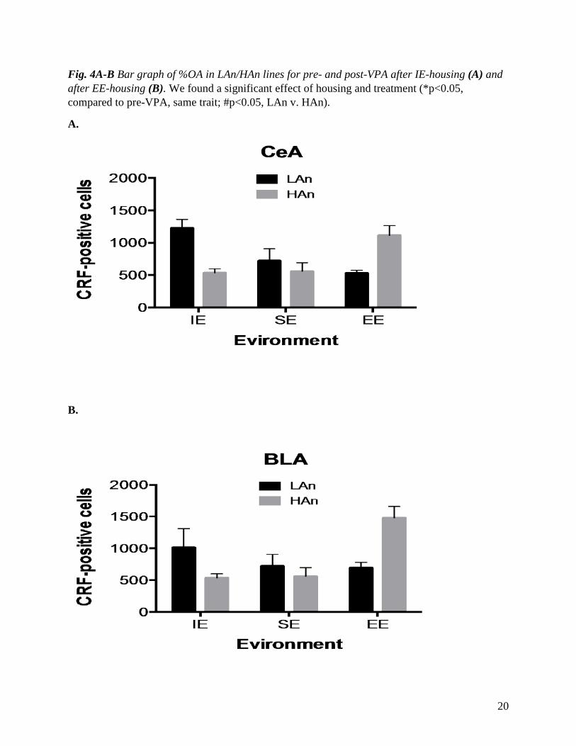
Fig. 5A-B. Bar graph of immunohistochemistry results for the central amygdala (3A) and basolateral amygdala (3B). A significant Trait x Environment effect indicated greater CRH-ir in the LAn IE and HAn EE rats for CeA and BLA (p<0.01).
Effect of Valproic Acid on Corticotropin Releasing Hormone.
To analyze the effects of valproic acid on stress hormones, we measured the level of CRH
in the different brain regions including the central amygdala (CeA) and basolateral amygdala
(BLA). As shown in Fig. 5A, there was a noticeable difference in the expression of CRH hormone
in the amygdala across the different housing settings. Our analysis revealed that the level of CRH-
positive cells was significantly decreased in LAn EE and increased in HAn EE housed Long Evan
rats. Within the IE housed rats, there were more CRH-positive cells in the LAn IE group as
opposed to HAn IE. In both brain regions, SE housed Long Evan rats did not show a significant
change in CRH-positive cell counts, with LAn SE having slightly more CRH cells than HAn SE.
Overall, comparisons between the housing conditions showed that there were more CRH-positive
cell counts in LAn IE housed rats in the CeA, as compared to BLA, which indicated more CRH-
positive cells in HAn EE. SE housed rats did not shift much in the CRH cell counts in either brain
region.
In the hypothalamus, CRH protein levels were significantly increased in HAn EE Long
Evan Rats as opposed to other housing groups. In contrast, there was a very noticeable difference
between LAn EE and Han EE, as the former had significantly less CRH levels measured. As with
the amygdala, SE housed Long Evan rats did not show much difference in the level of CRH across
anxiety levels in the hypothalamus. LAn SE had slightly more levels of CRH measured in the
hypothalamus than HAn SE. In addition, LAn IE had more CRF-positive cells than HAn IE in the
hypothalamus. CRH-positive cells observed from the hippocampus were not as diverse as the
22
results from the amygdala and hypothalamus across housing groups, but a significant difference
was observed when anxiety levels were compared within their distinct housing environments.
Almost all HAn trait Long Evan Rats had lower levels of CRH-positive cells measured compared
to their colleagues with Low Anxiety trait within the same housing environment. An exception
from the above statement was observed in SE housed Long Evan rats, which showed more CRH-
positive cells in High Anxiety Long Evan Rats. In comparison to IE, LAn EE had significantly
increased CRH-positive cell counts as opposed to LAn IE and HAn IE housed Long Evan rats. In
the hippocampus, SE housed Long Evan Rats showed a significant change in CRH-positive cell
counts, with HAn SE having more CRH-positive cells measured, than in LAn SE Long Evan Rats.
Discussion
Results from our experiment demonstrated that environmental enrichment has a positive
effect on 9th generation, Long Evans male rats selectively bred for trait anxiety. We demonstrated
that the enriched environment reversed the high anxiety behavioral and neural responses to
anxiogenic stimuli (Ravenelle et al. 2013). In addition, our results show that treatment with VPA,
presumably acting as a histone deacetylase (HDAC) inhibitor, conferred similar benefits for the
animals reared in the isolated environment. Finally, given the coordinating role of the CRH in
mammalian stress response, our investigations suggest that brain changes in CRH protein
expression following VPA treatment may underlie the benefits to anxiety of this treatment. VPA
has been shown to induce epigenetic action that includes interactions with brain CRH neuronal
systems. Our findings further implicate this phenomenon as correlative CRH protein level changes
in hypothalamic and extra-hypothalamic systems accompanied the modification(s) in VPA-treated
rats reared in isolated environment as opposed to those that are already enriched (i.e., housed in
EE)
23
Environmental conditions effect elevated plus maze activity
Prior to environmental housing, the test subjects were selectively separated based on a
median split separating lower quartile %OA (HAn) and higher quartile % OA (LAn). In general,
HAn subjects showed significantly less activity on the open arm as opposed to LAn subjects before
housing, after housing a reverse was observed. It was also observed, however, that both LAn and
HAn of IE decreased in activity tremendously after housing indicating an increase in anxiety level.
High Anxiety EE animals showed significantly less activity on the OA of EPM before housing
averaging 20 %; after housing, we observe that the trend significantly increases from 20% to 51.2%
OA time. In addition, SE Han animals showed a significant increase in % OA after housing from
25% to 35%. This trend demonstrates the significant impact of environment to the anxiety trait. In
summary, rearing conditions in this experiment exerted an influence on behavior on the EPM. Our
results were consistent with those of other studies when we examined the locomotor activity on
the elevated plus maze following environmental enrichment since Ravenelle et al. (2013) found
that EE conditions increased the percent open arm exploration as well as SE conditions, as opposed
to IE.
Enriching the environment with toys, ladders and socializing the test subjects provided
them an opportunity to interact and exercise all of which have been shown to improve the anxiety
and stress levels in affected subjects. This exposure to numerous activities works as a medium of
eliminating stressful anticipations hence motivating the animals to explore potentially stressful
environments such as OA on the EPM. The lack of sensorimotor, visual and social exposure in
isolated environments may explain the observed tremendous decrease in both LAn and Han
exploration of OA after housing. The rats in the IE did not have toys, ladders or fellow rats to
interact with and keep them busy hence increasing their stress and anxiety levels. This is observed
24
when the baseline EPM %OA time decreases from 40% in LAn and 35% HAn to 22% and 28%
respectively.
Valproic acid interaction with the environment and resulting effects on trait anxiety
In this research, we also examined the combined effect of environmental enrichment and
drug treatment in combating anxiety-like behavior in rats. Our findings from this research were
incompatible with other studies suggesting an increase in locomotor activity in anxiety trait rats
after treatment with VPA. Given that VPA is an HDAC inhibitor, we speculated that Valproic acid
would interact with the environment and affect the stress response system by altering the CRH
systems for the anxious Long Evans rats reared in isolation. There is supporting evidence in VPA
rats (rats treated prenatally with VPA on gestation day 12) are more anxious after environmental
enrichment have lower locomotor activity, enhanced exploratory activity and decreased anxiety
(Schneider et al. 2006; 2007; Ravenelle et al. 2013).
Behavioral results from our research, however, show that HDAC alterations do not benefit
environmental enrichment in reversing anxiety-like behavior. Before VPA treatment, EE rats had
a significant increase in %OA time, suggesting the successful effect of the enrichment on
decreasing anxiety-like behavior. However after VPA treatment, these patterns reversed as both
LAn EE and HAn EE decreased in %OA time, demonstrating an increase in anxiety level in the
Long Evans rats used in this experiment. It is, however, interesting to note that the rats that were
not enriched or reared in a social environment (IE) benefitted from VPA treatment. IE animals
spent more time on the OA after exposure to VPA, hence showing that the anxiety-like behavior
of the rats was reduced, but at a very low rate.
Valproic acid interaction with the environment and resulting effects on CRH proteins
25
Our suspicion that the combined treatment of VPA and EE would lower anxiety levels of
HAn rats was challenged. However, we did speculate that VPA and EE likely impacted protein
expression of CRH in brain areas important for anxiety. Analysis of post-mortem brains after VPA
treatment showed increased levels of CRH protein concentrations in multiple brain regions of HAn
EE housed animals, an indication of increased anxiety as was observed in our behavioral tests.
CRH is a neuropeptide mediator of the stress axis and physiological responses in the central
nervous system, and it is secreted by the neurons in the paraventricular nucleus of the
hypothalamus, amygdala and hippocampus. Clinically, CRH protein levels are increased in
individuals presenting with behavioral disorders including anxiety, and associated symptoms can
be reversed with treatment such as valproic acid. Valproic acid treatment, commonly used to treat
epileptic patients, and a novel treatment for anxiety and depression, was also successful in
improving anxiety in our LAn lines, which are also extreme anxiety animals since low anxiety is
abnormal and can adversely impact survival of animals. VPA was not successful in ameliorating
anxiety responses in the other extreme, our HAn lines, though it was successful for isolated
environment (IE) Long Evans rats in this study. From these findings, it would be safe to report that
both environmental enrichment and valproic acid are good treatments for anxiety disorders, but
not when used as co-treatments, particularly for those suffering from high anxiety. Based on the
brain analysis, it would be safe to add that if the combined treatment is considered, it might be
beneficial in treating low or moderate anxiety levels at a small scale. It is also important to report
that our findings may differ from other research results, further highlighting an important
therapeutic role for measuring pre-existing anxiety levels prior to treatment. For example,
Schneider et al. (2007) findings show that the combination of VPA and enriched environment
combat anxiety like behavior, while our results show the opposite. This difference may be due to
26
the fact that our research deals with postnatal treatment administered through drinking water, while
in the study by Schneider et al. (2007) the authors administered VPA during embryogenesis, and
that the authors did not determine pre-existing anxiety levels before treatment.
Conclusion
In summary, we found that Environment Enrichment may serve as a novel therapy and
treatment for anxiety-like behavior. These findings are in support of several recent studies that
reported an increase in EPM activity of HAn EE long Evan Rats (Ravenelle et al. 2013,) signifying
reduced indices of anxiety (Varman et al. 2012). Findings in the present study provide evidence
that administration of Valproic Acid may serve as a probable treatment for Lowered anxiety-like
behavior in IE animals. This effect was however not observed when Valproic acid and EE were
combined-there was no additive effect of combining VPA and EE treatment. The CRF/CRH-
mRNA levels in the in the LAn IE measured on the low end, while high levels of CRF/CRH-
mRNA expression was observed in HAn EE housed Long Evan rats after the combination. It is
possible that the mode of administration of VPA may have contributed to discrepancy observed.
It was administered through oral administration in solution (dissolved in water), which is prone to
Fast-past effect. Although pain-free, only part of the drug may have been absorbed hence explain
the trend in EPM behavior, and CRF/CRH-mRNA observed. May be if the drug was administered
through intracranial mode of administration, or if the concentration of the drug was different, our
hypothesis of seeing additive effect of combining the two treatments may have been supported,
but this is entirely a different procedure all together which was not the focus of this research, but
would make a great further study.
From the present study the combination of Valproic acid treatment and environment
enrichment, and how they affected CRH-mRNA expression in the examined brain regions shows
27
that combining VPA and EE treatment for anxiety-like behavior may not be of additive effect for
therapeutic purposes, but it may be beneficial in cases of low or moderate anxiety-like behavior.
A better understanding of effective mechanisms of administration, and time of treatment on how
VPA may alter anxiety-like behavior, will be required to achieve this goal.
ACKNOWLEDGEMENTS
I would like to thank Dr. Tiffany Donaldson who was not only my mentor and PI but also helped
and guided me every step of the way during this research, I thank Vanessa Romero, Robert O,
Joseph Lusse and all the other students in Dr. Donaldson’s lab for all their help. Lastly I would
also like to thank the Honors College department for sponsoring and financing my research.
28
References
Balfour, J. A., & Bryson, H. M. (1994). Valproic acid. CNS Drugs, 2(2), 144-173.
Chapillon, P., Manneche, C., Belzung, C., & Caston, J. (1999). Rearing environmental
enrichment in two in bred starins of mice: 1. effects on emotional reactivity. Behavior
Genetics, 29(1), 41-46.
Dautzenberg, Frank M., et al. "Molecular biology of the CRH receptors—in the
mood." Peptides 22.5 (2001): 753-760.
Dunn, Adrian J., and Artur H. Swiergiel. "Behavioral responses to stress are intact in CRF-
deficient mice." Brain research 845.1 (1999): 14-20.
Fakuchi, M., Nii, T., Ishinaru, N., Minamino, A., Hara, D., Takasaki, I., Tabuchi, A., & Tsuda, M.
(2009). Valproic acid induces up- or down-regulation of gene expression responsible for
the neuronal excitation and inhibition in rat cortical neurons through its epigenetic actions.
Neuroscience Research, 65(1), 35-43.
Gilmor, M. L., Skelton, K. H., Nemeroff, C. B., & Owens, M. J. (2003). The effects of chronic
treatment with the mood stabilizers Valproic acid and lithium on corticotropin-releasing
factor neuronal systems. Journal of Pharmacology and Experimental Therapeutics,
305(2), 434-439.
Grammatopoulos, Dimitris K., and George P. Chrousos. "Functional characteristics of CRH
receptors and potential clinical applications of CRH-receptor antagonists." Trends in
Endocrinology & Metabolism 13.10 (2002): 436-444.
29
Huising, M. O., et al. "Structural characterisation of a cyprinid (Cyprinus carpio L.) CRH, CRH-
BP and CRH-R1, and the role of these proteins in the acute stress response." Journal of
Molecular Endocrinology 32.3 (2004): 627-648.
Isaacson, J. S., Solis, J. M., & Nicoll, R. A. (1993). Local and diffuse synaptic actions of GABA
in the Hippocampus. Neuron, 10(2), 165-175.
Keck, M. E., Sartori, S. B., Welt, T., Müller, M. B., Ohl, F., Holsboer, F & Singewald, N. (2005).
Differences in serotonergic neurotransmission between rats displaying high or low
Anxiety/depression‐like behaviour: effects of chronic paroxetine treatment. Journal of
Neurochemistry, 92(5), 1170-1179.
Künzel, Heike E., et al. "Treatment of depression with the CRH-1-receptor antagonist R121919:
endocrine changes and side effects." Journal of psychiatric research 37.6 (2003): 525-533.
Le-Niculescu, H., Balaraman, Y., Patel, S. D., Ayalew, M., Gupta, J., Kuczenski, R., & Niculescu,
A. B. (2011). Convergent functional genomics of anxiety disorders: translational
identification of genes, biomarkers, pathways and mechanisms. Translational psychiatry,
1(5), e9.
Lovejoy, David A., et al. "Molecular evolution of GPCRS: CRH/CRH receptors." Journal of
molecular endocrinology 52.3 (2014): T43-T60.
Lowry, Christopher A., and Frank L. Moore. "Regulation of behavioral responses by corticotropin-
releasing factor." General and comparative Endocrinology 146.1 (2006): 19-27.
30
Machado‐Vieira, Rodrigo, Lobna Ibrahim, and Carlos A. Zarate Jr. "Histone Deacetylases and
Mood Disorders: Epigenetic Programming in Gene‐Environment Interactions." CNS
Neuroscience & Therapeutics 17.6 (2011): 699-704.
Mazon, de AF, et al. "Corticotropin-releasing hormone-receptor 1 (CRH-R1) and CRH-binding
protein (CRH-BP) are expressed in the gills and skin of common carp Cyprinus carpio L.
and respond to acute stress and infection." Journal of experimental biology 209.3 (2006):
510-517.
Morley‐Fletcher, S., Rea, M., Maccari, S., & Laviola, G. (2003). Environmental enrichment during
adolescence reverses the effects of prenatal stress on play behaviour and HPA axis
reactivity in rats. European Journal of Neuroscience, 18(12), 3367-3374.
Pariante, Carmine M., and Stafford L. Lightman. "The HPA axis in major depression: classical
theories and new developments." Trends in neurosciences 31.9 (2008): 464-468.
Ravenelle, R., Byrnes, E. M., Byrnes, J. J., McInnis, C., Park, J. H., & Donaldson, S. T. (2013).
Environmental enrichment effects on the neurobehavioral profile of selective outbred trait
anxiety rats. Behavioural Brain Research.
Schloesser, R. J., Lehmann, M., Martinowich, K., Manji, H. K., & Herkenham, M. (2010).
Environmental enrichment requires adult neurogenesis to facilitate the recovery from
psychosocial stress. Molecular psychiatry, 15(12), 1152-1163.
Schneider, T., Ziölkowska, B., Gieryk, A., Tyminska, A., Przewłocki, R. (2007). Prenatal
exposure to valproic acid disturbs the enkephalinergic system functioning, basal hedonic

31
tone, and emotional responses in an animal model of autism. Psychopharmacology 193:
547-55.
Schneider, T., Turczak, J., & Przewłocki, R. (2006). Environmental enrichment reverses
behavioral alterations in rats prenatally exposed to Valproic acid: issues for a
therapeutic approach in autism. Neuropsychopharmacology, 31(1), 36-46.
Soiza-Reilly, M., Anderson, W. B., Vaughan, C. W., & Commons, K. G. (2013). Presynaptic
gating of excitation in the dorsal raphe nucleus by GABA. Proceedings of the National
Academy of Sciences, 110(39), 15800-15805.
Stansbury, K., & Gunnar, M. R. (1994). Adrenocortical activity and emotion regulation.
Monographs of the Society for Research in Child Development, 59(2‐3), 108-134.
Stout, Steven C., et al. "Effects of sodium valproate on corticotropin-releasing factor systems in
rat brain." Neuropsychopharmacology 24.6 (2001): 624-631.
Tringali, Giuseppe, et al. "Valproic acid inhibits corticotropin-releasing factor synthesis and
release from the rat hypothalamus in vitro: evidence for the involvement of GABAergic
neurotransmission." Journal of Psychiatry and Neuroscience 29.6 (2004): 459.
Van Ameringen, M., Mancini, C., Pipe, B., & Bennett, M. (2004). Antiepileptic drugs in the
treatment of anxiety disorders. Drugs, 64(19), 2199-2220.
Varman, Durairaj Ragu, Ganapathy Marimuthu, and Koilmani Emmanuvel Rajan. "Environmental
enrichment exerts anxiolytic effects in the Indian field mouse (Mus booduga)." Applied
Animal Behaviour Science 136.2 (2012): 166-173.
Recommended