The role of SUPT6H in gliomagenesis
23/01/2015
PragueONCO2015
Rikke Darling Rasmussen Brain Tumor Biology
Danish Cancer Society Research Center
Genome Integrity Copenhagen, Denmark

Gliomas
Neuroglia, 2012
- 3000 cases primary CNS tumors/year
- 90% die within 2-5 years post diagnosis - the most malignant variant - GBM
Angiogenesis
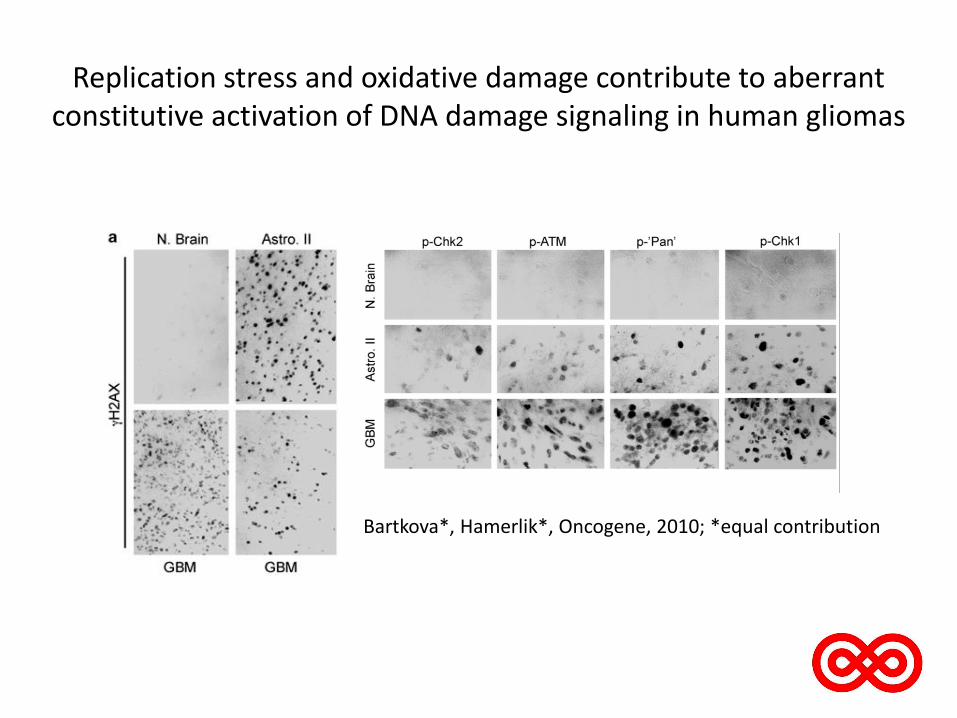
Replication stress and oxidative damage contribute to aberrant constitutive activation of DNA damage signaling in human gliomas
Bartkova*, Hamerlik*, Oncogene, 2010; *equal contribution
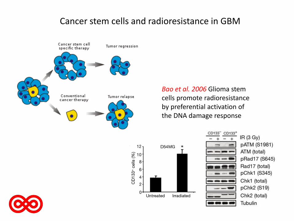
Cancer stem cells and radioresistance in GBM
Bao et al. 2006 Glioma stem cells promote radioresistance by preferential activation of the DNA damage response

CD133-positive glioma-derived cancer stem cells (GSCs) : - search for genes whose knock-down would result in changes: • proliferation rates (EdU pulse labeling) • spontaneous DNA damage (gH2AX)
i.e. candidate genes important for GSCs maintenance, repair efficiency and self-renewal
Readout: ScanR microscopy screening station
siRNA library: against 360 genes
3 validated independent siRNA performed as pooled siRNA screen

Experimental setup
Papain dissociation CD133+
CD133-
Confirmation of stem cell phenotype by qPCR
(GFAP,Sox2,Oct, Musashi, CD133)
Read-out on ScanR

Hit from siRNA screen: SUPT6H (suppressor of Ty 6 homolog)
• Encodes the protein Spt6 • Histone chaperone interacting with H3, H4 and H2b • Involved in both assembly and disassembly of the DNA • Spt6 regulates chromatin structure and gene expression
• Involved in the differentiation of stem cells (AH Wang et al., The EMBO journal, 2013, Kedes et al., J
Cell Physiol, 2003)
• Spt6 is required for proper activation of Notch signaling pathway genes shown in a zebrafish model(F.O. Kok et al., Developmental Biology, 2007)
HtH, helix-turn-helix domain, binds to double-stranded DNA; YqgFc, predicted to be a resolvase or ribonuclease, but in Spt6, catalytic residues are exchanged, thus probably not active; HhH, triple-helix-domain, binding to double-stranded DNA; S1, RNA-binding domain SH2-N, SH2-C, tandem SH2 domains, binds phosphorylated Ser residues
1 1726aa
Sun M et al. J. Biol. Chem. 2010;285:41597-41603

Con si34 si35 si36
Spt6 TUB
Spt6 is expressed in primary GBM
DAPI Emb-5
Spt6
LaminB
T1 T2 T3

GBM cells show decreased proliferation rates and increased DNA damage after Spt6 knock-down
EdU yH2AX DAPI
KD SUPT6H
EdU yH2AX DAPI
Con
0
100
200
300
400
500
600
siCon siEmb-5
Mean Intensity EdU
Spt6
0
100
200
300
400
500
600
siCon siEmb-5
Mean Intensity yH2AX
Spt6
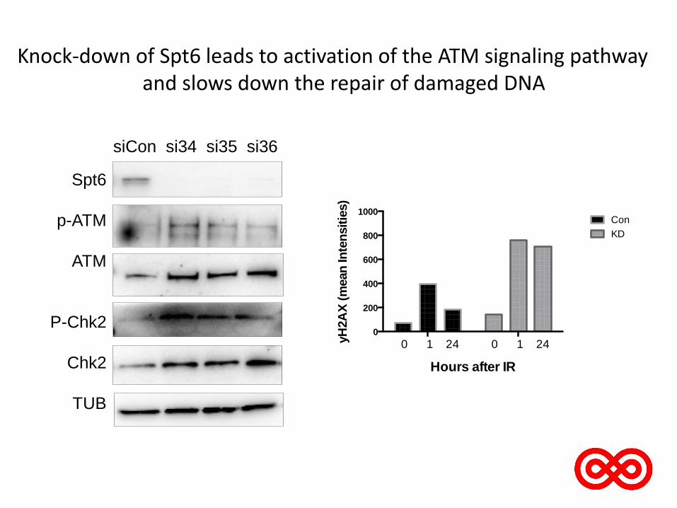
Knock-down of Spt6 leads to activation of the ATM signaling pathway and slows down the repair of damaged DNA
siCon si34 si35 si36
Spt6
p-ATM
ATM
P-Chk2
Chk2
TUB
Con
KD
0
200
400
600
800
1000
Hours after IR
yH
2A
X (
mean
In
ten
sit
ies)
Con
KD
0 1 24 0 1 24
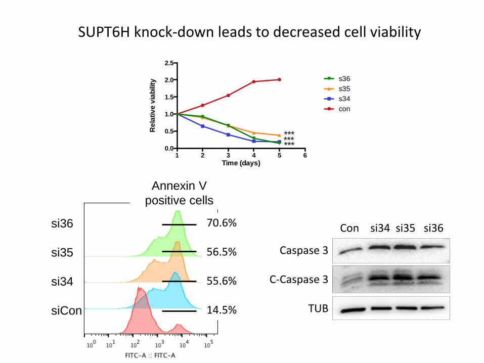
SUPT6H knock-down leads to decreased cell viability
Con si34 si35 si36
Caspase 3
C-Caspase 3
TUB
si36
si35
si34
siCon
Annexin V
positive cells
1 2 3 4 5 60.0
0.5
1.0
1.5
2.0
2.5
Time (days)
Re
lativ
e v
iab
ility
con
s34
s35
s36
*** *** ***
70.6% 56.5% 55.6% 14.5%

SUPT6H knock-down arrests GBM cells in G1 and G2-M phase of the cell cycle
DMSO Aph CDKi NOC
Spt6
Lamin b
Con S34 S35 S36 E
dU
-AF
647
EdU
-AF
647
EdU
-AF
647
Ce
ll cy
cle
EdU
-AF
647
Hoechst Hoechst Hoechst Hoechst
H3S
er1
0-A
F488
Mit
oti
c In
de
x
Hoechst Hoechst Hoechst Hoechst
H3S
er1
0-A
F488
H3S
er1
0-A
F488
H3S
er1
0-A
F488

Conclusion and working model
Cell cycle arrest and eventually apoptosis
Spt6 KD
Proliferation
DNA damage and activation of DDR

Thank you for your attention
Acknowledgement Brain Tumor Biology Petra Hamerlik Madhavsai Kirit Gajjar Elisabeth Anne Adanma Obara Julie la Cour Karottki Kamilla Ellermann Jensen Genome Integrity Group Jiri Bartek Jiri Bartek Jr.
Danish Cancer Society Research Center Dansk Kræftforsknings Fond
Danish Council For Independent Research
Recommended