The Past, Present and Future of Blood Gas and Electrolyte Testing. Thomas Koshy, Ph.D. Sr. Director, Scientific Affairs November 7, 2012
Disclaimers
• I work for Alere and am a scientific
representative for Epocal…
• Apologies to RTs in the audience…
• The “p” in pH is not italicized and is in pO2 and pCO2. I may not have it right in all of my slides
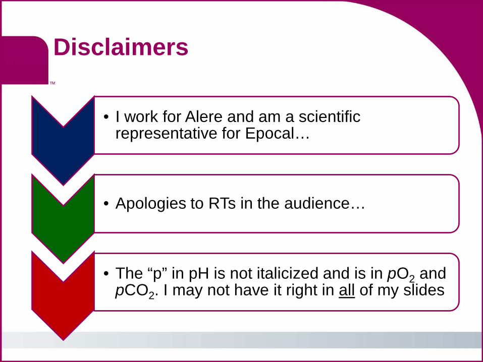
Agenda
• Why Do We Measure Blood Gasses • How Do We Measure Blood Gasses
• Why Do We Measure Electrolytes • How Do We Measure Electrolytes
• Instrument and Devices
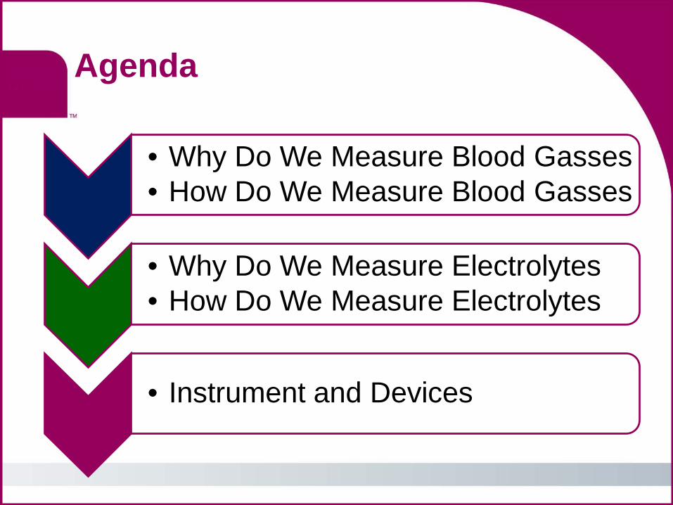
Why Do We Measure: ABGs
ABG studies may be helpful in the differential diagnosis of:
Unexplained tachypnea (increased respiration) or dyspnea
(shortness of breath)
Unexplained restlessness, drowsiness, confusion or
anxiety in bed patients or
those on O2
Assessment of surgical risk
Differential diagnosis of
possible metabolic
disturbances (i.e. sepsis,
diabetic keto-acidosis)
Before and during
prolonged oxygen
therapy and during
ventilator support of
patients (i.e. post-surgical
recovery)
Progression of cardio
pulmonary disease
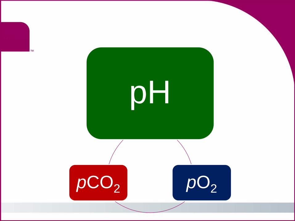
pH
pO2 pCO2
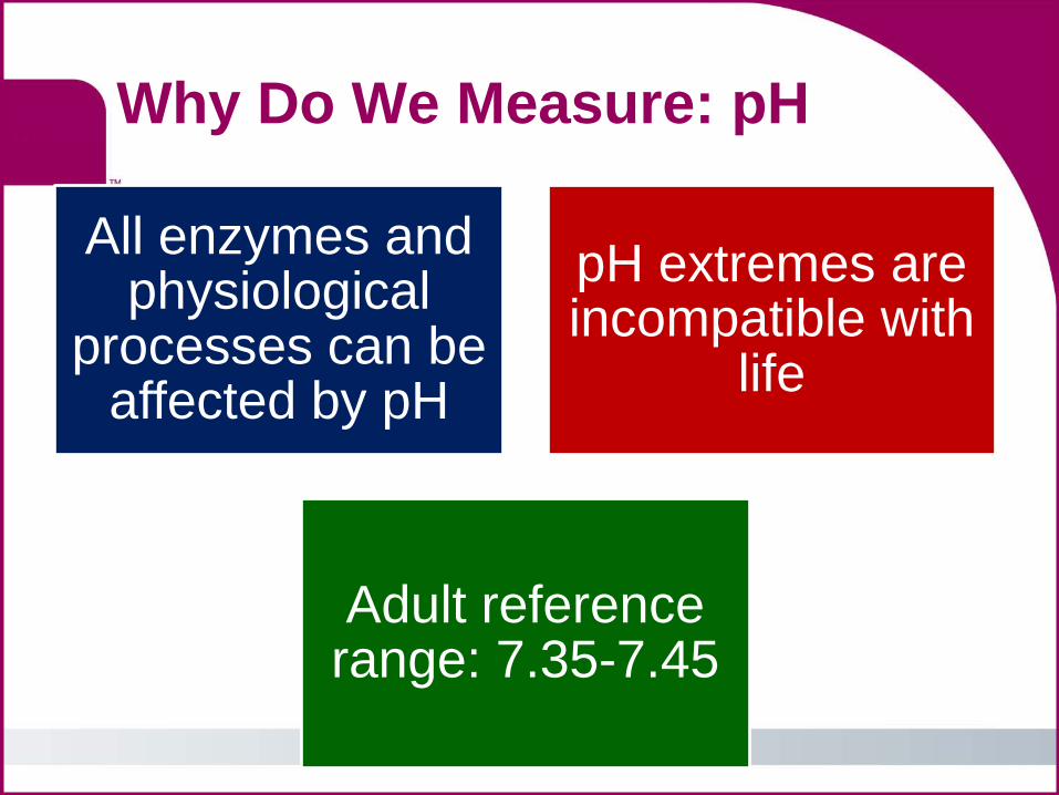
Why Do We Measure: pH
All enzymes and physiological
processes can be affected by pH
pH extremes are incompatible with
life
Adult reference range: 7.35-7.45
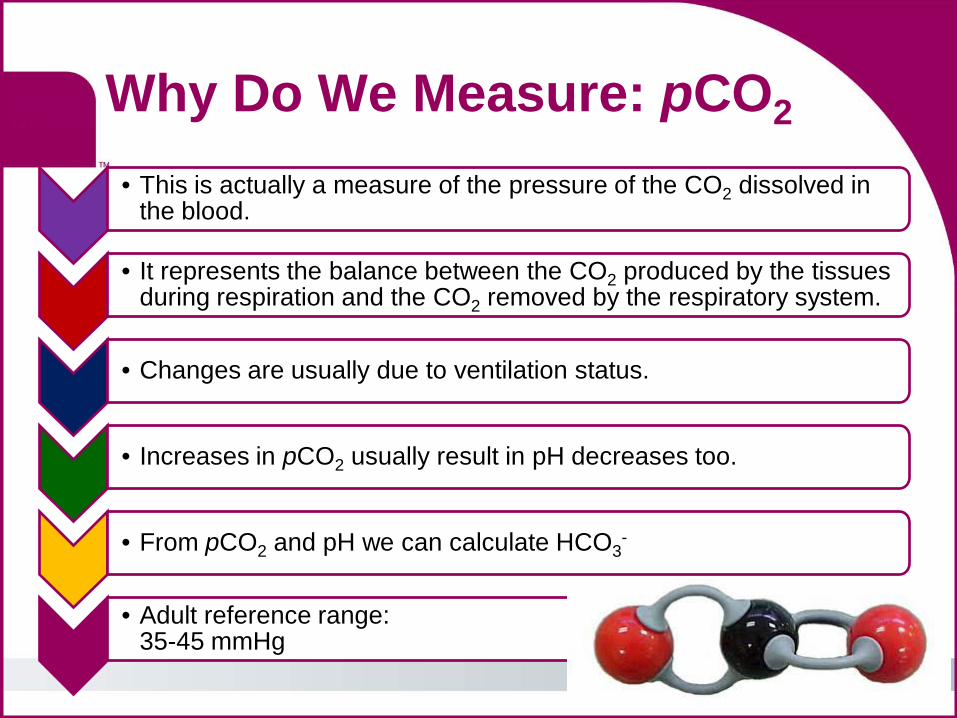
Why Do We Measure: pCO2 • This is actually a measure of the pressure of the CO2 dissolved in
the blood.
• It represents the balance between the CO2 produced by the tissues during respiration and the CO2 removed by the respiratory system.
• Changes are usually due to ventilation status.
• Increases in pCO2 usually result in pH decreases too.
• From pCO2 and pH we can calculate HCO3-
• Adult reference range: 35-45 mmHg
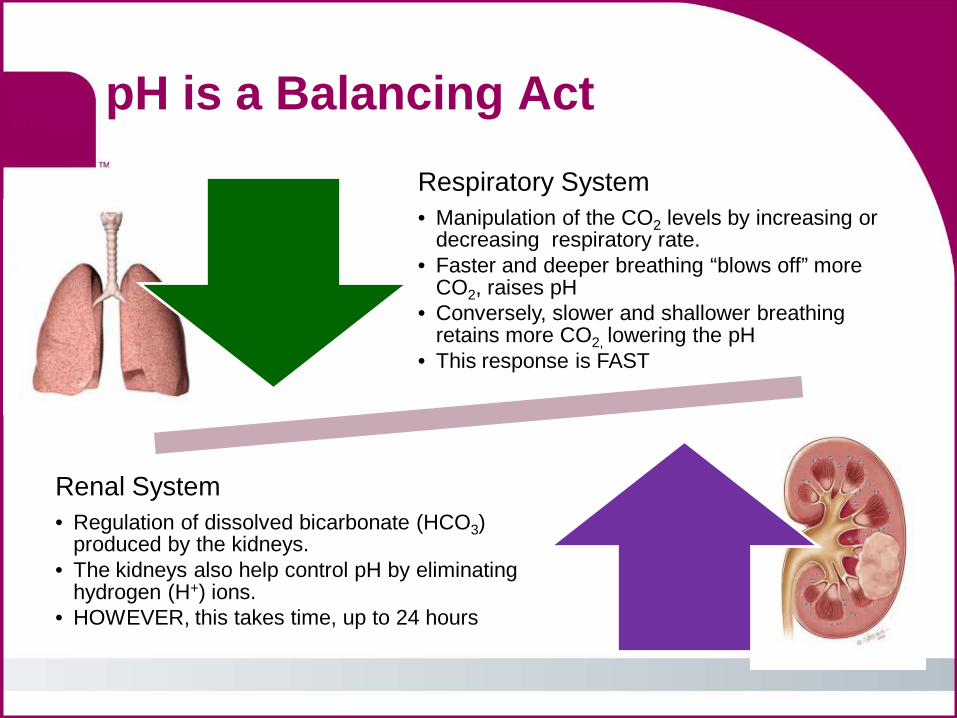
Renal System • Regulation of dissolved bicarbonate (HCO3)
produced by the kidneys. • The kidneys also help control pH by eliminating
hydrogen (H+) ions. • HOWEVER, this takes time, up to 24 hours
Respiratory System • Manipulation of the CO2 levels by increasing or
decreasing respiratory rate. • Faster and deeper breathing “blows off” more
CO2, raises pH • Conversely, slower and shallower breathing
retains more CO2, lowering the pH • This response is FAST
pH is a Balancing Act
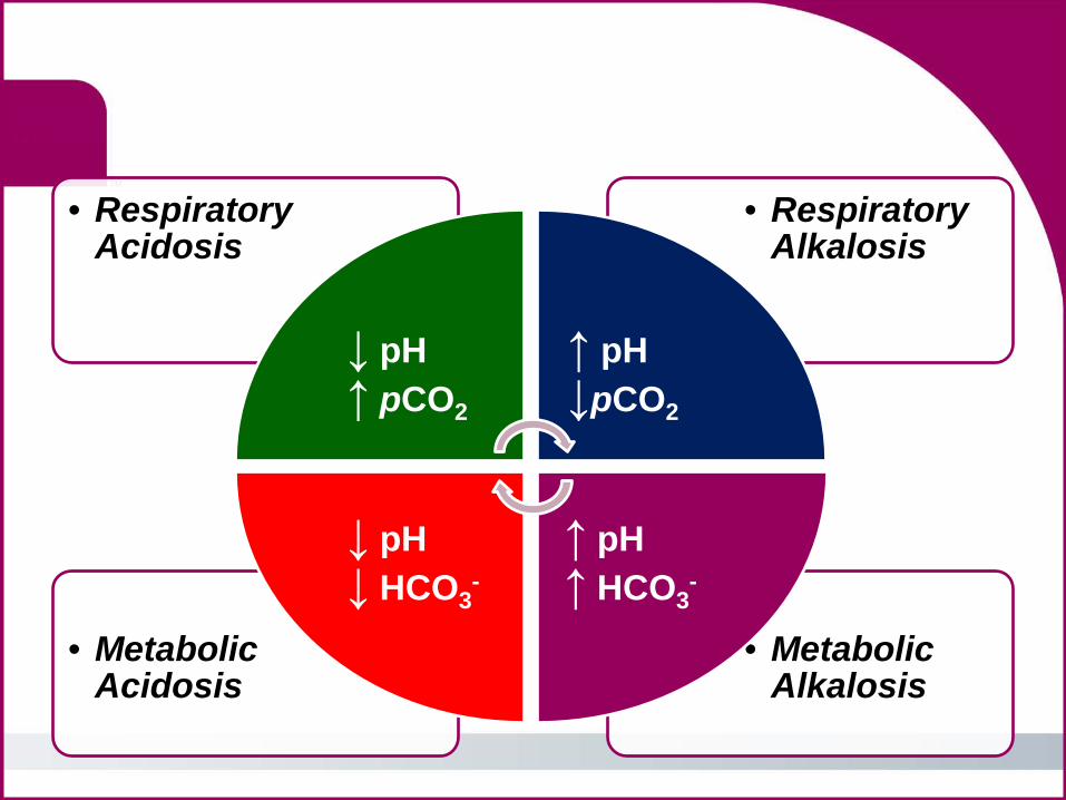
• Metabolic Alkalosis
• Metabolic Acidosis
• Respiratory Alkalosis
• Respiratory Acidosis
↓ pH ↑ pCO2
↑ pH ↓pCO2
↑ pH ↑ HCO3
- ↓ pH ↓ HCO3
-
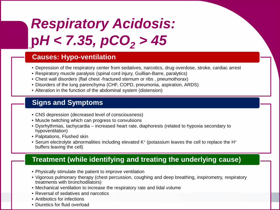
Respiratory Acidosis: pH < 7.35, pCO2 > 45 • Depression of the respiratory center from sedatives, narcotics, drug overdose, stroke, cardiac arrest • Respiratory muscle paralysis (spinal cord injury, Guillian-Barre, paralytics) • Chest wall disorders (flail chest -fractured sternum or ribs , pneumothorax) • Disorders of the lung parenchyma (CHF, COPD, pneumonia, aspiration, ARDS) • Alteration in the function of the abdominal system (distension)
Causes: Hypo-ventilation
• CNS depression (decreased level of consciousness) • Muscle twitching which can progress to convulsions • Dysrhythmias, tachycardia – increased heart rate, diaphoresis (related to hypoxia secondary to
hypoventilation) • Palpitations, Flushed skin • Serum electrolyte abnormalities including elevated K+ (potassium leaves the cell to replace the H+
buffers leaving the cell)
Signs and Symptoms
• Physically stimulate the patient to improve ventilation • Vigorous pulmonary therapy (chest percussion, coughing and deep breathing, inspirometry, respiratory
treatments with bronchodilators) • Mechanical ventilation to increase the respiratory rate and tidal volume • Reversal of sedatives and narcotics • Antibiotics for infections • Diuretics for fluid overload
Treatment (while identifying and treating the underlying cause)
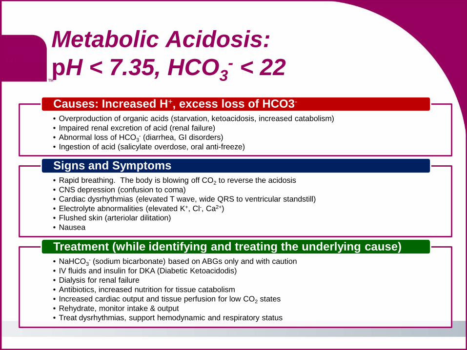
Metabolic Acidosis: pH < 7.35, HCO3
- < 22
• Overproduction of organic acids (starvation, ketoacidosis, increased catabolism) • Impaired renal excretion of acid (renal failure) • Abnormal loss of HCO3
- (diarrhea, GI disorders) • Ingestion of acid (salicylate overdose, oral anti-freeze)
Causes: Increased H+, excess loss of HCO3-
• Rapid breathing. The body is blowing off CO2 to reverse the acidosis • CNS depression (confusion to coma) • Cardiac dysrhythmias (elevated T wave, wide QRS to ventricular standstill) • Electrolyte abnormalities (elevated K+, Cl-, Ca2+) • Flushed skin (arteriolar dilitation) • Nausea
Signs and Symptoms
• NaHCO3- (sodium bicarbonate) based on ABGs only and with caution
• IV fluids and insulin for DKA (Diabetic Ketoacidodis) • Dialysis for renal failure • Antibiotics, increased nutrition for tissue catabolism • Increased cardiac output and tissue perfusion for low CO2 states • Rehydrate, monitor intake & output • Treat dysrhythmias, support hemodynamic and respiratory status
Treatment (while identifying and treating the underlying cause)
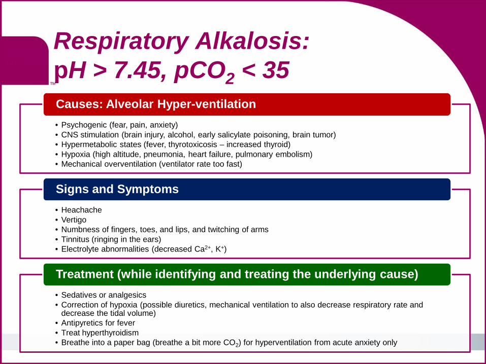
Respiratory Alkalosis: pH > 7.45, pCO2 < 35
• Psychogenic (fear, pain, anxiety) • CNS stimulation (brain injury, alcohol, early salicylate poisoning, brain tumor) • Hypermetabolic states (fever, thyrotoxicosis – increased thyroid) • Hypoxia (high altitude, pneumonia, heart failure, pulmonary embolism) • Mechanical overventilation (ventilator rate too fast)
Causes: Alveolar Hyper-ventilation
• Heachache • Vertigo • Numbness of fingers, toes, and lips, and twitching of arms • Tinnitus (ringing in the ears) • Electrolyte abnormalities (decreased Ca2+, K+)
Signs and Symptoms
• Sedatives or analgesics • Correction of hypoxia (possible diuretics, mechanical ventilation to also decrease respiratory rate and
decrease the tidal volume) • Antipyretics for fever • Treat hyperthyroidism • Breathe into a paper bag (breathe a bit more CO2) for hyperventilation from acute anxiety only
Treatment (while identifying and treating the underlying cause)
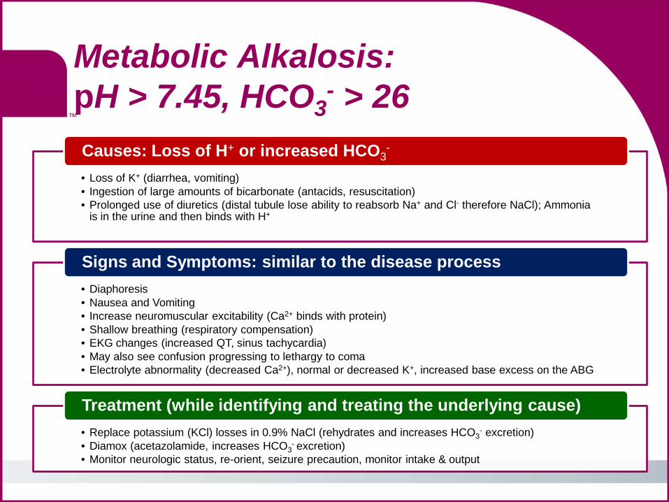
Metabolic Alkalosis: pH > 7.45, HCO3
- > 26
• Loss of K+ (diarrhea, vomiting) • Ingestion of large amounts of bicarbonate (antacids, resuscitation) • Prolonged use of diuretics (distal tubule lose ability to reabsorb Na+ and Cl- therefore NaCl); Ammonia
is in the urine and then binds with H+
Causes: Loss of H+ or increased HCO3-
• Diaphoresis • Nausea and Vomiting • Increase neuromuscular excitability (Ca2+ binds with protein) • Shallow breathing (respiratory compensation) • EKG changes (increased QT, sinus tachycardia) • May also see confusion progressing to lethargy to coma • Electrolyte abnormality (decreased Ca2+), normal or decreased K+, increased base excess on the ABG
Signs and Symptoms: similar to the disease process
• Replace potassium (KCl) losses in 0.9% NaCl (rehydrates and increases HCO3- excretion)
• Diamox (acetazolamide, increases HCO3- excretion)
• Monitor neurologic status, re-orient, seizure precaution, monitor intake & output
Treatment (while identifying and treating the underlying cause)
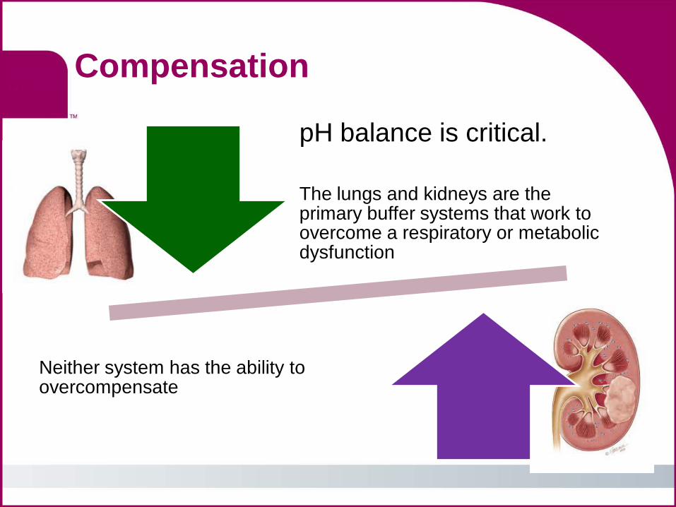
Neither system has the ability to overcompensate
pH balance is critical. The lungs and kidneys are the primary buffer systems that work to overcome a respiratory or metabolic dysfunction
Compensation
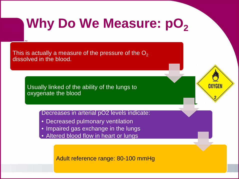
Why Do We Measure: pO2
This is actually a measure of the pressure of the O2 dissolved in the blood.
Usually linked of the ability of the lungs to oxygenate the blood
Decreases in arterial pO2 levels indicate: • Decreased pulmonary ventilation • Impaired gas exchange in the lungs • Altered blood flow in heart or lungs
Adult reference range: 80-100 mmHg
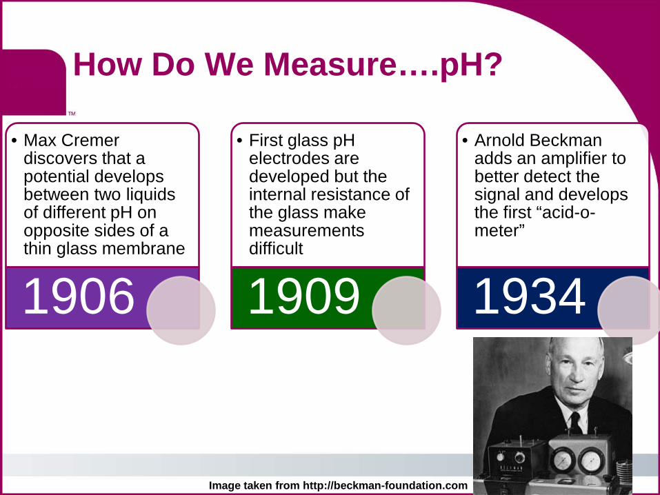
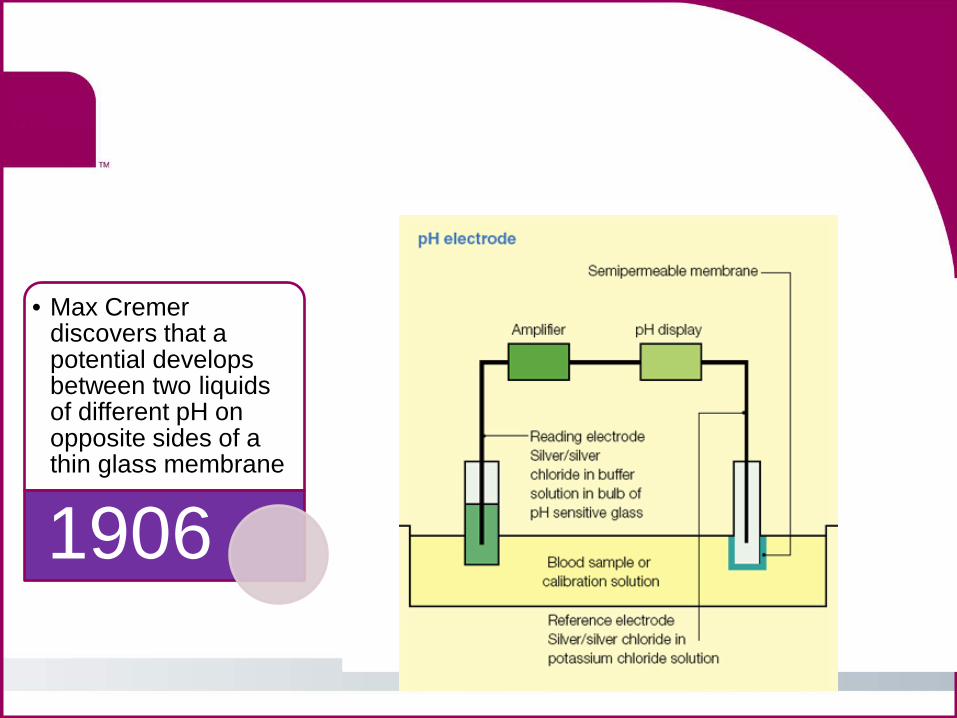
• Max Cremer discovers that a potential develops between two liquids of different pH on opposite sides of a thin glass membrane
1906
• First glass pH electrodes are developed but the internal resistance of the glass make measurements difficult
1909
• Arnold Beckman adds an amplifier to better detect the signal and develops the first “acid-o-meter”
1934
How Do We Measure….pH?
Image taken from http://beckman-foundation.com
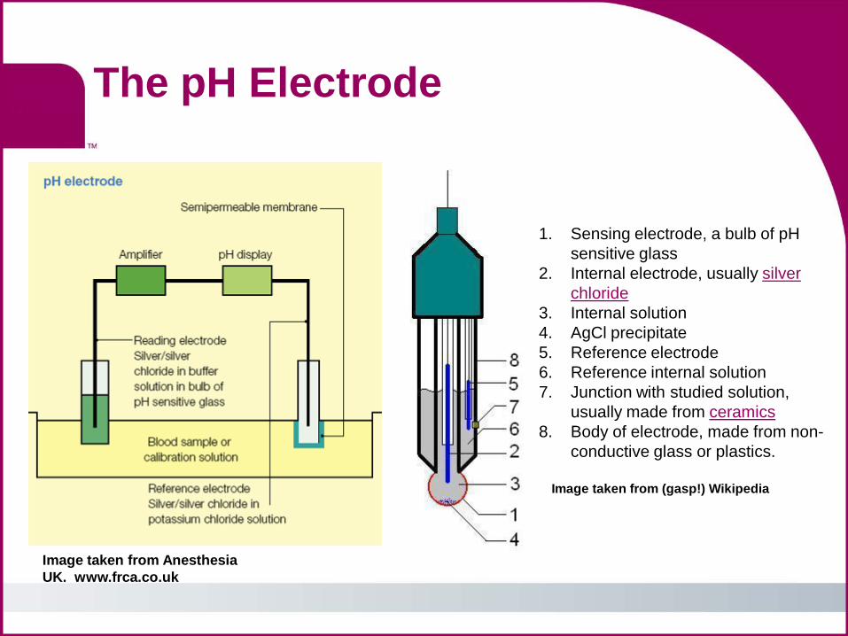
The pH Electrode
Image taken from Anesthesia UK. www.frca.co.uk
Image taken from (gasp!) Wikipedia
1. Sensing electrode, a bulb of pH sensitive glass
2. Internal electrode, usually silver chloride
3. Internal solution 4. AgCl precipitate 5. Reference electrode 6. Reference internal solution 7. Junction with studied solution,
usually made from ceramics 8. Body of electrode, made from non-
conductive glass or plastics.
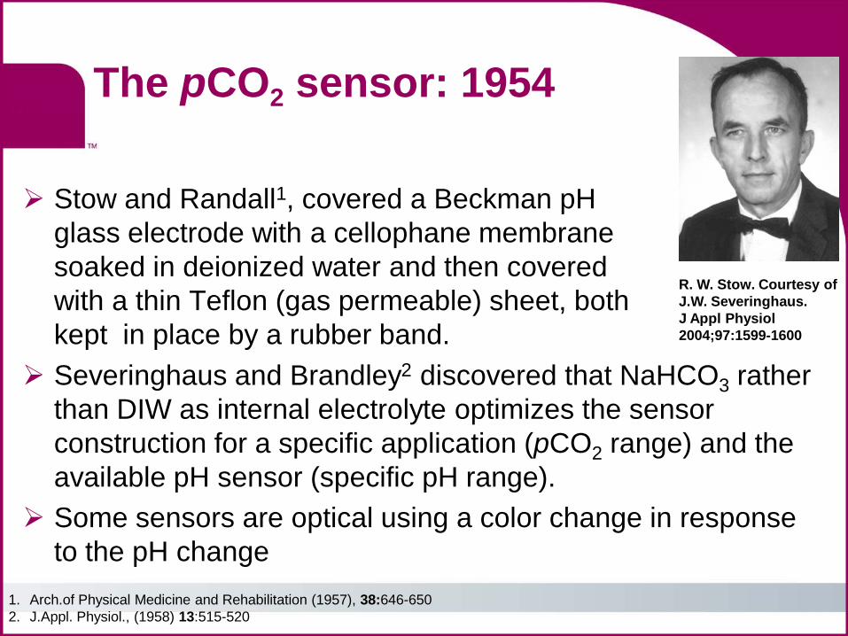
The pCO2 sensor: 1954
Stow and Randall1, covered a Beckman pH glass electrode with a cellophane membrane soaked in deionized water and then covered with a thin Teflon (gas permeable) sheet, both kept in place by a rubber band.
Severinghaus and Brandley2 discovered that NaHCO3 rather than DIW as internal electrolyte optimizes the sensor construction for a specific application (pCO2 range) and the available pH sensor (specific pH range).
Some sensors are optical using a color change in response to the pH change
R. W. Stow. Courtesy of J.W. Severinghaus. J Appl Physiol 2004;97:1599-1600
1. Arch.of Physical Medicine and Rehabilitation (1957), 38:646-650 2. J.Appl. Physiol., (1958) 13:515-520
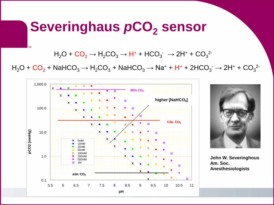
H2O + CO2 → H2CO3 → H+ + HCO3- → 2H+ + CO3
2-
H2O + CO2 + NaHCO3 → H2CO3 + NaHCO3 → Na+ + H+ + 2HCO3- → 2H+ + CO3
2-
0.1
1.0
10.0
100.0
1,000.0
5.5 6 6.5 7 7.5 8 8.5 9 9.5 10 10.5 11
pH
pCO
2 [m
mH
g]
5mM10mM20mM50mM100mM200mM500mM1M
higher [NaHCO3]
atm CO2
CAL CO2
95% CO2
Severinghaus pCO2 sensor
John W. Severinghous Am. Soc. Anesthesiologists
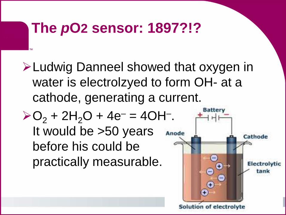
The pO2 sensor: 1897?!?
Ludwig Danneel showed that oxygen in water is electrolzyed to form OH- at a cathode, generating a current. O2 + 2H2O + 4e– = 4OH–.
It would be >50 years before his could be practically measurable.
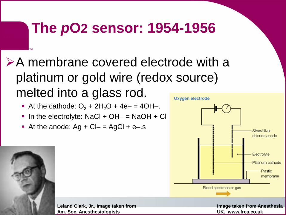
The pO2 sensor: 1954-1956
A membrane covered electrode with a platinum or gold wire (redox source) melted into a glass rod. At the cathode: O2 + 2H2O + 4e– = 4OH–. In the electrolyte: NaCl + OH– = NaOH + Cl At the anode: Ag + Cl– = AgCl + e–.s
Leland Clark, Jr., Image taken from Am. Soc. Anesthesiologists
Image taken from Anesthesia UK. www.frca.co.uk
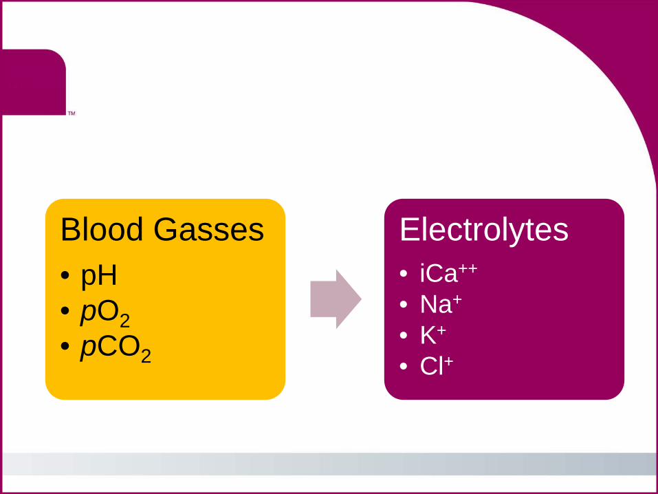
Blood Gasses • pH • pO2 • pCO2
Electrolytes • iCa++
• Na+
• K+
• Cl+
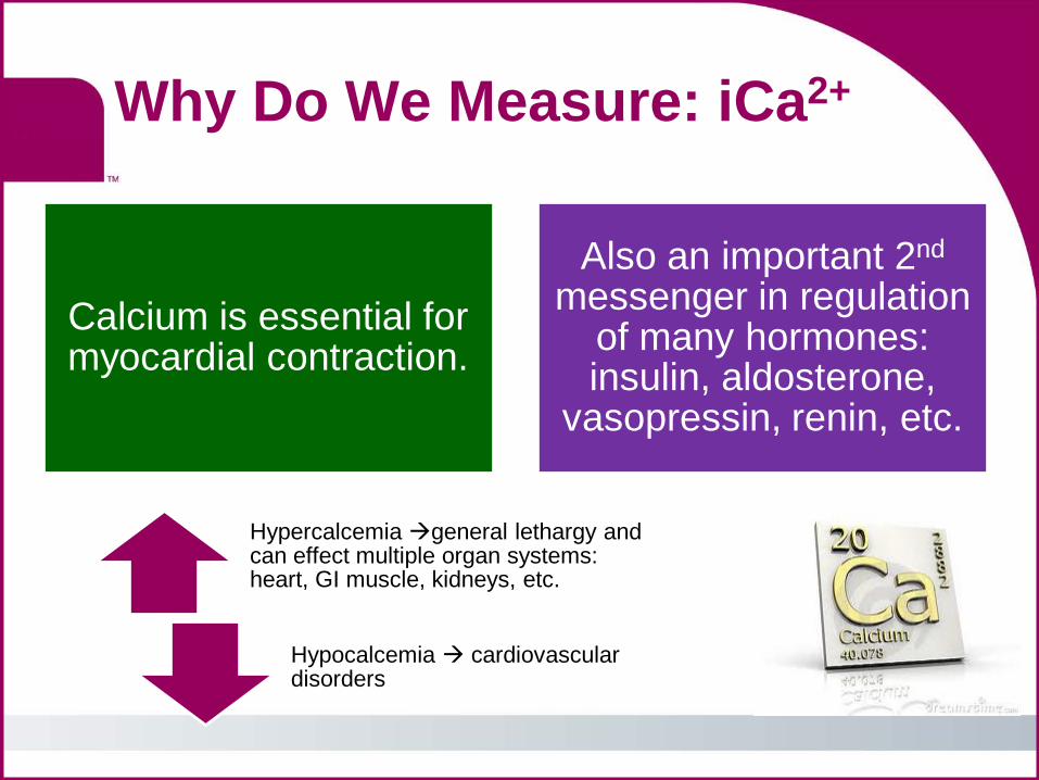
Why Do We Measure: iCa2+
Calcium is essential for myocardial contraction.
Also an important 2nd messenger in regulation
of many hormones: insulin, aldosterone,
vasopressin, renin, etc.
Hypercalcemia general lethargy and can effect multiple organ systems: heart, GI muscle, kidneys, etc.
Hypocalcemia cardiovascular disorders
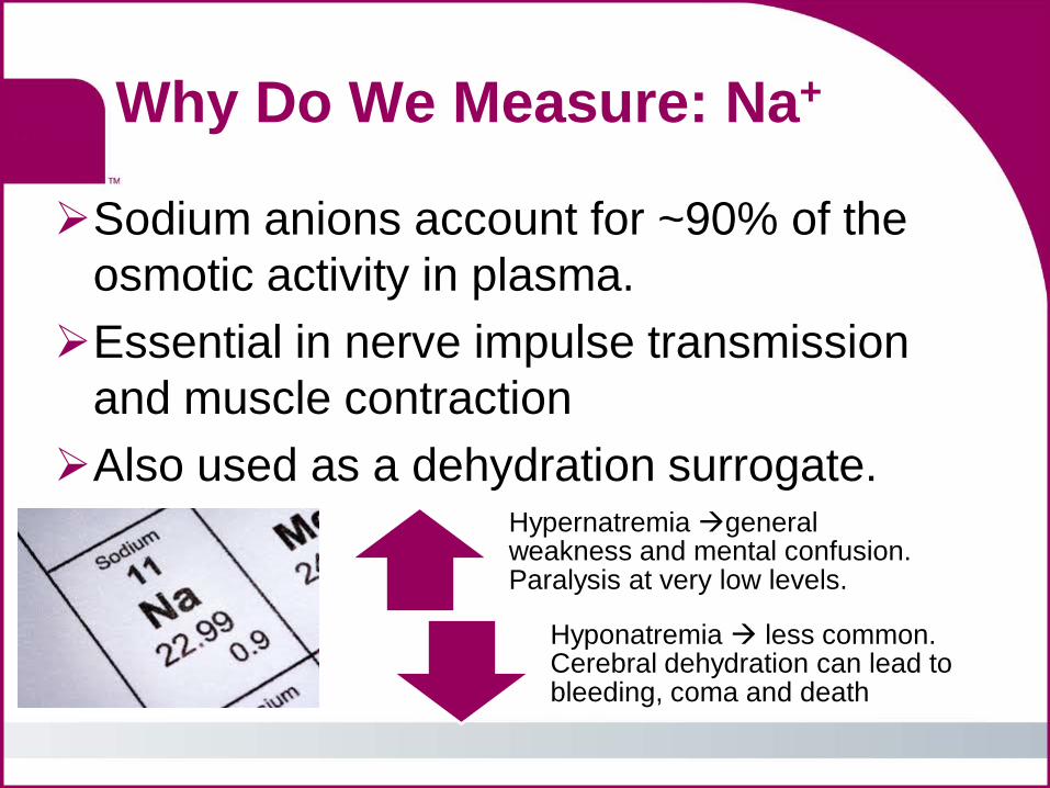
Hypernatremia general weakness and mental confusion. Paralysis at very low levels.
Hyponatremia less common. Cerebral dehydration can lead to bleeding, coma and death
Why Do We Measure: Na+
Sodium anions account for ~90% of the osmotic activity in plasma. Essential in nerve impulse transmission
and muscle contraction Also used as a dehydration surrogate.
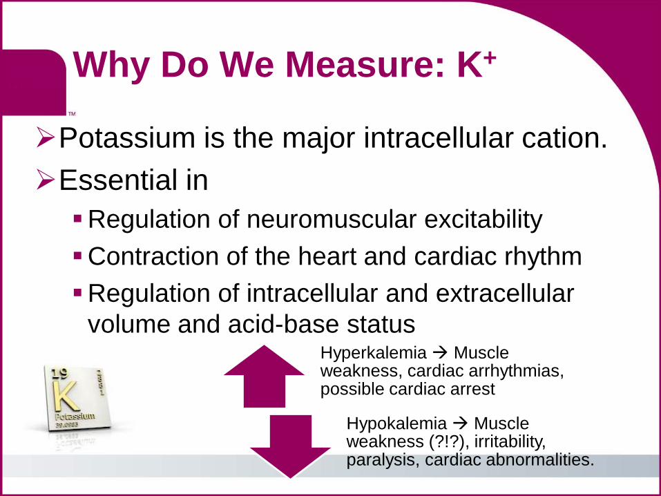
Why Do We Measure: K+
Potassium is the major intracellular cation. Essential in Regulation of neuromuscular excitability Contraction of the heart and cardiac rhythm Regulation of intracellular and extracellular
volume and acid-base status Hyperkalemia Muscle weakness, cardiac arrhythmias, possible cardiac arrest
Hypokalemia Muscle weakness (?!?), irritability, paralysis, cardiac abnormalities.
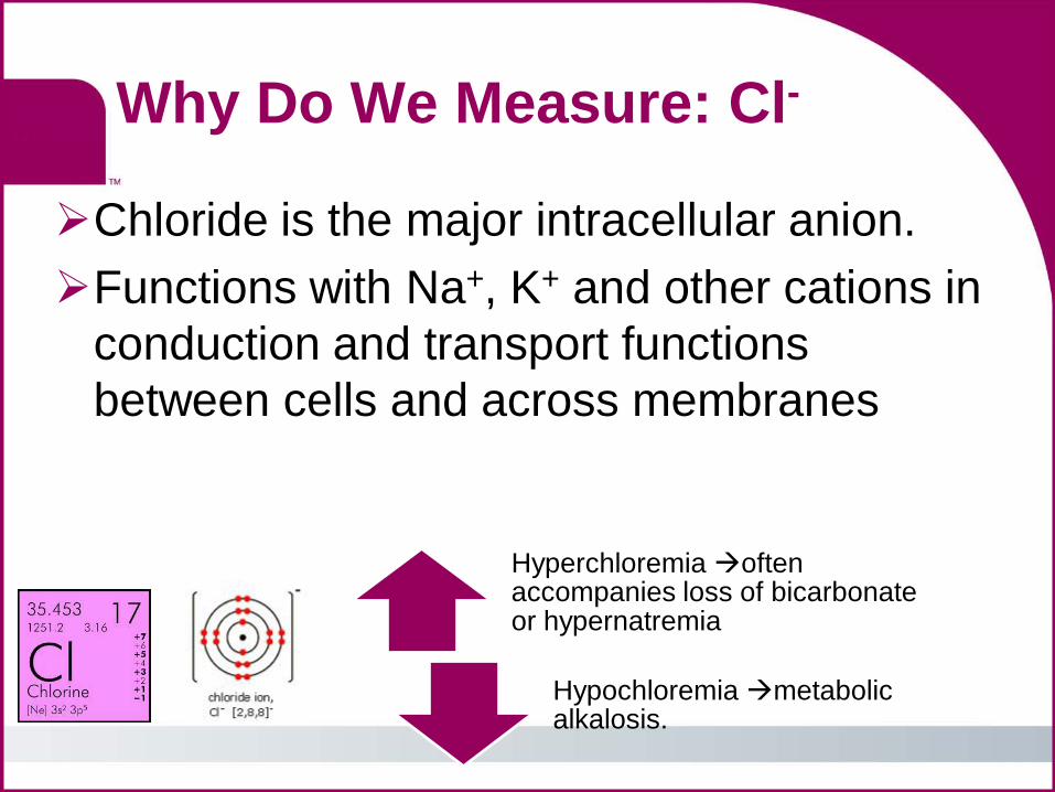
Why Do We Measure: Cl-
Chloride is the major intracellular anion. Functions with Na+, K+ and other cations in
conduction and transport functions between cells and across membranes
Hyperchloremia often accompanies loss of bicarbonate or hypernatremia
Hypochloremia metabolic alkalosis.
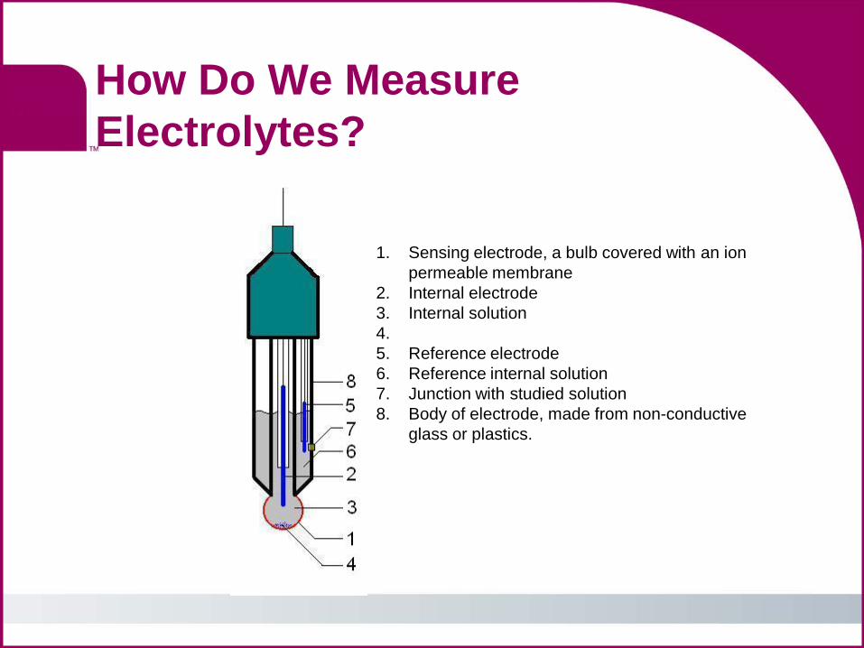
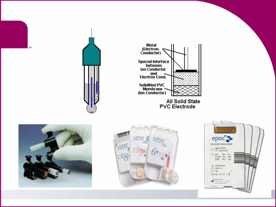
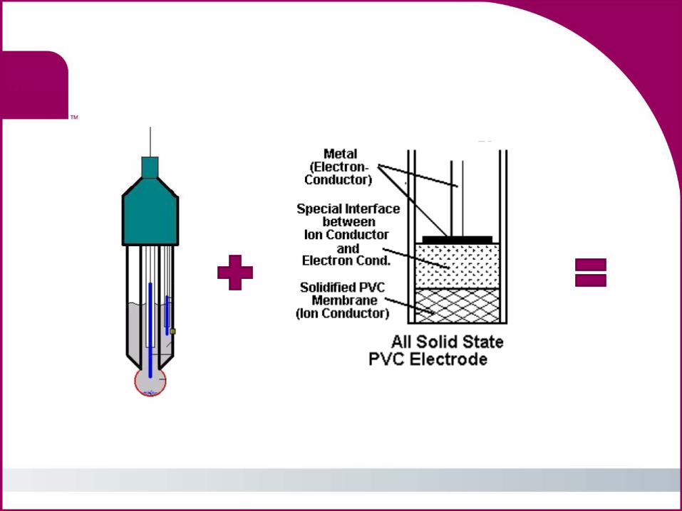
How Do We Measure Electrolytes?
1. Sensing electrode, a bulb covered with an ion permeable membrane
2. Internal electrode 3. Internal solution 4. 5. Reference electrode 6. Reference internal solution 7. Junction with studied solution 8. Body of electrode, made from non-conductive
glass or plastics.
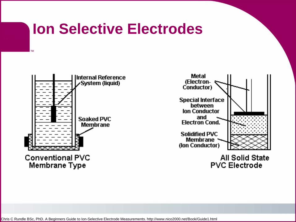
Ion Selective Electrodes
Chris C Rundle BSc, PhD. A Beginners Guide to Ion-Selective Electrode Measurements. http://www.nico2000.net/Book/Guide1.html
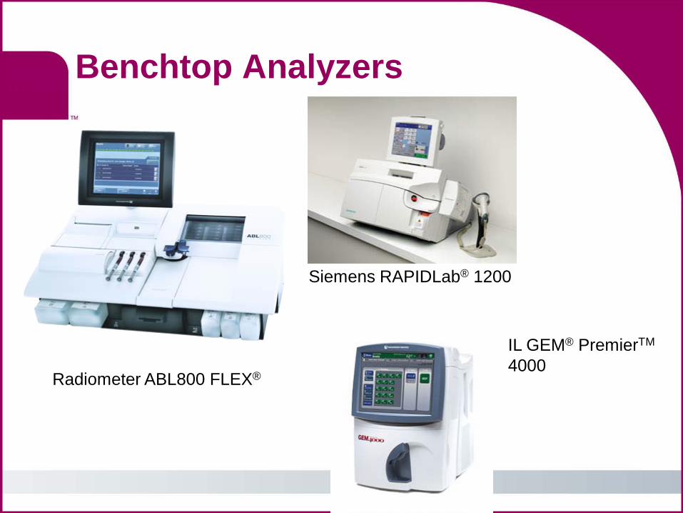
Benchtop Analyzers
Radiometer ABL800 FLEX®
Siemens RAPIDLab® 1200
IL GEM® PremierTM
4000
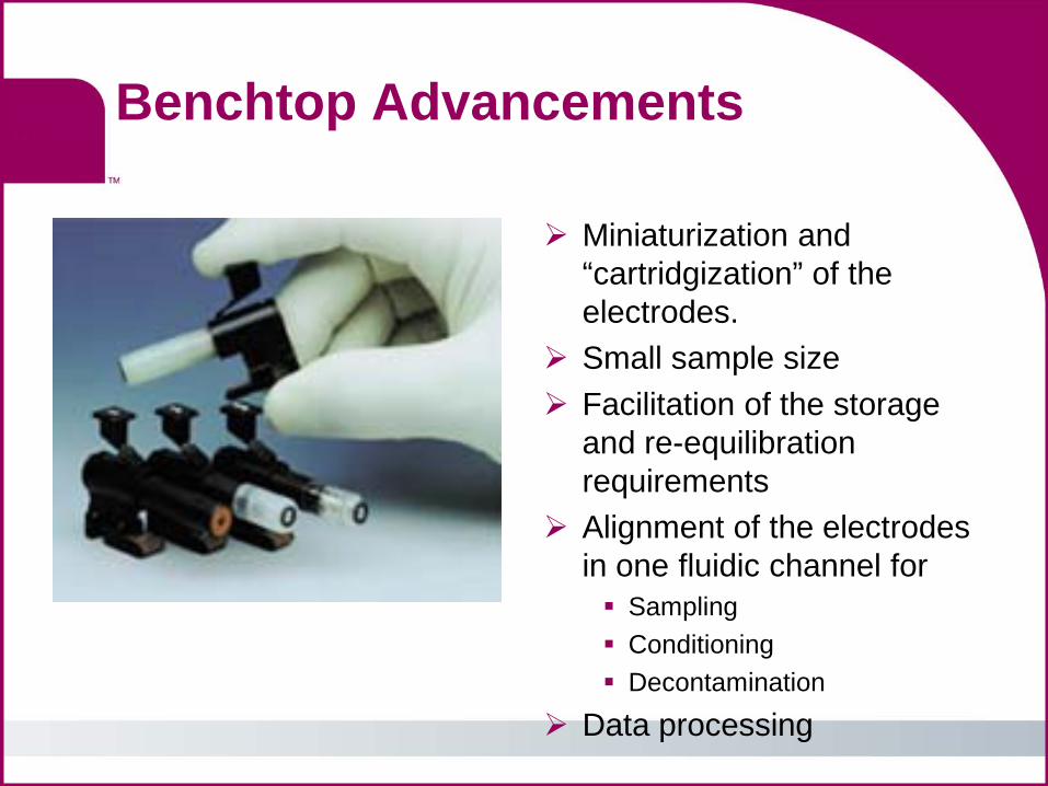
Benchtop Advancements
Miniaturization and “cartridgization” of the electrodes.
Small sample size Facilitation of the storage
and re-equilibration requirements
Alignment of the electrodes in one fluidic channel for Sampling Conditioning Decontamination
Data processing
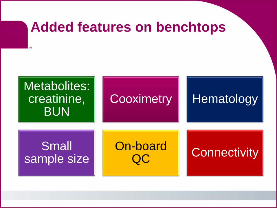
Added features on benchtops
Metabolites: creatinine,
BUN Cooximetry Hematology
Small sample size
On-board QC Connectivity
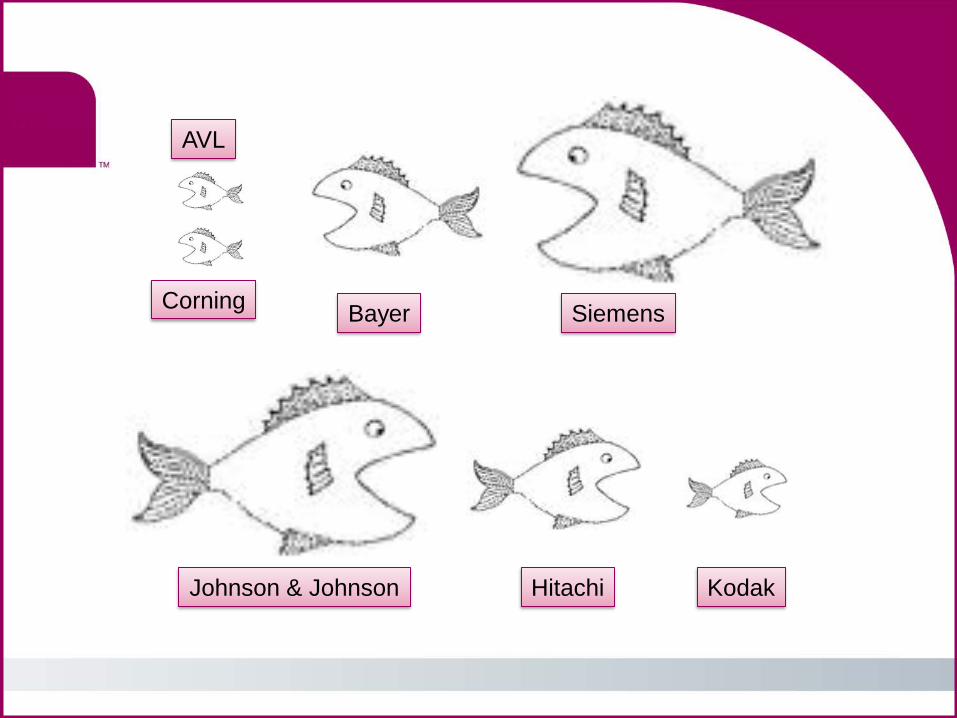
Corning Bayer Siemens
Johnson & Johnson Hitachi Kodak
AVL
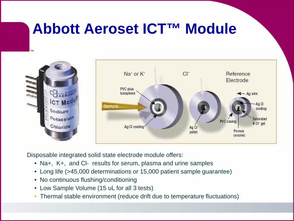
Abbott Aeroset ICT™ Module
Disposable integrated solid state electrode module offers: • Na+, K+, and Cl- results for serum, plasma and urine samples • Long life (>45,000 determinations or 15,000 patient sample guarantee) • No continuous flushing/conditioning • Low Sample Volume (15 uL for all 3 tests) • Thermal stable environment (reduce drift due to temperature fluctuations)
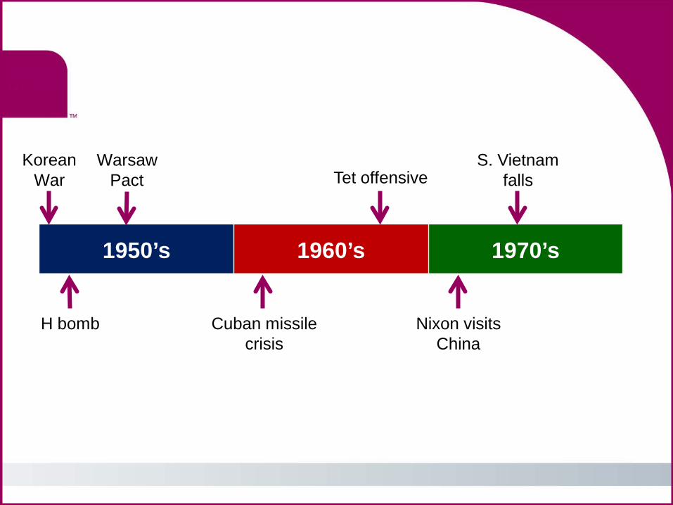
1950’s 1960’s 1970’s
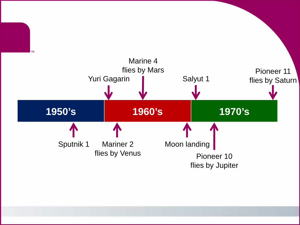
1950’s 1960’s 1970’s
Pioneer 11 flies by Saturn
Sputnik 1
Yuri Gagarin
Mariner 2 flies by Venus
Marine 4 flies by Mars
Moon landing
Salyut 1
Pioneer 10 flies by Jupiter
1950’s 1960’s 1970’s
H bomb Cuban missile crisis
Tet offensive
Nixon visits China
S. Vietnam falls
Korean War
Warsaw Pact
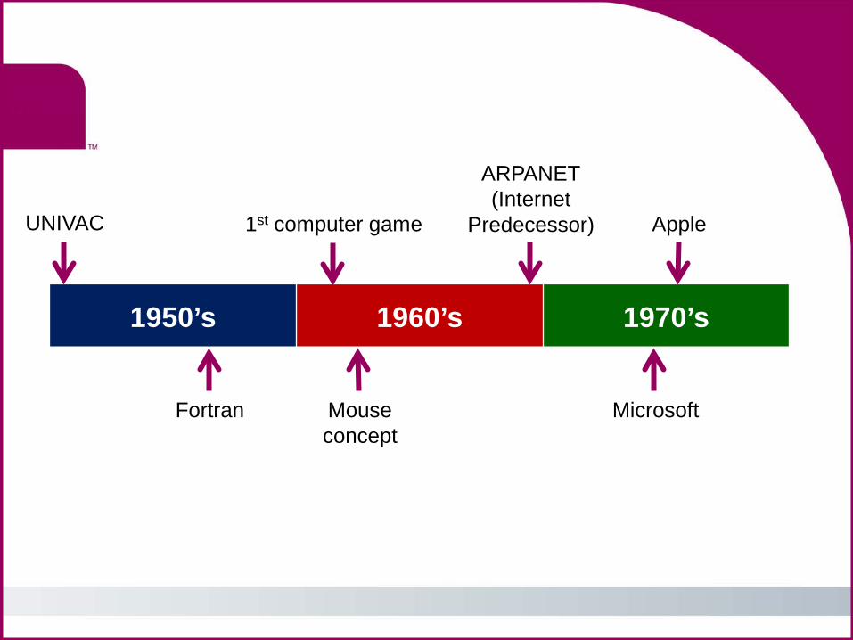
1950’s 1960’s 1970’s
UNIVAC
Fortran
1st computer game
Mouse concept
ARPANET (Internet
Predecessor)
Microsoft
Apple
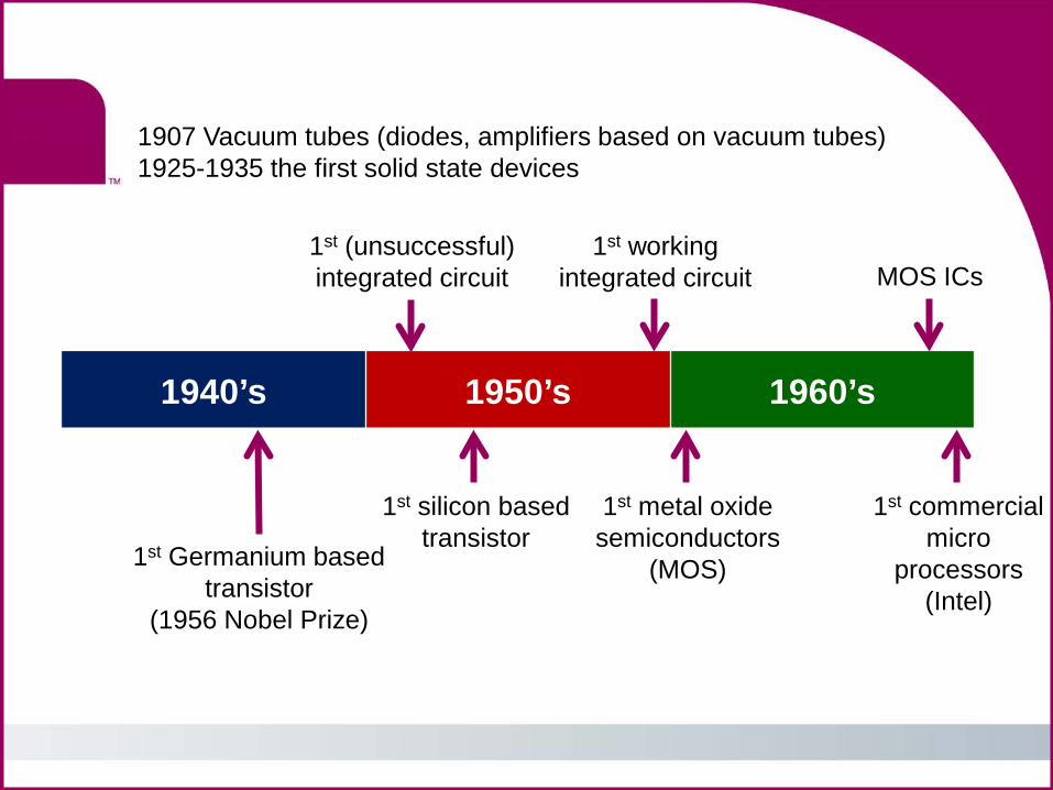
1940’s 1950’s 1960’s
1907 Vacuum tubes (diodes, amplifiers based on vacuum tubes) 1925-1935 the first solid state devices
1st Germanium based transistor
(1956 Nobel Prize)
1st (unsuccessful) integrated circuit
1st silicon based transistor
1st working integrated circuit
1st metal oxide semiconductors
(MOS)
MOS ICs
1st commercial micro
processors (Intel)
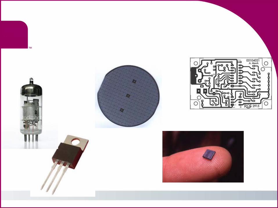
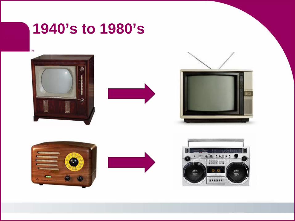
1940’s to 1980’s
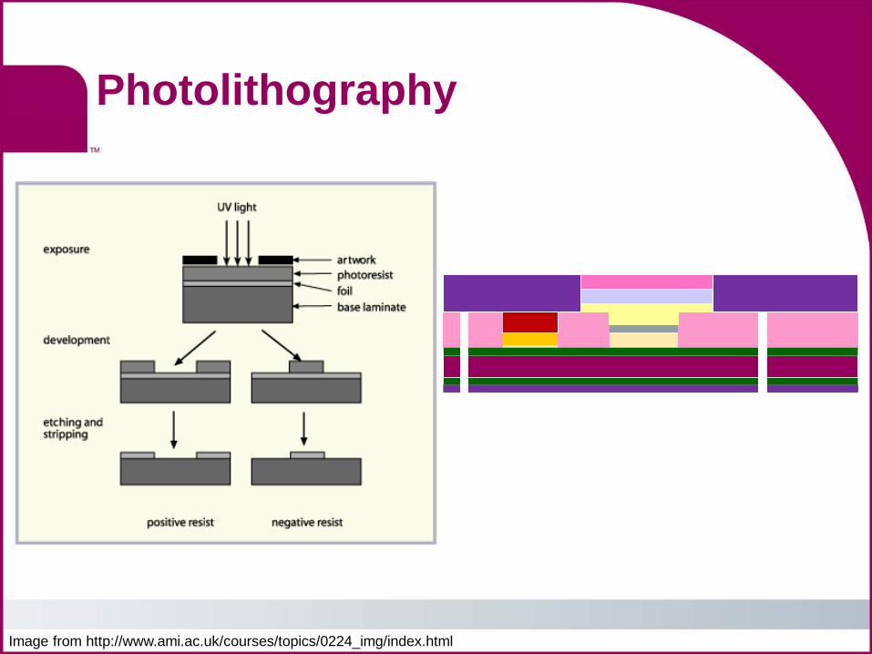
Photolithography
Image from http://www.ami.ac.uk/courses/topics/0224_img/index.html
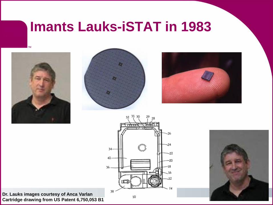
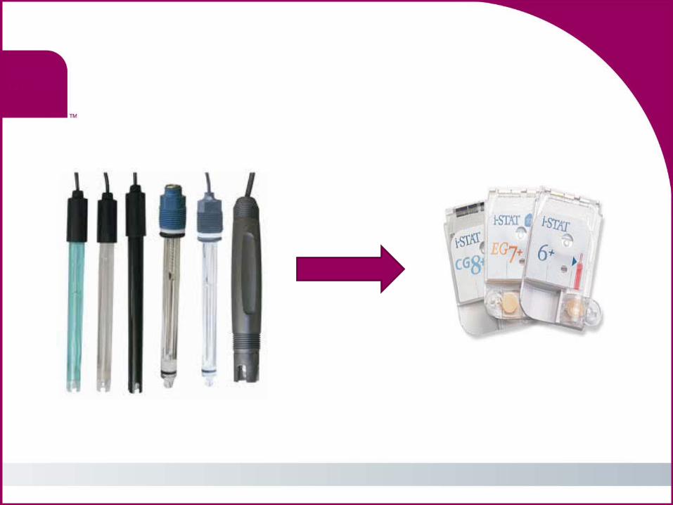
Imants Lauks-iSTAT in 1983
Dr. Lauks images courtesy of Anca Varlan Cartridge drawing from US Patent 6,750,053 B1
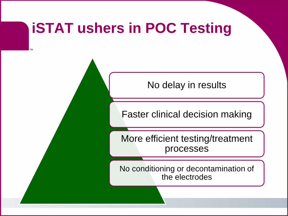
iSTAT ushers in POC Testing
No delay in results
Faster clinical decision making
More efficient testing/treatment processes
No conditioning or decontamination of the electrodes
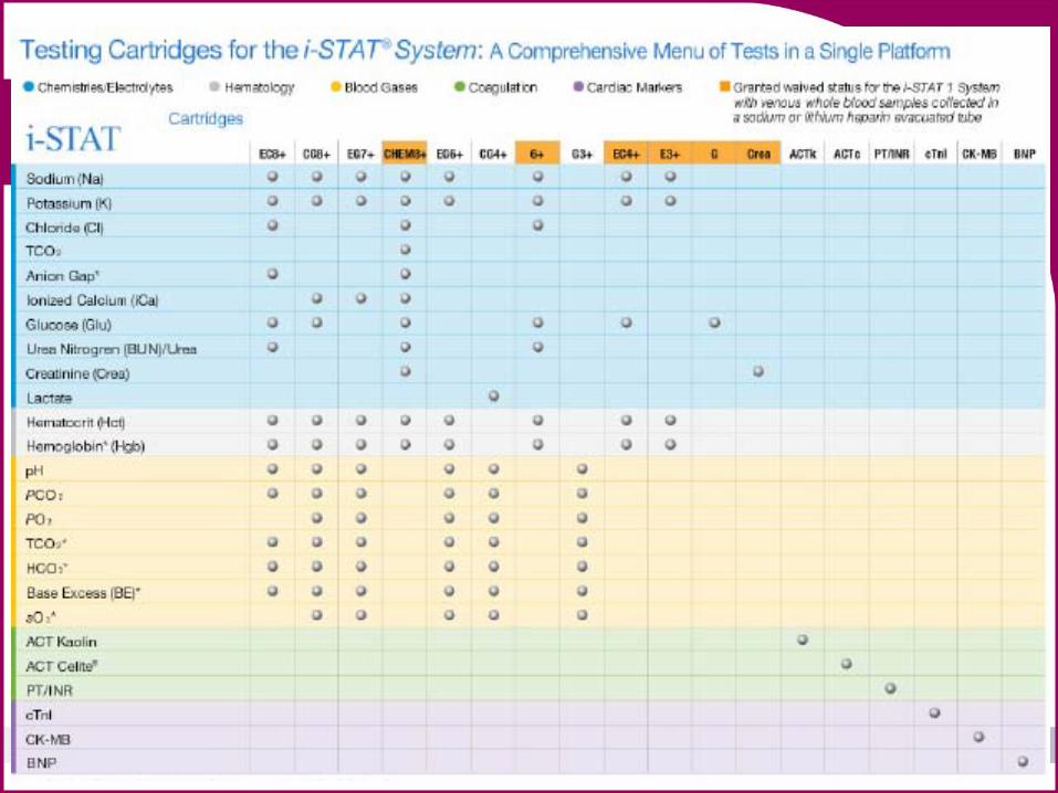
i-STAT menu
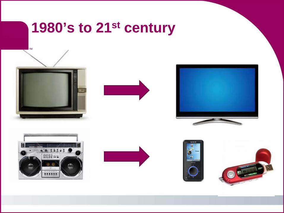
1980’s to 21st century
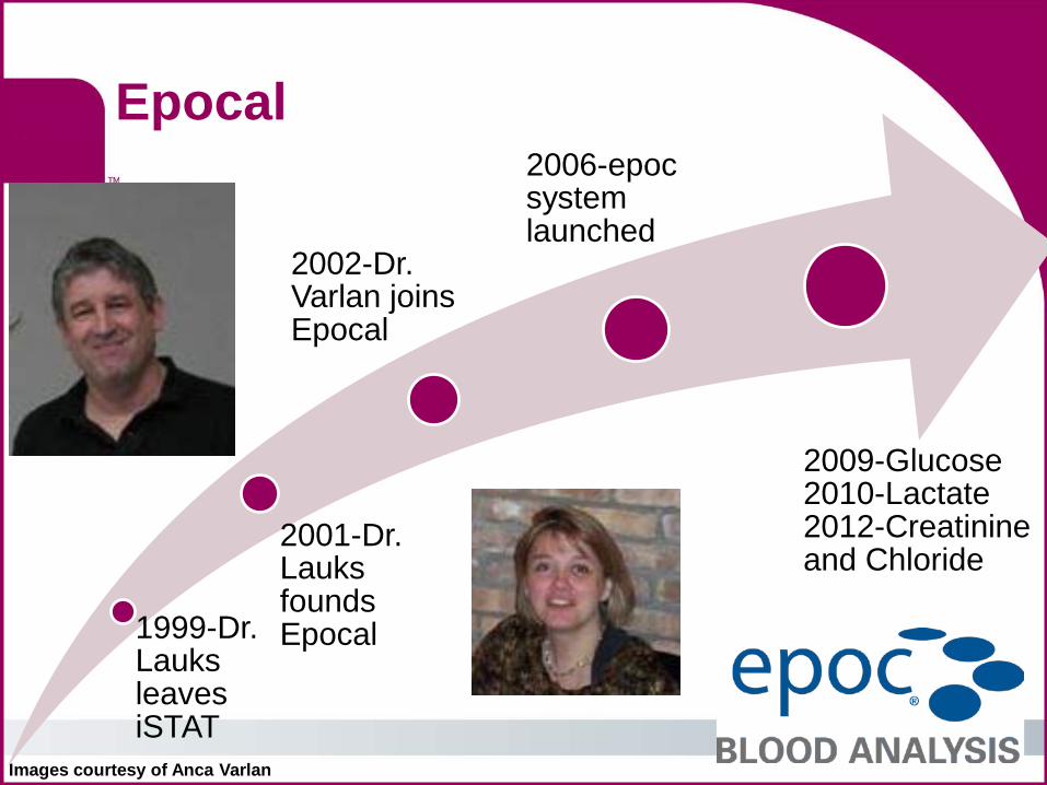
Epocal
Images courtesy of Anca Varlan
1999-Dr. Lauks leaves iSTAT
2001-Dr. Lauks founds Epocal
2002-Dr. Varlan joins Epocal
2006-epoc system launched
2009-Glucose 2010-Lactate 2012-Creatinine and Chloride
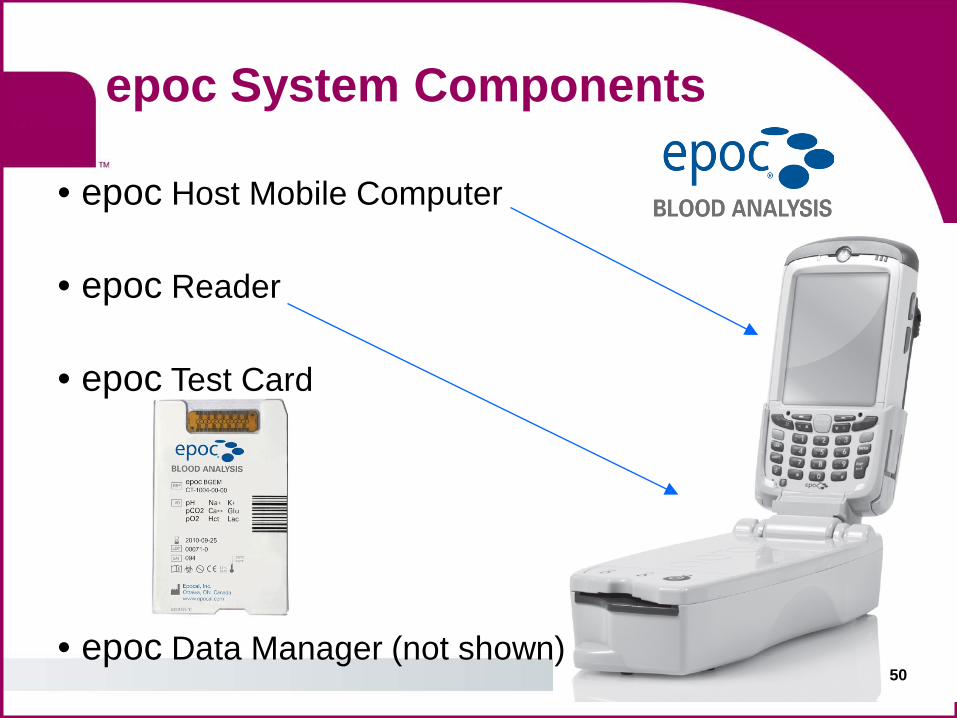
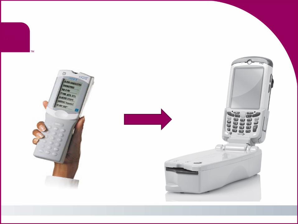
epoc System Components
50
• epoc Host Mobile Computer • epoc Reader
• epoc Test Card • epoc Data Manager (not shown)
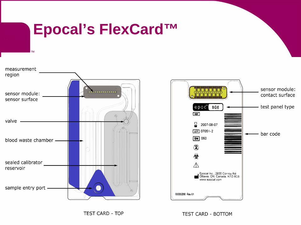
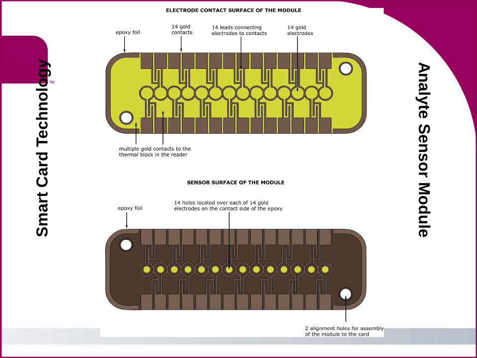
Epocal’s FlexCard™
51
Smar
t Car
d Te
chno
logy
Analyte Sensor Module
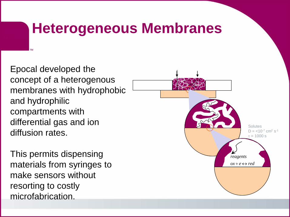
Heterogeneous Membranes
Epocal developed the concept of a heterogenous membranes with hydrophobic and hydrophilic compartments with differential gas and ion diffusion rates. This permits dispensing materials from syringes to make sensors without resorting to costly microfabrication.
Solutes D = <10-7 cm2 s-1
τ = 1000 s
redeoxreagents
↔+
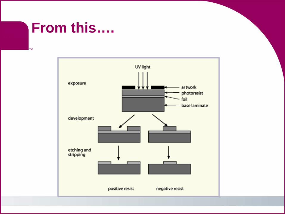
From this….
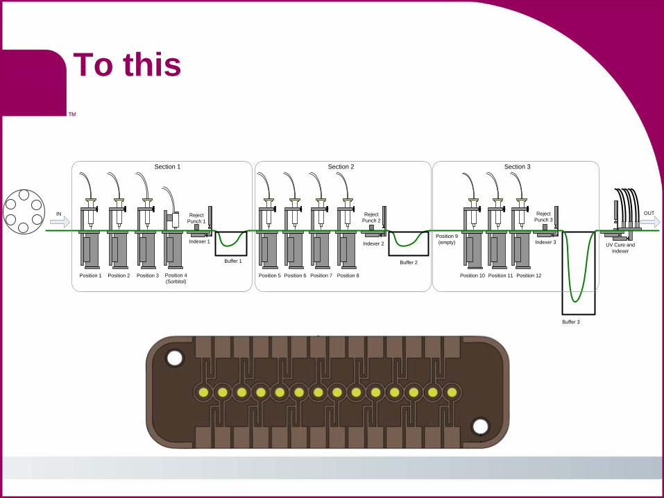
To this
Position 1 Position 2 Position 3 Position 4(Sorbitol)
Buffer 1
Position 5 Position 6 Position 7 Position 8
Indexer 1 Indexer 2
Buffer 2
Position 10 Position 11 Position 12
Indexer 3
Buffer 3
UV Cure and Indexer
OUTIN
Section 1 Section 2 Section 3
Position 9(empty)
Reject Punch 1
Reject Punch 2
Reject Punch 3
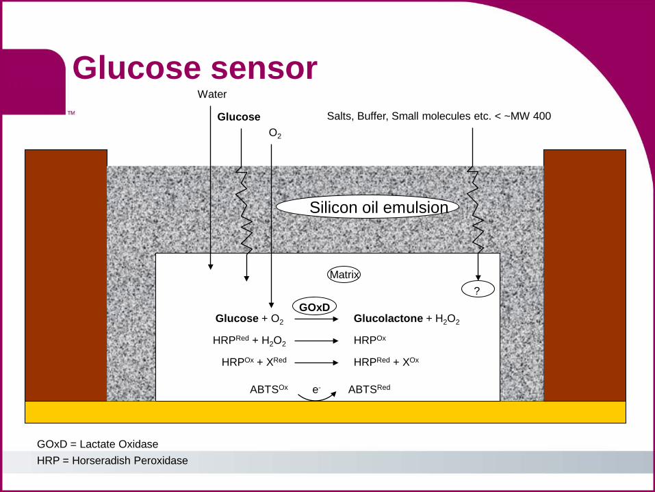
GOxD = Lactate Oxidase HRP = Horseradish Peroxidase
Glucose + O2 Glucolactone + H2O2 GOxD
HRPRed + H2O2 HRPOx
HRPOx + XRed HRPRed + XOx
ABTSOx ABTSRed e-
Matrix
Water
Glucose O2
?
Silicon oil emulsion
Salts, Buffer, Small molecules etc. < ~MW 400
Glucose sensor
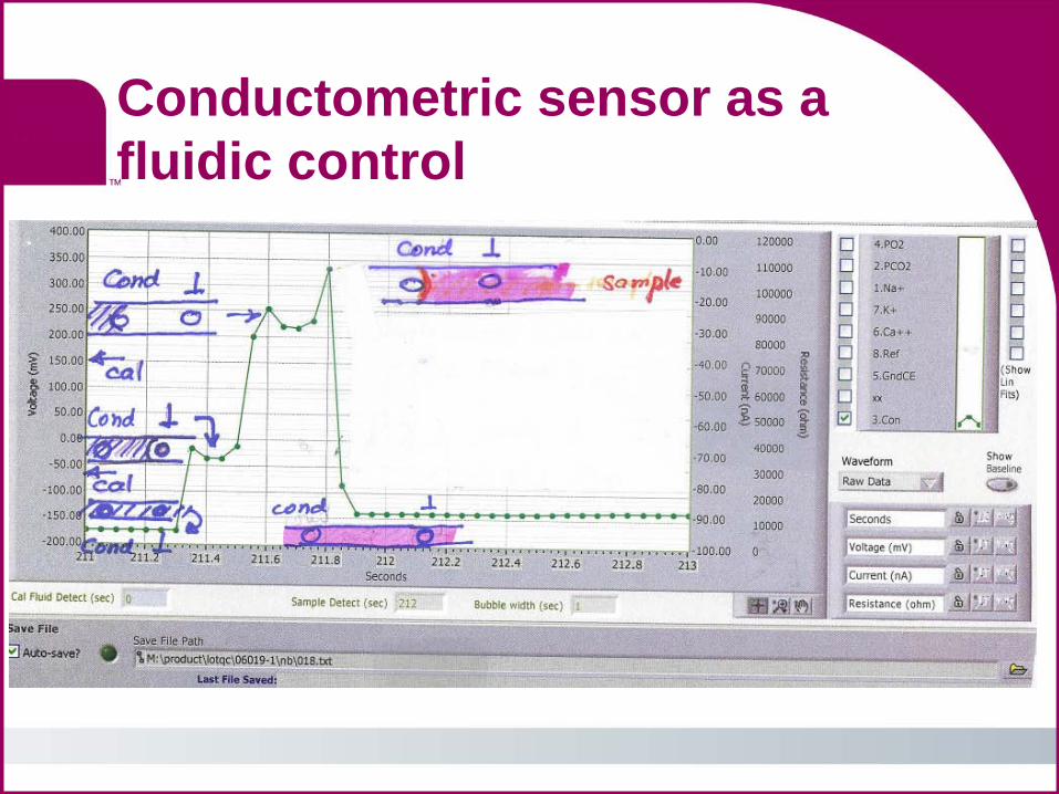
Conductometric sensor as a fluidic control
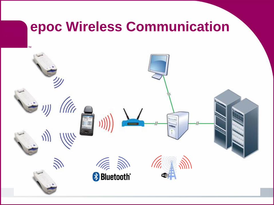
epoc Wireless Communication
58
• Max Cremer discovers that a potential develops between two liquids of different pH on opposite sides of a thin glass membrane
1906
• First glass pH electrodes are developed but the internal resistance of the glass make measurements difficult
1909
• Arnold Beckman adds an amplifier to better detect the signal and develops the first “acid-o-meter”
1934

Questions?
Thank You!
Today is the youngest you’ll be for the rest of your life. Act like it.
Recommended


![POC - Heska · Element POC Blood Gas & Electrolyte Analyzer B Preface Element POC [epoc ®] Blood Gas & Electrolyte Analyzer The Element POC® portable device consists of the blood](https://img.pdfslide.us/doc/110x75/5edb6ef2ad6a402d6665a6de/poc-heska-element-poc-blood-gas-electrolyte-analyzer-b-preface-element-poc.jpg)
![Blood glucose, acid–base and electrolyte changes during ... · followed by CRIs produce [Glu]B, acid-base and electrolyte changes. The clinical significance of the re-ported changes](https://img.pdfslide.us/doc/110x75/5fb313e7e2d54931e832a811/blood-glucose-acidabase-and-electrolyte-changes-during-followed-by-cris-produce.jpg)