Zanda Daneberga
THE FRAGILE X SYNDROME IN MENTALLY RETARDED
PATIENTS FROM LATVIA
Doctoral Thesis
Summary
Speciality: Medical Genetics
Riga, 2011
2
Elaboration of the Study: Medical Genetics Clinic, University Children`s Hospital, Riga, Latvia Scientific supervisor: dr. med. assoc. prof. Rita Lugovska, Medical Genetics Clinic, University Children`s Hospital, Riga, Latvia; Riga Stradins University, Scientific advisor: dr. med. Baiba Lāce, Medical Genetics Clinic, University Children`s Hospital, Riga, Latvia; Lavian BioMedical Research and Study Center, Riga, Latvia Official reviewers: dr. biol., assoc. prof. Edvīns Miklašēvičs, Riga Stradins University dr. habil. biol., prof. Nikolajs Sjakste, University of Latvia, Faculty of Medicine dr. med., assoc. prof. Jurate Kasnauskiene, Vilnius University, Faculty of Medicine Defence of the doctoral thesis will be held on 21st of December, 2011 at 14.00 o`clock in the Hippocratic lecture-hall of Riga Stradins University (RSU) Dzirciema Str.16. The Doctoral Thesis is available in the library of RSU and at www.rsu.lv The work was supported by ESF Project “Support to doctor’s studies and acquiring an academic degree in Riga Stradins University” No. 2009/ 0147/ 1DP/ 1.1.2.1.2/ 09/ IPIA/ VIAA/ 009.
Secretary of Promotion Council: Dr. habil.med., prof. Līga Aberberga - Augškalne
3
TABLE OF CONTENTS 1. INTRODUCTION......................................................................... 6
1.1. Aim of the Study........................................................................... 8
1.2. Tasks of the Study......................................................................... 8
1.3. Scientific Novelty of the Study..................................................... 9
1.4. Practical Novelty of the Study...................................................... 9
1.5. Author`s Contribution to the Work............................................... 10
1.6. Elaboration of the Study................................................................ 10
1.7. Outline of the Thesis..................................................................... 11
2. SUBJECTS AND METHODS..................................................... 11
2.1. Subjects......................................................................................... 11
2.1.1. Prevalence of the Fragile X Syndrome.......................... 11
2.1.2. Variation of CGG Trinucleotide Repeats....................... 12
2.1.3. The Case-Control Study………………………………. 12
2.1.4. Genotype-Phenotype Correlation................................... 13
2.2. Molecular Studies.......................................................................... 13
3.2.1. DNA Extraction.............................................................. 13
3.2.2. Routine Screening PCR Amplification.......................... 14
3.2.3. Fluorescent PCR............................................................ 14
3.2.4. Southern Blotting........................................................... 15
3.2.5. AGG Interspersion Pattern Analysis.............................. 15
3.2.6. Single Nucleotide Polymorphism Analysis.................... 16
3.2.7. Haplotype Analysis......................................................... 17
3.2.8. Statistical Data Analysis................................................. 19
3. RESULTS...................................................................................... 20
3.1. Prevalence of the Fragile X Syndrome......................................... 20
3.2. Variation of CGG Trinucleotide Repeats...................................... 21
4
3.3. ATL1 SNP..................................................................................... 22
3.4. Repeat Structures of Grey-Zone Alleles...................................... 24
3.5. DXS548-FRAXAC1-ATL1-FRAXAC2 Haplotypes.................. 25
3.6. Analysis of Molecular Variance................................................... 28
3.7. Genotype-Phenotype Correlation................................................. 29
4. DISCUSSION.............................................................................. 31
4.1. Prevalence of the Fragile X Syndrome........................................ 31
4.2. Variation of CGG Trinucleotide Repeats...................................... 32
4.3. ATL1 SNP..................................................................................... 33
4.4. Repeat Structures of Grey-Zone Alleles....................................... 33
4.5. DXS548-FRAXAC1-ATL1-FRAXAC2 Haplotypes................... 34
4.6. Genotype-Phenotype Correlation.................................................. 37
5. CONCLUSIONS........................................................................... 39
6. PUBLICATIONS.......................................................................... 40
7. ACKNOWLEDGMENTS............................................................ 42
8. REFERENCES.............................................................................. 44

5
ABBREVIATION AMOVA - Analysis of molecular variance
ARI - Acute respiratory infection
bp - Base pairs
CGG - Cytosine – guanine – guanine
CNS - Central nervous system
DNA - Deoxyribonucleic acid
EDTA - Ethylenediaminetetraacetic acid
FXS - Fragile X Syndrome
MR – Mental retardation
IQ - Intelligence Quotient
PCR - Polymerase Chain Reaction
PWS - Prader-Willy syndrome
RF – Relative frequency
SNP - Single-Nucleotide Polymorphism
STR – Short tandem repeat
6
1. INTRADUCTION Mental retardation (MR) is a complex phenotype, characterized by
suboptimal functioning of the central nervous system (CNS) resulting in
significant limitations both in intellectual functioning and in adaptive
behaviour. Mental retardation affects about 2–3% of people and about a quarter
of cases are caused by genetic disorders. Mental retardation is the most
frequent cause of severe handicap in children. Therefore ascertainment of
mental retardation aetiology is an important task in paediatrics.
Fragile X syndrome (FXS; MIM #300624; FRAXA, Xq27.3) is well
known and a common cause of X-linked mental retardation. The fragile X
syndrome is caused by an expanded CGG repeat (> 200 units, full mutation) at
the 5' end of the FMR1 gene, which is associated with methylation of a CpG
island upstream of the FMR1 gene and down regulation of the transcription
(Oberle et al., 1991; Poustka et al., 1991; Rousseau et al., 1991).
Amongst individuals from the general population, the polymorphic
CGG repeat ranges from 6 to 50 repeats and is usually interspersed every 9–10
repeats with an AGG (Eichler et al., 1996; Fu et al., 1991). Premutation alleles
have a moderate expansion of the repeat (from 50 to ~200 units), they are
unmethylated on an active X chromosome and do not affect FMR1 expression.
CGG repeat expansion over 200 is the basis for CpG island methylation,
leading to silencing of the FMR1 gene (de Vries et al., 1998). Intermediate or
grey zone alleles are poorly defined. Boundaries for the grey zone range vary
among studies, from 34 or 35 CGG repeats for the lower boundary to 58/60
repeats for the upper boundary (Moutou et al., 1997; Rife et al., 2004; Sherman
et al., 2005). These alleles usually have stable transmission, but are more likely
to exhibit unstable transmission with increasing size within this range.
The underlying mutational mechanism is not fully understood and
remains a topic of debate. The gender of the parent carrying an expanded repeat
7
(maternal imprinting), the number of repeats (dynamic mutation) and the
absence of AGG interruptions in long tracts of CGG repeats have been
described as the tree main factors related to this instability (Dombrowski et al.,
2002; Eichler et al., 1996; Rife et al., 2004). The microsatellite markers
DXS548-FRAXAC1-FRAXAC2 and the ATL1 SNP have previously been
reported as markers associated with FMR1 CGG repeat instability (Eichler et
al., 1996; Gunter et al., 1998; Kunst et al., 1996; Macpherson et al., 1994;
Murray et al., 1997; Oudet et al., 1993; Richards et al., 1991).
Haplotypes linked to FXS are widely described across Western
European and Scandinavian populations; however, less is known regarding
populations from North-Eastern Europe, including the Baltic States. This is the
first study in the Baltic States region regarding FMR1 linked haplotypes.
The first clinical indication of FXS is usually delay in child`s
developmental milestones and mental retardation. In addition to mental
retardation, speech and language skills are severely affected. Most speech is
poorly articulated and expressive language is often limited to three- or four-
word sentences. FXS patients often repeat words or phrases, an attribute
typically associated with autism. Indeed many FXS males present autistic type
behaviour – gaze aversion, shyness, hand biting, hand flapping and rocking
(Bardoni et al., 2000; Garber et al., 2008; Hernandez et al., 2009).
The phenotype is subtle in young children and evolves with age.
Hyperextensibility of finger joints, pectus excavatum, mitral valve prolapse and
strabismus are other possible prevalent features (de Vries et al., 1996;
Larbrisseau et al., 1982; Phadke 2005). The clinical manifestations of this
syndrome in adult males include an elongated and narrow face with a large
forehead and prominent chin, large and anteverted ears, joints with increased
mobility, and uni-or bilaterally large testes. Macroorchidism is an important
feature in the post-pubertal age. However, it is not presented in all FXS males,
8
but it is specific for FXS. Between 25-30% of all patients with FXS do not have
the typical faces of the syndrome. The secondary characteristics of FXS in turn
include tallness, a soft and silky skin, widened fingertips and flat feet (Ridaura-
Ruiz et al., 2009).
Early diagnosis of fragile X syndrome is crucial in order to inform other
members of the family of their risk of having affected offspring. Therefore it is
recommended that most fragile X diagnostic tests will be carried out on a very
broad range of patients regardless of a consequently low detection rate.
Ten years of experience with molecular diagnostic of the fragile X
syndrome in Latvia and number of diagnosed patients in this time period,
revealed a low pickup rate of patients and insufficient clinical recognition of
symptoms.
1.1.Aim of the Study Ascertain the prevalence of fragile X syndrome in Latvia, characterise
genetic and clinical variability of the FRAXA locus FMR1 gene in patients
with unclear aetiology of mental retardation.
1.2.Tasks of the Study 1. Estimate the prevalence of the fragile X syndrome in the entire Latvian
male population.
2. Perform a distribution and structure study of CGG repeats among X
chromosomes with normal CGG repeat alleles.
3. Characterise the ATL1 SNP/CGG repeat number correlation within
chromosomes with a normal CGG repeat number and chromosomes
with a full mutation.
4. Perform a case-control study of FMR1 gene linked haplotypes based on
STR and SNP markers, to identify specific haplotypes among Latvian
9
FXS patients and control group mentally retarded patients with a normal
number of CGG repeats with respect to allelic stability.
5. Identify the association of grey-zone allele structure and FMR1 linked
haplotypes.
6. Evaluate genotype-phenotype correlation in patients with full mutation
and/or repeat size/methylation mosaic.
1.3.Scientific Novelty of the Study This study is the first study in the Baltic States region regarding FMR1
linked haplotypes. Described haplotypes of Latvian fragile X syndrome patients
differ from published studies in populations of Western European descent.
Therefore this data provide evidence of different mutational pathways of CGG
repeat expansion in the North-Eastern European region.
The estimated prevalence of fragile X syndrome in the Latvian male
population contributes to the ascertainment of this disease distribution in our
geographical region.
1.4.Practical Novelty of the Study The estimated prevalence of fragile X syndrome in the Latvian male
population is in line with the prevalence of this syndrome in several other
European populations. The low number of confirmed patients with fragile X
syndrome in ten years, point to a low detection rate of patients in paediatrician,
child psychiatric and child neurology practices.
Haplotypes linked to unstable CGG repeat alleles in the Latvian FXS
male population are very useful in practical family cascade testing and for
consultation of families at risk.
10
The newly adapted clinical questionnaire form shall contribute to an
increase in the detection rate of patients with suspected fragile X syndrome by
paediatricians, child psychiatric and child neurology practices.
1.5.Author`s Contribution to the Work This PhD project was initiated in 2005, based on scientific elaboration
forerun of project “Genomic studies of the Latvian population, their application
for diagnosis and prevention of human pathology”.
The author of this thesis performed the following laboratory
investigations: DNA extraction (partly); routine screening PCR amplification;
fluorescent PCR; Southern blotting (partly); ATL1 SNP analysis; AGG
interspersion pattern analysis; fluorescent PCR of microsatellite markers and
haplotype analysis. Author performed retrospective data collection and data
analysis for prevalence study. All statistical data analysis and AMOVA were
done by the author of this thesis.
Clinical evaluation of patients was done by clinical geneticists and child
psychiatrists.
1.6. Elaboration of the Study The current study was carried out in the Medical Genetics Clinic,
University Children`s Hospital, Riga, Latvia in collaboration with Children`s
Psychiatric Department, University Children`s Hospital, Riga, Latvia.
Conformation of FXS diagnosis by Southern blot analysis was done in
the DNA Laboratory, Department of Medical Genetics, Ullevål University
Hospital, Oslo, Norway and in the DNA Diagnostic Laboratory, University
Medical Center Nijmegen, The Netherlands.
The Latvian Central Committee of Medical Ethics and the Riga Stradins
University Committee of Medical Ethics approved the study.
11
1.7. Outline of the Thesis The thesis is composed on 124 pages in English, following classical
scheme. The work is structured in ten chapters: Introduction; Literature review;
Subjects and Methods; Results; Discussion; Conclusions; Publications;
Acknowledgements; References and Appendixes. Text of thesis is
supplemented by 19 Tables; 19 Figures and 14 Appendixes. Reference list
consist of 131 cited references.
2. SUBJECTS AND METHODS
2.1. Subjects 2.1.1. Prevalence of the Fragile X Syndrome
The retrospective data of patients genotypes, analyzed in the Medical
Genetic Clinic, University Children`s Hospital between 1998 and 2007, were
summarized to assess the prevalence of FXS.
All patients were referred for exclusion/confirmation of fragile X
syndrome by clinical geneticist at the Medical Genetic Clinic, University
Children`s Hospital, by child psychiatrist for the hospitalized persons at the
Children`s Psychiatric Department, University Children`s Hospital and by
clinical geneticist at the children`s attending Social Care Centre Riga, Latvia.
Inclusion criteria for selecting patients' data were as follows:
patients with mental retardation in various degrees with or without
association with dysmorphic features
MR patients with autism, autistic spectrum disorders and any type of
behavioural disturbances
genotype data with exact number of CGG repeats
Exclusion criteria for selecting patients' data were as follows:
patient gender (female)
12
consanguinity
monogenic, chromosomal and metabolic diseases
The clinical features of the patients were assessed and family history
obtained by clinical geneticist. The ethnical background of patients was not
considered.
Based on inclusion/exclusion criteria 374 anonymous, unrelated male
patients data were selected for the prevalence study. The age of patients at the
time of the DNA diagnostic test varied between two and seventeen years.
2.1.2. Variation of CGG Trinucleotide Repeats
To assess distribution of normal CGG repeat alleles retrospective data of
patients genotypes, analyzed in the Medical Genetic Clinic, University
Children`s Hospital between 1998 and 2007, were used. Based on
inclusion/exclusion criteria, 374 anonymous, unrelated male patient data were
used. We considered selected data comparable, because for all 374 samples,
both routine screening with PCR and fluorescent PCR following Applied
Biosystems protocol for exact CGG repeat number detection, were performed.
2.1.3. The Case-Control Study
For case-control study of FMR1 linked haplotypes the control group of
122 unrelated male patients with normal number of CGG repeats were selected
based on inclusion/exclusion criteria.
Inclusion criteria for selecting control group were as follows:
parents or legal representatives of minors signed informed consent
according regulations issued by Ethics Committee for participation in this study
genotype data within a normal range of CGG repeats
patients with mental retardation in various degrees with or without
association with dysmorphic features
13
MR patients with autism, autistic spectrum disorders and any type of
behavioural disturbances
Exclusion criteria for selecting control group were as follows:
patient gender (female)
consanguinity
monogenic, chromosomal and metabolic diseases
The case group consisted of 11 unrelated male patients with confirmed
diagnosis (full mutation). Parents or legal representatives of minors signed
informed consent according regulations issued by Ethics Committee for
participation in this study.
2.1.4. Genotype-Phenotype Correlation
Genotype-phenotype correlation was assessed for 12 male patients with
confirmed diagnosis of FXS in time period from 1998 to 2010. In this group of
study siblings were included. The age of patients at the moment of diagnosis
varied between two and sixteen years (average = 7.33 ± 4.46). Clinical
information was obtained from case-records of patients by clinical geneticist or
child psychiatrist. Anthropometric data were measured according to the “Smith
recognizable patterns” and a methodology described by Krūmiņa, Kokare and
Biķis (2007). IQ tests were performed based on the Woodcock – Johnson test
and Wechsler Intelligence Scale for Children. Autistic spectrum disorders were
evaluated according to the Autism Diagnostic Observation Schedule (ADOS).
2.2.Molecular Studies 2.2.1. DNA Extraction
Five millilitres of peripheral blood were collected in EDTA-coated
tubes. Before DNA extraction, blood samples were kept frozen at -20°C. DNA
14
was extracted using “Genomic DNA Purification Kit” (Fermentas, Lithuania)
according to the manufacturer protocol.
2.2.2. Routine screening PCR amplification
For the amplification of normal CGG repeat allele within FMR1 gene,
primers sequence corresponding to position 212-241 and 599-571 at the 1kb
Pst1 fragment plasmid Ep5.1, containing CpG island and CGG repeat region,
were used. The reaction was performed according to the protocol described by
Chong et al. (1994).
2.2.3. Fluorescent PCR
For a precise determination of CGG repeat number, fluorescent PCR
was carried out according to the Applied Biosystems (USA) protocol. The
reagent solutions were in-house prepared. Approximately 100 diploid copies
(0.67 ng) per μl of genomic DNA were used to carry out the reaction. This
reaction was performed using the “Hot start” technique with Amli Wax
(Applied Biosystems, USA).
PCR products were separated on an ABI Prism® 310 genetic analyzer
(Applied Biosystems, USA) under two different electrophoresis conditions.
Genotyping results were analysed by GeneScan™ software (Applied
Biosystems, USA). The corresponding peaks length was calculated according
to the calibration curve of the fragile X size standard (50 bp-2500 bp).
Apolipoproteine E (ApoE) gene CG rich fragment was used as an internal
control for amplification. Presence of Apo E frgment indicates successful
amplification and template quality.
15
2.2.4. Southern blotting
The FXS diagnosis was confirmed by sizing of the repeat array using
methylation specific restriction enzyme digestion and genomic Southern blot
hybridization according to the described protocol (Dracopoli and Haines,
1994).
For Southern blot analysis, 4 - 6 μg of genomic DNA were used.
To perform sizing of the repeat array, two different digestion reactions
were used – EagI/EcoRI (methylation specific) and PstI. Products were
separated on 0.8% agarose gel (at 0.35 V/cm, overnight) and transferred to
positive charged Nylon membrane by capillary transfer. To detect DNA
fragment, labelled [32P] StB12.3 hybridisation probe was used. Unlabeled
StB12.3 probe obtained from Prof. J. L. Mandel, Strasbourg. Labelling of the
probe was done according to the described protocol (Sambrook and Russell,
2001). Analysis performed in the DNA Laboratory, Department of Medical
Genetics, Ullevål University Hospital, Oslo, Norway.
Southern blot analysis for patients and their family members using probe
pAO365 was performed in the DNA Diagnostic Laboratory, University
Medical Center Nijmegen, The Netherlands.
2.2.5. AGG interspersion pattern analysis
Twenty-six alleles of grey zone (35-50 CGG repeat) were analysed for
CGG repeat patterns. The AGG interspersion pattern was determined by
sequencing of the CGG-repeat array. In brief, the CGG repeat and surrounding
DNA sequences were amplified from genomic DNA by Pfu polymerase
(Fermentas, Lithuania) with the PCR protocol described previously (Chong et
al. 1994). The PCR products were run on 2.5% agarose gel at 5.5 V/cm for 60
min to check for amplification of a single allele. The PCR products were
16
concentrated and purified for sequencing by the Montage PCR centrifugal filter
device (Millipore, USA).
The sequencing reaction was performed using concentrated and purified
PCR products and the BigDye® Terminator v3.1 kit (Applied Biosystems,
USA) according to the manufacturer’s protocol.
All sequencing reactions were run on an ABI Prism® 310 genetic
analyzer using 61cm x 50µm (50 cm well-to-read) capillary with POP-6™
polymer and analyzed by ABI DNA™ sequencing software.
The sequence pattern of the CGG repeat array was red from the first
exon of FMR1 gene. DNA sequence was established by visual interpretation of
the electropherograms. Nucleotides were assigned in the sequence based on the
highest fluorescence signal at each position, provided that the nucleotide peak
exceeded the background level. To describe the sequence of the FMR1, the
number of CGG repeats is denoted as a number, while AGG interruption is
denoted as “+”.
2.2.6. Single nucleotide polymorphism analysis
The ATL1 polymorphism (alleles A/G located 5613bp upstream CGG
repeat) was analysed by the allele-specific oligonucleotide PCR protocol,
described by Dombrowski et al. (2002). PCR products were visualised on 1%
agarose gel using ethidium bromide staining. Presence of PCR product in
length of 385 bp was interpreted as a positive result for the specific allele. A
lack of PCR product was interpreted as a negative result for a specific allele.
The ATL1 SNP was identified by performing two PCR reactions for each
chromosome.
17
2.2.7. Haplotype Analysis
For haplotype analysis among normal and mutant chromosomes, the
microsatellite markers DXS548, FRAXAC1 and FRAXAC2 were used. The
DXS548 microsatellite is located 189895bp downstream CGG repeat. The
FRAXAC1 microsatellite is located 7221bp downstream CGG repeat and the
FRAXC2 microsatellite is located 12418bp upstream of the CGG repeat.
Multiplex fluorescent PCR for DXS548 and FRAXAC2 was performed
using primers sequences described by Chiurazzi et al. (1999). The FRAXAC1
microsatellite marker was amplified separately under same reaction conditions.
Primers sequences described by Chiurazzi et al. (1999) were used.
Genotyping results were analysed by GeneScan® Analysis software. The
corresponding fragment length was calculated according to the calibration
curve of the GeneScan™ ROX 500™ size standard. Nomenclature for alleles
was adjusted to the nomenclature recommended by Macpherson et al. (1994)
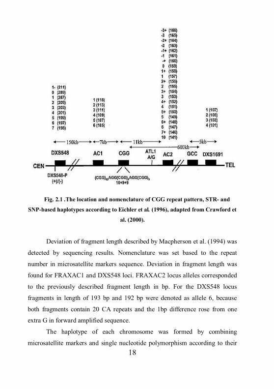
and Eichler et al. (1996) (Fig. 2.1.).
Validation of genotyping results was made by direct sequencing of
random alleles for each microsatellite marker. For alleles of each marker the
same PCR conditions, as described above for genotyping, was used. The
exception was reverse primers A, which was not labelled with fluorescent dye.
The PCR products obtained in reaction were concentrated and purified for
sequencing reaction by the Millipore Montage PCR filter device.
The sequencing reaction was performed using concentrated and purified
PCR products and the BigDye® Terminator v3.1 kit (Applied Biosystems,
USA) according to the manufacturer’s protocol.
All sequencing reactions were run on an ABI Prism® 310 genetic
analyzer using 61cm x 50µm (50 cm well-to-read) capillary with POP-6™
polymer and analyzed by ABI DNA™ sequencing software.
18
Fig. 2.1 .The location and nomenclature of CGG repeat pattern, STR- and
SNP-based haplotypes according to Eichler et al. (1996), adapted from Crawford et
al. (2000).
Deviation of fragment length described by Macpherson et al. (1994) was
detected by sequencing results. Nomenclature was set based to the repeat
number in microsatellite markers sequence. Deviation in fragment length was
found for FRAXAC1 and DXS548 loci. FRAXAC2 locus alleles corresponded
to the previously described fragment length in bp. For the DXS548 locus
fragments in length of 193 bp and 192 bp were denoted as allele 6, because
both fragments contain 20 CA repeats and the 1bp difference rose from one
extra G in forward amplified sequence.
The haplotype of each chromosome was formed by combining
microsatellite markers and single nucleotide polymorphism according to their
19
position at the FRAXA locus. STR and SNP marker haplotypes were combined
as follows: DXS548-FRAXAC1-ATL1-FRAXAC2.
2.3.Statistical Data Analysis For AMOVA analysis, level of heterozygosity for all polymorphisms
and calculation of pairwise genetic distances (Fst) the Arlequin 3.5 package
(Excoffier and Lischer, 2010) was used. The significance of the haplotype
results was tested by 10000 permutations. To detect critical value of Fst, the
on-line statistical calculator for critical values of F-statistics by BioKin, Ltd.
was used. The “degrees of freedom numerator” was set to 1. The
“denomenator” was set to 132.
Analysis of 27 haplotypes, derivates from a total 133 chromosomes, was
carried out by population splitting in two subgroups based on normal/mutated
FRAXA alleles.
The case-control study data was analysed by Fisher’s exact test of 2×2
contingency tables and chi-square using GraphPad QuickCalcs on-line
calculator. For statistical significance of results, the p-value was set less than
0.05. The Bonferroni correction was applied for multiple testing (Bland and
Altman, 1995). According to the Bonferroni correction in four markers, the
haplotype analysis p-value was set less than 0.0125.
For calculation of the 95% confidence interval the QuickCalcs on-line
calculator was used. For the proportion calculation CI 95% was applied
according to the modified Wald method by Agresti and Coull (1998).
In the retrospect study, estimation of FSX prevalence was based on data
from the Central Statistical Bureau of Latvia and data obtained from this study.
Prevalence per 100 000 males was calculated: number of estimated male patients during the period
average number of male population during the period X 100 000 Prevalence =
20
Prevalence expressed as one affected male to the number of male
persons in population was calculated:
average number of male population during the period
number of estimated male patients during the period
3. RESULTS 3.1. Prevalence of the Fragile X Syndrome
The prevalence of the fragile X syndrome in male individuals with
mental retardation and developmental disability was estimated retrospectively.
In this study the estimation of the population prevalence was restricted to the
data from male population, because females with a full mutation in the FMR1
gene show an intellectual development from severely retarded to normal and
cannot be identified solely by clinical data.
In the group of unrelated, mentally retarded males (n = 374), 10 (95% CI
4.80 – 18.39) in ten years time period newly diagnosed fragile X syndrome
patients had a relative prevalence of 0.0267 (2.67%). According to the data
from the Central Statistical Bureau of Latvia, during the ten years time period
1998 to 2007, 10503 patients with psychological development disorders or
behavioural and emotional disorders were diagnosed (with onset usually
occurring in childhood and adolescence, new cases excluded those caused by
alcoholism and dependency upon narcotic and psychoactive substances).
Gender structure for the diagnosed cases was not available. Based on a
theoretical gender structure of the population (1:1) and a developmental
disability diagnosis of 1.25 male to 1 female (Raymond, 2006), we estimated
that there were 6295 male patients (95% CI 5690-7430) with diagnosis of
developmental disability in Latvia. According to the calculated relative
Prevalence =
21
prevalence of disease from our laboratory data, we further estimated that this
population included 168 (95% CI 143 – 195) FXS male patients.
For Latvia, with an average 1 079 941 male residents (based on data of
the Central Statistical Bureau of Latvia for 1998 - 2007), and assumed 168
male patients with the fragile X syndrome, gives a result in prevalence of
1/6428 males (95% CI 5538-7552) or 15.55/100 000 males (95% CI 13.24 –
18.05).
3.2. Variation of CGG Trinucleotide Repeats
In total, 374 patients were analysed with PCR screening and for all those
patients an exact CGG repeat number within FMR1 gene were detected.
Distribution of alleles was as follow: 90.37% of alleles fell in group of normal
CGG repeat number, 6.95% were grey-zone alleles and 2.67% of alleles
revealed full CGG repeat expansion. The highest incidence among all analysed
chromosomes were observed for allele 30 (29.95%), allele 31 (13.10%) and
allele 29 (12.83%).
From 374 patients, analysed with PCR screening, 364 were detected having
CGG repeat alleles within a non-pathogenic range (5 – 50 repeats). Twenty-six
different alleles were observed. The smallest repeat size identified within the
normal range was 16 CGG repeats. There were absences of alleles with 17; 18;
19; 44; 46; 48 and 49 CGG repeats. The most common allele in the normal
range was allele 30 (30.77%). Comparably prevalent were alleles 29 (13.19%),
31 (13.46%), 23 (8.52%) and 24 (6.32%). Distribution of non-pathogenic range
CGG repeat alleles is shown in Fig. 3.1.
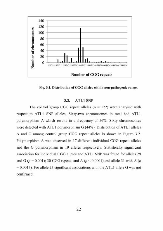
22
Fig. 3.1. Distribution of CGG alleles within non-pathogenic range.
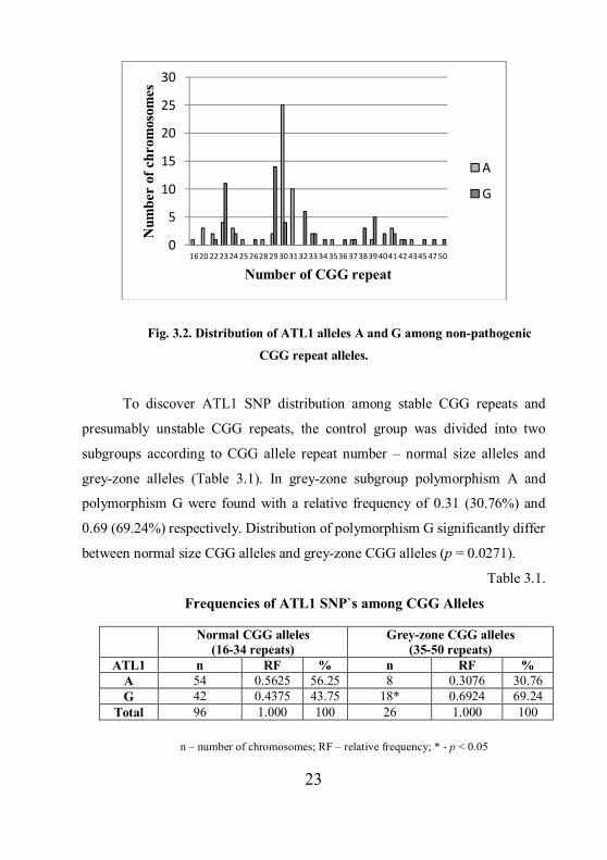
3.3. ATL1 SNP
The control group CGG repeat alleles (n = 122) were analysed with
respect to ATL1 SNP alleles. Sixty-two chromosomes in total had ATL1
polymorphism A which results in a frequency of 56%. Sixty chromosomes
were detected with ATL1 polymorphism G (44%). Distribution of ATL1 alleles
A and G among control group CGG repeat alleles is shown in Figure 3.2.
Polymorphism A was observed in 17 different individual CGG repeat alleles
and the G polymorphism in 19 alleles respectively. Statistically significant
association for individual CGG alleles and ATL1 SNP was found for alleles 29
and G (p = 0.001); 30 CGG repeats and A (p < 0.0001) and allele 31 with A (p
= 0.0013). For allele 23 significant associations with the ATL1 allele G was not
confirmed.
020406080
100120140
1617181920212223242526272829303132333435363738394041424344454647484950Num
ber
of c
hrom
osom
es
Number of CGG repeats
23
Fig. 3.2. Distribution of ATL1 alleles A and G among non-pathogenic
CGG repeat alleles.
To discover ATL1 SNP distribution among stable CGG repeats and
presumably unstable CGG repeats, the control group was divided into two
subgroups according to CGG allele repeat number – normal size alleles and
grey-zone alleles (Table 3.1). In grey-zone subgroup polymorphism A and
polymorphism G were found with a relative frequency of 0.31 (30.76%) and
0.69 (69.24%) respectively. Distribution of polymorphism G significantly differ
between normal size CGG alleles and grey-zone CGG alleles (p = 0.0271).
Table 3.1.
Frequencies of ATL1 SNP`s among CGG Alleles
n – number of chromosomes; RF – relative frequency; * - p < 0.05
0
5
10
15
20
25
30
1620 22 2324 25 2628 29 3031 3233 34 3536 37 3839 4041 42 4345 47 50
Num
ber
of c
hrom
osom
es
Number of CGG repeat
A
G
Normal CGG alleles (16-34 repeats)
Grey-zone CGG alleles (35-50 repeats)
ATL1 n RF % n RF % A 54 0.5625 56.25 8 0.3076 30.76 G 42 0.4375 43.75 18* 0.6924 69.24
Total 96 1.000 100 26 1.000 100
24
All FXS group chromosomes were exclusively found to be associated
with ATL1 allele G and this association was statistically significant (p =
0.0008).
3.4. Repeat Structure of Grey-Zone Alleles
26 grey-zone alleles were analysed using direct sequencing, to
characterise CGG repeat interruption by AGG trinucleotides. In twelve
chromosomes, a CGG interspersion pattern with three AGG`s was detected.
Twelve chromosomes with two AGG, one chromosome with one AGG and one
pure CGG tract were also detected. Ten different AGG interruption patterns
were detected (Table 3.2.). For all chromosomes, loss of AGG was detected on
3` end of the sequence.
Table 3.2. The Structure of Grey-Zone Allele CGG Array`s
Pattern of AGG interruption Number of chromosomes Rel. frequency
Pure 1 0.038 9+n 1 0.038
9+9+n 10 0.384 9+9+9+n 4 0.154
9+10+6+n 2 0.077 10+9+n 2 0.077 10+9+9+n 2 0.077 10+9+10+n 2 0.077 10+9+n+n 1 0.038 10+n+n+n 1 0.038
AGG pattern of CGG tract, the digit to the CGG repeat number, “n” corresponds to an
uninterrupted CGG repeat number and “+” denote the AGG interspersion position.
The CGG repeat structure was analysed with respect to ATL1 SNP
alleles. Significant associations were found, firstly for the allele A and a repeat
array with a 10+n structure (p = 0.001) and secondly for the allele G and a
repeat array with 9+n structure (p = 0.004).
25
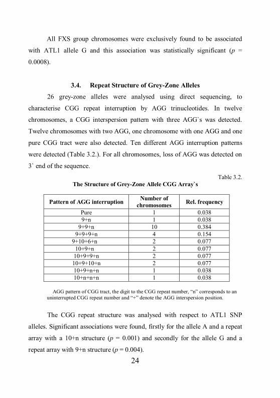
3.5. DXS548-FRAXAC1-ATL1-FRAXAC2 Haplotypes
Microsatellite markers and ATL1 SNP were analysed in both a control
group and FXS patient group (Fig. 3.3.)
Fig. 3.3. The location, nomenclature and frequencies of FMR1 linked STR- and SNP-
polymorphisms, tested in this study.
The haplotype of each chromosome was formed by combining
microsatellite markers and single nucleotide polymorphism according to their
position at the FRAXA locus. STR and SNP marker haplotypes were combined
as follows: DXS548-FRAXAC1-ATL1-FRAXAC2. In total 27 different
haplotypes were detected – 26 in the control group and three in the FXS group.
Only one haplotype from the FXS group was unique (Table 3.3.).
Among the FXS patients, haplotype 2-2-G-4 was found at a relative
frequency of 0.818 (p < 0.0001). The most common haplotype among control
group chromosomes was 7-4-A-5+ (RF = 0.327; p = 0.0336). Corrected by
Bonferroni this haplotype association is not significant for stable CGG repeat
alleles in our population.
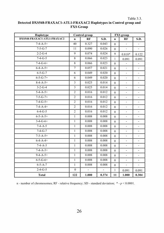
26
Table 3.3. Detected DXS548-FRAXAC1-ATL1-FRAXAC2 Haplotypes in Control group and
FXS Group
Haplotype Control group FXS group DXS548-FRAXAC1-ATL1-FRAXAC2 n RF S.D. n RF S.D.
7-4-A-5+ 40 0.327 0.043 0 - - 7-5-G-7 11 0.090 0.026 0 - - 2-2-G-4 9 0.074 0.024 9 0.818* 0.122 7-4-G-5 8 0.066 0.023 1 0.091 0.091
7-4-G-6+ 8 0.066 0.023 0 - - 6-4-A-5+ 7 0.057 0.021 0 - - 6-5-G-7 6 0.049 0.020 0 - -
6-5-G-7+ 6 0.049 0.020 0 - - 8-4-A-5+ 3 0.025 0.014 0 - - 3-2-G-4 3 0.025 0.014 0 - -
5-4-A-5+ 2 0.016 0.012 0 - - 7-5-G-7+ 2 0.016 0.012 0 - - 7-4-G-5+ 2 0.016 0.012 0 - - 7-4-A-4+ 2 0.016 0.012 0 - - 6-4-G-5 2 0.016 0.012 0 - -
6-5-A-5+ 1 0.008 0.008 0 - - 3-4-G-6+ 1 0.008 0.008 0 - - 7-4-A-5 1 0.008 0.008 0 - - 7-4-G-7 1 0.008 0.008 0 - -
7-3-A-5+ 1 0.008 0.008 0 - - 6-4-A-4+ 1 0.008 0.008 0 - - 7-4-A-3 1 0.008 0.008 0 - -
7-4-A-3+ 1 0.008 0.008 0 - - 9-4-A-5+ 1 0.008 0.008 0 - - 6-5-G-6+ 1 0.008 0.008 0 - - 6-5-A-7 1 0.008 0.008 0 - - 2-4-G-5 0 - - 1 0.091 0.091 Total 122 1.000 0.374 11 1.000 0.304
n - number of chromosomes; RF - relative frequency; SD - standard deviation; * - p < 0.0001.
27
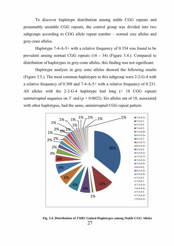
To discover haplotype distribution among stable CGG repeats and
presumably unstable CGG repeats, the control group was divided into two
subgroups according to CGG allele repeat number – normal size alleles and
grey-zone alleles.
Haplotype 7-4-A-5+ with a relative frequency of 0.354 was found to be
prevalent among normal CGG repeats (16 - 34) (Figure 3.4.). Compared to
distribution of haplotypes in grey-zone alleles, this finding was not significant.
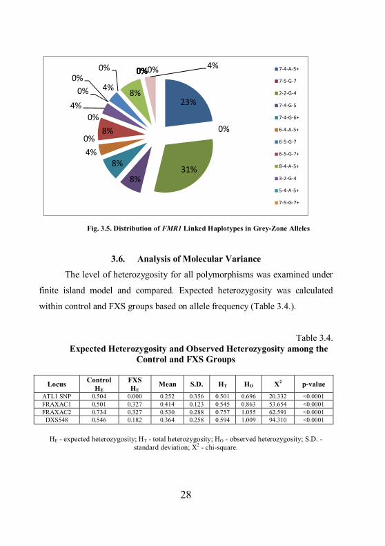
Haplotype analysis in grey zone alleles showed the following results
(Figure 3.5.). The most common haplotypes in this subgroup were 2-2-G-4 with
a relative frequency of 0.308 and 7-4-A-5+ with a relative frequency of 0.231.
All alleles with the 2-2-G-4 haplotype had long (> 18 CGG repeat)
uninterrupted sequence on 3` end (p = 0.0022). Six alleles out of 18, associated
with other haplotypes, had the same, uninterrupted CGG repeat pattern.
Fig. 3.4. Distribution of FMR1 Linked Haplotypes among Stable CGG Alleles
36%
12%
1%
6%6%
6%
6%
4%3%
2%
2% 2%1%
2% 0% 1%
1%1%
1%1% 1% 1% 1% 1% 7-4-A-5+
7-5-G-72-2-G-47-4-G-57-4-G-6+6-4-A-5+6-5-G-76-5-G-7+8-4-A-5+3-2-G-45-4-A-5+7-5-G-7+7-4-G-5+7-4-A-4+6-4-G-56-5-A-5+3-4-G-6+7-4-A-57-4-G-77-3-A-5+6-4-A-4+7-4-A-37-4-A-3+9-4-A-5+
28
Fig. 3.5. Distribution of FMR1 Linked Haplotypes in Grey-Zone Alleles
3.6. Analysis of Molecular Variance
The level of heterozygosity for all polymorphisms was examined under
finite island model and compared. Expected heterozygosity was calculated
within control and FXS groups based on allele frequency (Table 3.4.).
Table 3.4. Expected Heterozygosity and Observed Heterozygosity among the
Control and FXS Groups
Locus Control HE
FXS HE
Mean S.D. HT HO X2 p-value ATL1 SNP 0.504 0.000 0.252 0.356 0.501 0.696 20.332 <0.0001 FRAXAC1 0.501 0.327 0.414 0.123 0.545 0.863 53.654 <0.0001 FRAXAC2 0.734 0.327 0.530 0.288 0.757 1.055 62.591 <0.0001
DXS548 0.546 0.182 0.364 0.258 0.594 1.009 94.310 <0.0001
HE - expected heterozygosity; HT - total heterozygosity; HO - observed heterozygosity; S.D. -
standard deviation; X2 - chi-square.
23%
0%
31%8%
8%4%0%
8%0%
4%0%
0%4%
0%
8%
0%0%0%0%0%0%0%0%0%0% 4% 7-4-A-5+
7-5-G-7
2-2-G-4
7-4-G-5
7-4-G-6+
6-4-A-5+
6-5-G-7
6-5-G-7+
8-4-A-5+
3-2-G-4
5-4-A-5+
7-5-G-7+
29
AMOVA analysis revealed that molecular variation among groups was
27.04%. Molecular variation within groups was 72.96%. The fixation index
Fst was calculated based on haplotype frequencies between control and FXS
groups and found to be 0.27042 (p < 0.001). The critical value of Fst to confirm
the null hypothesis, was calculated to be 0.0683 (α = 0.01).
3.7. Genotype-Phenotype Correlation
Clinical data based on case-records of twelve confirmed FXS male
patients were analysed. The age of patients at the moment of diagnosis varied
between two and sixteen years (average = 7.33 ± 4.46). Molecular diagnostic
results for these patients revealed different patterns of CGG repeat expansion.
A full repeat size mutation (> 200 CGG repeats) with fully methylated gene
promoter region was found in nine patients. Two patients showed
premutation/full repeat size mutation mosaic with methylation mosaicism. One
patient had a full repeat size mutation with methylation mosaicism (up to 80%
unmethylated).
Major clinical symptoms of FXS were analysed for twelve patients.
Eight patients out of twelve were tested for IQ. Results revealed, IQ level of
patients ranged from 34 to 74 with an average IQ level of 52.75 (± 12.75). In
the group of psychomotor symptoms mental retardation, learning difficulties,
speech delay and attention-deficit/hyperactivity were observed all patients.
From other clinical symptom groups only hypotonia was found in all examined
patients.
In order to assess the genotype – phenotype correlation among full
mutation alleles and CGG repeat size and/or methylation mosaicism alleles,
clinical symptoms were compared between patients with full mutation in
lymphocytes and patients with repeat size and/or methylation mosaic. Genotype-phenotype comparison did not reveal significant differences among
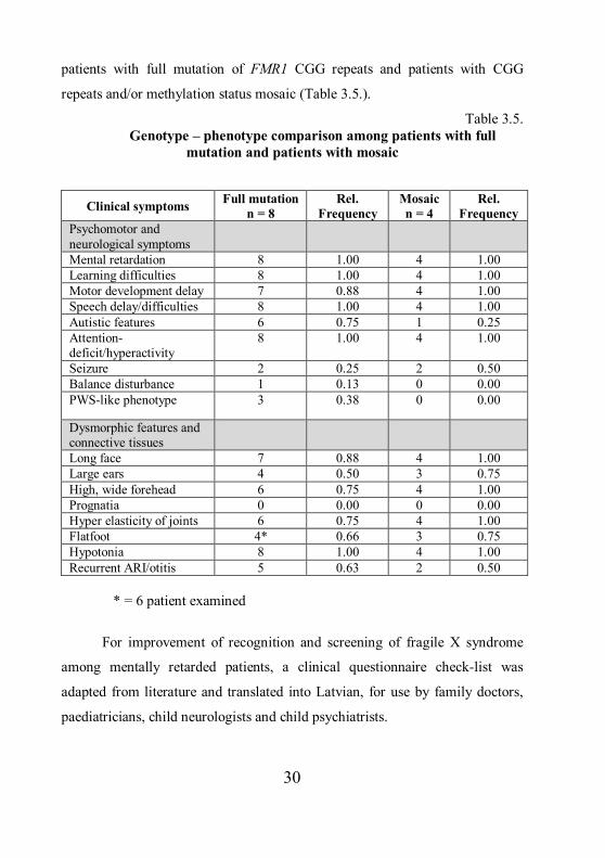
30
patients with full mutation of FMR1 CGG repeats and patients with CGG
repeats and/or methylation status mosaic (Table 3.5.).
Table 3.5. Genotype – phenotype comparison among patients with full
mutation and patients with mosaic
Clinical symptoms Full mutation n = 8
Rel. Frequency
Mosaic n = 4
Rel. Frequency
Psychomotor and neurological symptoms
Mental retardation 8 1.00 4 1.00 Learning difficulties 8 1.00 4 1.00 Motor development delay 7 0.88 4 1.00 Speech delay/difficulties 8 1.00 4 1.00 Autistic features 6 0.75 1 0.25 Attention-deficit/hyperactivity
8 1.00 4 1.00
Seizure 2 0.25 2 0.50 Balance disturbance 1 0.13 0 0.00 PWS-like phenotype 3 0.38 0 0.00
Dysmorphic features and connective tissues
Long face 7 0.88 4 1.00 Large ears 4 0.50 3 0.75 High, wide forehead 6 0.75 4 1.00 Prognatia 0 0.00 0 0.00 Hyper elasticity of joints 6 0.75 4 1.00 Flatfoot 4* 0.66 3 0.75 Hypotonia 8 1.00 4 1.00 Recurrent ARI/otitis 5 0.63 2 0.50
* = 6 patient examined
For improvement of recognition and screening of fragile X syndrome
among mentally retarded patients, a clinical questionnaire check-list was
adapted from literature and translated into Latvian, for use by family doctors,
paediatricians, child neurologists and child psychiatrists.
31
4. DISCUSSION 4.1. Prevalence of the Fragile X Syndrome
Ten years of experience with molecular diagnostics of the fragile X
syndrome in Latvia and a comparable low number of diagnosed patients with
this disease led to the question, how prevalent is fragile X syndrome in the
Latvian population? Lack of studies in our geographical region was one more
factor that inspired us for this study.
As with other published studies, the target population of our study were
patients with mental retardation and/or developmental disabilities, as these
characteristics are the main symptoms of FXS. It is of prime importance to
screen patients demonstrating symptoms of fragile X syndrome and whilst at
the same time increase the detection rate for this disease.
This study of mentally retarded males results in 2.67% of prevalence in
the target population. These results are in line with findings from other research
group studies of populations with a similar clinical symptom range. Our result
proves the importance of clinical symptoms recognition related to FXS
syndrome in clinical practices and necessity to suggest a check-list of
symptoms for clinical specialists to allow easier detection of patients with
suspected FXS.
In this study, to assess prevalence of FXS in entire male population, we
attributed our detected prevalence of FXS in target population to the total
number of patients with psychological development disorders or behavioural
and emotional disorders diagnosed in Latvia in same time period. These data
were obtained from published data source of the Central Statistical Bureau of
Latvia. The gender structure and a detailed overview of included diagnosis
were not available, but the overall patient description was the most appropriate
for the comparison with a target population of our study. This can be a source
of inaccuracy of calculated prevalence in entire male population. As it was
32
mentioned before, the calculated prevalence in different studies correlate with
the spectrum of clinical symptoms chosen for target population.
Our results are consistent with FXS prevalence in Europe. Crawford and
colleagues in their fragile X syndrome epidemiology review indicated the
necessity of a large population screening for the complete ascertainment of
disease prevalence. We completely agree with such a necessity and, even more
important, to find out the prevalence of the premutation carrier women in a
population of different ethnical backgrounds.
4.2. Variation of CGG Trinucleotide Repeats
In our study we analysed the distribution of normal CGG repeat alleles
among unrelated mentally retarded male patients. The prevalent allele detected
in this study agrees with reports from populations across Europe and Western
European descents from America, and it is allele 30. If we compare results from
our study with results from a study by Estonian colleagues, there are no
significant differences. Allele 30 was found in 29.30% of all chromosomes in
Estonian patients (Puusepp et al., 2008), and 29.95% of all chromosomes in
Latvian patients.
The distribution of normal CGG repeat alleles are described in different
populations. It is possible that discrepancy of data in one single repeat unit rose
from different methods used for CGG number detection and genotyping errors.
Distribution of CGG repeats in Latvian X chromosomes did not reveal
any significant differences among our data and data from European
populations. The total heterogeneity of CGG allele distribution in our
population was assumed to be in line with data from European populations.
33
4.3. ATL1 SNP
Previous studies have suggested linkage of CGG tract instability with
three factors: the G allele of ATL1 SNP; specific microsatellite marker
haplotypes; and a CGG tract AGG interspersion pattern exhibiting a long
uninterrupted CGG repeat at the 3’ end (Arrieta et al., 2003; Crawford et al.,
2000; Curlis et al., 2005; Dombrowski et al., 2002; Eichler et al., 1996; Gunter
et al., 1998; Zhou et al., 2006).
Interesting study proposed the hypothesis that polymorphism A
originated as a mutation in the 30 CGG repeat array linked with the haplotype
7-3-4+. Through either selection or genetic drift, the allele A become the
prevalent allele associated with normal CGG repeats in Western European
descent populations (Gunter et al., 1998). If we compare this finding with
results from our study, the haplotype 7-4-5+ is the most prevalent among the 30
CGG alleles (51.72%) and all alleles with this haplotype were associated with
ATL1 polymorphism A.
Our results revealed a statistically significant prevalence of the ATL1
polymorphism G among grey zone alleles and full mutation alleles, which is an
indicator of instability.
4.4. Repeat Structure of Grey-Zone Alleles
One of the tasks in this study was to detect the structure of CGG repeats
among X chromosomes with normal CGG repeat alleles. Since normal range
CGG repeats (5 – 34) is considered to be stable in transmission, we decided to
detect the CGG array just in grey-zone alleles (35 – 50). Grey-zone alleles are
normally expressed and do not leads to FXS phenotype but, these alleles may
show instability in transmission. For this reason these alleles are good a target
for the study of instability factors.
34
The absence of AGG interruptions in long tracts of CGG repeats have
been described as the main factors related to this instability (Rife et al., 2004).
It is hypothesised that CGG expansion occurs only at the 3` end of the triplet
array. There are various patterns of AGG interruptions of the array that are
believed to be responsible for “stabilizing” the alleles (Dombrowski et al.,
2002). In our study ten different CGG arrays for alleles with 35 to 50 CGG
repeats were found. One allele had a pure CGG tract. The most common pattern
of grey-zone alleles was 9+9+n. The association of CGG tract pattern results
and specific haplotypes is discussed in the next section.
4.5. DXS548-FRAXAC1-ATL1-FRAXAC2 Haplotypes
In the present study, we characterised the microsatellite markers
DXS548, FRAXAC1 and FRAXAC2, the ATL1 SNP and the corresponding
haplotypes in a mentally retarded male population from Latvia with normal and
expanded FMR1 gene CGG repeats. To achieve this task a case-control study
was made. The AMOVA data based on calculated Fst suggested that the
differences between detected haplotypes within the control and FXS groups
were significant and both our population subgroups show different genetical
backgrounds.
Several studies have identified specific haplotypes associated with FXS
patients chromosomes and normal CGG repeat alleles across European
populations (Arrieta et al., 2003; Dokic et al., 2008; Malmgren et al., 1994;
Peixoto et al., 1998; Pekarik et al., 1999; Rajkiewicz, 2008). In Caucasians,
haplotypes 6-4-4; 6-4-5 and 2-1-3 were reported as haplotypes positively
associated with full CGG repeat expansion (Eichler et al., 1996). However,
only a limited number of these studies focused on populations from Eastern and
North-eastern Europe.
35
Different microsatellite markers were used for these haplotype analyses
in European Caucasian populations. Thus, comparison of our results with these
analyses would prove difficult. As the analysed microsatellite loci and
nomenclature assigned to alleles in the literature are different, confusion arises,
which may lead to bias in the interpretation of literature data comparing
haplotypic results from different populations.
In Latvian population, 7-4-A-5+ was determined as the prevalent
haplotype for normal CGG alleles. However, after the Bonferroni correction,
this finding was not considered to be statistically significant. From literature
data the prevalent haplotypes in English population and Western European
descended populations of the USA were 7-3-4 and 7-3-4+. These haplotypes
were not detected in our control group. Though, taking in to account that most
published data from European populations analysed two microsatellite markers,
we can therefore compare our results with two marker haplotypes. The
haplotype 7-4 constructed from two microsatellite markers DXS548 and
FRAXAC1 respectively, was the prevalent one in studies from Basque valleys,
Czech, Croatia, Poland and Portugal (Arrieta et al., 2003; Dokic et al., 2008;
Pekarik et al., 1999; Peixoto et al. 1998; Rajkiewicz, 2008). In our study,
DXS548-FRAXAC1 haplotype 7-4 was found in 72 out of 122 control group
chromosomes. This finding shows prevalence of the haplotype 7-4 in our
control group.
Furthermore, haplotype 2-2-G-4 was found to be in positive
association with full mutation CGG alleles in Latvian FXS chromosomes. In
contrast, haplotypes 6-4-4; 6-4-5 and 2-1-3 were reported as positively
associated with FXS in Western European descents. These haplotypes were not
detected in our FXS group but, nevertheless haplotype 6-4-5 was detected in
two grey-zone alleles (41 and 42 CGG repeats respectively). Both alleles were
associated with ATL1 polymorphism G, and both had CGG tract pattern
36
9+9+n. All these findings for 6-4-5 haplotype linked alleles may provide
evidence of possible instability for these alleles in later generations.
Grey zone alleles featuring a long (≥18 repeats) uninterrupted CGG
tract at the 3’ end were found to be in positive association with the haplotype 2-
2-G-4. In grey-zone alleles haplotype 2-2-G-4 was detected in 8 alleles. All
these alleles had a CGG tract pattern where the first AGG interspersion
occurred after nine CGG repeats (9+ structure). This finding is in line with
Gunter`s and colleagues suggested “positively” associated haplotypes with the
fragile X mutation. Haplotype 7-4-A-5+ was found in six grey-zone alleles and
all alleles had CGG tract pattern where the first AGG interspersion occurred
after ten CGG repeats (10+ structure). This haplotype might be a “protective”
haplotype for CGG tract stability.
These findings imply that, in our population, haplotype 2-2-G-4 is a
marker of CGG tract instability. Grey zone alleles with a long uninterrupted
CGG tract at the 3’ end associated with this haplotype have a higher likelihood
of increasing the number of CGG repeats, leading to either premutation or
mutation over generations.
To the best of our knowledge, specific FMR1-linked haplotypes in the
Baltic State region and North-eastern Europe have not been previously
described. The present study is the first to report Latvian population FMR1
haplotype data. Comparison of the data with those obtained from
geographically close European populations highlights differences, particularly
with the FXS patient group. Indeed, haplotype 2-2-G-4 appears to be
exclusively found in Latvian FXS chromosomes. We conclude that a founder
effect could not be an explanation of our findings, on the basis of heterogeneity
exhibited in the Latvian population and on the basis of a lack of studies across
this geographical region. The small number of FXS chromosomes analysed in
this study was restricted by a low pick up rate of the fragile X syndrome in our
37
population. It could however, be a source of incomplete data for the FXS
chromosomes linked haplotypes. A larger study of FMR1-linked haplotypes in
Eastern and North-eastern European regions may provide more accurate data.
Nevertheless we consider that our data provide evidence of a specific
mutational pathway for unstable CGG alleles in our geographical region.
4.6. Genotype-Phenotype Correlation
Clinical symptoms are crucial for patients with fragile X syndrome
detection among the mentally retarded population. Recognition of these
symptoms is a first step towards a successful diagnosis of the fragile X
syndrome and the subsequent cascade testing among family members.
Assessment of a genotype – phenotype correlation among diagnosed patients
can help predict a prognosis for FXS patients and allow exploration for a
diversity of symptoms.
In our study we detected three patients with repeat size/methylation
mosaic. The observed unmethylated premutation repeat size varied from 78 to
150 CGG repeats. The phenotype of patients with repeat size/methylation
mosaic did not revealed significant differences from full mutation FXS
phenotype. The only remarkable observation was a lack of autistic features for
these patients. At the same time we should admit that for one patient with full
mutation autistic features also were also not observed. Neither of our patients
diagnosed with repeat size and /or methylation mosaicism showed any signs of
milder phenotype which was apparent for patients diagnosed with full mutation
and methylation. This can probably be explained by mitotic instability of the
expanded CGG tract and possible mosaic in different tissue. Based on these
observations we do not suggest making predictions on patient phenotype based
solely on genotype data obtained from leucocytes DNA study.
38
There is a report of eight FXS patients with “Prader-Willi like”
phenotype (de Vries et al., 1993). The patients had features resembling the
Prader-Willi syndrome (PWS), such as truncal obesity, hypogenitalism, and
small hands and feet. In our study three patient with “PW- like” phenotype
were diagnosed having fragile X syndrome. Three patients out of twelve
diagnosed with “PW- like” phenotype is remarkable number. In our opinion all
patients with PWS phenotype showing mental retardation and/or autistic
features should be tested for fragile X syndrome.
The low detection rate for patients with fragile X syndrome
demonstrated in our study led to the conclusion that fragile X syndrome is
generally clinically unrecognised. To help recognise patients with fragile X
syndrome among the mentally retarded male population we adapted clinical
check list based on a review of relevant literature and translated it into Latvian.
In our opinion, the low number of diagnosed patients was not only due
to the failure to clinically recognise fragile X syndrome, but also due to the
attitude of society toward mentally handicapped people and their families. In
our experience, families with diagnosed FXS patients refuse to inform relatives
at risk thereby preventing family genetic consultation. For clinical specialists, a
lack of specific treatment for fragile X syndrome place this diagnose in line
with other psychiatric diagnosis with symptomatic treatment.
39
5. CONCLUSIONS 1. The prevalence of fragile X syndrome in the Latvian male population was
estimated to be 1/6428 (95% CI 5538-7552) or 15.55/100 000 males (95%
CI 13.24 – 18.05). The prevalence of the fragile X syndrome among
mentally retarded male patients was estimated to be 2.67%.
2. The highest incidence among all analysed normal CGG repeat
chromosomes was observed for allele 30 (29.95%), allele 31 (13.10%) and
allele 29 (12.83%).
3. For individual CGG alleles with a normal CGG repeat number, a
statistically significant association with ATL1 SNP was found for allele 29
and G (p = 0.001); allele 30 and A (p < 0.0001) and allele 31 with A (p =
0.0013). Polymorphism G was found to be associated with grey-zone CGG
alleles (p = 0.0271) and exclusively associated with all FXS alleles.
4. In the case-control study, haplotype 7-4-A-5+ was determined as the
prevalent haplotype for normal CGG alleles in the Latvian population.
However, after Bonferroni correction, this finding was not considered to be
statistically significant. Results of this study imply that in the Latvian
population, haplotype 2-2-G-4 is a marker of CGG tract instability.
AMOVA results revealed distinct genetic background for FXS
chromosomes.
5. Analysing the structure of grey-zone alleles, revealed ten different CGG
arrays. The most common pattern of grey-zone alleles was 9+9+n. The
CGG array with a 9+n structure associated with the haplotype 2-2-G-4 was
recognised as the pattern positively associated with CGG repeat instability.
The CGG array with 10+n structure associated with the haplotype 7-4-A-
5+ was recognised as a “protective” pattern.
40
6. The results of genotype-phenotype analysis did not revealed significant
correlation among clinical symptoms, observed in FXS patients, and
distinct patterns of CGG repeat expansion.
6. PUBLICATIONS Publications
Zanda Daneberga, Zita Krūmiņa, Baiba Lāce, Daiga Bauze, Rita Lugovska.
Fragilās X hromosomas sindroms pacientiem ar neskaidras etioloģijas garīgo
atpalicību Latvijā. RSU Zinātniskie raksti 2007. RSU, 2008: 224-227
Zanda Daneberga, Zita Krūmiņa, Baiba Lāce, Daiga Bauze, Natālija Proņina,
Rita Lugovska. Fragile X syndrome in mentally retarded patients from Latvia
Proceedings of the Latvian Academy of Science, Section B, 2009 (63):70-72
Zanda Daneberga, Natalija Pronina, Baiba Lace, and Rita Lugovska. FMR1
Linked Haplotype Analysis in a Mentally Retarded Male Population. Central
European Journal of Medicine. 2011, 6(6):750-757
Zanda Daneberga, Zita Krūmiņa, Baiba Lāce, Daiga Bauze, Natālija Proņina,
Rita Lugovska. The Fragile X Syndrome: 13 Years of Experience. Proceedings
of the Latvian Academy of Science, Section B, 2011. In press.
Abstracts
Daneberga Z., Lace B., Pronina N., Lugovska R. FMR1 haplotype analysis in
mentally retarded male population. European Journal of Human Genetics Vol
19 Supp. 2, 2011, pp. 334.
Bauze D., Ķevere L., Kronberga Z., Riževs A., Jeļisejevs S., Daneberga Z.,
Dzalbs A., Ločmele Dz., Krūmiņa Z., Andrēziņa R., Lāce B. Autisma un
autiskā spektra traucējumu fenotipisko pazīmju, antropometrisko mērījumu un
bioķīmisko rādītāju salīdzinājums un analīze. RSU scientific conference,
Abstracts, 2011, pp.239.
41
Daneberga Z., Lāce B., Proņina N., Lugovska R. Ar FMR1 gēnu saistīto
haplotipu analīze. RSU scientific conference, Abstracts, 2011, pp.266.
Daiga Bauze, Zanda Daneberga, Arnis Riževs, Raisa Andrēziņa, Rita
Lugovska, Baiba Lāce. Fragilās X hromosomas sindroma (FXS) genotipa un
fenotipa salīdzinājums bērniem ar psihiskās attīstības traucējumiem. RSU
scientific conference, Abstracts, 2010, pp.113.
Daneberga Z., Krumina Z., Lace B., Bauze D., Pronina N., Lugovska R.
Prevalence of Fragile X syndrome in mentally retarded patients from Latvia.
European Journal of Human Genetics Vol 17 Supp. 2, 2009, pp. 134.
Daneberga Z., Krumina Z., Lace B., Bauze D., Pronina N., Lugovska R.
Molecular diagnostic of XLMR in mentally retarded males from Latvia.
European Journal of Human Genetics Vol 16 Supp. 2, 2008, pp. 62.
Daneberga Z., Krumina Z., Lace B., Bauze D., Pronina N., Lugovska R.
FRAXA locus investigation of mentally retarded patients. European Journal of
Human Genetics Vol 15 Supp. 1, 2007, pp.198
Daneberga Z., Krumina Z., Lace B., Pronina N., Lugovska R. Molecular
diagnosis of fragile X syndrome in mentally retarded patients in Latvia.
Laboratorine Medicina 1(29), 2006, pp.20
Daneberga Z., Krumina Z., Lace B., Pronina N., Lugovska R. Molecular
diagnosis of fragile X syndrome in mentally retarded patients in Latvia.
European Journal of Human Genetics Vol 14 Supp. 1, 2006, pp.251
Approbation
Pre-defence of this thesis was held in joint meeting of Department of Biology
and Microbiology, Riga Stradins University, Latvian Society of Medical
Genetics and Latvian Association of Human Genetics. July 1st, 2011.,
Department of Biology and Microbiology, Riga Stradins University, Riga,
Latvia.
42
Zanda Daneberga, Zita Krūmiņa, Baiba Lāce, Daiga Bauze, Natālija Proņina,
Rita Lugovska. Fragile X syndrome in Latvia: clinical, molecular and
population genetic aspects. 11th Conference of Baltic Child Neurology
Association. June 16th -18th, 2011, Riga, Latvia.
Zanda Daneberga, Baiba Lāce, Natālija Proņina, Rita Lugovska. Ar FMR1
gēnu saistīto haplotipu analīze. RSU scientific conference, April 14th - 15th,
2011. Riga, Latvia.
Zanda Daneberga, Zita Krūmiņa, Baiba Lāce, Daiga Bauze, Natālija Proņina,
Rita Lugovska. „Molecular diagnostic of X-linked mental retardation” 9th
Baltic Congress of Laboratory Medicine, September 18th - 20th, 2008. Jurmala,
Latvia.
Zanda Daneberga, Zita Krūmiņa, Baiba Lāce, Daiga Bauze, Natālija Proņina,
Rita Lugovska. „Fragile X syndrome in mentally retarded patients from Latvia”
IV Baltic Genetical Congress, October 9th - 12th, 2007. Daugavpils, Latvia
7. ACKNOWLEDGEMENTS Many people have contributed their time and knowledge into this thesis.
It is a pleasure to thank those who made this thesis possible.
I would like to thank my supervisor, assoc. prof. Rita Lugovska, for
making possible to develop my PhD thesis and grow up in both professional
and scientific field at the Medical Genetics Clinic, University Children`s
Hospital.
I owe my deepest gratitude to my scientific advisor, Dr. Baiba Lāce, for
the time she spent guiding me in statistical methods, reading my thesis and for
her selfless contribution to improve this work.
I would like to show my gratitude to Dr. Kristin Eiklid, Ulleval
University Hospital, Oslo, Norway for her open attitude, kindly offered
technical help with confirming diagnosis and hosting me in her laboratory.
43
It is an honour for me to express my thanks to Prof. R. A. Wevers and
Dr. H. Yntema, Radboud University Nijmegen Medical Centre, Nijmegen, The
Netherlands for technical help with confirming diagnosis and inspiration for
this work.
I am grateful to all clinicians at the Medical Genetic Clinic for
contribution to collect samples, evaluation of patient’s clinical symptoms and
my guidance in clinical data analysis. Special thanks to Dr. Zita Krūmiņa, Dr.
Daiga Bauze and Dr. Dzintra Ločmele.
I wish to thank Irēna Rogovska and Liāna Pliss for time and advices
they gave me in data statistical processing.
I would like to thank laboratory assistant of DNA laboratory, Kristina
Morozova, for the preliminary preparation of samples and technical assistance.
I am indebted to many of my colleagues in Laboratory of Genetical
Biochemistry and DNA laboratory for support, understanding, advice and
patience. Special thanks to Pārsla Vēvere and Natālija Proņina.
I wish to thank all patient families for participating in this research, their
cooperation are highly appreciated.
Finally, very special gratitude goes to my family, my partner and my
friends for faith in me, for understanding and patience during the many hours I
spent in laboratory and at my computer.
44
8. REFERENCES 1. Agresti A. and Coull B. A., Approximate is better than "Exact" for
interval estimation of binomial proportions. The American Statistician. 1998; 52:119-126.
2. Arrieta, I., O. Penagarikano, M. Telez et al. The FMR1 CGG repeat and linked microsatellite markers in two Basque valleys. Heredity 2003; 90:206-211.
3. Bardoni B., Mandel J-L. and Fish G. S. FMR1 gene and fargile X syndrome. Am. J. Med. Genet. 2000; 97:153-163.
4. Bland J. M., Altman D. G. Comparing methods of measurement: why plotting difference against standard method is misleading. Lancet 1995; 346: 1085-1087.
5. Chiurazzi, P. DNA panel to interlaboratory standardization of haplotype studies on the fragile X syndrome and proposal for a new allele nomenclature. Am. J. Med. Genet. 1999; 83:347-349.
6. Chong S. S., Eichler E. E., Nelson D. L. et al. Robust amplification and ethidium – visible detection of the fragile X syndrome CGG repeat using Pfu polymerase. Am. J. Med. Genet. 1994; 51:522-526.
7. Crawford D. C., Acuna J. M. and Sherman S. L. FMR1 and the fragile X syndrome: Human genome epidemiology review. Genetics in Medicine 2001; 3(5):359-371.
8. Crawford, D. C., C. E. Shwartz, K. L. Meadows et al. Survey of the Fragile X Syndrome CGG Repeat and the Short-Tandem-Repeat and Single-Nucleotide-Polymorphism Haplotypes in an African American Population. Am. J. Med. Genet. 2000; 66:480-493.
9. Curlis Y., Zhang C., Holden J. J. A., Kirkby K., Loeesch D., Mitchell R. J. Haplotype study of intermediate-length alleles at the fragile X (FMR1) gene: ATL1, FMRb, and microsatellite haplotypes differ from those found in common-size FMR1 alleles. Hum. Biol. 2005; 77(1):137-151.
10. de Vries B. B. A., Fryns J-P., Butler M. G., Canziani F., Wesby-van Swaay E., van Hemel J. O., Oostra B. A., Halley D. D. J, Niermeijer M. F. Clinical and molecular studies in fragile X patients with a Prader-Willi-like phenotype. J Med Genet 1993; 30: 761-766.
11. de Vries B. B. A., Jansen C. C. A. M., Duits A. A., Verheij C., Willemsen R., van Hemel J. O., van den Ouweland A. M. W., Niermeijer M. F., Oostra B. A., Halley D. D. J. Variable FMR1 gene methylation of large expansions leads to variable phenotype in three males from one fragile X family. J Med Genet 1996; 33:1007-1010.
12. de Vries, B. B. A., Halley D. J. J., Oostra B. A. et al. The fragile X syndrome. J. Med. Genet. 1998; 35:579-589.
45
13. Dokic, H., Barišic I., Čulic V., Lozic B., Hecimovic S. Haplotype and AGG interspersion analysis of FMR1 alleles in a Croatian population: no founder effect detected in patients with fragile X syndrome. Hum. Biol. 2008; 80:581-587.
14. Dombrowski, C., Levesque S., Morel M. L., Rouillard P., Morgan K., Rousseau F. Premutation and intermediate-size FMR1 alleles in 10 572 males from the general population: loss of an AGG interruption is a late event in the generation of fragile X syndrome alleles. Hum. Mol. Genet. 2002; 11:371-378.Dracopoli and Haines, 1994).
15. Eichler, E. E., Macpherson J. N., Murray A., Jacobs P. A., Chakravarti A., Nelson D. L. Haplotype and interspersion analysis of the FMR1 CGG repeat identifies two different mutational pathways for the origin of the fragile X syndrome. Hum. Mol. Genet. 1996; 5:319–330.
16. Excoffier, L. and Lischer H. E. L. Arlequin suite ver. 3.5: A new series of programs to perform population genetics analyses under Linux and Windows. Molecular Ecology Resources. 2010; 10:564-567.
17. Fu, Y. H., Kuhl D. P. A., Pizzuti A., Pieretti N., Sutcliffe J. S., Verkerk A. J. M. H., et al. Variation of the CGG repeat at the fragile X site results in genetic instability: Resolution of the Sherman paradox. Cell. 1991; 67:1047-1058.
18. Garber K. B., Visootsak J. and Warren S. T. Fragile X syndrome. Eur J Hum Genet. 2008; 16:666-672.
19. Gunter C., Paradee W., Crawford D. C., Meadows K. A., Newman J., Kunst C. B., et al. Re-examination of factors associated with expansion of CGG repeats using a single nucleotide polymorphism in FMR1. Hum. Mol. Genet. 1998; 7:1935-1946.
20. Hernandez R. N., Feinberg R. L., Vaurio R., Passanante N. M., Thompson R. E., Kaufmann W. E. Autism Spectrum Disorder in Fragile X Syndrome: A Longitudinal Evaluation. Am J Med Genet A. 2009; 149A(6):1125–1137.
21. Krūmiņa Dž., Kokare I. and Biķis E. Latvijas bērnu fiziskās attīstības novērtēšana. SIA Medicīnas apgāds, 2007, Rīga
22. Kunst, C. B., Zerylnick C., Karickhoff L. et al. FMR1 in Global Populations. Am. J. Hum. Genet. 1996; 58:513-522.
23. Larbrisseau A., Jean P., Messier B., Richer C-L. Fragile X chromosome and X-linked mental retardation. CMA Journal 1982; 127:123-126.
24. Macpherson, J. N., Bullman H., Youings S. A. et al. Insert size and flanking haplotype in fragile X and normal population: possible multiple origins for the fragile X mutations. Hum. Mol. Genet 1994; 3:399-405.
46
25. Malmgren, H., K. H. Gustavson, C. Oudet et al. Strong founder effect for the fragile X syndrome in Sweden. Eur. J. Hum. Genet. 1994; 2:103-109.
26. Moutou C., Vincent M. C., Biancalana V., Mandel J-L.. Transition from premutation to full mutation in fragile X syndrome is likely to be prezygotic. Hum. Mol. Genet. 1997; 3:971-979.
27. Murray A., Macpherson J. N., Pound M. C., Sharrock A., Youings S. A., Dennis N. R., et al. The role of size, sequence and haplotype in the stability of FRAXA and FRAXE alleles during transmission. Hum. Mol. Genet. 1997; 6:173-184.
28. Oberle I., Rousseau F., Heitz D., Kretz C., Devys D., Hanauer A., et al. Instability of a 550-Base Pair DNA Segment and Abnormal Methylation in Fragile X Syndrome. Science. 1991; 252:1097-1102.
29. Oudet C., Mornet E., Serre J. L., Lentes-Zengerling F. T. S., Kretz C., Deluchatt C., Tejada I., Bouet J., Bouet A., Mandel J. L. Linkage Disequilibrium between the Fragile X Mutation and Two Closely Linked CA Repeats Suggests That Fragile X Chromosomes Are Derived from a Small Number of Founder Chromosomes. Am. J. Hum. Genet. 1993; 52:297-304.
30. Peixoto A., dos Santos M. R., Seruca R. Amorim A., Castedo S. Analysis of FMR1 and flanking microsatellite markers in normal and fragile X chromosomes in Portugal: evidence for a “protector” haplotype. Eur. J. Hum. Genet. 1998; 6:518-522.
31. Pekarik V., Blazkova M. and Kozak L. Haplotype analysis of the fragile X syndrome gene FMR1 in the Czech Republic. Am. J. Med. Genet. 1999; 84:214-21.
32. Phadke S. R. Fragile X syndrome. Orphanet encyclopaedia. 2005; http://www.orpha.net/data/patho/GB/uk-Fragile-X.pdf
33. Poustka A., Dietrich A., Langenstein G., Toniolo D., Warren S. T., Lehrach H. Physical map of human Xq27-qter: Localizing the region of the fragile X mutation. Proc. Natl. Acad. Sci. USA 1991; 88:8302-8306.
34. Puusepp H., Kahre T., Sibul H., Soo V., Lind I., Raukas R., Õunap K. Prevalence of the Fragile X Syndrome Among Estonian Mentally Retarded and the Entire Children's Population J. Child. Neurol. 2008; 23:1400-1405.
35. Rajkiewicz, M. 2008. Molecular analysis of the FMR1 gene and searching for the premutations in the large group of Polish ataxia patients and group of women with premature ovarian failure. Ph.D. dissertation. Department of Genetics, Institute of Psychiatry and Neurology, Warsaw, Poland.
47
36. Richards R. I., Shen Y., Holman K., Kozman H., Hyland V. J., Mulley J. C., Sutherland G. R. Fragile X syndrome: genetic localisation by linkige mapping of two microsatellite repeats FRAXAC1 and FRAXAC2 which immediately flank the fragile site. J. Hum. Genet. 1991; 28:818-823.
37. Ridaura-Ruiz L., Quinteros-Borgarello M., Berini-Aytés L., Gay-Escoda C. Fragile X-syndrome: Literature review and report of two cases. Med. Oral. Patol. Oral. Cir. Bucal. 2009; 14(9):434-439.
38. Rife M., Badenas C., Quinto L. et al. Analysis of CGG variation through 642 meioses in Fragile X families. Mol. Hum. Reprod. 2004; 10:773-779.
39. Rousseau F., Heitz D., Oberle I., Mandel J-L. Selection in blood cells from female carriers of the fragile X syndrome: inverse correlation between age and proportion of active X chromosomes carrying the full mutation. J. Med. Genet. 1991; 28:830-836.
40. Sambrook J. and Russell D. W. Molecular Cloning: A Laboratory Manual, Cold Spring Harbor Laboratory, 2001. Cold Spring Harbor, NY.
41. Sherman S., Plecher B. A. and Driscoll D. A. Fragile X syndrome: Diagnostic and carrier testing. Genetics in Medicine 2005; 7:584-587.
42. Zhou Y., Tang K., Law H. Y., Ng I. S. L., Lee C. G. L., Chong S. S. FMR1 CGG repeat patterns and flanking haplotypes in three Asian populations and their relationship with repeat instability. Ann. Hum. Genet. 2006; 70:784-796.
Recommended