RUNNING HEAD: Bone Mineral Density 1
The Effect of Omeprazole on Bone Mineral Density in Wistar
Rats
Layla E. Borham*1,2, Magda M. Hagras3, Altaf A. Abdulkhaliq4, Mohammed
Badawood5, Gamal S. Abd El Aziz5
1Department of Pharmacology and Toxicology, Faculty of Medicine, Umm al-Qura
University, Makkah, Saudi Arabia
2Department of Clinical Pharmacology, Faculty of Medicine, Cairo University, Egypt
3Department of Clinical Pharmacology, Suez Canal University, Ismailia, Egypt
4Department of Biochemistry, Faculty of Medicine, Umm al-Qura University, Makkah, Saudi
Arabia
5Department of Anatomy, Faculty of Medicine, King Abdul Aziz University, Jeddah, Saudi
Arabia
*Corresponding Author: borhaml @hotmail.com
Tel: 00966503567843. Fax: 00966125282097
BONE MINERAL DENSITY 2
ABSTRACT
This study examined the association between use of Omeprazole and risk of
osteoporosis in rat sample. For this study, 180 adult male Wistar rats were assigned to 3
groups with 60 rats in each group. Group I (a, b) (controls) was given intraperitoneal (i.p.)
saline over the same durations as the test groups. Groups II and III (a, b) were given i.p.
Omeprazole at 5 and 10 mg/kg/day, respectively, for 4 and 8 weeks. Ten rats from each
subgroup were left without treatment for the following 4 weeks to detect any reversals in the
effects of the drug. Intraperitoneal mode of delivery of drug is chosen for this study because
intraperitoneal injection is suitable for smaller species like laboratory rodents for which
intravenous access is challenging and it can be used to administer large volumes of fluid
safely. Further, the bone mineral density and bone mineral content decreased in a dose and
time dependent method, with recovery. The serum calcium and phosphate decreased in the 10
mg/kg for 8 weeks subgroup, with recovery of calcium, but not phosphate. The parathyroid
hormone level increased, with no recovery. The Tartrate-Resistant Acid Phosphatase type 5b
(TRAP5b) increased in the 5 and 10 mg for 8 weeks subgroups, with no recovery, and the
Insulin-Like Growth Factor-1 (IGF-1) decreased in a dose and time dependent method, with
recovery in only the 5 mg for 4 weeks subgroup. Diverse deteriorated histopathological
changes were observed according to the dose and time of omeprazole treatment, which were
more apparent in 10 mg for 8 weeks group with recovery in low dose. The use of high doses
of omeprazole for 8 weeks durations could adversely affect bone homeostasis, and leads to
decrease in Bone Mineral Density (BMD) resulting in the increased risk of subsequent
fractures.
Keywords: Omeprazole, bone mineral density, osteoporosis, parathyroid hormone, calcium,
phosphorus.

BONE MINERAL DENSITY 3
Table of Contents
ABSTRACT .............................................................................................................................. 2
1. Introduction .......................................................................................................................... 4
2. Material and Methods ......................................................................................................... 5
2.1. Test Drugs ....................................................................................................................... 5
2.2. Animals ........................................................................................................................... 6
2.3. Study Design ................................................................................................................... 6
2.4. The following parameters were measured ...................................................................... 8
2.4.1. Bone Mineral Density (BMD) and Bone Mineral Content (BMC) ......................... 8
2.4.2. Measurement of Bone Turnover Markers ................................................................ 8
2.4.3. Measurement of serum calcium, phosphorus and parathyroid hormone ..................... 9
2.5. Bone histopathological procedures ................................................................................. 9
2.6. Statistical Analysis .......................................................................................................... 9
3. Results ................................................................................................................................. 10
3.1. Body weight .................................................................................................................. 10
3.2. Effect of Omeprazole on the BMD and BMC .............................................................. 10
3.3. Effects of Omeprazole on the Serum Concentrations of Ca, P04 and PTH ................. 13
3.4. Effect of Omeprazole on Bone Markers ....................................................................... 13
2.5. Histopathological results ............................................................................................... 15
4. Discussion............................................................................................................................ 21
5. Conclusion .......................................................................................................................... 28
Conflict of Interest ................................................................................................................... 28
Acknowledgments.................................................................................................................... 28
Funding .................................................................................................................................... 28
Statement of Animal Rights ..................................................................................................... 29
References ................................................................................................................................ 30
Lists of Figures
Figure 1: Experimental study design ........................................................................................ 7
Figure 2: A photomicrographs of different bone sections from rats of control group showing
outer compact bone with normal appearance ........................................................................... 17
Figure 3: A photomicrographs of bone sections from omeprazole treated groups ................. 18
Figure 4: A photomicrographs of bone sections from omeprazole treated groups ................. 19
Figure 5: A photomicrographs of bone sections from recovery groups after Omeprazole
treatment .................................................................................................................................. 20
Lists of Tables
Table 1: Effect of omeprazole treatment on rat body weight (g) ............................................ 10
Table 2: Intra and inter-assay coefficients .............................................................................. 11
Table 3: Effect of administration of Omeprazole for 4 and 8 weeks on rat BMD & BMC .... 11
Table 4: Effect of Omeprazole for 4-8 weeks on biochemical parameters ............................. 14
BONE MINERAL DENSITY 4
1. Introduction
According to Sermet-Gaudelus et al. (2011), bone maintains its optimal structure,
strength and mineralisation through a dynamic process that senses load, stress and repair
areas of micro-damage. Further, bone remodelling cycle mediates this homeostatic process, in
which old bone is replaced by newly formed one. The basic multicellular unit of bone
remodelling is comprised of osteocytes, osteoclasts and osteoblasts (Clarke, 2008; Seeman
and Delmas, 2006). Osteoporosis is a skeletal disease that approximately affects the entire
skeleton, in which there is an imbalance between bone formation and bone resorption during
the bone remodelling cycle. This imbalance results in bone micro-architectural deterioration
and reduction of BMD (Milas-Ahic, Prus, Kardum, & Kovacevic, 2014). It is also observed
that bone demineralisation disease does not become clinically apparent until a fracture occurs
(Drake et al., 2015). Osteoporotic fractures account an immense public health problem
among the elderly people worldwide (Cummings and Melton 2002). This problem may
reduce the quality of life and raise the risk of morbidity and mortality (Johnell and Kanis
2006).
Some studies have pointed out a relationship between the long use of Proton Pump
Inhibitors (PPIs) and elevated risk of osteoporosis-related fractures ( Targownik, Lix, Leung,
Leslie, 2010 & Ito and Jensen 2010). Niv, (2011) identified that PPIs are powerful drugs for
gastric acid suppression, and are effective against acid-related diseases. They inhibit H+/K+
Adenosine Triphosphatase irreversibly in the gastric parietal cells, leading to effective gastric
acid secretion suppression (Segawa, Nakazawa, Tsukamoto, Chujoh, Yamao, & Hase, 1987;
Cui, Syversen, Zhao, Chen, &Waldum, 2001; & Shin, Vagin, Munson, Kidd, Modlin, &
Sachs, 2008). These drugs show few adverse effects when administered correctly, so in
clinical practice they are used for both acute symptoms and for long term purposes, although
such indications are highly questionable (Yang, 2012). Despite outstanding efficacy and

BONE MINERAL DENSITY 5
negligible short-term adverse effects, increased interest is shown regarding the side effects of
the chronic use of PPIs (Thomson, Sauve, Kassam, & Kamitakahara, 2010; Vestergaard,
2012 & Reimer, 2013). Omeprazole, which is one of the most important PPIs, in a dose of 20
mg/day is able to decrease BMD significantly as instigated by Yang, Lewis, Epstein, Metz,
(2006).
However, studies conducted byYu et al., (2008) suggested that omeprazole diminishes
bone resorption and blocks the progression to osteoporosis. So, the relation between the PPIs
use and bone demineralisation and the increased risk of fractures associated with prolonged
use of Omeprazole remains obscure (Targownik, Lix, Leung & Leslie, 2010). Due to the
recurrent nature of acid-related diseases, many patients require long-term or lifelong therapy
with PPIs (Scholten, 2007). Thus, the safety of this famous class of medications is of great
public health concern. The widespread use of PPIs worldwide for almost two decades has
gradually increased concerns from physicians and the public with regard to their benefits
being compromised by a diversity of risks that, till now, have received little attention. The
aim of the present work was to study the association between the use of Omeprazole with two
different dosages and durations and BMD and Bone Mineral Content (BMC) together with
bone turnover markers and bone histopathology.
2. Material and Methods
2.1. Test Drugs
This study used Omeprazole injections from a 40 mg vial (Losec; AstraZeneca UK
Limited), ketamine-HCl and xylazine hydrochloride (Sigma-Aldrich, MO, USA).
BONE MINERAL DENSITY 6
2.2. Animals
For this study, 180 adult male Wister rates were selected each 10 weeks old. The
subjects were weighed at 275±25 g (King Fahd Research Centre, KAU, Jeddah). The rats
were housed in standard polypropylene cages and maintained in a 45 percent to 55 percent
humid and controlled environment at 23±2 degree celsius with 5 rates per cage. Further, rats
were exposed to a 12/12-hour modified dark-light cycle with transmission of light from 7:00
am to 7:00 pm. Moreover, before two weeks, the rats were adjusted to water and food
conditions where in food was provided ad libitum (normal rodent diet contains 1.35 percent
Calcium and 1.04 percent Phosphorus) (Smith et al., 2012). All the ethical considerations
concerning the safety and harm issue related to laboratory animals were conducted in
accordance with the ethically approved Umm al-Qura University Committee on the Ethics of
Animal Experiments guidelines (HAPO-02-K-012-2015-05-117), which comply with the
national, and international laws and policies in experimental field. It was also ensured that all
the animals were treated efficiently and best possible measures were taken into account to
reduce their sufferings.
2.3. Study Design
The rats were weighed and assigned to 3 groups of 60 rats per category. In group I (a,
b), the controls were administered i.p. saline over the same durations as the test drug.
Intraperitoneal mode of delivery of drug is chosen for this study because intraperitoneal
injection is suitable for smaller species like laboratory rodents for which intravenous access is
challenging and it can be used to administer large volumes of fluid safely. Group II (a, b) was
given 5 mg/kg/day i.p. Omeprazole for 4 and 8 weeks respectively at 10 a.m. daily till the
time of DXA. Group III (a, b) was given 10 mg/kg/day i.p. omeprazole for the same time
periods (Segawa et al., 1987). At the end of the treatment, 20 rats from each subgroup (Ia, Ib,
IIa, IIb, IIIa and IIIb) were weighed again and anaesthetised through the i.p. administration of

BONE MINERAL DENSITY 7
ketamine-HCl (80 mg/kg) and xylazine hydrochloride (10 mg/kg) (Kumar, Ramaswamy, &
Nath Mallick, 2013) for the radiological analyses of the BMD and BMC in the head, leg,
spine and the total. Blood samples were withdrawn from retroorbital venous plexus for
biochemical analysis. It is essential to note that the subjects were sacrificed and both femurs
from the body segments, the connective tissues and muscles were extracted and the samples
were prepared for histological mounting. The remaining 10 rats from each subgroup were left
without treatment for the next 4 weeks to detect the reversal of the effects of the drug (Figure
1).
Figure 1: Experimental study design
Rats: 20 10 20 10 20 10 20 10 20 10 20 10
4 w 8 w
Omeprazole i.p. 5 mg/kg/day
4 w 8 w
Omeprazole i.p. 10 mg/kg/day 4 w 8 w
Saline i.p.
a b a b a b
180 Rats
Group I (60 Rats) Group II (60 Rats) Group III (60 Rats)
Experimental design
Rats were left for 4 weeks without treatment for recovery
After 8 weeks rats were subjected to DXA imaging and biochemical testing
After 4 weeks rats were subjected to DXA imaging and biochemical testing
BONE MINERAL DENSITY 8
2.4. The following parameters were measured
2.4.1. Bone Mineral Density (BMD) and Bone Mineral Content (BMC)
The BMD and BMC were measured in head, spine, legs and total through dual-energy
x-ray absorptiometry (DXA) using a Hologic QDR-1000 (San Francisco, USA) adapted for
the measurement of small animals (Katikaneni, Ponnapakkam, Miller, Ponnapakkam,
Gensure, 2009). Later, the DXA software calculates the BMD automatically using the
equation (BMC/bone area). DXA were performed by the same technician and further the
anatomic regions typically scanned in the rat include the lumbar spine (L1-L4), total femur,
and tibia (Griffin, Kimble, Hopfer, & Pacifici, 1993). Total femur was measured including all
areas of interest the neck, trochanter and the diaphysis (Rosen et al., 1995). The anesthetised
rat is placed on the densitometer, usually in ventral or dorsal recumbency, with the legs
secured in place with tape. The hip, knee, and ankle typically are flexed at 90 (Rozenberg et
al., 1995). The scan is performed and lasts about 7 minutes depending on the size of the rat.
2.4.2. Measurement of Bone Turnover Markers
Blood samples were drawn from the retroorbital venous plexus 24 hours after DXA
measurements at 10 a.m. Samples were collected in vacutainer tubes and centrifuged for 15
min at 1500 x g. The resulting supernatant serum was aliquoted and stored at -80 oC until the
time of analysis. The bone resorption marker, TRAP5b was measured using an ELISA test
(Bone TRAP, SBA). In addition, to gain insight into the mechanisms behind the increase or
decrease in the bone formation markers, the IGF-1 was also measured via ELISA Kits.

BONE MINERAL DENSITY 9
2.4.3. Measurement of serum calcium, phosphorus and parathyroid hormone
Serum Calcium and Phosphorus assays were performed by means of diagnostic kits
supplied by ELISA. Parathyroid hormone was measured by immunoassay using ELISA kits
supplied by Sigma-Aldrich (Mybiosource Inc., San Diego, California, USA).
2.5. Bone histopathological procedures
Bones are excised completely from the soft tissue and is placed in a sealed containers
containing 10 percent phosphate buffered formalin solution (An YH 2003). Bone samples for
mounting were prepared for histopathology according to the method mentioned in a study by
Goldschlager, Abdelkader, Kerr, Boundy, Jenkin, 2001). The right femur from each rat was
cut from distal ends, with measurements from 5 mm proximally to the articular surface of
medial condyle. Later, the cut samples were mounted in the Neutral Buffered Formalin
(NBF) solution for 48 hours, demineralised in 7.5 percent ethylene diaminetetraacetic acid
(EDTA) in 0.1 mol/l cacodylate buffer over a period of 1 week, rinsed in buffer, and then
stored in 70 percent ethanol. The specimen were dehydrated by a series of ethanol and
acetone perfusions and later immersed in a solution of paraffin wax. For this mounting, 5 μm
thick frontal sections were cut from femurs using a rotatory microtome (Leica pharmaceutical
company, Germany) and then uploaded on glass slides. Specimen sections and samples were
stained with hematoxylin and eosin (H&E) for routine histological analysis (Morrisett, Jope,
Snead, 1987).
2.6. Statistical Analysis
The analysis of data and information collected were carried out by utilising the
commercially available software program SPSS 18.0 (SPSS Inc.), and the results and findings
were presented in the form of the mean ± standard deviation. One-way analysis of variance
(ANOVA) test was used to evaluate the results. The difference was considered to be
statistically significant when p≤0.05 was obtained.
BONE MINERAL DENSITY 10
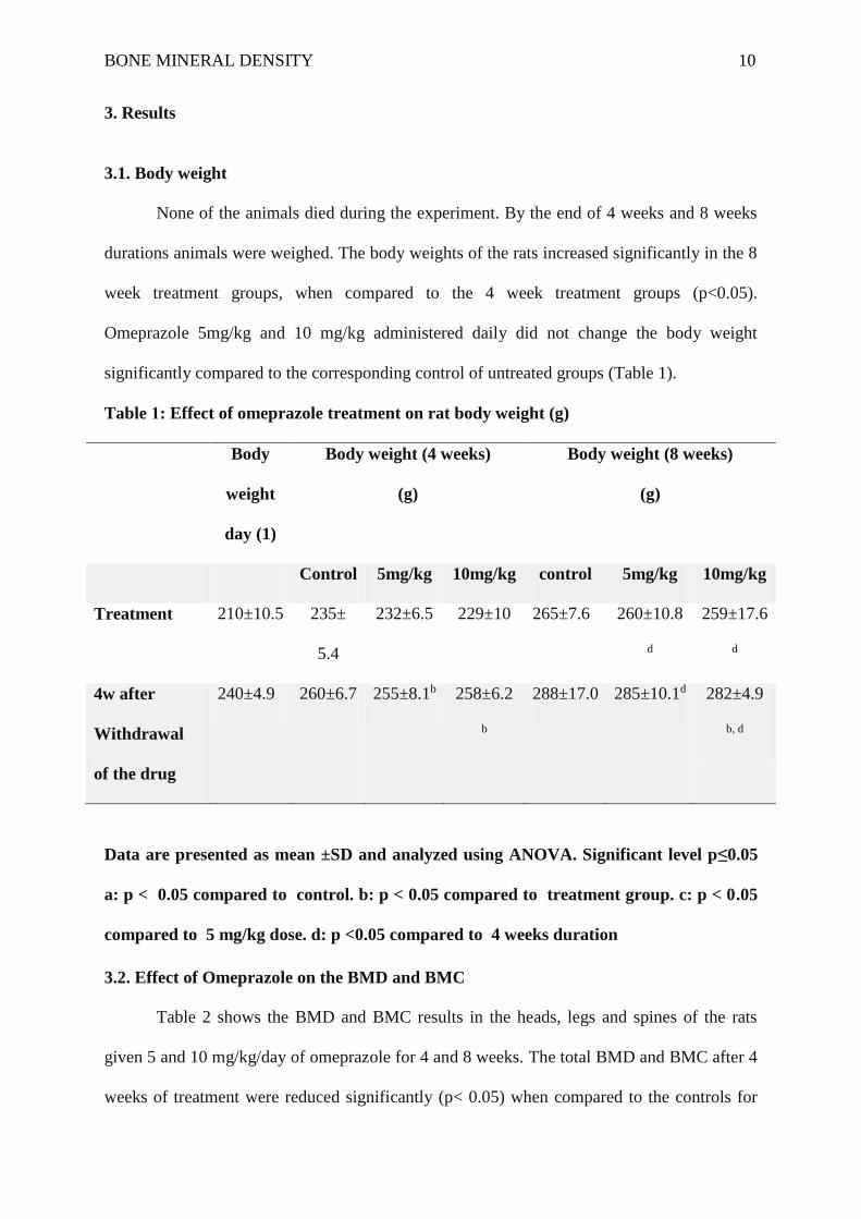
3. Results
3.1. Body weight
None of the animals died during the experiment. By the end of 4 weeks and 8 weeks
durations animals were weighed. The body weights of the rats increased significantly in the 8
week treatment groups, when compared to the 4 week treatment groups (p<0.05).
Omeprazole 5mg/kg and 10 mg/kg administered daily did not change the body weight
significantly compared to the corresponding control of untreated groups (Table 1).
Table 1: Effect of omeprazole treatment on rat body weight (g)
Body
weight
day (1)
Body weight (4 weeks)
(g)
Body weight (8 weeks)
(g)
Control 5mg/kg 10mg/kg control 5mg/kg 10mg/kg
Treatment 210±10.5 235±
5.4
232±6.5 229±10 265±7.6 260±10.8
d
259±17.6
d
4w after
Withdrawal
of the drug
240±4.9 260±6.7 255±8.1b 258±6.2
b
288±17.0 285±10.1d 282±4.9
b, d
Data are presented as mean ±SD and analyzed using ANOVA. Significant level p≤0.05
a: p < 0.05 compared to control. b: p < 0.05 compared to treatment group. c: p < 0.05
compared to 5 mg/kg dose. d: p <0.05 compared to 4 weeks duration
3.2. Effect of Omeprazole on the BMD and BMC
Table 2 shows the BMD and BMC results in the heads, legs and spines of the rats
given 5 and 10 mg/kg/day of omeprazole for 4 and 8 weeks. The total BMD and BMC after 4
weeks of treatment were reduced significantly (p< 0.05) when compared to the controls for

BONE MINERAL DENSITY 11
both doses. After 4 weeks from withdrawal of omeprazole, the BMD and BMC in the 5
mg/kg group showed significant improvement when compared to the end of treatment (p<
0.05), but the 10 mg/kg group showed no significant changes in the BMD or BMC when
compared to the end of treatment values. The coefficient of variation for BMD measurements
was 1.5 percent and 2 percent at femur spine and head, respectively. The total BMD and
BMC results after 8 weeks of treatment were significantly and dose-dependently decreased
when compared to the controls (p< 0.05). Recovery was observed in the BMD and BMC for
both doses when compared to the end of treatment values.
Table 2: Intra and inter-assay coefficients
Bone mineral
density (g/ cm2)
Experimental groups Control group p-value
Spine (L1-L4) 1.1.68±0.15 1.165±0.15 0.96
Femoral neck 1.029±0.17 1.061±0.17 0.49
trochanter 0.870±0.13 0.923±0.13 0.03
Total 1.051±0.15 1.111±0.15 0.04
Intra and inter-assay coefficients of variations of the measurements for BMD & BMC
Table 3: Effect of administration of omeprazole for 4 and 8 weeks on rat BMD & BMC
Control
untreated
5 mg/kg/d 10 mg/kg/d
Control
untreated
5
mg/kg/d
10
mg/kg/d
site BMD (g/cm2) BMC (g)
Head
4w
treatment
0.24± 0.01
0.23 ±
0.01 a
0.22 ± 0.01 a 2.3 ±0.14
2.05
±0.25 a
1.91
±0.17 a
4w after
Withdrawal
0.25±0.01
0.24 ±
0.03 b
0.24± 0.13 b 2.3±0.2
2.23 ±
0.16 b
2.16 ± 0.2
a,b

BONE MINERAL DENSITY 12
8w
Treatment
0.25± 0.03
0.22 ±
0.01a,d
0.20 ± 0.01a,d 2.4 ±0.17
2.04 ±
0.49 a
1.84 ±
0.20 a
4w after
Withdrawal
0.26±0.02
0.24 ±
0.01
0.24±0.01 2.4±0.2
2.14 ±
0.32
2.05
±0.30 a
Femur 4w
Treatment
0.13±0.009 0.13±.0.05 0.12 ± 0.01 1.86±0.11
1.51±
0.23 a
1.50 ±
0.46 a
4w after
Withdrawal
0.14±0.01
0.13
±0.002
0.13 ±0.07 1.9±0.2
1.70
±0.37
1.65±0.20
8w
Treatment
0.14±0.02
0.12 ±
0.01 a
0.12 ± 0.005 a 1.88±0.12
1.50 ±
0.52 a
1.55 ±
0.21 a
4w after
Withdrawal
0.15±0.01
0.13 ±
0.007 b
0.13 ± 0.009 b 2.01±0.1
1.60±
0.33
1.65±
0.41
Spine 4w
Treatment
0.15±0.015
0.13
±0.013 a
0.12±0.007 a, c 1.77±0.34
1.24
±0.13 a
1.31
±0.31 a
4w after
Withdrawal
0.15±0.02
0.14
±0.001 b
0.13±0.008a,b,c 1.80±0.3
1.50
±0.10 a, b
1.31
±0.19 a, c
8w
Treatment
0.16±0.015
0.12± 0.02
a
0.11 ± 0.012 a 1.79±0. 4
1.22 ±
0.28 a
1.20 ±
0.19 a
4w after
Withdrawal
0.16±0.01
0.13 ±
0.15 a
0.12 ± 0.01a,c 1.82±0.1
1.14 ±
0.38 a
1.13+
0.33 a
Total 4w
Treatment
0.15±0.007
0.14 ±0.04
a
0.14 ±0.007 a 9.17±1.16
7.24
±0.66 a
7.53
±1.15 a
4w after
Withdrawal
0.15±0.01
0.15 ±
0.00 b
0.14 ±0.004 a,
c
9.2±2.1
8.02 ±
0.52
8.23
±0.58 a, c
8w 0.16±0.007 0.14 ± 0.13± 0.008 a, 9.19±1.13 7.10 ± 7.07 ±
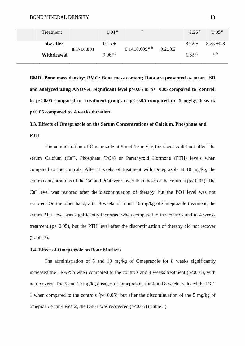
BONE MINERAL DENSITY 13
Treatment 0.01 a c 2.26 a 0.95 a
4w after
Withdrawal
0.17±0.001
0.15 ±
0.06 a,b
0.14±0.009 a, b 9.2±3.2
8.22 ±
1.62a,b
8.25 ±0.3
a, b
BMD: Bone mass density; BMC: Bone mass content; Data are presented as mean ±SD
and analyzed using ANOVA. Significant level p≤0.05 a: p< 0.05 compared to control.
b: p< 0.05 compared to treatment group. c: p< 0.05 compared to 5 mg/kg dose. d:
p<0.05 compared to 4 weeks duration
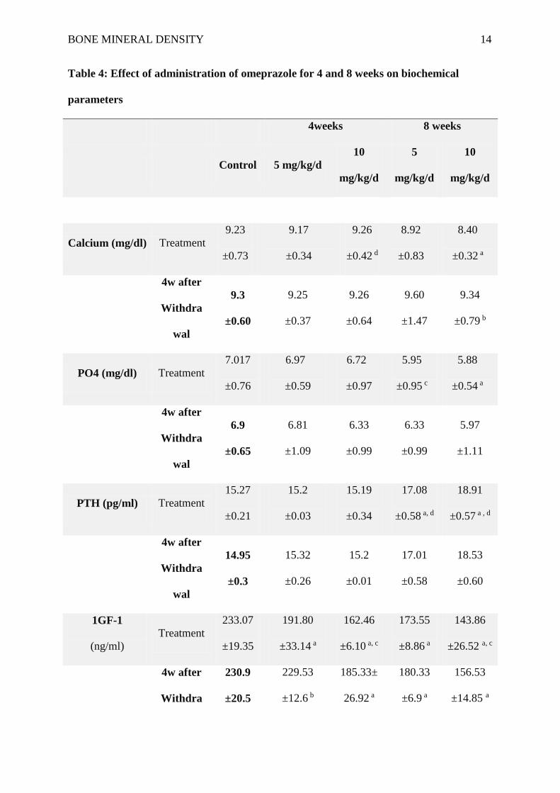
3.3. Effects of Omeprazole on the Serum Concentrations of Calcium, Phosphate and
PTH
The administration of Omeprazole at 5 and 10 mg/kg for 4 weeks did not affect the
serum Calcium (Ca+), Phosphate (PO4) or Parathyroid Hormone (PTH) levels when
compared to the controls. After 8 weeks of treatment with Omeprazole at 10 mg/kg, the
serum concentrations of the Ca+ and PO4 were lower than those of the controls (p< 0.05). The
Ca+ level was restored after the discontinuation of therapy, but the PO4 level was not
restored. On the other hand, after 8 weeks of 5 and 10 mg/kg of Omeprazole treatment, the
serum PTH level was significantly increased when compared to the controls and to 4 weeks
treatment (p< 0.05), but the PTH level after the discontinuation of therapy did not recover
(Table 3).
3.4. Effect of Omeprazole on Bone Markers
The administration of 5 and 10 mg/kg of Omeprazole for 8 weeks significantly
increased the TRAP5b when compared to the controls and 4 weeks treatment (p<0.05), with
no recovery. The 5 and 10 mg/kg dosages of Omeprazole for 4 and 8 weeks reduced the IGF-
1 when compared to the controls (p< 0.05), but after the discontinuation of the 5 mg/kg of
omeprazole for 4 weeks, the IGF-1 was recovered (p<0.05) (Table 3).
BONE MINERAL DENSITY 14
Table 4: Effect of administration of omeprazole for 4 and 8 weeks on biochemical
parameters
4weeks 8 weeks
Control 5 mg/kg/d
10
mg/kg/d
5
mg/kg/d
10
mg/kg/d
Calcium (mg/dl) Treatment
9.23
±0.73
9.17
±0.34
9.26
±0.42 d
8.92
±0.83
8.40
±0.32 a
4w after
Withdra
wal
9.3
±0.60
9.25
±0.37
9.26
±0.64
9.60
±1.47
9.34
±0.79 b
PO4 (mg/dl) Treatment
7.017
±0.76
6.97
±0.59
6.72
±0.97
5.95
±0.95 c
5.88
±0.54 a
4w after
Withdra
wal
6.9
±0.65
6.81
±1.09
6.33
±0.99
6.33
±0.99
5.97
±1.11
PTH (pg/ml) Treatment
15.27
±0.21
15.2
±0.03
15.19
±0.34
17.08
±0.58 a, d
18.91
±0.57 a , d
4w after
Withdra
wal
14.95
±0.3
15.32
±0.26
15.2
±0.01
17.01
±0.58
18.53
±0.60
1GF-1
(ng/ml)
Treatment
233.07
±19.35
191.80
±33.14 a
162.46
±6.10 a, c
173.55
±8.86 a
143.86
±26.52 a, c
4w after
Withdra
230.9
±20.5
229.53
±12.6 b
185.33±
26.92 a
180.33
±6.9 a
156.53
±14.85 a

BONE MINERAL DENSITY 15
wal
TRAP5b
(U/L)
Treatment
0.57±
0.04
0.54
±0.04
0.57±
0. 01
0.61
±0.01 a, d
0.74
±0.20 a, d
4w after
Withdra
wal
0.65±
0.06
0.50
±0.02
0.54
±0.04
0.60
±0.02
0.60
±0.84
PO4: Phosphate; PTH: Parathyroid hormone; 1GF-1: Insulin-like growth factor 1;
TRAP5b: Tartrate resistant acid phosphatase 5b; Data are presented as mean ±SD and
analyzed using ANOVA. Significant level p≤0.05 a: p< 0.05 compared to control. b: p<
0.05 compared to treatment group. c: p< 0.05 compared to 5 mg/kg dose. d: <0.05
compared to 4 weeks duration
2.5. Histopathological results
Examination of bone sections from control groups revealed its usual appearance,
which is formed of outer compact bone (CB) and inner spongy bone (SP). The CB showed
normal sized Haversian canals and regular arrangement of concentric lamellae and interstitial
lamellae. Osteocytes with their darkly stained nuclei inside their lacunae were seen between
different lamellae. Moreover, the covering periosteum showed subperiosteal bone deposition
appearing as a distinct basophilic line. The endosteal surface was lined with osteoblastic cells
and osteoclasts in their Howship’s lacunae. The inner spongy bone consisted of branching
and anastomosing thick bony trabeculae separated by interconnecting spaces containing bone
marrow. These trabeculae consisted of irregular and basophilic bone lamellae and osteocytes
within their lacunae in between the lamellae (Figure 2).
Examination of bone sections from different omeprazole treated groups (5
mg/kg/day/4w group, 10 mg/kg/day/4w group, 5 mg/kg/day/8w group, 10 mg/kg/day/8w

BONE MINERAL DENSITY 16
group), diverse deteriorated histopathological changes were observed according to the dose
and time of omeprazole treatment, which were more apparent in the groups of more dose and
longer time of treatment. In general, thinning of the CB with presence of many cavities,
tunnels and lightly stained areas were seen. The Haversian systems appeared irregular and
shrunken with some Haversian canals were dilated. Many osteocytes appeared darkly stained
and also, some empty lacunae were observed. In several sections, there was hypertrophy of
the periosteum especially in its fibrogenic layer. Moreover, the endosteum had irregular
surface with multiple notches and increase in number of osteoclasts. The trabeculae of the
inner spongy bone appeared thin and discontinuous with loss of their normal architecture and
widening of the interconnecting bone marrow spaces (Figures 3 and 4). Examination of bone
sections from different recovery groups (5 mg/kg/day/4w/Recovery, 10
mg/kg/day/4w/Recovery, 5mg/kg/day/8w/Recovery, 10mg/kg/day/8w/Recovery) presented
varied results where there was nearly complete to partial recovery. Almost complete recovery
took place in 5 mg/kg/day/4w/Recovery and 5mg/kg/day/8w/Recovery groups while the other
two groups showed an incomplete recovery. In general, there was a relative increase in the
thickness of the CB. The outer and inner bone surfaces appeared more or less regular. The
bone matrix presented some deeply stained areas indicating bone repair. The Haversian
systems with regular bone lamellae were seen. The inner spongy restored its normal
appearance which was comparable to that of the control group (Figures 5 & 6).

BONE MINERAL DENSITY 17
Figure 2: A photomicrographs of different bone sections (A, B, C, D) from rats of
control group showing outer compact bone (CB) with normal appearance. The bone is
covered from outside by the periosteum (Pr) and from inside by smooth endosteal
surface (En). Many lacunae containing osteocytes (Oc) were shown in between the bone
lamellae. Notice subperiosteal bone deposition appearing as distinct basophilic line
(arrow). The compact bone tissue is well organised, showing Haversian lamellae (HL)
arranged around the Haversian canals (HC). Also, the inner spongy bone was formed of
branching and anatomizing thick trabeculae (T) with bone marrow spaces (BM) in
between. These trabeculae were lined with cuboidal osteoblasts (Ob). VC: Volkmann’s
canals. (A: X100, B & D: X 200, C: X 400).

BONE MINERAL DENSITY 18
Figure 3: A photomicrographs of different bone sections from omeprazole treated
groups (A: 5 mg/kg/daily/4w group, B: 10 mg/kg/daily/4w group, C: 5 mg/kg/daily/8w
group, D: 10 mg/kg/daily/8w group) showing thinning of the compact bone (CB) with
absence of subperiosteal bone deposition, many cavities (C), tunnel formation (Tu) and
scattered pale areas (*). The periosteum (Pr) displays different degrees of thickness and
the endosteal surface (En) is irregular with many eroded areas. (A & B: X100, C & D: X
200).

BONE MINERAL DENSITY 19
Figure 4: A photomicrographs of different bone sections from omeprazole treated
groups (A & C: 5 mg/kg/daily/8w group, B & D: 10 mg/kg/daily/8w group) showing
Haversian systems with irregular lamellae (HL), dilated Haversian canals (HC) and
little number of osteocytes (Oc) inside empty lacunae in A and B. Also, notice that
number, size, and density of trabeculae (T) decreased with the appearance of widely
separated bony spicules. BM= bone marrow, especially in D. (A & B: X 400, C & D: X
200).

BONE MINERAL DENSITY 20
Figure 5: A photomicrographs of different bone sections from different recovery groups
after omeprazole treatment (A: 5 mg/kg/daily/4w/ recovery group, B: 10
mg/kg/daily/4w/ recovery group, C: 5 mg/kg/daily/8w/recovery group, D: 10
mg/kg/daily/8w/ recover group) showing nearly normal structure with increased
thickness of the outer compact bone (CB) to be comparable with the control and the
appearance of subperiosteal distinct basophilic line and the presence of many basophilic
areas () in A & B. Notice the existence of some cavities and hypertrophied periosteum
in C & D. BM= bone marrow, En = endosteum, HC = Haversian canal, Oc = osteocyte
(A & B: X100, C & D: X 200).

BONE MINERAL DENSITY 21
Figure 5: A photomicrographs of different bone sections from different recovery groups
after omeprazole treatment (A: 5 mg/kg/daily/4w/ recovery group, B& D: 10
mg/kg/daily/8w/ recovery group, C: 5 mg/kg/daily/8w/ recovery group) showing some
irregular bone lamellae around apparently normal Haversian canals (HC). Notice the
presence of distinct deep basophilic areas in the core of the thickened trabeculae (T) of
spongy bones with less apparent widening in the interconnecting bone marrow spaces
(BM). Oc = osteocytes (A & B: X400, C & D: X 200).
4. Discussion
According to the results, the acid-suppressive drugs are widely used for the treatment
of stomach acid-related disorders (Ali, Roberts & Tierney, 2009), but despite the outstanding
efficacy and negligible short-term adverse effects, increased concerns have been raised with

BONE MINERAL DENSITY 22
regard to the side effects of chronic PPI use (Thomson et al., 2010; Vestergaard, 2012; and
Reimer, 2013). In the present study, the BMD and BMC were decreased in a dose and time-
dependent manner, with recovery after stopping the use of Omeprazole. The Ca+ and PO4
were decreased with higher dose and longer duration, with recovery of the Ca+ levels, but not
the phosphate levels, after stopping Omeprazole. However, the PTH is increased when
compared to the controls, with no recovery. The long-term effects of intake of Omeprazole
(30mg/kg × 8weeks) are observed on bone turnover in a recent study by Al Subaie et al.,
(2016) in an experimental rat model with analysis of the signalling pathway involved in
osteoclast differentiation, bone resorption, and alteration of trabecular bone microstructure.
In our study, the mean PTH levels were reported as 15.27 for controls in relevance to
15.2±0.03 & 15.19±0.34 and 17.08±0.58 & 18.91±0.57 at 4 and 8 weeks, respectively.
According to the findings, PTH levels were reported to be higher in 8 weeks group compared
to 4 weeks group, with comparable low Ca+ levels (8.92±0.83 & 8.40±0.32 for 5 & 10
mg/Kg/day PPIs dose). Coinciding with these results, Assiri, Borham, Awad, Al-Maraghy,
(2003); Yang, (2008); Yu et al., (2008) found that the use of PPIs, especially at high dosages
and prolong time, was related to an increased risk of fracture owing to lower Ca+ and elevated
PTH levels.
In addition, a case-controlled study performed by Chiu, Huang, Chang, Yang, (2010)
and the results of Cai, Feng, and Jiang, (2015) showed that PPIs were linked to an increased
fracture risk, especially hip fractures, in a dose-dependent way. Similar to our findings
Krause et al., (2015) instigated, that the use of standard doses of PPIs for 3 months is
associated with significant changes in serum markers of Ca+ homeostasis or bone turnover.
Similarly, our results suggested that PO4 levels were reported at 6.97±0.59 & 6.72±0.97 at 4
weeks and 5.95±0.95 & 5.88±0.54 at 8 weeks interval signifying that the levels were
decreased with the increasing dose and time of use of Omeprazole; however, the levels were

BONE MINERAL DENSITY 23
not retrieved on withdrawal of the drug (6.81±1.09 & 6.33±0.99 at 4 weeks; and 6.33±0.99 &
5.97±1.11 at 8 weeks). Similar results were obtained in a study by Malluche, Davenport,
Cantor and Monier-Faugere, (2014), the lower intestinal phosphate absorption were observed
in patients with higher TRAP5b level and it has been previously shown that intestinal calcium
absorption was significantly decreased in patients treated with PPIs when compared with
healthy controls. It is imperative that hypergastrinemia has been shown to have a stimulatory
effect on the parathyroid glands. For example, experimental hypergastrinemia produced by
antral exclusion in rats led to hyperparathyroidism and an increase in volume and weight of
the parathyroid gland, due to parenchymal cells hyperplasia, resulting in increased bone
resorption ( Grimelius, Johansson, Lundqvist, Olazabal, Polak, Pearse, 1977). The
mechanism that links acid-suppressive medication with the risk of fractures remains largely
unknown (Cea-Soriano, Johansson, Garcia & Rodriguez 2013), although the effects of PPIs
on calcium absorption and metabolism have recently taken much concentration.
The intake of omeprazole has been reported to inhibit the absorption of calcium and
reduce the BMD in rat models (Chonan, Takahashi, Yasui, Watanuki, 1998). In addition
(Corley, Kubo, Zhao, & Quesenberry, 2010) demonstrated that the use of PPIs has been
correlated with calcium malabsorption and the loss of BMD, resulting in an increased risk of
fractures. Hypochlorhydria or achlorhydria in humans caused by some conditions, including
pernicious anaemia, gastrectomy and atrophic gastritis, are combined with an increased
incidence of osteoporosis and bone fractures; it is supposed that this is secondary to the
effects of the low gastric acid levels on calcium absorption (Yu et al., 2008; Lodato;
Sipponen and Harkonen, 2010). Some preclinical and clinical studies have shown that gastric
acid secretion can enhance the absorption of calcium and that the acidic medium in the
stomach promote the production of ionised calcium from insoluble calcium salts. Calcium
solubilisation is suggested to be crucial for calcium absorption. At the same time, gastric acid

BONE MINERAL DENSITY 24
suppression by a PPI can indirectly cause hypergastrinemia by suppressing the release of
somatostatin, and may cause the malabsorption of calcium. Both hypergastrinemia and
calcium malabsorption can negatively influence bone and mineral metabolism, at least in
part, through the induction of hyperparathyroidism, thus leading to a reduction in bone
mineral density (Insogna, 2009; Yang 2008 & 2012).
In the present study, the TRAP5b was increased in 0.54 ±0.04 & 0.57±0.01 after 4
weeks and 0.61±0.01 & 0.74±0.20 at 8 weeks, respectively. It is also observed that at 8 weeks
significant increase was reported both at 5 and 10 mg/kg/day with no recovery after 4 weeks
of withdrawal. From our findings, TRAP 5b showed a significant negative correlation with
BMD and could be useful as a marker in the serum for bone resorptive activity in
pathological conditions like osteoporosis (Halleen, Tiitinen, Ylipahkala, Fagerlund,
Vaananen, 2006; Oddie et al., 2000). The results predicted that when Omeprazole was used
for 8 weeks, the TRAP5b showed highest increment with no significant recovery after
withdrawal, hence, this suggests that TRAP5b is time dependent and chronic use of
Omeprazole may lead to significant rise in TRAP5b. It is interesting to note that TRAP5b is
significantly correlated with markers of resorption as well as with PTH and serve as a marker
of bone resorption in the assessment cases of osteodystrophy. With regard to the IGF-1, it
was decreased in this study in a dose and time dependent manner, with recovery only for the
low dose and short duration (4 weeks: 229.53±12.6 at 5 mg/kg/day & 185.33±26.9 at 10
mg/kg/day compared to 8 weeks: 180.33±6.9 at 5 mg/kg/day & 156.53±14.85 at 10
mg/kg/day).
The findings suggest the level of IGF-1 decreased when the dose of Omeprazole
increased from 5 mg/kg/day to 8 mg/kg/day; and on withdrawal levels recovered in only 4
weeks group. This is an indication that IGF-1 is dependent on the chronic use of Omeprazole
with depletion in concentration on long-term PPIs use. In a study by Courtland et al., (2013),

BONE MINERAL DENSITY 25
it is indicated that osteoclasts may react to the secreted IGF-1 from osteoblasts, which
indicates that this molecule acts also as a coupling factor for the remodelling unit. In addition,
IGF-1 is essential for normal osteoclast differentiation (Guntur and Rosen, 2013). Our results
were supported by Maggio et al., (2014) that inferred that the use of PPIs shows an
independent and negative association with IGF-1 levels. Moreover, acid-suppressive
medications have been reported to affect bone remodelling, and account for the increased risk
of fractures (Mizunashi, Furukawa, Katano, & Abe, 1993), while one multicentre cohort
study with 9,423 participants suggested a modest risk of fracture in PPI users (Fraser, Leslie,
Targownik, Papaioannou, Adachi, 2013). However, the studies assessing the association
between acid inhibition and bone mineral density have reported different conclusions. For
example, some studies found that a mildly reduced bone density was related to PPI use, but
there was no significant change in the levels of bone mineral density between the PPI users
and the controls in other studies (Yu et al., 2008). The authors further signified that there is a
higher incidence of hip fractures in patients treated with high doses of PPIs, and the risk
shows a progressive increase with the duration of the PPI treatment (Yang et al., 2006).
Experimentally, evidence has pointed out that PPIs may inhibit osteoclastic proton transport
system and affect bone resorption, which may alleviate the negative effect of the PPIs on
osteoporosis by decreasing calcium absorption (Yang et al., 2006; Wright, Proctor, Insogna,
Kerstetter, 2008; and Sheraly, Lickorish, Sarraf, & Davies, 2009).
The mechanism of this action focuses on bone turnover cells especially osteoclasts, as
there is a difference between proton pumps in gastric parietal cells (H+/K+ ATPase) and
osteoclasts (vacuolar H+ ATPase) Jefferies, Cipriano, Forgac, 2008). However, omeprazole
has been shown to block both H+/K+ ATPase and vacuolar H+ ATPase (Xu et al., 2007). It is
imperative to note that acid secretion through the H+ ATPase in osteoclasts is important for
bone resorption. Further, the hydrogen ions secreted cause decalcification of bone, and
BONE MINERAL DENSITY 26
activation of the proteolytic enzymes which cause bone matrix degradation. By blocking an
important step in bone resorption, there should be an increase in the bone mineral density,
leading to prevention or reduction of osteoporosis, and hence a decreased risk of bone
fractures. However, PPIs block the vacuolar H+ ATPase and inhibit the activity of osteoclasts
(Yu et al., 2008). This effect, together with their effect on calcium homeostasis, mentioned
previously, supports the assumption that PPIs can result in a condition resembling
osteopetrorickets, as reported by Schinke et al., (2009). Diverse deteriorated
histopathological changes were observed according to dose and time of omeprazole
treatment, which were more apparent in the groups of increased dose and longer duration of
treatment. There was nearly complete recovery in the low dose groups for both durations,
while the high dose groups showed an incomplete recovery.
Our data suggest that high-dose of Omeprazole appears to primarily affect
biomechanical strength parameters in metabolically active trabecular bone, owing to thinning
of CB with absence of subperiosteal bone deposition and cavity formation in 10
mg/kg/daily/8w group. Further, this is likely to be due to degenerated bone quality, according
to our histological data that indicate an increased content of Haversian systems with irregular
and dilated Haversian canals (HC) in Omeprazole-treated animals versus controls. It is
imperative to note that presence of cavities and hypertrophied periosteum denotes trabeculae
containing relatively more cartilage, and consequently less mineralised bone and lose in bone
strength. The microscopic examination of sections belonging to 10 mg/kg/daily/4w/ recovery
group compared with sections from the control group show the presence of obvious changes
and sometimes common ones but of different degrees, depending on the dose of drug
administered and time duration. The changes occurred at both the epithelial level (increased
thickness, different thicknesses, and appearance of subperiosteal distinct basophilic line) as
well as decreased density of trabeculae with the appearance of widely separated bony
BONE MINERAL DENSITY 27
spicules. In our study, the histological findings reveal that demineralisation of bone occurs
gradually; depending on the time elapsed since the start of Omeprazole administration. These
subsequent changes appeared in 5 mg/kg/daily group and 10 mg/kg/daily group after about 4
weeks of treatment and continued until the 8th week. In the control group, there was no
significant increase in tissue.
Additionally, results showed that BMC values 9.17±1.16 for control group; 7.24
±0.66 for 4 weeks group and 7.53 ±1.15 for 8 weeks group has been reported in this study.
These findings signified that BMC of 8.02 ± 0.52 & 8.23 ±0.58 for 4 weeks and 8 weeks,
respectively, is due to higher serum PTH levels and poses an important threat to bone mass of
the femur spine. In a study by Rhee et al., (2011), the excess PTH is reported to have anabolic
effects on trabecular bone. Moreover, our study suggested that studies have demonstrated an
increase in BMD in the femur head was reported as 0.15 ± 0.00 & 0.14 ±0.004 indicating that
BMD loss in the femur in chronic use of PPIs has higher PTH levels. These findings were
supported by studies by Sikjaer, Rejnmark, Rolighed, Heickendorff & Mosekilde; Siilin et
al.; and Pallan & Khan, (2011) by depicting that hyperparathyroidism affects both cortical
and trabecular bone leading to sclerotic thickening with increased PTH and simultaneously
stimulating resorption in cortical bone with reductions in BMD.
Some limitations were found in the present study, where the experiment was done on
adult male rats only. So, there were no data to point out if these results were related to
gender. In addition, mechanical properties, bone strength, bone macrometric parameters
(bone length, bone diameter of femur and tibia) were not measured. Further, duration of PPI
therapy was a major limitation of our study, with PPI use for a shorter period of time (8
weeks). Significant duration of time is a need to observe delayed recovery effects of
Omeprazole withdrawal in experimental samples.
BONE MINERAL DENSITY 28
5. Conclusion
The results of this study have demonstrated that the daily administration of
Omeprazole in rats was associated with decreased in the BMD and BMC in a dose and time
dependent manner, with recovery after stopping the use of the drug. These results were
accompanied with decrease in the serum Ca+ and PO4 concentrations, and an increase in the
PTH level. In addition, the TRAP5b was increased, with a decrease in the IGF-1 peptide.
Deteriorated histopathological changes were observed according to the dose and time of
omeprazole treatment, which were more apparent in the groups of increased dose and longer
time of treatment with recovery in low dose groups and with shorter time. These results
support the studies confirming that that chronic treatment of PPIs decreases the BMD and
could increase the risk of hip, spinal and radial weakness and fractures in femur. Clinicians
should keep in mind the risk of fracture when balancing the safety and efficacy of these
medications.
Conflict of Interest
Layla Borham, Magda Hagras, Altaf Abdulkhaliq, Mohammed Badawood and Gamal Abd El
Aziz declare that they have no conflict of interest.
Acknowledgments
We appreciate the efforts of Professor Soad Shaker Ali from the Anatomy Department in the
Faculty of Medicine at King Abdulaziz University for her help in the DXA analysis of the
rats.
Funding
This article was funded by the Medicine and Medical Sciences Research Centre at
Umm al-Qura University in Saudi Arabia (project # 43309029).

BONE MINERAL DENSITY 29
Statement of Animal Rights
The procedures involving the animals and their care were conducted in accordance
with the ethically approved Umm al-Qura University Committee on the Ethics of Animal
Experiments guidelines (HAPO-02-K-012-2015-05-117), which comply with the national
and international laws and policies. Every effort was made to minimise the number of
animals that were used, and their suffering.

BONE MINERAL DENSITY 30
References
Al Subaie, A., Emami, E., Tamimi, I., Laurenti, M., Eimar, H., Abdallah, M. N., & Tamimi,
F. (2016). Systemic administration of omeprazole interferes with bone healing and
implant osseointegration: an in vivo study on rat tibiae. Journal of clinical
periodontology, 43(2), 193-203.
Ali, T., Roberts, D.N., Tierney, W.M., 2009. Long-term safety concerns with proton pump
inhibitors. The American journal of medicine 122, 896-903.
An YH, M.K., 2003. Handbook of histology methods for bone and cartilage. . New Jersey,
USA: Humana Press Inc.
Assiri, A.M., Borham, L.E., Awad, H.A., Al-Maraghy, M.N., 2003. The Possible Protective
Effects of Fluoxetine and Paroxetine Against Experimental Myocardial Infraction.
Saudi Pharmaceutical Journal 11, 148-158.
Cai, D., Feng, W., Jiang, Q., 2015. Acid-suppressive medications and risk of fracture: an
updated meta-analysis. International journal of clinical and experimental medicine 8,
8893-8904.
Cea-Soriano, L., Johansson, S., Garcia Rodriguez, L.A., 2013. Risk factors for falls with use
of acid-suppressive drugs. Epidemiology (Cambridge, Mass.) 24, 600-607.
Chiu, H.F., Huang, Y.W., Chang, C.C., Yang, C.Y., 2010. Use of proton pump inhibitors
increased the risk of hip fracture: a population-based case-control study.
Pharmacoepidemiology and drug safety 19, 1131-1136.
Chonan, O., Takahashi, R., Yasui, H., Watanuki, M., 1998. Effect of L-lactic acid on calcium
absorption in rats fed omeprazole. Journal of nutritional science and vitaminology 44,
473-481.
Clarke, B., 2008. Normal bone anatomy and physiology. Clinical journal of the American
Society of Nephrology 3, S131-S139.
Corley, D.A., Kubo, A., Zhao, W., Quesenberry, C., 2010. Proton pump inhibitors and
histamine-2 receptor antagonists are associated with hip fractures among at-risk
patients. Gastroenterology 139, 93-101.
Courtland, H. W., Kennedy, O. D., Wu, Y., Gao, Y., Sun, H., Schaffler, M. B., & Yakar, S.
(2013). Low levels of plasma IGF-1 inhibit intracortical bone remodeling during
aging. Age, 35(5), 1691-1703.
Cui, G.L., Syversen, U., Zhao, C.M., Chen, D., Waldum, H.L., 2001. Long-term omeprazole
treatment suppresses body weight gain and bone mineralization in young male rats.
Scandinavian journal of gastroenterology 36, 1011-1015.
Cummings, S.R., Melton, L.J., 2002. Epidemiology and outcomes of osteoporotic fractures.
Lancet (London, England) 359, 1761-1767.
Drake, M.T., Clarke, B.L., Lewiecki, E.M., 2015. The Pathophysiology and Treatment of
Osteoporosis. Clinical therapeutics 37, 1837-1850.
Fraser, L.A., Leslie, W.D., Targownik, L.E., Papaioannou, A., Adachi, J.D., 2013. The effect
of proton pump inhibitors on fracture risk: report from the Canadian Multicenter
Osteoporosis Study. Osteoporosis international : a journal established as result of

BONE MINERAL DENSITY 31
cooperation between the European Foundation for Osteoporosis and the National
Osteoporosis Foundation of the USA 24, 1161-1168.
Goldschlager, T., Abdelkader, A., Kerr, J., Boundy, I., Jenkin, G., 2010. Undecalcified bone
preparation for histology, histomorphometry and fluorochrome analysis. Journal of
visualized experiments : JoVE.
Griffin, M.G., Kimble, R., Hopfer, W., Pacifici, R., 1993. Dual-energy x-ray absorptiometry
of the rat: accuracy, precision, and measurement of bone loss. Journal of bone and
mineral research : the official journal of the American Society for Bone and Mineral
Research 8, 795-800.
Grimelius, L., Johansson, H., Lundqvist, G., Olazabal, A., Polak, J.H., Pearse, G.E., 1977.
The parathyroid glands in experimentally induced hypergastrinemia in the rat.
Scandinavian journal of gastroenterology 12, 739-744.
Guntur, A.R., Rosen, C.J., 2013. IGF-1 regulation of key signaling pathways in bone.
BoneKEy Rep 2.
Halleen, J.M., Tiitinen, S.L., Ylipahkala, H., Fagerlund, K.M., Vaananen, H.K., 2006.
Tartrate-resistant acid phosphatase 5b (TRACP 5b) as a marker of bone resorption.
Clinical laboratory 52, 499-509.
Insogna, K.L., 2009. The effect of proton pump-inhibiting drugs on mineral metabolism. The
American journal of gastroenterology 104 Suppl 2, S2-4.
Ito, T., Jensen, R.T., 2010. Association of Long-term Proton Pump Inhibitor Therapy with
Bone Fractures and effects on Absorption of Calcium, Vitamin B12, Iron, and
Magnesium. Current gastroenterology reports 12, 448-457.
Jefferies, K.C., Cipriano, D.J., Forgac, M., 2008. Function, structure and regulation of the
vacuolar (H+)-ATPases. Archives of biochemistry and biophysics 476, 33-42.
Johnell, O., Kanis, J.A., 2006. An estimate of the worldwide prevalence and disability
associated with osteoporotic fractures. Osteoporosis international : a journal
established as result of cooperation between the European Foundation for
Osteoporosis and the National Osteoporosis Foundation of the USA 17, 1726-1733.
Katikaneni, R., Ponnapakkam, A., Miller, E., Ponnapakkam, T., Gensure, R.C., 2009. A new
technique for precisely and accurately measuring lumbar spine bone mineral density
in mice using clinical dual energy X-ray absorptiometry (DXA). Toxicology
Mechanisms and Methods 19, 225-231.
Krause, M., Keller, J., Beil, B., van Driel, I., Zustin, J., Barvencik, F., ... & Amling, M.
(2015). Calcium gluconate supplementation is effective to balance calcium
homeostasis in patients with gastrectomy. Osteoporosis International, 26(3), 987-995.
Kumar, R., Ramaswamy, R., Nath Mallick, B., 2013. Local Properties of Vigilance States:
EMD Analysis of EEG Signals during Sleep-Waking States of Freely Moving Rats.
PLoS ONE 8, e78174.
Lodato, F., Azzaroli, F., Turco, L., Mazzella, N., Buonfiglioli, F., Zoli, M., Mazzella, G.,
2010. Adverse effects of proton pump inhibitors. Best practice & research. Clinical
gastroenterology 24, 193-201.
Maggio, M., Lauretani, F., De Vita, F., Butto, V., Cattabiani, C., Masoni, S., Sutti, E., Bondi,
G., Dall'aglio, E., Bandinelli, S., Corsonello, A., Abbatecola, A.M., Lattanzio, F.,
Ferrucci, L., Ceda, G.P., 2014. Relationship between use of proton pump inhibitors
BONE MINERAL DENSITY 32
and IGF system in older subjects. The journal of nutrition, health & aging 18, 420-
423.
Malluche, H. H., Davenport, D. L., Cantor, T., & Monier-Faugere, M. C. (2014). Bone
mineral density and serum biochemical predictors of bone loss in patients with CKD
on dialysis. Clinical Journal of the American Society of Nephrology, 9(7), 1254-1262.
Milas-Ahic, J., Prus, V., Kardum, Z., Kovacevic, I., 2014. [Pathophysiology of osteoporosis].
Reumatizam 61, 65-69.
Mizunashi, K., Furukawa, Y., Katano, K., Abe, K., 1993. Effect of omeprazole, an inhibitor
of H+,K(+)-ATPase, on bone resorption in humans. Calcif Tissue Int 53, 21-25.
Morrisett, R.A., Jope, R.S., Snead, O.C., 3rd, 1987. Effects of drugs on the initiation and
maintenance of status epilepticus induced by administration of pilocarpine to lithium-
pretreated rats. Experimental neurology 97, 193-200.
Niv, Y. (2011). Gradual cessation of proton pump inhibitor (PPI) treatment may prevent
rebound acid secretion, measured by the alkaline tide method, in dyspepsia and reflux
patients. Medical hypotheses, 77(3), 451-452.
Oddie, G.W., Schenk, G., Angel, N.Z., Walsh, N., Guddat, L.W., de Jersey, J., Cassady, A.I.,
Hamilton, S.E., Hume, D.A., 2000. Structure, function, and regulation of tartrate-
resistant acid phosphatase. Bone 27, 575-584.
Pallan, S., & Khan, A. (2011). Primary hyperparathyroidism Update on presentation,
diagnosis, and management in primary care. Canadian Family Physician, 57(2), 184-
189.
Reimer, C., 2013. Safety of long-term PPI therapy. Best practice & research. Clinical
gastroenterology 27, 443-454.
Rhee, Y., Allen, M. R., Condon, K., Lezcano, V., Ronda, A. C., Galli, C., ... & Plotkin, L. I.
(2011). PTH receptor signaling in osteocytes governs periosteal bone formation and
intracortical remodeling. Journal of Bone and Mineral Research, 26(5), 1035-1046.
Rosen, H.N., Tollin, S., Balena, R., Middlebrooks, V.L., Beamer, W.G., Donohue, L.R.,
Rosen, C., Turner, A., Holick, M., Greenspan, S.L., 1995. Differentiating between
orchiectomized rats and controls using measurements of trabecular bone density: a
comparison among DXA, histomorphometry, and peripheral quantitative
computerized tomography. Calcif Tissue Int 57, 35-39.
Rozenberg, S., Vandromme, J., Neve, J., Aguilera, A., Muregancuro, A., Peretz, A.,
Kinthaert, J., Ham, H., 1995. Precision and accuracy of in vivo bone mineral
measurement in rats using dual-energy X-ray absorptiometry. Osteoporosis
International 5, 47-53.
Schinke, T., Schilling, A.F., Baranowsky, A., Seitz, S., Marshall, R.P., Linn, T., Blaeker, M.,
Huebner, A.K., Schulz, A., Simon, R., Gebauer, M., Priemel, M., Kornak, U.,
Perkovic, S., Barvencik, F., Beil, F.T., Del Fattore, A., Frattini, A., Streichert, T.,
Pueschel, K., Villa, A., Debatin, K.M., Rueger, J.M., Teti, A., Zustin, J., Sauter, G.,
Amling, M., 2009. Impaired gastric acidification negatively affects calcium
homeostasis and bone mass. Nature medicine 15, 674-681.
Scholten, T., 2007. Long-term management of gastroesophageal reflux disease with
pantoprazole. Therapeutics and clinical risk management 3, 231-243.
BONE MINERAL DENSITY 33
Seeman, E., Delmas, P.D., 2006. Bone Quality — The Material and Structural Basis of Bone
Strength and Fragility. New England Journal of Medicine 354, 2250-2261.
Segawa, K., Nakazawa, S., Tsukamoto, Y., Chujoh, C., Yamao, K., Hase, S., 1987. Effect of
omeprazole on gastric acid secretion in rat: evaluation of dose, duration of effect, and
route of administration. Gastroenterologia Japonica 22, 413-418.
Sermet-Gaudelus, I., Bianchi, M. L., Garabédian, M., Aris, R. M., Morton, A., Hardin, D. S.,
... & Wolfe, S. (2011). European cystic fibrosis bone mineralisation guidelines.
Journal of Cystic Fibrosis, 10, S16-S23.
Sheraly, A.R., Lickorish, D., Sarraf, F., Davies, J.E., 2009. Use of gastrointestinal proton
pump inhibitors to regulate osteoclast-mediated resorption of calcium phosphate
cements in vivo. Current drug delivery 6, 192-198.
Shin, J.M., Vagin, O., Munson, K., Kidd, M., Modlin, I.M., Sachs, G., 2008. Molecular
mechanisms in therapy of acid-related diseases. Cellular and molecular life sciences :
CMLS 65, 264-281.
Siilin, H., Lundgren, E., Mallmin, H., Mellström, D., Ohlsson, C., Karlsson, M., ... &
Ljunggren, Ö. (2011). Prevalence of primary hyperparathyroidism and impact on
bone mineral density in elderly men: MrOs Sweden. World journal of surgery, 35(6),
1266-1272.
Sikjaer, T., Rejnmark, L., Rolighed, L., Heickendorff, L., & Mosekilde, L. (2011). The effect
of adding PTH (1–84) to conventional treatment of hypoparathyroidism: a
randomized, placebo‐controlled study. Journal of Bone and Mineral Research,
26(10), 2358-2370.
Sipponen, P., Harkonen, M., 2010. Hypochlorhydric stomach: a risk condition for calcium
malabsorption and osteoporosis? Scandinavian journal of gastroenterology 45, 133-
138.
Smith, M.D., Baldassarri, S., Anez-Bustillos, L., Tseng, A., Entezari, V., Zurakowski, D.,
Snyder, B.D., Nazarian, A., 2012. Assessment of axial bone rigidity in rats with
metabolic diseases using CT-based structural rigidity analysis. Bone & Joint Research
1, 13-19.
Targownik, L.E., Lix, L.M., Leung, S., Leslie, W.D., 2010. Proton-pump inhibitor use is not
associated with osteoporosis or accelerated bone mineral density loss.
Gastroenterology 138, 896-904.
Targownik, L.E., Lix, L.M., Metge, C.J., Prior, H.J., Leung, S., Leslie, W.D., 2008. Use of
proton pump inhibitors and risk of osteoporosis-related fractures. CMAJ : Canadian
Medical Association journal = journal de l'Association medicale canadienne 179,
319-326.
Thomson, A.B., Sauve, M.D., Kassam, N., Kamitakahara, H., 2010. Safety of the long-term
use of proton pump inhibitors. World journal of gastroenterology 16, 2323-2330.
Vestergaard, P., 2012. Systematic review of observational studies finds increased risk of
fracture among older adults taking a proton pump inhibitor. Evidence-based medicine
17, 39-40.
Wright, M.J., Proctor, D.D., Insogna, K.L., Kerstetter, J.E., 2008. Proton pump-inhibiting
drugs, calcium homeostasis, and bone health. Nutrition reviews 66, 103-108.

BONE MINERAL DENSITY 34
Xu, J., Cheng, T., Feng, H.T., Pavlos, N.J., Zheng, M.H., 2007. Structure and function of V-
ATPases in osteoclasts: potential therapeutic targets for the treatment of osteolysis.
Histology and histopathology 22, 443-454.
Yang, Y.X., 2008. Proton pump inhibitor therapy and osteoporosis. Curr Drug Saf 3, 204-
209.
Yang, Y.X., 2012. Chronic proton pump inihibitor therapy and calcium metabolism. Current
gastroenterology reports 14, 473-479.
Yang, Y.X., Lewis, J.D., Epstein, S., Metz, D.C., 2006. Long-term proton pump inhibitor
therapy and risk of hip fracture. JAMA 296, 2947-2953.
Yu, E.W., Blackwell, T., Ensrud, K.E., Hillier, T.A., Lane, N.E., Orwoll, E., Bauer, D.C.,
Sof, Mr, O.S.R.G., 2008. Acid-Suppressive Medications and Risk of Bone Loss and
Fracture in Older Adults Running Title: Acid-Suppressive Drugs: Bone Loss &
Fracture Risk. Calcified tissue international 83, 251-259.
Recommended