THE ASSOCIATION BETWEEN OBESITY AND PERIODONTAL DISEASE
By
Ahmad Alshareef
BS in General Sciences, University of Pittsburgh, 2001
D.M.D, University of Pittsburgh, 2005
Submitted to the Graduate Faculty of the
School of Dental Medicine
in partial fulfillment
of the requirements for the degree of Master of Science in Dental Medicine
University of Pittsburgh
2012
ii
UNIVERSITY OF PITTSBURGH
SCHOOL OF DENTAL MEDICINE
This thesis was presented
by
Ahmad Alshareef D.M.D.
It was defended on
April 11, 2012
and approved by
Edward. P. Heinrichs D.M.D. Assistant Prof., Dept. of Periodontics and Preventive
Dentistry
Ali Seyedain D.M.D., M.D.S. Assistant Professor, Director of Pre-doctoral Periodontics
John M. Close MA, PMSD Assoc. Prof. Dept. of Dental Public Health/Information
Mgmt.
Thesis Director: Pouran Famili DMD, MS, MPH, PhD
Professor, Chair, Director of the Residency Program
Department of Periodontics and Preventive Dentistry

iii
Copyright © by Ahmad Alshareef
2012
iv
The American Journal of Public Health (2004) editorializes obesity as ‘the public
health challenge of our time.’ Obesity creates risk for chronic health problems, is
associated with increased mortality and exists in complexes of multiple, clustered
behavioral risk factors. Fine (2004) identifies obesity among the four most common risk
factors of chronic disease [cigarette smoking, risky drinking of alcohol, physical
inactivity, overweight]. That set of risk factors applied to the 2001 National Health
Interview Survey showed 17 percent (among 29,000) possessed three or more. Obesity
with raised cholesterol and hypertension is the major cause of mortality and disease in
Europe (James 2004) and is an issue for developing countries as well (Caballero 2001).
Adipocytes in the adipose (fatty) tissues of obese people produce quantities of
active molecules like leptin, important in regulating energy expenditure (thus body
weight). Adipocyte-derived active molecules (adipocytokines) are candidates for the
close association between obesity and multiple risk factor syndromes.
Similarly periodontal disease is one of the world’s most common chronic
diseases [possibly 35 percent of U.S. adults ages 30-40]. Increasing evidence
establishes periodontal disease as a significant risk factor in the etiology of diseases
with inflammatory components. [Severe periodontal disease is the well-established sixth
complication of diabetes (Lőe 1993) and the relationship may be two-way (Grossi
THE ASSOCIATION BETWEEN OBESITY AND PERIODONTAL DISEASE
Ahmad Alshareef D.M.D.
University of Pittsburgh, 2012
v
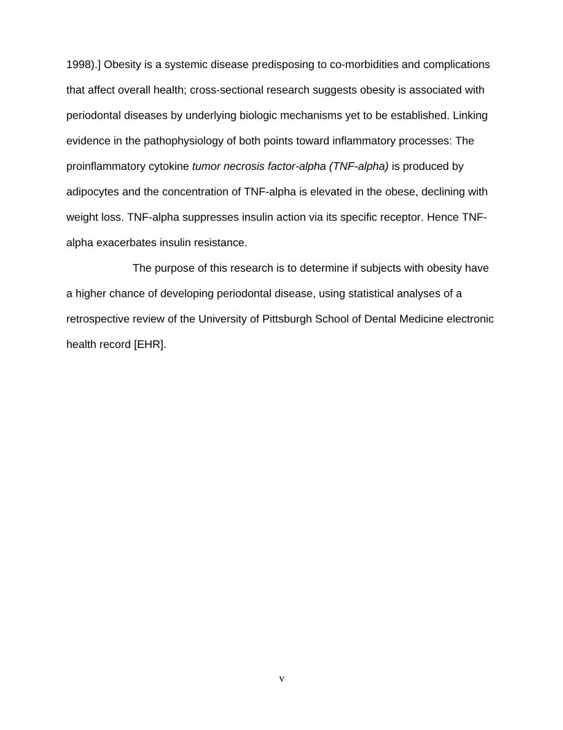
1998).] Obesity is a systemic disease predisposing to co-morbidities and complications
that affect overall health; cross-sectional research suggests obesity is associated with
periodontal diseases by underlying biologic mechanisms yet to be established. Linking
evidence in the pathophysiology of both points toward inflammatory processes: The
proinflammatory cytokine tumor necrosis factor-alpha (TNF-alpha) is produced by
adipocytes and the concentration of TNF-alpha is elevated in the obese, declining with
weight loss. TNF-alpha suppresses insulin action via its specific receptor. Hence TNF-
alpha exacerbates insulin resistance.
The purpose of this research is to determine if subjects with obesity have
a higher chance of developing periodontal disease, using statistical analyses of a
retrospective review of the University of Pittsburgh School of Dental Medicine electronic
health record [EHR].
vi
TABLE OF CONTENTS
PREFACE .................................................................................................................................... IX 1.0 INTRODUCTION..............................................................................................................1 1.1 REVIEW OF THE LITERATURE: PERIODONTAL DISEASE ...................2 1.1.1 The epidemiology of periodontal disease ....................................................2 1.1.2 Clinical assessment of periodontal disease .................................................3 1.1.3 The etiology of periodontal disease .............................................................4 1.1.4 Risk factors for periodontal disease ............................................................6 1.2 REVIEW OF THE LITERATURE: DIABETES MELLITUS .........................7 1.2.1 Types of diabetes mellitus ............................................................................8
1.2.1.1 Prediabetes......................................................................................8 1.2.1.2 Type I diabetes mellitus .................................................................8 1.2.1.3 Type II diabetes mellitus ...............................................................9
1.2.2 Symptoms of diabetes mellitus .....................................................................9 1.2.3 Diagnosis of diabetes mellitus ....................................................................10
1.2.4 Epidemiology and prevelance of diabetes .................................................13 1.3 REVIEW OF THE LITERATURE: OBESITY ...............................................15
1.3.1 Definition and classifications of obesity ....................................................16 1.3.2 Prevalence and trends in obesity ...............................................................18 1.3.3 Factors associated with BMI and obesity .................................................18 1.3.4 Inflammation and obesity...........................................................................20 1.3.5 Treatment of obesity ...................................................................................20 1.4 REVIEW OF THE LITERATURE: ESTABLISHING THE LINKING
RELATIONSHIP BETWEEN PERIODONTAL DISEASE AND OBESITY ..............................................................................................................21
2.0 MATERIALS AND METHODS ....................................................................................28
2.1 STUDY POPULATION ......................................................................................28 2.2 CRITERIA FOR INCLUSION ..........................................................................28 2.3 CRITERIA FOR EXCLUSION .........................................................................28 2.4 PERIODONTAL ASSESSMENT ......................................................................29 2.5 SPECIFIC DATA ELEMENTS EXTRACTED FROM THE ELECTRONIC
HEALTH RECORD ............................................................................................29 2.6 STUDY DESIGN ..................................................................................................29 2.7 STATISTICAL ANALYSIS ...............................................................................30
3.0 DISCUSSION ...................................................................................................................34 4.0 CONCLUSION ................................................................................................................37 BIBLIOGRAPHY ........................................................................................................................38
vii
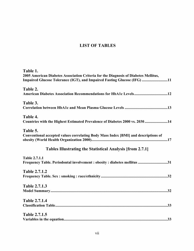
LIST OF TABLES
Table 1. 2005 American Diabetes Association Criteria for the Diagnosis of Diabetes Mellitus, Impaired Glucose Tolerance (IGT), and Impaired Fasting Glucose (IFG) ...........................11 Table 2. American Diabetes Association Recommendations for HbA1c Levels ...................................12 Table 3. Correlation between HbA1c and Mean Plasma Glucose Levels .............................................13 Table 4. Countries with the Highest Estimated Prevalence of Diabetes 2000 vs. 2030 ........................14 Table 5. Conventional accepted values correlating Body Mass Index [BMI] and descriptions of obesity (World Health Organization 2000)................................................................................17
Tables Illustrating the Statistical Analysis [from 2.7.1]
Table 2.7.1.1 Frequency Table. Periodontal involvement : obesity : diabetes mellitus ...............................31 Table 2.7.1.2 Frequency Table. Sex : smoking : race/ethnicity ......................................................................32 Table 2.7.1.3 Model Summary ...........................................................................................................................32 Table 2.7.1.4 Classification Table ......................................................................................................................33 Table 2.7.1.5 Variables in the equation.............................................................................................................33

viii
LIST OF FIGURES
No Figures are represented.

ix
PREFACE
The author extends his gratitude to Dr. Pouran Famili for her supervision and guidance
during the course of this study.
The author also extends his gratitude to Mr. John Close, for his valuable assistance
throughout the course of this study. Sincere appreciation is acknowledged also to Dr.
Edward P. Heinrichs and Dr. Ali Seyedain for their encouragement and assistance
throughout.
Lastly the author dedicates this work to his parents for their encouragement, and to
Dana.
1
1.0 INTRODUCTION
Obesity creates risk for chronic health problems, is associated with increased mortality
and exists in complexes of multiple, clustered behavioral risk factors. Similarly
periodontal disease is one of the world’s most common chronic diseases [possibly 35
percent of U.S. adults ages 30-40]. Increasing evidence establishes periodontal disease
as a significant risk factor in the etiology of diseases with inflammatory components.
Severe periodontal disease is the well-established sixth complication of diabetes (Lőe
1993) and the relationship may be two-way (Grossi 1998). Obesity is a systemic
disease predisposing to co-morbidities and complications that affect overall health;
cross-sectional research suggests obesity is associated with periodontal diseases by
underlying biologic mechanisms yet to be established. Linking evidence in the
pathophysiology of both points toward inflammatory processes: The proinflammatory
cytokine tumor necrosis factor-alpha (TNF-alpha) is produced by adipocytes and the
concentration of TNF-alpha is elevated in the obese, declining with weight loss. TNF-
alpha suppresses insulin action via its specific receptor. Hence TNF-alpha exacerbates
insulin resistance.
This research is a retrospective review of the University of Pittsburgh
School of Dental Medicine electronic health record [EHR].
2
1.1 REVIEW OF THE LITERATURE: PERIODONTAL DISEASE
1.1.1 The Epidemiology of Periodontal Disease
Epidemiology is the study of health and disease in populations and of how these states
are influenced by heredity, biology, physical environment, social environment, and
personal behavior. In the 1940s, early epidemiologic studies of periodontal disease
started with indices of gingivitis. By the 1980s the research focused more on the extent
and severity of the disease (Reddy and Jeffcoat 1993). Thirty-six percent (36%) of the
population in the United States aged 19 years and older was found to be affected with
peridontitis (pockets greater than 4mm) when Brown et al. in 1989 reported on a
national survey done by the Triangle Institute in 1981. Findings also included that 8% of
the population was affected with advanced periodontitis (pockets greater than 6mm),
and that 4% of the population required extraction of teeth due to end stage periodontal
destruction. Between 1985-1986, the NIDCR conducted a national survey of employed
adults. Of adults over 35 years of age, 18% showed attachment loss of greater than 4
mm (Brown et al. 1990). That same national survey also showed that 47% of males
and 39% of females exhibited at least one site that bled on probing. Findings from the
National Center for Health Statistics surveys in the 1960s, 1970s, and the 1985 and
1986 NIDR adult survey were compared by Capilouto and Douglass (1988). About 20%
of adults in the United States showed a level of periodontal disease severity that
required professional treatment. Multiple investigators recently have documented that
5% to 15% of any population exhibits severe generalized periodontitis (NIDR 1987,
3
CDC 1997, Oliver et al. 1998). Similar results have been shown when international
epidemiologic studies were conducted on the prevalence of periodontal disease.
1.1.2. Clinical Assessment of Periodontal Disease
Apart from gingival bleeding and radiographic assessment of bone loss, clinical
attachment loss (CAL) and probing depth (PD) have historically been the basic clinical
measures for periodontitis utilizing the manual probe, a protocol that was described
more than 45 years ago (Ramfjord 1959).
However, in an effort to quantify etiologic agents, gingival alterations or
alterations in the attachment apparatus, many periodontal indices have been used and
described. Such indices have been used to measure symptoms of disease, the etiologic
factors involved, to measure the disease, and also in the treatment of diseases.
According to Barnes et al. (1986), indices may be used in epidemiologic surveys,
clinical trials, or to evaluate the progress of an individual patient and patient motivation.
The PMA Index published by Schour and Massler (1947) was the first system to
classify gingivitis. The PMA Index determined the number of (1) inflamed papillae (P),
(2) gingival margins (M), and (3) areas of attached gingiva (A) at the labial surfaces of
anterior teeth. The PMA Index helped to examine and compare prevalence and severity
scores from different populations, considering its limitations (Lindhe 2008).
The Periodontal Index (PI) developed by Russel in 1956 examined both gingivitis
and periodontitis. The Periodontal Index was deemed useful for epidemiologic surveys
but not clinical trials due to its lack of sensitivity to small differences (Russel 1956).
4
Qualitative changes in the gingival soft tissue were measured by the gingival
index developed by Löe and Silness in 1963. Scores from 0-3 on the four smooth
surfaces of each tooth were used, with (0) being normal, and (3) describing severe
inflammation (Löe 1967).
More indices were developed in an effort to determine treatment needs. Some of
these indices include the Periodontal Treatment Need System (PTNS) developed by
Johansen et al. (1973). This index provided a quick measurement of treatment needs
without attention to details (Lindhe 2008). Another index used in epidemiologic studies
was the Community Periodontal Index of Treatment Needs (CPITN). It was developed
by Ainamo in 1982, and used a special World Health Organization (WHO) periodontal
probe to assess treatment needs (Ainamo et al. 1982).
Lastly is an index developed based on the CPITN, known as the Periodontal
Screening and Recording (PSR). Similar to the CPITN, the PSR index divides the
dentition into six sextants, and uses a color coded probe. The PSR examines all the
teeth but only records on a scale from 0-4 the worst score, and based on the severity of
the score (4 being most severe) the periodontal management and treatment are
determined (Lindhe 2008).
1.1.3. The Etiology of Periodontal Disease
The presence of bacterial plaque is required for the development of gingivitis (Löe et al.
1965, Theilade et al. 1966). It is thought that bacterial plaque will induce pathological
changes in the tissue directly and indirectly (Page 1986). The mass of that bacterial
plaque rather than specific periodontal pathogens causing periodontitis explains the
5
nonspecific plaque theory. More recently, research has shown that specific bacteria are
responsible for periodontitis, and that describes the specific plaque theory used today
(Jeffcoat 1993). Smalley in 1994 further explains the previous (nonspecific plaque)
theory suggesting that organisms display virulence factors giving them the ability to
mediate tissue destruction directly or indirectly.
In the periodontal pocket, more than 300 species of microorganisms have been
isolated, with only a small percentage being etiologic agents (Genco et al. 1990, Moore
et al. 1994). Bacterial constituents or metabolites capable of either causing disruption
of homeostatic or protective host mechanisms or causing the progression or initiation of
a disease are termed bacterial virulence factors. Such factors are one of the
characteristics to imply or deem an organism as etiologic (The American Academy of
Periodontology 1999). For periodontal microorganisms to act as pathogens, the
organism must exhibit 1) the capacity to colonize; 2) the ability to evade antibacterial
host defense mechanisms; and 3) the ability to produce substances that can directly
initiate tissue destruction (The American Academy of Periodontology 1999).
In 1992, Socransky and Haffajee concluded that for disease to result from a
pathogen, the potentially-pathogenic organism 1) must be a virulent clonal type; 2) must
possess the chromosomal and extra-chromosomal genetic factors to initiate disease; 3)
must discover a host susceptible to this particular pathogen; 4) must be in numbers
sufficient to exceed the threshold for that host; 5) must be located at the right place; 6)
must be fostered by other bacterial species, or at the very least not inhibited by other
bacteria, during the development of the pathologic process; and 7) must establish itself
in a local environment conducive to the expression the species’ virulence properties.
6
A dynamic equilibrium exists between periodontal disease and health, and the
equilibrium and its disruption may be influenced by many factors including local and
systemic (Smalley 1994).
1.1.4. Risk Factors for Periodontal Disease
A risk factor is an environmental exposure, aspect of behavior, or an inherent
characteristic associated with a disease (Last 2001).
Risk factors for periodontal disease include oral hygiene, education, systemic
factors, socioeconomic status, and age (Reddy and Jeffcoat 1993).
Systemic factors and modulation of host response will be discussed in detail in
later sections. An example of such systemic modulation of host response would be how
diabetes mellitus alters the host defense mechanisms and decreases the ability of the
host-body to fight infection (Reddy and Jeffcoat 1993). Further evidence for the
systemic modulation of host response in the case of risk factors for periodontal disease
is the conclusion by Grossi et al. (1994) that diabetic subjects experience twice more
odds for more attachment loss than non-diabetics.
An association between cigarette smoking and periodontitis has been shown in
both cross-sectional and longitudinal studies (Ismail and Lewis 1993). A history of
smoking is highly correlated with severity of attachment loss (Gross et al 1994) when a
cross sectional study of risk indicators for attachment loss was conducted.
Multiple investigators have shown that age was associated with severity of
attachment loss (Hugoson et al. 1992, Grossi et al. 1994). However other studies have
concluded that it is uncommon for elderly people with reasonably intact dentition to
7
exhibit sudden bursts of periodontitis (Page 1984). And when exhibited by little gingival
inflammation, and few deep pockets, tooth retention, good oral hygiene, and periodontal
health are closely associated, regardless of age (Burt et al. 1985, Abdellatif et al. 1987).
Lower socioeconomic status (SES) has been associated with periodontal disease
as a risk factor (National Center for Health Statistics 1965, 1979). Generally people
who have a higher SES tend to practice better oral hygiene, have more positive
attitudes toward oral hygiene, and visit the dentist more frequently.
1.2 REVIEW OF THE LITERATURE: DIABETES MELLITUS
Diabetes mellitus is a chronic metabolic disorder characterized by the inability to
maintain blood glucose levels within normal physiological limits. Diabetes mellitus
results from the inadequate production of insulin or insulin resistance, both creating a
physiologic condition in which the body cannot respond to the insulin formed within the
system leading to what is called hyperglycemia, the name of the condition deriving from
the state of accumulation of glucose in the blood. Insulin is a hormone produced by the
Islets of Langerhans in the pancreas, and is responsible for the absorption of glucose
into the cells for their energy needs, and into the liver and fat cells for storage.
Diabetes mellitus has been recognized as a serious public health problem
resulting in significant morbidity and mortality due to specific diabetes-related
microvascular complications, increased risk of macrovascular complications (ischemic
heart disease, stroke and peripheral vascular disease), and increased inflammatory
mediator response (American Diabetes Association 2005).
8
1.2.1 Types of Diabetes Mellitus
1.2.1.1 Prediabetes Prediabetes describes a condition in which blood glucose
levels are higher than normal but not high enough to be labeled diabetes. Prediabetes
occurs when the fasting blood sugar is between 101mg/dL and 126mg/dL or if the blood
sugar level two hours after glucose tolerance test is between 140mg/dL and 200mg/dL
(American Diabetes Association 2005). It has been shown that people with prediabetes
have an increased risk for future Type II diabetes as well as heart disease (American
Diabetes Association 2005). Borderline diabetes, Impaired Glucose Tolerance (IGT),
Impaired Fasting Glucose (IFG), Impaired Glucose Regulation (IGR), and Non-Diabetic
Hyperglycemia (NDH) are also medical terms that used to describe a prediabetes
condition (American Diabetes Association, 2005).
1.2.1.2 Type I Diabetes Mellitus Type I diabetes mellitus was formerly called
insulin-dependent diabetes or juvenile-onset diabetes. In this type, more than 90
percent of the pancreatic Beta cells are permanently destroyed due to cellular mediated
auto-immune destruction, leading to insulin deficiency (Mealey and Oates 2006). The
use of exogenous insulin is a must to compensate the lack of insulin production and to
sustain life, as those patients may develop diabetic ketoacidosis, a life-threatening
condition, hence the former name “insulin-dependent diabetes” (Mealey and Oates
2006). Although some studies showed 15-30 percent of all patients being diagnosed
9
after the age of 30, Type I diabetes usually occurs before that age, in children and
adolescents (Mealey and Oates 2006).
1.2.1.3 Type II Diabetes Mellitus Type II diabetes mellitus is also called non-
insulin-dependent diabetes or adult-onset diabetes. It is the most common type of
diabetes, which has a gradual onset and is often without symptoms; usually found in
people over 30, Type II diabetes mellitus becomes more common with the aging
process. However, in the presence of escalating numbers of overweight young people
and growing rates of obesity status, Type II diabetes mellitus increasingly occurs at
younger ages (American Diabetes Association 2005). Type II diabetes develops when
the body does not produce enough insulin. Type II diabetes also occurs in situations
where sometimes insulin is produced even at higher than normal levels but does not
work properly or as well as it should, so that there is not enough insulin to meet the
body needs. This condition is insulin resistance. Type II diabetes can be controlled by
lifestyle changes, careful dietary management, regular physical activity, weight
reduction, and oral anti-diabetic medications, but in some instances, insulin is required
(www.diabetes.org.uk/guide-to-diabetes).
1.2.2 Symptoms of Diabetes Mellitus
Both types of diabetes have very similar symptoms related to the direct effect of high
blood glucose levels. The body tries to flush the excess glucose in its system out in the
10
urine, resulting in these classic symptoms: polyuria (passing urine frequently),
polydipsia (increased thirst), polyphagia (increased appetite), blurred vision, extreme
fatigue, delayed wound healing, genital itching, weight loss, and regular episodes of
thrush (www.diabetes.org.uk/guide-to-diabetes).
In Type I diabetes, signs and symptoms are usually obvious and develop rapidly (over
few weeks). Once the condition is treated and under control, these symptoms are
quickly relieved. On the other hand, symptoms in Type II diabetes are not as obvious,
as the blood glucose level may rise so slowly that patients may not have symptoms for
years before they are diagnosed. Often Type II diabetes is diagnosed during an
appointment for another health problem or during a routine medical checkup
(diabetes.org.uk/guide-to-diabetes).
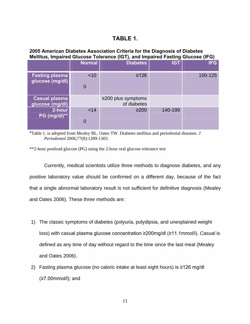
1.2.3. Diagnosis of Diabetes Mellitus
In 1965, the World Health Organization (WHO) published diagnostic criteria for diabetes
mellitus. The last review of these criteria (1998) was published as the guidelines for the
Definition, Diagnosis and Classification of Diabetes Mellitus and its Complications
(American Diabetes Association 2005). Since then, more information regarding the
diagnosis has become available. In 2003, the American Diabetes Association (ADA)
reviewed its diagnostic criteria, and the last review was in 2005. The following Table
shows the current diagnostic parameters for diabetes mellitus.
11
TABLE 1.
2005 American Diabetes Association Criteria for the Diagnosis of Diabetes Mellitus, Impaired Glucose Tolerance (IGT), and Impaired Fasting Glucose (IFG)
Normal Diabetes IGT IFG
Fasting plasma glucose (mg/dl)
<10
0
≥126 100-125
Casual plasma glucose (mg/dl)
≥200 plus symptoms of diabetes
2-hour PG (mg/dl)**
<14
0
≥200 140-199
*Table 1. is adopted from Mealey BL. Oates TW. Diabetes mellitus and periodontal diseases. J Periodontol 2006;77(8):1289-1303.
**2-hour postload glucose (PG) using the 2-hour oral glucose tolerance test
Currently, medical scientists utilize three methods to diagnose diabetes, and any
positive laboratory value should be confirmed on a different day, because of the fact
that a single abnormal laboratory result is not sufficient for definitive diagnosis (Mealey
and Oates 2006). These three methods are:
1) The classic symptoms of diabetes (polyuria, polydipsia, and unexplained weight
loss) with casual plasma glucose concentration ≥200mg/dl (≥11.1mmol/l). Casual is
defined as any time of day without regard to the time since the last meal (Mealey
and Oates 2006).
2) Fasting plasma glucose (no caloric intake at least eight hours) is ≥126 mg/dl
(≥7.00mmol/l); and
12
3) Two-hour post-load glucose ≥200mg/dl (≥11.1mmol/l) during the oral glucose
tolerance test (Mealey and Oates 2006).
In order to monitor the patient’s overall glycemic control (after being diagnosed
with diabetes), the hemoglobin A1c (HbA1c) test is used. Research has concluded that
to date no gold standard assay exists for the HbA1c and many countries do not have
ready access to the test. For those reasons, the HbA1c test is not recommended for
diagnosis of diabetes mellitus (Mealey and Oates 2006).
However the HbA1c test can be used to determine glycohemoglobin levels and
gives an estimate of the average blood glucose level over 30-90 days preceding the test
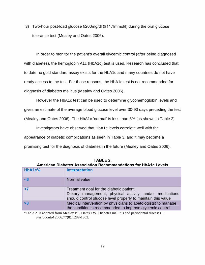
(Mealey and Oates 2006). The HbA1c ‘normal’ is less than 6% [as shown in Table 2].
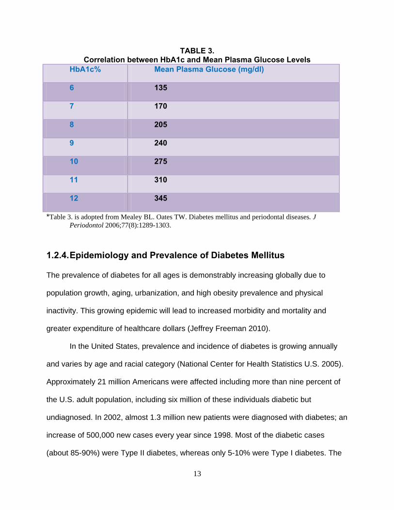
Investigators have observed that HbA1c levels correlate well with the
appearance of diabetic complications as seen in Table 3, and it may become a
promising test for the diagnosis of diabetes in the future (Mealey and Oates 2006).
TABLE 2.
American Diabetes Association Recommendations for HbA1c Levels HbA1c% Interpretation
<6 Normal value
<7 Treatment goal for the diabetic patient Dietary management, physical activity, and/or medications should control glucose level properly to maintain this value
>8 Medical intervention by physicians (diabetologists) to manage the condition is recommended to improve glycemic control
*Table 2. is adopted from Mealey BL. Oates TW. Diabetes mellitus and periodontal diseases. J Periodontol 2006;77(8):1289-1303.
13
TABLE 3. Correlation between HbA1c and Mean Plasma Glucose Levels
HbA1c% Mean Plasma Glucose (mg/dl)
6 135
7 170
8 205
9 240
10 275
11 310
12 345
*Table 3. is adopted from Mealey BL. Oates TW. Diabetes mellitus and periodontal diseases. J Periodontol 2006;77(8):1289-1303.
1.2.4. Epidemiology and Prevalence of Diabetes Mellitus
The prevalence of diabetes for all ages is demonstrably increasing globally due to
population growth, aging, urbanization, and high obesity prevalence and physical
inactivity. This growing epidemic will lead to increased morbidity and mortality and
greater expenditure of healthcare dollars (Jeffrey Freeman 2010).
In the United States, prevalence and incidence of diabetes is growing annually
and varies by age and racial category (National Center for Health Statistics U.S. 2005).
Approximately 21 million Americans were affected including more than nine percent of
the U.S. adult population, including six million of these individuals diabetic but
undiagnosed. In 2002, almost 1.3 million new patients were diagnosed with diabetes; an
increase of 500,000 new cases every year since 1998. Most of the diabetic cases
(about 85-90%) were Type II diabetes, whereas only 5-10% were Type I diabetes. The
14
increase in prevalence and incidence of both types of diabetes was mainly because of
dramatic increases in the rate of obesity among Americans (National Center for Health
Statistics U.S. 2005).
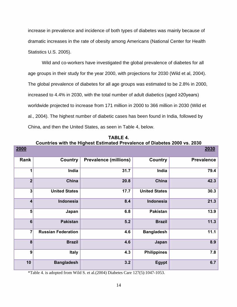
Wild and co-workers have investigated the global prevalence of diabetes for all
age groups in their study for the year 2000, with projections for 2030 (Wild et al, 2004).
The global prevalence of diabetes for all age groups was estimated to be 2.8% in 2000,
increased to 4.4% in 2030, with the total number of adult diabetics (aged ≥20years)
worldwide projected to increase from 171 million in 2000 to 366 million in 2030 (Wild et
al., 2004). The highest number of diabetic cases has been found in India, followed by
China, and then the United States, as seen in Table 4, below.
TABLE 4. Countries with the Highest Estimated Prevalence of Diabetes 2000 vs. 2030
2000 2030
Rank Country Prevalence (millions) Country Prevalence
1 India 31.7 India 79.4
2 China 20.8 China 42.3
3 United States 17.7 United States 30.3
4 Indonesia 8.4 Indonesia 21.3
5 Japan 6.8 Pakistan 13.9
6 Pakistan 5.2 Brazil 11.3
7 Russian Federation 4.6 Bangladesh 11.1
8 Brazil 4.6 Japan 8.9
9 Italy 4.3 Philippines 7.8
10 Bangladesh 3.2 Egypt 6.7
*Table 4. is adopted from Wild S. et al.(2004) Diabetes Care 127(5):1047-1053.
15
It has been observed (Wild 2004) that prevalence of diabetes is similar in men
and women worldwide, but there were prevalence differences by age in developing
countries. The prevalence of diabetic men younger than 60 years and women at older
ages was slightly higher (Wild et al. 2004). Moreover, the majority of diabetic people in
developing countries worldwide were aged between 45 and 64 years, whereas the
majority in developed countries was above the age of 64 years (Wild 2004).
Based on demographic changes, researchers have concluded that the “diabetes
epidemics” will continue even if the obesity prevalence will remain stable until 2030, a
scenario which seems unlikely. Wild estimates that the number of diabetic patients will
be more than double as a consequence of worldwide population aging and urbanization.
Prospective population studies have investigated relationships between plasma
glucose levels and the risk of (overall) mortality, cancer, inflammation, cardiovascular
disease, and of developing diabetes (Sorkin 2005). For instance, researchers in the
Baltimore Longitudinal Study found a statistically significant increase in all-cause
mortality in the presence of fasting plasma glucose test results at levels >6.1 mmol/l but
not lower levels. For the two-hour plasma glucose test, the risk was significantly
increased >7.8 mmol/l (Sorkin 2005).
1.3 REVIEW OF THE LITERATURE: OBESITY
One cannot dispute the negative effects of obesity. Obesity has been associated as a
strong risk factor for Type II diabetes, hypertension, osteoarthritis and cardiovascular
16
diseases (World Health Organization 2000). In addition, poor perceived health and
disability have been associated with obesity (Doll. 2000, Ford 2001).
1.3.1. Definitions and Classification of Obesity
Excess adipose tissue characterizes obesity. Multiple laboratory methods have been
used to quantify adipose tissue mass, including dual energy X-ray absorptiometer
(DEXA), magnetic resonance imaging (MRI), and computed tomography (CT) (Lusaki
1987, Seidell et al. 1987, Gray et al. 1991, Sobol et al. 1991). The previous methods
require costly equipment and have been found difficult to use in epidemiologic studies.
Body weight adjusted for stature (Body Mass Index) has been commonly used in
large-scale population surveys as a surrogate for body fat content (Revicki and Israel
1986, Gray and Fujioka 1991). Body mass index (BMI), also known as Quetelet’s
Index, is the most commonly used tool, the ratio defined as body weight (kg) divided by
height squared (m2). BMI has been shown to have strong correlation with body fatness,
and weak correlation with height (Keys et al. 1971, Revicki and Israel 1986).
However BMI has its limitations, as it fails to distinguish between lean body mass
and fat which will make the relationship between BMI and body fatness vary according
to body composition (Garn et al. 1986). Also height change related to age has an
influence on BMI and since the age of onset of puberty varies, estimating BMI in
children and adolescents is even more difficult to determine (World Health Organization
2000). BMI provides a simple measure of obesity in adults despite these limitations.
17
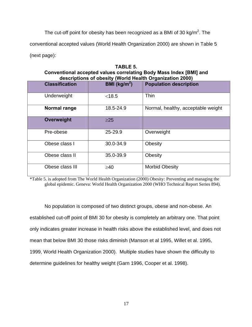
The cut-off point for obesity has been recognized as a BMI of 30 kg/m2. The
conventional accepted values (World Health Organization 2000) are shown in Table 5
(next page):
TABLE 5. Conventional accepted values correlating Body Mass Index [BMI] and
descriptions of obesity (World Health Organization 2000) Classification BMI (kg/m2) Population description
Underweight <18.5 Thin
Normal range 18.5-24.9 Normal, healthy, acceptable weight
Overweight ≥25
Pre-obese 25-29.9 Overweight
Obese class I 30.0-34.9 Obesity
Obese class II 35.0-39.9 Obesity
Obese class III ≥40 Morbid Obesity
*Table 5. is adopted from The World Health Organization (2000) Obesity: Preventing and managing the global epidemic. Geneva: World Health Organization 2000 (WHO Technical Report Series 894).
No population is composed of two distinct groups, obese and non-obese. An
established cut-off point of BMI 30 for obesity is completely an arbitrary one. That point
only indicates greater increase in health risks above the established level, and does not
mean that below BMI 30 those risks diminish (Manson et al 1995, Willet et al. 1995,
1999, World Health Organization 2000). Multiple studies have shown the difficulty to
determine guidelines for healthy weight (Garn 1996, Cooper et al. 1998).
18
1.3.2. Prevalence and Trends in Obesity
Various investigations have shown increases in prevalence of obesity in many countries
including the United States (Flegal 1998), Canada (Macdonald 1997) and Australia
(Eckersley 2001). Flegal in 1998 showed that in the United States, obesity prevalence,
more specifically in subjects with a BMI ≥ 35 kg/m2, remains higher than in other
countries (5 percent for white men, 10 percent for white women). Random-digit
telephone surveying of 195,005 US adults aged 18 years or older (Mokdad 2001)
concluded that the prevalence increase in obesity continues in both sexes, all ages, all
races, all educational levels, and all smoking levels. Mokdad also concluded that
obesity is strongly associated with several major health risk factors. It is worth
mentioning that, in that research, BMI was calculated based on self-reported weight and
height.
It has been suggested that obesity is a major avoidable contributor to the costs of
illness in the U.S. (Colditz 1992). Despite those facts, the prevalence of obesity
continues to grow, not in the United States alone but worldwide (Popkin Doak 1998).
1.3.3. Factors Associated with BMI and Obesity
Overconsumption of food and lack of physical activity have been shown to have a
causal relationship with obesity, but are not the only reasons for the obese condition.
Human physiological regulatory mechanisms can be overwhelmed by many societal
and environmental factors, as will be discussed.
Gender is one factor that has been associated with obesity. Multiple studies
provide evidence that women have a higher prevalence of obesity, and men have a

19
higher prevalence of overweight, especially after the age of 50 years (Flegal et al. 1998,
Stam-Moraga et al. 1999).
Several cross-sectional studies have documented increase in BMI with age
(Boyle 1994, Flegal 1998). A few longitudinal studies support the previously mentioned
finding, with age 60 years cited (Williamson 1990, Lewis 1997, Guo 1999).
Flegal (1998) showed variations across ethnic groups in the prevalence of
obesity. Genetic predisposition for obesity might be the reason for such variations
especially when individuals are exposed to an affluent lifestyle (World Health
Organization 2000).
Some studies have suggested a relationship between marital status and obesity,
although the relationship is not well established. Higher BMI has been associated with
married subjects than subjects living alone (Kahn 1991, Rosmond 1996). Other studies
suggest that no such link exists (Tavani 1994, Wamala 1997).
Studies on diet and obesity have reported inconsistent results, compromised by
factors such as weaknesses in study design, methodological errors in estimating energy
and nutrient intakes, and confounding factors (Lissner Heitmann 1995, Seidell 1998).
Obesity has been suggested in association with a large number of various dietary
factors but conclusive evidence is still lacking to prove than one diet would promote
obesity more than another.
According to the World Health Organization 2000, what is commonly known as
‘physical activity’ actually has three components: household chores, occupational work
and leisure-time physical pursuit. A large number of cross-sectional studies show an
inverse association between BMI and physical activity (Rosmond 1996, Stam-Moraga
20
1999). Less physical activity has been observed more often in obese subjects than non-
obese (Miller 1990, Cooper 2000). On the other hand, some studies have shown no
association between BMI and physical activity (Seidell 1991, Tremblay et al. 1995).
1.3.4. Inflammation and Obesity
This topic will be discussed further in following sections but it remains important to
introduce at this point the idea of adipose tissue and cells actively monitoring their
environment. Fat cells will respond to neural, paracrine, autocrine and endocrine inputs
such as prostaglandins, cytokines, steroids, cholesterol and fatty acids (Cottam 2004).
Secretion of inflammatory mediators from adipose tissue in response to chronic
inflammation associated with obesity was studied by Cottam et al. in 2004. Such
mediators include transforming growth factor beta (TGFβ), interleukin six (IL-6),
angiotensinogen (AGT), and tumor necrosis factor alpha (TNFα).
1.3.5. Treatment of Obesity
Treatment of obesity and maintaining weight loss are goals that in the literature have
been proven difficult to achieve. Prevention has been shown to possibly be most
effective to overcome or slow this growing epidemic (Bouchard 1996, Gill 1997).
Establishing an effective strategy to prevent obesity is hampered by many challenges,
including limited research and information. As suggested by James (1995), more
knowledge is needed about factors contributing to obesity. Until such knowledge is
obtained, changes in the prevalence of obesity and BMI over time should be observed
to determine more effective strategies in both treatment and prevention.
21
1.4 REVIEW OF THE LITERATURE:
ESTABLISHING THE LINKING RELATIONSHIP BETWEEN
OBESITY, DIABETES AND PERIODONTDAL DISEASE
The relationship between diabetes mellitus and periodontal disease has been
extensively studied by researchers, and there has been considerable interest in drawing
the association between diabetes mellitus and periodontal diseases, with conflicting
conclusions (Denis and Bartold, 2000). Globally both of these diseases have a relatively
high incidence in the general population with a number of common pathogeneses
through which periodontal infections contribute to the diabetes-related inflammatory
state (Malik 2011). Researchers have found a bi-directional relationship between the
two polygenic disorders with some degree of immuno-regulatory dysfunction. This
means that the presence of one condition is indicated as a risk factor for the other, as
well as that the meticulous treatment of either may result in improvement of the other
(Malik 2011).
Chronic periodontal disease is an inflammatory condition characterized by a shift
in the microbial ecology of subgingival plaque biofilms and the progressive host-
mediated destruction of tooth-supporting structures (Chaffee Weston 2010). Collectively
the most common diseases known to mankind, the complex classification of periodontal
diseases takes into account clinical presentation, age at diagnosis, rate of disease
progression, and systemic and local factors, all which may increase risk. Gingivitis
(inflammation is confined to the gingiva, and is reversible with good oral hygiene) and
22
periodontitis (inflammation continues, and results in tissue destruction and alveolar
bone resorption) (Preshaw et al. 2012) are the two current classifications of periodontal
disease.
In periodontal disease, destruction of the connective tissue mostly results from
the interaction of bacteria and their products with mononuclear cells (Denis Bartold
2000). The prevalence and severity of periodontal disease has been demonstrated
higher among individuals with diabetes due to the accumulation of glucose mediated
AGE (Advanced Glycation End-products) that would affect migration and phagocytic
activity of mononuclear and polymorphnuclear phagocytic cells, leading to formation of
more pathogenic sub-gingival flora (Denis and Bartold., 2000).
This inflammatory response will affect glucose-utilizing pathways by activating an
infection-mediated pathway of cytokine regulation, particularly with the secretion of
tumor necrosis factor (TNF-α), interleukin (IL-1β, IL-6), and prostaglandin (PGE2) and a
state of insulin resistance (Preshaw et al., 2012). The deregulated local secretion of
TNF-α and IL-1 also mediates inflammation and the breakdown of connective tissue and
alveolar bone in periodontal disease (Preshaw 2012).
In the case of diabetes mellitus (both Types I and II), the level of systemic
inflammatory markers is noticeably elevated, due to the state of hyperglycemia. The
elevated levels of systemic inflammatory markers contributes to both microvascular and
macrovascular complications, as well as to oxidative stress and apoptosis (Dandona
2004). For instance, Engebretson and co-workers have found that TNF- α serum levels
in people with Type II diabetes were considerably correlated with the severity of
periodontal destruction, plasma endotoxin, and IL-1b in the gingival crevicular fluid
23
(Engebretson 2007). Moreover, in a study of Type I diabetes, those (diabetic patients)
with either gingivitis or periodontitis have higher levels of gingival crevicular fluid (GCF;
fluid exudation from the gingival margins), PGE2, and IL-1β compared to the non-
diabetic group with the same periodontal disease level (Salvi 1997).
Accordingly, it has been indicated that diabetes is a risk factor for gingivitis and
periodontitis (Salvi 2008). Conversely, periodontitis may have a major role in worsening
glycemic control over time among diabetic individuals and may also increase the risk of
diabetic complications, by elevating levels of these cytokines closely linked to insulin
resistance (Salvi 2008).
Although some previous studies have not found a significant direct effect of
diabetes on periodontitis and gingivitis, many other studies have demonstrated that
diabetes is a major risk factor for periodontitis (Preshaw 2012). The first clear evidence
to support this hypothesis came from cross-sectional and longitudinal studies in the
1990s (Nelson) on the North American Pima Indian population (number: 2,273 subjects,
age 15- 54 years). Prevalence of periodontitis among Pima Indians with Type II
diabetes was 60 percent, and 36 percent in the non-diabetic patients. In a subgroup of
701 subjects (aged 15-54 years, follow up period over 2.5 years), the incidence of
periodontitis was increased by approximately three-fold compared with non-diabetic
Pima Indians (Nelson 1990).
Investigators have also concluded that all diabetic patients of both Type I and
Type II, including children and young adults, could be at risk for development of
periodontal diseases (Preshaw 2012). However the vast majority of studies were
focused on the effect of Type II diabetes on periodontal diseases, possibly due to the
24
fact that both diseases have historically tended to appear in patients in their 40s and
50s (Preshaw 2012). One early study reported that the prevalence of attachment loss,
bone loss, and gingivitis in around 10 percent of children (<18years) with Type I
diabetes was greater than the control group, despite comparable plaque scores
(Cianciola 1982). More recently it was found that the proportion of periodontitis in a
group of 350 diabetic children (6-18 years) was greater (>20) than the 350 non-diabetic
controls (8%) (Lalla 2007). In a large epidemiologic study in the United States, the risk
of periodontitis among patients with poorly controlled diabetes increased 2.9-fold
compared to the non-diabetic group. Conversely there was no significant increase in
risk among the well-controlled diabetics (Tsai 2002).
It is likewise well-recognized that the level of glycemic control is of key
importance in determining the development of chronic complications of diabetes, and
may play a role in the gingival response to bacterial plaque in diabetics (Preshaw 2012).
Analysis of data from the US National Health and Nutrition Examination Survey
(NHANES) III identified higher significant prevalence of severe periodontitis in adults
with HbA1c level >9% than those without diabetes (OR 2.90% CI 1.40, 6.03) after
adjusting for the effect of confounding variables such as age, sex, ethnicity, education,
and smoking (Tsai 2002). Although the metabolic control of diabetes may have an
important role in the onset and progression of periodontal diseases, researchers have
concluded that this association is difficult to define as it is similar to the association
between glycemic control and diabetic complications such as retinopathy and
nephropathy (Mealey and Oates 2006). More specifically there is marked heterogeneity
in the diabetic population (Tsai 2002). The body of evidence has reported that some
25
poorly-controlled diabetic patients may have extensive periodontal destruction, while
others may not. On the other hand, many patients with effective glycemic control may
have very good periodontal health, where others may develop periodontitis (Tsai 2002).
Results from a cross-sectional analysis of Type I diabetic individuals for a mean
duration greater than 16 years has demonstrated that poorly-controlled diabetics have a
greater prevalence of inter-proximal attachment loss and bone loss than well-controlled
Type I diabetic subjects (Mealey and Oates 2006). Corroborative longitudinal
epidemiological research among the Arizona Pima Native American community found
that the risk of progressive bone loss was increased 11-fold among subjects with poor
glycemic control of Type II diabetes compared to a non-diabetic control group (Taylor
1998).
Conversely, not only diabetes is a risk factor for periodontitis, but periodontitis
could have a negative effect that is considered a risk factor for worsening glycemic
control (Preshaw 2012).
The first clear evidence supporting this hypothesis was obtained from two-year
longitudinal research conducted in the Gila River Native American community (Taylor
1996). Diabetic patients with severe periodontitis at baseline had a six-fold increased
risk of poor glycemic control (HbA1c > 9.0%) over diabetic patients with no periodontitis
(Taylor 1996). Various investigations have reported that severe periodontitis may also
increase the risk of non- oral diabetes-related complications including retinopathy,
diabetic neuropathy, proteinuria, and cardiovascular complications (Preshaw 2012).
The impact of periodontitis on the development of overt nephropathy, defined as
macroalbuminuria and end stage renal disease (ESRD), in patients with Type II
26
diabetes was investigated by Shultis as well in Gila River Native Americans (2007).
Assessment of periodontal status was performed among 529 Type II diabetic patients
25 years or older, with a glomerular filtration rate of ≥60 ml min-1 1.73m-2 , and without
macroalbuminuria (urinary albumin: creatinine ration ≥600 mg/g) (Shultis et al. 2007).
Investigators found macroalbuminuria developed in 193 patients (median follow-up 9.4
years) and ESRD in 68 patients (median follow-up 14.9 years). The incidence of
macroalbuminuria was 2.0, 2.1, and 2.6 times greater in patients with moderate and
severe periodontitis, or in those who were edentulous, respectively, than in those with
mild or without periodontitis (p<0.05, after adjustment of age, sex, diabetes duration,
BMI, and smoking), whereas the incidence of ESRD was 2.3, 3.5, and 4.9 times greater
in the presence of moderate or severe periodontitis or edentulism respectively (p value
< 0.05). That research concluded that the development of overt nephropathy and ESRD
in a ‘dose–dependent’ manner in Type II diabetes was predicted with moderate and
severe periodontitis and with edentulism (Shultis 2007).
Malik et al (2011) conclude that “periodontitis may initiate or propagate insulin
resistance in a manner similar to that of obesity, by enhancing activation of overall
systemic immune response initiated by cytokines, and given these mechanisms
promoting insulin resistance.” The elevated chronic systemic inflammatory state induced
by periodontal disease in people with Type II diabetes may play a major role in
propagating insulin resistance and worsen glycemic control (Malik 2011). Emerging
evidence indicates that periodontits may contribute to the translocation of gram-negative
oral bacteria species and their products from the periodontal biofilm into the systemic
circulation, and to the translocation of cytokines from the periodontal space into the
27
circulation likewise. This may explain why in some studies glycemic control of diabetes
showed improvement after effective periodontal therapy, which is aimed to decrease
elevated serum inflammation mediators (such as TNF-α and IL-6) (Malik 2011).
Some researchers contend that these insights regarding effects of the treatment
of periodontal infection on glycemic control of diabetes investigates only non- surgical
periodontal treatment and derives from observational and treatment research in some
cases statistically significant and in others not (Kiran 2005). As a treatment footnote, to
date no clear evidence establishes a requirement for antibiotic use with non- surgical
periodontal treatment in order to observe the improvement in glycemic control that can
be associated with periodontal therapy (Kiran 2005; Chaffee and Weston 2010).
28
2.0 MATERIALS AND METHODS
2.1 STUDY POPULATION
Subjects were identified from a retrospective review of the records over a three-year
period (from August 2008 to November 1, 2011) using the electronic health record
(EHR) of the University of Pittsburgh School of Dental Medicine.
2.2 CRITERIA FOR INCLUSION
All subjects are adult (>age 18) patients of record of the University of Pittsburgh School
of Dental Medicine Department of Periodontics and Preventive Dentistry. Data were
collected from the electronic health record (EHR) of the University of Pittsburgh School
of Dental Medicine (axiUm database, Exan Systems, Las Vegas, Nevada) from the time
period from August 2008 to November 2011.
2.3 CRITERIA FOR EXCLUSION
Children are not included, as pediatric manifestations of periodontal disease or diabetes
are not within the scope of this research. Completely edentulous [toothless] individuals
would not be reflected in the electronic health record, as periodontal disease is not
present in conjunction with full edentulism. Subjects physically, mentally, or legally
incapacitated so that informed consent to treat cannot be obtained are not included.
29
2.4 PERIODONTAL ASSESSMENT
Pocket depth upon probing is the measure from the free gingival margin (FGM) to the
base of the sulcus/pocket with the use of a calibrated probe (HuFriedy Michigan 0
probe, diameter of probe is 0.5 mm). Pocket depth is probed on six sites per tooth
(distobuccal, midbuccal, mesiobuccal, distolingual, midlingual, and mesiolingual).
Clinical periodontal attachment level and the loss of periodontal attachment are defined
as the distance in millimeters (mm) from the cementoenamel junction (CEJ) to the base
of the pocket. Probing depth is the distance from the free gingival margin (FGM) to the
base of the sulcus/pocket.
2.5 SPECIFIC DATA ELEMENTS EXTRACTED FROM THE
ELECTRONIC HEALTH RECORD
• Height and weight to compute BMI.
• Patient’s diabetic status, as reported in the EHR.
• Smoking status.
• Race.
2.6 STUDY DESIGN
Subjects used in this study were 7171 dentate patients (n=7171), mean age 54 years.
Patients’ records with documented weight, height, age, sex, Type II diabetes status
(self-reported) and a completed periodontal evaluation were included. Periodontal
disease was classified in the presence of periodontal pocketing of 4mm or greater (any
30
surface or tooth). Body mass index (BMI) was calculated and patients were classified as
obese (BMI>=30) or non-obese (BMI<30).
2.7 STATISTICAL ANALYSIS
In this study, 7,171 records were reviewed, 5751 white or Caucasian to 1420 African-
American ethnicity. Among the total dental treatment records, twice as many, or
specifically 5104, had recorded periodontal disease versus 2067 medical records that
did not record periodontal disease. In the total set of electronic health records reviewed,
5263 did not calculate BMI to the level of obesity, while in 1908 of the EHR, height and
weight calculations fell into the category of calculated obesity.
The EHR population sample was split nearly 50:50 male to female, 3351 males
to 3820 females, the relationship of gender to periodontal disease was statistically
significant, p = 0.038. Similar to the calculated obesity, the total EHR were dominantly
non-smokers (4364) versus 2807 individuals who admitted cigarette smoking or other
tobacco use. The relationship of smoking to periodontal disease was not statistically
significant p = 0.089, and the relationship was not used in the final model.
Step-wise logistic regression analysis was performed, the final model including
the variables BMI, age, obesity, sex, and race. Relationships between periodontal
disease and those variables were significant, BMI p = .000; age p = .000; sex p = .038;
obesity p = .037; and African-American ethnicity p = .012. (Please see the tables for
more detail.)
31
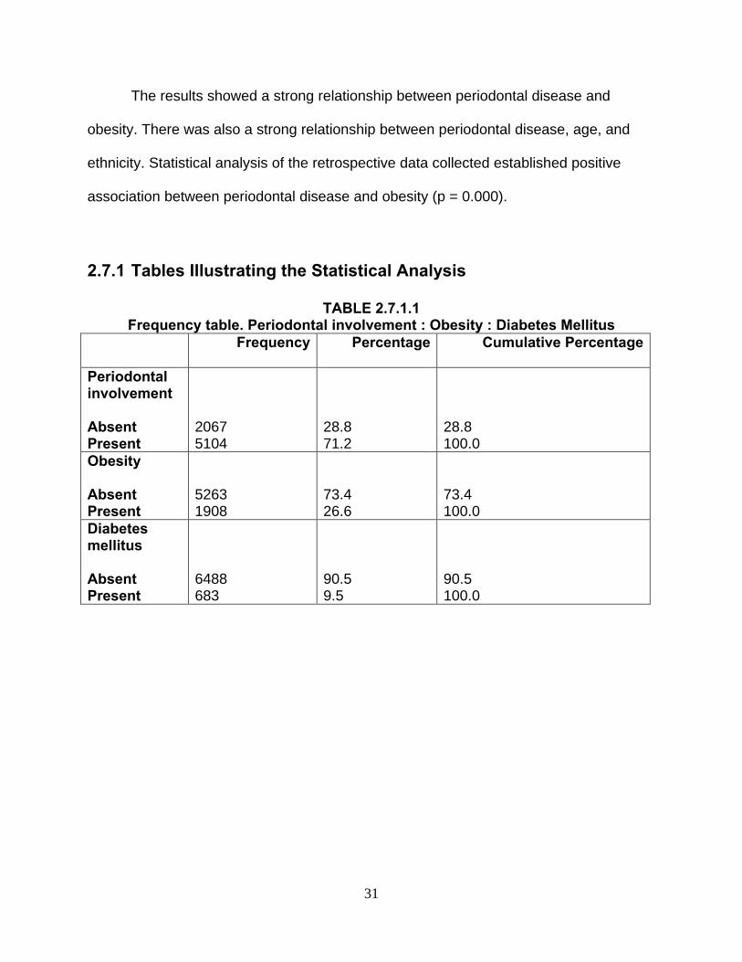
The results showed a strong relationship between periodontal disease and
obesity. There was also a strong relationship between periodontal disease, age, and
ethnicity. Statistical analysis of the retrospective data collected established positive
association between periodontal disease and obesity (p = 0.000).
2.7.1 Tables Illustrating the Statistical Analysis
TABLE 2.7.1.1 Frequency table. Periodontal involvement : Obesity : Diabetes Mellitus Frequency Percentage Cumulative Percentage
Periodontal involvement Absent Present
2067 5104
28.8 71.2
28.8 100.0
Obesity Absent Present
5263 1908
73.4 26.6
73.4 100.0
Diabetes mellitus Absent Present
6488 683
90.5 9.5
90.5 100.0
32
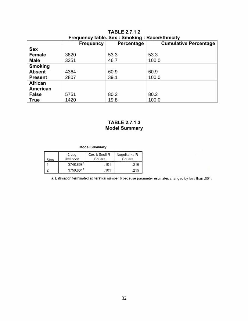
TABLE 2.7.1.2 Frequency table. Sex : Smoking : Race/Ethnicity
Frequency Percentage Cumulative Percentage Sex Female Male
3820 3351
53.3 46.7
53.3 100.0
Smoking Absent Present
4364 2807
60.9 39.1
60.9 100.0
African American False True
5751 1420
80.2 19.8
80.2 100.0
TABLE 2.7.1.3 Model Summary
33
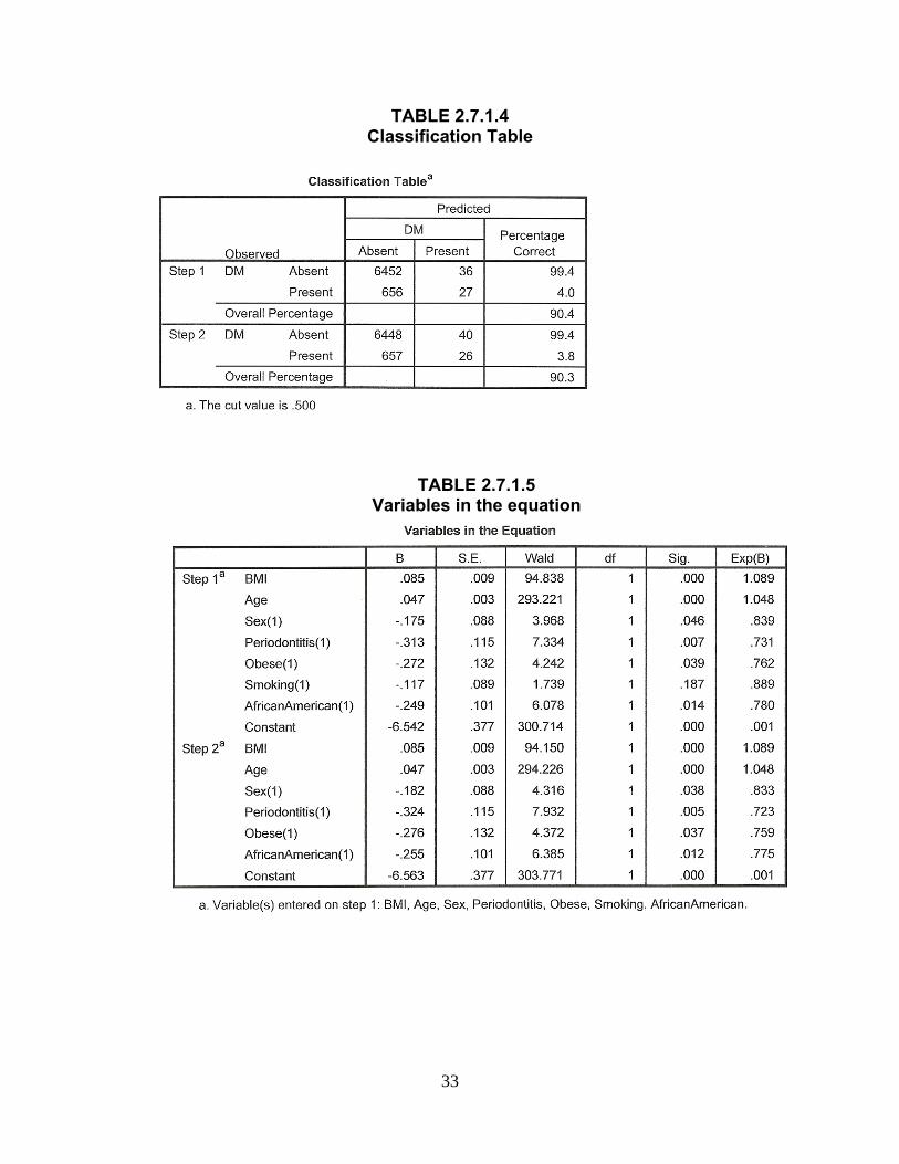
TABLE 2.7.1.4 Classification Table
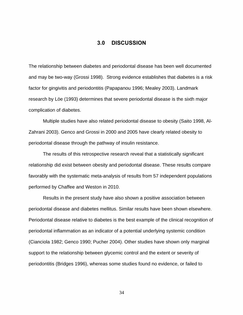
TABLE 2.7.1.5 Variables in the equation
34
3.0 DISCUSSION
The relationship between diabetes and periodontal disease has been well documented
and may be two-way (Grossi 1998). Strong evidence establishes that diabetes is a risk
factor for gingivitis and periodontitis (Papapanou 1996; Mealey 2003). Landmark
research by Löe (1993) determines that severe periodontal disease is the sixth major
complication of diabetes.
Multiple studies have also related periodontal disease to obesity (Saito 1998, Al-
Zahrani 2003). Genco and Grossi in 2000 and 2005 have clearly related obesity to
periodontal disease through the pathway of insulin resistance.
The results of this retrospective research reveal that a statistically significant
relationship did exist between obesity and periodontal disease. These results compare
favorably with the systematic meta-analysis of results from 57 independent populations
performed by Chaffee and Weston in 2010.
Results in the present study have also shown a positive association between
periodontal disease and diabetes mellitus. Similar results have been shown elsewhere.
Periodontal disease relative to diabetes is the best example of the clinical recognition of
periodontal inflammation as an indicator of a potential underlying systemic condition
(Cianciola 1982; Genco 1990; Pucher 2004). Other studies have shown only marginal
support to the relationship between glycemic control and the extent or severity of
periodontitis (Bridges 1996), whereas some studies found no evidence, or failed to
35
reach statistical significance establishing such a relationship, between glycemic control
and periodontal status (Barnett 1984; Sastrowijoto 1989).
The concept that insulin resistance appears to mediate the relationship between
obesity and periodontal disease, put forth by Genco and Grossi 2005, is supported by
the findings. Results show a statistically significant relationship between obese patients
with periodontal disease and a higher risk for diabetes mellitus. In obesity as in
periodontal disease, there is a chronic stimulation and secretion of pro-inflammatory
cytokines which could contribute to insulin resistance (Nishimura et al 2001).
For this study, smoking (pack years), age, sex and race were controlled and the
parameters again analyzed using multiple regression analysis. Results again showed a
statistically significant relationship between the studied parameters. Not only do
confounding variables make analysis difficult to interpret, but also the fact that some
variables were self-reported may introduce an inherent bias in the study.
This study investigated the association between obesity and periodontal disease,
and also the association of periodontal disease with diabetes. Specifically this research
attempted to determine whether obese individuals (assessed by variable BMI) are at
greater risk of developing periodontal disease (assessed by mean clinical attachment
loss), and whether obese individuals with periodontal disease are at greater risk for
diabetes mellitus (measured as poor glucose control). Electronic health records
(n=7171) obtained from the University of Pittsburgh School of Dental Medicine axiUm
database were analyzed. Review of the literature regarding periodontal disease,
obesity, diabetes mellitus and the relationships linking all three conditions together was
made. Due to the variability and complexity of the associated parameters, further

36
investigation is necessary to elicit and clarify whether such association exists beyond
reasonable doubt.
37
4.0 CONCLUSION
The records of 7171 patients aggregated from the electronic health record (EHR) at the
University of Pittsburgh School of Dental Medicine were reviewed and analyzed to
ascertain whether a correlation existed between obesity and periodontal disease. When
BMI values were compared to attachment loss a statistically significant relationship was
found (p=0.000). When the confounding variables of smoking, age, race, sex and
diabetic status were controlled for, a statistically significant relationship remained
between increasing body mass index and attachment loss (p=0.000).
Due to accumulating evidence in research linking periodontal disease to multiple
systemic conditions, the dental health-care provider must be cognizant of the
periodontal status of patients showing indication of such systemic conditions associated
with chronic inflammation (most specifically in this regard, diabetes and metabolic
syndrome). Numerous studies have identified obesity as a major public health concern
(Mokdad 1999) and the global incidence and prevalence of obesity has been on the rise
worldwide.
Further understanding of how obesity is linked to periodontal disease, along with
more well-designed prospective studies, is necessary to clarify how such relationships
between periodontal health and systemic health exist. Prevention is key for both chronic
conditions. Health-care providers should continue to underscore for their patients the
importance of reaching and maintaining a healthy weight, following the dietary and
nutritional recommendations of the U.S. National Institutes of Health, and having regular
oral health examinations and screenings for signs of periodontal disease.
38
BIBLIOGRAPHY
American Diabetes Association. Diagnosis and classification of diabetes mellitus. Position statement. Diabetes Care 2005;29(Suppl. 1):S37-S42.
American Diabetes Association. Standards of medical care in diabetes—2010 [published
correction appears in Diabetes Care 2010;33(3):692]. Diabetes Care 2010;33(Suppl 1):S11-S61.
Bouchard C. Can obesity be prevented? Nutr Rev 1996;54:S125-30. Boyle CA. Dobson AJ. Egger G. Magnus P. Can the increasing weight of Australians be
explained by the decreasing prevalence of cigarette smoking? Int J Obes Relat Metab Disord 1994;18:55-60.
Chaffee BW. Weston SJ (2010) Association between chronic periodontal disease and obesity: a
systematic review and meta-analysis. J Periodontol 81:1708–1724. Cianciola LJ. Park PH. Bruck E. Mosovich L. Genco RJ (1982) Prevalence of periodontal
disease in insulin-dependent mellitus (juvenile diabetes). J Am Dent Assoc 104:653–660. Colditz GA. Economic costs of obesity? Am J Clin Nutr 1992;55:S503-7. Cooper AR. Page A. Fox KR, Misson J. Physical activity patterns in normal, overweight and
obese individuals using minute-by-minute accelerometry. Eur J Clin Nutr 2000;54:887-94.
Cooper R. Durazo-Arvizu R. Luke A. McGee D. A contrary view on the search for weight
guidelines. Nutr Metab Cardiovasc Dis 1998;8:117-20. Dandona P. Aljada A. Bandyopadhyay A (2004) Inflammation: the link between insulin
resistance, obesity and diabetes. Trends Immunol 25:4–7. Denis FK. Bartold PM. Clinical relevance of the host responses of periodontitis. Perio 2000 43:
278-93. Doll HA. Petersen SEK. Stewart-Brown SL. Obesity and physical and emotional well-being:
Associations between body mass index, chronic illness, and the physical and mental components of the SF-36 questionnaire. Obes Res 2000;8:160-70.
39
Eckersley RM. Losing the battle of the bulge: causes and consequences of increasing obesity.
Med J Aust 2001;174:590-2. Engebretson S. Chertog R. Nichols A. Hey-Hadavi J. Celenti R. Grbic J (2007) Plasma levels
TNF-alpha in patients with chronic periodontitis and Type II diabetes. J Clin Perio 34:18-24.
Flegal KM. Carroll MD. Kuczmarski RJ. Johnson CL. Overweight and obesity in the United
States: prevalence and trends, 1960-1994. Int J Obes Relat Metab Disord 1998;22:39-47. Ford ES. Moriarty DG. Zack MM. Mokdad AH. Chapman DP. Self-reported body mass index
and health-related quality of life: Findings from the Behavioral Risk Factor Surveillance System. Obes Res 2001;9:21-31.
Garn SM. Fractionating healthy weight. Am J Clin Nutr 1996;63:S412-4. Gill TP. Key issues in the prevention of obesity. Br Med Bull 1997;53:359-88. Gray DS. Fujioka K. Use of relative weight and body mass index for the determination of
adiposity. J Clin Epidemiol 1991;44:545-50. Gray DS. Fujioka K. Colletti PM. Kim H. Devine W. Cuyegkeng T. Pappas T. Magnetic
resonance imaging used for determining fat distribution in obesity and diabetes. Am J Clin Nutr 1991;54:623-7.
Guo SS. Zeller C. Chumlea WC. Siervogel RM. Aging, body composition, and lifestyle: the Fels
Longitudinal Study. Am J Clin Nutr 1999;70:405-11. James WPT. A public health approach to the problem of obesity. Int J Obes Relat Metab Disord
1995;19:S37-45. Jeffrey S. Freeman DO. The increasing epidemiology of diabetes and review of current treatment
algorithm. [Published correction appears in J Am Osteopath Assoc.2010;110(10):572]. J Am Osteopath Assoc.2010;110(7 Suppl 7):eS2-eS6.
Kahn HS. Williamson DF. Stevens JA. Race and weight change in US women: The roles of
socioeconomic and marital status. Am J Public Health 1991;81:319-23. Keys A. Fidanza F. Karvonen MJ. Kimura N. Taylor HL. Indices of relative weight and obesity.
J Chronic Dis 1971;25:329-43. Kiran M. Arpak N. Unsal E. Erdogan MF (2005) The effect of improved periodontal health on
metabolic control in Type II diabetes mellitus. J Clin Periodontol 32:266–272.
40
Lalla E. Cheng B. Lal S et al (2007) Diabetes mellitus promotes periodontal destruction in children. J Clin Periodontol 34:294–298.
Lewis CE. Smith DE. Wallace DD. Williams OD. Bild DE. Jacobs DR. Seven-year trends in
body weight and associations with lifestyle and behavioral characteristics in black and white young adults: the CARDIA Study. Am J Public Health 1997;87:635-42.
Lissner L. Heitmann BL. Dietary fat and obesity: evidence from epidemiology. Eur J Clin Nutr
1995;49:79-90. Lukaski HC. Methods for the assessment of human body composition: traditional and new. Am J
Clin Nutr 1987;46:537-56. Macdonald SM. Reeder BA. Chen Y. Després J-P. Obesity in Canada: a descriptive analysis.
Can Med Assoc J 1997;157:S3-9. Malik G. Lehl G. Talwar M (2011) Association of periodontitis with diabetes mellitus: A review.
Journal of Medical College Chandigarh 1(1):10-13. Manson JE. Willett WC. Stampfer MJ. Colditz GA. Hunter DJ. Hankinson SE. Hennekens CH.
Speizer FE. Body weight and mortality among women. N Engl J Med 1995;333:677-85. Mealey BL. Oates TW. Diabetes mellitus and periodontal diseases. J Periodontol
2006;77(8):1289-1303. Miller WC. Lindeman AK. Wallace J. Niederpruem M. Diet composition, energy intake, and
exercise in relation to body fat in men and women. Am J Clin Nutr 1990;52:426-30. National Center for Health Statistics. United States 2005. Chartbook on Trends in the Health of
Americans Table 55. Hyattsville MD: National Center for Health Statistics 2005. Nelson RG. Shlossman M. Budding LM et al (1990) Periodontal disease and NIDDM in Pima
Indians. Diabetes Care 13:836–840. Popkin BM. Doak CM. The obesity epidemic is a worldwide phenomenon. Nutr
Rev1998;56:106-14. Preshaw PM. Alba AL. Herrera D. Jepsen S. Konstantinidis A. Makrilakis K. Taylor R. (2012)
Periodontitis and diabetes: a two- way relationship. Diabetologia 55: 21-31. Revicki DA. Israel RG. Relationship between body mass indices and measures of body
adiposity. Am J Public Health 1986;76:992-4. Rosmond R. Lapidus L. Björntorp P. The influence of occupational and social factors on obesity
and body fat distribution in middle-aged men. Int J Obes Relat Metab Disord 1996;20:599-607.
41
Salvi GE. Yalda B. Collins JG et al (1997) Inflammatory mediator response as a potential risk
marker for periodontal diseases in insulin-dependent diabetes mellitus populations. J Perio 68:127–135.
Salvi GE. Carollo-Bittel B. Lang NP (2008) Effects of diabetes mellitus on periodontal and peri-
implant conditions: update on associations and risks. J Clin Periodontol 35:398–409. Seidell JC. Dietary fat and obesity: an epidemiologic perspective. Am J Clin Nutr 1998;67:S546-
50. Seidell JC. Oosterlee A. Thijssen MA. Burema J. Deurenberg P. Hautvast JG. Ruijs JH.
Assessment of intra-abdominal and subcutaneous abdominal fat: Relation between anthropometry and computed tomography. Am J Clin Nutr 1987;45:7-13.
Seidell JC. Cigolini M. Deslypere J-P. Charzewska J. Ellisinger B-M. Cruz A. Body fat
distribution in relation to physical activity and smoking habits in 38-year-old European men. Am J Epidemiol 1991;133:257-65.
Shultis WA. Weil EJ. Looker HC et al (2007) Effect of periodontitis on overt nephropathy and
end-stage renal disease in Type II diabetes. Diabetes Care 30:306–311. Sobol W. Rossner S. Hinson B. Hiltbrandt E. Karstaedt N. Santago P. Wolfman N. Evaluation of
a new magnetic resonance imaging method for quantitating adipose tissue areas. Int J Obes Relat Metab Disord 1991;15:589-99.
Sorkin JD. Muller DC. Fleg JL. Andres R. The relation of fasting and 2-h postchallenge plasma
glucose concentrations to mortality. Data from the Baltimore Longitudinal Study of Aging with a critical review of the literature. Diabetes Care 2005; 28: 2626–2632.
Stam-Moraga MC. Kolanowski J. Dramaix M. De Backer G. Kornitzer MD. Sociodemographic
and nutritional determinants of obesity in Belgium. Int J Obes Relat Metab Disord 1999;23:S1-9.
Tavani A. Negri E. La Vecchia C. Determinants of body mass index: a study from Northern
Italy. Int J Obes Relat Metab Disord 1994;18:497-502. Taylor GW. Burt BA. Becker MP. Genco RJ. Shlossman M (1998) Glycemic control and
alveolar bone loss progression in Type II diabetes. Ann Periodontol 3:30–39. Taylor GW. Burt BA. Becker MP et al (1996) Severe periodontitis and risk for poor glycemic
control in patients with non-insulin-dependent diabetes mellitus. J Perio 67:1085–1093. Tremblay A. Buemann B. Thériault G. Bouchard C. Body fatness in active individuals reporting
low lipid and alcohol intake. Eur J Clin Nutr 1995;49:824-31.
42
Tsai C. Hayes C. Taylor GW (2002) Glycemic control of Type II diabetes and severe periodontal
disease in the US adult population. Community Dent Oral Epidemiol 30:182–192. Wamala SP. Wolk A. Orth-Gomér K. Determinants of obesity in relation to socioeconomic
status among middle-aged Swedish women. Prev Med 1997;26:734-44. WHO MONICA Project Principal Investigators. The World Health Organization MONICA
Project (monitoring trends and determinants of cardiovascular disease): A major international collaboration. J Clin Epidemiol 1988;41:105-14.
Wild S. Roglic G. Green A. Sicree R. King H. Global prevalence of diabetes: estimates for the
year 2000 and projections for 2030. Diabetes Care 2004;27(5):1047-1053. Willett WC. Manson JE. Stampfer MJ. Colditz GA. Rosner B. Speizer FE. Hennekens CH.
Weight, weight change and coronary heart disease in women. Risk within the 'normal' weight range. JAMA 1995;273:461-5.
Willett WC. Dietz WH. Colditz GA. Guidelines for healthy weight. N Engl J Med 1999;341:427-
34. Williamson DF. Kahn HS. Remington PL. Anda RF. The 10-year incidence of overweight and
major weight gain in US adults. Arch Intern Med 1990;150:665-72. World Health Organization. Obesity: Preventing and managing the global epidemic. Geneva:
World Health Organization 2000 (WHO Technical Report Series 894). www.cambridgeweightplan.co.uk www.diabetes.org.uk/guide-to-diabetes
Recommended