284 American Family Physician www.aafp.org/afp Volume 94, Number 4 ◆ August 15, 2016
Systemic lupus erythematosus is an autoimmune disease that affects many systems, including the skin, musculoskel-etal, renal, neuropsychiatric, hematologic, cardiovascular, pulmonary, and reproductive systems. Family physicians should be familiar with the manifestations of lupus to aid in early diagnosis, monitoring patients with mild disease, recognizing warning signs that require referral to a rheumatologist, and helping to monitor disease activity and treat-ment in patients with moderate to severe disease. The American College of Rheumatology has 11 classification criteria for lupus. If a patient meets at least four criteria, lupus can be diagnosed with 95% specificity and 85% sensitivity. All patients with lupus should receive education, counseling, and support. Hydroxychloroquine is the cornerstone of treatment because it reduces disease flares and other constitutional symptoms. Low-dose glucocorticoids can be used to treat most manifestations of lupus. The use of immunosuppressive and cytotoxic agents depends on the body sys-tems affected. Patients with mild disease that does not involve major organ systems can be monitored by their family physician. Patients with increased disease activity, complications, or adverse effects from treatment should be referred to a rheumatologist. To optimize treatment, it is important that a rheumatologist coordinate closely with the patient’s family physician to improve chronic care as well as preventive health services. (Am Fam Physician. 2016;94(4):284-294. Copyright © 2016 American Academy of Family Physicians.)
Systemic Lupus Erythematosus: Primary Care Approach to Diagnosis and ManagementNGUYET-CAM VU LAM, MD; MARIA V. GHETU, MD; and MARZENA L. BIENIEK, MD, St. Luke’s University Hospital, Bethlehem, Pennsylvania
Systemic lupus erythematosus (SLE) is an autoimmune disease that affects the skin and musculoskeletal, renal, neuropsychiatric, hematologic, car-
diovascular, pulmonary, and reproductive systems. Its course is typically recurrent, with periods of relative remission followed by flares. SLE can be fatal and significantly increases the risk of cardiovascular disease.
SLE affects about 300,000 persons in the United States.1 It is twice as prevalent in black persons as in white persons, and it is 10 times more common in females than in males.2 Over the past decade, the five-year survival rate of patients with SLE has improved to more than 95% because of more effective recognition and treatment of infectious and renal complications.3,4 Because patients with SLE are living longer, the focus of care should be comprehensive, including preventive ser-vices in addition to treatment.5
Role of the Primary Care Physician A qualitative study in the United Kingdom noted a lack of detailed knowledge about SLE by family physicians and the need for more cohesive health care.6 There is a need for
well-coordinated, multidisciplinary health care teams including subspecialists and fam-ily physicians to improve chronic care and preventive health services for these patients.7
A committee of the American College of Rheumatology (ACR) recommended that the role of primary care physicians is under-standing the manifestations of SLE to aid in early diagnosis, treating and monitor-ing patients with mild disease, recognizing warning signs to refer to a rheumatologist appropriately, and helping to monitor dis-ease activity and treatment in patients with moderate to severe disease.8
DiagnosisSLE is difficult to diagnose in primary care because many of the symptoms (e.g., fatigue, rash, joint pain) are nonspecific and over-lap with those of more common conditions. Furthermore, biomarkers are often negative or normal early in the course of the illness. The most common presenting symptoms are constitutional, such as fatigue, weight loss, and fever without a focal infection, occurring in up to 90% of patients.9 Other common presenting symptoms include arthralgia and
▲
See related Close-Up on page 304.
CME This clinical content conforms to AAFP criteria for continuing medical education (CME). See CME Quiz Questions on page 270.
Author disclosure: No rel-evant financial affiliations.
▲
Patient information: A handout on this topic, written by the authors of this article, is available at http://www.aafp.org/afp/2016/0815/p284-s1.html.
Downloaded from the American Family Physician website at www.aafp.org/afp. Copyright © 2016 American Academy of Family Physicians. For the private, noncom-mercial use of one individual user of the website. All other rights reserved. Contact [email protected] for copyright questions and/or permission requests.

Lupus
August 15, 2016 ◆ Volume 94, Number 4 www.aafp.org/afp American Family Physician 285
myalgia, which occur in up to 95% of patients with SLE.10 Less common presenting symptoms include malar rash (31%), photosensitivity (23%), pleuritic chest pain (16%), new-onset Raynaud phenomenon (16%), and mouth sores (12.5%).11 Table 1 lists the differential diagnosis.12
INITIAL EVALUATION
SLE should be suspected in a patient with symptoms in at least two of the following organ systems: consti-tutional, musculoskeletal, skin, renal, neuropsychiatric,
hematologic, cardiac, pulmonary, gastrointestinal, or reticuloendothelial.8 Specific symptoms are listed in Table 2.8,13 Discoid rash (positive likelihood ratio [LR+] = 18), malar rash (LR+ = 14), unexplained seizures or psychosis (LR+ = 13), and photosensitivity (LR+ = 11) provide the strongest evidence in favor of SLE.14
Once SLE is suspected, the initial evaluation should include an antinuclear antibody (ANA) test.15 This is a highly sensitive test, with positive results in about 94% of patients with SLE.15 SLE is unlikely in a patient
Table 1. Differential Diagnosis of Systemic Lupus Erythematosus
Differential diagnosis Distinguishing features Diagnostic approach
Adult-onset Still disease
Arthralgia, fever, lymphadenopathy, splenomegaly
Tests for elevated ESR, leukocytosis, and anemia
Behçet syndrome Aphthous ulcers, arthralgia, uveitis Recurrent oral ulcers plus two of the following: eye lesions, genital ulcers, skin lesions
Chronic fatigue syndrome
Persistent and unexplained fatigue that significantly impairs daily activities
Tests to rule out other diseases: complete blood count, ESR, CRP, complete metabolic panel, TSH, urinalysis
Endocarditis Arterial emboli, arthralgia, fever, heart murmur, myalgia
Positive echocardiography findings with vegetation on heart valve; positive blood culture
Fibromyalgia Poorly localized pain above and below waist on both sides, involving neck, back, and chest
11 of 18 sites (bilateral) perceived as painful. Posteriorly, the sites are: occiput, trapezius, supraspinatus, gluteal, greater trochanter. Anteriorly, they are: low cervical, second rib, lateral epicondyle, knee
HIV infection Arthralgia, fever, lymphadenopathy, malaise, myalgia, peripheral neuropathy, rash
Western blot assay for detection of HIV antibodies
Inflammatory bowel disease
Diarrhea, peripheral arthritis, rectal bleeding, tenesmus
Colonoscopy to assess disease activity; measure CRP level, platelets, and ESR; test for anemia
Lyme disease Arthritis, carditis, erythema migrans, neuritis
Serologic testing for Lyme disease
Mixed connective tissue disease
Arthralgia, myalgia, puffy fingers, Raynaud phenomenon, sclerodactyly
Tests for elevated ESR and hypergammaglobulinemia, positive anti-U1RNP antibodies
Psoriatic arthritis Psoriasis before joint disease, nail changes in fingers and toes
Inflammatory articular disease and more than three of the following: psoriasis, nail changes, negative rheumatoid factor, dactylitis, radiographic evidence of new bone formation in hand or foot
Reactive arthritis Acute nonpurulent arthritis from infection elsewhere in the body
Clinical diagnosis to identify triggers; serologic findings of recent infections may be present
Rheumatoid arthritis
Morning joint stiffness lasting more than one hour; affected joints are usually symmetric, tender, and swollen
Positive tests for rheumatoid factor and anticyclic citrullinated antibodies; synovial fluid reflects inflammatory state
Sarcoidosis Cough, dyspnea, fatigue, fever, night sweats, rash, uveitis
Chest radiography, bilateral adenopathy with biopsy revealing non-caseating granuloma, elevated angiotensin-converting enzyme level
Systemic sclerosis Arthralgia, decreased joint mobility, myalgia, Raynaud phenomenon, skin induration
Tests for specific autoantibodies
Thyroid disease Dry skin, fatigue, feeling cold, weakness Measure TSH
CRP = C-reactive protein; ESR = erythrocyte sedimentation rate; HIV = human immunodeficiency virus; TSH = thyroid-stimulating hormone.
Information from reference 12.
Lupus
286 American Family Physician www.aafp.org/afp Volume 94, Number 4 ◆ August 15, 2016
with negative results. However, it also has low specific-ity, and may be positive in healthy patients. Although most patients with SLE have positive ANA test results, most patients with positive ANA results do not have SLE. If results show a 1:40 titer or higher, more specific tests should be performed, including measurement of anti–double-stranded DNA (anti-dsDNA), anti-Smith, anti-RNP, anticardiolipin, and beta-2 gly-coprotein antibodies and lupus anticoagu-lant; elevated levels of one or more of these biomarkers increase the likelihood of SLE. Similarly, low levels of complements C3 and C4 increase the likelihood of SLE.8 Other tests that should be performed at initial evaluation include urinalysis, comprehen-sive metabolic panel, complete blood count, and direct Coombs test. The erythrocyte sedimentation rate (LR+ = 5.3 if greater than 100 mm per hour) and C-reactive protein level are useful to quantify disease activity.14
DIAGNOSTIC CRITERIA
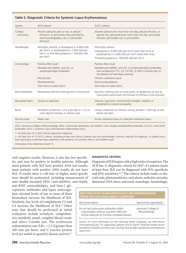
Diagnosis of SLE begins with a high index of suspicion. The ACR has 11 diagnostic criteria for SLE8; if a patient meets at least four, SLE can be diagnosed with 95% specificity and 85% sensitivity.16,17 The criteria include malar or dis-coid rash; photosensitivity; oral ulcers; arthritis; serositis; abnormal ANA titers; and renal, neurologic, hematologic,
Table 2. Diagnostic Criteria for Systemic Lupus Erythematosus
System ACR criteria* SLICC criteria†
Cardiac/pulmonary
Pleuritis (pleuritic pain or rub, or pleural effusion), or pericarditis (documented by electrocardiography, rub, or pericardial effusion)
Serositis (pleurisy for more than one day, pleural effusion, or pleural rub; pericardial pain more than one day, pericardial effusion, pericardial rub, or pericarditis)
Hematologic Hemolytic anemia, or leukopenia (< 4,000 cells per mm3), or lymphopenia (< 1,500 cells per mm3), or thrombocytopenia (< 100,000 cells per mm3)
Hemolytic anemia
Leukopenia (< 4,000 cells per mm3) more than once or lymphopenia (< 1,000 cells per mm3) more than once
Thrombocytopenia (< 100,000 cells per mm3)
Immunologic Positive ANA result Positive ANA result
Elevated anti-dsDNA, anti-Sm, or antiphospholipid antibodies
Elevated anti-dsDNA, anti-Sm, or antiphospholipid antibodies, low complement (C3, C4, CH 50), or direct Coombs test (in the absence of hemolytic anemia)
Discoid rash Chronic cutaneous lupus
Photosensitivity Nonscarring alopecia
Oral ulcers or nasal ulcers Oral ulcers or nasal ulcers
Musculoskeletal Nonerosive arthritis involving two or more joints Synovitis involving two or more joints, or tenderness at two or more joints and at least 30 minutes of stiffness in the morning
Neuropsychiatric Seizure or psychosis Seizures, psychosis, mononeuritis complex, myelitis, or peripheral or cranial neuropathy
Renal Persistent proteinuria > 0.5 g per day or > 3+ on urine dipstick testing, or cellular casts
Urinary creatinine (or 24-hour urinary protein) > 500 mg, or red blood cell casts
Skin/mucosal Malar rash Acute cutaneous lupus or subacute cutaneous lupus
ACR = American College of Rheumatology; ANA = antinuclear antibodies; anti-dsDNA = anti–double-stranded DNA antibodies; anti-Sm = anti-Smith antibodies; SLICC = Systemic Lupus International Collaborating Clinics.
*—At least four of 11 ACR criteria required for diagnosis.†—At least four of 13 SLICC criteria, including at least one clinical criterion and one immunologic criterion, required for diagnosis, or patient must have had biopsy-confirmed lupus nephritis in the presence of a positive ANA or anti-dsDNA result.
Information from references 8 and 13.
BEST PRACTICES IN RHEUMATOLOGY: RECOMMENDATIONS FROM THE CHOOSING WISELY CAMPAIGN
Recommendation Sponsoring organization
Do not test antinuclear antibodies (ANA) subserologies without a positive ANA and clinical suspicion of immune-mediated disease.
American College of Rheumatology
Source: For more information on the Choosing Wisely Campaign, see http://www.choosingwisely.org. For supporting citations and to search Choosing Wisely recom-mendations relevant to primary care, see http://www.aafp.org/afp/recommendations/search.htm.
Lupus
August 15, 2016 ◆ Volume 94, Number 4 www.aafp.org/afp American Family Physician 287
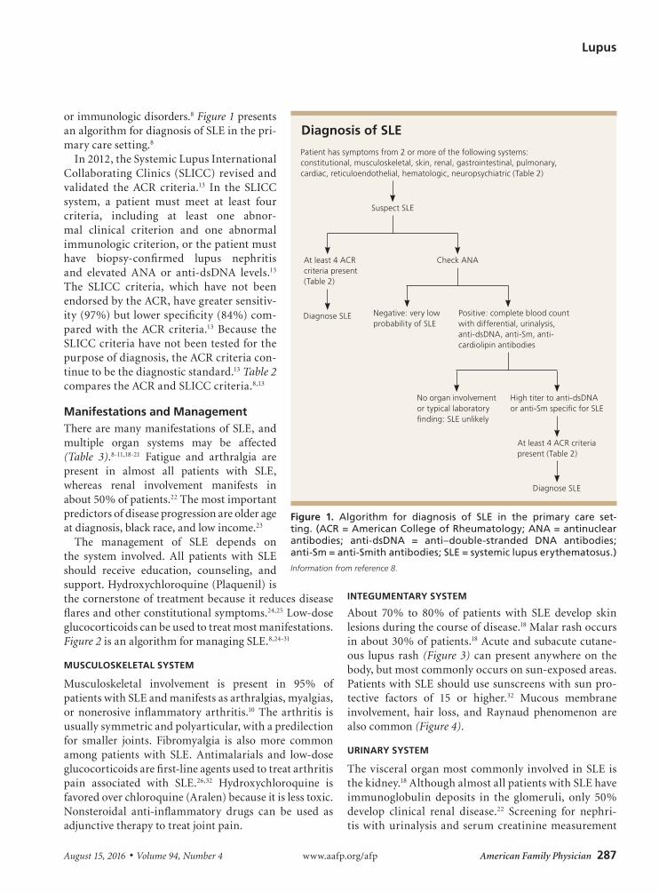
or immunologic disorders.8 Figure 1 presents an algorithm for diagnosis of SLE in the pri-mary care setting.8
In 2012, the Systemic Lupus International Collaborating Clinics (SLICC) revised and validated the ACR criteria.13 In the SLICC system, a patient must meet at least four criteria, including at least one abnor-mal clinical criterion and one abnormal immunologic criterion, or the patient must have biopsy-confirmed lupus nephritis and elevated ANA or anti-dsDNA levels.13 The SLICC criteria, which have not been endorsed by the ACR, have greater sensitiv-ity (97%) but lower specificity (84%) com-pared with the ACR criteria.13 Because the SLICC criteria have not been tested for the purpose of diagnosis, the ACR criteria con-tinue to be the diagnostic standard.13 Table 2 compares the ACR and SLICC criteria.8,13
Manifestations and Management There are many manifestations of SLE, and multiple organ systems may be affected (Table 3).8-11,18-21 Fatigue and arthralgia are present in almost all patients with SLE, whereas renal involvement manifests in about 50% of patients.22 The most important predictors of disease progression are older age at diagnosis, black race, and low income.23
The management of SLE depends on the system involved. All patients with SLE should receive education, counseling, and support. Hydroxychloroquine (Plaquenil) is the cornerstone of treatment because it reduces disease flares and other constitutional symptoms.24,25 Low-dose glucocorticoids can be used to treat most manifestations. Figure 2 is an algorithm for managing SLE.8,24-31
MUSCULOSKELETAL SYSTEM
Musculoskeletal involvement is present in 95% of patients with SLE and manifests as arthralgias, myalgias, or nonerosive inflammatory arthritis.10 The arthritis is usually symmetric and polyarticular, with a predilection for smaller joints. Fibromyalgia is also more common among patients with SLE. Antimalarials and low-dose glucocorticoids are first-line agents used to treat arthritis pain associated with SLE.26,32 Hydroxychloroquine is favored over chloroquine (Aralen) because it is less toxic. Nonsteroidal anti-inflammatory drugs can be used as adjunctive therapy to treat joint pain.
INTEGUMENTARY SYSTEM
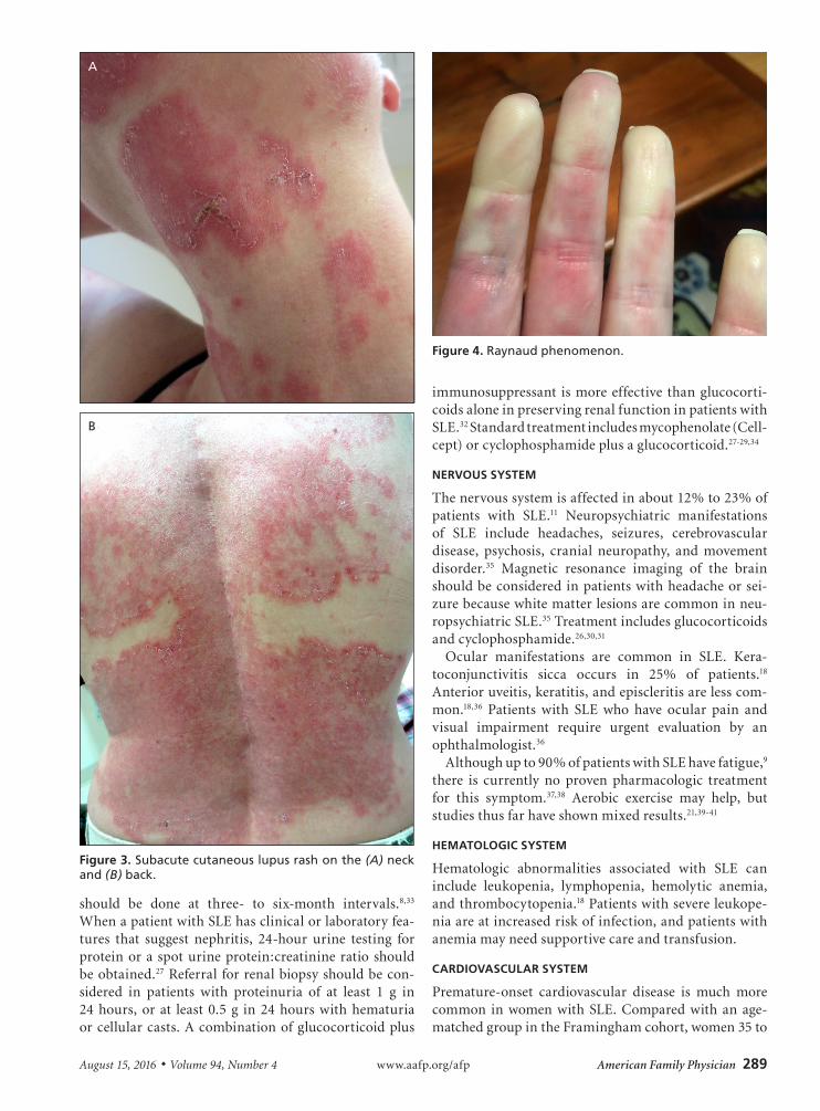
About 70% to 80% of patients with SLE develop skin lesions during the course of disease.18 Malar rash occurs in about 30% of patients.18 Acute and subacute cutane-ous lupus rash (Figure 3) can present anywhere on the body, but most commonly occurs on sun-exposed areas. Patients with SLE should use sunscreens with sun pro-tective factors of 15 or higher.32 Mucous membrane involvement, hair loss, and Raynaud phenomenon are also common (Figure 4).
URINARY SYSTEM
The visceral organ most commonly involved in SLE is the kidney.18 Although almost all patients with SLE have immunoglobulin deposits in the glomeruli, only 50% develop clinical renal disease.22 Screening for nephri-tis with urinalysis and serum creatinine measurement
Diagnosis of SLE
Patient has symptoms from 2 or more of the following systems: constitutional, musculoskeletal, skin, renal, gastrointestinal, pulmonary, cardiac, reticuloendothelial, hematologic, neuropsychiatric (Table 2)
Suspect SLE
Check ANAAt least 4 ACR criteria present (Table 2)
Diagnose SLE Negative: very low probability of SLE
Positive: complete blood count with differential, urinalysis, anti-dsDNA, anti-Sm, anti-cardiolipin antibodies
No organ involvement or typical laboratory finding: SLE unlikely
High titer to anti-dsDNA or anti-Sm specific for SLE
At least 4 ACR criteria present (Table 2)
Diagnose SLE
Figure 1. Algorithm for diagnosis of SLE in the primary care set-ting. (ACR = American College of Rheumatology; ANA = antinuclear antibodies; anti-dsDNA = anti–double-stranded DNA antibodies; anti-Sm = anti-Smith antibodies; SLE = systemic lupus erythematosus.)
Information from reference 8.

Lupus
288 American Family Physician www.aafp.org/afp Volume 94, Number 4 ◆ August 15, 2016
Table 3. Common Symptoms of Systemic Lupus Erythematosus
Symptoms Lifetime prevalence Treatment Prevention and screening
Arthralgia 95% Glucocorticoids, hydroxychloroquine (Plaquenil), methotrexate, nonsteroidal anti-inflammatory drugs
—
Fatigue 90% Aerobic exercise (possibly) Minimize exertion, get adequate sleep
Cutaneous lupus 70% to 80% Topical glucocorticoids, chloroquine (Aralen), hydroxychloroquine
Use sunscreen and wear protective clothing
Hematologic symptoms
Most patients Azathioprine (Imuran), mycophenolate (Cellcept), rituximab (Rituxan); transfusion for anemia (possibly)
Monitor and treat infection, especially in patients with leukopenia
Renal symptoms 50% Azathioprine, cyclophosphamide, glucocorticoids, mycophenolate
Urinalysis and serum creatinine test every three months
Cardiovascular symptoms
28% to 40% Antihypertensive agents, cholesterol-lowering agents Treat risk factors aggressively
Neuropsychiatric symptoms
12% to 23% Glucocorticoids; anticonvulsants, antidepressants, and antipsychotics for depression and headaches; antithrombotic agents and cyclophosphamide for thrombotic symptoms and cerebritis
Control aggravating factors and symptoms
Pulmonary symptoms
16% Depends on type of involvement and severity; azathioprine, cyclophosphamide, glucocorticoids, mycophenolate, plasmapheresis
—
Information from references 8 through 11, and 18 through 21.
Management of SLE
Figure 2. Algorithm for management of SLE. (IV = intravenous; NSAIDs = nonsteroidal anti-inflammatory drugs; SLE = systemic lupus erythematosus.)
Information from references 8, and 24 through 31.
Provide patient education, counseling, and support
Mild SLE (no major organ involvement) Moderate to severe SLE
Constitutional symptoms: hydroxychloroquine (Plaquenil), with or without low-dose systemic glucocorticoids or methotrexate; then azathioprine (Imuran), methotrexate, or mycophenolate (Cellcept); then rituximab (Rituxan) or belimumab (Benlysta)
Discoid lupus: sunscreen use; topical corticosteroid/tacrolimus (Protopic); topical acitretin (Soriatane), with or without hydroxychloroquine, with or without systemic glucocorticoids; then add azathioprine or switch to mycophenolate or methotrexate
Polyarthritis: hydroxychloroquine, with or without low-dose systemic glucocorticoids; then add methotrexate or rituximab; NSAIDs as adjunctive therapy
Uncomplicated digital or cutaneous vasculitis: systemic glucocorticoids, with or without hydroxy-chloroquine, with or without methotrexate; then add azathioprine or mycophenolate; then switch to IV cyclophosphamide
Alveolitis: systemic glucocorticoids plus mycophenolate or IV cyclophosphamide; then add rituximab or IV immune globulin; maintain with azathioprine or mycophenolate
Antiphospholipid syndrome: anticoagulation, with or without hydroxychloroquine, then add direct thrombin inhibitor
Lupus nephritis: systemic glucocorticoids plus mycophenolate; then IV cyclophos-phamide; then add rituximab or switch to azathioprine; angiotensin-converting enzyme inhibitor and hydroxy chloroquine as adjunctive therapy
Mononeuritis/central nervous system lupus: systemic glucocorticoids, with or without IV cyclophosphamide, then add rituximab, IV immune globulin, or plasmapheresis
Myocarditis: systemic glucocorticoids plus IV cyclophosphamide, with or without hydroxychloroquine; then rituximab, belimumab, or IV immune globulin
Pericarditis: NSAIDs; then systemic glucocorticoids plus hydroxychloroquine; then add mycophenolate, azathioprine, or methotrexate; then add belimumab or rituximab
Pulmonary artery hypertension: systemic glucocorticoids plus IV cyclophos phamide, or mycophenolate plus endothelin receptor antagonist; then add rituximab and phosphodiesterase-5 inhibitor; then add prostaglandin analogue; maintain with prostaglandin analogue
Thrombocytopenia: systemic glucocorticoids, with or without hydroxychloroquine; then add azathioprine or mycophenolate; then add rituximab or IV cyclophosphamide or IV immune globulin
Lupus
August 15, 2016 ◆ Volume 94, Number 4 www.aafp.org/afp American Family Physician 289
should be done at three- to six-month intervals.8,33 When a patient with SLE has clinical or laboratory fea-tures that suggest nephritis, 24-hour urine testing for protein or a spot urine protein:creatinine ratio should be obtained.27 Referral for renal biopsy should be con-sidered in patients with proteinuria of at least 1 g in 24 hours, or at least 0.5 g in 24 hours with hematuria or cellular casts. A combination of glucocorticoid plus
immunosuppressant is more effective than glucocorti-coids alone in preserving renal function in patients with SLE.32 Standard treatment includes mycophenolate (Cell-cept) or cyclophosphamide plus a glucocorticoid.27-29,34
NERVOUS SYSTEM
The nervous system is affected in about 12% to 23% of patients with SLE.11 Neuropsychiatric manifestations of SLE include headaches, seizures, cerebrovascular disease, psychosis, cranial neuropathy, and movement disorder.35 Magnetic resonance imaging of the brain should be considered in patients with headache or sei-zure because white matter lesions are common in neu-ropsychiatric SLE.35 Treatment includes glucocorticoids and cyclophosphamide.26,30,31
Ocular manifestations are common in SLE. Kera-toconjunctivitis sicca occurs in 25% of patients.18 Anterior uveitis, keratitis, and episcleritis are less com-mon.18,36 Patients with SLE who have ocular pain and visual impairment require urgent evaluation by an ophthalmologist.36
Although up to 90% of patients with SLE have fatigue,9 there is currently no proven pharmacologic treatment for this symptom.37,38 Aerobic exercise may help, but studies thus far have shown mixed results.21,39-41
HEMATOLOGIC SYSTEM
Hematologic abnormalities associated with SLE can include leukopenia, lymphopenia, hemolytic anemia, and thrombocytopenia.18 Patients with severe leukope-nia are at increased risk of infection, and patients with anemia may need supportive care and transfusion.
CARDIOVASCULAR SYSTEM
Premature-onset cardiovascular disease is much more common in women with SLE. Compared with an age-matched group in the Framingham cohort, women 35 to
A
B
Figure 3. Subacute cutaneous lupus rash on the (A) neck and (B) back.
Figure 4. Raynaud phenomenon.

Lupus
290 American Family Physician www.aafp.org/afp Volume 94, Number 4 ◆ August 15, 2016
44 years of age who had SLE had a greatly increased risk of myocardial infarction (rate ratio = 52; 95% confidence interval, 22 to 98).42 The increased risk of accelerated atherosclerosis suggests that there are other SLE-related factors, such as renal disease, cytokines, inflammatory mediators, antiphospholipid antibodies, oxidized low-density lipoprotein, and adverse effects of treatment, that cause accelerated cardiovascular disease.19,43 SLE is an independent risk factor for the development of ath-erosclerosis, and is identified as such by the American Heart Association.19,44
It is important to counsel patients to reduce tradi-tional cardiovascular risk factors such as smoking and obesity, and to have routine screenings for diabetes mel-litus, hypertension, and dyslipidemia. The recent Joint National Committee-8 guideline for hypertension and the Adult Treatment Panel IV guideline for hyperlip-idemia do not make separate recommendations for patients with SLE.45,46 Family physicians should be aware of the increased risk of cardiovascular events in patients with SLE, and should take steps to optimize risk in accor-dance with national guidelines. Although guidelines do not address this need, patients with SLE may benefit from treatment with a statin because of their increased 10-year risk of a cardiovascular event.47,48 Blood pressure should also be treated to a goal similar to that for patients with comorbid conditions such as diabetes (less than 140/90 mm Hg).44,45,47,49
RESPIRATORY SYSTEM
Lung involvement in SLE can vary from minor pleu-ritic pain in serositis to life-threatening complications
such as alveolar hemorrhage.50 Pleuritis occurs in 17% to 60% of patients with SLE.50,51 Treatment is based on the type and severity of lung involvement, and may include glucocorticoids, immunosuppressive agents, and plasmaphersis.32,52
REPRODUCTIVE SYSTEM
Pregnant women with SLE have an increased risk of spontaneous abortions, stillbirths, and fetal growth restriction.26 Pregnancy may also increase disease activity and precipitate disease flares.20 Although women with SLE can use most contraceptive meth-ods, those with antiphospholipid syndrome should not use estrogen-containing contraceptives because of an increased risk of thrombosis.53 Women with recurrent pregnancy loss should be screened for antiphospholipid syndrome.
Monitoring and Complications All patients with SLE should receive ongoing education, counseling, and support. Those with mild SLE that does not involve major organ systems can be monitored by primary care physicians.8 Patients with increased disease activity, complications, or adverse effects from treat-ment should be referred to a rheumatologist.8 Family physicians can monitor disease activity and therapy in patients with moderate to severe SLE.8
Measurement of anti-dsDNA antibodies, comple-ments, and creatinine; a complete blood count; and urinalysis should be performed every three to six months to monitor disease activity.8 Annual eye examinations are required for patients receiving hydroxychloroquine.36
SORT: KEY RECOMMENDATIONS FOR PRACTICE
Clinical recommendation Evidence rating References
The initial evaluation for suspected SLE should include an antinuclear antibody test. C 15
Patients diagnosed with SLE must meet at least four of the 11 American College of Rheumatology diagnostic criteria.
C 8
Treatment of mild SLE includes patient education, expectations of treatment, and counseling to avoid extensive ultraviolet light exposure and overexertion.
C 8
Hydroxychloroquine (Plaquenil) has been shown to reduce arthritis pain associated with SLE. A 32
A combination of glucocorticoid plus immunosuppressant is more effective than glucocorticoids alone in preserving renal function in patients with SLE.
A 32
A combination of glucocorticoid and mycophenolate (Cellcept) or cyclophosphamide is effective in achieving remission in patients with SLE nephritis.
A 28
SLE = systemic lupus erythematosus.
A = consistent, good-quality patient-oriented evidence; B = inconsistent or limited-quality patient-oriented evidence; C = consensus, disease-oriented evidence, usual practice, expert opinion, or case series. For information about the SORT evidence rating system, go to http://www.aafp.org/afpsort.
Lupus
August 15, 2016 ◆ Volume 94, Number 4 www.aafp.org/afp American Family Physician 291
Screening for dyslipidemia, diabetes, and osteoporo-sis should be performed regularly in patients receiving glucocorticoids. For patients with chronic kidney dis-ease who are receiving long-term immunosuppressive therapy, immunization with 13-valent pneumococcal conjugate vaccine (Prevnar) followed by 23-valent pneu-mococcal polysaccharide vaccine (Pneumovax) should be
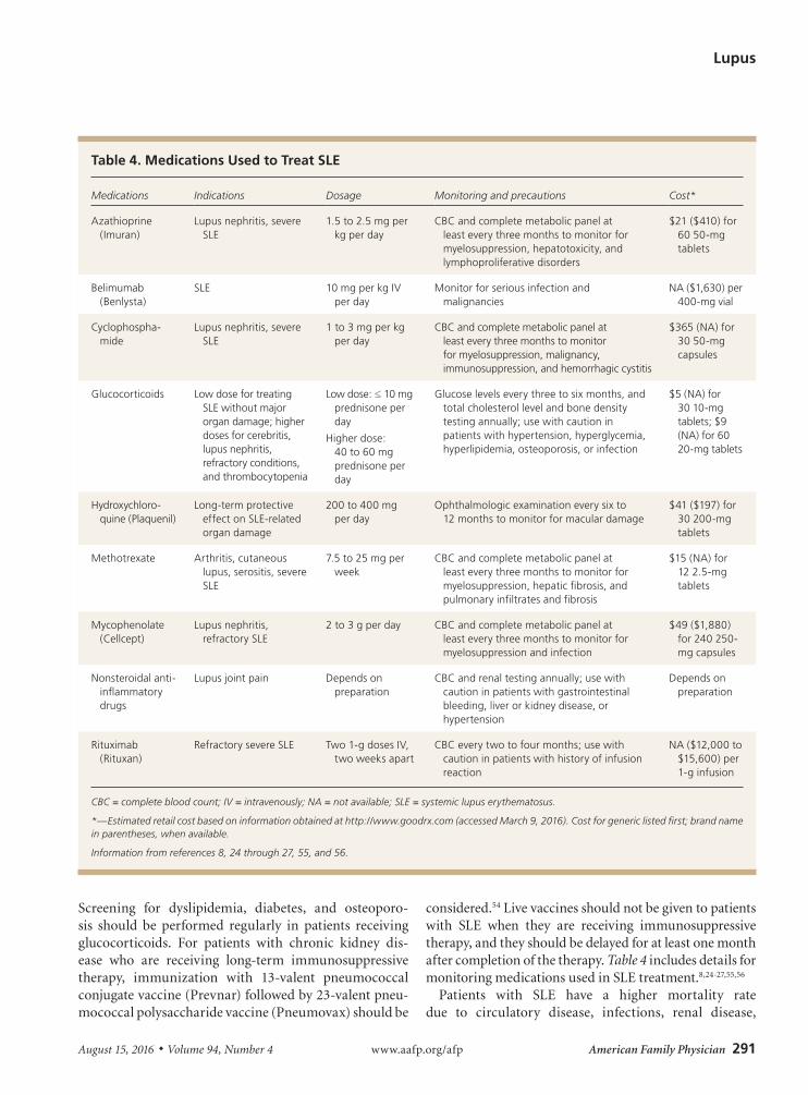
considered.54 Live vaccines should not be given to patients with SLE when they are receiving immunosuppressive therapy, and they should be delayed for at least one month after completion of the therapy. Table 4 includes details for monitoring medications used in SLE treatment.8,24-27,55,56
Patients with SLE have a higher mortality rate due to circulatory disease, infections, renal disease,
Table 4. Medications Used to Treat SLE
Medications Indications Dosage Monitoring and precautions Cost*
Azathioprine (Imuran)
Lupus nephritis, severe SLE
1.5 to 2.5 mg per kg per day
CBC and complete metabolic panel at least every three months to monitor for myelosuppression, hepatotoxicity, and lymphoproliferative disorders
$21 ($410) for 60 50-mg tablets
Belimumab (Benlysta)
SLE 10 mg per kg IV per day
Monitor for serious infection and malignancies
NA ($1,630) per 400-mg vial
Cyclophospha-mide
Lupus nephritis, severe SLE
1 to 3 mg per kg per day
CBC and complete metabolic panel at least every three months to monitor for myelosuppression, malignancy, immunosuppression, and hemorrhagic cystitis
$365 (NA) for 30 50-mg capsules
Glucocorticoids Low dose for treating SLE without major organ damage; higher doses for cerebritis, lupus nephritis, refractory conditions, and thrombocytopenia
Low dose: ≤ 10 mg prednisone per day
Higher dose: 40 to 60 mg prednisone per day
Glucose levels every three to six months, and total cholesterol level and bone density testing annually; use with caution in patients with hypertension, hyperglycemia, hyperlipidemia, osteoporosis, or infection
$5 (NA) for 30 10-mg tablets; $9 (NA) for 60 20-mg tablets
Hydroxychloro-quine (Plaquenil)
Long-term protective effect on SLE-related organ damage
200 to 400 mg per day
Ophthalmologic examination every six to 12 months to monitor for macular damage
$41 ($197) for 30 200-mg tablets
Methotrexate Arthritis, cutaneous lupus, serositis, severe SLE
7.5 to 25 mg per week
CBC and complete metabolic panel at least every three months to monitor for myelosuppression, hepatic fibrosis, and pulmonary infiltrates and fibrosis
$15 (NA) for 12 2.5-mg tablets
Mycophenolate (Cellcept)
Lupus nephritis, refractory SLE
2 to 3 g per day CBC and complete metabolic panel at least every three months to monitor for myelosuppression and infection
$49 ($1,880) for 240 250-mg capsules
Nonsteroidal anti-inflammatory drugs
Lupus joint pain Depends on preparation
CBC and renal testing annually; use with caution in patients with gastrointestinal bleeding, liver or kidney disease, or hypertension
Depends on preparation
Rituximab (Rituxan)
Refractory severe SLE Two 1-g doses IV, two weeks apart
CBC every two to four months; use with caution in patients with history of infusion reaction
NA ($12,000 to $15,600) per 1-g infusion
CBC = complete blood count; IV = intravenously; NA = not available; SLE = systemic lupus erythematosus.
*—Estimated retail cost based on information obtained at http://www.goodrx.com (accessed March 9, 2016). Cost for generic listed first; brand name in parentheses, when available.
Information from references 8, 24 through 27, 55, and 56.

Lupus
292 American Family Physician www.aafp.org/afp Volume 94, Number 4 ◆ August 15, 2016
non-Hodgkin lymphoma, and lung cancer.57 Damage related to SLE is most common in the musculoskeletal (15%), neuropsychiatric (11%), cardiovascular (9%), and hematologic (3%) systems.57 Osteoporosis is a potentially preventable complication of SLE.57 The inci-dence of non-Hodgkin lymphoma is increased three- to fourfold compared with the general population.57 Table 5
describes recommendations for monitoring manifesta-tions and complications related to SLE.8,27,36,49,54,57,58
Data Sources: Searches were done in Essential Evidence Plus, Clinical Evidence, the Cochrane Database of Systematic Reviews, and DynaMed. Key terms included systemic lupus erythematosus, manifestation, treat-ment, and management. Search dates: January 28 to February 15, 2015; September 23, 2015; and April 8, 2016.
Table 5. Follow-Up and Monitoring for Selected Complications of SLE
ComplicationFrequency of follow-up Prevention, monitoring, and management
None; mild, stable SLE8 Every three to six months
History for features of SLE, physical examination, CBC, creatinine level, urinalysis, anti-dsDNA, complements; keep all health maintenance screenings and vaccinations up to date
Cardiovascular abnormalities49,57
Every visit Optimal lupus control with minimal glucocorticoid use; judicious use of antimalarial and other immunosuppressive agents; smoking cessation, adequate exercise, low-cholesterol diet, lipid-lowering therapy, blood pressure control, screening for diabetes mellitus
Infection8,57 Every visit Assure that vaccinations are up to date; judicious use of immunosuppressive agents
Malignancy57,58 Yearly Assure that routine cancer screenings are up to date; screen for high-risk cancers (e.g., hematologic, non-Hodgkin lymphoma, lung, cervical)
Moderate to severe SLE with complications8
Frequent Monitor in conjunction with rheumatologist and lupus care subspecialists
New-onset nephritis8,27 Monthly or more frequently
Urinalysis, 24-hour urinary protein level, creatinine clearance, CBC, levels of cholesterol, calcium, phosphorus, alkaline phosphatase, sodium, and potassium; useful to assess complements and anti-dsDNA
Receiving high-dose glucocorticoids8,57
Every visit Consider steroid-sparing agent; use lowest dose possible to achieve optimum disease control; glucose testing every three to six months; cholesterol testing annually; DEXA every one to two years; maintain high index of suspicion for avascular necrosis if patient has acute joint pain
Receiving hydroxychloroquine (Plaquenil)8,36
Every six to 12 months Ophthalmologic examination to screen for retinal toxicity; keep dosage to no more than 6.5 mg per kg per day
Receiving immunosuppressive or cytotoxic agents8
Every one to two weeks initially, then every one to three months
CBC and liver function testing at baseline, then every one to two weeks at initiation of therapy, then one to three months; judicious use of immunosuppressive medications, vigilance for signs and symptoms of infection; routine cancer screening; avoidance of live vaccines; if live vaccines are needed, administer one month after therapy completion
Receiving low-dose glucocorticoids8
Every visit to every one to two years
Keep dosage as low as possible; healthy diet with adequate physical activity; smoking cessation; annual cholesterol and glucose testing; consider DEXA every one to two years for patients receiving long-term therapy
Renal abnormalities8,27,54 Every three months or more frequently, depending on disease state
Regular screening for proteinuria and hematuria; regular serum creatinine level; for patients with chronic kidney disease, vaccination with 13-valent pneumococcal conjugate vaccine (Prevnar) or 23-valent pneumococcal polysaccharide vaccine (Pneumovax), as indicated
Severe hemolytic anemia8 Weekly Hematocrit and reticulocyte count; may require transfusion
Severe thrombocytopenia (< 50,000 cells per mm3)8
Weekly Platelet count weekly initially; may require transfusion
Anti-dsDNA = anti–double-stranded DNA antibodies; CBC = complete blood count; DEXA = dual energy x-ray absorptiometry; SLE = systemic lupus erythematosus.
Information from references 8, 27, 36, 49, 54, 57, and 58.

Lupus
August 15, 2016 ◆ Volume 94, Number 4 www.aafp.org/afp American Family Physician 293
The authors thank Robert Langan, MD, FAAFP, for assistance with the manuscript.
This review updates a previous article on this topic by Gill, et al.59
The Authors
NGUYET-CAM VU LAM, MD, is associate program director at St. Luke’s Family Medicine Residency, St. Luke’s University Hospital, Bethlehem, Pa.
MARIA V. GHETU, MD, is clinical faculty and staff geriatrician at St. Luke’s Family Medicine Residency, St. Luke’s University Hospital, Bethlehem, Pa.
MARZENA L. BIENIEK, MD, is a rheumatologist in Whitehall, Pa., who is affiliated with St. Luke’s University Hospital.
Address correspondence to Nguyet-Cam Vu Lam, MD, St. Luke’s Family Medicine Residency, 2830 Easton Ave., Bethlehem, PA 18017 (e-mail: [email protected]). Reprints are not available from the authors.
REFERENCES
1. Helmick CG, Felson DT, Lawrence RC, et al.; National Arthritis Data Work-group. Estimates of the prevalence of arthritis and other rheumatic con-ditions in the United States. Part I. Arthritis Rheum. 2008;58(1):15-25.
2. Somers EC, Marder W, Cagnoli P, et al. Population-based incidence and prevalence of systemic lupus erythematosus: the Michigan Lupus Epidemiology and Surveillance program. Arthritis Rheumatol. 2014;66(2):369-378.
3. Bernatsky S, Boivin JF, Joseph L, et al. Mortality in systemic lupus erythe-matosus. Arthritis Rheum. 2006;54(8):2550-2557.
4. Jakes RW, Bae SC, Louthrenoo W, Mok CC, Navarra SV, Kwon N. Sys-tematic review of the epidemiology of systemic lupus erythematosus in the Asia-Pacific region: prevalence, incidence, clinical features, and mortality. Arthritis Care Res (Hoboken). 2012;64(2):159-168.
5. Burgos PI, Alarcón GS. Preventive health services for systemic lupus ery-thematosus patients: whose job is it? Arthritis Res Ther. 2010;12(3):124.
6. Hale ED, Treharne GJ, Lyons AC, et al. “Joining the dots” for patients with systemic lupus erythematosus: personal perspectives of health care from a qualitative study. Ann Rheum Dis. 2006;65(5):585-589.
7. Yazdany J, Tonner C, Trupin L, et al. Provision of preventive health care in systemic lupus erythematosus: data from a large observational cohort study. Arthritis Res Ther. 2010;12(3):R84.
8. American College of Rheumatology Ad Hoc Committee on Systemic Lupus Erythematosus Guidelines. Guidelines for referral and manage-ment of systemic lupus erythematosus in adults. Arthritis Rheum. 1999; 42(9): 1785-1796.
9. Zonana-Nacach A, Roseman JM, McGwin G Jr, et al. Systemic lupus ery-thematosus in three ethnic groups. VI: Factors associated with fatigue within 5 years of criteria diagnosis. LUMINA Study Group. LUpus in MInority populations: NAture vs Nurture. Lupus. 2000;9(2):101-109.
10. Zoma A. Musculoskeletal involvement on systemic lupus erythemato-sus. Lupus. 2004;13(11):851-853.
11. Cervera R, Khamashta MA, Font J, et al.; European Working Party on Systemic Lupus Erythematosus. Morbidity and mortality in systemic lupus erythematosus during a 10-year period: a comparison of early and late manifestations in a cohort of 1,000 patients. Medicine (Baltimore). 2003; 82(5):299-308.
12. Longo DL, Fauci AS, Kasper DL, Hauser SL, Jameson JL, Loscalzo J, eds. Harrison’s Principles of Internal Medicine. 18th ed. New York, NY: McGraw-Hill; 2012.
13. Petri M, Orbai AM, Alarcón GS, et al. Derivation and validation of the Sys-temic Lupus International Collaborating Clinics classification criteria for systemic lupus erythematosus. Arthritis Rheum. 2012;64(8):2677-2686.
14. Narain S, Richards HB, Satoh M, et al. Diagnostic accuracy for lupus and other systemic autoimmune diseases in the community setting. Arch Intern Med. 2004;164(22):2435-2441.
15. Hietarinta M, Lassila O. Clinical significance of antinuclear antibodies in systemic rheumatic diseases. Ann Med. 1996;28(4):283-291.
16. Tan EM, Cohen AS, Fries JF, et al. The 1982 revised criteria for the clas-sification of systemic lupus erythematosus. Arthritis Rheum. 1982; 25(11): 1271-1277.
17. Hochberg MC. Updating the American College of Rheumatology revised criteria for the classification of systemic lupus erythematosus. Arthritis Rheum. 1997;40(9):1725.
18. Cojocaru M, Cojocaru IM, Silosi I, Vrabie CD. Manifestations of systemic lupus erythematosus. Maedica (Buchar). 2011;6(4):330-336.
19. Sinicato NA, da Silva Cardoso PA, Appenzeller S. Risk factors in car-diovascular disease in systemic lupus erythematosus. Curr Cardiol Rev. 2013;9(1):15-19.
20. Bertsias G, Fanouriakis A, Boumpas DT. Treatment of systemic lupus erythematosus. In: Firestein GS, Budd RC, Gabriel SE, McInnes IB, O’Dell JR, eds. Kelly’s Textbook of Rheumatology. 9th ed. Philadelphia, Pa.: Saunders Elsevier; 2013:1304-1330.
21. Del Pino-Sedeño T, Trujillo-Martín MM, Ruiz-Irastorza G, Cuellar-Pompa L, de Pascual-Medina AM, Serrano-Aguilar P; Spanish Systemic Lupus Erythematosus CPG Development Group. Effectiveness of nonphar-macologic interventions for decreasing fatigue in adults with systemic lupus erythematosus: a systemic review. Arthritis Care Res (Hoboken). 2016; 68(1):141-148.
22. Drakoulogkona O, Barbulescu AL, Rica I, Musetescu AE, Ciurea PL. The outcome of patients with lupus nephritis and the impact of cardiovas-cular risk factors. Curr Health Sci J. 2011;37(2):70-74.
23. Petri M, Purvey S, Fang H, Magder LS. Predictors of organ damage in systemic lupus erythematosus: the Hopkins Lupus Cohort. Arthritis Rheum. 2012;64(12):4021-4028.
24. Ruiz-Irastorza G, Ramos-Casals M, Brito-Zeron P, Khamashta MA. Clini-cal efficacy and side effects of antimalarials in systemic lupus erythema-tosus: a systematic review. Ann Rheum Dis. 2010;69(1):20-28.
25. Wallace DJ, Gudsoorkar VS, Weisman MH, Venuturupalli SR. New insights into mechanisms of therapeutic effects of antimalarial agents in SLE. Nat Rev Rheumatol. 2012;8(9):522-533.
26. Muangchan C, van Vollenhoven RF, Bernatsky SR, et al. Treatment algo-rithms in systemic lupus erythematosus. Arthritis Care Res (Hoboken). 2015; 67(9):1237-1245.
27. Hahn BH, McMahon MA, Wilkinson A, et al. American College of Rheu-matology guidelines for screening, treatment, and management of lupus nephritis. Arthritis Care Res (Hoboken). 2012;64(6):797-808.
28. Henderson L, Masson P, Craig JC, et al. Treatment for lupus nephritis. Cochrane Database Syst Rev. 2012;(12):CD002922.
29. Bertsias GK, Tektonidou M, Amoura Z, et al. Joint European League Against Rheumatism and European Renal Association-European Dialysis and Transplant Association (EULAR/ERA-EDTA) recommendations for the management of adult and paediatric lupus nephritis. Ann Rheum Dis. 2012;71(11):1771-1782.
30. Fernandes Moça Trevisani V, Castro AA, Ferreira Neves Neto J, Atallah AN. Cyclophosphamide versus methylprednisolone for treat-ing neuropsychiatric involvement in systemic lupus erythematosus. Cochrane Database Syst Rev. 2013;(2):CD002265.
31. Bertsias GK, Ioannidis JP, Aringer M, et al. EULAR recommendations for the management of systemic lupus erythematosus with neuropsy-chiatric manifestations: report of a task force of the EULAR standing committee for clinical affairs. Ann Rheum Dis. 2010;69(12):2074-2082.
32. Madhok R, Wu O. Systemic lupus erythematosus. BMJ Clin Evid. 2009; 2009:1123.
33. Dall’era M, Wofsy D. Clinical features of systemic lupus erythematosus. In: Firestein GS, Budd RC, Gabriel SE, McInnes IB, O’Dell JR, eds. Kelly’s

Lupus
294 American Family Physician www.aafp.org/afp Volume 94, Number 4 ◆ August 15, 2016
Textbook of Rheumatology. 9th ed. Philadelphia, Pa.: Saunders Elsevier; 2013:1283-1303.
34. Appel GB, Contreras G, Dooley MA, et al.; Aspreva Lupus Management Study Group. Mycophenolate mofetil versus cyclophosphamide for induc-tion treatment of lupus nephritis. J Am Soc Nephrol. 2009; 20(5): 1103-1112.
35. Muscal E, Brey RL. Neurologic manifestations of systemic lupus erythe-matosus in children and adults. Neurol Clin. 2010;28(1):61-73.
36. Sivaraj RR, Durrani OM, Denniston AK, Murray PI, Gordon C. Ocu-lar manifestations of systemic lupus erythematosus. Rheumatology (Oxford). 2007;46(12):1757-1762.
37. Merrill JT, Burgos-Vargas R, Westhovens R, et al. The efficacy and safety of abatacept in patients with non-life-threatening manifestations of systemic lupus erythematosus: results of a twelve-month, multicenter, exploratory, phase IIb, randomized, double-blind, placebo-controlled trial. Arthritis Rheum. 2010;62(10):3077-3087.
38. Hartkamp A, Geenen R, Godaert GL, Bijl M, Bijlsma JW, Derksen RH. Effects of dehydroepiandrosterone on fatigue and well-being in women with quiescent systemic lupus erythematosus: a randomised controlled trial. Ann Rheum Dis. 2010;69(6):1144-1147.
39. Neill J, Belan I, Ried K. Effectiveness of non-pharmacological interven-tions for fatigue in adults with multiple sclerosis, rheumatoid arthritis, or systemic lupus erythematosus: a systematic review [published correction appears in J Adv Nurs. 2007;57(2):225]. J Adv Nurs. 2006; 56(6): 617-635.
40. Tench CM, McCarthy J, McCurdie I, White PD, D’Cruz DP. Fatigue in systemic lupus erythematosus: a randomized controlled trial of exercise. Rheumatology (Oxford). 2003;42(9):1050-1054.
41. Balsamo S, Santos-Neto LD. Fatigue in systemic lupus erythematosus: an association with reduced physical fitness. Autoimmun Rev. 2011; 10(9):514-518.
42. Manzi S, Meilahn EN, Rairie JE, et al. Age-specific incidence rates of myocardial infarction and angina in women with systemic lupus ery-thematosus: comparison with the Framingham Study. Am J Epidemiol. 1997;145(5):408-415.
43. Zuily S, Regnault V, Selton-Suty C, et al. Increased risk for heart valve disease associated with antiphospholipid antibodies in patients with systemic lupus erythematosus: meta-analysis of echocardiographic studies. Circulation. 2011;124(2):215-224.
44. Mosca L, Benjamin EJ, Berra K, et al. Effectiveness-based guide-lines for the prevention of cardiovascular disease in women—2011 update: a guideline from the American Heart Association [published corrections appear in Circulation. 2011;123(22):e624 and Circulation. 2011;124(16):e427]. Circulation. 2011;123(11):1243-1262.
45. James PA, Oparil S, Carter BL, et al. 2014 evidence-based guideline for the management of high blood pressure in adults: report from the panel members appointed to the Eighth Joint National Committee
(JNC 8) [published correction appears in JAMA. 2014;311(17):1809]. JAMA. 2014;311(5):507-520.
46. Stone NJ, Robinson JG, Lichtenstein AH, et al. 2013 ACC/AHA guideline on the treatment of blood cholesterol to reduce atherosclerotic cardio-vascular risk in adults: a report of the American College of Cardiology/American Heart Association Task Force on Practice Guidelines [pub-lished correction appears in J Am Coll Cardiol. 2014;63(25 pt B):3024-3025]. J Am Coll Cardiol. 2014;63(25 pt B):2889-2934.
47. Esdaile JM, Abrahamowicz M, Grodzicky T, et al. Traditional Framingham risk factors fail to fully account for accelerated atherosclerosis in sys-temic lupus erythematosus. Arthritis Rheum. 2001;44(10):2331-2337.
48. Bruce IN, Urowitz MB, Gladman DD, Hallet DC. The natural history of hypercholesterolaemia in systemic lupus erythematosus. J Rheumatol. 1999;26(10):2137-2143.
49. McMahon M, Hahn BH, Skaggs BJ. Systemic lupus erythematosus and cardiovascular disease: prediction and potential for therapeutic inter-vention. Expert Rev Clin Immunol. 2011;7(2):227-241.
50. Keane MP, Lynch JP III. Pleuropulmonary manifestations of systemic lupus erythematosus. Thorax. 2000;55(2):159-166.
51. Todd NW, Wise RA. Respiratory complications in the collagen vascular diseases. Clin Pulm Med. 1996;3(2):101-112.
52. Carmier D, Marchand-Adam S, Diot P, Diot E. Respiratory involvement in systemic lupus erythematosus. Rev Mal Respir. 2010;27(8):e66-e78.
53. Culwell KR, Curtis KM, del Carmen Cravioto M. Safety of contraceptive method use among women with systemic lupus erythematosus: a sys-tematic review. Obstet Gynecol. 2009;114(2 pt 1):341-353.
54. Centers for Disease Control and Prevention (CDC). Use of 13-valent pneumococcal conjugate vaccine and 23-valent pneumococcal polysac-charide vaccine for adults with immunocompromising conditions: rec-ommendations of the Advisory Committee on Immunization Practices (ACIP). MMWR Morb Mortal Wkly Rep. 2012;61(40):816-819.
55. Goldberg A, Katzap E. Belimumab for the treatment of systemic lupus erythematosus. Int J Clin Rheum. 2010;5(4):407-413.
56. U.S. Food and Drug Administration. FDA news release. FDA approves Benlysta to treat lupus. http://www.fda.gov/NewsEvents/Newsroom/PressAnnouncements/ucm246489.htm. Accessed April 1, 2015.
57. Gordon C. Long-term complications of systemic lupus erythematosus. Rheumatology (Oxford). 2002;41(10):1095-1100.
58. Bernatsky S, Boivin JF, Joseph L, et al. An international cohort study of cancer in systemic lupus erythematosus. Arthritis Rheum. 2005; 52(5): 1481-1490.
59. Gill JM, Quisel AM, Rocca PV, Walters DT. Diagnosis of systemic lupus erythematosis. Am Fam Physician. 2003;68(11):2179-2187.
Recommended