A Clear Edge in Imaging
System Microscope
BX63/BX53BX3 Series
Photos courtesy of:
Junko Kyozuka, Associate Professor, Graduate School of Agricultural and Life Sciences,
The Tokyo University (P.11 below)
Prof. Nakatani, Biochemistry course, School of medicine, Showa University (P.13 above)
Prof. Tadokoro, Pathology class, School of medicine, St. Marianna University (P.15)
Printed in Japan M1697E-0310B
• OLYMPUS CORPORATION is ISO14001 certified.• OLYMPUS CORPORATION is FM553994/ISO9001 certified.• OLYMPUS CORPORATION is MD540624/ISO13485 certified.• Illumination devices for microscope have suggested lifetimes.
Periodic inspections are required. Please visit our web site for details.• All company and product names are registered trademarks and/or trademarks of their respective owners.• Images on the PC monitors are simulated.• Specifications and appearances are subject to change without any notice or obligation on the part of the manufacturer.
Images are courtesy of:
Dr. Shigeo Hayashi, Dr. Kagayaki Kato, Dr. Reiko Tajiri and Mr. Hosei WadaLaboratory for Morphogenetic SignalingRIKEN Center for Developmental Biology (P.12, P.15 top right, P.16 top left)
Shigenobu Yonemura, Ph.D.Electron Microscope LaboratoryRIKEN Center for Developmental Biology (P.6 lower right, P.7, P.15 lower right, P.17 Phase contrast, P.19 top right)
Guojun Sheng, Ph.D., Yukiko Nakaya, Ph.D.Laboratory for Early EmbryogenesisRIKEN Center for Developmental Biology (P.2, P.15 lower left, P.16 lower left)
Fumio Matsuzaki, Ph.D., Daijiro Konno, Ph.D. Laboratory for Cell Asymmetry RIKEN Center for Developmental Biology (P.6 lower left, P.15 top left, P.16 top right)
Junko Kyozuka, Associate ProfessorGraduate School of Agricultural and Life SciencesThe Tokyo University (P.16 lower right)
21
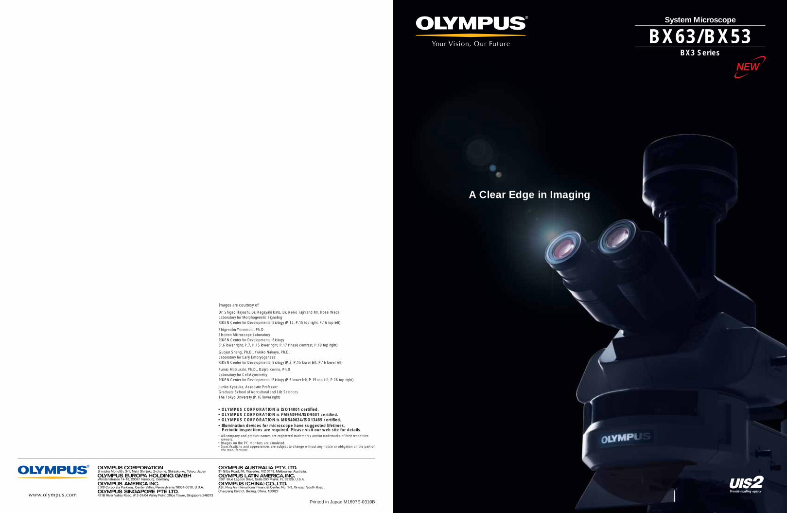
User-friendly design assures outstanding operating ease and flexibility forbrightfield/darkfield and fluorescence imaging in a wide range of researchsettings. With a choice of models and configuration options, there's anOlympus research microscope to meet your individual needs.
A Revolutionary New Standard in Accuracy and Imaging Efficiency
43
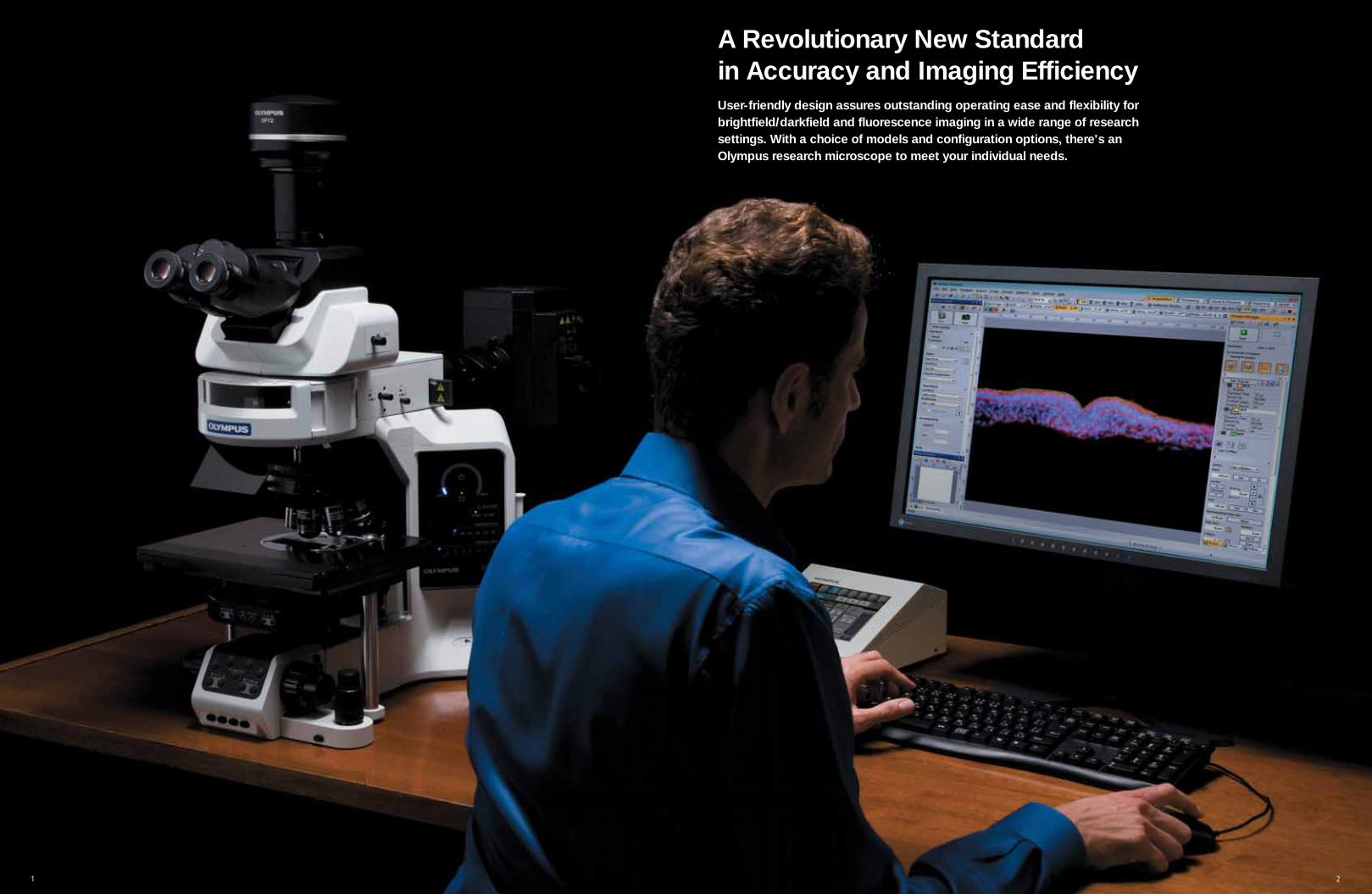
Advanced Sensitivity in Fluorescence Imaging
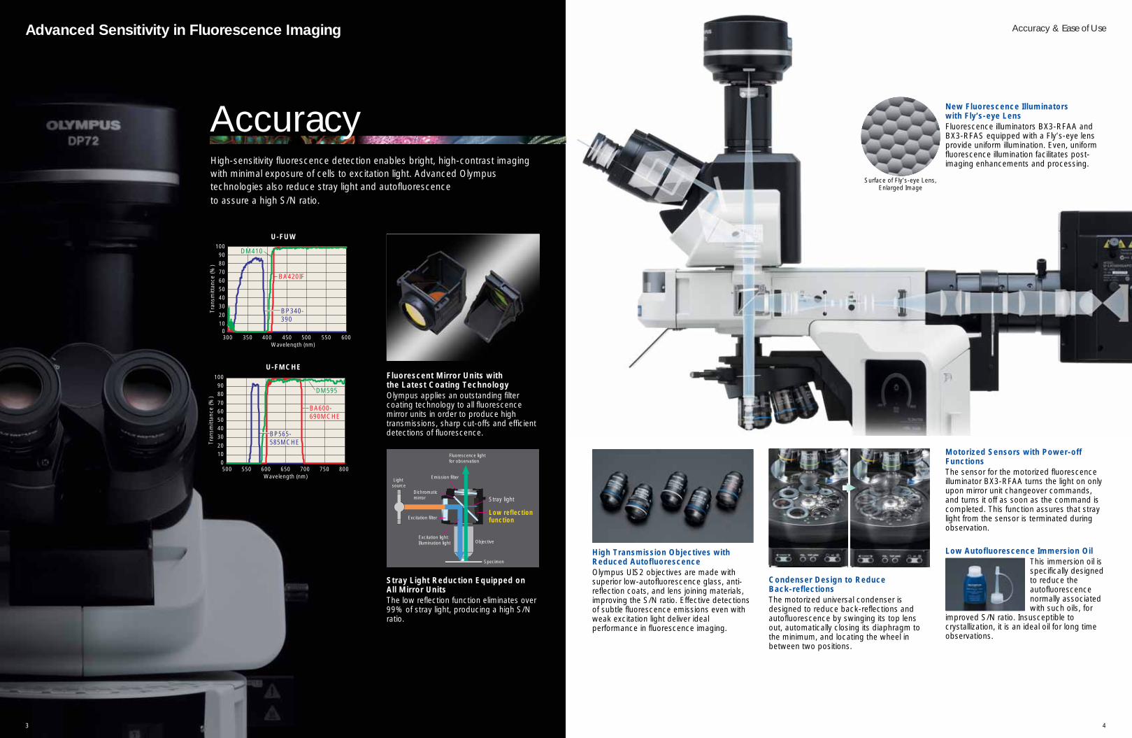
Motorized Sensors with Power-offFunctionsThe sensor for the motorized fluorescenceilluminator BX3-RFAA turns the light on onlyupon mirror unit changeover commands,and turns it off as soon as the command iscompleted. This function assures that straylight from the sensor is terminated duringobservation.
New Fluorescence Illuminators with Fly’s-eye LensFluorescence illuminators BX3-RFAA andBX3-RFAS equipped with a Fly’s-eye lensprovide uniform illumination. Even, uniformfluorescence illumination facilitates post-imaging enhancements and processing.
Accuracy & Ease of Use
High Transmission Objectives withReduced AutofluorescenceOlympus UIS2 objectives are made withsuperior low-autofluorescence glass, anti-reflection coats, and lens joining materials,improving the S/N ratio. Effective detectionsof subtle fluorescence emissions even withweak excitation light deliver idealperformance in fluorescence imaging.
Condenser Design to Reduce Back-reflectionsThe motorized universal condenser isdesigned to reduce back-reflections andautofluorescence by swinging its top lensout, automatically closing its diaphragm tothe minimum, and locating the wheel inbetween two positions.
Low Autofluorescence Immersion OilThis immersion oil isspecifically designedto reduce theautofluorescencenormally associatedwith such oils, for
improved S/N ratio. Insusceptible tocrystallization, it is an ideal oil for long timeobservations.
Fluorescent Mirror Units with the Latest Coating TechnologyOlympus applies an outstanding filtercoating technology to all fluorescencemirror units in order to produce hightransmissions, sharp cut-offs and efficientdetections of fluorescence.
Stray Light Reduction Equipped on All Mirror UnitsThe low reflection function eliminates over99% of stray light, producing a high S/Nratio.
Low reflection function
Stray light
Excitation filter
Emission filter
Fluorescence light for observation
Objective
Specimen
Excitation light: Illumination light
Dichromatic mirror
Light source
High-sensitivity fluorescence detection enables bright, high-contrast imagingwith minimal exposure of cells to excitation light. Advanced Olympustechnologies also reduce stray light and autofluorescence to assure a high S/N ratio.
U-FMCHE
01020
3040506070
8090
100
500 550Wavelength (nm)
Tran
smitt
ance
(%)
600 650 700 750 800
BP565-BP565-585MCHE585MCHEBP565-585MCHE
DM595DM595DM595
BA600-BA600-690MCHE690MCHEBA600-690MCHE
BP340-390
DM410
BA420IF
Tran
smitt
ance
(%)
BP565-585MCHE
DM595
BA600-690MCHE
BP340-BP340-390390BP340-390
DM410DM410DM410
BA420IFBA420IFBA420IF
010
20
304050
60
708090
100
300 350 400 450 500 550Wavelength (nm)
600
U-FUW
AccuracySurface of Fly’s-eye Lens,
Enlarged Image
6
Accuracy & Ease of Use Reliability and Precision in an Easy-to-use Package
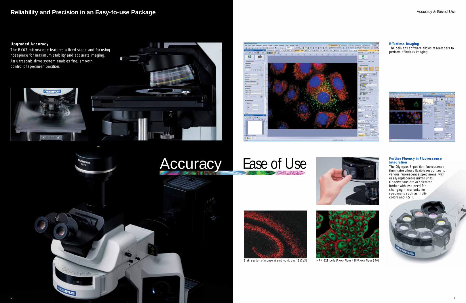
Effortless Imaging The cellSens software allows researchers toperform effortless imaging.
NRK-52E cells (Alexa Fluor 488/Alexa Fluor 546)Brain section of mouse at embryonic day 15 (Cy3)
Further Fluency in FluorescenceIntegration The Olympus 8-position fluorescenceilluminator allows flexible responses tovarious fluorescence specimens, witheasily replaceable mirror units.Observations are acceleratedfurther with less need forchanging mirror units forspecimens such as multi-colors and FISH.
Upgraded AccuracyThe BX63 microscope features a fixed stage and focusingnosepiece for maximum stability and accurate imaging. An ultrasonic drive system enables fine, smooth control of specimen position.
Accuracy Ease of Use
5
7 8
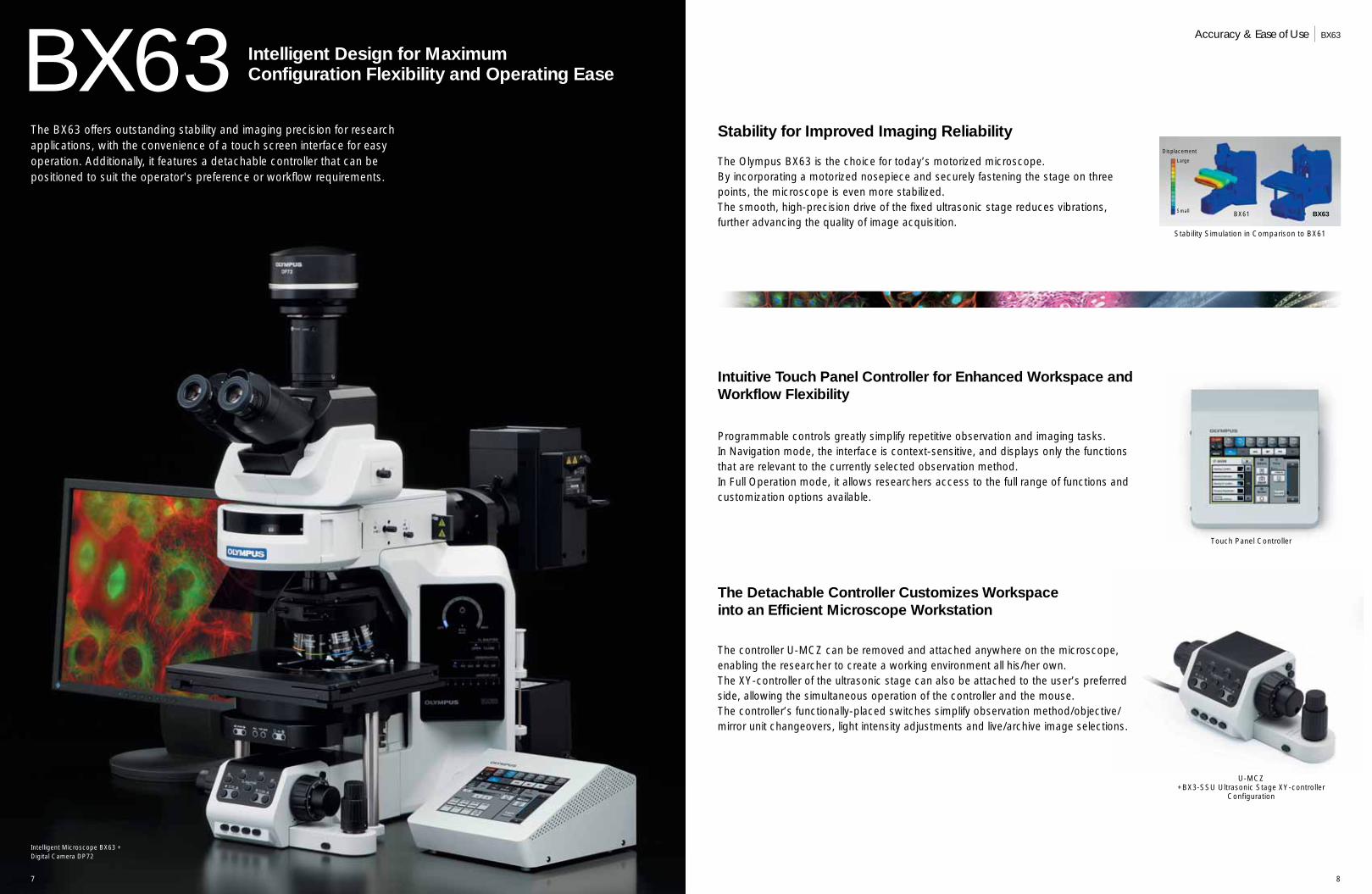
BX63 Intelligent Design for MaximumConfiguration Flexibility and Operating Ease
The BX63 offers outstanding stability and imaging precision for researchapplications, with the convenience of a touch screen interface for easyoperation. Additionally, it features a detachable controller that can bepositioned to suit the operator's preference or workflow requirements.
Programmable controls greatly simplify repetitive observation and imaging tasks.In Navigation mode, the interface is context-sensitive, and displays only the functionsthat are relevant to the currently selected observation method.In Full Operation mode, it allows researchers access to the full range of functions andcustomization options available.
The Olympus BX63 is the choice for today’s motorized microscope.By incorporating a motorized nosepiece and securely fastening the stage on threepoints, the microscope is even more stabilized. The smooth, high-precision drive of the fixed ultrasonic stage reduces vibrations, further advancing the quality of image acquisition.
Intuitive Touch Panel Controller for Enhanced Workspace andWorkflow Flexibility
Stability for Improved Imaging Reliability
The Detachable Controller Customizes Workspace into an Efficient Microscope Workstation
The controller U-MCZ can be removed and attached anywhere on the microscope,enabling the researcher to create a working environment all his/her own. The XY-controller of the ultrasonic stage can also be attached to the user’s preferredside, allowing the simultaneous operation of the controller and the mouse.The controller’s functionally-placed switches simplify observation method/objective/mirror unit changeovers, light intensity adjustments and live/archive image selections.
Accuracy & Ease of Use BX63
Intelligent Microscope BX63 + Digital Camera DP72
Touch Panel Controller
Stability Simulation in Comparison to BX61
U-MCZ+BX3-SSU Ultrasonic Stage XY-controller
Configuration
BX61
Displacement
Large
Small BX63
9 10
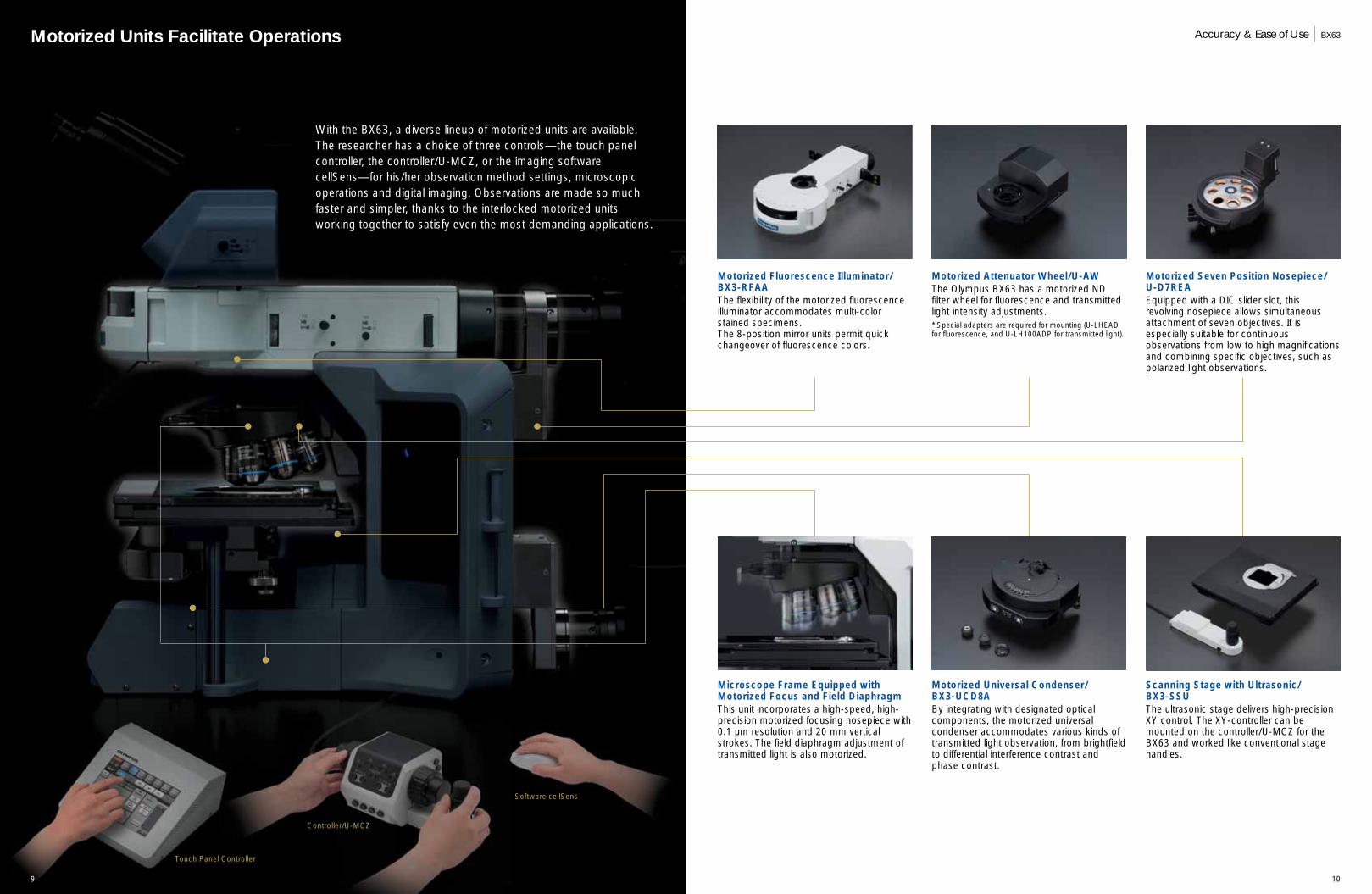
Motorized Fluorescence Illuminator/BX3-RFAAThe flexibility of the motorized fluorescenceilluminator accommodates multi-colorstained specimens. The 8-position mirror units permit quickchangeover of fluorescence colors.
Motorized Attenuator Wheel/U-AWThe Olympus BX63 has a motorized NDfilter wheel for fluorescence and transmittedlight intensity adjustments.* Special adapters are required for mounting (U-LHEAD for fluorescence, and U-LH100ADP for transmitted light).
Touch Panel Controller
Controller/U-MCZ
Software cellSens
Motorized Seven Position Nosepiece/U-D7REAEquipped with a DIC slider slot, thisrevolving nosepiece allows simultaneousattachment of seven objectives. It isespecially suitable for continuousobservations from low to high magnificationsand combining specific objectives, such aspolarized light observations.
Motorized Units Facilitate Operations
Scanning Stage with Ultrasonic/BX3-SSUThe ultrasonic stage delivers high-precisionXY control. The XY-controller can bemounted on the controller/U-MCZ for theBX63 and worked like conventional stagehandles.
Motorized Universal Condenser/BX3-UCD8ABy integrating with designated opticalcomponents, the motorized universalcondenser accommodates various kinds oftransmitted light observation, from brightfieldto differential interference contrast andphase contrast.
With the BX63, a diverse lineup of motorized units are available.The researcher has a choice of three controls—the touch panelcontroller, the controller/U-MCZ, or the imaging softwarecellSens—for his/her observation method settings, microscopicoperations and digital imaging. Observations are made so muchfaster and simpler, thanks to the interlocked motorized unitsworking together to satisfy even the most demanding applications.
Microscope Frame Equipped withMotorized Focus and Field DiaphragmThis unit incorporates a high-speed, high-precision motorized focusing nosepiece with0.1 µm resolution and 20 mm verticalstrokes. The field diaphragm adjustment oftransmitted light is also motorized.
Accuracy & Ease of Use BX63
11 12
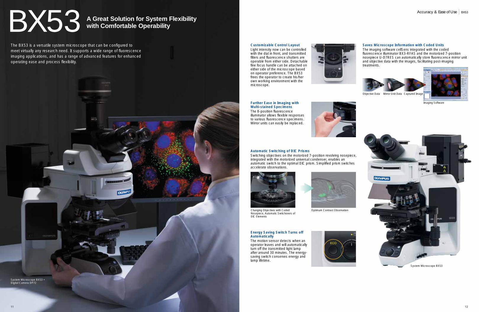
BX53
Energy Saving Switch Turns offAutomaticallyThe motion sensor detects when anoperator leaves and will automaticallyturn off the transmitted light lampafter around 30 minutes. The energy-saving switch conserves energy andlamp lifetime.
Further Ease in Imaging withMulti-stained SpecimensThe 8-position fluorescenceilluminator allows flexible responsesto various fluorescence specimens.Mirror units can easily be replaced.
Automatic Switching of DIC PrismsSwitching objectives on the motorized 7-position revolving nosepiece,integrated with the motorized universal condenser, enables anautomatic switch to the optimal DIC prism. Simplified prism switchesaccelerate observations.
Customizable Control LayoutLight intensity now can be controlledwith the dial in front, and transmittedfilters and fluorescence shutters areoperable from either side. Detachablefine focus handle can be attached oneither side of the microscope basedon operator preference. The BX53frees the operator to create his/herown working environment with themicroscope.
Saves Microscope Information with Coded UnitsThe imaging software cellSens integrated with the codedfluorescence illuminator BX3-RFAS and the motorized 7-positionnosepiece U-D7RES can automatically store fluorescence mirror unitand objective data with the images, facilitating post-imagingtreatments.
A Great Solution for System Flexibility with Comfortable Operability
The BX53 is a versatile system microscope that can be configured tomeet virtually any research need. It supports a wide range of fluorescenceimaging applications, and has a range of advanced features for enhancedoperating ease and process flexibility.
Changing Objectives with CodedNosepiece, Automatic Switchovers ofDIC Elements
Optimum Contrast Observation
Accuracy & Ease of Use BX53
Objective Data Mirror Unit Data Captured Image
Imaging Software
System Microscope BX53 + Digital Camera DP72
System Microscope BX53
13 14
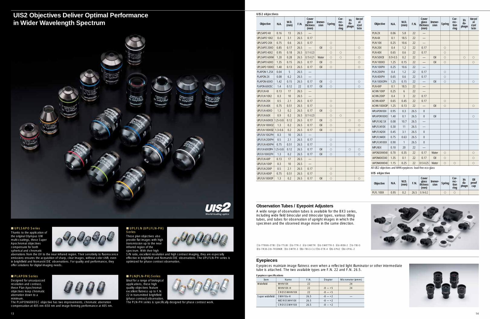
UIS2 Objectives Deliver Optimal Performance in Wider Wavelength Spectrum
■ PLAPON SeriesDesigned for unsurpassedresolution and contrast,these Plan Apochromatobjectives keep chromaticaberration down to aminimum. The PLAPON60XOSC objective has two improvements, chromatic aberrationcompensation at 405 nm–650 nm and image-forming performance at 405 nm.
■ UPLSAPO SeriesThanks to the application ofthe original Olympus UWmulti-coatings, these SuperApochromat objectivescompensate for bothspherical and chromaticaberrations from the UV to the near infrared region. Their sensitivity to fluorescenceemissions ensures the acquisition of sharp, clear images, without color shift, evenin brightfield and Nomarski DIC observations. For quality and performance, theyoffer solutions for digital imaging needs.
■ PLN(PLN-PH) SeriesIdeal for a range of biologicalapplications, these highquality objectives featureexcellent flatness up to F.N.22 in transmitted brightfield(phase contrast) observation.The PLN-PH series is specifically designed for phase contrast work.
■ UPLFLN (UPLFLN-PH)SeriesThese plan objectives alsoprovide flat images with hightransmission up to the nearinfrared region of thespectrum. With their highS/N ratio, excellent resolution and high contrast imaging, they are especiallyeffective in brightfield and Nomarski DIC observations. The UPLFLN-PH series isoptimized for phase contrast observation.
Item Name F.N. Diopter Micrometer (ømm)
Widefield WHN10X 22 24
WHN10X-H 22 -8 — +5 24
CROSSWHN10X 22 -8 — +5
Super widefield SWH10x-H 26.5 -8 — +2 —
MICROSWH10X 26.5 -8 — +2
CROSSSWH10X 26.5 -8 — +2
Cover Cor- Water proof
Objective N.A.W.D.
F.N.glass Immer-
Springrec- and
(mm) thickness sion tion oil proof(mm) ring function
UPLSAPO 4X 0.16 13 26.5 —
UPLSAPO 10X2 0.4 3.1 26.5 0.17
UPLSAPO 20X 0.75 0.6 26.5 0.17 _
UPLSAPO 20XO 0.85 0.17 26.5 — Oil _ _
UPLSAPO 40X2 0.95 0.18 26.5 0.11-0.23 _ _
UPLSAPO 60XW 1.20 0.28 26.5 0.15-0.21 Water _ _ _
UPLSAPO 60XO 1.35 0.15 26.5 0.17 Oil _ _
UPLSAPO 100XO 1.40 0.13 26.5 0.17 Oil _ _
PLAPON 1.25X 0.04 5 26.5 —
PLAPON 2X 0.08 6.2 26.5 —
PLAPON 60XO 1.42 0.15 26.5 0.17 Oil _ _
PLAPON 60XOSC 1.4 0.12 22 0.17 Oil _ _
UPLFLN 4X 0.13 17 26.5 —
UPLFLN 10X2 0.3 10 26.5 —
UPLFLN 20X 0.5 2.1 26.5 0.17 _
UPLFLN 40X 0.75 0.51 26.5 0.17 _
UPLFLN 40XO 1.3 0.2 26.5 0.17 Oil _ _
UPLFLN 60X 0.9 0.2 26.5 0.11-0.23 _ _
UPLFLN 60XOI 1.25-0.65 0.12 26.5 0.17 Oil _ _ _
UPLFLN 100XO2 1.3 0.2 26.5 0.17 Oil _ _
UPLFLN 100XOI2 1.3-0.6 0.2 26.5 0.17 Oil _ _ _
UPLFLN 10X2PH 0.3 10 26.5 —
UPLFLN 20XPH 0.5 2.1 26.5 0.17 _
UPLFLN 40XPH 0.75 0.51 26.5 0.17 _
UPLFLN 60XOIPH 1.25-0.65 0.12 26.5 0.17 Oil _ _ _
UPLFLN 100XO2PH 1.3 0.2 26.5 0.17 Oil _ _
UPLFLN 4XP 0.13 17 26.5 —
UPLFLN 10XP 0.3 10 26.5 —
UPLFLN 20XP 0.5 2.1 26.5 0.17 _
UPLFLN 40XP 0.75 0.51 26.5 0.17 _
UPLFLN 100XOP 1.3 0.2 26.5 0.17 Oil _ _
Cover Cor- Water proof
Objective N.A.W.D.
F.N.glass Immer-
Springrec- and
(mm) thickness sion tion oil proof(mm) ring function
PLN 2X 0.06 5.8 22 —
PLN 4X 0.1 18.5 22 —
PLN 10X 0.25 10.6 22 —
PLN 20X 0.4 1.2 22 0.17 _
PLN 40X 0.65 0.6 22 0.17 _
PLN 50XOI 0.9-0.5 0.2 22 — Oil _ _ _
PLN 100XO 1.25 0.15 22 — Oil _ _
PLN 10XPH 0.25 10.6 22 —
PLN 20XPH 0.4 1.2 22 0.17 _
PLN 40XPH 0.65 0.6 22 0.17 _
PLN 100XOPH 1.25 0.15 22 — Oil _ _
PLN 4XP 0.1 18.5 22 —
ACHN 10XP 0.25 6 22 —
ACHN 20XP 0.4 3 22 0.17
ACHN 40XP 0.65 0.45 22 0.17 _
ACHN 100XOP 1.25 0.13 22 — Oil _ _
MPLAPON100X 0.95 0.3 26.5 0
MPLAPON100XO 1.40 0.1 26.5 0 Oil _
MPLFLN2.5X 0.08 10.7 26.5 —
MPLFLN10X 0.30 11 26.5 —
MPLFLN20X 0.45 3.1 26.5 0
MPLFLN40X 0.75 0.63 26.5 0
MPLFLN100X 0.90 1 26.5 0
MPLN5X 0.10 20 22 —
UAPON20XW340 0.70 0.35 22 0.17 Water _ _
UAPON40XO340 1.35 0.1 22 0.17 Oil _ _
UAPON40XW340 1.15 0.25 22 0.13-0.25 Water _ _ _
Cover Cor-
Objective N.A.W.D.
F.N.glass Immer-
Springrec-
(mm) thickness sion tion(mm) ring
PLFL 100X 0.95 0.2 26.5 0.14-0.2 _ _
UIS2 objectives
UIS objective
A wide range of observation tubes is available for the BX3 series,including wide field binocular and trinocular types, various tiltingtubes, and tubes for observation of upright images in which thespecimen and the observed image move in the same direction.
Observation Tubes / Eyepoint Adjusters
Eyepieces maintain image flatness even when a reflected light illuminator or other intermediatetube is attached. The two available types are F.N. 22 and F.N. 26.5.
Eyepieces
Eyepiece specifications
q
we r
t
y
u
i
o
!0!1 !2
!3
qU-TTBI/U-ETBI wU-TTLBI eU-TTR-2 rU-SWETR tU-SWETTR-5 yU-BI30-2 uU-TBI-3
iU-TR30-2/U-TR30NIR oU-SWTR-3 !0U-TBI-3-CLI !1U-ETR-4 !2U-EPA2 !3U-EPAL-2
All UIS2 objectives and WHN eyepieces: lead-free eco-glass
Irisdia-
phragm
Irisdia-
phragm
Irisdia-
phragm
Oilproofcap
15 16
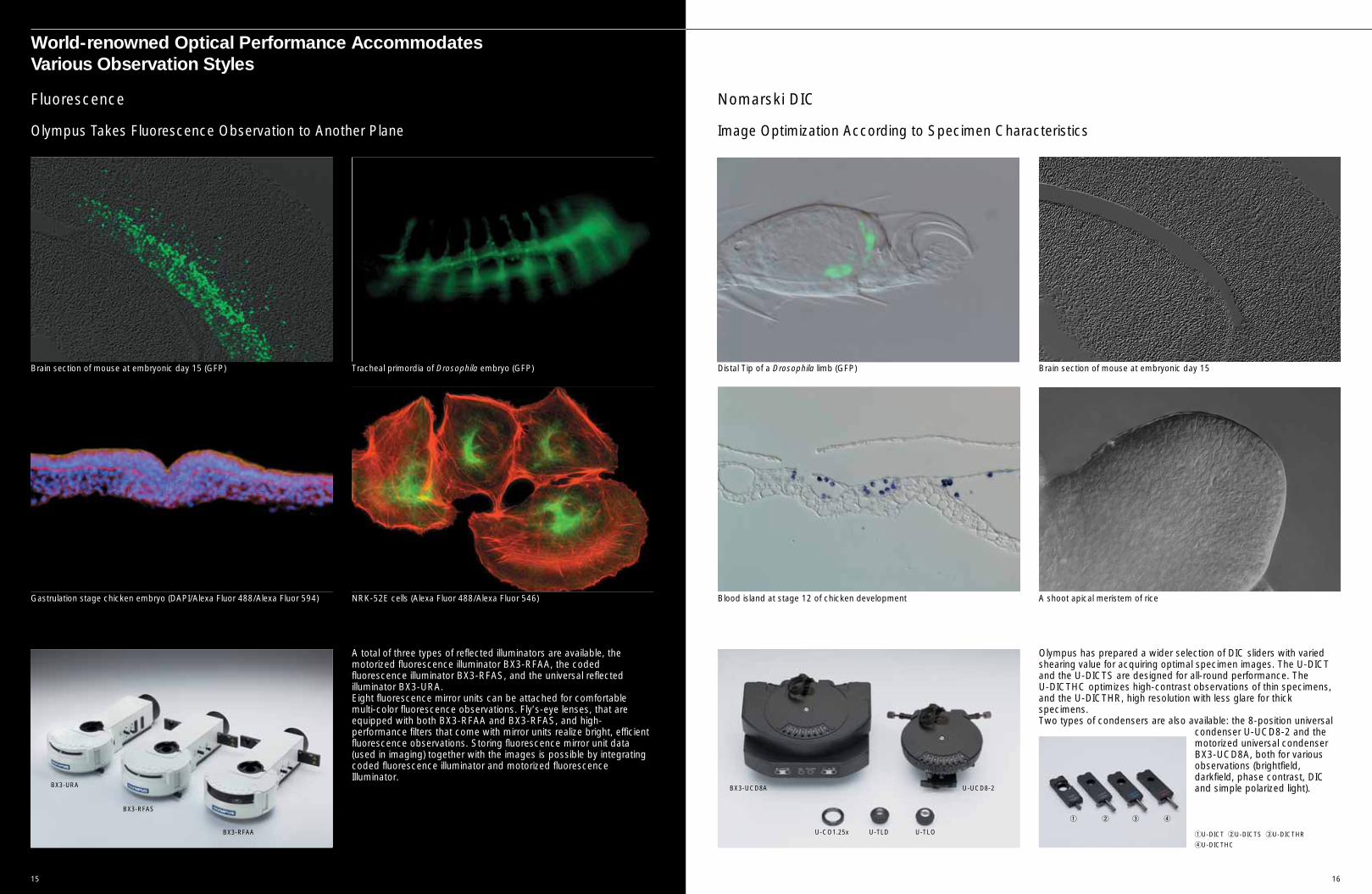
Olympus Takes Fluorescence Observation to Another Plane Image Optimization According to Specimen Characteristics
Brain section of mouse at embryonic day 15 (GFP) Tracheal primordia of Drosophila embryo (GFP)
NRK-52E cells (Alexa Fluor 488/Alexa Fluor 546)Gastrulation stage chicken embryo (DAPI/Alexa Fluor 488/Alexa Fluor 594)
Fluorescence
Olympus has prepared a wider selection of DIC sliders with variedshearing value for acquiring optimal specimen images. The U-DICTand the U-DICTS are designed for all-round performance. The U-DICTHC optimizes high-contrast observations of thin specimens,and the U-DICTHR, high resolution with less glare for thickspecimens. Two types of condensers are also available: the 8-position universal
condenser U-UCD8-2 and themotorized universal condenserBX3-UCD8A, both for variousobservations (brightfield,darkfield, phase contrast, DICand simple polarized light).
Distal Tip of a Drosophila limb (GFP) Brain section of mouse at embryonic day 15
A shoot apical meristem of riceBlood island at stage 12 of chicken development
Nomarski DIC
A total of three types of reflected illuminators are available, themotorized fluorescence illuminator BX3-RFAA, the codedfluorescence illuminator BX3-RFAS, and the universal reflectedilluminator BX3-URA. Eight fluorescence mirror units can be attached for comfortablemulti-color fluorescence observations. Fly’s-eye lenses, that areequipped with both BX3-RFAA and BX3-RFAS, and high-performance filters that come with mirror units realize bright, efficientfluorescence observations. Storing fluorescence mirror unit data(used in imaging) together with the images is possible by integratingcoded fluorescence illuminator and motorized fluorescenceIlluminator.
World-renowned Optical Performance Accommodates Various Observation Styles
BX3-RFAA
BX3-RFAS
BX3-URA BX3-UCD8A
U-CO1.25x U-TLD U-TLO
U-UCD8-2
q w e r
qU-DICT wU-DICTS eU-DICTHR
rU-DICTHC
17 18
Spirogyra
Water Flea
Polarized Light
High-quality Darkfield Effect at All Magnifications
Two darkfield condensers are provided: the dry darkfield condenser(U-DCD), for magnifications from 10x to 100x (up to N.A. 0.80); andthe oil immersion darkfield condenser (U-DCW), for magnificationsfrom 20x to 100x (up to N.A. 1.2).* Please consult your nearest Olympus representative for applicable objectives.
Tooth, bone, muscle tissue, nerve tissue, actomyosin fiber and mitoticspindle can all be observed, without staining. There are intermediateattachments (U-OPA/U-CPA) for orthoscopic andorthoscopic/conoscopic viewing.Various compensators make itpossible to observe a wide range ofretardation. Also available is acondenser exclusively for polarizationobservation, revolving nosepiece,rotating stage, objectives and simplepolarizing attachment.
Darkfield
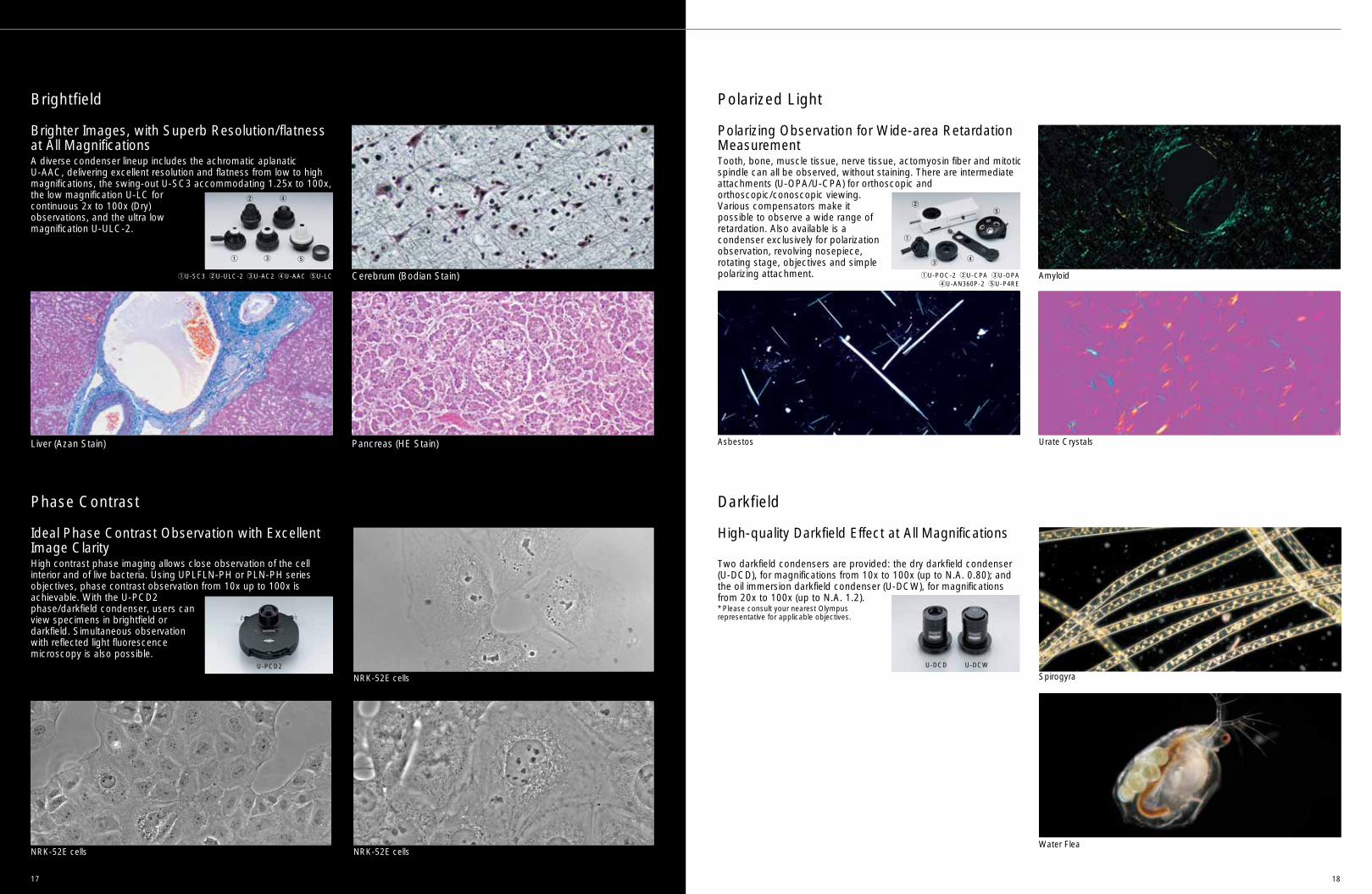
Cerebrum (Bodian Stain)
Liver (Azan Stain) Pancreas (HE Stain)
Amyloid
Urate CrystalsAsbestos
Brighter Images, with Superb Resolution/flatnessat All Magnifications
Brightfield
A diverse condenser lineup includes the achromatic aplanatic U-AAC, delivering excellent resolution and flatness from low to highmagnifications, the swing-out U-SC3 accommodating 1.25x to 100x,the low magnification U-LC forcontinuous 2x to 100x (Dry)observations, and the ultra lowmagnification U-ULC-2.
NRK-52E cells
NRK-52E cells NRK-52E cells
Ideal Phase Contrast Observation with ExcellentImage ClarityHigh contrast phase imaging allows close observation of the cellinterior and of live bacteria. Using UPLFLN-PH or PLN-PH seriesobjectives, phase contrast observation from 10x up to 100x isachievable. With the U-PCD2phase/darkfield condenser, users canview specimens in brightfield ordarkfield. Simultaneous observationwith reflected light fluorescencemicroscopy is also possible.
Phase Contrast
qU-SC3 wU-ULC-2 eU-AC2 rU-AAC tU-LC
U-DCD U-DCW
qU-POC-2 wU-CPA eU-OPA rU-AN360P-2 tU-P4RE
q
w
er
t
Polarizing Observation for Wide-area RetardationMeasurement
q
w
e
r
t
U-PCD2
High-speed, High-definition Image Capture Provides Smooth Fluorescence Imaging
High-definition, High-resolution 5-million Pixel Technology
The Optimal Stand-alone Model for Conferences
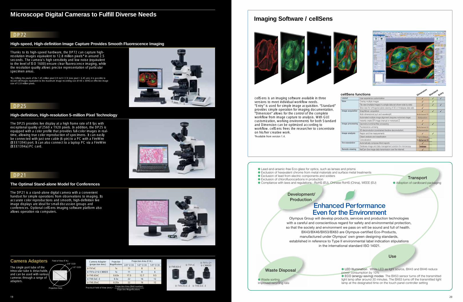
Microscope Digital Cameras to Fulfill Diverse Needs
DP72
DP25
DP21
Thanks to its high-speed hardware, the DP72 can capture high-resolution images equivalent to 12.8 million pixels* in around 2.5seconds. The camera's high sensitivity and low noise (equivalentto the level of ISO 1600) ensure clear fluorescence imaging, whilethe resolution quality allows precise representation of particularspecimen areas.
*By shifting the pixels of the 1.45 million pixel 2/3 inch CCD (one pixel = 6.45 um), it is possible torecord still images equivalent to the maximum image recording size (4140 x 3096) or effective imagesize of 12.8 million pixels.
The DP25 provides live display at a high frame rate of 8 fps withexceptional quality of 2560 x 1920 pixels. In addition, the DP25 isequipped with a color profile that provides full-color images in real-time, allowing true color reproduction of specimens. It can easilybe connected with just one cable (6-pin) to a PC with a FireWire(IEEE1394) port. It can also connect to a laptop PC via a FireWire(IEEE1394a) PC card.
The DP21 is a stand-alone digital camera with a convenienthandset for simple operations from observations to imaging. Itsaccurate color reproductions and smooth, high-definition liveimage displays are ideal for small discussion groups andconferences. Optional cellSens imaging software platform alsoallows operation via computers.
2019
Imaging Software / cellSens
cellSens is an imaging software available in threeversions to meet individual workflow needs. "Entry" is used for simple image acquisition. “Standard”provides simple operation for imaging documentation.“Dimension” allows for the control of the completeworkflow from image capture to analysis. With GUIcustomization, working environments for both Standardand Dimension can be optimized according toworkflow. cellSens frees the researcher to concentrateon his/her creative work.*Available from version 1.4.
The single port tube of thetrinocular tube is detachable,and can be used with variouscameras through a range ofadapters.
Camera Adapters
Practical Field of View (mm) =Projection Area (field number)
Objective Magnifications
2/3" CCD 1/2" CCD 1/3" CCDU-TV1xC 1x 11 8 6
U-TV1x-2+U-CMAD3 1x 11 8 6
U-TV0.63xC 0.63x 17.5 12.7 9.5
U-TV0.5xC-3 0.5x 22 16 12
U-TV0.35xC 0.35x 22 16 12
Camera Adapter(projection lens)
Projection Area (F.N.)ProjectionMagnifications
U-TV0.63×C
U-TV0.35×C-2 U-TV0.5×C-3
U-TV1×CU-TV1×-2+U-CMAD3
LayoutView
Image acquisition
Image analysis
Documentation
Remote viewing
User experience customization
Tile view (multiple images in a single data set shown side by side)
Snap/Movie acquisitionMulti-dimensional (xyzt and wavelength)
Instantly create EFI image (manual or motorized Z)
Fluorescence unmixing3D deconvolution (constrained iterative deconvolution)Region and line measurementsObject analysis and classification
Automatically compose Word reports
NetCam (live image data transmission over the internet)
cellSens functions EntryDim
ension
Standard
✓
✓
✓
Multichannel 5D
✓
✓
Multichannel 5D
CI Deconvolution
✓
Count & Measure
✓
✓
✓
✓
✓
✓
✓
✓
✓
2221
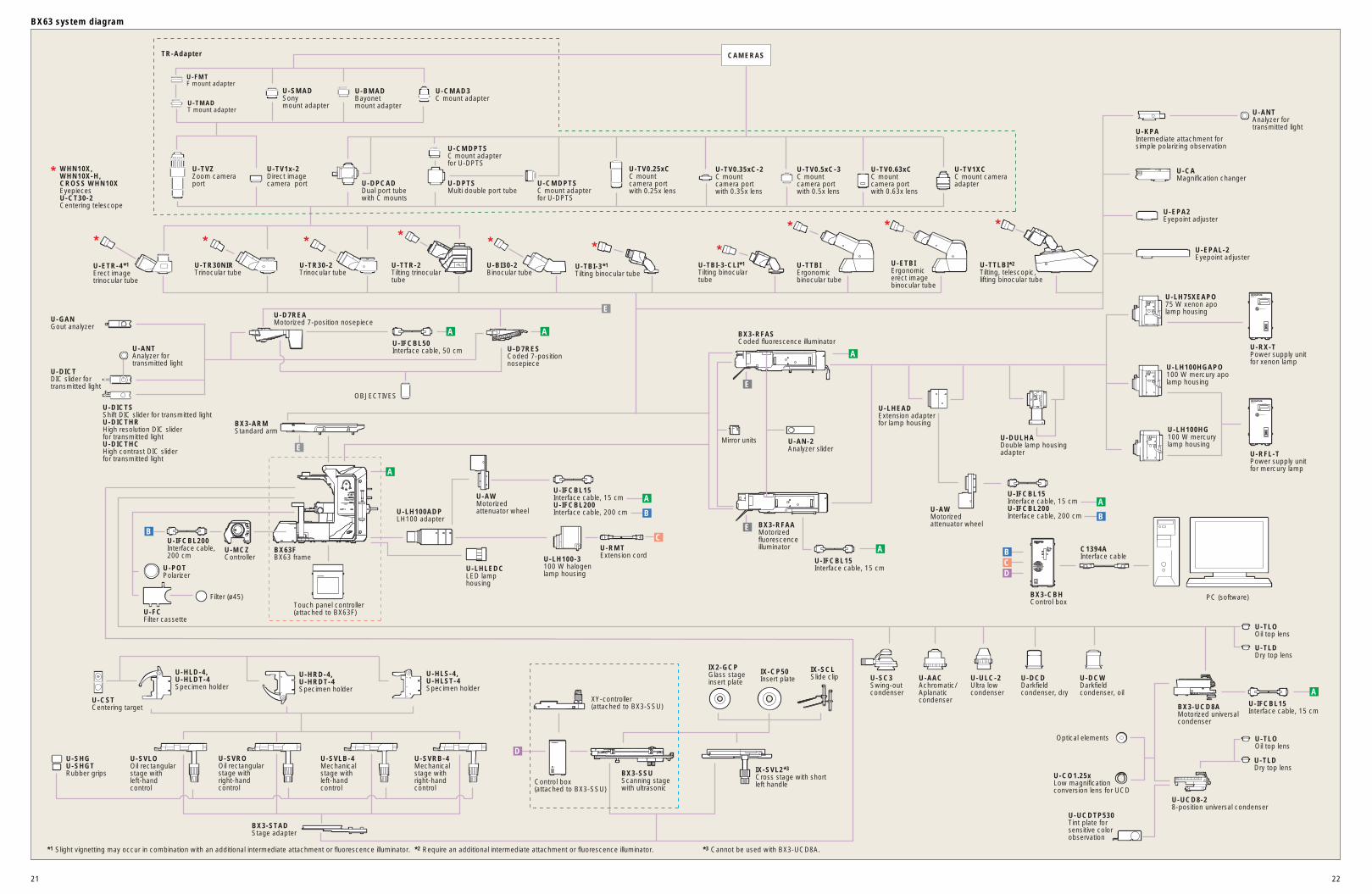
BX63 system diagram
LIFE TIME
BURNER ON
U-RFL-T
LIFE TIME
BURNER ON
U-RFL-T
Mirror units
D
D
E
E
B
BB
C
A
A
A
A A
A
A
A
C
E
E
B
U-FCFilter cassette
Filter (ø45) PC (software)
U-TVZZoom camera port
U-TV1x-2Direct image camera port
U-TV0.63xCC mount camera port with 0.63x lens
U-TV1XCC mount camera adapter
U-TV0.5xC-3C mount camera port with 0.5x lens
U-TV0.35xC-2C mount camera port with 0.35x lens
U-TV0.25xCC mount camera port with 0.25x lens
U-CMAD3C mount adapter
U-BMADBayonetmount adapter
U-LH100-3100 W halogen lamp housing
U-AN-2Analyzer slider
BX3-RFASCoded fluorescence illuminator
U-D7REAMotorized 7-position nosepiece
U-IFCBL50Interface cable, 50 cmU-ANT
Analyzer fortransmitted light
U-DICTDIC slider fortransmitted light
OBJECTIVES
U-D7RESCoded 7-position nosepiece
U-HLS-4, U-HLST-4Specimen holder
U-HLD-4, U-HLDT-4Specimen holder
U-HRD-4, U-HRDT-4Specimen holder
U-POTPolarizer
U-AACAchromatic/Aplanaticcondenser
U-SC3Swing-out condenser
U-ULC-2Ultra low condenser
U-DCDDarkfieldcondenser, dry
U-DCWDarkfieldcondenser, oil
BX63FBX63 frame
Touch panel controller(attached to BX63F)
U-MCZController
U-LH100ADPLH100 adapter
U-LHLEDCLED lamp housing
BX3-ARMStandard arm
U-TR30-2Trinocular tube
U-TR30NIRTrinocular tube
U-BI30-2Binocular tube
U-TBI-3*1
Tilting binocular tubeU-TBI-3-CLI*1
Tilting binocular tube
U-ETBIErgonomic erect image binocular tube
U-TTLBI*2
Tilting, telescopic, lifting binocular tube
U-TTBIErgonomic binocular tube
U-FMTF mount adapter
TR-Adapter
U-TMADT mount adapter
U-SMADSonymount adapter
U-CAMagnification changer
U-KPAIntermediate attachment forsimple polarizing observation
U-ANTAnalyzer for transmitted light
U-EPA2Eyepoint adjuster
U-EPAL-2Eyepoint adjuster
U-CST Centering target
U-TLOOil top lens
U-TLDDry top lens
U-TLOOil top lens
U-TLDDry top lens
U-UCD8-28-position universal condenser
BX3-UCD8AMotorized universal condenser
U-CO1.25xLow magnification conversion lens for UCD
Optical elements
U-DICTSShift DIC slider for transmitted light U-DICTHRHigh resolution DIC slider for transmitted light U-DICTHCHigh contrast DIC sliderfor transmitted light
U-SVROOil rectangularstage with right-hand control
U-SVLOOil rectangular stage with left-hand control
U-SVRB-4Mechanical stage withright-handcontrol
U-SVLB-4Mechanical stage withleft-handcontrol
BX3-STADStage adapter
U-ETR-4*1
Erect imagetrinocular tube
U-TTR-2Tilting trinoculartube
U-LH100HG100 W mercury lamp housing
U-LH75XEAPO75 W xenon apo lamp housing
U-LH100HGAPO100 W mercury apo lamp housing
BX3-RFAAMotorized fluorescence illuminator
U-GANGout analyzer
*1 Slight vignetting may occur in combination with an additional intermediate attachment or fluorescence illuminator. *2 Require an additional intermediate attachment or fluorescence illuminator. *3 Cannot be used with BX3-UCD8A.
U-SHGU-SHGTRubber grips
CAMERAS
U-DPTSMulti double port tube
U-DPCADDual port tube with C mounts
U-CMDPTSC mount adapter for U-DPTS
U-CMDPTSC mount adapter for U-DPTS
U-DULHADouble lamp housing adapter
WHN10X, WHN10X-H, CROSS WHN10XEyepiecesU-CT30-2Centering telescope
U-RX-TPower supply unit for xenon lamp
U-RFL-TPower supply unit for mercury lamp
U-AWMotorized attenuator wheel
U-IFCBL15Interface cable, 15 cmU-IFCBL200Interface cable, 200 cm
U-IFCBL200Interface cable, 200 cm
U-LHEADExtension adapter for lamp housing
U-AWMotorized attenuator wheel
Control box(attached to BX3-SSU)
XY-controller(attached to BX3-SSU)
BX3-CBH Control box
IX-SVL2*3
Cross stage with short left handle
BX3-SSUScanning stage with ultrasonic
IX2-GCPGlass stage insert plate
IX-CP50Insert plate
IX-SCLSlide clip
U-RMTExtension cord
U-IFCBL15Interface cable, 15 cmU-IFCBL200Interface cable, 200 cm
U-IFCBL15Interface cable, 15 cm
U-IFCBL15Interface cable, 15 cm
C1394AInterface cable
U-UCDTP530 Tint plate for sensitive color observation
2423
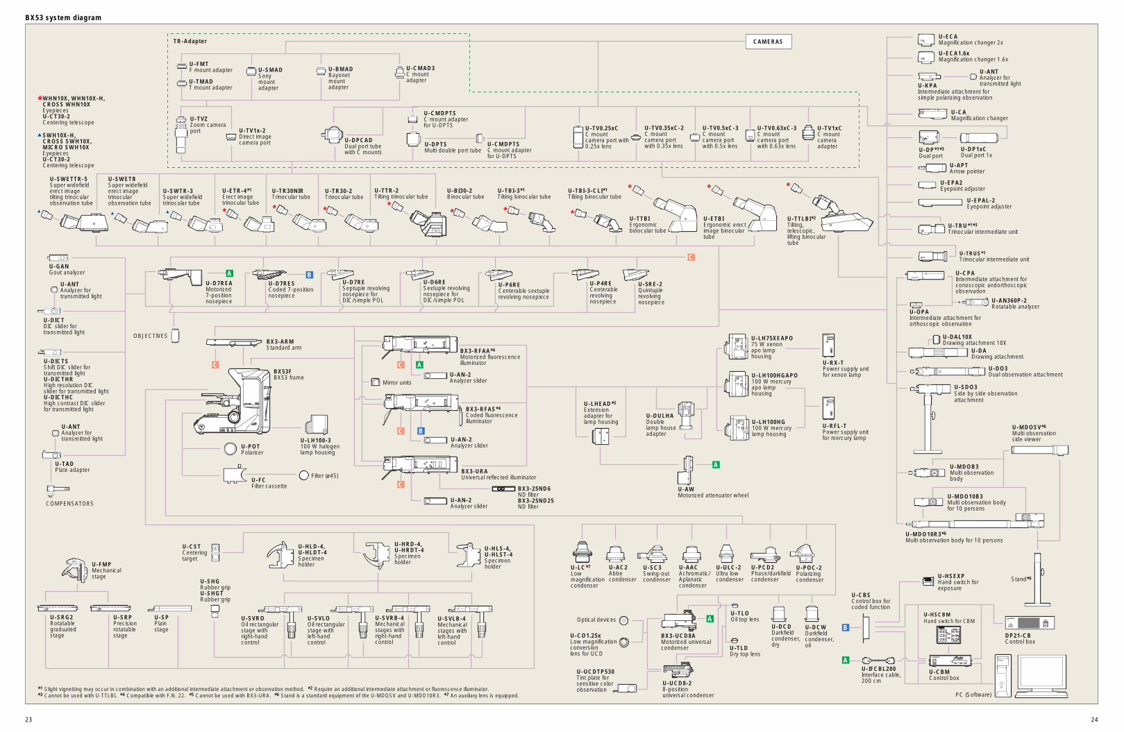
BX53 system diagram
* * *
*
* * * *
**
LIFE TIME
BURNER ON
U-RFL-T
LIFE TIME
BURNER ON
U-RFL-T
U-SRPPrecision rotatable stage
A
A
AB
BA
C
ACC
C
C
B
U-HLS-4, U-HLST-4Specimen holder
U-HLD-4, U-HLDT-4Specimen holder
U-HRD-4, U-HRDT-4Specimen holder
U-CST Centeringtarget
U-SPPlain stage
U-FMPMechanical stage
U-SVROOil rectangularstage with right-hand control
U-SVLOOil rectangular stage with left-hand control
U-SVRB-4Mechanical stages withright-handcontrol
U-SVLB-4Mechanical stages withleft-handcontrol
U-SHGRubber grip U-SHGTRubber grip
U-SRG2 Rotatable graduated stage U-CO1.25x
Low magnification conversionlens for UCD
U-PCD2Phase/darkfield condenser
U-POC-2Polarizingcondenser
U-AACAchromatic/Aplanaticcondenser
U-AC2Abbe condenser
U-SC3Swing-out condenser
U-ULC-2Ultra low condenser
U-DCDDarkfieldcondenser,dry
U-DCWDarkfieldcondenser,oil
U-TLOOil top lens
U-TLDDry top lens
U-UCD8-28-positionuniversal condenser
Optical devices
U-UCDTP530 Tint plate for sensitive color observation
BX3-UCD8AMotorized universal condenser
U-LC*7
Low magnificationcondenser
U-CBSControl box forcoded function
U-HSEXPHand switch forexposure
U-CBMControl box
U-HSCBMHand switch for CBM
*1 Slight vignetting may occur in combination with an additional intermediate attachment or observation method. *2 Require an additional intermediate attachment or fluorescence illuminator.*3 Cannot be used with U-TTLBI. *4 Compatible with F.N. 22. *5 Cannot be used with BX3-URA. *6 Stand is a standard equipment of the U-MDOSV and U-MDO10R3. *7 An auxiliary lens is equipped.
U-TRUS*1
Trinocular intermediate unit
U-SWETRSuper widefield erect image trinocular observation tube
U-SWETTR-5Super widefield erect image tilting trinocular observation tube
U-IFCBL200Interface cable, 200 cm
* WHN10X, WHN10X-H,
CROSS WHN10X Eyepieces U-CT30-2 Centering telescope ▲ SWH10X-H, CROSS SWH10X, MICRO SWH10X Eyepieces U-CT30-2 Centering telescope
U-TBI-3-CLI*1
Tilting binocular tube
U-CMAD3C mountadapter
U-BMADBayonetmountadapter
U-FMTF mount adapter
U-TMADT mount adapter
U-SMADSonymountadapter
CAMERAS
U-TV1xCC mount camera adapter
U-TVZZoom camera port U-TV1x-2
Direct image camera port
U-TV0.63xC-3C mountcamera port with 0.63x lens
U-TV0.5xC-3C mountcamera port with 0.5x lens
U-TV0.35xC-2C mountcamera port with 0.35x lens
U-TV0.25xCC mountcamera port with0.25x lensU-DPTS
Multi double port tube
U-DPCADDual port tube with C mounts
U-CMDPTSC mount adapter for U-DPTS
U-CMDPTSC mount adapter for U-DPTS
U-TTR-2Tilting trinocular tube
U-TTLBI*2
Tilting, telescopic, lifting binocular tube
U-SWTR-3Super widefieldtrinocular tube
U-TR30NIRTrinocular tube
U-TR30-2Trinocular tube
U-BI30-2Binocular tube
U-ETBIErgonomic erect image binocular tube
U-TTBIErgonomic binocular tube
U-ETR-4*1
Erect imagetrinocular tube
U-TBI-3*1
Tilting binocular tube
TR-Adapter
U-D7RESCoded 7-positionnosepiece
U-D7REAMotorized 7-positionnosepiece
U-P4RECenterable revolvingnosepiece
U-P6RECenterable sextuple revolving nosepiece
U-D6RESextuple revolvingnosepiece forDIC/simple POL
U-D7RESeptuple revolvingnosepiece forDIC/simple POL
U-5RE-2Quintuplerevolvingnosepiece
U-ANTAnalyzer fortransmitted light
U-DICTDIC slider fortransmitted light
U-DICTSShift DIC slider fortransmitted light U-DICTHRHigh resolution DICslider for transmitted light U-DICTHCHigh contrast DIC sliderfor transmitted light
U-GANGout analyzer
U-TADPlate adapter
COMPENSATORS
U-ANTAnalyzer fortransmitted light
OBJECTIVES
U-POTPolarizer
U-FCFilter cassette
Filter (ø45)
BX53FBX53 frame
U-LH100-3100 W halogenlamp housing
U-AWMotorized attenuator wheel
BX3-ARMStandard arm
U-AN-2Analyzer slider
U-AN-2Analyzer slider
U-AN-2Analyzer slider
BX3-URAUniversal reflected illuminator
BX3-RFAS*4
Coded fluorescence illuminator
BX3-RFAA*4
Motorized fluorescence illuminator
BX3-25ND6ND filterBX3-25ND25ND filter
Mirror units
U-ECA1.6xMagnification changer 1.6x
U-ECAMagnification changer 2x
U-TRU*1*3
Trinocular intermediate unit
U-CAMagnification changer
U-KPAIntermediate attachment forsimple polarizing observation
U-ANTAnalyzer fortransmitted light
U-EPA2Eyepoint adjuster
U-EPAL-2Eyepoint adjuster
U-APTArrow pointer
U-DP*1*3
Dual port U-DP1xCDual port 1x
U-CPAIntermediate attachment for conoscopic andorthoscopic observation
U-AN360P-2Rotatable analyzer
U-OPAIntermediate attachment fororthoscopic observation
U-DO3Dual observation attachment
U-DADrawing attachment
U-DAL10XDrawing attachment 10X
U-SDO3Side by side observationattachment
U-MDO10B3Multi observation body for 10 persons
U-MDOB3Multi observation body
U-MDOSV*6
Multi observation side viewer
Stand*6
U-MDO10R3*6
Multi observation body for 10 persons
U-LH100HG100 W mercury lamp housing
U-LH75XEAPO75 W xenon apo lamp housing
U-LH100HGAPO100 W mercury apo lamp housing
U-RX-TPower supply unit for xenon lamp
U-RFL-TPower supply unit for mercury lamp
U-DULHADouble lamp house adapter
U-LHEAD*5
Extension adapter for lamp housing
PC (Software)
DP21-CBControl box
2625
Microscope frame Optical system UIS2 optical system
Focus Built-in motorized nosepiece focusStroke: 20 mm, minimum increment: 0.01 µm, maximum nosepiece movement speed: 3 mm/s
Illuminator Built-in Koehler illumination for transmitted light, Light intensity LED indicator, Built-in motorized field stop• High color reproductivity LED light source •12 V 100 W halogen bulb (pre-centered)
Revolving nosepiece • Motorized septuple revolving nosepiece • Interchangeable reversed coded septuple nosepiece
Observation tube Widefield (F.N. 22) • Widefield tilting trinocular • Widefield trinocular • Widefield erect image trinocular • Widefield tilting binocular • Widefield tilting,Telescopic,Lifting binocular tube • Widefield ergo binocular • Widefield binocular
Stage • Ultrasonic stage (Stage stroke: X:76 mm x Y: 52 mm, maximum stage movement speed: 30 mm/s
• Ceramic-coated coaxial stage with left or right hand low drive control: with rotating mechanism and torque adjustment mechanism, optional rubber grips available
• Cross stage with short left handle
Condenser • Motorized universal condenser (N.A. 0.9, motorized 8-position turret, Aperture stop, polarizing filter in/out mechanism and top lens swing out mechanism), for 1.25x–100x [swing-out 1.25x-4x, with oil top lens: (N.A. 1.4)]
• Swing out Achromatic (N.A. 0.9), for 1.25x–100x (swing-out: 1.25x–4x)• Achromatic Aplanatic (N.A. 1.4), for 10x–100x• Universal (N.A. 0.9), for 1.25x–100x [swing-out: 1.25x–4x, with oil top lens:(N.A. 1.4)]• Ultra low (N.A. 0.16), for 1.25x–4x• Darkfield dry (N.A. 0.8–0.92), for 10x–100x• Darkfield oil (N.A. 1.20–1.40), for 10x–100x
ND filter wheel • Motorized 6-position ND filter wheel
Fluorescence illuminator • Motorized multi-purpose coded type (F.N. 22, motorized 8-position mirror unit turret, 4-position ND slider)• Multi-purpose coded type (F.N. 22, 8-position mirror unit turret, 4-position ND slider)
Fluorescence light source • 100 W Hg apo lamp housing and transformer• 100 W Hg lamp housing and transformer• 75 W Xe lamp housing and transformer
Controller • High-performance control box ( I/F: FireWire)
*This device is designed for use in industrial environments for the EMC performance (EC61326-1 Class A device). Using it in a residential environment may affect other equipment in the environment.
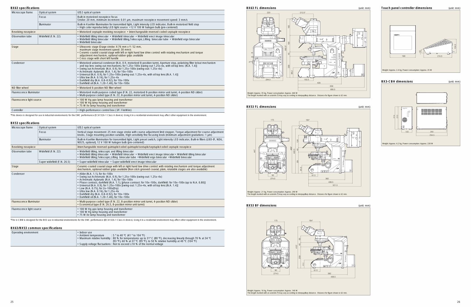
BX63 specifications
Microscope frame Optical system UIS2 optical system
Focus Vertical stage movement: 25 mm stage stroke with coarse adjustment limit stopper, Torque adjustment for coarse adjustmentknobs, Stage mounting position variable, High sensitivity fine focusing knob (minimum adjustment gradations: 1 µm)
Illuminator Built-in Koehler illumination for transmitted light, Light preset switch, Light intensity LED indicator, Built-in filters (LBD-IF, ND6,ND25, optional), 12 V 100 W halogen bulb (pre-centered)
Revolving nosepiece Interchangeable reversed quintuple/coded quintuple/sextuple/septuple/coded septuple nosepiece
Observation tube Widefield (F.N. 22) • Widefield tilting, telescopic and lifting binocular • Widefield tilting trinocular • Widefield trinocular • Widefield erect image trinocular • Widefield tilting binocular• Widefield tilting,Telescopic,Lifting binocular tube •Widefield ergo binocular •Widefield binocular
Super widefield (F.N. 26.5) • Super widefield trinocular • Super widefield erect image trinocular
Stage Ceramic-coated coaxial stage with left or right hand low drive control: with rotating mechanism and torque adjustmentmechanism, optional rubber grips available (Non stick grooved coaxial, plain, rotatable stages are also available)
Condenser • Abbe (N.A. 1.1), for 4x–100x• Swing out Achromatic (N.A. 0.9), for 1.25x–100x (swing-out: 1.25x–4x)• Achromatic Aplanatic (N.A. 1.4), for 10x–100x• Phase contrast, darkfield (N.A. 1.1), [phase contrast: for 10x–100x, darkfield: for 10x–100x (up to N.A. 0.80)]• Universal (N.A. 0.9), for 1.25x–100x [swing-out: 1.25x–4x, with oil top lens:(N.A. 1.4)]• Low (N.A. 0.75), for 2x–100x(Dry)• Ultra low (N.A. 0.16), for 1.25x–4x• Darkfield dry (N.A. 0.8–0.92), for 10x–100x• Darkfield oil (N.A. 1.20–1.40), for 10x–100x
Fluorescence illuminator • Multi-purpose coded type (F.N. 22, 8-position mirror unit turret, 4-position ND slider)• Economical type (F.N. 26.5, 8-position mirror unit turret)
Fluorescence light source • 100 W Hg apo lamp housing and transformer• 100 W Hg lamp housing and transformer• 75 W Xe lamp housing and transformer
*The U-CBM is designed for the BX3 use in industrial environments for the EMC performance (IEC61326-1 Class A device). Using it in a residential environment may affect other equipment in the environment.
BX53 specifications
Operating environment • Indoor use• Ambient temperature : 5 º to 40 ºC (41 º to 104 ºF)• Maximum relative humidity : 80 % for temperatures up to 31º C (88 ºF), decreasing linearly through 70 % at 34 ºC
(93 ºF), 60 % at 37 ºC (99 ºF), to 50 % relative humidity at 40 ºC (104 ºF)• Supply voltage fluctuations : Not to exceed ±10 % of the normal voltage
BX63/BX53 common specifications
209
4516
416
0
582.
5*
152.5
429
212.5*
656
680.5
294.5
220
96.2
OPEN
OFF
DF
UNIT
8
TURRET POL. TOP LENS AS
3
MAXECOmode
CLOSE
FL SHUTTER
FS AS
OFFECOmode
MAX
FL SHUTTER
OPEN
OBSERVATION
FL MIRROR UNIT
1 2 3 4 5 6 7 8
FL
OLYMPUS
PH DIC BF PO DF
CLOSE
190 210.5
104.
5
25––
Weight: Approx. 35 kg, Power consumption: Approx. 440 WThe length marked with an asterisk (*) may vary according to interpupillary distance. Distance for figure shown is 62 mm.
Weight: Approx. 2.4 kg, Power consumption: Approx. 25 W
81.5
209
4565
410*
184*
362
91.5
436.5
175
90
274.5
Weight: Approx. 16 kg, Power consumption: Approx. 140 WThe length marked with an asterisk (*) may vary according to interpupillary distance. Distance for figure shown is 62 mm.
SHUTTER
81.5
209
4512
4
440.
5469*
91.5
362
436.5
521.5
175
447
90
274.5
184*
FS AS
Weight: Approx 21 kg, Power consumption: Approx. 290 WThe length marked with an asterisk (*) may vary according to interpupillary distance. Distance for figure shown is 62 mm.
BX63 FL dimensions (unit: mm)
BX53 FL dimensions (unit: mm)
Touch panel controller dimensions (unit: mm)
100
210
214
280
294.5
BX3-CBH dimensions (unit: mm)
BX53 BF dimensions (unit: mm)
Weight: Approx. 4.2 kg, Power consumption: Approx. 230 W
Recommended