Synthesis, surface characterization and electrokinetic propertiesof colloidal silica nanoparticles with magnetic core
Karolina Gdula1 • Andrzej Dabrowski1 • Ewa Skwarek2
Received: 30 October 2015 / Revised: 21 December 2015 / Accepted: 30 December 2015 / Published online: 7 January 2016
� The Author(s) 2016. This article is published with open access at Springerlink.com
Abstract In order to obtain functionalized silica colloids
with magnetic properties, the three-step reaction, was car-
ried out. The aqueous suspension of iron oxide nanoparti-
cles as magnetic core (maghemite—c-Fe2O3) was
synthesized according to the Massart’s method (Massart in
IEEE Trans Magn 17:1247–1248, 1981). A silica shell was
obtained onto maghemite employing a modified sol–gel
method by Salgueirino (Salgueirino-Maceira et al. in Adv
Mater 19:4131–4144, 2006). As-synthesized magnetic sil-
ica core/shell nanocomposites were further functionalized
by the post-grafting approach, using (3-Aminopropyl)-
trimethoxysilane (APTMS) (Pham et al. in Langmuir
18:4915–4920, 2002). The obtained materials were char-
acterized by nitrogen adsorption/desorption measurements,
X-ray powder diffractometry (XRD), transmission electron
microscopy (TEM). Particle sizes were measured by
diffraction laser (DL). In order to establish colloidal sta-
bility of the system, zeta potential were measured. The
syntheses routes led to obtain both, good purity of
maghemite particles (more than 90 %) and their good
coverage by outer silica shell. Thanks to their nanometer
size and surface properties (especially, presence of amino
groups which can ensure the electrostatic interaction
between surface of adsorbent and adsorbate molecules), as-
prepared nanocomposites can be considered as promising
adsorbents. It can be found in the literature, that such
systems are used in medicine as targeted drug delivery
systems (Chen et al. in J Magn Magn Mater
322:2439–2445, 2010; Bele et al. in J Chromatogr B
867:160–164, 2008) or in environment protection as mag-
netically removable adsorbents (Giakisikli and Anthemidis
in Anal Chim Acta 789:1–16, 2013; Tural et al. in J Phys
Chem Solids 72:968–973, 2011).
Keywords Magnetic nanoparticles � Core/shell
structures � Zeta potential � Size
1 Introduction
In the last two decades, an increased development in the
field of meso- and nanoscience has been achieved. Espe-
cially, in the field of colloidal science, in which materials
have at least one characteristic dimension in the range of
1 nm to 1 lm (Xia et al. 2000). Inorganic colloidal parti-
cles are small objects which are dispersed in solvent.
Nowadays, so-called core/shell structures are of great
interest among researchers in many scientific groups. Such
objects can deliver many interesting properties, which
cannot be ensured by singular particles. Core/shell hybrid
materials can be composed of different core materials,
which are characterized by many interesting properties,
such as: magnetic (Hyeon 2002; Deng et al. 2008; Kim
et al. 2008), fluorescence (Tovmachenko et al. 2006; Reiss
et al. 2009), luminescent (Mahmoudi et al. 2011) etc.
Especially, superparamagnetic iron oxide nanoparticles
(SPIONs) (Salgueirino-Maceira and Correa-Duarte 2007)
are promising materials for many biomedical applications,
such as hyperthermia treatment (Laurent et al. 2011),
magnetic resonance imaging (MRI) (Schlorf et al. 2011),
& Karolina Gdula
1 Department of Theoretical Chemistry, Maria Curie-
Sklodowska University, Maria Curie-Sklodowska Sq. 3,
20 031 Lublin, Poland
2 Department of Radiochemistry and Colloid Chemistry, Maria
Curie-Sklodowska University, Maria Curie-Sklodowska Sq.
3, 20 031 Lublin, Poland
123
Adsorption (2016) 22:681–688
DOI 10.1007/s10450-015-9755-8
targeted drug delivery systems, especially in cancer treat-
ment (Laurent and Mahmoudi 2011; Rosena et al. 2012;
Wahajuddin and Arora 2012), magnetically removable
catalysts (Lee and Lee 2008) and, more recently, as mag-
netically removable adsorbents (Tang and Lo 2013; Zhang
et al. 2008). Nevertheless, there is a need to modify their
surface in order to improve their biocompatibility and
biodegradability. Thus, the above-mentioned magnetic
cores can be modified with a broad range of outer shells,
e.g. silica (Yi et al. 2005; Guerrero-Martinez et al. 2010) or
polymers (Gupta and Gupta 2005; Rudzka et al. 2012; Guo
et al. 2005). The surface modification of magnetic
nanoparticles is crucial for the control of their chemical
and physical properties, and colloidal stability, which is
very important in their further applications. As mentioned
above, SPIONs are very attractive in many biological
applications due to the fact that they are highly biocom-
patible and biodegradable, as well as suitable for surface
modification in order to improve those properties. What is
more, the surface functionalization by outer shells provides
them with a steric barrier against agglomeration.
A form of matter arrangement called the double elec-
trical layer is created at the solid/electrolyte interface.
There are some literature reports about a-Fe2O3. The
novelty of the paper is characterization of the core/sell
structures/electrolyte interface (Chibowski and Janusz
2002).
In this work, we present synthesis, surface characteri-
zation and zeta potential measurement of the synthesized
nanoparticles composed of magnetic core and silica shell,
as well as those core/shell structures functionalized with
amine groups. Effects of pH and ionic strength on the zeta
potential properties of the above-mentioned nanocompos-
ites, were examined.
2 Experimental
2.1 Reagents
The following compounds were used: FeCl3�6H2O
(Chempur), FeCl2�4H2O (Chempur), NH4OH (30 %,
Chempur), HCl (37 %, Chempur), FeNO3�9H2O (POCH),
HNO3 (65 %, POCH), tetraethoxysilane (TEOS 99 %,
Sigma), ethanol (96 %, STANLAB), (3-Aminopropyl)-
trimethoxysilane (APTMS 95 %, Acros). All chemicals
were used as received, without further purification. For the
preparation of all suspensions and solutions, deionized
water (Millipore, USA), was used.
The Fig. 1 presents schematic synthesis route of func-
tionalized core/shell nanocomposites, in respect to their
surface nature.
2.2 Synthesis of c-Fe2O3
The maghemite nanoparticles were obtained by co-pre-
cipitation of iron(II) and (III) salts in a basic medium,
reported by Massart (1981). Briefly, 20 mL of a 1 M
solution of FeCl3 and 5 mL of a 2 M solution of FeCl2 (in
2 M HCl), were added at room temperature to 250 mL of a
0.7 M ammonia solution. The black magnetite precipitate
was produced in a few seconds and kept under mechanical
stirring during 5 min. After this time, the magnetic
nanoparticles were decanted by using the permanent
magnet. The supernatant was removed, and moist precipi-
tate was heated up to about 90 �C, while stirring on a hot
plate. After about 5 min, magnetite nanoparticles were
oxidized by adding 20 mL of 2 M HNO3 and 30 mL of
0.33 M FeNO3 to the solution. After about 1 h at 90 �C,
under mechanical stirring, the oxidation step was
Fig. 1 Schematic synthesis route of obtaining amine-functionalized magnetic-core/silica-shell nanocomposites, illustrating the nature of the
surface of the synthesized structures, in terms of the functional groups
682 Adsorption (2016) 22:681–688
123
terminated. The dispersed maghemite nanoparticles was
centrifuged and redispersed in water. The cleaning process
was repeated several times. The clean naoparticles were
redispersed in distilled water, to obtain the final concen-
tration 4.72 mg/mL.
2.3 Synthesis of c-Fe2O3/SiO2
A silica shell was produced on maghemite cores using the
modified well-known Stober method proposed by Sal-
gueirino (Salgueirino-Maceira et al. 2006). Briefly,
12.44 mL of a maghemite suspension was added to the
previously prepared solution containing 57.6 mL of H2O,
55 mL of EtOH and 9.7 mL of NH4OH (28 %). Finally, a
solution containing 4 mL of TEOS in 60 mL of ethanol,
was added to the maghemite solution. The condensation of
silica shell onto magnetic nanoparticles was terminated
after 4 h. In order to minimize particle aggregation, the
synthesis was carried out under mechanical stirring in an
ultrasonic bath. In order to remove unreacted compounds
and impurities, the magnetic-core/silica-shell structures
were cleaned by repeated cycles of centrifugation, and
redispersion in distilled water, to obtain the final concen-
tration 9.9 mg/mL.
2.4 Synthesis of c-Fe2O3/SiO2/N
In order to modify their surface, the magnetic silica
nanoparticles were treated with (3-aminopropyl)
trimethoxysilane (APTMS) (Pham et al. 2002). Briefly,
60 mL of silica-coated maghemite nanoparticles solution
was mechanically stirred in an ultrasound bath during
10 min. Afterwards, 0.03 mL of APTMS was added, and the
resulting solution was stirred and sonicated for 3 h. After this
time, the nanoparticles solution was cleaned three times by
cycles of centrifugation, and redispersion in water. In the
final cycle, ethanol was used as a suspending liquid.
2.5 Characterization of the obtained nanoparticles
In order to characterize surface of the obtained nanoparti-
cles, several analytical techniques were employed. The
nitrogen adsorption/desorption isotherms were measured at
-196 �C using Nova 1200e Quantachrome Instruments
analyzer. All samples were degassed at 120 �C for at least
2.5 h in a vacuum, prior to each measurement. The specific
surface area (SBET) was evaluated in the 0.03–0.35 range of
relative pressures. The average pore diameter (d) was
calculated using the DFT method (Landers et al. 2013).
The total pore volume (Vp) was calculated by converting
the amount adsorbed at a relative pressure about 0.99 to the
volume of liquid adsorbate. The powder X-ray diffraction
(XRD) patterns were recorded using a XRPD diffrac-
tometer (Empyrean, PANalytical). The parameters chosen
for the measurement were 2h steps of 0.013�, 8 s of
counting per step, and 2h range from 5� to 90� at room
temperature.
In order to measure the size of the obtained nanoparti-
cles both, the microscopic observations and the diffraction
laser technique (DL), were used. Transmission electron
micrographs were obtained by transmission electron
microscope (ZEISS Libra 120) at 80 keV electron beam
energy.
Zeta potential were measured in ZetaSizer Nano ZS
(Malvern Instruments, U.K.). All measurements were per-
formed in diluted suspensions (at concentration 100 ppm)
in two types of media: water and electrolyte (with the fixed
ionic strength—0.001 M NaCl and 0.01 M NaCl) at dif-
ferent pH values. NaOH and HCl were employed to adjust
the pH values of the suspensions. All measurements were
performed at room temperature.
Depending on the particle size, which was measured
before, three following equations were used in order to
measure the zeta potential: Smoluchowski’s, Henry’s and
Huckel’s. In apparatus (NanoZetaSizer) in which the
measurements of zeta potential were carried out, there is a
possibility of choice the proper equation, depending on
particle radius (a) and Debye–Huckel coefficient (j). In our
studies, we used, as follows:
1. Huckel’s equation (when ja\0:1)
le ¼2e0en
3g
2. Henry’s equation (when 0:1\ja\100)
le ¼2e0en
3gf1ðjaÞ
3. Smoluchowski’s equation (when ja[ 100)
le ¼e0eng
where e—relative permittivity, e0—relative permit-
tivity in vacuum, g—viscosity of liquid, surrounding
the particle; j ¼ 2000F2 12
Pciz
2ið Þ
ee0RT
� �12
Sizes of particles were measured using the device
Mastersizer 2000, produced by the Malvern Instruments,
U.K. Monodispersity of the examined systems is confirmed
by particles size measurements determined on Mastersizer.
Particle sizing was determined based on the results
obtained from the diffraction laser (DL) measurements.
Adsorption (2016) 22:681–688 683
123
3 Results and discussion
3.1 N2 adsorption/desorption measurements
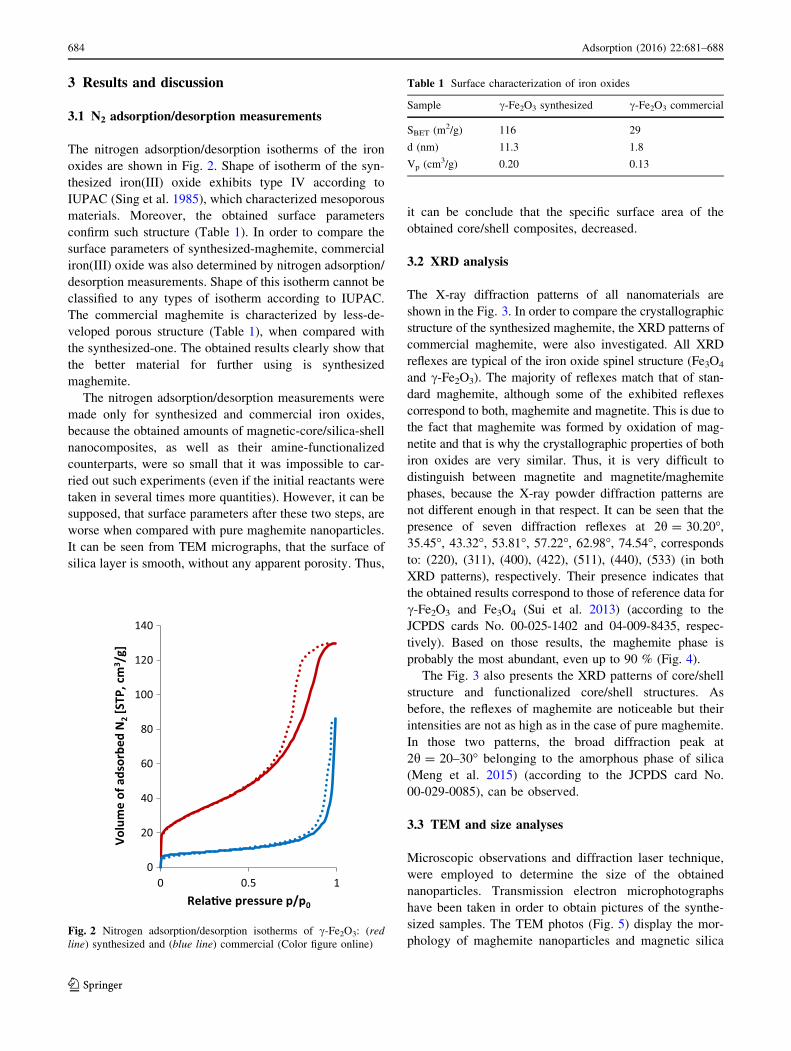
The nitrogen adsorption/desorption isotherms of the iron
oxides are shown in Fig. 2. Shape of isotherm of the syn-
thesized iron(III) oxide exhibits type IV according to
IUPAC (Sing et al. 1985), which characterized mesoporous
materials. Moreover, the obtained surface parameters
confirm such structure (Table 1). In order to compare the
surface parameters of synthesized-maghemite, commercial
iron(III) oxide was also determined by nitrogen adsorption/
desorption measurements. Shape of this isotherm cannot be
classified to any types of isotherm according to IUPAC.
The commercial maghemite is characterized by less-de-
veloped porous structure (Table 1), when compared with
the synthesized-one. The obtained results clearly show that
the better material for further using is synthesized
maghemite.
The nitrogen adsorption/desorption measurements were
made only for synthesized and commercial iron oxides,
because the obtained amounts of magnetic-core/silica-shell
nanocomposites, as well as their amine-functionalized
counterparts, were so small that it was impossible to car-
ried out such experiments (even if the initial reactants were
taken in several times more quantities). However, it can be
supposed, that surface parameters after these two steps, are
worse when compared with pure maghemite nanoparticles.
It can be seen from TEM micrographs, that the surface of
silica layer is smooth, without any apparent porosity. Thus,
it can be conclude that the specific surface area of the
obtained core/shell composites, decreased.
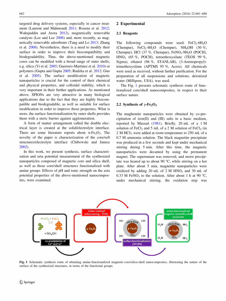
3.2 XRD analysis
The X-ray diffraction patterns of all nanomaterials are
shown in the Fig. 3. In order to compare the crystallographic
structure of the synthesized maghemite, the XRD patterns of
commercial maghemite, were also investigated. All XRD
reflexes are typical of the iron oxide spinel structure (Fe3O4
and c-Fe2O3). The majority of reflexes match that of stan-
dard maghemite, although some of the exhibited reflexes
correspond to both, maghemite and magnetite. This is due to
the fact that maghemite was formed by oxidation of mag-
netite and that is why the crystallographic properties of both
iron oxides are very similar. Thus, it is very difficult to
distinguish between magnetite and magnetite/maghemite
phases, because the X-ray powder diffraction patterns are
not different enough in that respect. It can be seen that the
presence of seven diffraction reflexes at 2h = 30.20�,35.45�, 43.32�, 53.81�, 57.22�, 62.98�, 74.54�, corresponds
to: (220), (311), (400), (422), (511), (440), (533) (in both
XRD patterns), respectively. Their presence indicates that
the obtained results correspond to those of reference data for
c-Fe2O3 and Fe3O4 (Sui et al. 2013) (according to the
JCPDS cards No. 00-025-1402 and 04-009-8435, respec-
tively). Based on those results, the maghemite phase is
probably the most abundant, even up to 90 % (Fig. 4).
The Fig. 3 also presents the XRD patterns of core/shell
structure and functionalized core/shell structures. As
before, the reflexes of maghemite are noticeable but their
intensities are not as high as in the case of pure maghemite.
In those two patterns, the broad diffraction peak at
2h = 20–30� belonging to the amorphous phase of silica
(Meng et al. 2015) (according to the JCPDS card No.
00-029-0085), can be observed.
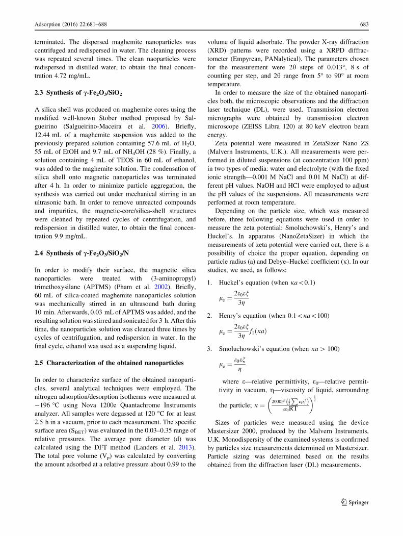
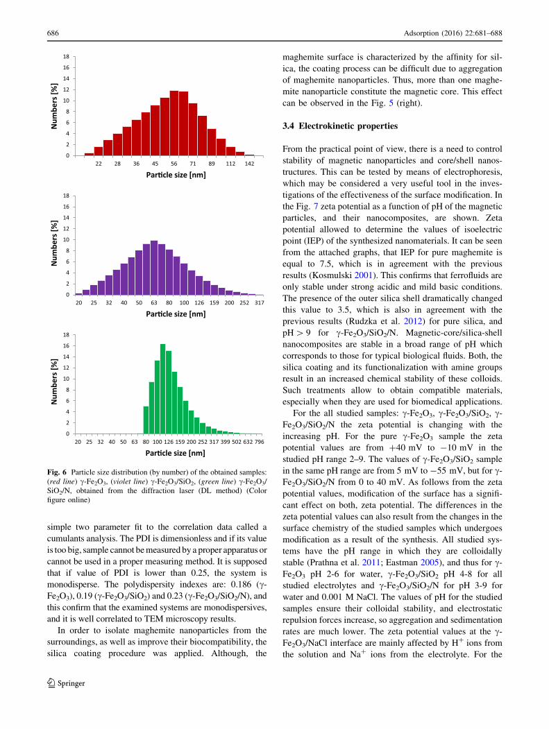
3.3 TEM and size analyses
Microscopic observations and diffraction laser technique,
were employed to determine the size of the obtained
nanoparticles. Transmission electron microphotographs
have been taken in order to obtain pictures of the synthe-
sized samples. The TEM photos (Fig. 5) display the mor-
phology of maghemite nanoparticles and magnetic silica
0
20
40
60
80
100
120
140
0 0.5 1
Volu
me
of a
dsor
bed
N2 [
STP,
cm
3 /g]
Rela�ve pressure p/p0
Fig. 2 Nitrogen adsorption/desorption isotherms of c-Fe2O3: (red
line) synthesized and (blue line) commercial (Color figure online)
Table 1 Surface characterization of iron oxides
Sample c-Fe2O3 synthesized c-Fe2O3 commercial
SBET (m2/g) 116 29
d (nm) 11.3 1.8
Vp (cm3/g) 0.20 0.13
684 Adsorption (2016) 22:681–688
123
structures. It can be seen, that the obtained magnetic
nanoparticles and core/shell nanostructures, are almost
spherical and monodispersed.
The size histograms for the synthesized materials obtained
from diffraction laser are presented in the Fig. 6. The average
size of particles obtained for c-Fe2O3 using the DL method is
56 nm and is smaller compared to c-Fe2O3/SiO2, where the
average size of particles is 62, and c-Fe2O3/SiO2/N where the
average size of particles is 100 nm. The polydispersity index
(PDI) is very important in the practical point of view and is
used as a measure of the breadth of the particle size distri-
bution. PDI can be explained as a number calculated from a
(220
)
(311
)
(400
)
(422
)
(511
)
(440
)
(533
)
5 10 15 20 25 30 35 40 45 50 55 60 65 70 75 80 85 90
Inte
nsity
[j.a
u.]
[°]
Fig. 3 XRD patterns of the
synthesized nanoparticles and
nanocomposites: (red line) c-
Fe2O3-synthesized, (blue line)
c-Fe2O3-commercial, (violet
line) c-Fe2O3/SiO2, (green line)
c-Fe2O3/SiO2/N (Color
figure online)
5 10 15 20 25 30 35 40 45 50 55 60 65 70 75 80 85 90
Inte
nsity
[j.a
u.]
[°]
Fig. 4 XRD patterns of the
synthesized iron oxide
nanoparticles. Two phases
(magnetite (green line) and
maghemite (grey line)) are
compared (Color figure online)
Fig. 5 TEM micrographs of
maghemite (a) and magnetic-
core/silica-shell (b) structures.
Bar length: 500 nm
Adsorption (2016) 22:681–688 685
123
simple two parameter fit to the correlation data called a
cumulants analysis. The PDI is dimensionless and if its value
is too big, sample cannot be measured by a proper apparatus or
cannot be used in a proper measuring method. It is supposed
that if value of PDI is lower than 0.25, the system is
monodisperse. The polydispersity indexes are: 0.186 (c-
Fe2O3), 0.19 (c-Fe2O3/SiO2) and 0.23 (c-Fe2O3/SiO2/N), and
this confirm that the examined systems are monodispersives,
and it is well correlated to TEM microscopy results.
In order to isolate maghemite nanoparticles from the
surroundings, as well as improve their biocompatibility, the
silica coating procedure was applied. Although, the
maghemite surface is characterized by the affinity for sil-
ica, the coating process can be difficult due to aggregation
of maghemite nanoparticles. Thus, more than one maghe-
mite nanoparticle constitute the magnetic core. This effect
can be observed in the Fig. 5 (right).
3.4 Electrokinetic properties
From the practical point of view, there is a need to control
stability of magnetic nanoparticles and core/shell nanos-
tructures. This can be tested by means of electrophoresis,
which may be considered a very useful tool in the inves-
tigations of the effectiveness of the surface modification. In
the Fig. 7 zeta potential as a function of pH of the magnetic
particles, and their nanocomposites, are shown. Zeta
potential allowed to determine the values of isoelectric
point (IEP) of the synthesized nanomaterials. It can be seen
from the attached graphs, that IEP for pure maghemite is
equal to 7.5, which is in agreement with the previous
results (Kosmulski 2001). This confirms that ferrofluids are
only stable under strong acidic and mild basic conditions.
The presence of the outer silica shell dramatically changed
this value to 3.5, which is also in agreement with the
previous results (Rudzka et al. 2012) for pure silica, and
pH[ 9 for c-Fe2O3/SiO2/N. Magnetic-core/silica-shell
nanocomposites are stable in a broad range of pH which
corresponds to those for typical biological fluids. Both, the
silica coating and its functionalization with amine groups
result in an increased chemical stability of these colloids.
Such treatments allow to obtain compatible materials,
especially when they are used for biomedical applications.
For the all studied samples: c-Fe2O3, c-Fe2O3/SiO2, c-
Fe2O3/SiO2/N the zeta potential is changing with the
increasing pH. For the pure c-Fe2O3 sample the zeta
potential values are from ?40 mV to -10 mV in the
studied pH range 2–9. The values of c-Fe2O3/SiO2 sample
in the same pH range are from 5 mV to -55 mV, but for c-
Fe2O3/SiO2/N from 0 to 40 mV. As follows from the zeta
potential values, modification of the surface has a signifi-
cant effect on both, zeta potential. The differences in the
zeta potential values can also result from the changes in the
surface chemistry of the studied samples which undergoes
modification as a result of the synthesis. All studied sys-
tems have the pH range in which they are colloidally
stable (Prathna et al. 2011; Eastman 2005), and thus for c-
Fe2O3 pH 2-6 for water, c-Fe2O3/SiO2 pH 4-8 for all
studied electrolytes and c-Fe2O3/SiO2/N for pH 3-9 for
water and 0.001 M NaCl. The values of pH for the studied
samples ensure their colloidal stability, and electrostatic
repulsion forces increase, so aggregation and sedimentation
rates are much lower. The zeta potential values at the c-
Fe2O3/NaCl interface are mainly affected by H? ions from
the solution and Na? ions from the electrolyte. For the
0
2
4
6
8
10
12
14
16
18
22 28 36 45 56 71 89 112 142
Num
bers
[%]
Num
bers
[%]
Par�cle size [nm]
0
2
4
6
8
10
12
14
16
18
20 25 32 40 50 63 80 100 126 159 200 252 317
Par�cle size [nm]
0
2
4
6
8
10
12
14
16
18
20 25 32 40 50 63 80 100 126 159 200 252 317 399 502 632 796
Num
bers
[%]
Par�cle size [nm]
Fig. 6 Particle size distribution (by number) of the obtained samples:
(red line) c-Fe2O3, (violet line) c-Fe2O3/SiO2, (green line) c-Fe2O3/
SiO2/N, obtained from the diffraction laser (DL method) (Color
figure online)
686 Adsorption (2016) 22:681–688
123
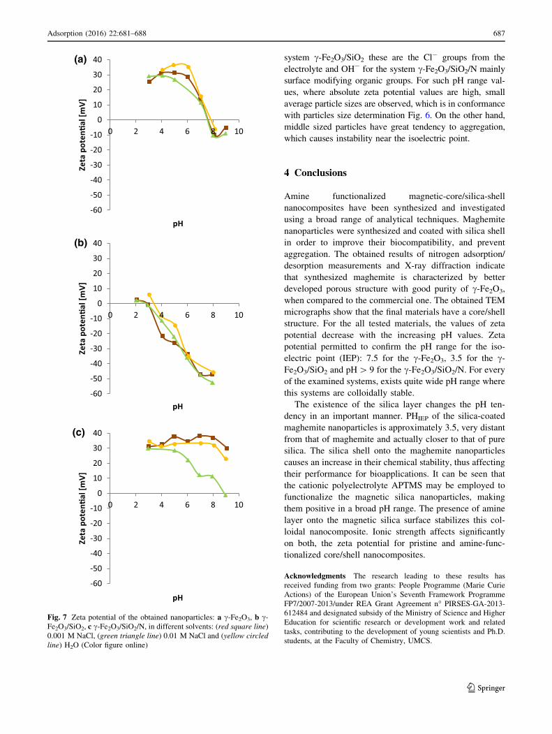
system c-Fe2O3/SiO2 these are the Cl- groups from the
electrolyte and OH- for the system c-Fe2O3/SiO2/N mainly
surface modifying organic groups. For such pH range val-
ues, where absolute zeta potential values are high, small
average particle sizes are observed, which is in conformance
with particles size determination Fig. 6. On the other hand,
middle sized particles have great tendency to aggregation,
which causes instability near the isoelectric point.
4 Conclusions
Amine functionalized magnetic-core/silica-shell
nanocomposites have been synthesized and investigated
using a broad range of analytical techniques. Maghemite
nanoparticles were synthesized and coated with silica shell
in order to improve their biocompatibility, and prevent
aggregation. The obtained results of nitrogen adsorption/
desorption measurements and X-ray diffraction indicate
that synthesized maghemite is characterized by better
developed porous structure with good purity of c-Fe2O3,
when compared to the commercial one. The obtained TEM
micrographs show that the final materials have a core/shell
structure. For the all tested materials, the values of zeta
potential decrease with the increasing pH values. Zeta
potential permitted to confirm the pH range for the iso-
electric point (IEP): 7.5 for the c-Fe2O3, 3.5 for the c-
Fe2O3/SiO2 and pH[ 9 for the c-Fe2O3/SiO2/N. For every
of the examined systems, exists quite wide pH range where
this systems are colloidally stable.
The existence of the silica layer changes the pH ten-
dency in an important manner. PHIEP of the silica-coated
maghemite nanoparticles is approximately 3.5, very distant
from that of maghemite and actually closer to that of pure
silica. The silica shell onto the maghemite nanoparticles
causes an increase in their chemical stability, thus affecting
their performance for bioapplications. It can be seen that
the cationic polyelectrolyte APTMS may be employed to
functionalize the magnetic silica nanoparticles, making
them positive in a broad pH range. The presence of amine
layer onto the magnetic silica surface stabilizes this col-
loidal nanocomposite. Ionic strength affects significantly
on both, the zeta potential for pristine and amine-func-
tionalized core/shell nanocomposites.
Acknowledgments The research leading to these results has
received funding from two grants: People Programme (Marie Curie
Actions) of the European Union’s Seventh Framework Programme
FP7/2007-2013/under REA Grant Agreement n� PIRSES-GA-2013-
612484 and designated subsidy of the Ministry of Science and Higher
Education for scientific research or development work and related
tasks, contributing to the development of young scientists and Ph.D.
students, at the Faculty of Chemistry, UMCS.
-60
-50
-40
-30
-20
-10
0
10
20
30
40
0 2 4 6 8 10
Zeta
pot
en�a
l [m
V]
pH
-60
-50
-40
-30
-20
-10
0
10
20
30
40
0 2 4 6 8 10
Zeta
pot
en�a
l [m
V]
pH
-60
-50
-40
-30
-20
-10
0
10
20
30
40
0 2 4 6 8 10
Zeta
pot
en�a
l [m
V]
pH
(c)
(a)
(b)
Fig. 7 Zeta potential of the obtained nanoparticles: a c-Fe2O3, b c-
Fe2O3/SiO2, c c-Fe2O3/SiO2/N, in different solvents: (red square line)
0.001 M NaCl, (green triangle line) 0.01 M NaCl and (yellow circled
line) H2O (Color figure online)
Adsorption (2016) 22:681–688 687
123
Open Access This article is distributed under the terms of the
Creative Commons Attribution 4.0 International License (http://crea
tivecommons.org/licenses/by/4.0/), which permits unrestricted use,
distribution, and reproduction in any medium, provided you give
appropriate credit to the original author(s) and the source, provide a
link to the Creative Commons license, and indicate if changes were
made.
References
Bele, M., Hribar, G., Campelj, S., Makovec, D., Gaberc-Porekar, V.,
Zorko, M., Gaberscek, M., Jamnik, J., Venturi, P.: Zinc-
decorated silica-coated magnetic nanoparticles for protein bind-
ing and controlled release. J. Chromatogr. B 867, 160–164
(2008)
Chen, F., Shi, R., Xue, Y., Chen, L., Wan, Q.H.: Templated synthesis
of monodisperse mesoporous maghemite/silica microspheres for
magnetic separation of genomic DNA. J. Magn. Magn. Mater.
322, 2439–2445 (2010)
Chibowski, S., Janusz, W.: Specific adsorption of Zn(II) and Cd(II)
ions at the a-Fe2O3/electrolyte interface structure of electrical
double layer. App. Surf. Sci. 196, 343–355 (2002)
Deng, Y., Qi, D., Deng, C., Zhang, X., Zhao, D.: Superparamagnetic
high-magnetization microspheres with an Fe3O4@SiO2 core
and perpendicularly aligned mesoporous SiO2 shell for removal
of microcystins. J. Am. Chem. Soc. 130, 28–29 (2008)
Eastman, J.: Colloid science: Principles. Methods and Applications.
Blackwell Publishing Ltd, Bristol (2005)
Giakisikli, G., Anthemidis, A.N.: Magnetic materials as sorbents for
metal/metalloid preconcentration and/or separation. A review.
Anal. Chim. Acta. 789, 1–16 (2013)
Guerrero-Martinez, A., Perez-Juste, J., Liz-Marzan, L.M.: Recent
progress on silica coating of nanoparticles and related nanoma-
terials. Adv. Mater. 22, 1182–1195 (2010)
Guo, J., Yang, W., Deng, Y., Wang, C., Fu, S.: Organic-dye-coupled
magnetic nanoparticles encaged inside thermoresponsive PNI-
PAM microcapsules. Small 7, 737–743 (2005)
Gupta, A.K., Gupta, M.: Synthesis and surface engineering of iron
oxide nanoparticles for biomedical applications. Biomaterials
26, 3995–4021 (2005)
Hyeon, T.: Chemical synthesis of magnetic nanoparticles. Chem.
Commun. 9, 927–934 (2002)
Kim, J., Kim, H.S., Lee, N., Kim, T., Kim, H., Yu, T., Song, I.C.,
Moon, W.K., Hyeon, T.: Multifunctional uniform nanoparticles
composed of a magnetite nanocrystal core and a mesoporous
silica shell for magnetic resonance and fluorescence imaging and
for drug delivery. Angew. Chem. Int. Ed. 47, 8438–8441 (2008)
Kosmulski, M.: Chemical Properties of Material Surfaces. Marcel
Dekker Inc, New York (2001)
Landers, J., Gor, G.Y., Neimark, A.V.: Density functional theory
methods for characterization of porous materials. Colloids Surf.
A 437, 3–32 (2013)
Laurent, S., Dutz, S., Hafeli, U.O., Mahmoudi, M.: Magnetic fluid
hyperthermia: focus on superparamagnetic iron oxide nanopar-
ticles. Adv. Colloid Interface Sci. 166, 8–23 (2011)
Laurent, S., Mahmoudi, M.: Superparamagnetic iron oxide nanopar-
ticles: promises for diagnosis and treatment of cancer. Int.
J. Mol. Epidemiol. Genet. 2, 367–390 (2011)
Lee, J., Lee, Y., Youn, J.K., Na, H.B., Yu, T., Kim, H., Lee, S.M.,
Koo, Y.M., Kwak, J.H., Park, H.G., Chang, H.N., Hwang, M.,
Park, J.G., Kim, J., Hyeon, T.: Simple synthesis of functional-
ized superparamagnetic magnetite/silica core/shell nanoparticles
and their application as magnetically separable high-perfor-
mance biocatalysts. Small 4, 143–152 (2008)
Mahmoudi, M., Sant, S., Wang, B., Laurent, S., Sen, T.: Superpara-
magnetic iron oxide nanoparticles (SPIONs): development,
surface modification and applications in chemotherapy. Adv.
Drug Deliv. Rev. 63, 24–46 (2011)
Massart, R.: Preparation of aqueous magnetic liquids in alkaline and
acidic media. IEEE Trans. Magn. 17, 1247–1248 (1981)
Meng, S.C., Wang, H., Qing, M., Qiu, C.W., Yang, Y., Li, Y.W.:
Preparation and characterization of SiO2@Fe2O3 core-shell
catalysts. J. Fuel Chem. Technol. 43, 692–700 (2015)
Pham, T., Jackson, J.B., Halas, N.J., Lee, T.R.: Preparation and
characterization of gold nanoshells coated with self-assembled
monolayers. Langmuir 18, 4915–4920 (2002)
Prathna, T.C., Chandrasekaran, N., Mukherjee, A.: Studies on
aggregation behaviour of silver nanoparticles in aqueous
colloids. Surf. Physicochem. Eng. Asp. 390, 216–224 (2011)
Reiss, P., Protiere, M., Li, L.: Core/shell semiconductor nanocrystals.
Small 5, 154–168 (2009)
Rosena, J.E., Chana, L., Shieh, D.B., Gu, F.X.: Iron oxide nanopar-
ticles for targeted cancer imaging and diagnostics. Nanomed.
Nanotechnol. Biol. Med. 8, 275–290 (2012)
Rudzka, K., Delgado, A.V., Viota, J.L.: Maghemite functionalization
for antitumor drug vehiculization. Mol. Pharm. 9, 2017–2028
(2012)
Salgueirino-Maceira, V., Correa-Duarte, M.A., Farle, M., Lopez-
Quintela, A., Sieradzki, K., Diaz, R.: Bifunctional gold-coated
magnetic silica spheres. Chem. Mater. 18, 2701–2706 (2006)
Salgueirino-Maceira, V., Correa-Duarte, M.A.: Increasing the com-
plexity of magnetic core/shell structured nanocomposites for
biological applications. Adv. Mater. 19, 4131–4144 (2007)
Schlorf, T., Meincke, M., Kossel, E., Gluer, C.C., Jansen, O.,
Mentlein, R.: Biological properties of iron oxide nanoparticles
for cellular and molecular magnetic resonance imaging. Int.
J. Mol. Sci. 12, 12–23 (2011)
Sing, K.S.W., Everett, D.H., Haul, R.A.W., Moscou, L., Pierotti,
R.A., Rouquerol, J., Siemieniewska, T.: Reporting physisorption
data for gas/solid systems with special reference to the deter-
mination of surface area and porosity. Pure Appl. Chem. 57,
603–619 (1985)
Sui, J., Li, J., Yang, S., Li, Z., Cai, W.: A facile method to fabricate
superparamagnetic c-Fe2O3/silica nanotubes using multi-walled
carbon nanotubes as template. Mater. Lett. 100, 32–35 (2013)
Tang, S.C.N., Lo, I.M.C.: Magnetic nanoparticles: essential factors
for sustainable environmental applications. Water Res. 47,
2613–2632 (2013)
Tovmachenko, O.G., Graf, C., van den Heuvel, D.J., van Blaaderen,
A., Gerritsen, H.C.: Fluorescence enhancement by metal-core/
silica-shell nanoparticles. Adv. Mater. 18, 91–95 (2006)
Tural, B., Sopaci, S.B., Ozkan, N., Demir, A.S., Volkan, M.:
Preparation and characterization of surface modified c-Fe2O3
(maghemite)—silica nanocomposites used for the purification of
benzaldehyde lyase. J. Phys. Chem. Solids 72, 968–973 (2011)
Wahajuddin, Arora: S.: superparamagnetic iron oxide nanoparticles:
magnetic nanoplatforms as drug carriers. Int. J. Nanomed. 7,
3445–3471 (2012)
Xia, Y., Gates, B., Yin, Y., Lu, Y.: Monodispersed colloidal spheres:
old materials with new applications. Adv. Mater. 12, 693–713
(2000)
Yi, D.K., Selvan, S.T., Lee, S.S., Papaefthymiou, G.C., Kundaliya,
D., Ying, J.Y.: Silica-coated nanocomposites of magnetic
nanoparticles and quantum dots. J. Am. Chem. Soc. 127,
4990–4991 (2005)
Zhang, B., Xing, J.M., Liu, H.Z.: Synthesis and characterization of
superparamagnetic poly(urea-formaldehyde) adsorbents and
their use for adsorption of flavonoids from Glycyrrhiza uralensis
Fisch. Adsorption 14, 65–72 (2008)
688 Adsorption (2016) 22:681–688
123
Recommended