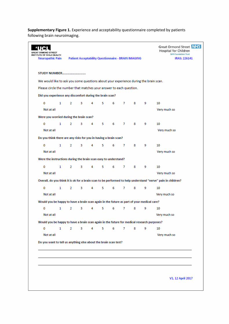
Supplementary Figure 1. Experience and acceptability questionnaire completed by patients
following brain neuroimaging.
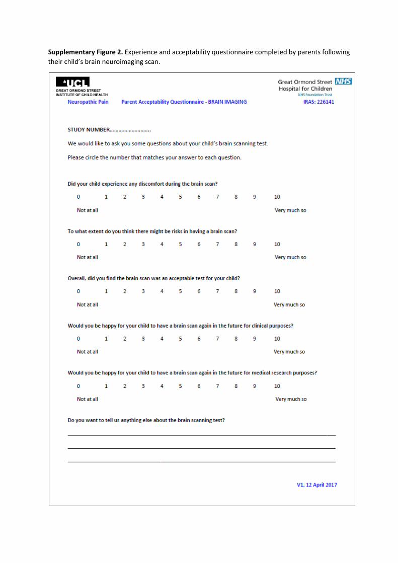
Supplementary Figure 2. Experience and acceptability questionnaire completed by parents following
their child’s brain neuroimaging scan.
1
Supplementary Methods
Manuscript Title:
The feasibility and acceptability of research magnetic resonance imaging in adolescents with
moderate‐severe neuropathic pain
Manuscript Authors:
Madeleine Verriotis, Massieh Moayedi, Clarissa Sorger, Judy Peters, Kiran Seunarine, Christopher A.
Clark, and Suellen M. Walker
2
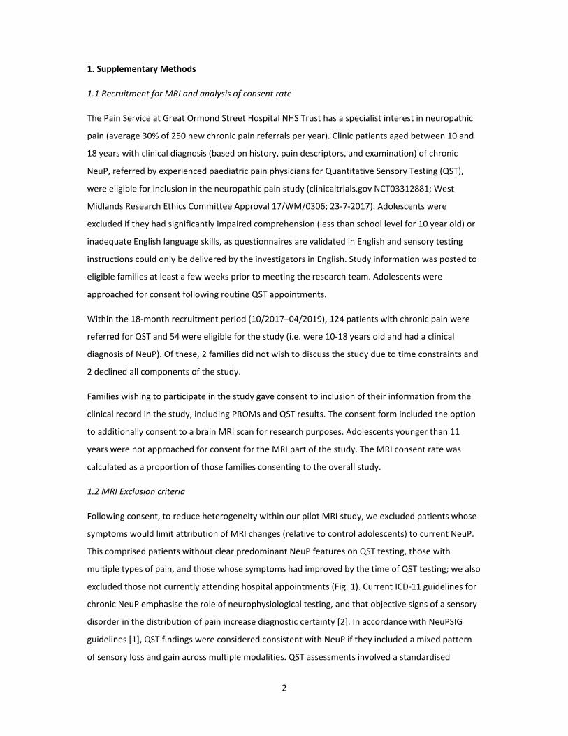
1. Supplementary Methods
1.1 Recruitment for MRI and analysis of consent rate
The Pain Service at Great Ormond Street Hospital NHS Trust has a specialist interest in neuropathic
pain (average 30% of 250 new chronic pain referrals per year). Clinic patients aged between 10 and
18 years with clinical diagnosis (based on history, pain descriptors, and examination) of chronic
NeuP, referred by experienced paediatric pain physicians for Quantitative Sensory Testing (QST),
were eligible for inclusion in the neuropathic pain study (clinicaltrials.gov NCT03312881; West
Midlands Research Ethics Committee Approval 17/WM/0306; 23‐7‐2017). Adolescents were
excluded if they had significantly impaired comprehension (less than school level for 10 year old) or
inadequate English language skills, as questionnaires are validated in English and sensory testing
instructions could only be delivered by the investigators in English. Study information was posted to
eligible families at least a few weeks prior to meeting the research team. Adolescents were
approached for consent following routine QST appointments.
Within the 18‐month recruitment period (10/2017–04/2019), 124 patients with chronic pain were
referred for QST and 54 were eligible for the study (i.e. were 10‐18 years old and had a clinical
diagnosis of NeuP). Of these, 2 families did not wish to discuss the study due to time constraints and
2 declined all components of the study.
Families wishing to participate in the study gave consent to inclusion of their information from the
clinical record in the study, including PROMs and QST results. The consent form included the option
to additionally consent to a brain MRI scan for research purposes. Adolescents younger than 11
years were not approached for consent for the MRI part of the study. The MRI consent rate was
calculated as a proportion of those families consenting to the overall study.
1.2 MRI Exclusion criteria
Following consent, to reduce heterogeneity within our pilot MRI study, we excluded patients whose
symptoms would limit attribution of MRI changes (relative to control adolescents) to current NeuP.
This comprised patients without clear predominant NeuP features on QST testing, those with
multiple types of pain, and those whose symptoms had improved by the time of QST testing; we also
excluded those not currently attending hospital appointments (Fig. 1). Current ICD‐11 guidelines for
chronic NeuP emphasise the role of neurophysiological testing, and that objective signs of a sensory
disorder in the distribution of pain increase diagnostic certainty [2]. In accordance with NeuPSIG
guidelines [1], QST findings were considered consistent with NeuP if they included a mixed pattern
of sensory loss and gain across multiple modalities. QST assessments involved a standardised
3
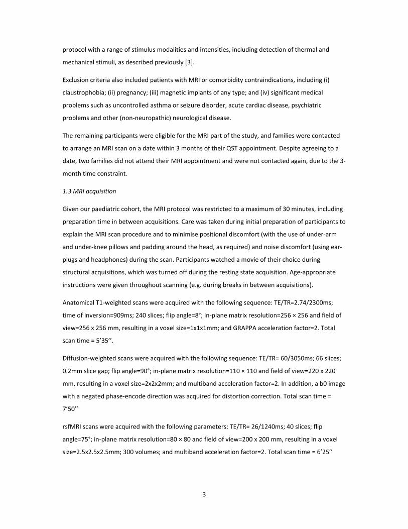
protocol with a range of stimulus modalities and intensities, including detection of thermal and
mechanical stimuli, as described previously [3].
Exclusion criteria also included patients with MRI or comorbidity contraindications, including (i)
claustrophobia; (ii) pregnancy; (iii) magnetic implants of any type; and (iv) significant medical
problems such as uncontrolled asthma or seizure disorder, acute cardiac disease, psychiatric
problems and other (non‐neuropathic) neurological disease.
The remaining participants were eligible for the MRI part of the study, and families were contacted
to arrange an MRI scan on a date within 3 months of their QST appointment. Despite agreeing to a
date, two families did not attend their MRI appointment and were not contacted again, due to the 3‐
month time constraint.
1.3 MRI acquisition
Given our paediatric cohort, the MRI protocol was restricted to a maximum of 30 minutes, including
preparation time in between acquisitions. Care was taken during initial preparation of participants to
explain the MRI scan procedure and to minimise positional discomfort (with the use of under‐arm
and under‐knee pillows and padding around the head, as required) and noise discomfort (using ear‐
plugs and headphones) during the scan. Participants watched a movie of their choice during
structural acquisitions, which was turned off during the resting state acquisition. Age‐appropriate
instructions were given throughout scanning (e.g. during breaks in between acquisitions).
Anatomical T1‐weighted scans were acquired with the following sequence: TE/TR=2.74/2300ms;
time of inversion=909ms; 240 slices; flip angle=8°; in‐plane matrix resolution=256 × 256 and field of
view=256 x 256 mm, resulting in a voxel size=1x1x1mm; and GRAPPA acceleration factor=2. Total
scan time = 5’35’’.
Diffusion‐weighted scans were acquired with the following sequence: TE/TR= 60/3050ms; 66 slices;
0.2mm slice gap; flip angle=90°; in‐plane matrix resolution=110 × 110 and field of view=220 x 220
mm, resulting in a voxel size=2x2x2mm; and multiband acceleration factor=2. In addition, a b0 image
with a negated phase‐encode direction was acquired for distortion correction. Total scan time =
7’50’’
rsfMRI scans were acquired with the following parameters: TE/TR= 26/1240ms; 40 slices; flip
angle=75°; in‐plane matrix resolution=80 × 80 and field of view=200 x 200 mm, resulting in a voxel
size=2.5x2.5x2.5mm; 300 volumes; and multiband acceleration factor=2. Total scan time = 6’25’’
4
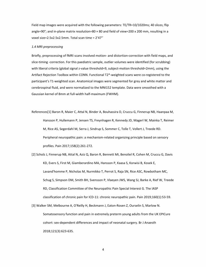
Field map images were acquired with the following parameters: TE/TR=10/1020ms; 40 slices; flip
angle=90°; and in‐plane matrix resolution=80 × 80 and field of view=200 x 200 mm, resulting in a
voxel size=2.5x2.5x2.5mm. Total scan time = 2’47’’
1.4 MRI preprocessing
Briefly, preprocessing of fMRI scans involved motion‐ and distortion‐correction with field maps, and
slice‐timing ‐correction. For this paediatric sample, outlier volumes were identified (for scrubbing)
with liberal criteria (global signal z‐value threshold=9, subject‐motion threshold=2mm), using the
Artifact Rejection Toolbox within CONN. Functional T2*‐weighted scans were co‐registered to the
participant’s T1‐weighted scan. Anatomical images were segmented for grey and white matter and
cerebrospinal fluid, and were normalized to the MNI152 template. Data were smoothed with a
Gaussian kernel of 8mm at full‐width half‐maximum (FWHM).
References[1] Baron R, Maier C, Attal N, Binder A, Bouhassira D, Cruccu G, Finnerup NB, Haanpaa M,
Hansson P, Hullemann P, Jensen TS, Freynhagen R, Kennedy JD, Magerl W, Mainka T, Reimer
M, Rice AS, Segerdahl M, Serra J, Sindrup S, Sommer C, Tolle T, Vollert J, Treede RD.
Peripheral neuropathic pain: a mechanism‐related organizing principle based on sensory
profiles. Pain 2017;158(2):261‐272.
[2] Scholz J, Finnerup NB, Attal N, Aziz Q, Baron R, Bennett MI, Benoliel R, Cohen M, Cruccu G, Davis
KD, Evers S, First M, Giamberardino MA, Hansson P, Kaasa S, Korwisi B, Kosek E,
Lavand'homme P, Nicholas M, Nurmikko T, Perrot S, Raja SN, Rice ASC, Rowbotham MC,
Schug S, Simpson DM, Smith BH, Svensson P, Vlaeyen JWS, Wang SJ, Barke A, Rief W, Treede
RD, Classification Committee of the Neuropathic Pain Special Interest G. The IASP
classification of chronic pain for ICD‐11: chronic neuropathic pain. Pain 2019;160(1):53‐59.
[3] Walker SM, Melbourne A, O'Reilly H, Beckmann J, Eaton‐Rosen Z, Ourselin S, Marlow N.
Somatosensory function and pain in extremely preterm young adults from the UK EPICure
cohort: sex‐dependent differences and impact of neonatal surgery. Br J Anaesth
2018;121(3):623‐635.

5

1
STROBE Statement—Checklist of items that should be included in reports of cohort studies
Item No Recommendation
Page reference and Details
Text taken directly from manuscript highlighted in italics Title and abstract 1 (a) Indicate the study’s design with
a commonly used term in the title or the abstract
Title: The feasibility and acceptability of research magnetic resonance imaging in adolescents with moderate-severe neuropathic pain Abstract: prospective cohort study.
(b) Provide in the abstract an informative and balanced summary of what was done and what was found
Structured abstract within required word limit, with clear headings for background, objective, methods, results, and conclusions.
Introduction Background/rationale 2 Explain the scientific background
and rationale for the investigation being reported
p.1, Introduction section, paragraphs 1-2. Lack of evidence regarding feasibility and practical or ethical burden of MRI in such cohorts [28] may represent barriers to research study planning, ethical approval, and/or recruitment [33].
Objectives 3 State specific objectives, including any prespecified hypotheses
Within a larger clinical cohort of adolescents with moderate-severe NeuP, a pilot study assessed MRI consent rate, post-scan acceptability, and data quality.
Methods Study design 4 Present key elements of study
design early in the paper
p.1, Methods section. Adolescents aged 10-18 years with clinically diagnosed NeuP were recruited from the Great Ormond Street Hospital Chronic Pain Management Service. … Age-matched healthy participant data with the same MRI protocol and scanner were available for comparison.
Setting 5 Describe the setting, locations, and relevant dates, including periods of recruitment, exposure, follow-up, and data collection
p.1, Methods section. The MRI pilot forms part of an ongoing cohort study evaluating PROMs and QST. … families were given the option to additionally consent to an MRI scan, which required one additional hospital visit within 3 months of QST testing and recruitment p.2, Methods section. Multimodal neuroimaging was performed using a 3T Siemens Prisma MRI scanner with a 64-channel coil at Great Ormond Street Hospital. Supplementary Material, p.2 Within the 18-month recruitment period (10/2017–04/2019), 124 patients with chronic pain were referred for QST and 54 were eligible for the study
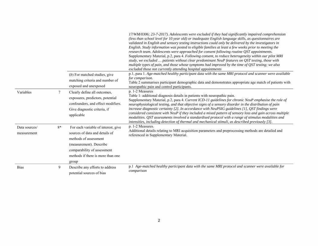
Participants 6 (a) Give the eligibility criteria, and the sources and methods of selection of participants. Describe methods of follow-up
Fig. 1 Recruitment flow chart Supplementary Material, p.2, para 1. Clinic patients aged between 10 and 18 years with clinical diagnosis (based on history, pain descriptors, and examination) of chronic NeuP, referred by experienced paediatric pain physicians for Quantitative Sensory Testing (QST), were eligible for inclusion in the neuropathic pain study (clinicaltrials.gov NCT03312881; West Midlands Research Ethics Committee Approval
2
17/WM/0306; 23-7-2017). Adolescents were excluded if they had significantly impaired comprehension (less than school level for 10 year old) or inadequate English language skills, as questionnaires are validated in English and sensory testing instructions could only be delivered by the investigators in English. Study information was posted to eligible families at least a few weeks prior to meeting the research team. Adolescents were approached for consent following routine QST appointments. Supplementary Material, p.2, para 4. Following consent, to reduce heterogeneity within our pilot MRI study, we excluded … patients without clear predominant NeuP features on QST testing, those with multiple types of pain, and those whose symptoms had improved by the time of QST testing; we also excluded those not currently attending hospital appointments
(b) For matched studies, give matching criteria and number of exposed and unexposed
p.1, para 1. Age-matched healthy participant data with the same MRI protocol and scanner were available for comparison. Table 2 summarises participant demographic data and demonstrates appropriate age match of patients with neuropathic pain and control participants.
Variables 7 Clearly define all outcomes, exposures, predictors, potential confounders, and effect modifiers. Give diagnostic criteria, if applicable
p. 1-2 Measures Table 1: additional diagnosis details in patients with neuropathic pain. Supplementary Material, p.2, para 4. Current ICD-11 guidelines for chronic NeuP emphasise the role of neurophysiological testing, and that objective signs of a sensory disorder in the distribution of pain increase diagnostic certainty [2]. In accordance with NeuPSIG guidelines [1], QST findings were considered consistent with NeuP if they included a mixed pattern of sensory loss and gain across multiple modalities. QST assessments involved a standardised protocol with a range of stimulus modalities and intensities, including detection of thermal and mechanical stimuli, as described previously [3].
Data sources/ measurement
8* For each variable of interest, give sources of data and details of methods of assessment (measurement). Describe comparability of assessment methods if there is more than one group
p. 1-2 Measures. Additional details relating to MRI acquisition parameters and preprocessing methods are detailed and referenced in Supplementary Material.
Bias 9 Describe any efforts to address potential sources of bias
p.1 Age-matched healthy participant data with the same MRI protocol and scanner were available for comparison

3
Study size 10 Explain how the study size was arrived at
The primary aim of the pilot MRI study within the overall descriptive cohort study was to assess feasibility of research MRI in adolescents with moderate-severe neuropathic pain, and estimate recruitment rate. Therefore, we aimed to recruit the maximum available subjects, and no a priori power analysis was performed.
Quantitative variables
11 Explain how quantitative variables were handled in the analyses. If applicable, describe which groupings were chosen and why
p. 2 MRI acquisition and analysis. FD values were compared between adolescents with NeuP and controls. p. 2 Data analyses The following measures are descriptive within the cohort of adolescents with neuropathic pain: Supplementary Material, p.2, para 3. The MRI consent rate was calculated as a proportion of those families consenting to the overall study. Mean (SD) demographic characteristics, pain ratings, and questionnaire scores are reported in Table 1. Range of acceptability and discomfort ratings are presented in the text and Fig. 2.
Statistical methods 12 (a) Describe all statistical methods, including those used to control for confounding
p. 2 MRI acquisition and analysis. p. 2 Data analyses Figure and Table Legends include additional details. Supplementary Material, p.3, para 1
(b) Describe any methods used to examine subgroups and interactions
Figure 3 presents correlation between head motion and age per group; details in Legend.
(c) Explain how missing data were addressed
The only variables with missing data are questionnaire measures. Table 1 presents mean questionnaire scores, and the Legend indicates number of participants completing questionnaire measures
(d) If applicable, explain how loss to follow-up was addressed
n/a (not a longitudinal design)
(e) Describe any sensitivity analyses n/a
Results Participants 13* (a) Report numbers of individuals at
each stage of study—eg numbers potentially eligible, examined for eligibility, confirmed eligible, included in the study, completing follow-up, and analysed
Figure 1 Supplementary Material, p.2-3 provides further details on eligibility and exclusion criteria, including number potentially eligible
(b) Give reasons for non-participation at each stage
Figure 1 presents all reasons for non-participation at each stage Supplementary Material, p.2-3 provides further details on eligibility and exclusion criteria,
(c) Consider use of a flow diagram Figure 1 is presented as a flow diagram of participant numbers at each stage of the study. Descriptive data 14* (a) Give characteristics of study
participants (eg demographic, Tables 1 & 2
4
clinical, social) and information on exposures and potential confounders (b) Indicate number of participants with missing data for each variable of interest
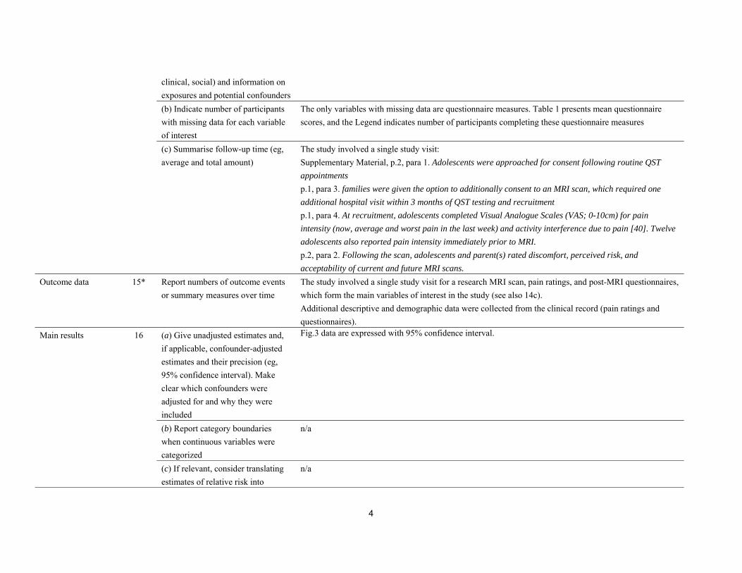
The only variables with missing data are questionnaire measures. Table 1 presents mean questionnaire scores, and the Legend indicates number of participants completing these questionnaire measures
(c) Summarise follow-up time (eg, average and total amount)
The study involved a single study visit: Supplementary Material, p.2, para 1. Adolescents were approached for consent following routine QST appointments p.1, para 3. families were given the option to additionally consent to an MRI scan, which required one additional hospital visit within 3 months of QST testing and recruitment p.1, para 4. At recruitment, adolescents completed Visual Analogue Scales (VAS; 0-10cm) for pain intensity (now, average and worst pain in the last week) and activity interference due to pain [40]. Twelve adolescents also reported pain intensity immediately prior to MRI. p.2, para 2. Following the scan, adolescents and parent(s) rated discomfort, perceived risk, and acceptability of current and future MRI scans.
Outcome data 15* Report numbers of outcome events or summary measures over time
The study involved a single study visit for a research MRI scan, pain ratings, and post-MRI questionnaires, which form the main variables of interest in the study (see also 14c). Additional descriptive and demographic data were collected from the clinical record (pain ratings and questionnaires).
Main results 16 (a) Give unadjusted estimates and, if applicable, confounder-adjusted estimates and their precision (eg, 95% confidence interval). Make clear which confounders were adjusted for and why they were included
Fig.3 data are expressed with 95% confidence interval.
(b) Report category boundaries when continuous variables were categorized
n/a
(c) If relevant, consider translating estimates of relative risk into
n/a
5
absolute risk for a meaningful time period
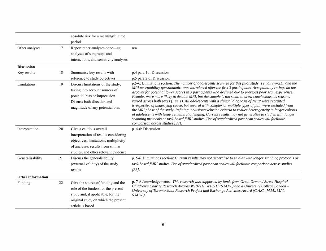
Other analyses 17 Report other analyses done—eg analyses of subgroups and interactions, and sensitivity analyses
n/a
Discussion Key results 18 Summarise key results with
reference to study objectives p.4 para 1of Discussion p.5 para 2 of Discussion
Limitations 19 Discuss limitations of the study, taking into account sources of potential bias or imprecision. Discuss both direction and magnitude of any potential bias
p.5-6, Limitations section: The number of adolescents scanned for this pilot study is small (n=21), and the MRI acceptability questionnaire was introduced after the first 3 participants. Acceptability ratings do not account for potential lower scores in 3 participants who declined due to previous poor scan experience. Females were more likely to decline MRI, but the sample is too small to draw conclusions, as reasons varied across both sexes (Fig. 1). All adolescents with a clinical diagnosis of NeuP were recruited irrespective of underlying cause, but several with complex or multiple types of pain were excluded from the MRI phase of the study. Refining inclusion/exclusion criteria to reduce heterogeneity in larger cohorts of adolescents with NeuP remains challenging. Current results may not generalize to studies with longer scanning protocols or task-based fMRI studies. Use of standardized post-scan scales will facilitate comparison across studies [33].
Interpretation 20 Give a cautious overall interpretation of results considering objectives, limitations, multiplicity of analyses, results from similar studies, and other relevant evidence
p. 4-6: Discussion
Generalisability 21 Discuss the generalisability (external validity) of the study results
p. 5-6. Limitations section: Current results may not generalize to studies with longer scanning protocols or task-based fMRI studies. Use of standardized post-scan scales will facilitate comparison across studies [33].
Other information Funding 22 Give the source of funding and the
role of the funders for the present study and, if applicable, for the original study on which the present article is based
p. 7 Acknowledgements. This research was supported by funds from Great Ormond Street Hospital Children’s Charity Research Awards W1071H, W1071I (S.M.W.) and a University College London – University of Toronto Joint Research Project and Exchange Activities Award (C.A.C., M.M., M.V., S.M.W.).
6
*Give information separately for exposed and unexposed groups. Note: An Explanation and Elaboration article discusses each checklist item and gives methodological background and published examples of transparent reporting. The STROBE checklist is best used in conjunction with this article (freely available on the Web sites of PLoS Medicine at http://www.plosmedicine.org/, Annals of Internal Medicine at http://www.annals.org/, and Epidemiology at http://www.epidem.com/). Information on the STROBE Initiative is available at http://www.strobe-statement.org.
Recommended