Date and version No: FUTUREGB_Protocol_V4.0_14Oct2020.docx
Clinical Research Protocol Template version 14.0 Page 1 of 52
© Copyright: The University of Oxford and Oxford University Hospitals NHS Foundation Trust 2018 NIHR EME COPY
Study Title: FUTURE-GB – Functional and Ultrasound guided Resection of Glioblastoma.
A two Stage trial.
Stage 1 – Non-randomised learning evaluation of participating centres (an IDEAL study), followed by
Stage 2 – A multi-centre randomised trial with 2 mechanistic sub-studies.
Internal Reference Number / Short title: FUTURE-GB
Ethics Ref: 20/LO/0840
IRAS Project ID: 264482
Date and Version No: 14th October 2020, V4.0
ISRCTN: 38834571
Chief Investigator: Professor Puneet Plaha, Nuffield Department of Surgical Sciences,
University of Oxford
Co-Chief Investigators: Ms Sophie Camp, Imperial College Healthcare NHS Trust
Professor Dipankar Nandi, Imperial College Healthcare NHS Trust

Date and version No: FUTUREGB_Protocol_V4.0_14Oct2020.docx
Clinical Research Protocol Template version 14.0 Page 2 of 52
© Copyright: The University of Oxford and Oxford University Hospitals NHS Foundation Trust 2018 NIHR EME COPY
Investigators: Professor Jonathan Cook, Oxford Clinical Trials Research Unit,
University of Oxford
Professor Adrian Lim, Imperial College Healthcare NHS Trust
Professor Martin Taphoorn, Haaglanden Medical Center, University of
Leiden, Netherlands
Professor Linda Driven, Haaglanden Medical Center, University of
Leiden, Netherlands.
Professor Peter McCulloch, Nuffield Department of Surgical Sciences,
University of Oxford
Professor Natalie Voets, Nuffield Department of Surgical Sciences,
University of Oxford
Dr Matthew Grech-Sollars, Imperial College Healthcare NHS Trust
Professor Colin Watts, University of Birmingham
Dr Helen Bulbeck, Brainstrust
Professor Michael Jenkinson, University of Liverpool & The Walton
Centre
Dr Matt Williams, Imperial College Healthcare NHS Trust
Professor Colin Watts, University Hospitals Birmingham Foundation
NHS Trust
Sponsor: University of Oxford
Joint Research Office, 1st Floor, Boundary Brook House, Churchill Drive,
Headington, OX3 7GB
Funder: National Institute for Health Research – Efficacy and Mechanism
Evaluation Programme (EME)
Conflicts of Interest: None of the investigators have any conflicts of interest.
Confidentiality Statement
This document contains confidential information that must not be disclosed to anyone other than the
Sponsor, the Investigator Team, HRA, host organisation, and members of the Research Ethics
Committee, unless authorised to do so.
Date and version No: FUTUREGB_Protocol_V4.0_14Oct2020.docx
Clinical Research Protocol Template version 14.0 Page 3 of 52
© Copyright: The University of Oxford and Oxford University Hospitals NHS Foundation Trust 2018 NIHR EME COPY
TABLE OF CONTENTS
Contents
1. KEY CONTACTS ................................................................................................................................... 7
2. LAY SUMMARY................................................................................................................................... 8
2.1. Aims and Background ........................................................................................................................ 8
2.2. Design ................................................................................................................................................ 8
2.3. Public and Patient Involvement ........................................................................................................ 9
2.4. Dissemination: ................................................................................................................................... 9
3. SYNOPSIS ........................................................................................................................................... 9
4. ABBREVIATIONS ............................................................................................................................... 13
5. BACKGROUND AND RATIONALE ...................................................................................................... 14
5.1. Health problem to be addressed ..................................................................................................... 14
5.2. Health Related Quality of Life (HRQoL) ........................................................................................... 15
5.3. Knowledge Gap ................................................................................................................................ 15
5.4. Current surgical standard of care .................................................................................................... 16
5.5. Combined NiUS and DTI may improve outcome ............................................................................. 16
5.5. Pilot data shows that combining techniques is the way forward ................................................... 17
5.6. The need for the FUTURE-GB trial ................................................................................................... 17
6. OBJECTIVES AND OUTCOME MEASURES ......................................................................................... 18
6.1. PRIMARY OUTCOME MEASURES ..................................................................................................... 18
6.1.1. Stage 1 ......................................................................................................................................... 18
6.1.2. Stage 2 ......................................................................................................................................... 18
6.2. Secondary Outcome Measures – Stage 2 only ................................................................................ 19
7. STUDY DESIGN ................................................................................................................................. 22
7.1. Stage 1: non-randomised multicentre learning and evaluation stage (IDEAL Stage IIB study) ...... 22
7.2. Stage 2: prospective, Stage III, multicentre randomised controlled trial with internal pilot .......... 23
8. PARTICIPANT IDENTIFICATION ........................................................................................................ 23
8.1. Study Participants ............................................................................................................................ 23
8.2. Inclusion Criteria .............................................................................................................................. 23

Date and version No: FUTUREGB_Protocol_V4.0_14Oct2020.docx
Clinical Research Protocol Template version 14.0 Page 4 of 52
© Copyright: The University of Oxford and Oxford University Hospitals NHS Foundation Trust 2018 NIHR EME COPY
8.3. Exclusion Criteria ............................................................................................................................. 24
9. PROXY INCLUSION (Stage 2 only) .................................................................................................... 24
9.1. Inclusion Criteria .............................................................................................................................. 24
10. PROTOCOL PROCEDURES ................................................................................................................ 25
10.1. Stage 1: non-randomised multicentre learning and evaluation Stage (IDEAL Stage IIB study) ...... 25
10.1.1. Pre-Stage 1 Stage Webinar or Symposium .................................................................................. 25
10.1.2. IDEAL IIB Study (Quality assurance, mentoring and trial centres learning). ............................... 25
10.1.3. 1-Day Pre-RCT Webinar or Symposium. ...................................................................................... 26
10.2. Recruitment ..................................................................................................................................... 26
10.2.1. Participants .................................................................................................................................. 26
10.2.2. Sites ............................................................................................................................................. 26
10.3. Screening and Eligibility Assessment ............................................................................................... 27
10.4. Informed Consent - Participant ....................................................................................................... 27
10.4.1. Informed Consent – Proxy (Stage 2 only) .................................................................................... 28
10.5. Randomisation ................................................................................................................................. 28
10.6. Blinding and code-breaking ............................................................................................................. 29
10.7. Description of study intervention(s), comparators and study procedures (clinical)....................... 29
10.7.1. Description of study intervention(s)............................................................................................ 29
10.7.2. Description of comparator(s) ...................................................................................................... 29
10.7.3. Description of study procedure(s) – Stage 1 ............................................................................... 30
10.7.4. Description of study procedure(s) – Stage 2 ............................................................................... 30
10.11. Imaging Schedule for both Stage 1 and 2 ........................................................................................ 32
10.12. Sample Handling .............................................................................................................................. 32
10.13. Early Discontinuation/Withdrawal of Participants .......................................................................... 33
10.13.1. Stage 1 ......................................................................................................................................... 33
10.13.2. Stage 2 ......................................................................................................................................... 33
10.14. Definition of End of Study ............................................................................................................... 34
10. SAFETY REPORTING ......................................................................................................................... 34
10.1. Definitions ....................................................................................................................................... 34

Date and version No: FUTUREGB_Protocol_V4.0_14Oct2020.docx
Clinical Research Protocol Template version 14.0 Page 5 of 52
© Copyright: The University of Oxford and Oxford University Hospitals NHS Foundation Trust 2018 NIHR EME COPY
10.1.1. Adverse Event (AE) ...................................................................................................................... 34
10.1.2. Serious Adverse Event (SAE) ........................................................................................................ 34
10.2. Reporting Procedures ...................................................................................................................... 34
10.2.1. Events exempt from being reported as SAEs .............................................................................. 35
10.3. Death during the study .................................................................................................................... 35
10.4. Elective admissions and supportive care ........................................................................................ 36
11. STATISTICS AND ANALYSIS ............................................................................................................... 36
11.1. Statistical Analysis Plan (SAP) .......................................................................................................... 36
11.2. Description of the Statistical Methods ............................................................................................ 36
11.3. Sample Size Determination for IDEAL IIB Study (Stage 1) ............................................................... 37
11.4. Sample Size Determination for the RCT (Stage 2) ........................................................................... 37
11.5. Recruitment Predictions for the RCT (Stage 2)................................................................................ 37
11.6. Analysis populations ........................................................................................................................ 38
11.7. Decision points................................................................................................................................ 38
11.7.1. Stage 1 (IDEAL IIB study).............................................................................................................. 38
11.7.2. Stage 2 (RCT) ................................................................................................................................ 38
11.8. Stopping rules ................................................................................................................................. 39
11.8.1. Stage 1 (IDEAL study) ................................................................................................................... 39
11.8.2. Stage 2 (RCT) ................................................................................................................................ 39
11.9. The Level of Statistical Significance ................................................................................................. 39
11.10. Procedure for Accounting for Missing, Unused, and Spurious Data. .............................................. 39
11.11. Procedures for Reporting any Deviation(s) from the Original Statistical Plan ................................ 39
11.12. Health Economics Analysis .............................................................................................................. 39
12. DATA MANAGEMENT ...................................................................................................................... 40
12.1. Source Data ..................................................................................................................................... 40
12.2. Access to Data ................................................................................................................................. 40
12.3. Data Recording and Record Keeping ............................................................................................... 40
13. QUALITY ASSURANCE PROCEDURES ............................................................................................... 41
13.1. Risk assessment ............................................................................................................................... 41

Date and version No: FUTUREGB_Protocol_V4.0_14Oct2020.docx
Clinical Research Protocol Template version 14.0 Page 6 of 52
© Copyright: The University of Oxford and Oxford University Hospitals NHS Foundation Trust 2018 NIHR EME COPY
13.2. Study monitoring ............................................................................................................................. 41
13.3. Study Committees ........................................................................................................................... 41
13.3.1. Data & Safety Monitoring Committee (DSMC) for Stage 2 ......................................................... 41
13.3.2. Trial Steering Committee (TSC) for Stage 2 ................................................................................. 41
13.3.3. Core Trial Management Group (TMG) for all Stages ................................................................... 42
14. PROTOCOL DEVIATIONS .................................................................................................................. 42
15. SERIOUS BREACHES ......................................................................................................................... 42
16. ETHICAL AND REGULATORY CONSIDERATIONS ............................................................................... 42
16.1. Declaration of Helsinki .................................................................................................................... 42
16.2. Guidelines for Good Clinical Practice .............................................................................................. 43
16.3. Approvals ......................................................................................................................................... 43
16.4. Other Ethical Considerations ........................................................................................................... 43
16.5. Reporting ......................................................................................................................................... 43
16.6. Participant Confidentiality ............................................................................................................... 43
16.7. Expenses and Benefits ..................................................................................................................... 43
17. FINANCE AND INSURANCE .............................................................................................................. 43
17.1. Funding ............................................................................................................................................ 43
17.2. Insurance ......................................................................................................................................... 44
17.3. Contractual arrangements .............................................................................................................. 44
18. PUBLICATION POLICY ....................................................................................................................... 44
19. DISSEMINATION POLICY .................................................................................................................. 44
20. DEVELOPMENT OF A NEW PRODUCT/ PROCESS OR THE GENERATION OF INTELLECTUAL
PROPERTY .................................................................................................................................................... 44
21. ARCHIVING....................................................................................................................................... 45
22. REFERENCES .................................................................................................................................... 46
23. APPENDIX A: STUDY FLOW CHART ................................................................................................. 51
24. APPENDIX B: AMENDMENT HISTORY ............................................................................................. 52

Date and version No: FUTUREGB_Protocol_V4.0_14Oct2020.docx
Clinical Research Protocol Template version 14.0 Page 7 of 52
© Copyright: The University of Oxford and Oxford University Hospitals NHS Foundation Trust 2018 NIHR EME COPY
1. KEY CONTACTS
Chief Investigator Professor Puneet Plaha
Consultant Neurosurgeon
Associate Professor
Nuffield Department of Surgical Sciences, University of Oxford
Level 3, West Wing, John Radcliffe Hospital, Headley Way, Oxford OX3 9DU
Co-Chief
Investigators
Miss Sophie Camp
Consultant Neurosurgeon
Imperial College HealthCare NHS Trust
Fulham Palace Road, Hammersmith, London W6 8RF
Professor Dipankar Nandi
Consultant Neurosurgeon
Imperial College HealthCare NHS Trust
Fulham Palace Road, Hammersmith, London W6 8RF
Sponsor University of Oxford
Joint Research Office, 1st Floor, Boundary Brook House, Churchill Drive, Headington, OX3
7GB
Funder(s) National Institute for Health Research
Efficacy and Mechanism Evaluation (EME) Programme, an MRC and NIHR partnership
Clinical Trials Unit Oxford Clinical Trials Research Unit (OCTRU)
University of Oxford
Botnar Research Centre, Windmill Road, Headington, Oxford OX3 7LF
Statistician Ariel Wang
University Lecturer in Statistics
Oxford Clinical Trials Research Unit, University of Oxford
Botnar Research Centre, Windmill Road, Headington, Oxford OX3 7LF
Committees TRIAL STEERING COMMITTEE (TSC)
Chair: Mr Philip Weir
Consultant Neurosurgeon
Belfast Trust
DATA SAFETY AND MONITORING COMMITTEE (DSMC)
Chair: Mr Ian Sabin
Consultant Neurosurgeon
Wellington Hospital, London

Date and version No: FUTUREGB_Protocol_V4.0_14Oct2020.docx
Clinical Research Protocol Template version 14.0 Page 8 of 52
© Copyright: The University of Oxford and Oxford University Hospitals NHS Foundation Trust 2018 NIHR EME COPY
2. LAY SUMMARY
2.1. Aims and Background
Glioblastoma (GB) is the most common primary brain tumour and is incurable. It grows very quickly from
the brain tissue itself, rather than from a cancer elsewhere in the body. It is expected that the number of
people with a brain tumour will rise by 6% in the UK between 2014 and 20351. However, prognosis
(outcome) remains extremely poor, with most people surviving just over 12 months, and as a patient’s
tumour grows patients experience a reduction (decline) in their quality of life. Therefore, we need to
ensure quality of life, which remains difficult. The main treatments for GB are surgery, radiotherapy and
chemotherapy, given in combination.
For patients where it is thought that surgery will benefit, a surgeon often removes as much tumour as
possible, whilst limiting the risk of causing damage, such as weakness, speech, or cognitive difficulties.
However, which technology a surgeon should use during surgery to remove the tumour safely is unclear.
This can affect how soon the cancer returns, what effects of surgery or symptoms a patient develops,
and how a patient feels.
High frequency sound waves that create an image, called Ultrasound (US), is one of the tools a surgeon
can use during the operation to find the tumour and see how much is removed. Another technology,
Diffusion Tensor Imaging (DTI), allows important nerve pathways involved in certain functions, for
example, speech/language, vision and movement, to be avoided in surgery.
This trial aims to see if GB surgery with these extra technologies (tools) added to the standard ones,
increases a patient’s good functioning quality of life, so-called Deterioration Free Survival (DFS).
2.2. Design
FUTURE-GB is a two Stage trial.
Stage 1 a non-randomised cohort study using the IDEAL Stage 2b design format2 . It will evaluate
standard care surgery with the addition of DTI imaging and the ultrasound imaging during the operation.
This Stage will ensure standardisation of the use of the technologies across all trial centres by expert
mentoring, and will evaluate quality of delivery, including monitoring of the learning curve for the group
as a whole.
Stage 2 is a randomised controlled trial. This means those who agree to take part will be allocated by
chance (like the tossing of a coin). The trial plans to enrol 357 newly diagnosed patients to receive either
brain surgery with standard methods without US and DTI, or surgery with the addition of US and DTI as
well as standard tools. Patients will not know into which group they have been placed, nor will the
research team assessing them before and after surgery. They will be recruited from at least 15 NHS
hospitals that routinely undertake GB surgery and have access to these tools. The trial will result in only
minor changes to the present care pathway. After agreeing to take part, participants will be asked to
complete questionnaires about their quality of life, such as their walking, ability to look after their
personal hygiene, how they feel. They will also have a brief physical and cognitive/functional assessment
before their surgery. Afterwards, the questionnaires and assessments will be repeated, before leaving
hospital, and at three monthly intervals until 24 months after agreeing to take part (consenting). These
will be combined with planned hospital visits. How long a patient lives will also be recorded.
Date and version No: FUTUREGB_Protocol_V4.0_14Oct2020.docx
Clinical Research Protocol Template version 14.0 Page 9 of 52
© Copyright: The University of Oxford and Oxford University Hospitals NHS Foundation Trust 2018 NIHR EME COPY
2.3. Public and Patient Involvement
The trial focuses on keeping good quality of life for people living with a GB for as long as possible. It has
been designed with the help of patient support groups at the Brain Tumour Charity and Brainstrust, the
Patient Relative Advisory Group at the Oxford University Hospitals NHS Foundation Trust and the Brain
Tumour PPI Group at Imperial College Healthcare NHS Trust. Dr Helen Bulbeck (Brainstrust’s Director)
has been part of the trial proposal and is one of the trial’s investigators.
2.4. Dissemination:
Trial results will be published and widely shared via a variety of channels. If this trial shows that patients
benefit, it is expected that the tools will become standard care and help GB patients in all 24 UK NHS
neurosurgery units, and worldwide.
3. SYNOPSIS
Study Title FUTURE-GB – Functional and Ultrasound guided Resection of GlioBlastoma (GB):
A two-Stage trial. Stage 1: non-randomised learning Stage evaluation of
participating centres (an IDEAL study). Stage 2: a multicentre definitive trial with
2 mechanistic sub-studies.
Internal ref. no. / short title FUTURE-GB
Study registration ISRCTN: 38834571
Sponsor University of Oxford
Funder NIHR EME Programme
Study Design FUTURE-GB is a 2-Stage trial: Stage 1 is a non-randomised multicentre learning
and evaluation Stage (IDEAL IIB study), and Stage 2 a prospective, multicentre
definitive randomised controlled trial.
Study Participants Newly diagnosed glioblastoma (GB) patients
Sample Size Up to 75 patients will participate in Stage 1, (up to 5 per centre).
357 patients will participate in the Stage 2, randomised controlled trial
Planned Study Period Patients participating in the trial will be followed at 3 monthly intervals up to 24
months after randomisation.
Planned Recruitment period 6-9 months Stage 1 and then 27 months for Stage 2.
Intervention(s) Surgery to resect the GB using Diffusion Tensor Imaging (DTI) and Navigated
intraoperative Ultrasound (NiUS) (where available) in addition to standard care
(i.e. neuronavigation based on preoperative MRI and intraoperative use of 5-
aminolevulinic acid)
Comparator The comparator is standard care as per current NICE guidelines (i.e.
neuronavigation based on preoperative MRI and intraoperative use of 5-
aminolevulinic acid).
Date and version No: FUTUREGB_Protocol_V4.0_14Oct2020.docx
Clinical Research Protocol Template version 14.0 Page 10 of 52
© Copyright: The University of Oxford and Oxford University Hospitals NHS Foundation Trust 2018 NIHR EME COPY
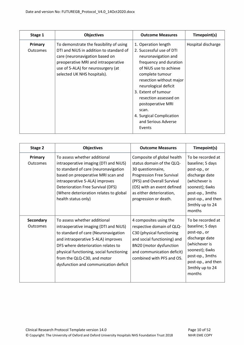
Stage 1 Objectives Outcome Measures Timepoint(s)
Primary
Outcomes
To demonstrate the feasibility of using
DTI and NiUS in addition to standard of
care (neuronavigation based on
preoperative MRI and intraoperative
use of 5-ALA) for neurosurgery (at
selected UK NHS hospitals).
1. Operation length
2. Successful use of DTI
neuronavigation and
frequency and duration
of NiUS use to achieve
complete tumour
resection without major
neurological deficit
3. Extent of tumour
resection assessed on
postoperative MRI
scan.
4. Surgical Complication
and Serious Adverse
Events
Hospital discharge
Stage 2 Objectives Outcome Measures Timepoint(s)
Primary
Outcomes
To assess whether additional
intraoperative imaging (DTI and NiUS)
to standard of care (neuronavigation
based on preoperative MRI scan and
intraoperative 5-ALA) improves
Deterioration Free Survival (DFS)
(Where deterioration relates to global
health status only)
Composite of global health
status domain of the QLQ-
30 questionnaire,
Progression Free Survival
(PFS) and Overall Survival
(OS) with an event defined
as either deterioration,
progression or death.
To be recorded at
baseline; 5 days
post-op., or
discharge date
(whichever is
soonest); 6wks
post-op., 3mths
post-op., and then
3mthly up to 24
months
Secondary
Outcomes
To assess whether additional
intraoperative imaging (DTI and NiUS)
to standard of care (Neuronavigation
and intraoperative 5-ALA) improves
DFS where deterioration relates to
physical functioning, social functioning
from the QLQ-C30, and motor
dysfunction and communication deficit
4 composites using the
respective domain of QLQ-
C30 (physical functioning
and social functioning) and
BN20 (motor dysfunction
and communication deficit)
combined with PFS and OS.
To be recorded at
baseline; 5 days
post-op., or
discharge date
(whichever is
soonest); 6wks
post-op., 3mths
post-op., and then
3mthly up to 24
months

Date and version No: FUTUREGB_Protocol_V4.0_14Oct2020.docx
Clinical Research Protocol Template version 14.0 Page 11 of 52
© Copyright: The University of Oxford and Oxford University Hospitals NHS Foundation Trust 2018 NIHR EME COPY
Stage 2 Objectives Outcome Measures Timepoint(s)
Secondary
Outcomes
To assess whether additional
intraoperative imaging (DTI and NiUS)
to standard of care (Neuronavigation
and intraoperative 5-ALA) improves
time to deterioration
Defined similar to DFS with
the exception that
progression is excluded as
an event (i.e. only
deterioration or death are
considered). There will be
five time to deterioration
outcomes, one for each of
the domains utilized utilised
in the primary and
secondary DFS outcomes,
used in turn to define
deterioration.
To be recorded at
5 days post-op., or
discharge date
(whichever is
soonest); 6wks
post-op., 3mths
post-op., and then
3mthly up to 24
months
Secondary
Outcomes
To assess whether additional
intraoperative imaging (DTI and NiUS)
to standard of care (Neuronavigation
and intraoperative 5-ALA) improves
Overall Survival (OS)
OS (time from diagnosis to
death or trial closure)
To be recorded at
24 months
Secondary
Outcomes
To assess whether additional
intraoperative imaging (DTI and NiUS)
to standard of care (Neuronavigation
and intraoperative 5-ALA) improves
Progression Free Survival (PFS)
PFS (time from diagnosis to
radiological tumour
progression on imaging, as
agreed in local MDT
MRI at 6 months
post-op., and then
3mthly up to 24
months or an MRI
performed outside
protocol if patient
is symptomatic
Secondary
Outcomes
To assess whether additional
intraoperative imaging (DTI and NiUS)
to standard of care (Neuronavigation
and intraoperative 5-ALA) improves
the Extent of tumour resection
Extent of resection as % of
pre-operative tumour
volume on postoperative
contrast enhanced MRI
Post-operative
review
Secondary
Outcomes
To assess whether additional
intraoperative imaging (DTI and NiUS)
to standard of care (Neuronavigation
and intraoperative 5-ALA) improves
the incidence of surgical complications
Number and type of
surgical complications
To be recorded at
5 days post-op., or
discharge date
(whichever is
soonest); 6wks
post-op., 3mths
post-op., and then
3mthly up to 24
months

Date and version No: FUTUREGB_Protocol_V4.0_14Oct2020.docx
Clinical Research Protocol Template version 14.0 Page 12 of 52
© Copyright: The University of Oxford and Oxford University Hospitals NHS Foundation Trust 2018 NIHR EME COPY
Stage 2 Objectives Outcome Measures Timepoint(s)
Secondary
Outcomes
To assess whether additional
intraoperative imaging (DTI and NiUS)
to standard of care (Neuronavigation
and intraoperative 5-ALA) improves
the number of patients eligible for
adjuvant treatment following surgery
Number of patients eligible
for adjuvant treatment
3mths post-op.
Secondary
Outcomes
To assess whether additional
intraoperative imaging (DTI and NiUS)
to standard of care (Neuronavigation
and intraoperative 5-ALA) improves
functional outcome postoperatively
WHO performance status
MOCA
Barthel Index
MRC grading of power in all
4 limbs
To be recorded at
5 days post-op., or
discharge date
(whichever is
soonest); 6wks
post-op., 3mths
post-op., and then
3mthly up to 24
months
Secondary
Outcomes
Assess the correlation of proxy to
participant classification assessment of
quality of life
At a minimum, answers to
questions 29 and 30 of the
QLQ-C3. Ideally answers
will be provided to all of the
QLQ-C30 and BN20.
Baseline, 5 days
post-op., or
discharge date
(whichever is
soonest); 6wks
post-op., 3mths
post-op., and then
3mthly up to 24
months. Proxy will
not complete
questionnaires
when participant
stops completing
them.
Tertiary
Mechanistic
Study Objectives
– on a sub set of
participants –
To assess the sensitivity and specificity
of the anatomico-spatial location of
DTI fibre tracts compared with
intraoperative direct electrical
stimulation in patients undergoing
awake surgery
Sensitivity and specificity
calculation using pre and
post-surgery MRI images
Analysis will be
undertaken post-
surgery.
Tertiary
Mechanistic
Study Objectives
– on a sub set of
participants –
To assess the sensitivity and specificity
of NiUS to identify the tumour
boundary when compared with 5-ALA,
navigated biopsies will be taken from
tumour boundary tissue planned for
resection.
Intra operative NiUS images
and post-operative MRI
scans and Intraoperative
biopsy samples
Analysis will be
undertaken post-
surgery when
biopsy results are
available.

Date and version No: FUTUREGB_Protocol_V4.0_14Oct2020.docx
Clinical Research Protocol Template version 14.0 Page 13 of 52
© Copyright: The University of Oxford and Oxford University Hospitals NHS Foundation Trust 2018 NIHR EME COPY
4. ABBREVIATIONS
5-ALA 5-AminoLevulinic Acid – also known as the Pink Drink
CI Chief Investigator
CRF Case Report Form
CTRG Clinical Trials & Research Governance, University of Oxford
DFS Deterioration Free Survival
DSMC Data Safety Monitoring Committee
DTI Diffusion Tensor Imaging
EORTC European Organisation for Research and Treatment of Cancer
FUTURE-GB FUncTional and Ultrasound guided Rsection of GlioBlastoma
GB Glioblastoma
GCP Good Clinical Practice
GP General Practitioner
GTR Gross Total Resection
HRA Health Research Authority
HRQoL Health Related Quality of Life
ICF Informed Consent Form
IDEAL Idea, Development, Exploration, Assessment, Long-term study
ITT Intention To Treat
iUS Intraoperative Ultrasound
MDT Multi Disciplinary Team
MHz Megahertz
mm millimetre
MOCA The Montreal Cognitive Assessment
MRI Magnetic Resonance Imaging
NHS National Health Service
NiUS Navigated Intraoperative Ultrasound
OS Overall Survival
PFS Progression Free Survival

Date and version No: FUTUREGB_Protocol_V4.0_14Oct2020.docx
Clinical Research Protocol Template version 14.0 Page 14 of 52
© Copyright: The University of Oxford and Oxford University Hospitals NHS Foundation Trust 2018 NIHR EME COPY
PI Principal Investigator
PIL Participant/ Patient Information Leaflet
PP Protocol Population
R&D NHS Trust R&D Department
REC Research Ethics Committee
SOP Standard Operating Procedure
QLQ Quality of Life Questionnaire
QLQ-BN20 Quality of Life Questionnaire Brain
QLQ-C30 Quality of Life Questionnaire Cancer
QoL Quality of Life
TMZ Temozolomide
TSC Trial Steering Committee
US Ultrasound
WHO World Health Organisation
5. BACKGROUND AND RATIONALE
5.1. Health problem to be addressed
Glioblastoma (GB) is a cancer with unmet needs.
GB is the most frequent and aggressive form of brain cancer, with an incidence of 4.64/100,000/year in
the UK.3 Prognosis remains extremely poor with median survival just over 12 months4 and as the tumour
grows patients experience a progressive decline in health-related quality of life (HRQoL), and caregivers
report high levels of distress and carer burden.5 Resistance to treatment leads to poor survival,6 with
high costs to the patient, relatives, society, and the economy.6-8
Although primary brain tumours represent only 3% of all cancers, a brain tumour reduces life expectancy
by an average of 20 years, the highest of any cancer, and accounts for more average years of life lost
than any other cancer.6-8 GB affects adults in their economic prime, and is a leading cause of death in
those under 40 years, costing the economy £578M per year.6-8 To date, there has been little progress in
improving outcome, with many trials failing to show an effect.9 Furthermore, the Department of Health
and Cancer Research UK have identified research in this field as being under resourced,10 and recently
allocated additional funding of £45 million.11 The Brain Tumour Charity have also recently invested
£2.8M into the Tessa Jowell BRAIN-MATRIX (British feasibility study of molecular stratification and
targeted therapy to optimise the clinical management of patients with glioma by enhancing clinical
outcomes, reducing avoidable toxicity, improving management of post-operative residual and recurrent
disease and improving survivorship), a trial aimed at radically increasing opportunities for brain tumour

Date and version No: FUTUREGB_Protocol_V4.0_14Oct2020.docx
Clinical Research Protocol Template version 14.0 Page 15 of 52
© Copyright: The University of Oxford and Oxford University Hospitals NHS Foundation Trust 2018 NIHR EME COPY
patients to access non-standard treatments.12 This is timely, as the incidence of brain tumours is
projected to rise by 6% in the UK between 2014 and 2035.1
A recent James Lind Alliance Priority Setting Partnership13 revealed two of the top ten priorities in Neuro-
oncology are concerned with the long-term physical and cognitive effects of treatment, and the impact
of extent of resection on survival. These themes are reflected in the comments from engagement with
the Neuro-oncology Patient and Public Involvement (PPI) Groups at Imperial College Healthcare NHS
Trust and Oxford University Hospitals NHS Foundation Trust, the Brainstrust PPI group and the Brain
Tumour Charity-Research Involvement Network (BTC-RIN). It is apparent that extending survival without
functional compromise, and maintaining HRQoL is optimum for patients and relatives, given GB is an
incurable cancer with extremely short survival.
5.2. Health Related Quality of Life (HRQoL)
Health Related Quality of Life (HRQoL) is important for patients as GB is incurable and median survival is
short. Of late, the role of Quality of Life (QoL) measurement has become increasingly important in
oncology trials. HRQoL is a key outcome of interest to patients, and more recently has been used as a
secondary outcome measure in a number of randomised trials of GB treatment.14 For a patient with GB,
QoL is particularly significant, given there is substantial potential for a negative impact on HRQoL due to
surgical resection, as well as deterioration due to disease progression. Therefore, it is critical that
assessments of QoL are relevant to the patient population and the effect of their condition. The EORTC
QLQ-C30 has been widely used in oncology trials for over 20 years (currently version 3), to assess the
quality of life of cancer patients.15,16 More recently, an additional brain cancer specific module (QLQ BN-
20) has been produced which complements the main QLQ-C30, with more brain cancer specific
symptoms and additional QoL domains to ensure a comprehensive and relevant assessment for these
patients.17
In newly diagnosed GB, QoL may be relatively high, close to that of a healthy population, it can however
deteriorate rapidly. The ultimate aim of treatment, therefore, is to prolong survival in a way that is
clinically meaningful to the patient, i.e. whilst maintaining their reported HRQoL at as high a level as
possible. Accordingly, in the FUTURE-GB trial the primary outcome is the patient centric Deterioration
Free Survival (DFS),18 which takes into account decline in QoL, as well as survival, and disease
progression.
5.3. Knowledge Gap
There is currently a knowledge gap: surgery is the mainstay of treatment for GB but optimum surgical
technologies remain unclear. Surgery to resect GB is integral to maximum first line treatment, with a
greater impact on survival than non-operative treatments (radiotherapy and chemotherapy).19 It
improves symptom control, reduces dependence on dexamethasone, and increases progression free and
overall survival.20,21 However, maximising the extent of surgical resection must be balanced against the
potential risk of causing neurological deficit. It remains unclear which technologies should be employed
intraoperatively, without increasing physical and/or cognitive dysfunction. A recent Cochrane review
emphasized the lack of high-quality evidence to support the use of any specific intraoperative imaging
technology.22 Research regarding which technologies have the greatest efficacy is of poor quality, with
few details concerning the impact of more radical surgical resection on neurological function.
Furthermore, there are no ongoing or new trials evaluating Navigated intraoperative UltraSound (NiUS),

Date and version No: FUTUREGB_Protocol_V4.0_14Oct2020.docx
Clinical Research Protocol Template version 14.0 Page 16 of 52
© Copyright: The University of Oxford and Oxford University Hospitals NHS Foundation Trust 2018 NIHR EME COPY
Diffusion Tensor Imaging (DTI), or 5-aminolevulinc acid (5-ALA), that will influence treatment guidelines
and policy.
5.4. Current surgical standard of care
The current standard of care advises gross total resection (GTR), i.e. removing all enhancing tumour seen
on the preoperative MRI scan), where appropriate, using neuronavigation, based on a preoperative MRI
scan, and intraoperative 5-aminolevulinic acid (5-
ALA), which is an oral solution administered prior
to surgery to facilitate tumour visualisation and
differentiation from surrounding normal brain
tissue during surgery.5 However there is wide
variations of surgical standard of care across the
UK. Neuronavigation systems allow the
craniotomy to be placed accurately, and the
surgeon to locate the tumour. However, once the
dura has been open, cerebrospinal fluid (CSF)
and/or cyst fluid is drained, and, as tumour is
removed, a phenomenon known as ‘brain shift
occurs’,23 rendering standard neuronavigation
inaccurate when identifying the tumour margins. Consequently, a surgeon may halt the resection at
what is perceived to be tumour margin, inadvertently leaving residual disease. 5-ALA use increases the
extent of tumour resection and progression free survival (PFS).20,24 However, 5-ALA only identifies
fluorescing tumour immediately apparent in the surrounding tissue, without consideration of volume, or
of tumour hidden due to an irregular resection cavity. Furthermore, it does not inform of tissue function.
A survey of all 24 adult UK neurosurgical centres (Telephone and Email survey conducted in 2018 by
Oxford), showed wide variation in the use of technologies employed during GB resection. Whilst all
centres employ standard neuronavigation and 5-ALA, only 75% have access to iUS, 62% to DTI, and 16%
to an intraoperative MRI scanner. However, most of these technologies are not regularly used for
tumour resection, with surgeons unclear of the efficacy of each, and what is the optimum combination.
Indeed, the National Institute for Health and Care Excellence (NICE) guidance5 has suggested that the
available range of intraoperative technologies are considered, as appropriate, in addition to standard
techniques, for tumour resection.
5.5. Combined NiUS and DTI may improve outcome
The desire to achieve a safe, maximal resection, particularly in eloquent regions, has led to an increase in
the popularity of intraoperative imaging. This attempts to eliminate the error produced by brain shift, an
inherent problem in navigation systems based on preoperative imaging,23 to demonstrate residual
tumour at operation, and to visualise accurately relevant white matter tracts and tumour margins. Two
technologies which facilitate surgical resection intraoperatively are NiUS and DTI.
1. NiUS accommodates for brain shift as it is linked to neuronavigation systems and allows the
surgeon to track tumour resection in real time. It permits multiple, real time image acquisitions,
and at each stage, comparison with the preoperative MRI navigation sequence, to evaluate brain
shift and residual disease. NiUS minimally augments operative time,25 allowing precise

Date and version No: FUTUREGB_Protocol_V4.0_14Oct2020.docx
Clinical Research Protocol Template version 14.0 Page 17 of 52
© Copyright: The University of Oxford and Oxford University Hospitals NHS Foundation Trust 2018 NIHR EME COPY
visualization of tumour resection. It is user friendly, widely available, and a pragmatic and cost-
effective alternative to intraoperative MRI, which is prohibitively expensive for many UK units.
Intraoperative ultrasound, and more recently NiUS, has a long history in brain tumour surgery,26
facilitating/extending resection,27-31 and improving survival.32 It has also been evaluated with
respect to histology.33,34 However, there is a learning curve, and image interpretation, especially
once resecting, can be challenging.26 NiUS demonstrates residual tumour in real time. Indeed, it
has been reported that NiUS and 5-ALA provide different information of tumour extent, and
when combined, enhance extent of resection.35 Despite this, there are no randomised trials
assessing its efficacy.
2. DTI is a special magnetic resonance imaging (MRI) technique that can identify the location of
white matter nerve tracts important for speech/language/visual/motor functions. The location of
white matter fibre pathways is the most frequent reason why surgery is halted early, to avoid
compromising patient function.36 DTI is the only method available to visualise functionally
important white matter tracts in the vicinity of a tumour before surgery, and can be fused with
standard intraoperative navigation systems to enable visualisation of the spatial location of the
tracts during surgery, allowing removal of tumour in close proximity. DTI’s usefulness in brain
tumour surgery has recently been reviewed.36 Intraoperative visualisation of DTI is reported to
contribute to maximising safe resection,37-39 reducing visual field deficits,40 and predicting
longterm language problems after surgery.41 A single centre, DTI randomised control trial
showed significantly better gross total resection rates, a lower risk of movement loss, and
improved life expectancy.42 Furthermore, DTI-informed awake surgery reduced the occurrence
and severity of behavioural problems postoperatively, leading to faster recovery, and shorter
hospital stay43 DTI requires the collection of additional MRI data, specialist software for analysis,
and detailed knowledge of white matter anatomy and function. In addition, tract visualisation
may be limited where there is peritumoural oedema. As a result, there is only limited data
available on the sensitivity and specificity of DTI in GB surgery, particularly with reference to its
value as an intraoperative tool and in predicting DFS.
5.5. Pilot data shows that combining techniques is the way forward
Whilst the current aim for GB surgery is resection of all the contrast-enhancing tumour, several small,
single centre studies have reported that extending tumour resection beyond the contrast enhanced
margin, i.e. suptratotal resection, can increase overall survival without additional disability.44-46 A recent
pilot study,47 incorporating DTI neuronavigation, 5-ALA guidance, and, where indicated, awake surgery
has shown that supratotal resection prolongs Progression Free Survival (PFS). Furthermore, preliminary
data in 80 patients, using DTI neuronavigation, in addition to 5-ALA guided surgery, achieved complete
tumour resection in 72% of cases (65% using 5-ALA alone), with 3% postoperative disability (5-10%
reported in the literature).48 In 15% of cases the residual tumour on the postoperative MRI scan was
“hidden/missed” during surgery and would possibly have been detected using NiUS.
5.6. The need for the FUTURE-GB trial
Surgery to maximally remove tumour is the initial and most important step of the patient pathway for
GB patients, and despite the current standard of care,5 there remains great scope for surgical trials and
improvement in outcome.47 Together with the implementation of the revised brain tumour

Date and version No: FUTUREGB_Protocol_V4.0_14Oct2020.docx
Clinical Research Protocol Template version 14.0 Page 18 of 52
© Copyright: The University of Oxford and Oxford University Hospitals NHS Foundation Trust 2018 NIHR EME COPY
classification,49 which permits more accurate patient categorisation, further enhancing surgical
techniques will facilitate additional potential gain for patients.
Intraoperative tools which provide information of residual disease and function are invaluable.
Combining intraoperative techniques enhances the extent of resection.35,47 With the use of NiUS to
identify residual disease in real time, and the functional information provided by DTI, in addition to
standard care, it is anticipated that these two surgical techniques will prolong GB survival without
increasing disability, and whilst maintaining a good quality of life. Increased survival without functional
compromise has obvious benefit for patients and relatives, substantial health gains for society, and
significant economic implications. Proven intraoperative techniques would be applied as standard care
across the NHS, in all 24 hospitals performing adult neurosurgery. In addition, such surgical technologies
have clear global appeal and worldwide impact.
However, considering that this wide use of technologies inevitably varies amongst surgeons, major
technical variations and significant deficits in competence or experience with trial procedures can
compromise Randomised Controlled Trials (RCTs) by increasing “noise”, and introducing performance
bias in relation to the new method/tool. Therefore, this trial has adopted an initial IDEAL IIB study
format,50,51 a prospective non-randomised multicentre learning and harmonisation stage in which quality
control measures and mentoring will be employed prior to the definitive randomised controlled stage of
the trial.
6. OBJECTIVES AND OUTCOME MEASURES
6.1. PRIMARY OUTCOME MEASURES
6.1.1. Stage 1
The IDEAL study will determine whether surgeons using the technologies employed in the randomised
controlled trial demonstrate acceptable expertise in delivering the new approach prior to proceeding
with the randomisation stage.
Note: There are no set levels for acceptable expertise – this will be an evolving process. Factors used to
evaluate expertise will include operation length, successful use of DTI neuronavigation, frequency and
duration of NiUS use to achieve complete tumour resection without major neurological deficit; and extent
of tumour resection.
6.1.2. Stage 2
The primary outcome is Deterioration Free Survival (DFS)18 using only the global health domain to
indicate whether or not deterioration has occurred. Specifically, DFS is defined as the time to a 10-point
deterioration from baseline in the global health status domain of the QLQ-C30 version 3 scores (regarded
as clinically meaningful)53, without subsequent 10-point improvement in scores compared with baseline;
or progressive disease (radiological tumour progression assessed from scan); or death in the absence of
previous definitive deterioration.
The other four domains will be used to define corresponding DFS secondary outcomes.
Note: It is imperative that the primary outcome measure is completed by those participating in the trial,
therefore in addition to the participant completing the questionnaires as far as possible, a proxy will be
asked to complete a separate set of questionnaires throughout the length of the trial. A record will be

Date and version No: FUTUREGB_Protocol_V4.0_14Oct2020.docx
Clinical Research Protocol Template version 14.0 Page 19 of 52
© Copyright: The University of Oxford and Oxford University Hospitals NHS Foundation Trust 2018 NIHR EME COPY
taken of whether participants or proxies or both have completed each of the questionnaires at each data
point.
6.2. Secondary Outcome Measures – Stage 2 only
• DFS as defined for the primary outcome except that physical functioning, social functioning from
the QLQ-C30, and motor dysfunction and communication deficit from the QLQ-BN20 are each
used instead of global health status to determine deterioration.
Note: The QLQ-C3015,16 and QLQ-BN2017 will be collected for participants throughout the follow-
up period. The 5 HRQoL scale scores from these two questionnaires which are most pertinent to
patients with GB are global health status, physical functioning, social functioning from the QLQ-
C30, and motor dysfunction and communication deficit from the QLQ-BN20. Both the QLQ-C30
and the QLQ-BN20 questionnaires will be scored according to the EORTC scoring manual15
following the strategy employed in the placebo arm of the AVAglio trial.18
• Time to Deterioration – defined similar to DFS with the exception that progressive disease is
excluded as an event.
Note: There will be five time to deterioration outcomes, one for each of the scales utilised in the
primary and secondary outcome DFS, used in turn to define deterioration.
• Overall Survival (OS) – defined as time from diagnosis to death or trial closure.
• Progression Free Survival (PFS) – defined as time from diagnosis to radiological tumour
progression (scan date), as identified by local neuro-oncology MDT agreement.
Note: Each MDT will investigate as per local protocols to differentiate true versus pseudo
progression. Scan date confirming progression will be used as date of progression.
• Extent of tumour resection on postoperative contrast enhanced MRI – measured as a % of the
tumour volume when compared to the preoperative MRI scan.
• Surgical complications and serious adverse events – recorded postoperatively and at follow up
visits.
• Number of patients eligible for adjuvant treatment following surgery (radiotherapy and/or
chemotherapy).
• Functional outcome postoperatively: World Health Organisation (WHO) performance status,55
MOCA,56 Barthel Index57 and MRC power grading in all 4 limbs.
Mechanistic component outcomes:
• Sensitivity and specificity of the anatomico-spatial location of DTI fibre tracts compared with
intraoperative direct electrical stimulation in patients undergoing awake surgery.
• Sensitivity and specificity of NiUS to identify the tumour boundary when compared with 5-
ALA, navigated biopsies will be taken from tumour boundary tissue planned for resection.

Date and version No: FUTUREGB_Protocol_V4.0_14Oct2020.docx
Clinical Research Protocol Template version 14.0 Page 20 of 52
© Copyright: The University of Oxford and Oxford University Hospitals NHS Foundation Trust 2018 NIHR EME COPY
Objectives Outcome Measures Timepoint(s)
Primary Stage 2 To assess whether additional
intraoperative imaging (DTI and
NiUS) to standard of care
(neuronavigation based on
preoperative MRI scan and
intraoperative 5-ALA) improves
Deterioration Free Survival (DFS)
(Where deterioration relates to
global health status only)
Composite of global health
status domain of the QLQ-30
questionnaire, Progression
Free Survival (PFS) and Overall
Survival (OS) with an event
defined as either
deterioration, progression or
death.
Baseline; 5 days post-
op., or discharge date
(whichever is soonest);
6wks post-op., 3mths
post op., and then
3mthly up to 24 months
Secondary To assess whether additional
intraoperative imaging (DTI and
NiUS) to standard of care
(neuronavigation based on
preoperative MRI scan and
intraoperative 5-ALA) improves DFS
where deterioration relates to
physical functioning, social
functioning from the QLQ-C30, and
motor dysfunction and
communication deficit
4 composites using the
respective domain of QLQ-C30
(physical functioning and social
functioning) and BN20 (motor
dysfunction and
communication deficit)
combined with PFS and OS.
Baseline; 5 days post-
op., or discharge date
(whichever is soonest);
6wks post-op., 3mths
post op., and then
3mthly up to 24 months
Secondary To assess whether additional
intraoperative imaging (DTI and
NiUS) to standard of care
(neuronavigation based on
preoperative MRI scan and
intraoperative 5-ALA) improves time
to deterioration
Defined similar to DFS with the
exception that progression is
excluded as an event (i.e. only
deterioration or death are
considered). There will be five
time to deterioration
outcomes, one for each of the
domains utilized utilised in the
primary and secondary DFS
outcomes, used in turn to
define deterioration.
5 days post-op., or
discharge date
(whichever is soonest);
6wks post-op., 3mths
post op., and then
3mthly up to 24 months
Secondary To assess whether additional
intraoperative imaging (DTI and
NiUS) to standard of care
(neuronavigation based on
preoperative MRI scan and
intraoperative 5-ALA improves
Overall Survival (OS)
OS (time from diagnosis to
death or trial closure)
24 months

Date and version No: FUTUREGB_Protocol_V4.0_14Oct2020.docx
Clinical Research Protocol Template version 14.0 Page 21 of 52
© Copyright: The University of Oxford and Oxford University Hospitals NHS Foundation Trust 2018 NIHR EME COPY
Objectives Outcome Measures Timepoint(s)
Secondary To assess whether additional
intraoperative imaging (DTI and
NiUS) to standard of care
(neuronavigation based on
preoperative MRI scan and
intraoperative 5-ALA) improves
Progression Free Survival (PFS)
PFS (time from diagnosis to
radiological tumour
progression on imaging, as
agreed in local MDT).
MRI at 6 months post-
surgery and then
3mthly up to 24 months
or an MRI performed
outside protocol if
patient is symptomatic.
Secondary To assess whether additional
intraoperative imaging (DTI and
NiUS) to standard of care
(neuronavigation based on
preoperative MRI scan and
intraoperative 5-ALA) improves the
Extent of tumour resection
extent of resection as % of pre-
operative tumour volume on
postoperative contrast
enhanced MRI
Post-operative review
Secondary To assess whether additional
intraoperative imaging (DTI and
NiUS) to standard of care
(neuronavigation based on
preoperative MRI scan and
intraoperative 5-ALA) improves the
incidence of surgical complications
number and type of surgical
complications
3mths post-op.
Secondary To assess whether additional
intraoperative imaging (DTI and
NiUS) to standard of care
(neuronavigation based on
preoperative MRI scan and
intraoperative 5-ALA) improves the
number of patients eligible for
adjuvant treatment following surgery
number of patients eligible for
adjuvant treatment
5 days post-op., or
discharge date
(whichever is soonest);
6wks post-op.,3mths
post op., and then
3mthly up to 24 months
Secondary To assess whether additional
intraoperative imaging (DTI and
NiUS) to standard of care
(neuronavigation based on
preoperative MRI scan and
intraoperative 5-ALA) improves the
functional outcome postoperatively
WHO performance status
MOCA
Barthel Index
MRC grading of power in all 4
limbs
5 days post-op., or
discharge date
(whichever is soonest);
6wks post-op.,3mths
post op. and then
3mthly up to 24 months
Secondary Assess the correlation of proxy to
participant classification assessment
of quality of life
At a minimum, answers to
questions 29 and 30 of the
QLQ-C30 Ideally answers will
be provided to all of the QLQ-
C30 and BN20.
Baseline, 5 days post op
or discharge date
(whichever is soonest);
6wks post op, 3mths
post op. and then
3mthly up to 24

Date and version No: FUTUREGB_Protocol_V4.0_14Oct2020.docx
Clinical Research Protocol Template version 14.0 Page 22 of 52
© Copyright: The University of Oxford and Oxford University Hospitals NHS Foundation Trust 2018 NIHR EME COPY
Objectives Outcome Measures Timepoint(s)
months. Proxy will not
complete
questionnaires when
participant stops
completing them.
Tertiary –
Mechanistic
Study Objectives
– on a sub-set of
participants
Sensitivity and specificity of the
anatomico-spatial location of DTI
fibre tracts compared with
intraoperative direct electrical
stimulation or Motor evoked
potential changes in patients
undergoing awake surgery
Sensitivity and specificity
calculation using pre and post-
surgery MRI images
Analysis will be
undertaken post-
surgery.
Tertiary –
Mechanistic
Study Objectives
– on a sub-set of
participants
Sensitivity and specificity of NiUS to
identify the tumour boundary when
compared with 5-ALA and post-
operative MRI scan. Navigated
biopsies will be taken from tumour
boundary tissue planned for
resection.
Intra operative NiUS images
and post-operative MRI scans
and Intraoperative biopsy
samples
Analysis will be
undertaken post-
surgery when biopsy
results are available.
7. STUDY DESIGN
7.1. Stage 1: non-randomised multicentre learning and evaluation stage (IDEAL Stage IIB
study)
This is a non-randomised multicentre learning and harmonisation stage in which quality control
measures and mentoring will be employed to improve and evaluate standards of practice (IDEAL Stage
IIB Study).50,51 The IDEAL Stage IIB study will determine whether surgeons using the technologies
employed in the randomised controlled trial demonstrate acceptable expertise in delivering the new
approach prior to proceeding with the randomisation stage.
Stage 1 is divided into 3 components:
1. Pre-trial Webinar (see section 9.1.1)
2. IDEAL Stage IIB (Quality assurance, Mentoring and Trial centres evaluation) (see section 9.1.2)
3. End of Stage 1, Pre-Stage 2 RCT, 1-day Symposium (see section 9.1.3)
The IDEAL Stage IIB study will comprise the following:

Date and version No: FUTUREGB_Protocol_V4.0_14Oct2020.docx
Clinical Research Protocol Template version 14.0 Page 23 of 52
© Copyright: The University of Oxford and Oxford University Hospitals NHS Foundation Trust 2018 NIHR EME COPY
• Mentoring for local site surgeons.
• Quality assurance of operative procedure.
Mentoring by the CI and Co-CIs will be provided through visits to participating centres and frequent
participant meetings, together with a helpline for individual advice sessions from the CI and Co-Cis and
co-applicants, as appropriate. Neurosurgeons will contribute data to ensure standardisation of the
protocol and acceptable expertise in delivering the new approach. This will be evaluated using the
following metrics: operation length; successful use of DTI neuronavigation and frequency and duration of
NiUS use to achieve complete tumour resection without major neurological deficit; and extent of tumour
resection assessed on postoperative MRI scan. The number of cases required for this may vary, but
is expected to be small as most surgeons are already familiar with the component techniques and are
not anticipated to require substantial assessment. Ensuring all participating surgeons are ready to take
part will minimize performance bias in Stage 2 and ensure standardisation of intraoperative technique.
7.2. Stage 2: prospective, Stage III, multicentre randomised controlled trial with internal
pilot
This is a parallel group two arm, multicentre, randomised controlled trial. See Appendix A for a
Flowchart depicting the flow through the trial.
Population: 357 participants with GB suitable for maximally resective surgery (attempted gross total
resection of all enhancing tumour), as agreed at the local Neuro-oncology Multi-Disciplinary Team
meeting.
Intervention: standard care surgery (neuronavigation based on preoperative imaging and intraoperative
use of 5-ALA) with the addition of DTI neuronavigation and NiUS.
Control: standard care surgery (neuronavigation based on preoperative imaging and intraoperative use
of 5-ALA)
Outcome: Deterioration Free Survival
Setting: UK NHS Trusts undertaking GB surgery
8. PARTICIPANT IDENTIFICATION
8.1. Study Participants
Patients aged 18-75 year with a primary GB tumour which is deemed maximally resectable (attempted
gross total resection of all enhancing tumour) by the local Neuro-oncology Multi-Disciplinary Team
(MDT) meeting, will be potentially suitable for inclusion in the trial.
8.2. Inclusion Criteria
• Age 18-75 years
• Neuro-oncology Multi-Disciplinary Team (MDT) decision that the imaging shows a primary GB
tumour which is maximally resectable (attempted gross total resection of all enhancing tumour)

Date and version No: FUTUREGB_Protocol_V4.0_14Oct2020.docx
Clinical Research Protocol Template version 14.0 Page 24 of 52
© Copyright: The University of Oxford and Oxford University Hospitals NHS Foundation Trust 2018 NIHR EME COPY
• Patient is suitable for concomitant 6 weeks adjuvant radiotherapy and Temozolamide (TMZ)
chemotherapy or adjuvant TMZ at the time of MDT decision
• Willing and able to give informed consent
• Able to understand written English to enable completion of trial questionnaires (Stage 2 only)
Note: This is a requirement as one of the outcome questionnaires in not available in all of the UK
national languages; specifically BN-20 is currently not available in Welsh
• Able to provide a proxy who is willing to complete questionnaires as requested (Stage 2 only).
8.3. Exclusion Criteria
The participant may not enter the trial if ANY of the following apply:
• Midline/basal ganglia/cerebellum/brainstem GB
• Multifocal GB
• Recurrent GB
• Suspected secondary GB
• Contraindication to MRI
9. PROXY INCLUSION (Stage 2 only)
It is widely recognised in quality of life research that an individual may measure their quality of life differently from how another person would do, even if that person is close to the individual (e.g. carer, partner etc.). We have therefore set the study up so that we can collect both the participants own assessments (as they are able to provide them), and also the proxy/participant’s assessment. By doing so we will be able to explore potential difference in assessment between the two particularly with regards to deterioration of quality of life. This require both assessments to be collected whenever possible.
The proxy/participant assessment is also important as it will be collected for the small subset of individuals who are not able to complete the quality of life questionnaires but do not lose capacity, have disease progression or die until sometime later. Thus, providing a measure of the quality of life which in the absence of the patients is the next best measure.
It is vitally important in terms of the study's validity to be able to collect data on quality of life, death and
time to progression to the point of death to the end of the follow-up, hence the need to include a Proxy
in this study and has start and end questionnaire data.
Proxies will receive questionnaires as per the schedule in 10.7.4. The quality of life questionnaires will be
the same as those given to the participant except in the third rather than first person.
(Note where a participant dies, loses capacity or withdraws from the study – this will also automatically
cease the proxy’s involvement in the study).
9.1. Inclusion Criteria
• Age 18-75 years
• Nominated by an individual who has consented to participate in Stage 2
• Willing and able to give informed consent
• Able to understand written English to enable completion of trial questionnaires

Date and version No: FUTUREGB_Protocol_V4.0_14Oct2020.docx
Clinical Research Protocol Template version 14.0 Page 25 of 52
© Copyright: The University of Oxford and Oxford University Hospitals NHS Foundation Trust 2018 NIHR EME COPY
10. PROTOCOL PROCEDURES
10.1. Stage 1: non-randomised multicentre learning and evaluation Stage (IDEAL Stage IIB
study)
Patients will be recruited at each centre following the same procedures as detailed in Sections 10.2 to
10.6 (see below for variation in consent information). They will undergo the same pre-operative
assessment as patients in Stage 2. All Stage 1 patients will undergo standard care surgery and in addition
NiUS and DTI, which will be used in the experimental arm of the Stage 2 trial. Awake surgery will be used
at the discretion of the operating surgeon. Postoperative assessment by MRI scan and assessment of
any complications will be performed the same as in Stage 2, including evaluation of DTI and iUS images
versus post-operative MRI assessment.
Stage 1 is divided into the following three components:
10.1.1. Pre-Stage 1 Stage Webinar or Symposium
At the start of Stage 1, a Webinar or Symposium will be held at which expert providers and practitioners
will demonstrate and discuss the use of DTI and NiUS expected in the study. In the case of DTI,
neurosurgeons will be updated on the latest MRI/DTI sequences, intraoperative DTI neuronavigation
software (BrainLAB Elements or Medtronic Stealth), and the clinical application with case examples and
white fibre tract surgical anatomy refreshed. Variations in clinical MRI hardware mean it will not be
possible to fully eliminate variance in diffusion data collected across all study sites. However, every effort
will be made to standardise the sequence acquisition parameters; each site will be asked to acquire a
routine diffusion acquisition with 32 evenly-distributed diffusion directions at a b-value of 1000 s/mm2
and a resolution of 2x2x2mm.
Guidance will be provided on minimum necessary diffusion processing and analysis using the BrainLAB
Elements and Medtronic Stealth software in use at each site to maximise standardisation in tractography
and image fusion to the neuronavigation system. With respect to NiUS, all recruiting centres already
deploy iUS as appropriate, with the use of a range of ultrasound machines. The centres also all have
access to probes within the range of 5-10MHz. As probe selection is dependent on the location of the
lesion and the overlying craniotomy, it is not possible to stipulate use of the same equipment uniformly.
However, this is actually a strength of the study, as should the use of iUS be shown to be beneficial, it
allows far more generalisability of the results, and hence global appeal.
The symposium or webinar will permit surgeons to refresh their ultrasonography skills, case study
examples from the lead centres, and the opportunity to consult with the ultrasound expert and the Co-
CIs using NiUS routinely for tumour resection.
10.1.2. IDEAL IIB Study (Quality assurance, mentoring and trial centres learning).
Mentoring will take place at participating centres through visits as necessary from the expert trial faculty,
and arrangements made for e-mail and telephone support where participating surgical teams are less
familiar with aspects of the protocol.
Participants will be recruited at each centre following the same procedures as detailed in Sections 10.2
to 10.6. The number of cases required for this may vary, but is expected to be small for most surgeons,
as the participating centres are already familiar with the component techniques.

Date and version No: FUTUREGB_Protocol_V4.0_14Oct2020.docx
Clinical Research Protocol Template version 14.0 Page 26 of 52
© Copyright: The University of Oxford and Oxford University Hospitals NHS Foundation Trust 2018 NIHR EME COPY
10.1.3. 1-Day Pre-RCT Webinar or Symposium.
One month prior to the planned completion of Stage 1, Co-applicants, all participating centre Principal
Investigators, and Industry supporting partners (Brainlab Pvt Ltd and Medtronic Plc) will meet via a
Webinar or symposium hosted by the Lead Investigators and the Surgical Interventional Trials Unit
(SITU). This will review progress and initial data from Stage 1, with particular focus on the experimental
interventions, namely DTI neuronavigation and NiUS. Surgeons will have the opportunity to discuss the
quality standards for the interventions. The discussion of early results and experiences will permit group
learning, which may be used to update/refine the protocol for the Stage 2 part of the trial. In addition,
any concerns raised by experience during Stage 1 about (a) participant inclusion criteria (b) acceptable
variations in intra-operative practice will be discussed in the light of the early results, and modifications
made to this protocol if necessary.
10.2. Recruitment
10.2.1. Participants
Recruitment into the trial will be undertaken in two phases in conjunction with the separate stages of
the trial. There will be a separate Patient Information Sheet and Consent Form for patients entering
Stage 1 (IDEAL IIb) and Stage 2 (RCT).
Note: The stages are sequential at participating sites and the stages cannot be recruited to in parallel.
Posters advertising the trial will be displayed in electronic and paper formats as allowed in participating
sites. All potentially eligible participants will have the trial mentioned at the same time the options
regarding their surgery are discussed in dedicated neuro-oncology clinics. Depending upon the site, the
resources available, and most importantly how the participant is dealing with their diagnosis, the
recruitment process and approach may vary across and within sites. Potential participants may straight
away be provided with the trial participant information sheet and asked to consider the trial, and that a
member of the local research team will contact them. It may be the case that individuals are asked if it
would be acceptable for their name to be passed to the research team who will make contact at a later
timepoint, or potential participants may be given the participant information sheet and asked to call the
number on it if they wish to find out more about the trial.
Note: When a potential participant is approached for verbal consent for their details to be passed onto
the local research team – if this consent is given this should be recorded in their clinical notes.
The local and co-ordinating team’s details will be on the participant information sheet so that individuals
and their families can make contact to have any questions/queries answered. In addition, brain tumour
charities will be contacted to promote and display information regarding FUTURE-GB. Furthermore, both
OCTRU and SITU have twitter feeds which will be utilised to promote the trial, and acknowledge when
milestones are met (i.e. sites open to recruitment, first recruitment at a site, 10% of recruitment, 100
patients recruited etc.) Also, it is anticipated that patient bodies who have their own social media feeds
may post about the trial.
10.2.2. Sites
Sites that would like to participate in the trial will be required to complete a site feasibility questionnaire,
deliver GB surgery, and have access to the technology being utilised in the trial. Site feasibility will be
assessed on an individual unit basis, however research experienced and research naïve sites will be

Date and version No: FUTUREGB_Protocol_V4.0_14Oct2020.docx
Clinical Research Protocol Template version 14.0 Page 27 of 52
© Copyright: The University of Oxford and Oxford University Hospitals NHS Foundation Trust 2018 NIHR EME COPY
included. Those sites assessed as being suitable will recruit participants initially into Stage 1 and then
proceed to recruit participants to Stage 2.
10.3. Screening and Eligibility Assessment
Following screening and consent, participants will be entered into the Stage that is currently open at
their site.
Note: Screening should only be undertaken by those listed on the trial screening log, where individual site
Principal Investigators’ have delegated this trial responsibility. Eligibility checks will involve checking that
participants meet all inclusion criteria and none of the exclusion criteria.
There will be no waivers regarding eligibility, i.e. each participant must satisfy all the approved inclusion
criteria of the protocol. Changes to the approved inclusion and exclusion may only be made by a
substantial amendment to the protocol.
10.4. Informed Consent - Participant
The participant must personally sign and date the latest approved version of the Consent form before
any trial specific procedures will be performed. Due to different timelines used at sites, the exact timing
and location of the consent may vary – however as a minimum, consent must be given before a
participant undergoes the routine pre-operative imaging as the participants needs to have been
randomised by this Stage to ensure that all imaging required is undertaken. Patient Information Sheets
will differ slightly between Stage 1 and Stage 2: In Stage 1 the purpose of the Stage within the overall
Trial will be explained and it will be made clear that randomised allocation of treatment will not occur –
all entrants will receive the experimental treatment protocol, but reduced follow-up.
Written and verbal versions of the Participant Information and Informed Consent will be presented to
the participants detailing: the exact nature of the trial; what it will involve for the participant; the
implications and constraints of the protocol; the known side effects and any risks involved in taking part.
It will be clearly stated that the participant is free to withdraw from the trial at any time for any reason
without prejudice to future care, without affecting their legal rights, and with no obligation to give the
reason for withdrawal.
The potential participants will be allowed as much time as desired to consider the information and will
also be given the opportunity to question the Investigator/ a member of the research team, their GP or
other independent parties to decide whether they will participate in the trial. However, neither the trial,
nor time to think about participating in the trial, should cause a delay to any surgery to be undertaken.
Written Informed Consent will then be obtained by means of participant dated signature and dated
signature of the person who presented and obtained the Consent. The person who obtains the consent
must be suitably qualified and experienced and have been authorised to take consent by the site’s
Principal Investigator. A copy of the signed Consent Form will be given to the participant, a copy placed
in the medical notes, and the original will be retained at the study site.
At the same time as taking consent in Stage 2 only, the participant will be asked to provide the name of a
proxy, such as a friend/relative/spouse who they would nominate to also answer questions about a
patient’s quality of life. This individual will be given a separate Information Leaflet and asked to sign a
Date and version No: FUTUREGB_Protocol_V4.0_14Oct2020.docx
Clinical Research Protocol Template version 14.0 Page 28 of 52
© Copyright: The University of Oxford and Oxford University Hospitals NHS Foundation Trust 2018 NIHR EME COPY
separate proxy consent form. To enter Stage 2, the participant must have a proxy who agrees to consent
to also participate in FUTURE-GB, by questionnaire completion.
Note: If at some point a patient wishes to change their proxy, or the proxy needs to change, for example
their contact reduces with the patient, the proxy can suggest another proxy to be approached for consent
and questionnaire completion.
10.4.1. Informed Consent – Proxy (Stage 2 only)
The proxy must personally sign and date the latest approved version of the Consent form. Due to
different timelines used at sites, the exact timing and location of the consent may vary – however as a
minimum, consent must have been given before the individual they are the proxy to undergoes the
routine pre-operative imaging.
Written and verbal versions of the Proxy Participant Information and Informed Consent will be presented
to the potential proxy participants detailing: the exact nature of the trial; what it will involve for
themselves; and any risks involved in taking part. It will be clearly stated that the participant is free to
withdraw from the trial at any time for any reason without prejudice to future care, without affecting
their legal rights, and with no obligation to give the reason for withdrawal. It is anticipated that these
will initially be given to the participant to decide who they will ask to be their potential proxy. It is also
anticipated that the potential proxy may well be the person attending the standard clinic appointments
with the participant.
Note: There is no specific requirements on who can act as a proxy for a participant – this includes
relationship to the participant, time known to the participant, time spent with the participant each week
10.4.2. COVID consenting – all stages
Due to pandemic-related changes in clinic operations and people permitted to accompany patients, the
option of eConsenting is being given in both stages of the trial, for the duration of the need for COVID-
safe practices at participating Trusts. This means that interested participants and proxies would have the
option of completing the consent online, via a secure link, as well as the option of completing it in clinic
as previously detailed. Participants (and proxies if COVID-measures still in place) would be asked for their
email address after initial approach from the clinical research team, and an email sent through which
they could complete the eConsent as a secure online form, and also to have a copy emailed to
themselves as a pdf. This would allow more time for participants to consider the trial and talk to their
proxy if needed. Robust procedures would be in place to ensure that participants would not be able to
be registered/randomise if this consent was not already in place.
10.5. Randomisation
Randomisation of patients will only occur in Stage 2 of the trial. Every centre and each participating
surgeon will offer surgery under both arms of the trial.
Randomisation will be via the web-based service provided by the Oxford Clinical Trials Research Unit
(OCTRU), using the method of minimisation. The minimization factors will be trial site, age (≤ 55 yrs or >
55 yrs), expected surgery status (under general anaesthesia or awake), and eloquence of tumour location
(non-eloquent or eloquent).

Date and version No: FUTUREGB_Protocol_V4.0_14Oct2020.docx
Clinical Research Protocol Template version 14.0 Page 29 of 52
© Copyright: The University of Oxford and Oxford University Hospitals NHS Foundation Trust 2018 NIHR EME COPY
Participants will be randomised after having given written consent, however they will remain blinded as
to which arm of the trial they have been allocated. The local clinical team at site will receive an email
from the randomisation system detailing the arm of the trial to which a participant has been
randomised. Randomisation has to occur before the pre-operative imaging takes place so that the
assigned trial pre-operative imaging can be undertaken.
Participants will be randomised 1:1 to either:
• Standard care surgery (neuronavigation based on preoperative imaging and intraoperative use of
5-ALA) (Control arm)
• Standard care surgery (neuronavigation based on preoperative imaging and intraoperative use of
5-ALA) AND of DTI neuronavigation and NiUS (Intervention arm)
The participant’s identifiable information will be recorded on the randomisation form, and will be
uploaded to an encrypted, separate database at the University of Oxford.
10.6. Blinding and code-breaking
Stage 1 is not blinded; the participants will be receiving all of the technologies during their surgery.
In Stage 2, the participant will be blinded to the allocation (intervention or control arm), and the treating
clinician will be aware, as he/she needs to perform the surgery with the intraoperative technologies as
allocated. In addition to the participant, the radiologist (reviewing the postoperative MRI) will be blinded
to the trial arm. Given this, only on the operation CRF will data of the allocation be included.
There is no code breaking procedure required, as the intervention is a single time point application for
the duration of the participants’ surgery. If there are any complications/issues that arise post-
operatively, the arm to which the patient has been allocated will not affect any subsequent actions
undertaken by the participant’s clinical care team.
10.7. Description of study intervention(s), comparators and study procedures (clinical)
The intervention comprises standard care surgery (neuronavigation based on preoperative imaging and
intraoperative use of 5-ALA) with DTI neuronavigation and NiUS. The comparator is the control, which is
standard care surgery (neuronavigation based on preoperative imaging and intraoperative use of 5-ALA).
Surgery will be undertaken, using the technology allocated.
10.7.1. Description of study intervention(s)
The planned experimental intervention is: GB tumour resection using standard care surgery (i.e.
neuronavigation based on preoperative imaging and intraoperative use of 5-ALA), with the addition of
DTI neuronavigation and NiUS. All patients entering Stage 1 will receive this intervention.
Note: An imaging manual will be available to sites.
10.7.2. Description of comparator(s)
The control intervention is: GB tumour resection using standard care surgery (i.e. neuronavigation based
on preoperative imaging and intraoperative use of 5-ALA).
Date and version No: FUTUREGB_Protocol_V4.0_14Oct2020.docx
Clinical Research Protocol Template version 14.0 Page 30 of 52
© Copyright: The University of Oxford and Oxford University Hospitals NHS Foundation Trust 2018 NIHR EME COPY
10.7.3. Description of study procedure(s) – Stage 1
All patients will undergo a routine preoperative neuronavigation scan. All patients will have a DTI scan
(additional 5 minutes).
There will be minimal deviation from the standard care pathway for GB patients undergoing resective
surgery. Awake surgery and/or that using intraoperative electrophysiological monitoring is permitted as
clinically indicated. Surgery will be performed by a dedicated neuro-oncology surgeon. There is no
alteration to the standard radiotherapy and chemotherapy dosing regimen or treatment schedule for
participants. Postoperative assessments will be undertaken pre-discharge (5 days post-operatively, or at
discharge).
Within 72 hours of surgery, patients will undergo an MRI with contrast, permitting a volumetric
evaluation of residual contrast enhancing tumour, which will be recorded. Further follow up MRI scans
will be undertaken at 6 months postoperatively, and 3 monthly thereafter as per standard care pathway.
Time point from Operation Data Data Collection Method
At discharge. Surgical complications and serious
adverse events
NIHR research nurse / local clinical team
at routine appointment on trial CRFs
6months Surgical complications and serious
adverse events
NIHR research nurse / local clinical team
at routine appointment on trial CRFs
10.7.4. Description of study procedure(s) – Stage 2
All participants will undergo a routine preoperative neuronavigation scan. Those participants
randomised to the experimental arm, will have a DTI scan (additional 5 minutes).
All participants will have preoperative assessments that will comprise: HRQoL (EORTC QLQ-C3015,16 and
BN2017) and functional performance status (WHO performance status,55 MOCA,56 Barthel Index,57 MRC
power grading of limbs). Additionally, baseline data will be collected including sex, weight, height, MGMT
methylation status, hand dominance, date of primary diagnosis and tumour location.
Proxies will be asked to provide their relationship to the participant, the time on average they spend
with the participant and any comments they wish to make about the participant’s cancer journey.
Within 72 hours of surgery, patients will undergo an MRI with contrast, permitting a volumetric
evaluation of residual contrast enhancing tumour, which will be recorded. Further follow up MRI scans
will be undertaken at 6 months postoperatively, and 3 monthly thereafter as per standard care pathway.
The patient, radiologist (reviewing the postoperative MRIs), will be blinded to study arm.
Surgery will be undertaken, using the technology allocated. Awake surgery and/or that using
intraoperative electrophysiological monitoring is permitted in either group, as clinically indicated.
Surgery will be performed by a dedicated neuro-oncology surgeon, who has participated in Stage 1 of the
trial. There will be minimal deviation from the standard care pathway for GB patients undergoing
resective surgery, and no alteration to the standard radiotherapy and chemotherapy dosing regimen or
treatment schedule for participants. Data will be collected on the operation performed and histology
reported locally at site. Postoperative assessments will be undertaken pre-discharge (5 days post-

Date and version No: FUTUREGB_Protocol_V4.0_14Oct2020.docx
Clinical Research Protocol Template version 14.0 Page 31 of 52
© Copyright: The University of Oxford and Oxford University Hospitals NHS Foundation Trust 2018 NIHR EME COPY
operatively, or at discharge), and at six weeks post randomisation, with 3 monthly assessments
subsequently. Survival will be tracked for 24 months.
Once the patient has been randomised, the chance of intra operative crossover is very small, and would
only arise if there was an unexpected surgical complication, whereby the surgeon during the operation
considered he/she required the use of ultrasound. If this occurred, this would be a protocol deviation,
and should be reported to the trial team.
Note: DTI cannot be performed during surgery, and therefore this does not apply.
The Strummer et al. surgical trial,24 which evaluated a similar change in surgery-related practice, did not
report any crossover due to surgeon equipoise, and only in 2 of 322 patients was there a technology
issue.
The schedule of trial assessments and methods for data collection are described in the table below after
consent has been given. As per standard practice, those undertaking any trial assessments/data
collection – should establish that the participant still has capacity to continue to decide to participate in
the trial:
Time point from
Randomisation
Data Data Collection Method
Baseline HRQoL (EORTC QLQ-C30 and BN20)
Functional performance status (WHO
performance status, MOCA, Barthel
Index, MRC power grading of limbs)
Participant and proxy will complete questionnaires
NIHR research nurse / local clinical team
5 days post-op., or
at discharge, 6
weeks,
3,6,9,12,15,18,21
and 24 months
HRQoL (EORTC QLQ-C30 and BN20) *
Functional performance status (WHO
performance status, MOCA, Barthel
Index, MRC power grading of limbs)
Surgical complications and serious
adverse events
Participant* and proxy will self-complete
questionnaires in the hospital
NIHR research nurse / local clinical team on trial CRFs
NIHR research nurse / local clinical team on trial CRFs
24 months Mortality data§
NIHR research nurse / local clinical team on trial CRFs
* NOTE: Due to the natural progression of this disease, a participant may not be able to complete the questionnaires at
all or in their entirety. It is hoped that the full questionnaire pack is completed, however for the primary outcome, only
2 of the questions in the questionnaires are required to be answered as the primary outcome is based on just one of the
DFS domains that is part of the questionnaire. Proxies will be contacted for completion of the questionnaires for each
time point applicable to the participants for whom they are a proxy
§ Progression of disease and occurrence of death are the universal outcome measures by which cancer treatments are assessed to enable the study to report overall survival rates for the study. These have to be collected to fulfill the widely held standards for cancer-based research in this area.

Date and version No: FUTUREGB_Protocol_V4.0_14Oct2020.docx
Clinical Research Protocol Template version 14.0 Page 32 of 52
© Copyright: The University of Oxford and Oxford University Hospitals NHS Foundation Trust 2018 NIHR EME COPY
The specific questions that need to be answered for the primary outcome are:
Please circle the number between 1 and 7 that best applies to you
29. How would you rate your overall health during the past week?
1 2 3 4 5 6 7 Very poor to excellent
30. How would you rate your overall quality of life during the past week?
1 2 3 4 5 6 7 Very poor to excellent
10.11. Imaging Schedule for both Stage 1 and 2
Time point Imaging Part of Standard Care?
Preoperative Neuronavigation MRI scan (T1 with contrast)
Aim: to facilitate surgery
Yes, however, if part of Stage 1 or
randomised to experimental arm, this will
include a DTI scan (which may add up to 5
minutes to the MRI scan)
Postoperatively
(within 48-72
hours of surgery)
Diagnostic MRI (T1 +/- contrast)
Aim: to determine extent of resection as a % of
preoperative contrast enhancing tumour volume
Yes
6 months Diagnostic MRI (T1 +/- contrast)
Aim: to assess response following concomitant
radiotherapy and chemotherapy
Yes
Subsequent 3
monthly follow
up scans
Diagnostic MRI (T1 +/- contrast)
Aim: to assess response following adjuvant
chemotherapy and detect early tumour recurrence
Yes
10.12. Sample Handling
All participants in both Stage 1 and Stage 2 of the trial will have biopsy samples taken from their tumour
as standard of care (tumour surface, tumour core and margin). This includes, as appropriate,
samples from tumour margin tissue planned for resection, where no fluorescence is seen, but the NiUS
image shows signal change suggestive of possible tumour/abnormal tissue (those patients in Stage 1
and in the experimental arm of Stage 2). These biopsies will be sent to the pathology lab as per standard
protocols/guidelines at each participating centre. No special precautions are required when handling
these tissue samples.

Date and version No: FUTUREGB_Protocol_V4.0_14Oct2020.docx
Clinical Research Protocol Template version 14.0 Page 33 of 52
© Copyright: The University of Oxford and Oxford University Hospitals NHS Foundation Trust 2018 NIHR EME COPY
10.13. Early Discontinuation/Withdrawal of Participants
10.13.1. Stage 1
Principal investigators may withdraw participants for the following reasons:
1. If, following consent but prior to surgery, a participant decides to refuse surgery.
2. If, following consent but prior to surgery, a clinician decides that resective surgery is no longer
considered in the participant’s best interests.
If participants are withdrawn from their interventions for these reasons, they will continue to be
followed up in line with the protocol.
Participants may also withdraw from the trial intervention and some or all of the follow-up at any time,
without this affecting their clinical care. Where appropriate, remote follow-up and reasons for
withdrawal will be collected where possible. As there is no set sample size for this part of the study,
participants that withdraw will not be replaced.
All data and samples collected for the purposes of the study will be retained by the trial team.
10.13.2. Stage 2
Principal investigators may withdraw participants from their randomised intervention for the following
reasons:
1. If, following consent but prior to surgery, a participant decides to refuse surgery.
2. If, following consent but prior to surgery, a clinician decides that resective surgery is no longer
considered in the participant’s best interests.
3. During surgery if, for an unforeseen reason, the surgeon decides it is in the best interest of the
participant that the use of NiUS or iUS will facilitate surgery above that which is otherwise possible.
If participants are withdrawn from their interventions for these reasons, they will continue to be
followed up in line with the protocol.
Participants may also withdraw from their randomisation intervention and or some or all of the follow-
up at any time, without this affecting their clinical care. Where appropriate, remote follow-up, and
proxy-completed data collection will be encouraged. Reasons for withdrawal will be collected where
possible. The type of withdrawal and reason will be recorded in the CRF. All data and samples collected
for the purposes of the study will be retained by the trial team.
Participants who withdraw will not be replaced, as a loss to follow-up allowance has been included to
allow for sufficient numbers to be analysed. Replacements will however be recruited if requested after a
DSMC review.
If a participant withdraws from the study or loses capacity, this will cease also all contact with their proxy
and no further contact will be made.

Date and version No: FUTUREGB_Protocol_V4.0_14Oct2020.docx
Clinical Research Protocol Template version 14.0 Page 34 of 52
© Copyright: The University of Oxford and Oxford University Hospitals NHS Foundation Trust 2018 NIHR EME COPY
10.14. Definition of End of Study
The end of study is the point at which all the study data have been entered, any queries resolved, and a
hard data lock has taken place of stage 2.
10. SAFETY REPORTING
The trial will be run in accordance with OCTRU’s Standard Operating Procedures (SOPs) and operational
policies, which all adhere to applicable UK regulatory requirements. An independent Data and Safety
Monitoring Committee (DSMC) and Trial Steering Committee (TSC) will be appointed. The DSMC will
monitor data arising from the trial, review confidential interim reports of accumulating data, and
recommend whether there are any ethical or safety reasons why the trial should not continue. The TSC
will monitor the trial’s progress and will provide independent advice. Both committees will comprise
independent clinicians, statisticians, health service researchers and patient representatives. The project
will also be monitored by the Sponsor (University of Oxford), and progress reports will be submitted to
the Funder. Given the nature of the primary outcome and the interest in key secondary outcomes
(namely, OS and PFS), no formal interim analyses are planned.
10.1. Definitions
10.1.1. Adverse Event (AE)
An adverse event is any untoward medical occurrence in a clinical trial participant.
Note: An adverse event can therefore be any unfavourable and unintended sign (including an abnormal
laboratory finding, for example), symptom or disease temporarily associated with the trial procedures,
whether or not considered related to the procedures.
10.1.2. Serious Adverse Event (SAE)
A serious adverse event is any untoward medical occurrence that:
• results in death
• is life-threatening
• requires inpatient hospitalisation or prolongation of existing hospitalisation
• results in persistent or significant disability/incapacity
Other ‘important medical events’ may also be considered a serious adverse event when, based upon
appropriate medical judgment, the event may jeopardise the participant and may require medical or
surgical intervention to prevent one of the outcomes listed above.
Note: The term "life-threatening" in the definition of "serious" refers to an event in which the participant
was at risk of death at the time of the event; it does not refer to an event which hypothetically might
have caused death if it were more severe.
10.2. Reporting Procedures
It is important to consider the natural history of a high-grade brain tumour affecting each participant
enrolled, the expected sequelae of the illness, and the relevance of these complications to the trial
treatment. All eligible participants have a poor prognosis, and due to the complexity of their condition

Date and version No: FUTUREGB_Protocol_V4.0_14Oct2020.docx
Clinical Research Protocol Template version 14.0 Page 35 of 52
© Copyright: The University of Oxford and Oxford University Hospitals NHS Foundation Trust 2018 NIHR EME COPY
are at increased risk of experiencing multiple adverse events. Consequently, only Serious Adverse Events
(SAE) will be recorded in this trial. This is limited to serious adverse events, which might reasonably
occur as a consequence of the trial treatment (i.e. not events that are part of the natural history of the
primary disease process or expected complications of a brain tumour).
SAEs, as defined above, experienced by a participant from their enrolment until their completion of the
trial must be reported in the participant’s medical notes, on the trial CRF, and reported to the CTU using
the SAE Reporting Form, within 24 hours of observing or learning of the SAE(s). All sections of the SAE
Reporting Form must be completed.
A SAE occurring to a participant will be reported to the REC that gave a favourable opinion of the study
where in the opinion of the Chief Investigator the event was ‘related’ (resulted from administration of
any of the research procedures) and ‘unexpected’ in relation to those procedures. Reports of related and
unexpected SAEs should be submitted within 15 working days of the Chief Investigator becoming aware
of the event, using the HRA report of serious adverse event form
10.2.1. Events exempt from being reported as SAEs
The following hospitalisations are not considered a SAE:
o a visit to the emergency room or other hospital department < 24 hours, that does not result in
admission (unless considered an important medical or life-threatening event)
o admissions as per protocol for a planned medical/surgical procedure
o admissions for planned chemotherapy and/or radiotherapy and any related sequelae
o routine health assessment requiring admission for baseline/trending of health status (e.g.
routine colonoscopy)
o medical/surgical admission other than to remedy ill health and planned prior to entry into the
study
o admission encountered for another life circumstance that carries no bearing on health status and
requires no medical/surgical intervention (e.g., lack of housing, economic inadequacy, caregiver
respite, family circumstances, administrative reason)
o admission for administration of anticancer therapy in the absence of any other SAEs (applies to
oncology protocols)
o disease progression where not considered to be related to study intervention
10.3. Death during the study
Death due to disease under study is to be recorded on the Death CRF form providing the death is not
unexpected or if a causal relationship suspected. The investigator must clearly state whether the death
was expected or unexpected and whether a causal relationship to the study intervention or other protocol
treatment intervention is suspected.

Date and version No: FUTUREGB_Protocol_V4.0_14Oct2020.docx
Clinical Research Protocol Template version 14.0 Page 36 of 52
© Copyright: The University of Oxford and Oxford University Hospitals NHS Foundation Trust 2018 NIHR EME COPY
10.4. Elective admissions and supportive care
Elective admissions to hospital for patient convenience or for planned procedures or investigations or
treatment as specified in this protocol and standard supportive care are not SAEs, and do not require SAE
reporting.
11. STATISTICS AND ANALYSIS
11.1. Statistical Analysis Plan (SAP)
The statistical aspects of the study are summarised here, with details fully described in a statistical
analysis plan (SAP). The SAP will be agreed to by the TSC, with input from the DSMC, prior to unblinding
of trial results to the team. The SAP will be finalised prior to the final analysis taking place, and prior to
unblinding of trial results to the team.
11.2. Description of the Statistical Methods
The analysis of the primary outcome will be a time-to-event analysis using a mixed effect Cox
proportional hazard regression model. Minimisation factors (age, site, anticipated patient operative state
and tumour location), radiotherapy, and MethylGuanine-DNA MethylTransferase (MGMT) status will be
adjusted for as fixed effects. Centre will be included as a random effect.
The assumption of proportional hazard for the Cox model will be examined. If the proportional hazard
assumption is not met, parametric survival analysis, such as the accelerated failure time method will be
considered. A sensitivity analysis will look at the impact of adjusting for surgeon instead of centre.
Secondary analysis will explore the influence of progression as an event by assessing DFS minus
progression. An unadjusted comparison using a log-rank test will also be carried out. Kaplan-Meier
curves will also be generated. Secondary Time-to-event outcomes (e.g. Overall Survival) will be analysed
in a similar manner.
Quality of life amongst survivors will be quantified without a formal statistical comparison between
treatment groups.
There are multiple factors that may influence how a patient rates his/her HRQoL, which may be related
to factors other than the intervention. However, by using a randomized trial design, it is assumed that
patients in both treatment arms are comparable on all aspects, both measured (e.g. age, performance
status) and unmeasured (e.g. mood, coping strategy, personality). This means that the impact of the
psychological state on the evaluation of HRQoL is treated as similar for the two trial arms. Thus, the
study will be able to measure whether the experimental intervention has an impact on HRQoL when
compared to patients receiving standard treatment.

Date and version No: FUTUREGB_Protocol_V4.0_14Oct2020.docx
Clinical Research Protocol Template version 14.0 Page 37 of 52
© Copyright: The University of Oxford and Oxford University Hospitals NHS Foundation Trust 2018 NIHR EME COPY
11.3. Sample Size Determination for IDEAL IIB Study (Stage 1)
There is no formal sample size for the IDEAL study. Participants will be recruited at each centre, the
number of cases required from each centre will vary depending upon caseload numbers and the number
of neurosurgeons, but is expected to be small for most sites (<5), as the participating centres are already
familiar with the component techniques.
11.4. Sample Size Determination for the RCT (Stage 2)
The sample size is based on the primary outcome of DFS based upon the global health status domain in
the QLQ-C30 questionnaire, version 3,18,53,54 as previously defined, and achieving a statistical power of
90% for the primary analysis (see below) with 2-sided significance level of 5%.
Assuming a Hazard Ratio (HR) of 0.7, median DFS survival time of 5 months in the control arm, 24
months follow-up on all participants and allowing for 5% loss to follow-up occurring by month 3, this
yields an overall target of 357 participants (178/179 per arm; 335 events overall) (Stata “artsurv”
www.stata.com). The global health status DFS outcome had median survival times of 6 months in the
standard treatment arm (surgical resection with standard radiotherapy and chemotherapy) group in a
recent trial.18 Additionally, the observed HR was 0.64, 95% CI (0.56, 0.74) for the DFS measures in this
trial suggesting that a HR of 0.7 as assumed above is a plausible magnitude of effect to be observed for
this population.58 It would also be one which would be considered important to clinicians and patients
given the definition of a DFS event (death, progression or a patient anchor determined clinically
meaningful deterioration of 10 points).
For key secondary outcomes (i.e. the other four DFS outcomes, PFS and OS) there is over 80% power for
this size of study, assuming a median overall survival time of 6-9, 7 and 15 months respectively in the
control arm,5 a HR of 0.70 for both, and other inputs as per above.
11.5. Recruitment Predictions for the RCT (Stage 2)
15 NHS Trusts have already agreed participation in the study. These centres collectively review
approximately 1600-1866 GB patients per year (2531 cases diagnosed in 2015 with projected 6% annual
rise).59 Assuming 40% of patients are deemed suitable for maximal resective surgery, and allowing trial
exclusion criteria, this creates a potential annual pool of 600-700 patients. Brain tumour research
recruitment rates are often over 50%, higher than the typical 33%. Therefore, conservatively, it is
anticipated that 250-300 patients/year could enrol. 15 centres have up to 25 dedicated neuro-oncology
surgeons and it is predicted that each site may recruit on average 1 participant per month. A total of 357
patients will be recruited across a minimum of 15 sites. The trial documentation and procedures will be
developed together with patient representative input, and OCTRU involvement, to ensure optimum
recruitment rates.

Date and version No: FUTUREGB_Protocol_V4.0_14Oct2020.docx
Clinical Research Protocol Template version 14.0 Page 38 of 52
© Copyright: The University of Oxford and Oxford University Hospitals NHS Foundation Trust 2018 NIHR EME COPY
11.6. Analysis populations
The principal analysis will be performed once data collection is completed and on the intention to treat
(ITT) population, whereby participants will be analysed according to their randomisation allocation,
irrespective of compliance with the protocol. If appropriate, additional analysis population, such as a per
protocol population (PP), will be defined in the statistical analysis plan. A PP population may exclude
participants who deviate from specific aspects of the protocol.
11.7. Decision points
11.7.1. Stage 1 (IDEAL IIB study)
Up to 5 participants need to have been recruited from a site and evaluated by the IDEAL team for the
site to be allowed to proceed to open to recruitment of Stage 2.
11.7.2. Stage 2 (RCT)
Built into the trial is an internal pilot of recruitment to the RCT (Stage 2). There will be a formal stop/go
review after 12 months of recruitment to the RCT to review the number of randomisations over the pilot
period. If the target of at least 80 randomisations has been met, the trial will continue to recruit for a
further 15 months. Data from the 80 patients will be included in the final analysis. All Stage 2 patients
will be followed up to 24 months after randomisation.
The following stop-go criteria are proposed for the Trial Steering Committee (TSC) after 12 months of
recruitment:

Date and version No: FUTUREGB_Protocol_V4.0_14Oct2020.docx
Clinical Research Protocol Template version 14.0 Page 39 of 52
© Copyright: The University of Oxford and Oxford University Hospitals NHS Foundation Trust 2018 NIHR EME COPY
actual recruitment after 12 months of recruitment
target = 80 >80 participants 65 - 80 participants <65 participants
recruitment rate
(per centre per month) 0.6 0.45 0.37
stop-go criteria
recruitment feasible
proceed with study
review recruitment strategies
report to TSC
continue but modify and monitor
closely
recruitment not feasible
decision not to proceed
The TSC and the funder would make the final decision to terminate the trial.
11.8. Stopping rules
11.8.1. Stage 1 (IDEAL study)
There are no formal stopping rules for this Stage of the study.
11.8.2. Stage 2 (RCT)
Given the nature of the primary, and key secondary, outcomes and the planned study length, no formal
interim analyses with stopping guidelines are planned. An independent Data Safety and Monitoring
Committee (DSMC) will review the accumulating data at regular intervals and may recommend pausing
or stopping the trial in the event of safety concerns.
11.9. The Level of Statistical Significance
All principal analyses will be performed at the 2-sided 5% significance level.
11.10. Procedure for Accounting for Missing, Unused, and Spurious Data.
The procedure for handling spurious or missing data will be described in the Statistical Analysis Plan, and
the Data Monitoring Plan. The trial will attempt to collect data as completely as possible.
The main analysis will include participants for whom endpoint data are available, with other participants
being censored after their last available relevant outcome measure. Sensitivity analyses will examine the
effects of alternative assumptions about the missing data.
11.11. Procedures for Reporting any Deviation(s) from the Original Statistical Plan
Any deviation(s) from the original statistical plan will be described and justified in the final report, should
these occur.
11.12. Health Economics Analysis
There are no health economic analyses to be undertaken as part of the trial.

Date and version No: FUTUREGB_Protocol_V4.0_14Oct2020.docx
Clinical Research Protocol Template version 14.0 Page 40 of 52
© Copyright: The University of Oxford and Oxford University Hospitals NHS Foundation Trust 2018 NIHR EME COPY
12. DATA MANAGEMENT
A data management and sharing plan will be produced for the trial in accordance with OCTRU Standard
Operating Procedures (SOPs), this will include reference to confidentiality, access and security
arrangements.
All data will be processed following the SOPs, which have been written in line with all applicable
regulatory requirements. All trial-specific documents, except for the signed consent form and follow-up
contact details, will refer to the participant with a unique study participant number/code and not by
name. Participant identifiable data will be stored separately from study data, and in accordance with
OCTRU SOPs. All trial data will be stored securely in offices only accessible by swipe card by the central
coordinating team staff in Oxford, and authorised personnel.
Data will be collected from participants and proxies via questionnaires and case report forms that will be
returned to the central trial office in Oxford, via post using a pre-addressed freepost envelope, or NHS
email as appropriate, or directly into an online secure database – the study’s dedicated instance of
REDCap. In addition, participant images will be stored within the cloud database Quentry (BrainLab AG).
As a third-party processor BrainLab will not receive any data that could identify participants.
Participant data will be stored and transported in accordance to SOPs. Upon completion of the trial, fully
de-identified research data may be shared with other organisations at the behest of the funder.
12.1. Source Data
Source documents are where data are first recorded, and from which participants’ CRF data are
obtained. These include, but are not limited to, hospital records (from which medical history and
previous and concurrent medication may be summarised into the CRF), clinical and office charts,
laboratory and pharmacy records, scans, diaries, microfiches, radiographs, and correspondence.
CRF entries will be considered source data if the CRF is the site of the original recording (e.g. there is no
other written or electronic record of data). All documents will be stored safely in confidential conditions.
On all study-specific documents, other than the signed consent and follow-up contact details, the
participant and proxy will be referred to by the study participant/proxy number/code, not by name.
12.2. Access to Data
Direct access will be granted to authorised representatives from the Sponsor, host institution and the
regulatory authorities to permit trial-related monitoring, audits and inspections.
Where data is submitted directly to the trial office, contemporaneous access by local research teams to
the online database will enable the local research teams at sites to download copies of their participants’
data.
12.3. Data Recording and Record Keeping
The data will be stored and used in compliance with the relevant, current data protection laws (Data
Protection Act 2018; General Data Protection Regulation (GDPR) 2018). The trial data (including data for
SAEs) will be entered onto a validated REDCap study database developed and maintained by OCTRU and

Date and version No: FUTUREGB_Protocol_V4.0_14Oct2020.docx
Clinical Research Protocol Template version 14.0 Page 41 of 52
© Copyright: The University of Oxford and Oxford University Hospitals NHS Foundation Trust 2018 NIHR EME COPY
which can only be accessed by authorised users via the application. The application resides on a
webserver hosted and managed by Oxford University’s IT Services division. The server is on the
university’s backbone network and is backed up nightly to a secure off-site location.
After closure of the trial and data analyses, the data will be made publicly available at the time of
publication. The Trial Master File will be archived for five years from the end of the study.
13. QUALITY ASSURANCE PROCEDURES
The study may be monitored, or audited in accordance with the current approved protocol, GCP,
relevant regulations and CTU standard operating procedures. This research will be coordinated by the
Surgical Interventional Trials Unit (SITU), which falls under the Oxford Clinical Trials Research Unit
(OCTRU) and SITU personnel work according to OCTRU SOPs. The OCTRU SOPs and related quality
assurance and control procedures will be used by SITU to ensure that the study procedures are assessed
and carried out as defined in this protocol.
13.1. Risk assessment
A risk assessment and monitoring plan will be prepared before the study opens and will be reviewed as
necessary over the course of the study to reflect significant changes to the protocol or outcomes of
monitoring activities.
13.2. Study monitoring
Monitoring will be performed following a central monitoring and audit plan. Data will be evaluated for
compliance with the protocol and accuracy in relation to source documents.
13.3. Study Committees
13.3.1. Data & Safety Monitoring Committee (DSMC)
The DSMC will meet regularly throughout the trial at time-points agreed by the Chair of the Committee
and the CI. At a minimum this will be on an annual basis. The DSMC will review the safety data
generated, including all adverse events, and make recommendations as to whether the protocol should
be amended to protect patient safety. Recommendations of the DSMC will be discussed between the CI,
TSC, and the Sponsor.
13.3.2. Trial Steering Committee (TSC)
The role of the Trial Steering Committee (TSC) is to provide the overall supervision of the trial. The TSC
will monitor trial progress and conduct, and will advise on scientific credibility. The TSC will consider and
act, as appropriate, upon the recommendations of the Data and Safety Monitoring Committee (DSMC)
and ultimately carries the responsibility for deciding whether the trial needs to be stopped on grounds of
safety or efficacy.

Date and version No: FUTUREGB_Protocol_V4.0_14Oct2020.docx
Clinical Research Protocol Template version 14.0 Page 42 of 52
© Copyright: The University of Oxford and Oxford University Hospitals NHS Foundation Trust 2018 NIHR EME COPY
13.3.3. Core Trial Management Group (TMG) for all Stages
The Trial Management Group (TMG) consists of those individuals responsible for the operational
management of the trial such as the chief investigators, the clinical trial manager, Chair and Vice Chair of
the Surgical Trainee Network and representatives from OCTRU.
The TMG will meet every month throughout the lifetime of the Stage 2 study and will:
• Supervise the conduct and progress of the study, and adherence to the study protocol
• Assess the safety as compiled by SITU and assessed by the DSMC, and efficacy of the
interventions during the study
• Evaluate the quality of the study data
• Review relevant information from other sources (e.g. related studies)
• Escalate any issues for concern to SITU, specifically where the issue could compromise patient
safety or the integrity of the study or quality of the study data
14. PROTOCOL DEVIATIONS
A study related deviation is a departure from the ethically approved study protocol or other study
document or process (e.g. consent process or administration of study intervention) or from the
principles of Good Clinical Practice (GCP) or any applicable regulatory requirements. Any deviations from
the protocol will be documented in a protocol deviation form and filed in the trial master file.
15. SERIOUS BREACHES
A “serious breach” is a breach of the protocol or of the conditions or principles of Good Clinical Practice
which is likely to affect to a significant degree –
(a) the safety or physical or mental integrity of the trial subjects; or
(b) the scientific value of the research.
In the event that a serious breach is suspected the Sponsor must be contacted within 1 working day. In
collaboration with the Chief Investigator, the serious breach will be reviewed by the Sponsor and, if
appropriate, the Sponsor will report it to the approving REC committee and the relevant NHS host
organisation within seven calendar days.
16. ETHICAL AND REGULATORY CONSIDERATIONS
16.1. Declaration of Helsinki
The Investigator will ensure that this study is conducted in accordance with the principles of the
Declaration of Helsinki.

Date and version No: FUTUREGB_Protocol_V4.0_14Oct2020.docx
Clinical Research Protocol Template version 14.0 Page 43 of 52
© Copyright: The University of Oxford and Oxford University Hospitals NHS Foundation Trust 2018 NIHR EME COPY
16.2. Guidelines for Good Clinical Practice
The Investigator will ensure that this study is conducted in accordance with the principles of Good
Clinical Practice.
16.3. Approvals
Following Sponsor approval, the protocol, consent form, participant information sheet and any proposed
advertising material will be submitted to an appropriate Research Ethics Committee (REC), and HRA and
host institutions for written approval.
The Investigator will submit and, where necessary, obtain approval from the above parties for all
substantial amendments to the original approved documents.
16.4. Other Ethical Considerations
In the unlikely event of identifying any additional structural abnormalities on imaging, the scan will be
checked by a clinical specialist. If the specialist feels that the abnormality is medically important, they will
discuss the implications with the participant, and arrange for further investigations as necessary.
Participants will not be informed unless the doctor considers the finding has clear implications for their
current or future health.
16.5. Reporting
The Chief Investigator will submit once a year throughout the study, or on request, an Annual Progress
report to the REC Committee, HRA (where required) host organisation, Sponsor and funder (where
required). In addition, an End of Study notification and final report will be submitted to the same parties.
16.6. Participant Confidentiality
The study will comply with the General Data Protection Regulation (GDPR) and Data Protection Act 2018,
which require data to be de-identified as soon as it is practical to do so. The processing of the personal
data of participants will be minimised by making use of a unique participant study number only on all
study documents and any electronic database(s). All documents will be stored securely and only
accessible by study staff and authorised personnel. The study staff will safeguard the privacy of
participants’ personal data.
16.7. Expenses and Benefits
There is no budget to pay for any expenses incurred as a result of the study. However, study visits at the
hospital have been scheduled to coincide with routine clinical appointments.
17. FINANCE AND INSURANCE
17.1. Funding

Date and version No: FUTUREGB_Protocol_V4.0_14Oct2020.docx
Clinical Research Protocol Template version 14.0 Page 44 of 52
© Copyright: The University of Oxford and Oxford University Hospitals NHS Foundation Trust 2018 NIHR EME COPY
This trial is funded by NIHR Efficacy and Mechanism Evaluation (EME) Programme, an MRC and NIHR
partnership. The views expressed in this publication are those of the author(s) and not necessarily those
of the MRC, NIHR or the Department of Health and Social Care.
The trial is supported by Industry partners BrainLab Pvt Ltd and Medtronic Plc. The Industry partners will
provide software/hardware/personnel support during the trial. Contract agreements have been put in
place between BrainLab, Medtronic and the University of Oxford as sponsor of the trial.
17.2. Insurance
The University has a specialist insurance policy in place which would operate in the event of any
participant suffering harm as a result of their involvement in the research (Newline Underwriting
Management Ltd, at Lloyd’s of London). NHS indemnity operates in respect of the clinical treatment that
is provided.
17.3. Contractual arrangements
Appropriate contractual arrangements will be put in place with all third parties.
18. PUBLICATION POLICY
The Investigators will be involved in reviewing drafts of the manuscripts, abstracts, press releases and
any other publications arising from the study. Authors will acknowledge that the study was funded by
the National Institute for Health Research – Efficacy and Mechanism Evaluation Programme. Authorship
will be determined in accordance with the ICMJE guidelines and other contributors will be
acknowledged.
19. DISSEMINATION POLICY
The trial has been prospectively registered, prior to ethics approval, on the International Standard
Randomised Controlled Trial Number register. The trial protocol will be published in an open-access
peer-reviewed journal in accordance with the Standard Protocol Items: Recommendations for
Interventional Trials statement (SPIRIT, www.spirit-statement.org/). The trial results will be published in
an open-access journal, in accordance with the NIHR’s policy on open-access research. The study will be
reported following the Consolidated Standards of Reporting Trials guideline (CONSORT, www.consort-
statement.org), in particular the extensions for non-pharmacological interventions, patient-reported
outcomes and pilot and feasibility studies. The Template for Intervention Description and Replication
(TIDieR) statement will be used for reporting the intervention,57 ensuring that replication is possible.
20. DEVELOPMENT OF A NEW PRODUCT/ PROCESS OR THE GENERATION OF INTELLECTUAL
PROPERTY
Ownership of IP generated by employees of the University vests in the University. The protection and
exploitation of any new IP is managed by the University’s technology transfer office, Oxford University
Innovations.

Date and version No: FUTUREGB_Protocol_V4.0_14Oct2020.docx
Clinical Research Protocol Template version 14.0 Page 45 of 52
© Copyright: The University of Oxford and Oxford University Hospitals NHS Foundation Trust 2018 NIHR EME COPY
21. ARCHIVING
During the clinical trial and after trial closure the Investigator will maintain adequate and accurate
records to enable the conduct of the clinical trial and the quality of the research data to be evaluated
and verified. All essential documents will be stored in such a way that ensures that they are readily
available, upon request for the minimum period required by national legislation or for longer if needed.
The medical files of trial subjects will be retained in accordance with applicable national legislation and
the host institution policy.
Retention and storage of laboratory records for clinical trial samples will also follow these guidelines.
It is the University of Oxford’s policy to store data for a minimum of 3 years from publication.
Investigators may not archive or destroy study essential documents or samples without written
instruction from the trial office.

Date and version No: FUTUREGB_Protocol_V4.0_14Oct2020.docx
Clinical Research Protocol Template version 14.0 Page 46 of 52
© Copyright: The University of Oxford and Oxford University Hospitals NHS Foundation Trust 2018 NIHR EME COPY
22. REFERENCES
1) Smittenaar CR, Petersen KA, Stewart K, Moitt N. Cancer incidence and mortality projections in
the UK until 2035. Br J Cancer. 2016;115(9):1147-55.
2) Hirst A, Philippou Y, Blazeby J, Campbell B Campbell M, Feinberg J, et al. No Surgical Innovation
Without Evaluation: Evolution and Further Development of the IDEAL Framework and
Recommendations. Ann Surg. 2019;269(2):211-220.
3) Brodbelt A, Greenberg D, Winters T, Williams M, Vernon S, Collins VP, et al. Glioblastoma in
England: 2007-2011. Eur J Cancer. 2015;51:533-42.
4) Stupp R, Mason WP, van den Bent MJ, Weller M, Fisher B, Taphoorn MJ, et al. Radiotherapy plus
concomitant and adjuvant temozolomide for glioblastoma. N Engl J Med. 2005;352(10):987-96.
5) NICE Guideline [NG99] : Brain tumours (primary) and brain metastases in adults. July, 2018.
6) Cancer Research UK "Beating Cancer Sooner" - Our Research Strategy. 2014, CRUK.
7) Burnet NG, Jefferies SJ, Benson RJ, Hunt DP, Treasure FP. Years of life lost (YLL) from cancer is an
important measure of population burden and should be considered when allocating research
funds. Br J Cancer. 2005;92(2):241-45.
8) Creighton H, Beach B, Bamford SM. Rethinking Cancer The big 'c':quantifying the social and
economic impact. September, 2015.
9) Cihoric N, Tsikkinis A, Minniti G, Lagerwaard FJ, Herrlinger U, Mathier E, et al. Current status and
perspective of interventional clinical trials for glioblastoma - analysis of clinical trials.gov.
Radiation Oncology. 2017;12(1):1.
10) House of Commons Petitions Committee Funding for research into brain tumours: Government
Responses to the Petitions Committee's First Report of Session 2015-2016. First Special Report of
Session 2016-2017.
11) Report of the Task and Finish Working Group on Brain Tumour Research. Department of Health
and Social Care, 2018.
12) Tessa Jowell Brain-MATRIX: British feasibility study of molecular stratification and targeted
therapy to optimise the clinical management of patients with glioma by enhancing clinical
outcomes, reducing avoidable toxicity, improving management of post-operative residual and
recurrent disease and improving survivorship. The Brain Tumour Charity.
13) James Lind Alliance Neuro-oncology Top 10 Priorities.
14) Dirven L, Koekkoek JAF, Reijneveld JC, Taphoorn MJB. Health-related quality of life in brain
tumour patients:as an endpoint in clinical trials and its value in clinical care. Expert Review of
Quality of Life in Cancer Care. 2016;1(1):37-44.
15) Fayers PM, Aaronson NK, Bjordal K, Groenvold M, Curran D, Bottomley A, on behalf of the EORTC
Quality of Life Group. EORTC QLQ-C30 Scoring Manual (3rd edition). Brussels: EORTC, 2001.

Date and version No: FUTUREGB_Protocol_V4.0_14Oct2020.docx
Clinical Research Protocol Template version 14.0 Page 47 of 52
© Copyright: The University of Oxford and Oxford University Hospitals NHS Foundation Trust 2018 NIHR EME COPY
16) Aaronson NK, Ahmedzai S, Bergman B, Bullinger M, Cull A, Duez NJ. et al. The European
Organization for Research and Treatment of Cancer QLQ-C30: A quality-of-life instrument for use
in international clinical trials in oncology. J Natl Cancer Inst. 1993;85(5):365-76.
17) Taphoorn MJ, Claassens L, Aaronson NK, Coens C, Mauer M, Osoba D, et al. An international
validation study of the EORTC brain cancer module (EORTC QLQ-BN20) for assessing health-
related quality of life and symptoms in brain cancer patients. Eur J Cancer. 2010;46(6):1033-40.
18) Taphoorn MJB, Henriksson R, Bottomley A, Cloughesy T, Wick W, Mason WP, et al. Health-
related quality of life in a randomized phase III Study of bevacizumab, temozolomide, and
radiotherapy in newly diagnosed glioblastoma. J Clin Oncol. 2015;33(19):2166-75.
19) Li YM, Suki D, Hess K, Sawaya R. The influence of maximum safe resection of glioblastoma on
survival in 1229 patients: Can we do better than gross-total resection? J Neurosurg.
2016;124(4):977-88.
20) Stummer W, Reulen HJ, Meinel T, Pichlmeier U, Schumacher W, Tonn JC, et al. Extent of
resection and survival in glioblastoma multiforme: identification of and adjustment for bias.
Neurosurgery. 2008;62(3):564-76;discussion 564-76.
21) Brown TJ, Brennan MC, Li M, Church EW, Brandmeir NJ, Rakszawski KL, et al. Association of the
extent of resection with survival in glioblastoma: A systematic review and meta-analysis. JAMA
Oncol. 2016;2(11):1460-69.
22) Jenkinson MD, Barone DG, Bryant A, Vale L, Bulbeck H, Lawrie TA, et al. Intraoperative imaging
technology to maximise extent of resection for glioma. Cochrane Systematic Review, 2018.
23) Hajioff D, Dorward NL, Wadley JP, Crockard HA, Palmer JD, et al. Post-imaging brain distortion:
magnitude, correlates, and impacts on neuronavigation. J Neurosurgery. 1998;88:656-62.
24) Stummer W, Pichlmeier U, Meinel T, Wiestler OD, Sanella F, Reulen HJ, et al. Fluorescence-
guided surgery with 5-aminolevulinic acid for resection of malignant glioma: a randomised
controlled multicentre phase III trial. Lancet Oncol. 2006;7(5):392-401.
25) Sastry R, Bi WL, Pieper S, Frisken S, Kapur T, Wells W 3rd, et al. Applications of ultrasound in the
resection of brain tumours. J Neuroimaging. 2017;27(1):5-15.
26) Bal J, Camp SJ, Nandi D. The use of ultrasound in intracranial tumour surgery. Acta Neurochir
(Wien). 2016;158(6):1179-85.
27) Unsgaard G, Gronningsaeter A, Ommedal S, Nagelhus Hernes TA. Brain operations guided by
real-time two-dimensional ultrasound: new possibilities as a result of improved image quality.
Neurosurgery. 2002; 51(2): 402-11.
28) Unsgaard G, Ommedal S, Muller T, Gronningsaeter A, Nagelhus Hernes TA. Neuronavigation by
intraoperative three dimensional ultrasound: initial experience during brain tumour resection.
Neurosurgery. 2002;50(4):804-12.
29) Unsgaard G, Rygh OM, Selbekk T, Muller TB, Kolstad F, Lindseth F, et al. Intra-operative 3D
ultrasound in neurosurgery. Acta Neurochir (Wien). 2006;148(3):235-53.
Date and version No: FUTUREGB_Protocol_V4.0_14Oct2020.docx
Clinical Research Protocol Template version 14.0 Page 48 of 52
© Copyright: The University of Oxford and Oxford University Hospitals NHS Foundation Trust 2018 NIHR EME COPY
30) Mahboob S, McPhillips R, Qiu Z, Jiang Y, Meggs C, Schiavone G, et al. Intraoperative ultrasound-
guided resection of gliomas: A meta-analysis and review of the literature. World Neurosurg.
2016;92:255-63.
31) Serra C, Stauffer A, Actor B, Burkhardt JK, Ulrich NH, Bernays RL, et al. Intraoperative high
frequency ultrasound in intracerebral high-grade tumours. Ultraschall Med. 2012;33(7):306-12.
32) Saether CA, Torsteinsen M, Torp SH, Sundstrom S, Unsgard G, Solheim O. Did survival improve
after the implementation of intraoperative neuronavigation and 3D ultrasound in glioblastoma
surgery? A retrospective analysis of 192 primary operations. J Neurol Surg A Cen Eur Neurosurg.
2012;73(2):73-8.
33) Woydt M, Krone A, Becker G, Schmidt K, Roggendorf W, Roosen K. Correlation of intra-operative
ultrasound with histopathologic findings after tumour resection in supratentorial gliomas. A
method to improve gross total resection. Acta Neurochir (Wien). 1996;138(12):1391-8.
34) Camp SJ, Apostolopoulos V, Raptopoulos V, Mehta A, O’Neill K, Awad M, et al. Objective image
analysis of real-time three-dimensional intraoperative ultrasound for intrinsic brain tumour
surgery. J Ther Ultrasound. 2017;16:5:2.
35) Moiyadi A, Shetty P. Navigable intraoperative ultrasound and fluorescence-guided resections are
complementary in resection control of malignant gliomas: One size does not fit all. J Neurol Surg
A Cent Eur Neurosurg. 2014;75(06):434-41.
36) Voets NL, Bartsch A, Plaha P. Brain white matter fiber tracts: a review of functional neuro-
oncological relevance. J Neurol Neurosurg Psychiatry. 2017;88:1017-25.
37) Herbert G, Maheu M, Costi E, Lafargue G, Duffau H. Mapping neuroplastic potential in brain-
damaged patients. Brain. 2016;139(Pt 3):829-44.
38) Vassal F, Schneider F, Sontheimer A, Lemaire JJ, Nuti C. Intraoperative visualisation of language
fascicles by diffusion tensor imaging-based tractography in glioma surgery. Acta Neurochir
(Wien). 2013;155(3):437-48.
39) Bello L, Riva M, Fava E, Ferpozzi V, Castellano A, Raneri F, et al. Tailoring neurophysiological
strategies with clinical context enhances resection and safety and expands indications in gliomas
involving motor pathways. Neuro Oncol. 2014;16(8):1110-28.
40) Winston GP, Daga P, White MJ, Micallef C, Miserocchi A, Manicini L, et al. Preventing visual field
deficits from neurosurgery. Neurology. 2014;83(7):604-11.
41) Caversazi E, Hervey-Jumper SL, Jordan KM, Lobach IV, Li J, Panara V, et al. Identifying
preoperative language tracts and predicting postoperative functional recovery using HARDI q-
ball fiber tractography in patients with gliomas. J Neurosurg. 2016;125(1):33-45.
42) Wu JS, Zhou LF, Tang WJ, Mao Y, Hu J, Song YY, et al. Clinical evaluation and follow-up outcome
of diffusion tensor imaging-based functional neuronavigation: a prospective, controlled study in
patients with gliomas involving pyramidal tracts. Neurosurgery 2007;61(5):935-48; discussion
948-9.
Date and version No: FUTUREGB_Protocol_V4.0_14Oct2020.docx
Clinical Research Protocol Template version 14.0 Page 49 of 52
© Copyright: The University of Oxford and Oxford University Hospitals NHS Foundation Trust 2018 NIHR EME COPY
43) Ma R, Livermore LJ, Plaha P. Fast track recovery program after endoscopic and awake
intraparenchymal brain tumor surgery. World Neurosurg. 2016;93:246-52.
44) Chaichana KL, Cabrera-Aldana EE, Jusue-Torres I, Wijesekera O, Olivi A, Rahman M, et al. When
gross total resection of a glioblastoma is possible, how much resection should be achieved?
World Neurosurg. 2014;82(1-2):e257-65.
45) Esquenazi Y, Friedman E, Liu Z, Zhu JJ, Hsu S, Tandon N. The survival advantage of “supratotal”
resection of glioblastoma using selective cortical mapping and the subpial technique.
Neurosurgery. 2017;81(2):275-88.
46) Li YM, Suki D, Hess K, Sawaya R. The influence of maximum safe resection of glioblastoma on
survival in 1229 patients: Can we do better than gross-total resection? J Neurosurg.
2016;124(4):977-88.
47) Plaha P. et al. Functionally Guided Supramaximal resection of IDH Wild Type High grade glioma.
Abstract and Oral presentation. British Neuro Oncology Society Annual Meeting, Winchester, UK
4-6th July 2018.
48) Lau D, Hervey-Jumper SL, Chang S, Molinaro AM, McDermott MW, Phillips JJ, et al. A prospective
phase II clinical trial of 5-aminolevulinic acid to assess the correlation of intraoperative
fluorescence intensity and degree of histologic cellularity during resection of high-grade gliomas.
J Neurosurg. 2016;124(5):1300-9.
49) Louis DN, Perry A, reifenberger G, Von Deimling A, Figarella-Branger D, Cavenee WK, et al. The
2016 World Health Organization Classification of Tumours of the Central Nervous System: a
summary. Acta Neuropathol. 2016;131(6):803-20.
50) McCulloch P, Altman DG, Campbell WB, Flum DR, Glasziou P, Marshall JC, et al. No surgical
innovation without evaluation: the IDEAL recommendations. Lancet. 2009;374(9695):1105-12.
51) McCulloch P, Cook JA, Altman DG, Heneghan C, Diener MK, IDEAL Group. IDEAL framework for
surgical innovation 1: the idea and development stages. BMJ. 2013; 346:f3012.
52) Biau DJ, Williams SM, Schlup MM, Nizard RS, Prorcher R. Quantitative and individualised
assessment of the learning curve using LC-CUSUM. B J Surg. 2008; 95(7):925-9.
53) Osoba D, Rodrigues G, Myles J, Zee B, Pater J. Interpreting the significant of changes in health-
related quality-of-life scores. J Clin Oncol. 1998;16(1):139-44.
54) Taphoorn MJB, Dirven L, Kanner AA, Lavy-Shahaf G, Weinberg U, Taillibert S, et al. Influence of
treatment with tumour-treating fields on health-related quality of life of patients with newly
diagnosed glioblastoma:a secondary analysis of a randomized clinical trial. JAMA Oncol.
2018;4(4):495-504.
55) West HJ, Jin JO. Performance status in patients with cancer. JAMA Oncol. 2015;1(7)998.
56) Nasreddine ZS, Phillips NA, Bedirian V, Charbonneau S, Whitehead V, Collin I, et al. The Montreal
Cognitive Assessment, MoCA: a brief screening tool for mild cognitive impairment. J Am Geriatr
Soc. 2005;53(4):695–9.

Date and version No: FUTUREGB_Protocol_V4.0_14Oct2020.docx
Clinical Research Protocol Template version 14.0 Page 50 of 52
© Copyright: The University of Oxford and Oxford University Hospitals NHS Foundation Trust 2018 NIHR EME COPY
57) Mahoney F, Barthel D. Functional evaluation: the Barthel Index. Md State Med J. 1965;14:61–5.
58) Cook JA, Julious SA, Sones W, Hampson LV, Hewitt C, Berlin JA, et al. DELTA2 guidance on
choosing the target difference and undertaking and reporting the sample size calculation for a
randomised controlled trial. BMJ. 2018;363:k3570.
59) Philip A, Henshaw DL, Lamburn G, O’Carroll MJ. Brain tumours:rise in glioblastoma multiforme
incidence in England 1995-2015 suggests an adverse environmental or lifestyle factor. Journal of
Environmental and Public Health 2018;article ID 7910754, 10 pages.

Date and version No: FUTUREGB_Protocol_V4.0_14Oct2020.docx
Clinical Research Protocol Template version 14.0 Page 51 of 52
© Copyright: The University of Oxford and Oxford University Hospitals NHS Foundation Trust 2018 NIHR EME COPY
23. APPENDIX A: STUDY FLOW CHART
1-7 days post surgery
QoL and Physical ability
Follow up at 6 months MRI (standard of care), HR-QoL, Complications
and adverse events, Functional performance
status.
Follow up at 72 hours MRI (standard care)
Follow up at 9, 12,15,
18,21,24 months MRI (standard of care),
HR-QoL, Complications
and adverse events,
Functional performance
status. Mortality status
1-7 days post surgery
QoL and Physical ability
Follow up at 72 hours MRI (standard care)
Abbreviations: 5-ALA – Aminolevulinic acid; DTI – Diffusion tensor imaging; iUS –
Intraoperative ultrasound; MRI – magnetic resonance imaging; QoL – quality of life.
OUTCOMES: (Tracked for 24 months)
Primary- Deterioration Free Survival ӿ ‡
Secondary:
• Overall survival ӿ
• Time to Deterioration ӿ ‡
• Progression free survival †
• Extent of tumour resection on
postoperative contrast enhanced MRI †
• Surgical complications and Adverse
Events ӿ*
• Number of patients eligible for Adjuvant
therapy following surgery (Radiotherapy
and Chemotherapy) ӿ*
• Functional outcome post surgery
(WHO) performance status, Cognitive
ability (MOCA), Physical ability (Barthel
Index and MRC power grading in all 4
limbs) ӿ*
• Mechanistic study outcomes
• HR-Quality of Life Questionnaires:
EORTC QLQ-C30 and BN20 ‡
* Notes review, † Radiology report, ‡ Patient
(carer) reported, ӿ Trial Office tracked
Stage 1
Non-randomised multicentre learning curve and evaluation phase (IDEAL Phase IIB study)
Stage 2
Prospective, Phase III, multicentre randomised controlled trial with internal pilot Randomised (n=357)
Adults (18-75 yrs) scheduled to undergo maximal surgery for a primary high-grade brain tumour
(Glioblastoma)
Two Mechanistic Sub Studies:
1) Sensitivity and specificity of the
anatomico-spatial location of DTI fibre
tracts compared with intraoperative
findings in patients undergoing awake
surgery.
2) Sensitivity and specificity of iUS to
identify the tumour boundary when
compared with 5-ALA, navigated
biopsies will be taken from tissue
planned for resection.
Follow up at 6 months MRI (standard of care), HR-QoL, Complications
and adverse events, Functional performance
status.
Follow up at 9, 12,15,
18,21,24 months MRI (standard of care),
HR-QoL, Complications
and adverse events,
Functional performance
status. Mortality status
Control Group (n=178) Surgery using standard
neuronavigation and 5-ALA
Treatment Group
(n=179) Surgery using Standard
neuronavigation and 5-ALA
with the addition of Intraop
DTI and iUS

Date and version No: FUTUREGB_Protocol_V4.0_14Oct2020.docx
Clinical Research Protocol Template version 14.0 Page 52 of 52
© Copyright: The University of Oxford and Oxford University Hospitals NHS Foundation Trust 2018 NIHR EME COPY
24. APPENDIX B: AMENDMENT HISTORY
Amendment
No.
Protocol Version No.
Date issued Author(s) of changes Details of Changes made
N/A V2.0 07Aug2020 V2.0 was first accepted version of the
protocol by REC
SA001 V3.0 13Oct2020 Amy Jones Addition of wording for eConsenting during
the COVID-19 pandemic
SA002 V4.0 14Oct2020 Amy Jones, Puneet Plaha, Sophie Camp
Correction of typos and grammar for
clarification. Removal of 6 week and 3
monthly SAE/mortality status check from
Stage 1
Recommended