UNIVERSIDADE DA BEIRA INTERIOR Faculdade de Ciências
Study of atopic markers in allergic rhinitis in the
elderly
Joana Gonçalves Monteiro
Tese para obtenção do Grau de Mestre em
Bioquímica (2º ciclo de estudos)
Orientador: Prof. Doutora Olga Lourenço
Covilhã, Junho de 2011

Study of Atopic Markers in Allergic Rhinitis in the Elderly
ii

Study of Atopic Markers in Allergic Rhinitis in the Elderly
iii
Dedicatória
À minha família: ao meu pai, às minha irmãs, aos meus avós e, apesar de já não se encontrar
entre nós, à minha mãe.

Study of Atopic Markers in Allergic Rhinitis in the Elderly
iv

Study of Atopic Markers in Allergic Rhinitis in the Elderly
v
Agradecimentos
Um profundo agradecimento à minha orientadora, Professora Doutora Olga, pela orientação
constante, ajuda, disponibilidade e atenção que sempre teve comigo.
À Professora Doutora Mafalda Fonseca, pelo seu aconselhamento e ajuda sempre que precisei.
Ao Professor Doutor Luís Taborda Barata, por todos os conhecimentos que me transmitiu e por
me ter ajudado a evoluir ao longo deste trabalho.
À minha família: ao meu pai, pelo seu apoio incondicional; às minhas irmãs, que à sua
diferente maneira me apoiaram e deram força para continuar e aos meus avós, que me
apoiaram sempre e tudo fizeram para que conseguisse atingir esta meta.
A todos os meus colegas e amigos. Um obrigado especial à Cleide, à Estela, à Eunice e ao
Paulo, que foram os quatro fantásticos que estiveram sempre por perto nesta etapa e que me
ajudaram de todas as formas que lhes foram possíveis. Obrigada também à Inês, pela sua
amizade e pelo seu discernimento, que me apoiaram sempre. E, finalmente, obrigada ao
Sérgio, meu colega neste estudo, pelo seu companheirismo.
A todas as pessoas que aceitaram participar neste estudo, sem elas o mesmo não teria sido
possível. Obrigada também aos funcionários do Centro de Saúde da Covilhã que nos
receberam com simpatia e que providenciaram tudo para que nada nos faltasse.

Study of Atopic Markers in Allergic Rhinitis in the Elderly
vi

Study of Atopic Markers in Allergic Rhinitis in the Elderly
vii
Resumo Alargado
A Rinite Alérgica (RA) é uma doença com elevada prevalência a nível mundial,
afectando cerca de 10 a 20% da população. A sua prevalência tem vindo a aumentar durante
as últimas décadas. A rinite é definida como sendo uma inflamação da mucosa nasal e é
caracterizada por um ou mais dos seguintes sintomas: congestão nasal, rinorreia, comichão
nasal, e espirros. A rinite pode ser classificada etiologicamente como alérgica ou não
alérgica. A RA é induzida pela exposição a alergénios que desencadeiam uma inflamação das
vias nasais mediada pela imunoglobulina E (IgE) e que dá origem aos sintomas. A RA está
muitas vezes acompanhada de conjuntivite, denominando-se rinoconjuntivite. As normas ARIA
classificaram a RA como “intermitente” se os sintomas estão presentes durante menos de 4
dias por semana ou menos de 4 semanas consecutivas e como “persistente” se os sintomas
estão presentes mais de quatro dias por semana e durante mais de 4 semanas consecutivas. A
severidade dos sintomas é classificada como “moderada” se eles estão presentes mas não
causam problemas e como “moderada/severa” se existirem distúrbios de sono, prejuízo das
actividades diárias ou dias perdidos de escola ou trabalho. A atopia é conhecida como um
factor de risco no desenvolvimento de patologias alérgicas como a RA; esta é a tendência em
produzir anticorpos IgE, em resposta a baixas doses de aeroalergénios. O aumento dos níveis
de IgE sérica é observado em doentes atópicos. A determinação da IgE total sérica é um dos
marcadores de inflamação alérgica e é usada frequentemente. O Phadiatop é um teste de IgE
específica para uma mistura de alergénios. Vários estudos concluíram que a atopia diminui
com o aumento da idade e que há uma maior prevalência nas mulheres do que nos homens.
O objectivo deste estudo foi investigar a relação entre os três marcadores atópicos
(Phadiatop, IgE total e testes cutâneos de alergia por picada, TCA) com a rinite alérgica
numa população de idosos e jovens adultos da Cova da Beira. Este estudo realizou-se na
Faculdade de Ciências da Saúde e no Centro de Saúde da Covilhã. A população deste estudo
consistiu em dois grupos de indivíduos da Beira Interior: um grupo de adultos jovens
(nascidos entre Janeiro de 1976 e Dezembro de 1993) e outro de idosos (nascidos antes de
1944). Um questionário validado foi aplicado a todos os voluntários. Para além disso estes
foram avaliados através de três marcadores atópicos de uso comum: TCA, IgE sérica total e
Phadiatop. A amostra da população em estudo incluiu 403 voluntários. Embora todos os
voluntários tenham respondido ao questionário, apenas 381 foram avaliados face à resposta
a 5 aeroalergénicos regionais comuns através TCA e apenas 356 aceitaram proceder à
colheita de sangue para determinação da IgE total e do Phadiatop. A análise dos dados foi
baseada nos resultados do Phadiatop. De entre os 356 indivíduos escolhidos aleatoriamente,
239 eram idosos (média das idades = 73; 141 indivíduos do sexo feminino) e 117 adultos
jovens (média das idades = 28; 70 indivíduos do sexo feminino). Destes, 96 voluntários (38
idosos e 58 jovens) tiveram Phadiatop positivo e 48 (21 idosos e 27 jovens) apresentaram

Study of Atopic Markers in Allergic Rhinitis in the Elderly
viii
testes cutâneos positivos. Para além disso, encontraram-se diferenças estatisticamente
significativas nos dois grupos, no que diz respeito ao grau académico, classe social e tipo de
residência. Este estudo sugere que os idosos são menos atópicos em relação aos jovens; em
ambos os grupos a prevalência de atopia, de acordo com o Phadiatop, revelou ser maior nos
homens; obteve-se uma correlação significativa na relação entre a positividade do
Phadiatop e a sensibilização demonstrada por TCA. No que diz respeito à IgE total, não se
verificou uma diferença significativa dos valores da sua concentração com a idade dos
voluntários nem se conseguiu relacionar com a concentração do Phadiatop. A RA foi definida
com o Phadiatop positivo e presença de sintomas da doença. 69 voluntários tinham RA (42
jovens e 27 idosos). Destes, os idosos são significativamente mais sensibilizados aos
alergénios outdoor do que os jovens e menos sensibilizados aos alergénios indoor, mas não
significativamente. No geral, a população que tem resposta ao TCA para alergénios outdoor
sente que a época polínica agrava os seus sintomas. Com este estudo foi possível concluir
que a RA tem maior prevalência nos adultos jovens e afecta mais os homens de ambos os
grupos; para além disso, a valor médio da concentração da IgE total da população é mais
elevado nos adultos jovens. Foi ainda possível concluir que os idosos são mais sensibilizados
para os alergénios outdoor, enquanto que os jovens são mais sensibilizados aos alergénios
indoor.

Study of Atopic Markers in Allergic Rhinitis in the Elderly
ix
Abstract
Allergic rhinitis (AR) is a prevalent disease worldwide, affecting 10% to 25% of the world
population. AR’s prevalence has increased during the past few decades. Rhinitis is defined as
an inflammation of the nasal mucosa and is characterized by one or more of the following
symptoms: congestion, rhinorrhea, itching of the nose, postnasal drip, and sneezing. Atopy is
known to increase the risk of developing allergic disorders such as AR; this is the tendency to
produce the IgE antibody in response to low doses of aeroallergens. The aim of this study was
to evaluate atopy through different methods in an elderly population in Covilhã. The
population of this study consisted in two groups of individuals of Beira Interior: one of young
adults (born between January 1976 and December 1993) and another of elderly (born before
1944). A standardised questionnaire was carried out in all volunteers. In addition, they were
evaluated by three commonly used approaches: Skin Prick Tests (SPT), total serum IgE and
Phadiatop. The study sample included 403 volunteers. Although all patients answered the
questionnaire, only 381 patients were evaluated for SPT reaction to 5 common regional
aeroallergens and only 356 accepted to collect blood for total IgE and Phadiatop
determination. Data analysis was based on Phadiatop results. Among the 356 random subjects
involved, 239 were elderly (mean age = 73; 141 females) and 117 were young adults (mean
age= 28; 70 females). Of these, 96 volunteers (38 elderly and 58 young adults) had positive
Phadiatop and 48 (21 elderly and 27 young adults) had positive SPT. Furthermore, significant
differences were found on both groups, concerning to academic degree, social class and
residence. This study suggests that elderly subjects are less atopic than younger subjects in
the population under study. In both groups atopy prevalence according to Phadiatop, was
higher in men; a significant correlation was found between Phadiatop positivity and positive
SPT. In what concerns total IgE, no significant relation exists between was found on its
concentration values and volunteers’ age. AR was defined with positive Phadiatop and
presence of disease symptoms. 69 volunteers had AR (42 young adults and 27 elderly). Of
those, elderly are more sensitized to outdoor allergens than young adults and less sensitized
to indoor allergens, but not significantly. Overall, the population that has positive TCA for
outdoor allergens feels like pollen season increases their symptoms. With this study was
possible to conclude that AR has a higher prevalence on young adults and affects more men in
both groups.
Keywords
Allergic rhinitis, elderly, atopy, IgE, Phadiatop.

Study of Atopic Markers in Allergic Rhinitis in the Elderly
x
Study of Atopic Markers in Allergic Rhinitis in the Elderly
xi
Index
Chapter 1 - Introduction .................................................................................... 1
1.1 Allergic Rhinitis ........................................................................................ 1
1.1.1 Classification ..................................................................................... 1
1.1.2 Physiological changes ........................................................................... 2
1.1.3 Pathophysiology and Clinical Presentation of Rhinitis .................................... 2
1.1.4 Atopy and IgE ..................................................................................... 3
1.1.5 Epidemiology ..................................................................................... 5
1.1.6 Risk factors ....................................................................................... 6
1.1.7 Effects of rhinitis on quality of life ........................................................... 6
1.2 Objective of the study ................................................................................ 7
Chapter 2 - Methods ......................................................................................... 9
2.1 Study Design and Selection or subjects............................................................ 9
2.2 Subject Recruitment .................................................................................. 9
2.3 Questionnaires ......................................................................................... 9
2.4 Skin Prick Tests ....................................................................................... 10
2.5 Definition of rhinitis and AR ........................................................................ 10
2.6 Sample Processing for Total IgE and Phadiatop ................................................. 11
2.7 Phadiatop .............................................................................................. 11
2.8 Total IgE ................................................................................................ 12
2.9 Statistical analysis .................................................................................... 13
Chapter 3 - Results .......................................................................................... 15
3.1 Characterization of the Study Population ........................................................ 15
3.2 Phadiatop results and SPT .......................................................................... 17
3.3 Phadiatop and total IgE concentration ........................................................... 22
3.4 Clinical Characteristics .............................................................................. 24
Chapter 4 – Discussion and conclusions ................................................................ 27
4.1 Future Perspectives .................................................................................. 31
Chapter 5 - Bibliography ................................................................................... 33

Study of Atopic Markers in Allergic Rhinitis in the Elderly
xii
Annexes ....................................................................................................... 37
I-Informed Consent ....................................................................................... 37
II-Questionnaire ........................................................................................... 39
III-SPT information ........................................................................................ 46

Study of Atopic Markers in Allergic Rhinitis in the Elderly
xiii
Figures List
Figure 1.1-Example of an allergic response ............................................................... 3
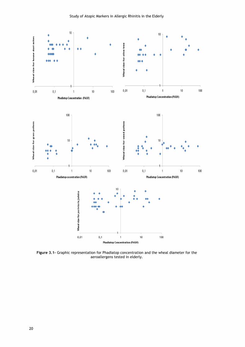
Figure 3.1- Graphic representation for Phadiatop concentration and the wheal diameter for
the aeroallergens tested in the elderly .................................................................. 20
Figure 3.2- Graphic representation for Phadiatop concentration and the wheal diameter for
the aeroallergens tested in young adults ................................................................ 21
Figure 3.3- Graphic representation of the distribution of the Phadiatop and total IgE
concentrations in all volunteers........................................................................... 22
Figure 3.4- Graphic representation of the distribution of the Phadiatop and total IgE
concentrations in the young adults ....................................................................... 22
Figure 3.5- Graphic representation of the distribution of the Phadiatop and total IgE
concentrations in the elderly .............................................................................. 23
Figure 3.6- Graphic representation of volunteers age versus total IgE ............................ 23

Study of Atopic Markers in Allergic Rhinitis in the Elderly
xiv

Study of Atopic Markers in Allergic Rhinitis in the Elderly
xv
List of Tables
Table 3.1- Number of completed questionnaires, skin prick tests, Phadiatop and total IgE
analysis ........................................................................................................ 15
Table 3.2- Academic degree of the study population ................................................. 15
Table 3.3- Jobs classification, stratified according to the National Reader Survey Scale,
United Kingdom .............................................................................................. 16
Table 3.4- Residence of the study population .......................................................... 17
Table 3.5- Distribution of Phadiatop in the two groups .............................................. 17
Table 3.6- Phadiatop distribution by gender in young and elderly ................................. 18
Table 3.7- Comparison between Phadiatop and SPT results ......................................... 18
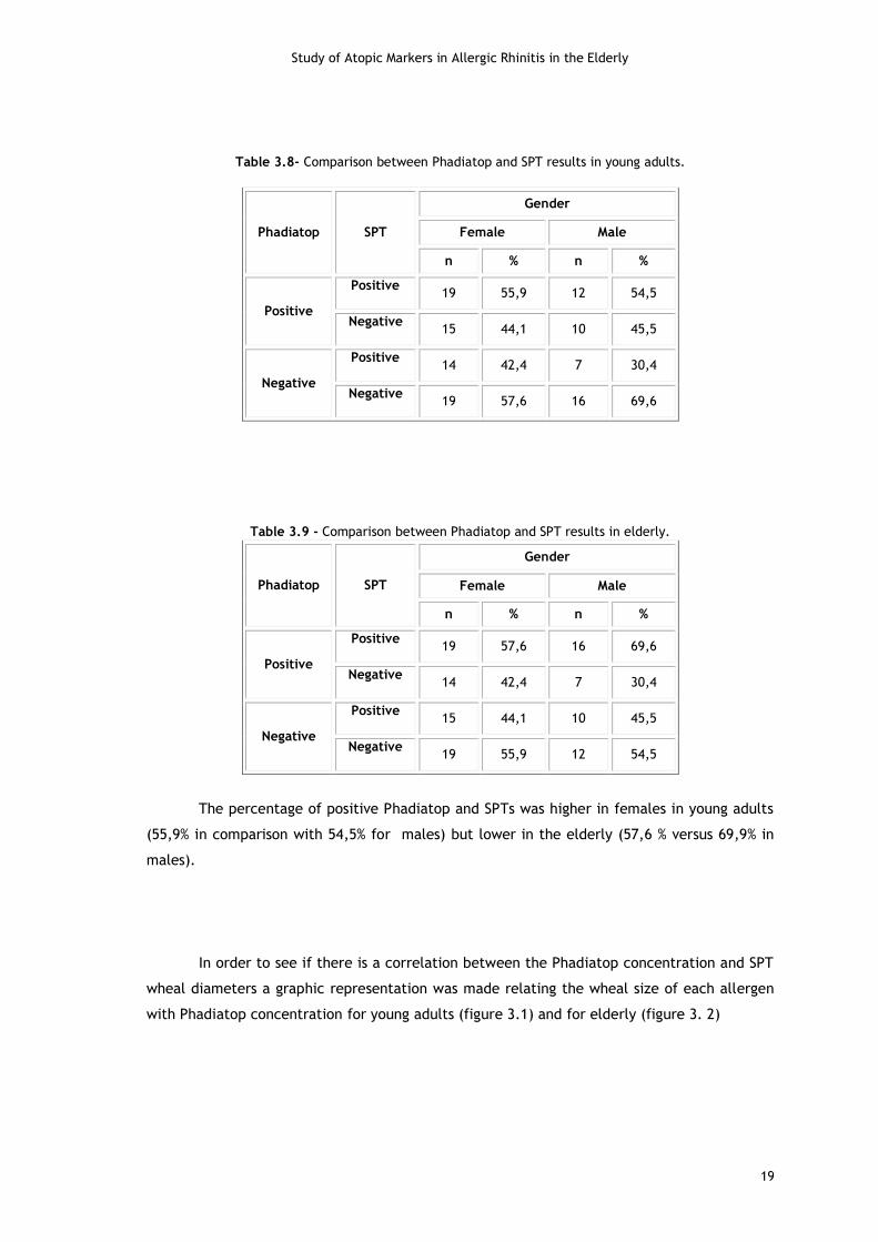
Table 3.8- Comparison between Phadiatop and SPT results in young adults...................... 19
Table 3.9- Comparison between Phadiatop and SPT results in the elderly ........................ 19
Table 3.10- Volunteers with AR ........................................................................... 24
Table 3.11- Distribution of SPT in the two groups ..................................................... 24
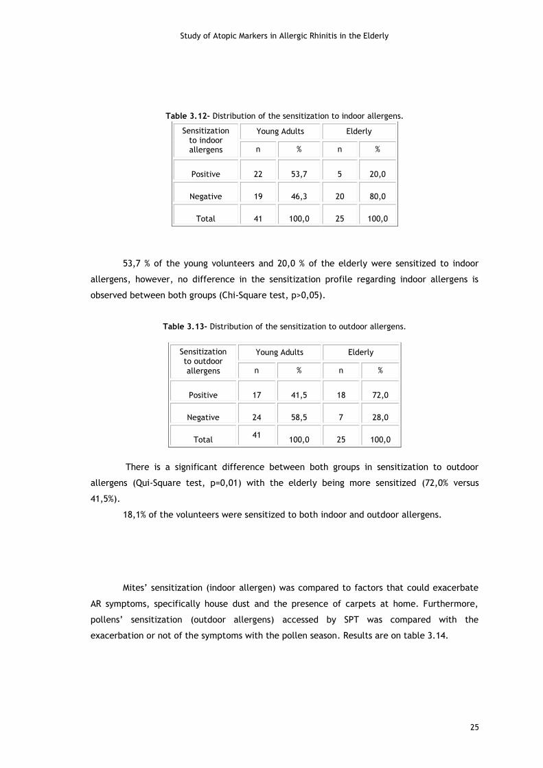
Table 3.12- Distribution of the sensitization to indoor allergens ................................... 25
Table 3.13- Distribution of the sensitization to indoor allergens ................................... 25
Table 3.14- Relation between positive SPT for mites and symptoms worsened by house dust and pollens .................................................................................................................. 26

Study of Atopic Markers in Allergic Rhinitis in the Elderly
xvi

Study of Atopic Markers in Allergic Rhinitis in the Elderly
xvii
List of Abbreviations
AR Allergic Rhinitis
ARIA Allergic Rhinitis and its Impact on Asthma
IgE Immunoglobulin E
IgG Immunoglobulin G
IL-2 Interleukin 2
ISSAC International Study of Asthma and Allergies in Childhood
PAQUID Personnes Agées Quid
QRESERCH Quantum Marquet Research
SAPALDIA Swiss Study on Air Pollution and Lung diseases in Adults
SPT Skin Prick Test
TH1 Helper T 1
TH2 Helper T 2
IFN Interferon

Study of Atopic Markers in Allergic Rhinitis in the Elderly
xviii
Study of Atopic Markers in Allergic Rhinitis in the Elderly
1
Chapter 1
Introduction
1.1 Allergic Rhinitis
1.1.1 Classification
Rhinitis is defined as an inflammation of the nasal mucosa and is characterized by one
or more of the following symptoms: congestion, rhinorrhea, itching of the nose, postnasal
drip, and sneezing1,2. In the geriatric population, a broad interpretation of this symptom
complex may also include crusting within the nose, cough, excessive drainage, olfactory loss,
and nasal dryness.
Rhinitis can be classified by etiology as allergic or nonallergic and differentiated from
conditions that mimic symptoms of rhinitis3.
Nonallergic rhinitis is characterized by non-immunoglobulin E (IgE)-mediated
symptoms typical of rhinitis, such as congestion and clear rhinorrhea, with less proeminence
of sneezing and ocular/nasal pruritus. The associated symptoms may be perennial or
sporadic, lacking a clear seasonality, and may be exacerbated by nonspecific triggers such as
odors, food, emotions, or change in atmospheric conditions1.
Approximately 50% of all cases of rhinitis are caused by allergy4. The condition
originates when individuals are exposured to allergens they are sensitized to, like airborne
agents such as pollens, mold spores and dust-born mites5,6. Allergic rhinitis (AR) is induced by
exposure to allergens that trigger an IgE-mediated inflammation of the nasal passageways
that can result in chronic or recurrent symptoms of rhinorrhea, congestion and sneezing1,5.
Itching of the ears and throat can also be associated with allergic rhinitis7. AR is often
accompanied by allergic rhinoconjuctivitis (a complex sometimes referred to as allergic
rhinoconjuctivitis) that results in conjunctival injection and chemosis and symptoms of itchy
eyes and tearing7.
Symptoms of AR may be classified as seasonal or perennial. An international working
group modified this classification scheme due to potential difficulties in differentiating
between seasonal and perennial symptoms and created the Allergic Rhinitis and its Impact on
Asthma (ARIA) Report. The ARIA guidelines temporally classify AR as 'intermittent' if symptoms
are present less than four days per week or less than four consecutive weeks, or as 'persistent'
if symptoms are present more than four days per week and for more than four consecutive
weeks. Severity of symptoms is graded as 'mild' if they are present but not troublesome, and
as 'moderate/severe' if they lead to sleep disturbance, impairment of daily activities, or
impairment of school or work1’8.
Study of Atopic Markers in Allergic Rhinitis in the Elderly
2
1.1.2 Physiological changes
Rhinitis is an inflammatory disease, as such, mechanisms and presentation of the
condition are altered as immune function changes with age, a concept entitled
immunosenescence. A critical component of the immune system is the thymus, which rapidly
involutes from adolescence to near middle age, followed by an approximate 1% cellular loss
per year thereafter. The decline in functional mass causes depressed production of naïve T-
cells leading to impaired cell-mediated immunity. Despite thymic involution, the total T-cell
pool remains constant due to an increase in production of memory T-cells. With the aging
process T-cell responsiveness to growth factors decreases, lymphocyte response to specific
antigens is altered, and IL-2 production and receptor expression are diminished. B-cells also
change with age; although the peripheral B-cell population remains constant, there is less IgG
isotype class switching, and the number of antigen-specific antibodies decreases while the
number of autoantibodies and circulating immune complexes increase. These changes might
also contribute to the milder symptoms as well as the decreased incidence of allergic rhinitis
in the geriatric population. Furthermore, as individuals age, several changes in nasal anatomy
and physiology occur which may affect the development and expression of rhinitis1.
1.1.3 Pathophysiology and Clinical Presentation of Rhinitis
AR is the result of type I hypersensitivity reactions whereby exposure to allergens in
susceptible individuals leads to sensitization by production of specific IgE antibodies directed
against these extrinsic proteins. This antibody then binds to the surface of mast cells, and
when the allergen is reintroduced, IgE cross-binding to the antigen leads to mast cell
degranulation. Within seconds of contact, inflammatory mediators such as histamine,
leukotrienes, and prostaglandin D2 are released causing vascular endothelial dilation, which
subsequently causes leakage and mucosal edema. This leads to nasal obstruction and
symptoms of congestion, redness, tearing, swelling, ear pressure, and postnasal drip. Irritant
receptors are stimulated by the allergen causing itching and sneezing1.
Within four to eight hours of initial exposure, cytokines attracted by previously
released mediators lead to recruitment of other inflammatory cells to the mucosa, such as
neutrophils, eosinophils, lymphocytes, and macrophages (see figure 1.1). The inflammation
persists and this stage is termed the late-phase response. The late-phase response presents
similarly to the early phase, however, sneezing and itching are less proeminent, whereas
congestion and mucus production are more severe. The late phase may persist for hours or
days1.

Study of Atopic Markers in Allergic Rhinitis in the Elderly
3
When allergen challenges are given repeatedly, the amount of allergen required to
induce an immediate response decreases. This priming effect is thought to be a result of the
release of inflammatory mediators from effector cells during ongoing, prolonged allergen
exposure and repeated late-phase responses. Consequently, at the end of a pollen season,
symptoms may decline at a slower rate than the pollen count. Therefore, it is important to
know the full spectrum of aeroallergens to which the patient responds as well as seasonal
variations in symptoms3.
Individual host sensitivity to an aeroallergen influences the intensity of symptoms; for
example, the pollen counts that cause symptoms may vary on the basis of an individual’s
degree of sensitivity and may be different for different pollens. Studies have not been
consistently able to demonstrate symptom and/or medication reduction with any of the
commonly used environmental control measures in patients with rhinitis3.
Patients with AR caused by pollens may be exposed to allergen from nonpollen plant
fragments, allergenic bioaerosols without intact pollen grains, and even high pollen
concentrations of insect-pollinated plants. Pollen counts are generally highest on sunny,
windy days with low humidity. Because the interplay of different weather factors (eg, wind,
temperature, rain, and humidity) is complex, it may not be possible to reliably predict levels
of outdoor aeroallergens on the basis of the influence of a single weather factor3.
1.1.4 Atopy and IgE
Atopy is a predisposition to develop an IgE-mediated immune response to
environmental allergens that do not sensitize nonatopic individuals. The expression of an
atopic phenotype requires the interaction of a partly genetic predisposition with
environmental allergen exposure9.
Atopy is known to increase the risk of developing allergic disorders such as AR. Atopy,
as measured by allergen skin prick testing, is an important attribute of allergic disease. Not
all atopic individuals are symptomatic and not all those with allergic symptoms are atopic10.
According to Gold and Kemp, the distinction between atopy and atopic disease is
important. A child with atopy produces specific IgE antibodies after exposure to common
environmental allergens and is said to be sensitized to that allergen. Eczema, asthma and
Figure 1.1- Example of an allergic response. Adapted from Togias A. et al 27

Study of Atopic Markers in Allergic Rhinitis in the Elderly
4
rhinoconjunctivitis are clinical syndromes each defined by a collection of symptoms and signs
and are commonly referred to as the atopic diseases. While most children with these
conditions are atopic, some are not, and, conversely, some children with atopy may not
manifest atopic disease11.
IgE concentration in the serum is the lowest of the 5 immunoglobulin subtypes, has
the shortest half-life (approximately 2 days) and its expression is tightly regulated in the
absence of disease. IgE shows no transplacental transfer. In the absence of disease, IgE levels
in cord blood are low (<2 kIU/L; < 4.8 mg/L), gradually increase throughout childhood with a
peak at 10 to 15 years of age, and then decrease throughout adulthood. Total IgE levels are
also influenced by genetic makeup, race, immune status, and environmental factors (eg,
pollen exposure)9.
Increased IgE levels are seen in patients with atopic diseases, with the highest levels
generally being seen in patients with atopic dermatitis, followed by those with atopic asthma,
perennial allergic rhinitis, and seasonal allergic rhinitis. For seasonal allergens, peak IgE
levels occur 4 to 6 weeks after the peak of the pollen season. Increased IgE levels are also
seen in other disorders, including parasitic infections (eg, strongyloidiasis, ascariasis, and
schistosomiasis), nonparasitic infections (eg, EBV, cytomegalovirus, HIV, and Mycobacterium
tuberculosis), inflammatory diseases (eg, Kimura disease, Churg-Strauss vasculitis, and
Kawasaki disease), hematologic malignancies (eg, Hodgkin´s lymphoma and IgE myeloma),
cutaneous diseases (eg, Netherton syndrome and bullous pemphigoid), cystic fibrosis,
nephrotic syndrome, and primary immunodeficiency diseases. Increased IgE levels are also
detected after hematopoietic stem cell transplantation, in smokers (particularly male
smokers), and in those with alcoholism9.
The presence of one allergic disorder significantly increases the risk of developing
other allergic disorders, affecting different organ systems12.
Patients with AR have allergen-specific IgE demonstrable both systemically (e.g.,
positive skin tests) as well as local IgE produced in the nasal mucosa3.
Serum total IgE measurement was one of the first allergic inflammation marker tests.
It has been extensively used for the diagnostic of allergy23. The Phadioatop test is a specific
IgE test for multiple allergens being its main use as a screening test6.
For evaluation and diagnosis a thorough allergy history remains the best diagnostic
tool available. The history will include the patient’s chief concerns and symptoms and often
includes the pattern, chronicity, seasonality, and triggers of nasal and related symptoms,
family history, current medications, occupational exposure, and a detailed environmental
history. Questions relating symptoms to pollen and animal exposure have been shown to have
positive predictive value for diagnosing allergic rhinitis3.
Determination of specific IgE, preferably by skin testing, is indicated to provide
evidence of an allergic basis for the patient’s symptoms, confirm suspected causes of the
patient’s symptoms, or assess the sensitivity to a specific allergen for avoidance measures

Study of Atopic Markers in Allergic Rhinitis in the Elderly
5
and/or allergen immunotherapy3. Blood eosinophil count and total serum IgE level tend to be
elevated in allergic rhinitis13.
The prevalence of allergic sensitization is lower in the most advanced ages14. Many
studies concluded that atopy decreases with increasing age either in the general population
samples or in population samples of healthy individuals (i.e. with no allergy-related
symptoms). There is also a higher prevalence in females than in males, in spite of that,
lifetime prevalence of AR is higher in males before the teenage prevalence peak15’1’16. The
observation of lower IgE in older subjects compared with younger subjects has been reported
in cross-sectional studies. Morais-Almeida and colleagues estimated a prevalence of rhinitis of
26% in a large population sample selected in the primary care centres of Mainland Portugal
including 6859 questionnaire responses corresponding to a mean age of 48.3 years16.
According to results from SAPALDIA Study, which studied the influence of sex, age and
smoking habits on total serum IgE and allergen-specific IgE antibody concentrations (assessed
by means of the Phadiatop test) on the prevalence of hay fever; Phadiatop tests, positive skin
tests and atopy decreased significantly with age. This study also demonstrated that tobacco
smoking is associated with increased IgE levels and negatively related to atopy and hay
fever29. Another study analyzed results from 8329 randomized adults from SAPALDIA Study
and revealed that the prevalence of positive Phadiatop, positive SPT (at least, one out of
eight SPT to common aeroallergens with a wheal of > or = 3 mm), and positive total IgE (IgE >
or = 100 kU/L) were 29, 23, and 23%, respectively 17. Some studies have shown a significant
age-related decline of IgE for both genders, with levels in females being significantly lower
than in males18. A study conducted in France (PAQUID cohort) with subjects aged 65 years and
over also found an association between smoking and IgE level independent of allergic
reactivity to common allergens in the elderly. Questionnaires were applied by telephone or
letter and total serum IgE and Phadiatop were determined. A positive Phadiatop test was not
related to gender and smoking but significantly associated with total IgE and rhinitis. The
study demonstrates persistence of respiratory allergies after age 65 years and confirms an
association between smoking and IgE level independent of allergic reactivity to common
allergens for elderly people. Furthermore, IgE level was significantly higher in males than in
females18.
Another study performed in Germany analyzed questionnaires, total IgE and specific
IgE and concluded that the total IgE had a negative correlation with age in all patients and
also allergen specific IgE was significantly decreased in the elderly suffering from AR19.
1.1.5 Epidemiology
Allergic rhinitis (AR) is a prevalent disease worldwide, affecting 10% to 25% of the
world population1,2,5. AR prevalence has increased during the past few decades. Though its
peak incidence is during young adulthood, AR is prevalent among older people. In fact, the
2005 National Center for Health Statistics report stated that 10.7% of individuals between 45-

Study of Atopic Markers in Allergic Rhinitis in the Elderly
6
64 years of age, 7.8% of patients 65-75 years of age, and 5.4% of patients older than 75 are
affected by AR. Along with the anatomic and physiologic changes of the nose, non-specific
immune changes such as decreased mucus production and ineffective cough mechanisms are
all thought to contribute to persistent or late-onset allergic disease in older people, as these
processes are necessary for clearance of allergens and irritants.
A cross-sectional study, based on questionnaires and SPT, made in East Germany
showed that the prevalence of hay fever and atopic sensitization increased significantly
between 1991 and 199620. In England, Ghouri and co-workers analyzed the incidence rate per
1000 person-years of AR for each of five years from the QRESEARCH database. These data
showed an overall 33.0% increase during the period 2001-200515. In Belgium, the prevalence
of AR was 29.8%21.
Phase 1 of the ISAAC study reported worldwide rates of rhinoconjunctivitis in the
range of 1.4-39.7% in adolescents of 13-14 years of age, and between 0.8-14.9% in children
aged 6-7 years22.
1.1.6 Risk factors Several studies have shown that the frequency of AR increases with age and positive
allergy skin tests are significant risk factors for the development of new symptoms of hay
fever4. Risk factors for AR include family history of atopy, serum IgE>100 IU/mL before age 6
years and the presence of a positive allergy SPT3. There appears to be a higher prevalence of
rhinitis in higher socioeconomic classes, in nonwhites, in some polluted areas and in
individuals born during the pollen season. Additionally, studies in children in the first year of
life have shown that the risk of rhinitis was higher in those youngsters with early introduction
of foods or formula, heavy maternal cigarette smoking in the first year of life, exposure to
indoor allergens such as animal dander and dust mites and parental disorders4. Children with
a bilateral family history of atopy may develop symptoms more frequently and at a younger
age than those with an unilateral family history. Aeroallergen sensitization rarely begins
before 6 months of age but may start between 6 months and 2 years of life. Infants born to
atopic families are sensitized to pollen aeroallergens more frequently than to indoor
aeroallergens in the first year of life. Seasonal allergic rhinitis symptoms generally do not
develop until 2 to 7 years of age. The prevalence of seasonal AR is higher in children and
adolescents, whereas perennial allergic rhinitis has a higher prevalence in adults3.
1.1.7 Effects of rhinitis on quality of life
AR can be a considerable source of morbidity in poorly managed patients. It impairs
social and work functions, and can significantly affect the patient’s quality of life13. Several
studies have shown the deleterious effects of rhinitis on the quality of life in symptomatic
patients1. Nasal obstruction can cause sleep disturbances that reduce a patient’s daytime
concentration and lead to daytime sleepiness. Complaints of poor sleep are already common

Study of Atopic Markers in Allergic Rhinitis in the Elderly
7
among older individuals due to various sleep disorders as well as the normal aging process,
thus AR may exacerbate these problems23. Lack of sleep can alter physiological processes such
as glucose metabolism, cognition, appetite control, and endocrine function, all critical
physiologic processes in older people1.
AR is also capable of markedly altering the patient’s performance, learning and
productivity. In addition, AR is commonly associated with other respiratory diseases, like
asthma (ARIA), and the cost resulting from these comorbidities increases even more the
socioeconomic impact of the disease 24.
1.2 Objective of the study
Although AR has a high prevalence and greatly impacts on daily life, there are only
few studies if we compare them with those on other allergic diseases. Therefore the aim of
this study was to investigate the relationship between Phadiatop, serum total IgE
concentration and SPT and allergic rhinitis symptoms in a population of elderly and young
adults of Cova da Beira.

Study of Atopic Markers in Allergic Rhinitis in the Elderly
8

Study of Atopic Markers in Allergic Rhinitis in the Elderly
9
Chapter 2
Methods
2.1 Study Design and Selection of Subjects
This study was carried out at the Faculty of Health Sciences of the University of Beira
Interior and at the Covilhã Primary Health Care Centre.
This was a cross-sectional study using a random sample. The population consisted of 2
groups of individuals living in Cova da Beira: one of young adults (born between January 1976
and December 1993) and another of elderly individuals (born before 1944).
Participants were randomly selected using a stratified strategy from individuals who
were registered in the list of all General Practitioners (GP) at Covilhã Primary Health Care
Centre. Stratification implied selecting the patients according to the 2 age groups.
The study was approved by the Ethics Committee of Sub-Regional Health
Administration of Castelo Branco and all volunteers signed a written informed consent, in
accordance with the Declaration of Helsinki (Annex I).
2.2 Subject Recruitment
Volunteers’ recruitment started on June 2008 through 2011. Data analysis includes all
information collected on that period (questionnaires, skin prick tests, Total IgE and
Phadiatop).
Initially, volunteers were contacted by post mail. If they didn’t reply to the letters
they lately were contacted by phone. Those who were not currently living in Cova da Beira,
who had died or that we were unable to contact were excluded from the study.
Patients who refused to participate in the study or were not able to go to the Primary
Health Care Centre were asked to answer the questionnaire by phone.
2.3 Questionnaires
A standardized questionnaire was given to all selected individuals (Annex II). It
contained validated questions regarding signs and symptoms of allergic rhinitis and atopy-
related risk factors like animal ownership, housing conditions or smoking exposure. We also
collected data about demographic, clinical situation, personal and familiar history.
Study of Atopic Markers in Allergic Rhinitis in the Elderly
10
Furthermore, the questionnaires included relevant data about age, gender, place of
residence, smoking habits, education and current and past professions. The completion of the
questionnaires was made by the investigators.
2.4 Skin Prick Tests
SPT are employed for screening allergic sensitisation in patients with suspected
allergic diseases. SPTs were performed on the volar aspects of both forearms, using a battery
containing 3 single and 2 mixtures of the most prevalent aeroallergens in Cova da Beira
(Annex III). Allergens used in this study were chosen according to a previous study that
revealed the most prevalent allergens in Cova da Beira and include house dust mites
(Dermatophagoides pteronyssinus), olive tree (Olea europea), grass pollen, weed pollen and
Parietaria judaica. Histamine (10mg/ml) and allergen dilluent were used as positive and
negative controls, respectively. Allergen extracts were manufactured by Leti (Barcelona,
Spain) and all belonged to the same batch.
The skin was disinfected with ethanol and numbers were written on it to indicate
where the allergen extracts would be located. A drop of each allergen extract was placed
upon the epidermis and then pricked through using a 1.5 mm-long lancet tip (Stallergenes,
France).
The mean wheal size was recorded after 15 minutes with a wheal size reader. SPT
was regarded as positive with a wheal size minimum of 3 mm and if the response was less
than 2 mm diameter, the SPT was regarded as negative. A skin test panel was considered
valid if the correct outcomes for the controls were verified, including a histamine wheal
greater than 3mm in diameter and an absence of wheal at the negative control site.
Otherwise tests were considered inconclusive and if possible were repeated at least one week
afterwards.
In the case volunteers had taken tricyclic antidepressants or antihistamines or if they
had applied any product on the skin containing corticosteroids within the previous 7 days, skin
prick tests would be postponed.
2.5 Definition of rhinitis and AR
Rhinitis was defined by a positive response to the question number 2.1 and current
rhinitis by a positive response to the question number 2.2 of the questionnaire, or if the
volunteer was under medication for the treatment of allergic rhinitis.
When rhinitis symptoms were reported by volunteers (positive response to the 2.2
question of the questionnaire) with both negative SPTs and Phadiatop they were regarded as

Study of Atopic Markers in Allergic Rhinitis in the Elderly
11
having non-allergic rhinitis. If patients had symptoms of rhinitis and atopy (positive response
to the 2.2 question of the questionnaire) and they were confirmed by positive SPTs and/or
positive Phadiatop, they were regarded as having AR.
2.6 Sample Processing for Total IgE and Phadiatop
A sample of 10 ml of peripheral blood was collected from patients by venopuncture,
into a biochemistry tube containing coagulation accelerator (SARSTEDT, Germany) and two
hemogram tubes containing EDTA (SARSTEDT, Germany).
Venous blood from the biochemistry tube was centrifuged at 4000 rpm for 10 minutes.
Serum samples were stored at -20ºC until tested. Both Total IgE and Phadiatop tests were
made in Hospital Sousa Martins, Guarda. Leucocytes were isolated from the hemogram tubes,
after red blood cells lysis, washing and subsequent frizzing in liquid nitrogen for posterior
analysis.
2.7 Phadiatop
ImmunoCAP Phadiatop (Phadia, Sweden) is a blood test designed to differentiate
between atopic and non-atopic patients. Results indicate high or low probability for atopy. A
negative result indicates that the symptoms are not caused by common environmental
allergens, and the physician may explore other possibilities. The manufacturer has not
revealed the precise formulation of the test.
Phadiatop assay is graded for the determination of atopy with semiquantitative or
qualitative results.
Semiquantitative Phadiatop results are expressed as Phadia Arbitrary Units/L (PAU/L)
indicating the degree of sensitization. A Phadiatop PAU/L value above the limit of
quantification indicates that the patient is atopic (positive), i.e. measurable levels of specific
IgE antibodies to common inhalant allergens have been detected. A Phadiatop PAU/L value
below the limit of quantification indicates that the patient is non-atopic (negative), i.e. the
level of specific IgE antibodies is undetectable. Higher Phadiatop PAU/L values indicate a
higher degree of sensitization, i.e. higher levels of specific IgE antibodies to common inhalant
allergens.
Qualitative Phadiatop results are expressed as positive or negative. A positive
Phadiatop result indicates that the patient is atopic, a negative result indicates that the
patient is non-atopic, i.e. not sensitized to inhalant allergens.
The technology of the test is based on an extremely high total binding capacity,
achieved through a high binding capacity per mg cellulose in combination with an optimal

Study of Atopic Markers in Allergic Rhinitis in the Elderly
12
amount of cellulose in each solid phase. This ensures binding of all relevant antibodies,
regardless of antibody affinity, still giving low non-specific binding.
The ImmunoCAP solid phase consists of a cellulose derivative enclosed in a capsule.
The hydrophilic, highly branched polymer provides an ideal microenvironment for allergens,
binding them irreversibly while maintaining their native structure.
The test is designed as a sandwich immunoassay. A balanced mixture of relevant
inhalant allergens, covalently coupled to the solid phase, reacts with the specific IgE in the
patient serum sample. After washing away non-specific IgE, enzyme-labelled antibodies
against IgE are added to form a complex. After incubation, unbound enzyme-labelled anti-IgE
is washed away and the bound complex is then incubated with a developing agent. After
stopping the reaction, the fluorescence of the eluate is measured. The higher the
flouresence, the more specific IgE is present in the sample.
2.8 Total IgE
ImmunoCAP Total IgE is an in-vitro test for quantitative measurement of the total
amount of circulating IgE in human serum or plasma samples. IgE antibodies appear as a result
of sensitisation to allergens and measurement of circulating total IgE provides an aid in the
clinical diagnosis of IgE-mediated allergic disorders. Elevated levels of circulating total IgE
are usually seen in patients suffering from extrinsic asthma, hay fever or atopic eczema.
The serum concentration of total IgE is age-related. It increases during childhood and,
at about 10 years of age, serum total IgE reaches values that are maintained during adult life.
The technology is based on a similar system as that of Phadiatop.
Serum total IgE determination was made automatically in ADVIA Centaur® Bayer
analytic system and consisted in an sandwich immunoassay in two steps, using direct
chemiluminescence technology that uses constant amounts of two human anti-IgE antibodies.
The first one is a caprine anti-human IgE antibody labeled with acridine ester; the second one
is in the solid phase (covalently binded to paramagnetic particles) and is an anti-human IgE
mouse antibody. Volunteers’ samples were incubated with both antibodies and after a
washing step, equal amounts of acid and basic reagent were added to initiate the
chemiluminescent reaction. Light relative unities amount (RLUs) detected by the system is
proportional to total IgE quantity on the sample. Calibration curve values were introduced in
the system through a code bar reader and after that, an adjustment to the curve was made
using two calibrators standardized by the preparation of 75/502 World Health Organization
reference.

Study of Atopic Markers in Allergic Rhinitis in the Elderly
13
2.9 Statistical analysis
All statistical analyses were performed using Statistical Package for the Social
Sciences (SPSS).
Data obtained were studied based on the relative and absolute frequencies of each
studied variable (descriptive statistics). Chi square tests were used to compare the
distribution of the factors between both groups (young adults and elderly adults). A p value of
less than 0.05 was considered statistically significant.

Study of Atopic Markers in Allergic Rhinitis in the Elderly
14

Study of Atopic Markers in Allergic Rhinitis in the Elderly
15
Chapter 3
Results
3.1 Characterization of the Study Population
There were 1835 patients randomized into this study (926 young adults and 909
elderly). From those, 416 accepted to participate in this study. Of the 416 volunteers 403
patients answered to a validated questionnaire and 381 patients were evaluated for their skin
prick test response to 5 common regional aeroallergens. In addition, 356 were analyzed for
their serum total IgE and Phadiatop. Following analysis were carried out in this subgroup of
volunteers.
Table 3.1- Number of completed questionnaires, skin prick tests, Phadiatop and total IgE analysis
Elderly adults Young Adults Total
Questionnaires 270 132 402
Skin Prick Tests 252 128 380
Phadiatop 239 117 356
Total IgE 239 111 350
Among the 357 random subjects involved, there were 239 elderly subjects (mean age
= 73; 141 females) and 117 young adult subjects (mean age= 28; 70 females).
Both groups were paired regarding gender. Data on academic degree, jobs and
residence can be found on the following tables.
Table 3.2- Academic degree of the study population
Elderly Young
Adults
% n % n
Academic degree
No studies
14,23 34 0% 0
Less than 4 years
35,15 84 0% 0
4-9 years
37,66
90 14,5% 16

Study of Atopic Markers in Allergic Rhinitis in the Elderly
16
There is a significant difference between academic degree of both groups (Chi-square
test, p<0,000). All young adults studied more than 4 years and 45% studied more than 12
years. In contrast, 14% of the elderly didn’t study at all and only 4% studied more than 12
years.
Table 3.3- Jobs classification, stratified according to the National Reader Survey Scale, United
Kingdom26
Elderly Young Adults
% n % n
Jobs
A 7,4 17 19,3 18
B 11,7 27 15,0 14
C1 10,0 23 19,3 18
C2 23,0 53 10,7 10
D 29,0 67 12,9 12
E 16,5 38 22,5 21
Total 100,0 231 100,0 93
A- Upper middle class; B-middle class; C1- lower middle class: C2-skilled working class; D- working
Class; E- those at the lower level of subsistence
There is a significant difference between jobs stratification of both groups (Chi-square
test, p<0,001). Young adults have a major percentile at “A” (19%) and “E” (23%) classification
and the elderly have a major percentile to the “C2” (23%) and “D” (29%) classification.
9-12 years
9,21 22 40,9% 45
More than 12 years
3,77
9 44,5% 49
Total
100%
239
100%
110
Didn’t
answered
7

Study of Atopic Markers in Allergic Rhinitis in the Elderly
17
Table 3.4: Residence of the study population
Elderly Young Adults
% n % n
Residence
Urban 81,5 194 82,9 97
Rural village
6,7 16 10,2 12
rural farm
10,9 26 1,7 2
Rural and
urban 0,8 2 5,1 6
Total 100,0 238 100,0 117
The majority of the volunteers have urban residence (83% and 82%, for young adults
and elderly, respectively). However there are statistically significant differences between
young adults and elderly regarding to residence, probably due to the different distribution on
the rural residence (Chi-square, p= 0,005).
3.2 Phadiatop results and SPT
We next analyzed the data regarding Phadiatop test. Of the 239 elderly volunteers,
38 had positive Phadiatop (16%). On the young adults group, 58 of the 117 had positive
Phadiatop (50%), which means that the prevalence of atopy measured by Phadiatop was
significantly higher in the young adults group (Chi-Square test, p=0,000).
Table 3.5 - Distribution of Phadiatop in the two groups
Young Adults Elderly
n % n %
Positive Phadiatop
58 49,6 38 15,9
Negative Phadiatop
59 50,4 201 84,1
Total 117 100,0 239 100,0

Study of Atopic Markers in Allergic Rhinitis in the Elderly
18
Significant difference was found in the distribution of Phadiatop with gender in young
adults (Chi-Square test, p=0,02) and elderly (Chi-Square test, p=0,05); with higher
frequencies being found in males (53,2% and 21,2 %) (Table 3.6 ).
Table 3.6- Phadiatop distribution by gender in young and elderly.
Young Adults Elderly
Male Female Male Female
n % n % n % n %
Phadiatop Positive 25 53,2 33 47,1 21 21,2 17 12,1
Negative 22 46,8 37 52,9 78 78,8 123 87,9
Total 47 100 70 100 99 100 140 100
Assuming Phadiatop results as the gold standasr we found the existence of 83 false
positives and 28 false negatives (see table 3.7) when evaluating SPT.
Table 3.7- Comparison between Phadiatop and SPT results.
SPT
Positive Negative
Phadiatop Positive 63 28
Negative 83 165
Comparing sensitization to the SPTs and a positive Phadiatop, we obtained 62,5 % of
the positive Phadiatop young adults with also positive SPT (at least one positive SPT with a
wheal size minimum 3 mm). In regard to the elderly group, we obtained 80% of the Phadiatop
positive patients with positive SPTs.
The relation between Phadiatop and SPT is organized by gender for young adults and
elderly on tables 3.8 and 3.9, respectively.
Study of Atopic Markers in Allergic Rhinitis in the Elderly
19
Table 3.8- Comparison between Phadiatop and SPT results in young adults.
Phadiatop SPT
Gender
Female Male
n % n %
Positive
Positive
19 55,9 12 54,5
Negative
15 44,1 10 45,5
Negative
Positive
14 42,4 7 30,4
Negative
19 57,6 16 69,6
Table 3.9 - Comparison between Phadiatop and SPT results in elderly.
Phadiatop SPT
Gender
Female Male
n % n %
Positive
Positive
19 57,6 16 69,6
Negative
14 42,4 7 30,4
Negative
Positive
15 44,1 10 45,5
Negative
19 55,9 12 54,5
The percentage of positive Phadiatop and SPTs was higher in females in young adults
(55,9% in comparison with 54,5% for males) but lower in the elderly (57,6 % versus 69,9% in
males).
In order to see if there is a correlation between the Phadiatop concentration and SPT
wheal diameters a graphic representation was made relating the wheal size of each allergen
with Phadiatop concentration for young adults (figure 3.1) and for elderly (figure 3. 2)
Study of Atopic Markers in Allergic Rhinitis in the Elderly
20
Figure 3.1- Graphic representation for Phadiatop concentration and the wheal diameter for the aeroallergens tested in elderly.

Study of Atopic Markers in Allergic Rhinitis in the Elderly
21
Figure 3.2- Graphic representation for Phadiatop concentration and the wheal diameter for
tested aeroallergens in young adults.
Using Spearman’s test no correlation is observed for Phadiatop concentration and the
wheal diameter for aeroallergens in elderly or in young adults.

Study of Atopic Markers in Allergic Rhinitis in the Elderly
22
3.3 Phadiatop and total IgE concentration
Some studies state that there is not a clear correlation between Phadiatop positivity
and total IgE values. To access if this was also true in our population we performed
correlations between Phadiatop and total IgE values.
Figure 3.3- Graphic representation of the distribution of the Phadiatop and total IgE concentrations in all volunteers.
Using Spearman’s test no correlation is observed between Phadiatop and IgE concentration
in the total sample (young adults plus elderly). 0,35 is the value from which Phadiatop was
considered positive.
The distribution of the Phadiatop and total IgE concentrations in young adults and elderly
patients separately is represented on the following graphics.
Figure 3.4- Graphic representation of the distribution of Phadiatop and total IgE concentrations in the young adults.

Study of Atopic Markers in Allergic Rhinitis in the Elderly
23
Figure 3.5 Graphic representation of the distribution of Phadiatop and total IgE concentrations in the elderly.
Applying the Spearman test to the stratified population didn’t show any correlation
between Phadiatop and total IgE values (figures 3.4 and 3.5 respectively).
Figure 3.6 shows the values of total serum IgE and volunteers’ age.
Figure 3.6 – Graphic representation of volunteers’ age versus total IgE.
Applying Spearman test no correlation was found for the relation between total IgE
concentration and the age of volunteers. However, the mean total IgE concentration in young
adults is higher than in the elderly (81,6 and 71,7 respectively).

Study of Atopic Markers in Allergic Rhinitis in the Elderly
24
3.4 Clinical characteristics
As stated in the methods section, AR was defined as a positive answer to question 2.2 or use
of anti-allergic medication and a positive Phadiatop value. Thus 69 volunteers (27 elderly and 42
young adults) had AR (see table 3.10).
Table 3.10- Volunteers with AR
Young Adults Elderly
n
%
n
%
Have AR
42
48,9
27
16,6
Have not
AR
44
51,1
136
83,4
Total
86
100,0
163
100,0
There is a significant difference between both groups (Qui-Square test, p=0,000). Young
adults have a higher prevalence of AR than the elderly (48,9% versus 16,6%).
Comparing positive SPT of the volunteers with rhinitis of the two groups, we obtained 65,9 %
of young adults and 87,5 % of elderly with positive SPT (see table 3.11)
Table 3.11- Distribution of SPT in the two groups
Young Adults Elderly
n % n %
Positive SPT 27 65,9 21 87,5
Negative SPT
14 34,1 3 12,5
Total 41 100 24 100
There is a significant difference between both groups regarding the result of SPT in
volunteers with AR (Qui-Square test, p=0,05).
In order to access triggering factors for AR, positive SPTs were compared to reported
symptoms. Allergens were classified as indoor (mites) and outdoor (pollens). The distribution
of these allergens is on table 3.12 and 3.13, respectively.
Study of Atopic Markers in Allergic Rhinitis in the Elderly
25
Table 3.12- Distribution of the sensitization to indoor allergens.
Sensitization to indoor allergens
Young Adults Elderly
n % n %
Positive
22
53,7
5
20,0
Negative
19
46,3
20
80,0
Total
41
100,0
25
100,0
53,7 % of the young volunteers and 20,0 % of the elderly were sensitized to indoor
allergens, however, no difference in the sensitization profile regarding indoor allergens is
observed between both groups (Chi-Square test, p>0,05).
Table 3.13- Distribution of the sensitization to outdoor allergens.
Sensitization to outdoor
allergens
Young Adults Elderly
n % n %
Positive
17
41,5
18
72,0
Negative
24
58,5
7
28,0
Total
41
100,0
25
100,0
There is a significant difference between both groups in sensitization to outdoor
allergens (Qui-Square test, p=0,01) with the elderly being more sensitized (72,0% versus
41,5%).
18,1% of the volunteers were sensitized to both indoor and outdoor allergens.
Mites’ sensitization (indoor allergen) was compared to factors that could exacerbate
AR symptoms, specifically house dust and the presence of carpets at home. Furthermore,
pollens’ sensitization (outdoor allergens) accessed by SPT was compared with the
exacerbation or not of the symptoms with the pollen season. Results are on table 3.14.

Study of Atopic Markers in Allergic Rhinitis in the Elderly
26
Table 3.14- Relation between positive SPT for mites and symptoms worsened by house dust and pollens.
Symptoms worsened by triggering factors
Triggering factors
Yes No
n % n %
House
dust
Positive SPT for house dust mites
Yes 6 66,7 2 33,3
No 3 33,3 4 66,7
Carp
ets
at
hom
e
Positive SPT for
house dust mites
Yes 3 33,3 24 42,1
No 6 66,7 33 57,9
Pollens
Positive SPT
Yes 13 86,7 0 0
No 2
13,3 0
0
Symptoms worsened by triggering factors were assessed by a positive answer to question 2.4
(which includes house dust and pollens) and 5.2 (carpets at home) of the questionnaire.
Mites’ sensitization (indoor allergen) was not related to factors that could exacerbate AR
symptoms, specifically house dust and the presence of carpets at home (Chi-Square test, p>0,05).
Similarly, AR symptoms were not related to the presence of carpets or house dust (Chi-Square test,
p>0,05). However, a correlation was found for pollen sensitization (outdoor allergens) as all the
volunteers sensitized to pollens complained of worsening of symptoms in the pollen season.

Study of Atopic Markers in Allergic Rhinitis in the Elderly
27
Chapter 4
Discussion and conclusions
In comparison with asthma, allergic rhinitis have been less extensively investigated,
although this does not mean that it should be regarded as a minor disorder but rather as an
important pathology that affects the quality of life of the patients and their families,
generating considerable direct and indirect costs.
This is a multifactor disorder without a single causal agent, in which the most
important component is the genetic predisposition of the patient (atopy), modulated by
environmental factors, exposure to allergens, infections and irritants, among others 22.
Rhinitis in the elderly may be caused by types of rhinitis common in other age groups but
may also be influenced by age-related physiologic changes such as cholinergic hyperactivity,
anatomic changes and medications taken for other medical conditions (usually elderly take
many medication). Over the past few years, several studies supporting the hygiene
hypothesis have suggested that early exposure to viral and bacterial infections, such as day
care attendance or more siblings may reduce the incidence of atopic disease by redirecting
the immune system away from the allergic T-helper 2(TH2) pattern to the T-Helper 1(TH1)
pattern. One early explanation proposed that the increased incidence of atopy as explained
by the hygiene hypothesis is a result of the reduced production of IL-12 and interferons
(IFNs) by cells of the innate immune system that are normally stimulated by bacterial
products via their Toll-like receptors30.
From a clinical perspective, supported by epidemiological investigations, there
would appear to be a decline with age in both the incidence and severity of atopic diseases,
particularly among the elderly population who are 60 years or older. Atopic incidence
declines, symptoms severity declines, and there would appear to be a general humoral
alteration of the propensity for atopy reflected by age-associated declines in serum total IgE
values. The decline of the onset of allergic symptoms observed in ageing might result from a
decrease of serum total IgE due to an unbalance of cytokines and soluble factors involved in
its production28.
The natural course of allergy is based on persistent antigen exposure; the repeated
stimulation with identical antigenic proteins induces on one hand the expression of new
surface molecules and, on the other hand, the expansion of effector and memory cells. It
follows that, with aging, the number of naive cells decreases and the sensitized cells
increase.

Study of Atopic Markers in Allergic Rhinitis in the Elderly
28
Cross-sectional studies show that the prevalence of IgE sensitization is lower in older
age groups than younger age groups. This could reflect either a decrease in sensitization with
aging or a higher prevalence of sensitization in more recent birth cohorts.
Although IgE sensitization and total IgE are both associated with an increased risk of
allergic disease in individuals, they exhibit very different changes with aging and with
cohort.This may reflect their differing association with environmental factors29. Evidence that
the prevalence of atopic diseases, including asthma and hay fever, has increased over the
past 20-30 years comes mainly from questionnaire based surveys; objective measurements
being limited. Atopy can be demonstrated by SPT or increase in IgE measured as serum total
IgE or specific IgE. We measured SPTs and serological markers of atopic sensitization in stored
serum samples that had been collected from the population stated on Results. The serological
markers included total IgE and Phadiatop. Both measure IgE, but whilst total IgE provides a
measurement for all produced IgE independently of the underlying disorder, Phadiatop gives a
sensitisation screening by measuring the concentration of specific serum IgE against common
aeroallergens. We also used skin prick tests to assess atopy, but we must consider that they
measure not IgE, but skin mast cell degranulation. Skin prick test are the most useful single
modality for demonstrating an IgE-mediated underlying mechanism in suspected allergic
diseases. The test is reliable, cheap, and easy to perform and they offer a prompt result.
However, SPT may be subjected to a number of problems such as choice and storage of
allergens, prick test technique and individual interpretation. Advantages of serum specific IgE
assays are convenience for the patient, lack of risks and the possibility of testing subjects
unable to stop medication that could alter the results of SPT. A major disadvantage of serum
specific IgE assays is their high cost, especially in case of assays for multiples allergens.
Although the test developed successive variants, all of them have as the common principle to
test for serum specific IgE to a mixture of relevant allergens causing common inhalant
allergies. Diagnostic accuracy of Phadiatop may vary not only with the prevalence of allergic
sensitization in the studied population, but also with the aeroallergen profile of a given area.
In this sense is should be noted that the aeroallergen composition of Phadiatop is fixed and
not stated by the manufacturer.
In this study we used Phadiatop as our gold standard and results were based on its
positivity. The test is well characterized and has been used in other studies. In the PAQUID
cohort, Raherison and colleagues collected data by questionnaire and based their study on
total IgE concentration and Phadiatop to investigate the specific relationship of serum IgE and
Phadiatop with rhinitis and smoking habits using a random sample of 352 elderly subjects18.
Jarvis and co-workers in the ECRHS study tried to identify environmental risk factors for the
development of IgE sensitization in adult life determining serum specific IgE to common
allergens and total IgE, measured in 2 occasions about 9 years apart. In an observational,
descriptive, cross-sectional study, Navarro and co-workers studied a sample of allergic
patients treated in consultations in the Spain health system. The diagnosis of AR was mainly
based of clinical history, physical examination and skin prick tests29. Mediaty and colleagues

Study of Atopic Markers in Allergic Rhinitis in the Elderly
29
evaluated the effects of age on total and specific IgE in patients with AR across a population
of 559 individuals randomly selected19.
Our study showed that there were significant differences in demographic data of
young adults and elderly. All young adults studied more than 4 years whereas 14% of elderly
didn´t study at all and only 4 % studied more than 12 years unlike young adults that have a
higher percentage (45 %). These results are in agreement with the results achieved for jobs
stratification, which also have a significant difference between both groups. A higher
academic degree corresponds to a higher social class. Individuals who are better educated are
more likely to understand the pathophysiological basis of AR and hence the importance of
treatment, avoidance of allergens and prevention of episodes of disease. It has already been
proven that lower level of education is an independent predictor of a poor quality of life in
individuals with AR30.
Regarding residence, the majority of our volunteers have urban residence; however
there are significant differences in both groups, probably due to the different distribution on
the rural residence.
We also found that the Phadiatop positivity is significantly higher in the young adults
when compared with the elderly. This result is concordant with other studies in other
countries which have also reported that young adults have a higher sensitization than older
adults 19.
Furthermore, comparing genders, Phadiatop positivity is significantly higher in men of
both groups, 53,2% in young adults and 21,2% in elderly. These results are in conformity with
some studies that also found a higher prevalence in men18; however other studies found a
higher prevalence in women15, 6 . Regarding the association between the Phadiatop result and
the SPT we found the existence of 83 false positives and 28 false negatives. They were
classified as false positives if a SPT result was positive but Phadiatop was negative; false
negatives were those with a negative SPT but positive Phadiatop (considering Phadiatop our
gold standard). This could happen because SPT and Phadiatop don’t measure exactly the
same (as said before). Criteria for interpreting skin test results differ between younger and
older patients and the incorrect interpretation of skin testing may lead to false negative
responses. One possible explanation can be found in the age-associated reduction in skin
reactivity of the elderly to histamine and allergens.
We also looked for a correlation between the Phadiatop concentration and SPT wheal
diameters; graphic representations were made relating the wheal size of each allergen with
Phadiatop concentration for young adults and for elderly, but no correlation was observed.
Once again, this could be due to the different parameters each test determines.
Another atopic marker is serum total IgE. No significant correlation between
Phadiatop concentration and total IgE concentration was found neither for all population
either for young adults or elderly. Some studies state that there is not a clear correlation
between Phadiatop positivity and total IgE values. One possible explanation is that total IgE is
genetically determined while sensitization can be environmentally determined.

Study of Atopic Markers in Allergic Rhinitis in the Elderly
30
Many studies affirmed that total IgE decreases with increasing age 19’29. We obtained
no correlation. However, the mean total IgE concentration in young adults is higher than in
elderly (81,6 and 71,7 respectively). This could reflect either a decrease in sensitization with
aging or a higher prevalence of sensitization in more recent birth cohorts.
Defining AR with positive Phadiatop and presence of symptoms (question 2.2 of the
questionnaire) or use of anti-allergic medication, we found 69 volunteers with AR (42 young
adults and 27 elderly). The prevalence of rhinitis is higher in young adult, as reported in other
studies6’16.
Volunteers with rhinitis also showed a significant difference regarding to SPTs. There
is a significant difference between both groups regarding the result of SPT in volunteers with
AR. 87,5% of the elderly had positive SPTs and young adults had 65,9%. This could mean that
in elderly the combination of these two tests is more significant than in the young adults and
is an interesting result because is contradictory with less skin sensibilization that is common
in elderly.
Genetic factors may influence immunologic development. However current rapid
rise in allergic diseases cannot be fully explained only by genetic factors. The complex
interplay between immune responses of the host, the level and variety of the environmental
exposure, and the interactions between the genetic background and the range of exposures
are likely to affect the development of allergic diseases. To assess the involvement of the
gene-environment interaction in the onset of allergic disorders, Tanaka and colleagues
believed that it would be useful to list candidate environmental factors associated with
allergic disorders 30. In this study we chose three possible triggering factors: house dust and
presence of carpets at home (for indoor allergens) and pollens (for outdoor allergens). In
what concerns to outdoor allergens, we found a correlation for aggravation of the symptoms
in the pollen season when compared to the SPT sensitization for pollens; everyone who had
a positive SPT for pollens complained that pollens worsened their symptoms. Indeed, elderly
had a major prevalence of sensitization for outdoor allergens than for indoor (72% versus
20%). On reverse, young adults had major sensibilization for indoor allergens (53,7% versus
41,5%). However we found no correlation for indoor allergens. 66,7% of volunteers with
positive SPT for house dust mites also reported aggravation of rhinitis symptoms. This
difference could be due to the fact that the elderly belong to a decade in which people
worked more outdoor and then they were more exposure to outdoor allergens.
This study had some limitations. Participation in the study was volunteer, which may
have skewed the results as individuals are more likely to respond if they identify themselves
with the problem of the study. Increased awareness of the previously diagnosed patients as
well as the presence of symptoms of allergy probably explains thar the majority of
responding young adults reported symptoms. The sample selection was limited to individuals
registered at the Health Care Centre, and data refering to contact is incomplete or
outdated, especially for young adults. Another limitation is that potential cognitive

Study of Atopic Markers in Allergic Rhinitis in the Elderly
31
impairment can lead the subjects to give approximate responses in the questionnaire
because of memory problems.
Although there are some weaknesses in the present study, there were also some
strong points.Our study compared demographic, clinical and sensitisation characteristic
between elderly and young adults. In spite of a potential selection bias, our sample was
random and paired regarding gender. Finally, our methodological approach was thorough as
is involved not only a validated questionnarie, but also skin prick tests, total IgE and
Phadiatop evaluation.
In summary our study showed that young adults have a higher prevalence of allergic
rhinitis when compared with the elderly group and it is more prevalent in men in both
groups. Furthermore, the mean IgE concentration of the population was also higher in young
adults and we found that elderly are more sensitized to outdoor allergens while young
adults are more sensitized to indoor allergens.
4.1 Future perspectives
As this is an ongoing study we will continue to recruit and analyse volunteers in order to
achieve a bigger sample to allow us to extrapolate our data to the general population.
Furthermore, volunteers with positive qusetionnaires for AR will be frther studied in terms
of symptoms features, as recommended by the ARIA guidelines and using other
complementary diagnostic means to confirmate the diagnosis.

Study of Atopic Markers in Allergic Rhinitis in the Elderly
32

Study of Atopic Markers in Allergic Rhinitis in the Elderly
33
Chapter 5
Bibliography
1. Pinto JM, Jeswani S. Rhinitis in the geriatric population. Immunology And Allergy Clinics
Of North America. 2010:1-12.
2. Ashaʼ ari ZA, Yusof S, Ismail R, Che Hussin CM. Clinical features of allergic rhinitis and skin
prick test analysis based on the ARIA classification: a preliminary study in Malaysia. Annals of
the Academy of Medicine, Singapore. 2010;39(8):619-24.
3. Wallace DV, Dykewicz MS, Bernstein DI, et al. The diagnosis and management of rhinitis: an
updated practice parameter. The Journal of allergy and clinical immunology. 2008;122(2
Suppl):S1-84.
4. Skoner D. Allergic rhinitis: Definition, epidemiology, pathophysiology, detection, and
diagnosis*1. Journal of Allergy and Clinical Immunology. 2001;108(1):S2-S8.
5. Saleem T, Khalid U, Sherwani UUR, Ghaffar S. Clinical profile, outcomes and improvement
in symptoms and productivity in rhinitic patients in Karachi, Pakistan. BMC ear, nose, and
throat disorders. 2009;9:12.
6. Navarro a, Colás C, Antón E, et al. Epidemiology of allergic rhinitis in allergy consultations
in Spain: Alergológica-2005. Journal of investigational allergology & clinical immunology :
official organ of the International Association of Asthmology (INTERASMA) and Sociedad
Latinoamericana de Alergia e Inmunología. 2009;19 Suppl 2:7-13.
7. Dykewicz MS, Hamilos DL. Rhinitis and sinusitis. The Journal of allergy and clinical
immunology. 2010;125(2 Suppl 2):S103-15.
8. Bousquet J, Khaltaev N, Cruz a a, et al. Allergic Rhinitis and its Impact on Asthma (ARIA)
2008 update (in collaboration with the World Health Organization, GA(2)LEN and AllerGen).
Allergy. 2008;63 Suppl 8:8-160.
9. Stone KD, Prussin C, Metcalfe DD. IgE, mast cells, basophils, and eosinophils. The Journal
of allergy and clinical immunology. 2010;125(2 Suppl 2):S73-80.
10. Sibbald B, Rink E, Dʼ Souza M. Is the prevalence of atopy increasing? The British journal of
general practice : the journal of the Royal College of General Practitioners. 1990; 40 (337) :
338-40.
11. Gold MS, Kemp AS, JA. Practice Essencials-Paedriatrics Journal M. M 2005;182(6):298-304.

Study of Atopic Markers in Allergic Rhinitis in the Elderly
34
12. Simpson CR, Newton J, Hippisley-Cox J, Sheikh A. Incidence and prevalence of multiple
allergic disorders recorded in a national primary care database. Journal of the Royal Society
of Medicine. 2008;101(11):558-63.
13. Lim MY, Leong JL. Allergic rhinitis: evidence-based practice. Singapore medical journal.
2010;51(7):542-50.
14. Scichilone N, Callari A, Augugliaro G, et al. The impact of age on prevalence of positive
skin prick tests and specific IgE tests. Respiratory medicine. 2011;105(5):651-8.
15. Ghouri N, Hippisley-Cox J, Newton J, Sheikh A. Trends in the epidemiology and
prescribing of medication for allergic rhinitis in England. Journal of the Royal Society of
Medicine. 2008;101(9):466-72.
16. Loureiro C, Todo-bom A, Nunes C, Pereira C. Avaliação da prevalência e caracterização da
rinite em utentes dos cuidados de saúde primários de Portugal Continental - estudo ARPA.
2005:69-80.
17. Tschopp JM, Sistek D, Schindler C, et al. Current allergic asthma and rhinitis: diagnostic
efficiency of three commonly used atopic markers (IgE, skin prick tests, and Phadiatop).
Results from 8329 randomized adults from the SAPALDIA Study. Swiss Study on Air Pollution
and Lung Diseases in Adults. Allergy. 1998;53(6):608-13.
18. Raherison C, Nejjari C, Marty ML, et al. IgE level and Phadiatop in an elderly population
from the PAQUID cohort: relationship to respiratory symptoms and smoking. Allergy.
2004;59(9):940-5.
19. Mediaty A, Neuber K. Total and specific serum IgE decreases with age in patients with
allergic rhinitis, asthma and insect allergy but not in patients with atopic dermatitis.
Immunity & ageing : I & A. 2005;2(1):9.
20. Mutius E von, Weiland SK, Fritzsch C, Duhme H, Keil U. Increasing prevalence of hay fever
and atopy among children in Leipzig, East Germany. Lancet. 1998;351(9106):862-6.
21. Bachert C, Cauwenberge PV, Olbrecht J, Schoor JV. Original article Prevalence,
classification and perception of allergic and nonallergic rhinitis in Belgium. Allergy. 2006:693-
698.
22. Torres-Borrego J, Molina-Terán a B, Montes-Mendoza C. Prevalence and associated factors
of allergic rhinitis and atopic dermatitis in children. Allergologia et immunopathologia.
2008;36(2):90-100.
23. Mullol J. A survey of the burden of allergic rhinitis in Spain. Journal of investigational
allergology & clinical immunology : official organ of the International Association of

Study of Atopic Markers in Allergic Rhinitis in the Elderly
35
Asthmology (INTERASMA) and Sociedad Latinoamericana de Alergia e Inmunología.
2009;19(1):27-34.
24. Oʼ Connor PM, Crock CT, Dhillon RS, Keeffe JE. Review article: Resources for the
management of ocular emergencies in Australia. Emergency medicine Australasia : EMA.
2011;23(3):331-6.
25. Fajraoui N, Charfi MR, Khouani H, Abouda M, KerKenil Y, Zaouari B. Contribution of serum
total immunoglobulin E measurement in the diagnosis of respiratory allergic diseases. Tunis
Medical Journal. 2008 Jan; 86 (1):32-7.
26. Lifestyle Data [Online]. [cited 2011 June 20]; [1 page]. Available from URL:
www.nrs.co.uk/lifestyle.html.
27. Togias A. Unique mechanistic features of allergic rhinitis. Journal of Allergy and Clinical
Immunology. 2000;105(6):S599-S604.
28. Lorenzo G. A study of age-related IgE pathophysiological changes. Mechanisms of Ageing
and Development. 2003; 124:445-448.
29. Jarvis D. Change in prevalence of IgE sensitization and mean total IgE with age and
cohort. Journal of Allergy and Clinical Immunology. 2005; 116(3):675-682.
30. Tanaka K, Miyake Y, Kiyohara C. Environmental factors and allergic disorders. Allergology
international : official journal of the Japanese Society of Allergology. 2007;56(4):363-96.

Study of Atopic Markers in Allergic Rhinitis in the Elderly
36

Study of Atopic Markers in Allergic Rhinitis in the Elderly
37
Annexes
I- Informed consent
ESTUDO DA PREVALÊNCIA DE ATOPIA NUMA POPULAÇÃO DE IDOSOS E
ADULTOS JOVENS.
FOLHA DE INFORMAÇÃO DOS VOLUNTÁRIOS (conforme “Declaração de Helsínquia”, da Associação Médica Mundial, 1964)
A alergia é uma reacção exagerada do sistema imunitário ao contacto com proteínas
comuns do meio ambiente que, na maior parte das pessoas, não provocam reacção. Pode
manifestar-se, entre outras doenças, por asma, rinite, conjuntivite ou dermatite atópica.
Como é importante saber a percentagem de pessoas na região da Beira Interior que têm
alergias, levamos a cabo o presente estudo, desenvolvido pela Universidade da Beira Interior,
para o qual agradecemos a sua participação.
Para o estudo necessitamos da sua colaboração, através do preenchimento de um
questionário, da realização de testes cutâneos de alergia e ainda da colheita de uma pequena
quantidade de sangue (20 ml).
Os testes cutâneos de alergia são uma técnica muito segura, frequentemente usada.
Consistem na colocação de uma pequena gota de proteínas do ambiente no antebraço. Uma
lanceta com uma ponta de 1mm é então usada para introduzir a gota na pele. Caso haja alergia
formar-se-á uma pequena pápula associada a alguma comichão, que desaparecem passado
pouco tempo.
A colheita de sangue é uma técnica de rotina, sem riscos, que acarreta um desconforto
mínimo.
Os testes e a colheita de sangue serão efectuados por médicos com vasta experiência.
Este estudo poderá ajudar esclarecer melhor a frequência e tipo de doenças alérgicas na
região da Beira Interior.
Caso assim o deseje, poderá recusar participar neste estudo a qualquer altura, sem que isso
prejudique os seus direitos em termos de assistência hospitalar.
Os resultados deste estudo poderão ser consultados pelos responsáveis científicos do
projecto de investigação e ser publicados em revistas científicas. No entanto, os dados de
carácter pessoal serão mantidos confidenciais.

Study of Atopic Markers in Allergic Rhinitis in the Elderly
38
ESTUDO DA PREVALÊNCIA DE ATOPIA NUMA POPULAÇÃO DE IDOSOS E
ADULTOS JOVENS.
Eu, abaixo assinado (nome completo do voluntário)
_____________________________________________________________________________
_, compreendi a explicação que me foi fornecida acerca do meu caso clínico e do método ou
tratamento que se tenciona instituir, tendo-me sido dada a oportunidade de discutir e fazer as
perguntas que julguei necessárias.
Por isso, consinto que me seja aplicado os métodos propostos para o estudo actual.
Data: ____/___/______
Assinatura: ________________________________________________________
Testemunha (caso haja)
Data: ____/___/______
Assinatura: ________________________________________________________
Eu, abaixo assinado, _________________________________________________, investigador
responsável, certifico que foram postas à disposição, informações respeitantes ao estudo
supracitado, “de modo simples, inteligível e leal”, conforme o disposto no Decreto-Lei nº 97/94,
de 09 de Abril.
Data: ____/___/______
Assinatura: ________________________________________________________

Study of Atopic Markers in Allergic Rhinitis in the Elderly
39
II- Questionnaire
DADOS PESSOAIS
CÓDIGO: _______________
NOME:
___________________________________________________________
MORADA: ___________________________________________________________
TELEFONE: __________________ DATA NASCIMENTO: _______________
QUESTIONÁRIO: Sim Não
TESTES CUTÂNEOS: Sim Não Inconclusivos
COLHEITA DE SANGUE: Sim Não
ARMAZENADOS:
CÉLULAS: Sim Não ______________________
SORO: Sim Não ______________________

Study of Atopic Markers in Allergic Rhinitis in the Elderly
40
DADOS PESSOAIS
CÓDIGO: ___________
Sexo: Feminino Masculino
Local de residência:
Locais onde viveu anteriormente:
Infância: Campo Cidade
Idade Adulta: Campo Cidade
Habilitações Académicas (anos de estudo):
Não estudou
- de 4 anos
De 4 a 9 anos
De 9 a 12 anos
+ de 12 anos
1. QUESTIONÁRIO SOBRE ASMA
(Todas estas perguntas se referem a situações em que não está constipado/a ou com
gripe)
1.1 Alguma vez teve pieira ou ‘gatinhos no peito’?
Sim Não (Se respondeu “Não”, por favor passe à pergunta 2.)
1.2 Nos últimos 12 meses, teve “gatinhos” ou “pieira” no peito?
Sim Não
1.3 Nos últimos 12 meses tomou medicamentos para tratar a asma?
Sim Não
1.4 Que factores agravam os seus “gatinhos” ou pieira?
Alterações de temperatura
(frio/quente) Pólenes
Pó da casa Comidas/Bebidas
Fumo do Tabaco Outros Fumos
Emoções Roupa de Lã

Study of Atopic Markers in Allergic Rhinitis in the Elderly
41
Medicamentos Cheiros intensos (perfumes, detergentes,
lixívia)
Constipações/Gripes Animais de estimação
Trabalho Exercício Físico
Outros Quais? ____________________
1.5 Os seus sintomas de pieira ou falta de ar surgem:
Durante todo o ano Só em parte do ano:
Inverno (altura do Natal e do frio) Primavera (rebentar da flor)
Verão (tempo quente)
Outono (cair da folha)
1.6 Alguma vez teve “pieira”, tosse seca ou falta de ar durante ou depois de fazer
exercício?
Sim Não
1.7 Nos últimos 12 meses teve “pieira”, tosse seca ou falta de ar durante ou depois
de fazer exercício?
Sim Não
2. QUESTIONÁRIO SOBRE RINITE E CONJUNTIVITE
(Todas estas perguntas se referem a situações em que não está constipado/a ou com
gripe)
2.1 Alguma vez teve espirros, o nariz “a correr” ou o nariz tapado sem estar
constipado ou com gripe?
Sim Não
(Se respondeu “Não” por favor passe à pergunta 3.)
2.2 Nos últimos 12 meses teve espirros, o nariz “a correr” ou o nariz tapado sem
estar constipado ou com gripe?
Sim Não
2.3 Nos últimos 12 meses estes problemas de nariz eram acompanhados de
comichão nos olhos?
Sim Não
2.4 Que factores agravam estes problemas do nariz?
Alterações de temperatura
(frio/quente) Pólenes
Pó da casa Comidas/Bebidas
Fumo do Tabaco Outros Fumos
Roupa de Lã Medicamentos

Study of Atopic Markers in Allergic Rhinitis in the Elderly
42
Cheiros intensos (perfumes, detergentes,lixívia)
Animais de estimação
Trabalho
Outros Quais? ____________________
2.5 Os seus sintomas do nariz surgem:
Durante todo o ano Só em parte do ano:
Inverno (altura do Natal e do frio) Primavera (rebentar da flor)
Verão (tempo quente)
Outono (cair da folha)
3. OUTRAS ALERGIAS
3.1 Alguma vez teve alergias a algum alimento?
Sim Não
3.2 Alguma vez teve alergias a algum medicamento?
Sim Não
3.3 Alguma vez teve alguma reacção exagerada à picada de uma abelha ou vespa?
Sim Não
4. QUESTIONÁRIO SOBRE PROFISSÃO E PASSATEMPOS
4.1 Qual é a sua profissão? _________________________________________

Study of Atopic Markers in Allergic Rhinitis in the Elderly
43
4.2 Neste momento encontra-se:
No activo Reformado Desempregado
4.3 Alguma vez trabalhou em:
Indústria têxtil Minas Agricultura
4.4 Que passatempos tem?
Outdoor 1 (caça, pesca, caminhadas, desp. ar livre)
Outdoor 2 (jardinagem)
Indoor (trab. com lãs, arraiolos)
5. QUESTIONÁRIO SOBRE RESIDÊNCIA
5.1 Como é a sua residência?
Urbana
Rural (aldeia/vila)
Rural (quinta)
5.2 A sua casa é alcatifada?
Sim Não
5.3 A sua casa tem fungos/bolores nas paredes/tecto?
Sim Não
5.4 Tem animais?
Não
Sim, no quintal
Sim, em casa
5.5 Que animais tem?
Cão
Gato
Pássaros
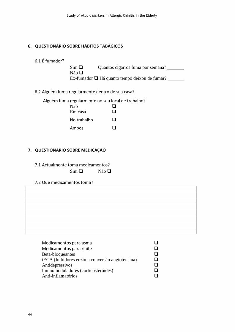
Study of Atopic Markers in Allergic Rhinitis in the Elderly
44
6. QUESTIONÁRIO SOBRE HÁBITOS TABÁGICOS
6.1 É fumador?
Sim Quantos cigarros fuma por semana? _______
Não
Ex-fumador Há quanto tempo deixou de fumar? _______
6.2 Alguém fuma regularmente dentro de sua casa?
Alguém fuma regularmente no seu local de trabalho? Não
Em casa
No trabalho
Ambos
7. QUESTIONÁRIO SOBRE MEDICAÇÃO
7.1 Actualmente toma medicamentos?
Sim Não
7.2 Que medicamentos toma?
Medicamentos para asma Medicamentos para rinite Beta-bloqueantes
iECA (Inibidores enzima conversão angiotensina)
Antidepressivos
Imunomoduladores (corticosteróides)
Anti-inflamatórios

Study of Atopic Markers in Allergic Rhinitis in the Elderly
45
8. QUESTIONÁRIO SOBRE ANTECEDENTES FAMILIARES
Assinale na tabela com um X as alergias que conheça na sua família:
Familiar Asma/bronquite asmática Rinite alérgica
Pai
Mãe
Irmãos
Avós Paternos
Avós Maternos
Chegou ao fim do nosso questionário.
Obrigado pela sua colaboração.

Study of Atopic Markers in Allergic Rhinitis in the Elderly
46
III- SPT information
TESTES CUTÂNEOS
CÓDIGO: _____________
DATA: _____________
Tomou antihistamínicos ou antidepressivos tricíclicos há menos de 7 dias?
Sim Não
Tem aplicado corticosteróides tópicos na pele?
Sim Não
Controlo Negativo (Diluente)
Controlo Positivo (Histamina)
Dermatophagoides pteronyssinus
Olea europea
Mix Pólenes IV (Gramineas) Grass
Mix Pólenes II (Ervas) Weed
Parietaria judaica
Voluntário sensibilizado a aerolergénios?
Sim Não Testes Inconclusivos
Recommended