1
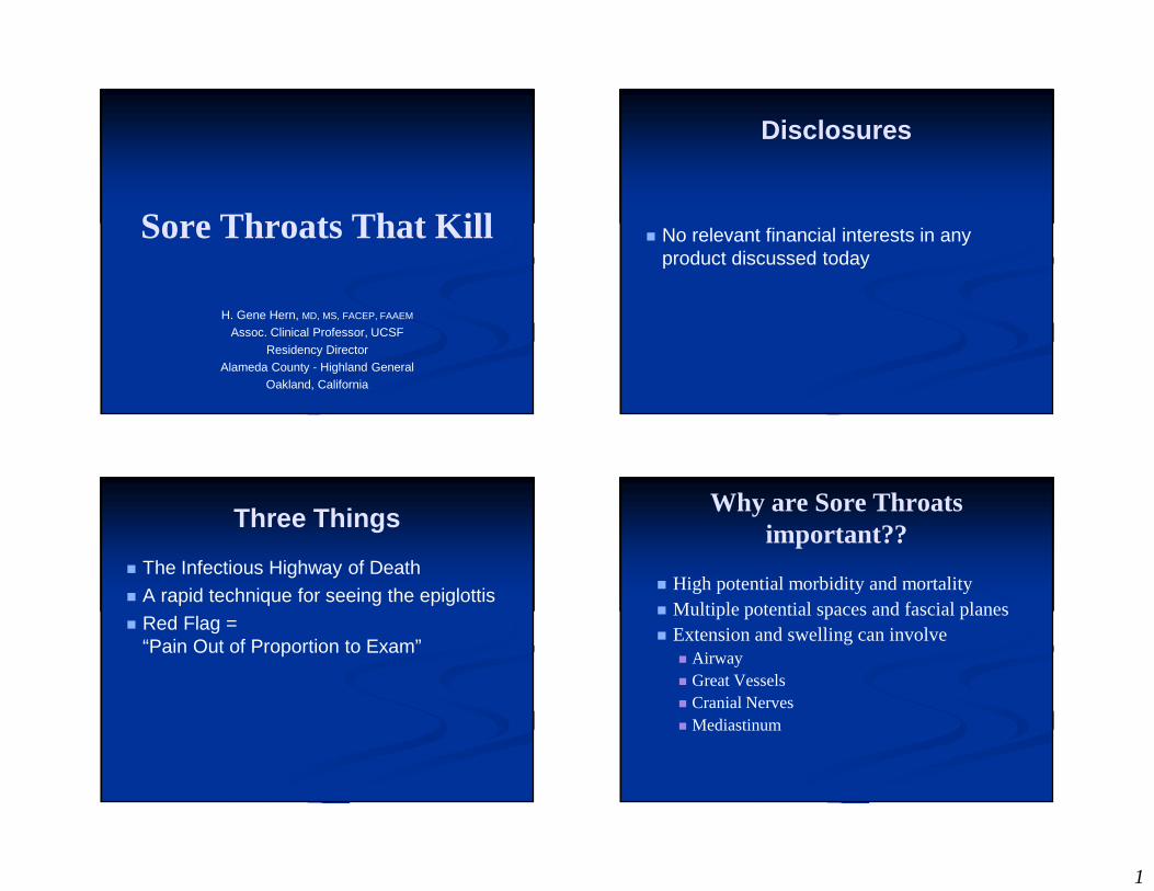
Sore Throats That Kill
H. Gene Hern, MD, MS, FACEP, FAAEM
Assoc. Clinical Professor, UCSF
Residency DirectorAlameda County - Highland General
Oakland, California
Disclosures
� No relevant financial interests in any product discussed today
Three Things
� The Infectious Highway of Death
� A rapid technique for seeing the epiglottis
� Red Flag = “Pain Out of Proportion to Exam”
Why are Sore Throats important??
� High potential morbidity and mortality� Multiple potential spaces and fascial planes� Extension and swelling can involve
� Airway� Great Vessels� Cranial Nerves� Mediastinum
2
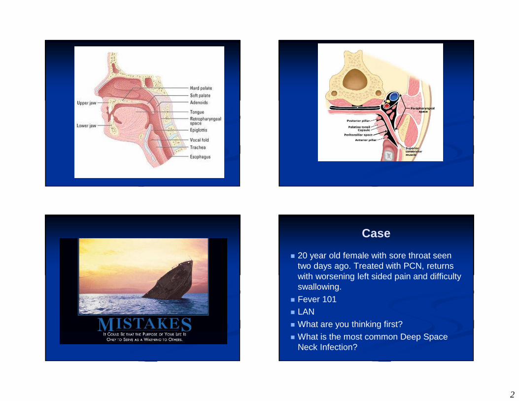
Case
� 20 year old female with sore throat seen two days ago. Treated with PCN, returns with worsening left sided pain and difficulty swallowing.
� Fever 101� LAN
� What are you thinking first?
� What is the most common Deep Space Neck Infection?
3
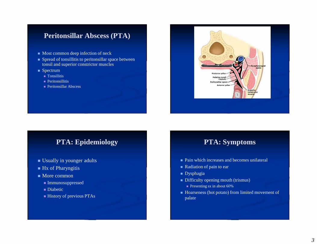
Peritonsillar Abscess (PTA)
� Most common deep infection of neck� Spread of tonsillitis to peritonsillar space between
tonsil and superior constrictor muscles� Spectrum
� Tonsillitis� Peritonsillitis� Peritonsillar Abscess
PTA: Epidemiology
� Usually in younger adults
� Hx of Pharyngitis
� More common � Immunosuppressed
� Diabetic
� History of previous PTAs
PTA: Symptoms
� Pain which increases and becomes unilateral
� Radiation of pain to ear
� Dysphagia
� Difficulty opening mouth (trismus)� Presenting sx in about 60%
� Hoarseness (hot potato) from limited movement of palate
4
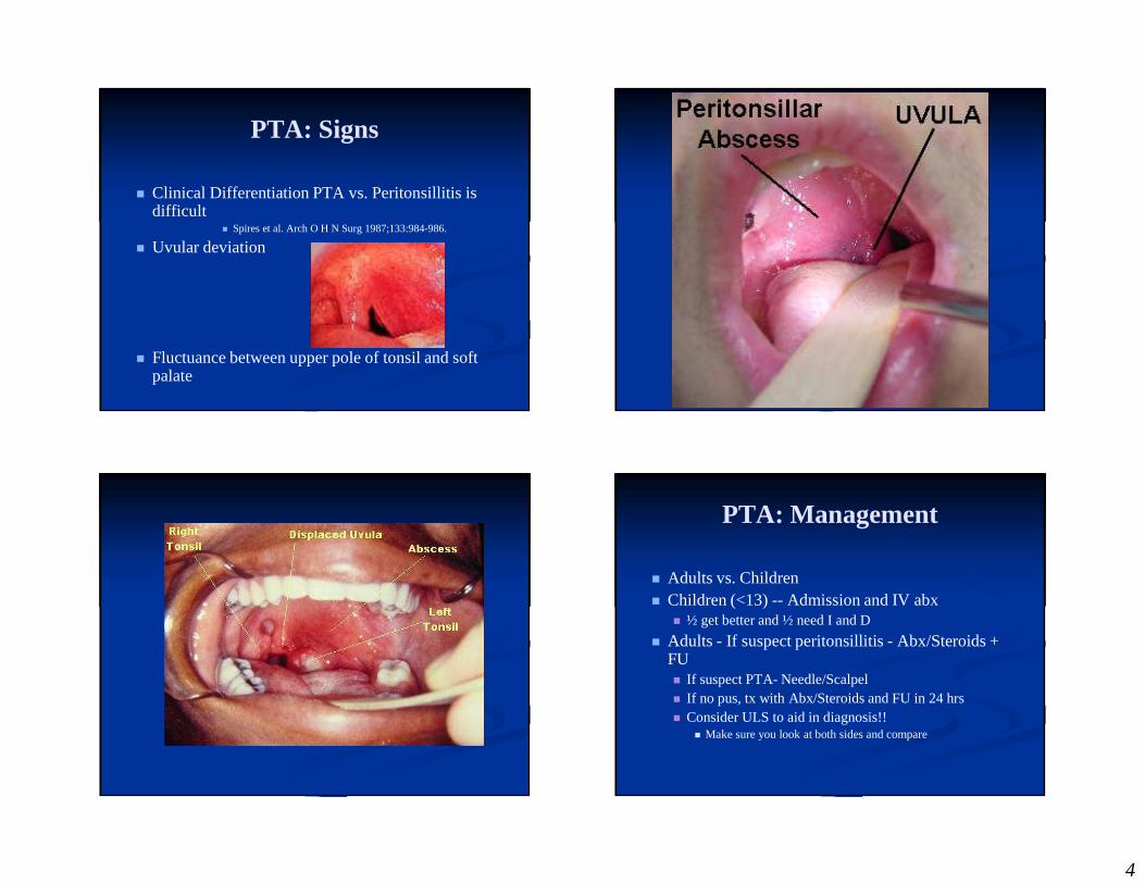
PTA: Signs
� Clinical Differentiation PTA vs. Peritonsillitis is difficult
� Spires et al. Arch O H N Surg 1987;133:984-986.
� Uvular deviation
� Fluctuance between upper pole of tonsil and soft palate
PTA: Management
� Adults vs. Children� Children (<13) -- Admission and IV abx
� ½ get better and ½ need I and D
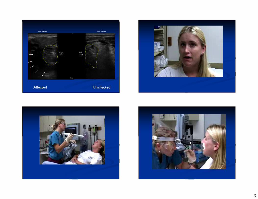
� Adults - If suspect peritonsillitis - Abx/Steroids + FU� If suspect PTA- Needle/Scalpel� If no pus, tx with Abx/Steroids and FU in 24 hrs� Consider ULS to aid in diagnosis!!
� Make sure you look at both sides and compare
5
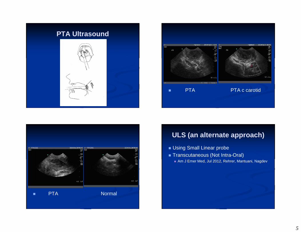
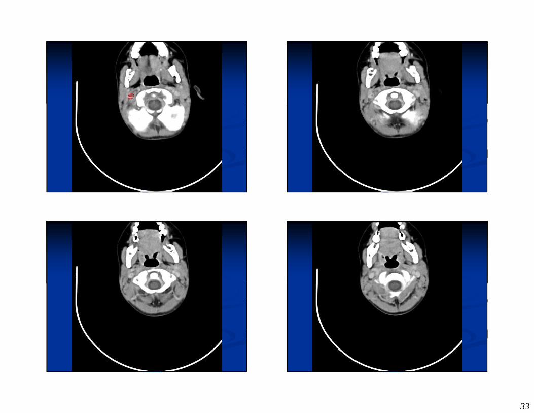
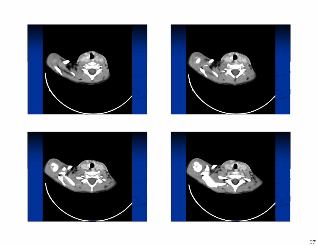
PTA Ultrasound
� PTA PTA c carotid
� PTA Normal
ULS (an alternate approach)
� Using Small Linear probe
� Transcutaneous (Not Intra-Oral)� Am J Emer Med, Jul 2012, Rehrer, Mantuani, Nagdev
6
7
PTA drainage
� Ultrasound localized
� Anesthetize with lido, or bupivicaine
� 18 g needle aspiration � +/- scalpel incision� Mehanna 2002, 60% pref Asp, 25% pref I&D� Powell, 2012, Wide variation, little data� Equal Re-accumulation rates
Peritonsillar Abscess Summary
� Spectrum� Tonsillitis
� Peritonsillitis
� Peritonsillar Abscess
� Antibiotics (PCN or Clinda)� Steroids (Decadron 6-10 mg IM)
� Drainage (with or without ULS)
Case
� 20 year old Male with fatigue malaise and pharyngitis…� Fever� Some LAN� Worse throat exam you have seen in weeks
8
Which of the following is true re: Infectious Mononucleosis?
� Splenic Rupture occurs in 5% of all cases
� Male: Female predominance is 2:1
� Splenic rupture occurs predominately in males
� Caused by EBV 100% of the time
Infectious Mononucleosis
� First described in 1920
� Most commonly EBV (CMV< Adeno, HIV)
� Mode of transmission… infected saliva
Infectious Mononucleosis
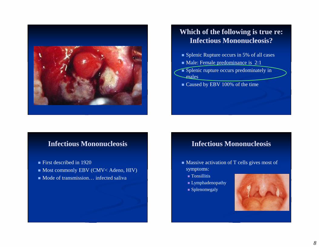
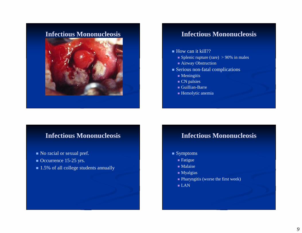
� Massive activation of T cells gives most of symptoms:� Tonsillitis
� Lymphadenopathy
� Splenomegaly
9
Infectious Mononucleosis Infectious Mononucleosis
� How can it kill??� Splenic rupture (rare) > 90% in males� Airway Obstruction
� Serious non-fatal complications� Meningitis� CN palsies� Guillian-Barre� Hemolytic anemia
Infectious Mononucleosis
� No racial or sexual pref.
� Occurrence 15-25 yrs.
� 1.5% of all college students annually
Infectious Mononucleosis
� Symptoms� Fatigue
� Malaise
� Myalgias
� Pharyngitis (worse the first week)
� LAN
10
Infectious Mononucleosis
� Signs: Low Grade Fevers� Pharyngitis
� HSM 10-30%
� Peri-orbital Edema 15-35%
� Palatal petechiae
� Occ. jaundice
Infectious Mononucleosis
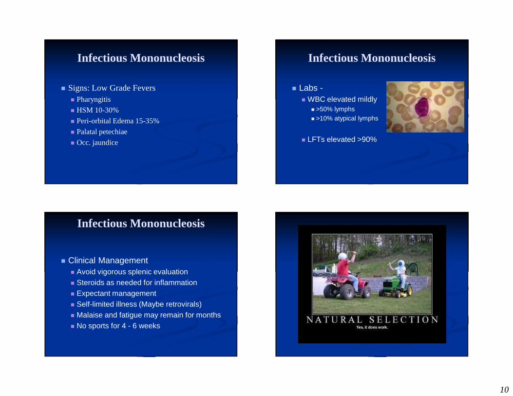
� Labs -� WBC elevated mildly
� >50% lymphs� >10% atypical lymphs
� LFTs elevated >90%
Infectious Mononucleosis
� Clinical Management� Avoid vigorous splenic evaluation� Steroids as needed for inflammation� Expectant management� Self-limited illness (Maybe retrovirals)� Malaise and fatigue may remain for months� No sports for 4 - 6 weeks
11
Case
� 3 Year old unvaccinated male presents with 1 day of sore throat illness, decr. POs
� Worsening over the last few hours
� Febrile
� Tripoding� Drooling
Pedi Epiglottitis
� Prototypical Pediatric Airway Disease� Decrease in significance since 1985
� HIB Vaccine
� Incidence decreased from 10.9:10,000 admits to 1.8:10,000
� Is it disappearing???
Epiglottitis: Who, What…
� Children between 3-7 (older after HIB vacc)
� Non-immunized� Invasive bacterial disease
� H. influenza B (less now… 25%)
� GAβHS (increasing… 75%)
� Minor players-� S. aureus, S. pneumoniae
Epiglottitis: Clinical
� Acute -- No prodrome� High fever
� Sore throat
� Toxicity
� Rapid progression� 85% sick less than 24 hrs
� Losek JD et al: Epiglottitis : comparison of signs and symptoms in children less than 2 years old and older, Ann Emerg Med 19:55–58, 1990.
12
Epiglottitis: Clinical
� Anxious� Sniffing position (jaw forward): tripod � Drooling: Inability to manage secretions
Epiglottitis: Management
� The “STABLE” airway should not be jeopardized.� IVs, Xrays, etc. should not be attempted w/o airway
personnel
� Never send to XRAY
� ?? portable XR with no/little movement
� Careful transport to OR for definitive airway
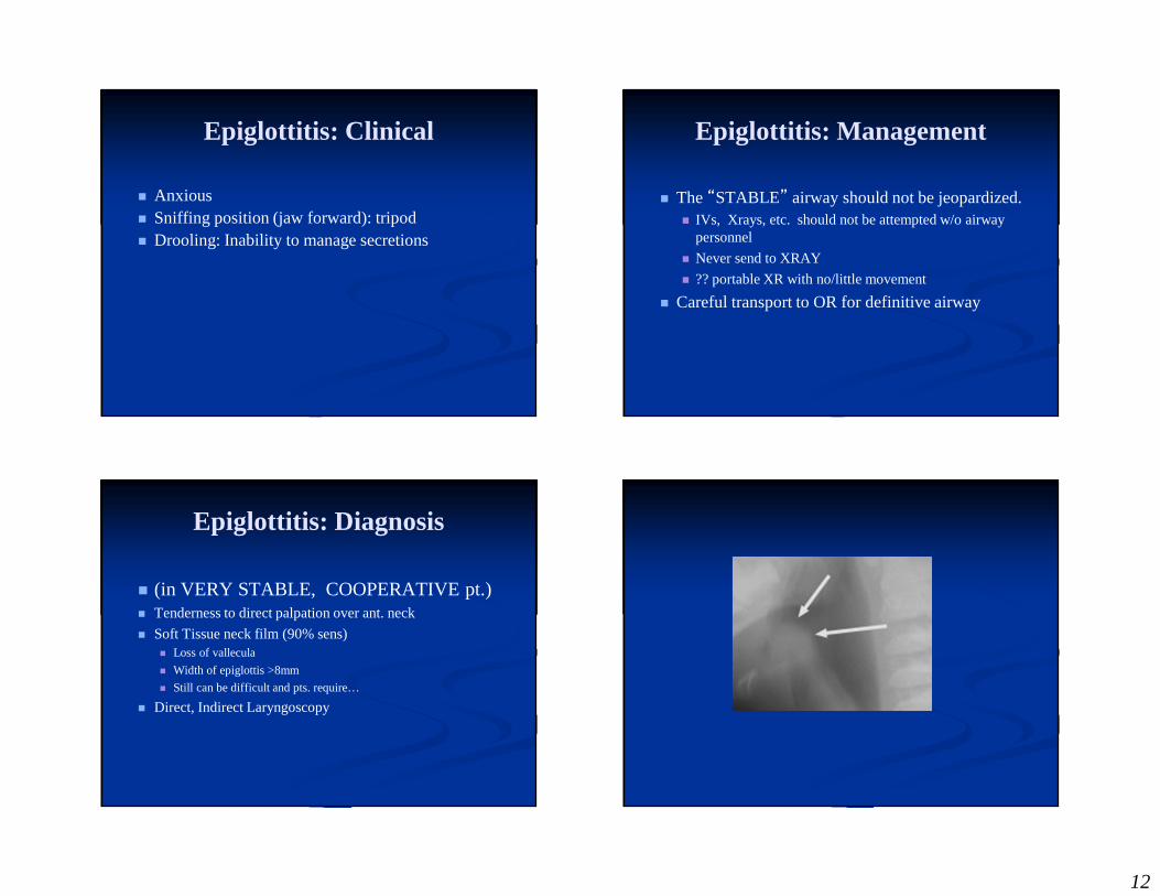
Epiglottitis: Diagnosis
� (in VERY STABLE, COOPERATIVE pt.)� Tenderness to direct palpation over ant. neck
� Soft Tissue neck film (90% sens)� Loss of vallecula
� Width of epiglottis >8mm
� Still can be difficult and pts. require…
� Direct, Indirect Laryngoscopy
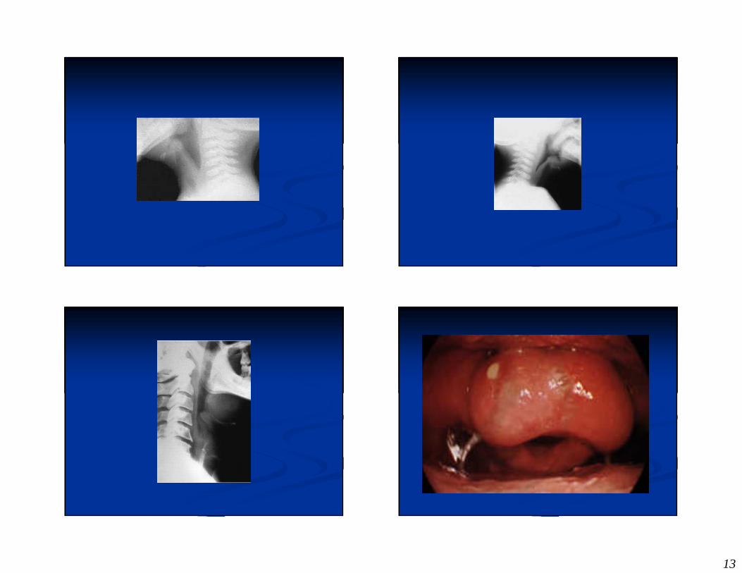
13
14
Epiglottitis: Diagnosis
� Direct visualization with larynx-vue or fiberoptic scope may help
� If PEDs � ONLY in VERY STABLE patients� ONLY when advanced airway personnel present
� If Adult…
Epiglottitis: an adults only disease??
� This may only of historical significance…� Shah, RK, Laryngoscope 2010. Avg age of
epiglottitis = 45 years old & mort. rate < 1%
Adult Epiglottitis or Supraglottitis
� Cellulitis of supraglottic structures� Base of the tongue
� Vallecula
� Aryepiglottic folds
� Arytenoid soft tissues
� Lingual tonsils
� Epiglottis
Adult Epiglottitis: Supraglottitis… Who, What
� Any Age, Any Season
� Male, Smokers greater risk
� Bugs: Same, but often none isolated� H. influenza B
� GAβHS� S. aureus, S. pneumoniae
15
Adult Epiglottitis: Supraglottitis… Clinical
� Prodrome 1-2 days� Dysphagia� Odynophagia� Sore throat
� Pain disproportionate to clinical
� Dysphonia, Muffled voice � Hoarseness is usually not found. � Fever is absent in up to 50% of cases (only in the
later stages of the disease)
Adult Epiglottitis: Supraglottitis… Diagnosis
� Misdiagnosed: Up to 1/3 of pts. seen but misdiagnosed with Strep.
� Tender to direct palp over ant. neck (80%)
� Soft tissue neck film (90% sens)� Loss of vallecula
� Width of epiglottis >8mm
� Still can be difficult and pts. require…
� Direct, Indirect Laryngoscopy…
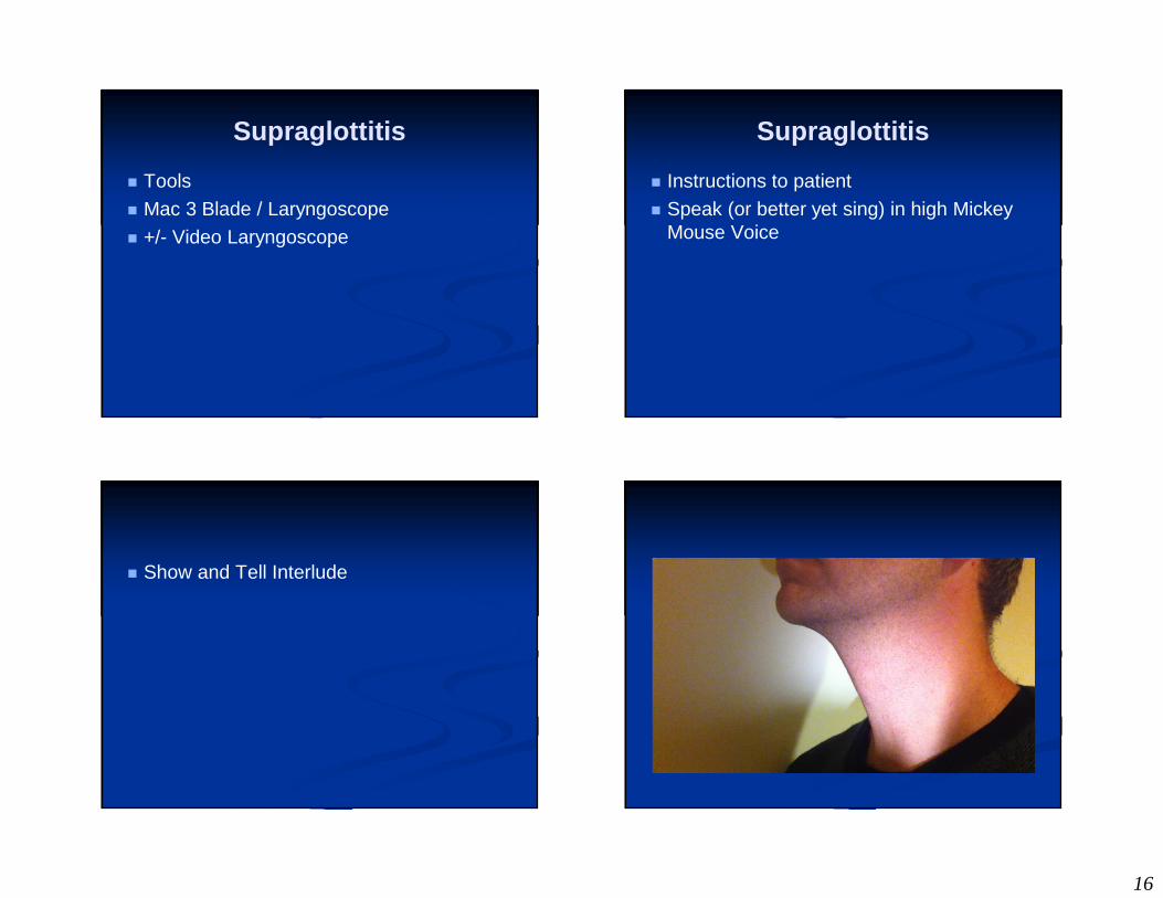
Supraglottitis
� How would you directly visualize the Epiglottis???� Patient Set Up� Tools� Instructions to patient
Supraglottitis
� Patient set up� Sitting up in bed� After Lidocaine Nebs� Facing you
16
Supraglottitis
� Tools
� Mac 3 Blade / Laryngoscope
� +/- Video Laryngoscope
Supraglottitis
� Instructions to patient
� Speak (or better yet sing) in high Mickey Mouse Voice
� Show and Tell Interlude
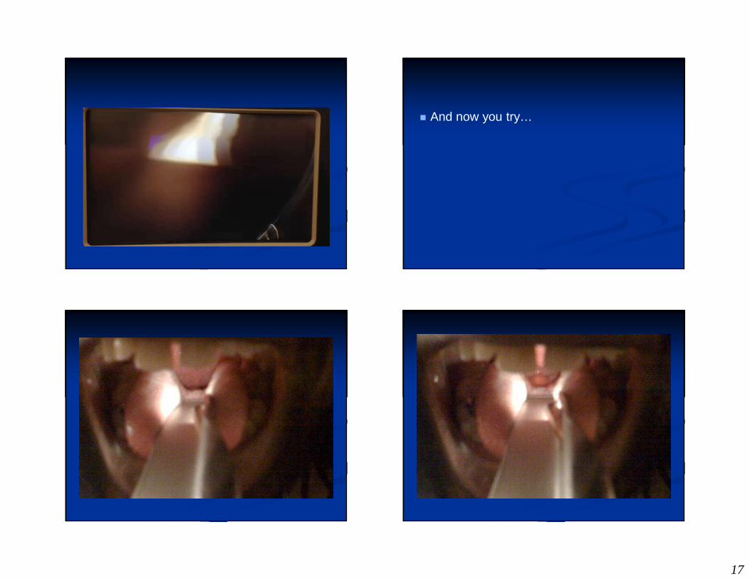
17
� And now you try…
18
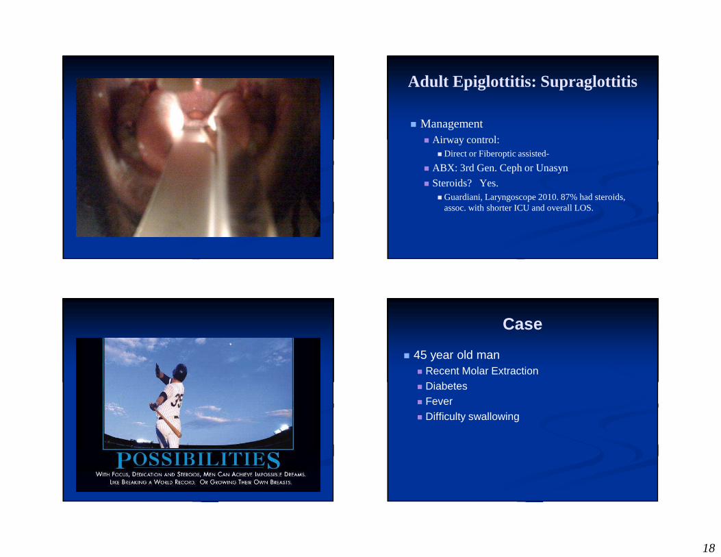
Adult Epiglottitis: Supraglottitis
� Management � Airway control:
� Direct or Fiberoptic assisted-
� ABX: 3rd Gen. Ceph or Unasyn
� Steroids? Yes. � Guardiani, Laryngoscope 2010. 87% had steroids,
assoc. with shorter ICU and overall LOS.
Case
� 45 year old man� Recent Molar Extraction� Diabetes� Fever� Difficulty swallowing
19
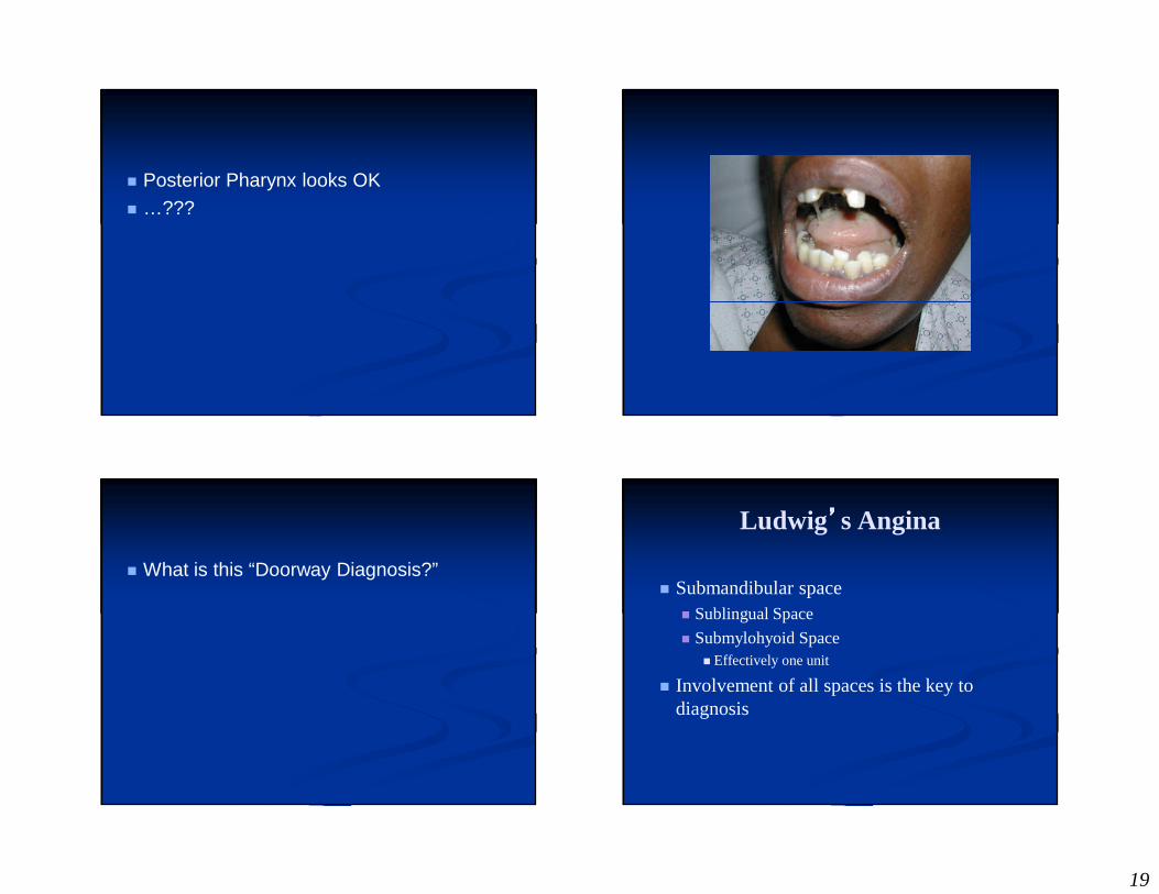
� Posterior Pharynx looks OK
� …???
� What is this “Doorway Diagnosis?”
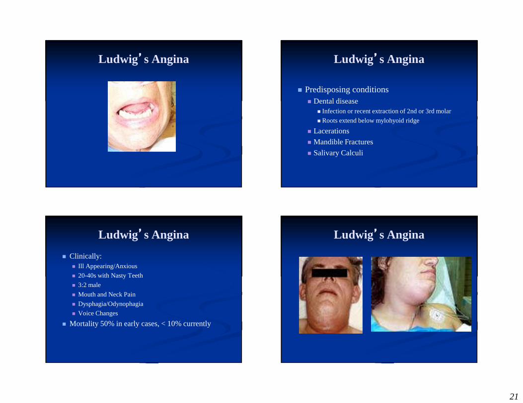
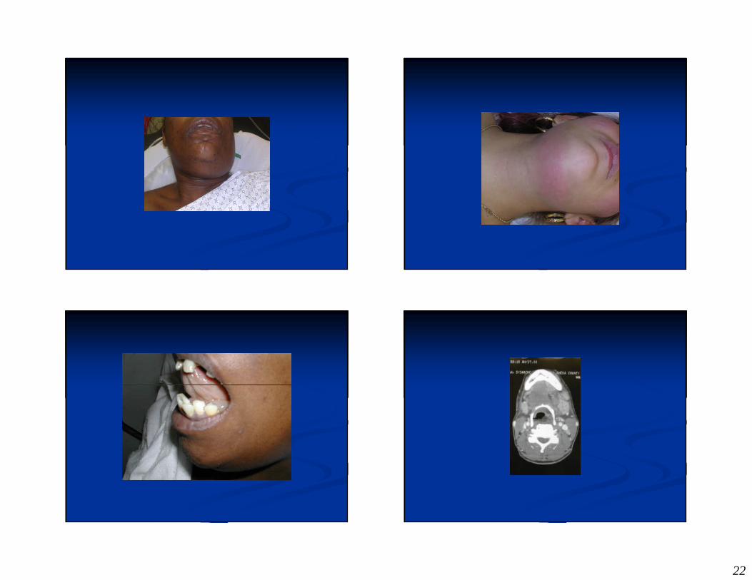
Ludwig’’’’s Angina
� Submandibular space� Sublingual Space
� Submylohyoid Space� Effectively one unit
� Involvement of all spaces is the key to diagnosis
20
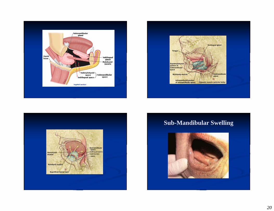
Sub-Mandibular Swelling
21
Ludwig’’’’s Angina Ludwig’’’’s Angina
� Predisposing conditions� Dental disease
� Infection or recent extraction of 2nd or 3rd molar
� Roots extend below mylohyoid ridge
� Lacerations
� Mandible Fractures
� Salivary Calculi
Ludwig’’’’s Angina
� Clinically:� Ill Appearing/Anxious
� 20-40s with Nasty Teeth
� 3:2 male
� Mouth and Neck Pain
� Dysphagia/Odynophagia
� Voice Changes
� Mortality 50% in early cases, < 10% currently
Ludwig’’’’s Angina
22
23
Ludwig’’’’s Angina
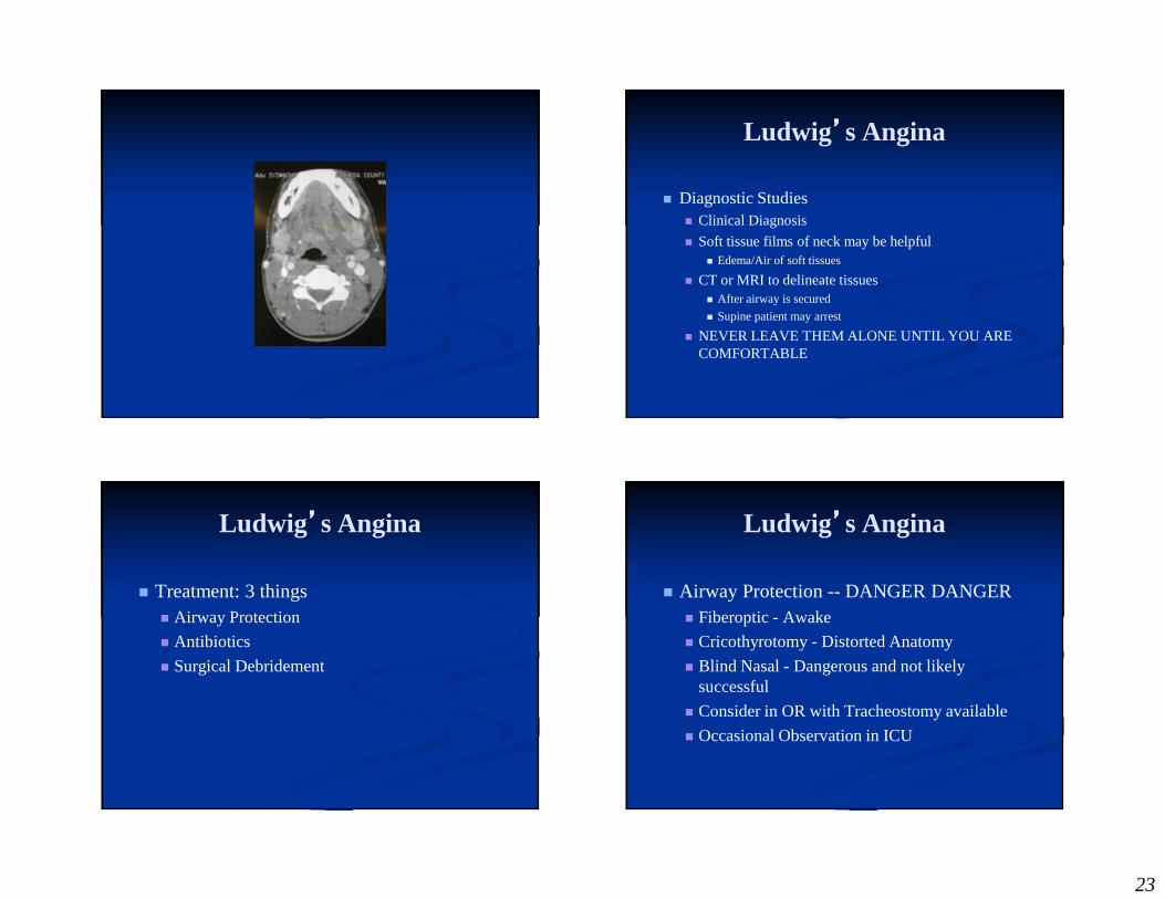
� Diagnostic Studies� Clinical Diagnosis
� Soft tissue films of neck may be helpful� Edema/Air of soft tissues
� CT or MRI to delineate tissues� After airway is secured
� Supine patient may arrest
� NEVER LEAVE THEM ALONE UNTIL YOU ARE COMFORTABLE
Ludwig’’’’s Angina
� Treatment: 3 things� Airway Protection
� Antibiotics
� Surgical Debridement
Ludwig’’’’s Angina
� Airway Protection -- DANGER DANGER� Fiberoptic - Awake
� Cricothyrotomy - Distorted Anatomy
� Blind Nasal - Dangerous and not likely successful
� Consider in OR with Tracheostomy available
� Occasional Observation in ICU
24
Ludwig’’’’s Angina
� Antibiotics� 3rd gen. Cephalosporin plus flagyl or
� Unasyn
� Once airway is protected - 50% will resolve only with antibiotics
Ludwig’’’’s Angina
� Surgical Debridement� Once recommended for all patients
� Reserved for refractory cases
� CT results can guide drainage
Question:
� Retropharyngeal abscess occurs in which age group primarily?� 1-2 year olds
� 2-6 year olds
� 6-12 year olds
� 12-25 year olds
� Over 25
25
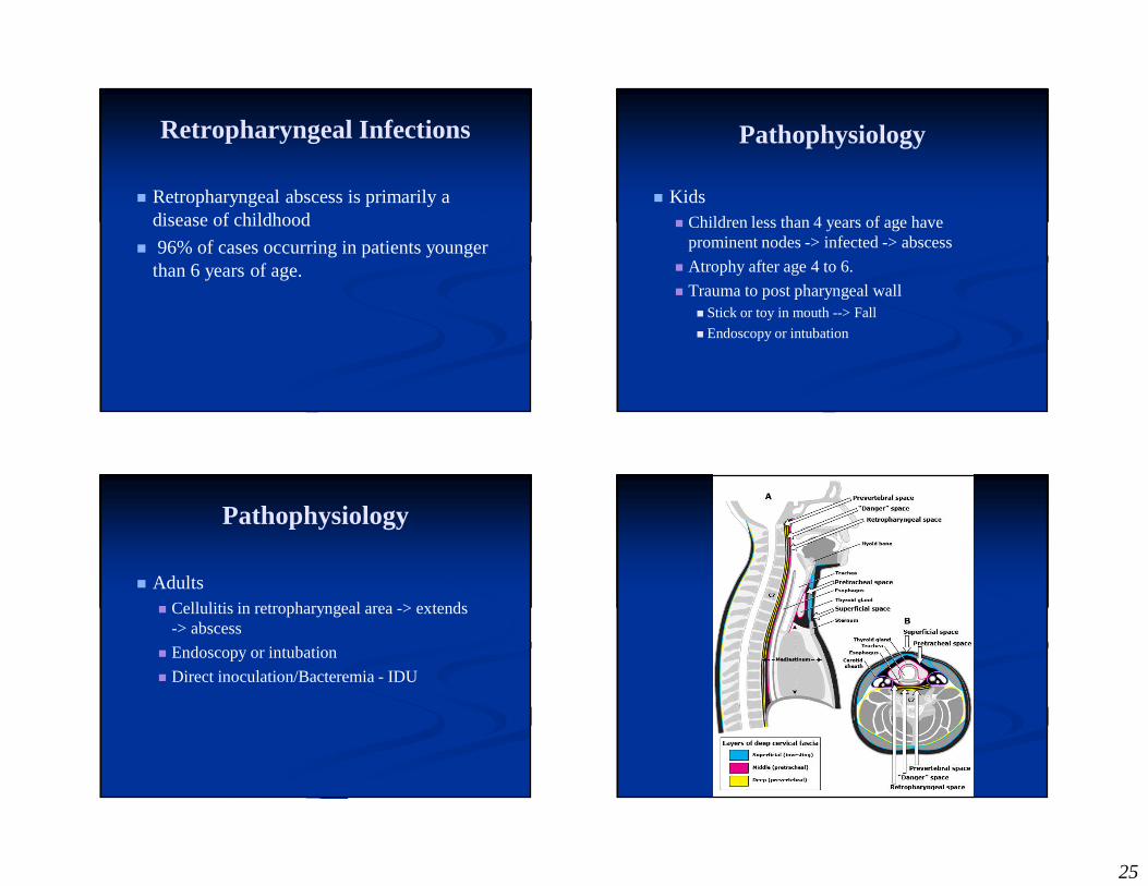
Retropharyngeal Infections
� Retropharyngeal abscess is primarily a disease of childhood
� 96% of cases occurring in patients younger than 6 years of age.
Pathophysiology
� Kids� Children less than 4 years of age have
prominent nodes -> infected -> abscess
� Atrophy after age 4 to 6.
� Trauma to post pharyngeal wall� Stick or toy in mouth --> Fall
� Endoscopy or intubation
Pathophysiology
� Adults� Cellulitis in retropharyngeal area -> extends
-> abscess
� Endoscopy or intubation
� Direct inoculation/Bacteremia - IDU
26
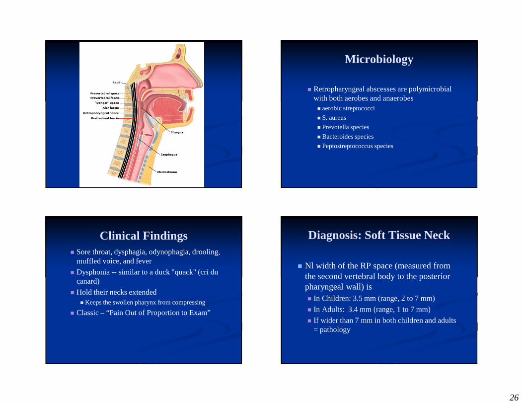
Microbiology
� Retropharyngeal abscesses are polymicrobial with both aerobes and anaerobes� aerobic streptococci
� S. aureus
� Prevotella species
� Bacteroides species
� Peptostreptococcus species
Clinical Findings� Sore throat, dysphagia, odynophagia, drooling,
muffled voice, and fever
� Dysphonia -- similar to a duck "quack" (cri du canard)
� Hold their necks extended� Keeps the swollen pharynx from compressing
� Classic – “Pain Out of Proportion to Exam”
Diagnosis: Soft Tissue Neck
� Nl width of the RP space (measured from the second vertebral body to the posterior pharyngeal wall) is � In Children: 3.5 mm (range, 2 to 7 mm)
� In Adults: 3.4 mm (range, 1 to 7 mm)
� If wider than 7 mm in both children and adults = pathology
27
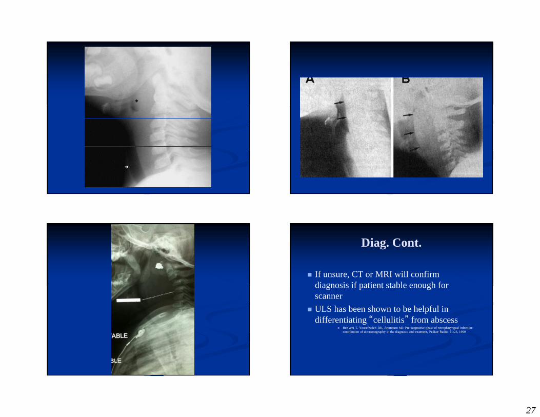
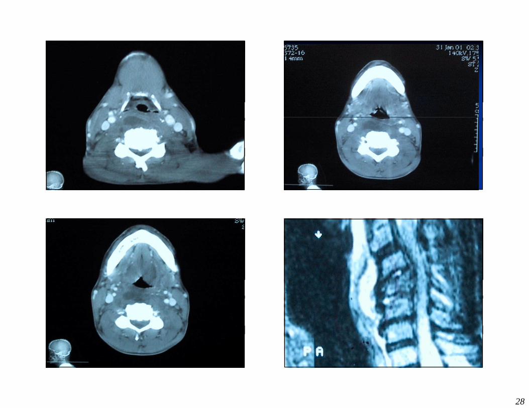
Diag. Cont.
� If unsure, CT or MRI will confirm diagnosis if patient stable enough for scanner
� ULS has been shown to be helpful in differentiating “cellulitis” from abscess
� Ben-ami T, Yousefzadeh DK, Aramburo MJ: Pre-supprative phase of retropharyngeal infection: contribution of ultrasonography in the diagnosis and treatment, Pediatr Radiol 21:23, 1990
28
29
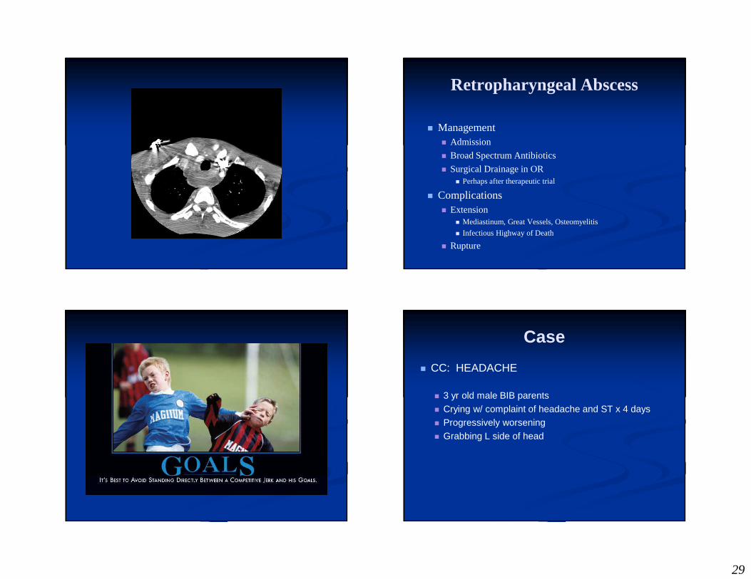
Retropharyngeal Abscess
� Management� Admission
� Broad Spectrum Antibiotics
� Surgical Drainage in OR� Perhaps after therapeutic trial
� Complications� Extension
� Mediastinum, Great Vessels, Osteomyelitis
� Infectious Highway of Death
� Rupture
Case
� CC: HEADACHE
� 3 yr old male BIB parents� Crying w/ complaint of headache and ST x 4 days� Progressively worsening� Grabbing L side of head
30
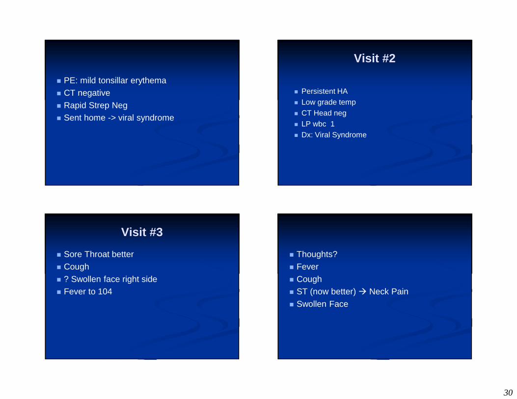
� PE: mild tonsillar erythema
� CT negative
� Rapid Strep Neg� Sent home -> viral syndrome
� Persistent HA� Low grade temp� CT Head neg� LP wbc 1� Dx: Viral Syndrome
Visit #2
Visit #3
� Sore Throat better
� Cough
� ? Swollen face right side� Fever to 104
� Thoughts?
� Fever
� Cough� ST (now better) � Neck Pain
� Swollen Face
31
32
33
34
35
36
37
38
39
40
41
42
43
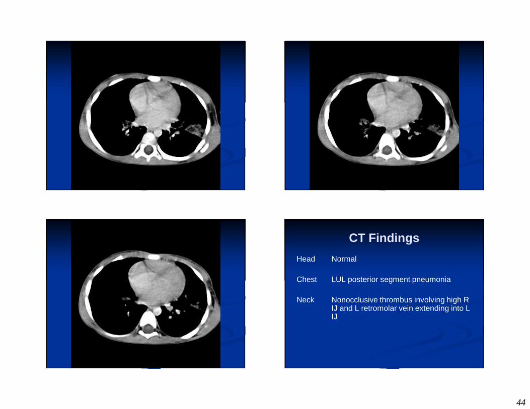
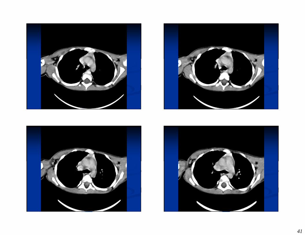
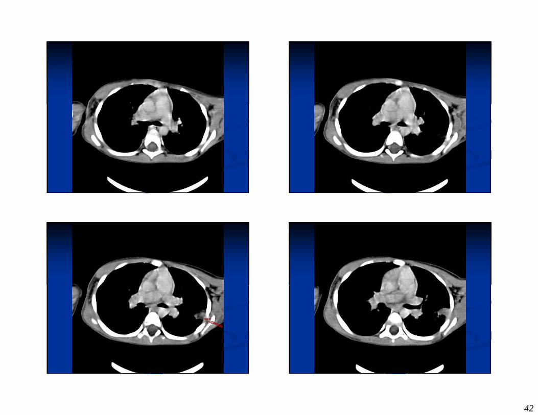
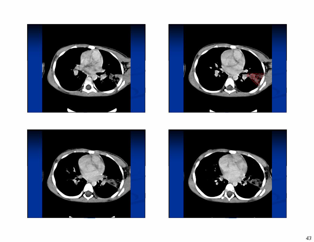
44
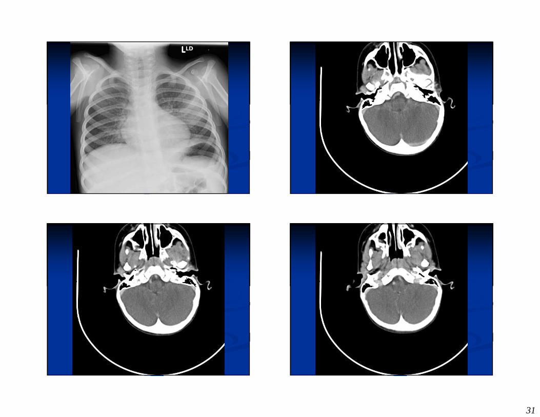
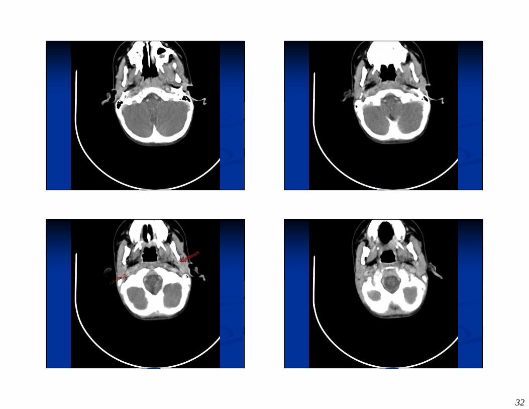
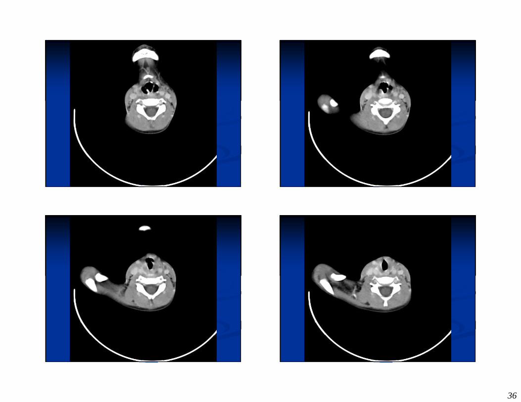
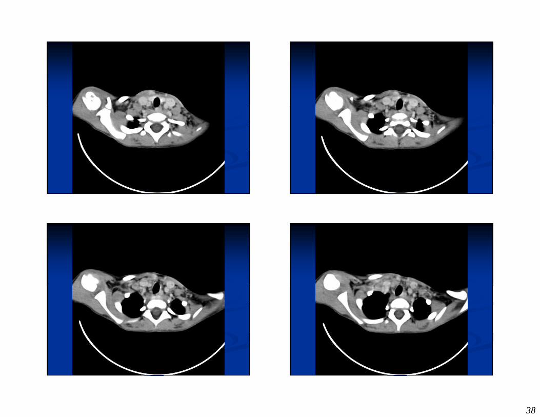
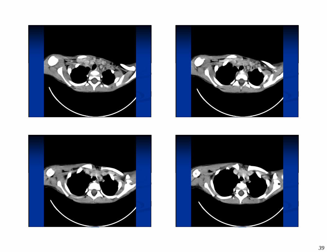
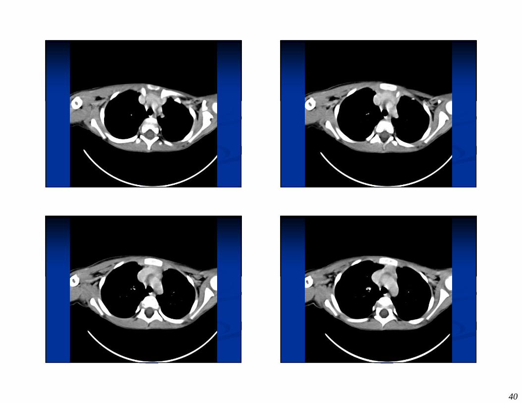
CT Findings
Head Normal
Chest LUL posterior segment pneumonia
Neck Nonocclusive thrombus involving high R IJ and L retromolar vein extending into L IJ
45
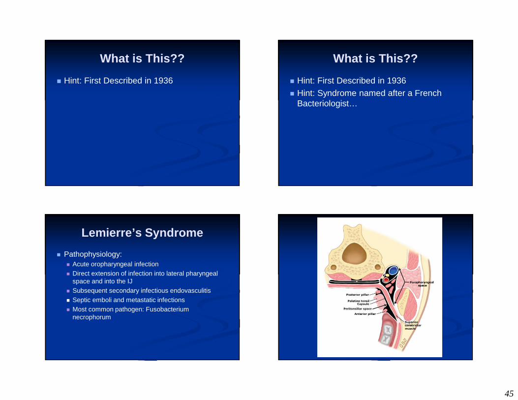
What is This??
� Hint: First Described in 1936
What is This??
� Hint: First Described in 1936
� Hint: Syndrome named after a French Bacteriologist…
Lemierre’s Syndrome
� Pathophysiology:� Acute oropharyngeal infection� Direct extension of infection into lateral pharyngeal
space and into the IJ � Subsequent secondary infectious endovasculitis� Septic emboli and metastatic infections� Most common pathogen: Fusobacterium
necrophorum
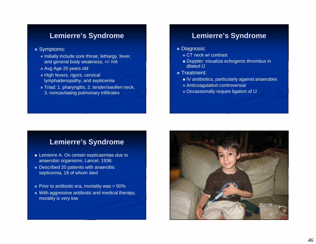
46
Lemierre’s Syndrome
� Symptoms:� Initially include sore throat, lethargy, fever,
and general body weakness, +/- HA� Avg Age 20 years old� High fevers, rigors, cervical
lymphadenopathy, and septicemia� Triad: 1. pharyngitis, 2. tender/swollen neck,
3. noncavitating pulmonary infiltrates
Lemierre’s Syndrome
� Diagnosis:� CT neck w/ contrast� Doppler: visualize echogenic thrombus in
dilated IJ
� Treatment:� IV antibiotics, particularly against anaerobes� Anticoagulation controversial� Occassionally require ligation of IJ
Lemierre’s Syndrome
� Lemierre A. On certain septicaemias due to anaerobic organisms. Lancet. 1936.
� Described 20 patients with anaerobic septicemia, 18 of whom died
� Prior to antibiotic era, mortality was > 50%� With aggressive antibiotic and medical therapy,
morality is very low

47
Summary of what this lecture covered
� Rapidly running differential for life-threatening sore throats� PTA� Mononucleosis� Epiglottitis� Ludwig’s� Retro-Pharyngeal Abscess� Lemierre’s
Closing Thoughts…
� Management of the sore throat that kills� Critical monitoring
� Don’t leave the room until YOU are comfortable
� Airway protection early
� When the pain is out of proportion to exam… worry about the dangerous kinds...
Three Things
� The Infectious Highway of Death
� A rapid technique and tips for seeing the epiglottis
� Red Flag = “Pain Out of Proportion to Exam”
49
Acute Pharyngitis
� 2% of all visits to EDs, outpt. clinics� 50-80% Viral� 5-36% GAβHS� 1-10% EBV� 2-5% Chlamydia, Mycoplasma, GC
� Antibiotics Rx’d to 75% of adult pts. with pharyngitis
Why do physicians give Rx?
� MDs believe…
� Pts. expect them
� Pts. will come back if they don’t get Rx
� Pts. will be unsatisfied without Rx
� Quicker to write Rx than to explain
Problems with this assumption
� MDs are not good at predicting which pts. expect Rx
� Pt. satisfaction � Less on Rx, more on MDs showing concern and
reassurance� Little et al, BMJ 1999;319:736-7� Ann Fam Med 4(6):494, November/December 2006
� Delaying Rx does NOT increase chance of pt. returning in next few days
� Adverse pt. consequences and public health issues
Evidence for Abx RxSo why give it?
� Prevent Rheumatic Fever
� Prevent Acute Glomerulonephritis
� Prevent Complications
� Decrease Contagion
� Relieve Symptoms
50
Acute Rheumatic Fever: Old Assumptions
� Early trials show reduction in ARF with PCN (RR=.28)� However, number needed to treat (NNT)
= 63 pts. to prevent 1 case of ARF� Now the incidence is MUCH lower (60x) so
NNT 4000?� Widespread antibiotic use for strep� Less virulent strains� Improved living conditions
Acute Rheumatic Fever: Old Assumptions
� Most clinically significant problem is myocarditis
� Myocarditis incidence 50-90% in kids, <33% in adults � Most of these cases based on echo evidence� Not clear how many were CLINICALLY
symptomatic� NNT much higher --> 10000? 20000?
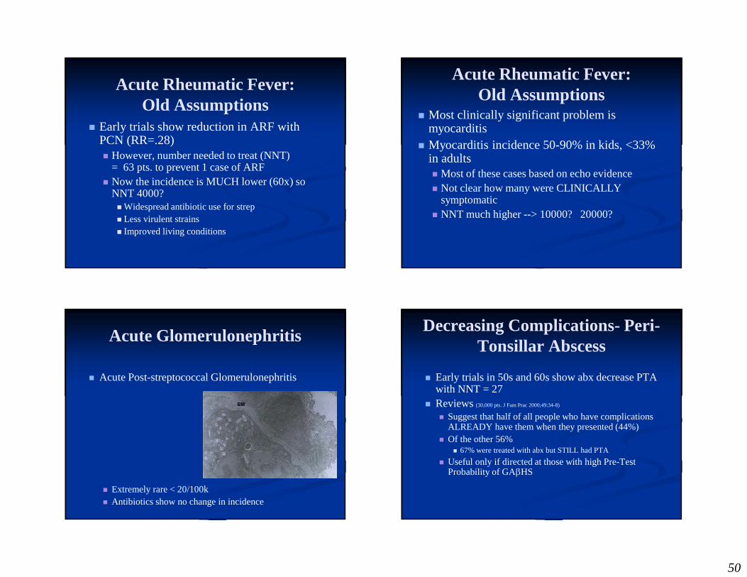
Acute Glomerulonephritis
� Acute Post-streptococcal Glomerulonephritis
� Extremely rare < 20/100k� Antibiotics show no change in incidence
Decreasing Complications- Peri-Tonsillar Abscess
� Early trials in 50s and 60s show abx decrease PTA with NNT = 27
� Reviews (30,000 pts. J Fam Prac 2000;49:34-8)
� Suggest that half of all people who have complications ALREADY have them when they presented (44%)
� Of the other 56% � 67% were treated with abx but STILL had PTA
� Useful only if directed at those with high Pre-Test Probability of GAβHS
51
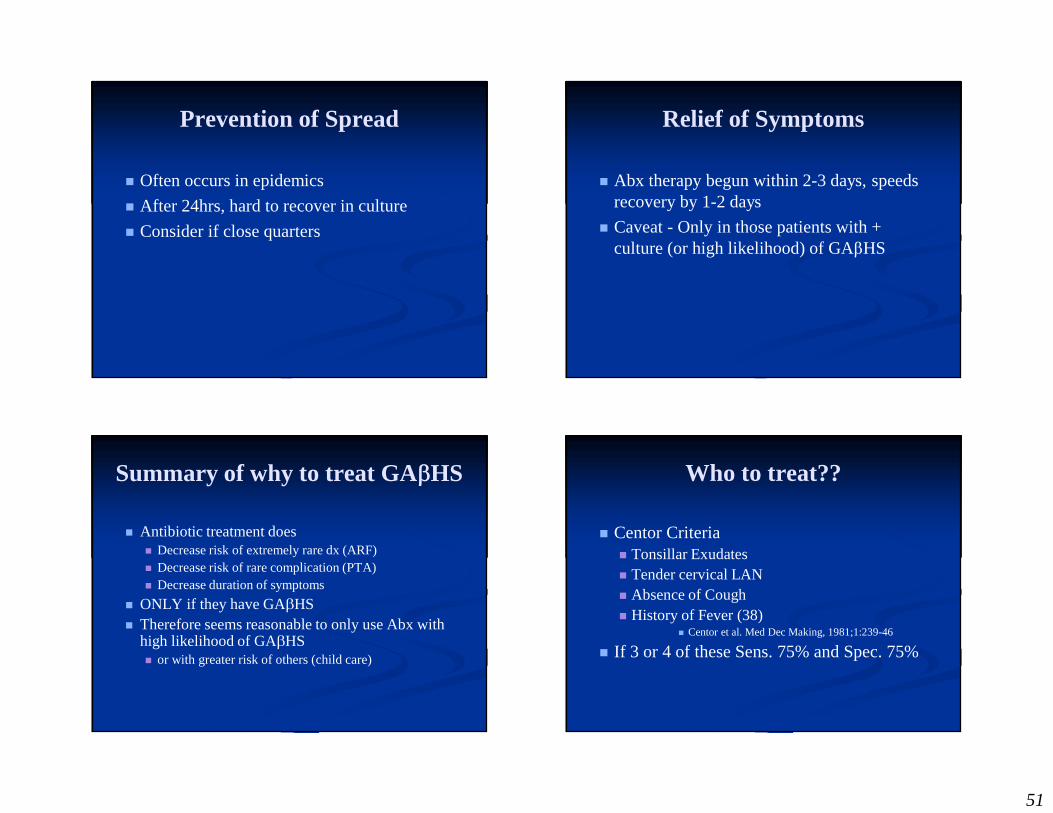
Prevention of Spread
� Often occurs in epidemics
� After 24hrs, hard to recover in culture
� Consider if close quarters
Relief of Symptoms
� Abx therapy begun within 2-3 days, speeds recovery by 1-2 days
� Caveat - Only in those patients with + culture (or high likelihood) of GAβHS
Summary of why to treat GAβHS
� Antibiotic treatment does� Decrease risk of extremely rare dx (ARF)� Decrease risk of rare complication (PTA)� Decrease duration of symptoms
� ONLY if they have GAβHS� Therefore seems reasonable to only use Abx with
high likelihood of GAβHS� or with greater risk of others (child care)
Who to treat??
� Centor Criteria� Tonsillar Exudates� Tender cervical LAN� Absence of Cough� History of Fever (38)
� Centor et al. Med Dec Making, 1981;1:239-46
� If 3 or 4 of these Sens. 75% and Spec. 75%
52
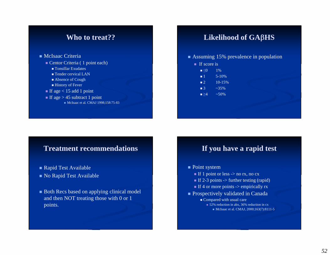
Who to treat??
� McIsaac Criteria� Centor Criteria ( 1 point each)
� Tonsillar Exudates� Tender cervical LAN� Absence of Cough� History of Fever
� If age < 15 add 1 point� If age > 45 subtract 1 point
� McIsaac et al. CMAJ 1998;158:75-83
Likelihood of GAβHS
� Assuming 15% prevalence in population� If score is
� ≤0 1%
� 1 5-10%
� 2 10-15%
� 3 ~35%
� ≥4 ~50%
Treatment recommendations
� Rapid Test Available
� No Rapid Test Available
� Both Recs based on applying clinical model and then NOT treating those with 0 or 1 points.
If you have a rapid test
� Point system� If 1 point or less -> no rx, no cx� If 2-3 points -> further testing (rapid)� If 4 or more points -> empirically rx
� Prospectively validated in Canada� Compared with usual care
� 52% reduction in abx, 36% reduction in cx� McIsaac et al. CMAJ, 2000;163(7):8111-5
53
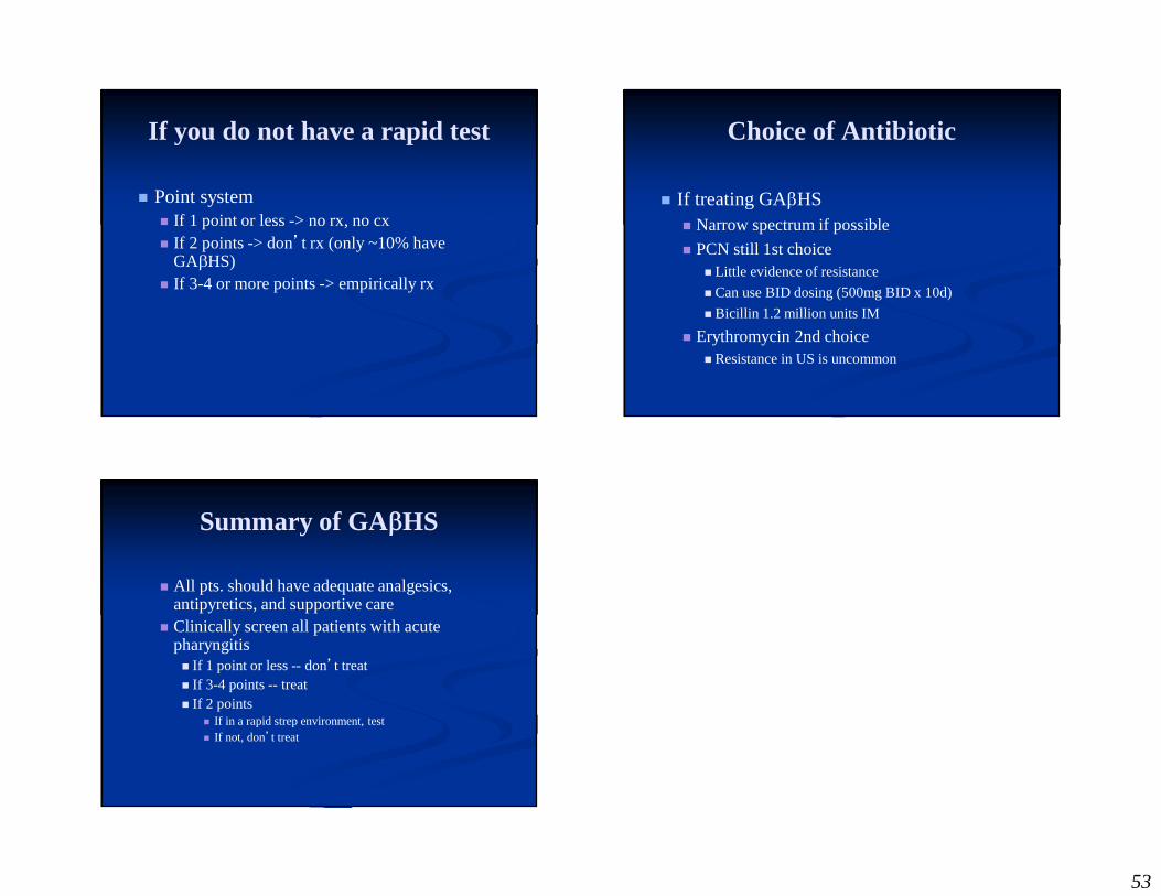
If you do not have a rapid test
� Point system� If 1 point or less -> no rx, no cx� If 2 points -> don’t rx (only ~10% have
GAβHS)� If 3-4 or more points -> empirically rx
Choice of Antibiotic
� If treating GAβHS � Narrow spectrum if possible
� PCN still 1st choice� Little evidence of resistance
� Can use BID dosing (500mg BID x 10d)
� Bicillin 1.2 million units IM
� Erythromycin 2nd choice� Resistance in US is uncommon
Summary of GAβHS
� All pts. should have adequate analgesics, antipyretics, and supportive care
� Clinically screen all patients with acute pharyngitis� If 1 point or less -- don’t treat� If 3-4 points -- treat� If 2 points
� If in a rapid strep environment, test� If not, don’t treat
Recommended