Live Cell RNA DetectionSmartFlare™ RNA Detection Probes
Go live. It’s smart.
EMD Millipore is a division of Merck KGaA, Darmstadt, Germany
2
Live cell RNA detection with SmartFlare™ RNA Detection Probes
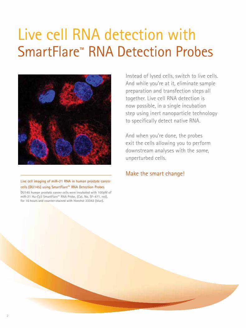
Instead of lysed cells, switch to live cells. And while you’re at it, eliminate sample preparation and transfection steps all together. Live cell RNA detection is now possible, in a single incubation step using inert nanoparticle technology to specifically detect native RNA.
And when you’re done, the probes exit the cells allowing you to perform downstream analyses with the same, unperturbed cells.
Make the smart change!Live cell imaging of miR-21 RNA in human prostate cancer cells (DU145) using SmartFlare™ RNA Detection ProbesDU145 human prostate cancer cells were incubated with 100pM of miR-21 Hu-Cy3 SmartFlare™ RNA Probe, (Cat. No. SF-471, red), for 16 hours and counter-stained with Hoechst 33342 (blue).

3
Use a SmartFlare™ probe to analyze gene expression in live cells with no sample prep, cell lysis or toxicity, and you’ll see just how smart a tiny particle can be.
• Live Cell Detection See real-time RNA expression in live cells, giving you more physiologically relevant data.
• Native RNA No need for amplification, targeting only native, endogenous RNA, avoiding artifacts with traditional amplification methods.
• No Sample Prep Only a single incubation step, eliminating all sample preparation.
• Easy Open-platform Detection Detect the fluorescent signals using a platform of your choice: microscope, personal cell analyzer, flow cytometer, cell sorter, etc.
• No Toxicity, Leaving Cells Unchanged Non-toxic nanoparticles that enter and exit cells via natural mechanisms, allowing the same live cells to be used for downstream assays.
What’s smart
Capture Strand
Reporter Strand
-AAAAAAATCAACCATACACCGTGACTTTGCTTGACCC
-AGTTGGTATGTGGCACT
Gold-quenchedfluorophore on reporter strand
Gold nanoparticle
Capture strand/reporter strandduplex
Figure 1. The SmartFlare™ Probe Anatomy
SmartFlare™ probes consist of a gold nanoparticle conjugated to double stranded oligos, one of which contains a fluorophore that is quenched by its proximity to the gold core.
The oligo duplexes are designed with an RNA “capture” sequence and a complementary “reporter” sequence.

4
1. Plate cells at desired density (typical example: 30,000 cells in 200 µL complete medium in each well of a 96-well plate)
2. Dilute reconstituted SmartFlare™ reagent 1:20 in sterile phosphate-buffered saline to create working solution
3. Add 4 µL diluted SmartFlare™ reagent to each well of cells (which are 60-80% confluent)
4. Incubate overnight (16 hours) at appropriate temperature (37 °C), CO2 (5%) and relative humidity.
5. Detect fluorescence using platform of choice: flow cytometer, imaging cytometer, fluorescence microscope, etc.
Cell testing protocol (recommended protocol for testing a cell-probe combination for the first time):
• No sample prep
• No RNA amplification
• No transfection reagents
• No cell lysis
• No toxicity
RNA detection often requires laborious sample prep, RNA amplification and detection based on standard curves. SmartFlare™ RNA Detection Probes are endo-cytosed by live cells using existing cellular endocytosis machinery. Sample prep is unnecessary; simply add SmartFlare™ probes to your cultures, incubate over-night and detect the next day. Over time, the probes will exit the cell, without any adverse effects, allowing for subsequent downstream assays.
Eliminate the complexity and uncertainty associated with other RNA detection techniques. SmartFlare™ RNA Detection Probes make discovery effortless.
What’s simple

5
Figure 2. Enter, bind, fluoresce, exit. SmartFlare™ probes enter the cell using the cell’s own endocytosis process. The probes circulate within the cell and bind to the complementary RNA sequence. This binding event releases a fluorophore, illuminating the cells for detection. Over time, the probe exits the cell, leaving the cell unchanged and free for downstream analyses.
Probes enter cell via natural endocytosis
Probe binds targetand flare releases for detection.
Probe approaches RNA target.
Probes exit cell via exocytosis. The same cell can be reused for other experiments.
obe binds targett
Enter Bind Fluoresce Exit
+
In absence of target, probeis quenched by gold and does
not fluoresceTarget
When target is present, it will bindthe capture strand, releasing
the reporter strand, which fluoresces
The SmartFlare™ probe’s simple mechanism reveals the RNA content of live cells while keeping cells alive and intact.

6
Applications using SmartFlare™ Probes
• Track changes in RNA dynamically, over time
• Sort cell types that are traditionally difficult to isolate, using intracellular RNA markers
• Quantify miRNAs
• Assess RNA and protein in the same cellular preparations
You can take a peek at your RNA without your cells ever knowing you were there. SmartFlare™ probes enable novel experimental designs that can change how you do science. Imagine the possibilities.
Figure 3. Sorting based on intracellular RNA markers. Cells were sorted based on miR-155 expres-sion. miR-155 high and miR-155 low cells were isolated and then used for downstream experiments examining protein levels in the same cells.
What’s powerful
0
100 101 102 103 104
Forw
ard
S ca
tter
51
102
153
204
256
105
miR-155 Expression Level (Cy5 Fluorescence)
• Reuse your SmartFlare™ probe-treated cells for downstream assays
• Live cell detection via flow cytometry
• Assess multiple RNA targets simultaneously (multiplex)
You can now sort your cells based on levels of RNA targets. After sorting, these cells are alive, unchanged, and ready for further analysis. The ability to detect and separate live cells based on the level of a specific RNA target provides new opportunities to correlate gene expression with cellular functions and to identify rare cell types such as certain tumor cells and cancer stem cells.
Sort live cells based on RNA content

7
Quantify RNA using flow cytometry Quantifying RNA has never been easier. Because flow cytometry interrogates single cells for fluorescence expression, amplification is not necessary. Get specific, single-cell, relative quantification of RNA in intact cells using EMD Millipore’s guava easyCyte™ or Muse™ flow cytometry platforms with SmartFlare™ Probes. Or really enhance your detection capabilities using the Amnis® Imaging Flow Cytometry systems.
Fluo
resc
ence
(M5-
510)
Cycles1 2 6 9 10 1613 19 22 25 29 4034 37314
Fluo
resc
ence
(M5-
510)
Cycles
30
40
50
60
70
80
90
100
110
40
50
60
70
80
90
100
110
1 2 6 9 10 1613 19 22 25 29 4034 37314
MCF-7No probe HeLa
MCF-7 HeLa
0
20
100 101 102 103 104
40
60
80
100
120
140
EGFR mRNA probe (RED2-HLog)
FGF2 mRNA probe (RED2-HLog)
Coun
t
No probe HT1376
0
100 101 102 103 104
40
20
60
80
100
120140
160
Coun
t
HUVEC
HT1376 HUVEC
180
Figure 5.
Probe detection of mRNA levels correlates to RT-PCR data. Using SmartFlare™ technology to determine the mRNA levels of EGFR mRNA (A) in HeLa and MCF-7 cells as well as FGF2 mRNA (B) in HUVEC and HT1376 cells. RNA levels as detected by flow cytometry show good correlation to trends in cycle numbers and RNA levels determined by quantitative RT-PCR.
Using live cells means you are looking at native, unamplified RNA, which can reveal meaningful links between gene expression profiles and phenotypes. Choose the way you discover what’s really happening in your cells.
Scrambled Control
MCF
-7 C
ells
HeL
a Ce
lls
EGFR Target
What’s really happening
A.
B.
Figure 4. Microscopy shows specificity and dynamic range of EGFR SmartFlare™ probe. Varying levels of EGFR expression (red) shown in MCF7 (low expression) and HeLa (high expression) cells when incubated with an EGFR-specific SmartFlare™ probe. No signal is seen in cells treated with a sequence-scrambled control probe (left column).
Visualize RNA using microscopy Using SmartFlare™ RNA Detection Probes via microscopy is simple. You can see RNA target expression in living cells right under the microscope, track changes in real time, monitor gene expression profiles in mixed cell populations, and get quick, yes/no answers about target expression in living cells.

8
SmartFlare™ RNA Detection Probes are compatible with almost any fluorescence detection system. After a simple overnight incubation, you are ready to analyze your cell’s RNA content on the platform of your choice.
Choose from a wide range of fluorescence detection platforms based on your research needs.
MicroscopyPersonalized Cell Analyzer Flow Cytometry
Imaging Flow Cytometry Cell Sorter
Live Cell Analysis √ √ √ √ √
Visualization √ √
Relative Quantitation
√ √ √ √
Single Cell Analysis
√ √ √ √ √
Sorting Capabilities
√
96-well Format √ √ √
What’s the best detection platform

9
www.millipore.com/muse
www.millipore.com/guava
www.amnis.com
Muse™ Cell Analyzer• Compact and affordable
• Intuitive software and touchscreen interface allow for easy operation - no flow expertise necessary • Green laser for flow-based detection of Cy3 fluorophore • Optimized assays for cell health, cell signalling and immunology
guava easyCyte™ Benchtop Flow Cytometer • Automated sampling for 96-well plates
• No sheath fluid—microcapillary system lets you
use less sample and less reagents for analysis
• Powerful InCyte™ software with capabilities for heatmapping and real-time adjustment of data analysis
Amnis® Brand Imaging Flow Cytometers • Combines the power of digital fluorescence
microscopy with the speed and sensitivity of
flow cytometry
• Perform phenotypic and functional studies at
the same time using up to seven lasers and 12
images per cell
EMD Millipore offers powerful and innovative options for single cell fluorescence detection.
Simplified analysis of miR-196 in single cells with SmartFlare™ probes and the Muse™ Cell Analyzer.
Population distribution with respect to APRIL expression in two cell lines as determined using the guava easyCyte™ Benchtop Flow Cytometer and SmartFlare™ probes.
Imaging flow cytometry using the Amnis® brand instrument and SmartFlare™ probes reveals the pattern and intensity of Twist expression in individual Hs578t cells.

10
Interested in testing SmartFlare™ probes in your system? Start with our controls. We recommend starting with an Uptake and Housekeeping control to see how SmartFlare™ RNA detection probes can help your research. Whether you’re an experienced user or just interested in performing some initial proof of concept experiments, we have the right control for you. Be confident in your results by having the proper SmartFlare™ controls in your experiments.
B. Scramble Control
C. Housekeeping Control
A. Update Control
Flare release upon target binding.
Flare always on, does not bind target.
No flare release.
Figure 6. SmartFlare™ Control:
A. Uptake Control - These controls are always fluorescing and are a good way of confirming the presence
and relative amount of SmartFlare™ probes that enter your cell of interest.
B. Scramble Control - These controls help you account for any background fluorescence within the cell.
C. Housekeeping Control - These controls target commonly-expressed genes, are great for
introductory experiments and serve as positive controls for your target SmartFlare™ probes.
What’s the first step

11
Description Qty. Cat. No.
Housekeeping Controls
18S Hu, Ms, Rt-Cy3 SmartFlare™ RNA Detection Probe 250 reactions SF-143
18S Hu, Ms, Rt-Cy5 SmartFlare™ RNA Detection Probe 250 reactions SF-142
Beta Actin Hu-Cy3 SmartFlare™ RNA Detection Probe 250 reactions SF-145
Beta Actin Hu-Cy5 SmartFlare™ RNA Detection Probe 250 reactions SF-144
Cyclophilin A Hu-Cy3 SmartFlare™ RNA Detection Probe 250 reactions SF-150
Cyclophilin A Hu-Cy5 SmartFlare™ RNA Detection Probe 250 reactions SF-139
Cyclophilin A Ms-Cy3 SmartFlare™ RNA Detection Probe 250 reactions SF-129
Cyclophilin A Ms-Cy5 SmartFlare™ RNA Detection Probe 250 reactions SF-128
GAPDH Hu-Cy3 SmartFlare™ RNA Detection Probe 250 reactions SF-126
GAPDH Hu-Cy5 SmartFlare™ RNA Detection Probe 250 reactions SF-136
GAPDH Ms-Cy3 SmartFlare™ RNA Detection Probe 250 reactions SF-125
GAPDH Ms-Cy5 SmartFlare™ RNA Detection Probe 250 reactions SF-138
GAPDH Rt-Cy3 SmartFlare™ RNA Detection Probe 250 reactions SF-141
GAPDH Rt-Cy5 SmartFlare™ RNA Detection Probe 250 reactions SF-140
HPRt1 Hu-Cy3 SmartFlare™ RNA Detection Probe 250 reactions SF-133
HPRt1 Hu-Cy5 SmartFlare™ RNA Detection Probe 250 reactions SF-132
HPRt1 Ms-Cy3 SmartFlare™ RNA Detection Probe 250 reactions SF-135
HPRt1 Ms-Cy5 SmartFlare™ RNA Detection Probe 250 reactions SF-134
HPRt1 Rt-Cy3 SmartFlare™ RNA Detection Probe 250 reactions SF-112
HPRt1 Rt-Cy5 SmartFlare™ RNA Detection Probe 250 reactions SF-189
RNA Pol II Hu-Cy3 SmartFlare™ RNA Detection Probe 250 reactions SF-131
RNA Pol II Hu-Cy5 SmartFlare™ RNA Detection Probe 250 reactions SF-130
RNA Pol II Ms-Cy3 SmartFlare™ RNA Detection Probe 250 reactions SF-123
RNA Pol II Ms-Cy5 SmartFlare™ RNA Detection Probe 250 reactions SF-124
RPLP0 Hu-Cy5 SmartFlare™ RNA Detection Probe 250 reactions SF-482
RPLP0 Ms-Cy3 SmartFlare™ RNA Detection Probe 250 reactions SF-187
RPLP0 Ms-Cy5 SmartFlare™ RNA Detection Probe 250 reactions SF-199
Description Qty. Cat. No.
Uptake Controls
Uptake-Cy3 SmartFlare™ RNA Detection Probe
250 reactions SF-114
Uptake-Cy5 SmartFlare™ RNA Detection Probe
250 reactions SF-137
Scramble Controls
Scramble-Cy3 SmartFlare™ RNA Detection Probe
250 reactions SF-103
Scramble-Cy5 SmartFlare™ RNA Detection Probe
250 reactions SF-102
miRNA Scramble-Cy3 SmartFlare™ RNA Detection Probe
250 reactions SF-147
miRNA Scramble-Cy5 SmartFlare™ RNA Detection Probe
250 reactions SF-146
Uptake/Scramble Combination Controls
Scramble Cy3+Uptake Cy5 SmartFlare™ RNA Detection Probe
250 reactions SF-105
Scramble Cy5+Uptake Cy3 SmartFlare™ RNA Detection Probe
250 reactions SF-104
Please visit our website for the complete list of SmartFlare™ RNA detection probes. More performance data and application notes can also be found on our website.
www.millipore.com/smartflare
Related Products - SmartFlare™ Controls

To learn more and order online, visit:www.millipore.com/smartflare
www.emdmillipore.com/offices
To Place an Order or ReceiveTechnical AssistanceIn the U.S. and Canada, call toll-free 1-800-221-1975
For other countries across Europe and the world, please visit: www.emdmillipore.com/offices
For Technical Service, please visit: www.emdmillipore.com/techservice
Get Connected! Join EMD Millipore Bioscience on your favorite social media outlet for the latest updates, news, products, innovations, and contests!
facebook.com/EMDMilliporeBioscience
twitter.com/EMDMilliporeBio
EMD Millipore, the M mark, Muse, guava easyCyte and SmartFlare are trademarks of Merck KGaA, Darmstadt, Germany. Amnis is a registered trademark of Amnis Corporation.Lit No. PB4438EN00 Rev. B BS-GEN-13-07968 03/2013 Printed in the USA. © 2013 EMD Millipore Corporation, Billerica MA USA. All rights reserved.
Recommended