SHOT Haemolytic Transfusion Reactions
Interactive Cases Including Clinical Outcomes
Nicci Wilkes Consultant Clinical Scientist Trainee
RCI-NHSBT
Starter for 10….
Q: How confident are you in getting all of these questions correct?
A:
1. No hope at all
2. It could go either way
3. Fairly
4. Confidence is my middle name
5. I’m willing to bet my Christmas bonus on it!
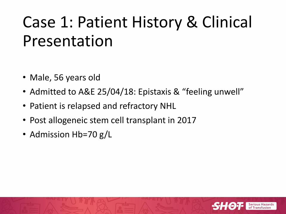
Case 1: Patient History & Clinical Presentation
• Male, 56 years old
• Admitted to A&E 25/04/18: Epistaxis & “feeling unwell”
• Patient is relapsed and refractory NHL
• Post allogeneic stem cell transplant in 2017
• Admission Hb=70 g/L
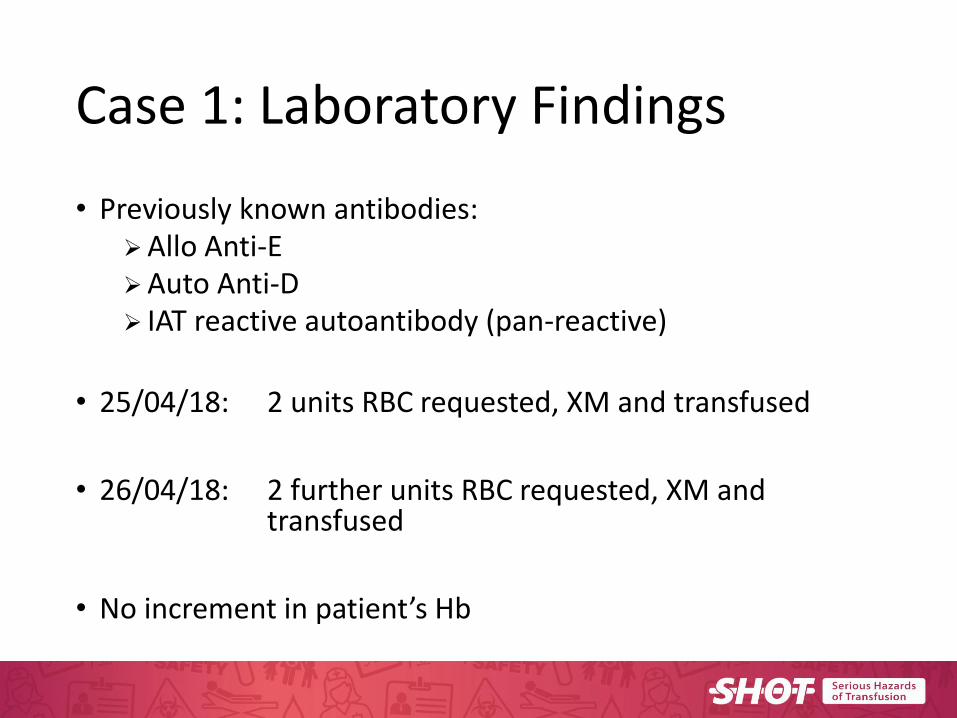
Case 1: Laboratory Findings
• Previously known antibodies: Allo Anti-E Auto Anti-D IAT reactive autoantibody (pan-reactive)
• 25/04/18: 2 units RBC requested, XM and transfused
• 26/04/18: 2 further units RBC requested, XM and transfused
• No increment in patient’s Hb
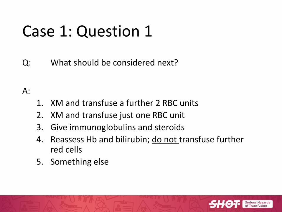
Case 1: Question 1
Q: What should be considered next?
A:
1. XM and transfuse a further 2 RBC units
2. XM and transfuse just one RBC unit
3. Give immunoglobulins and steroids
4. Reassess Hb and bilirubin; do not transfuse further red cells
5. Something else
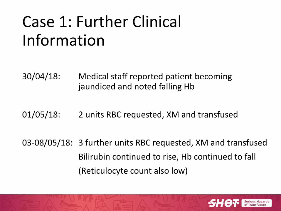
Case 1: Further Clinical Information
30/04/18: Medical staff reported patient becoming jaundiced and noted falling Hb
01/05/18: 2 units RBC requested, XM and transfused
03-08/05/18: 3 further units RBC requested, XM and transfused
Bilirubin continued to rise, Hb continued to fall
(Reticulocyte count also low)
Case 1: Question 2
Q: What possible diagnosis should be considered in light of this information?
A:
1. TACO
2. Delayed haemolytic transfusion reaction
3. Hyperhaemolysis
4. Bacterial infection
5. TRALI
Case 1: Question 3
Q: What is the likely mechanism of hyperhaemolysis?
A:
1. Activation of the coagulation cascade
2. Activated macrophages leading to destruction of both patient & donor red cells
3. I have no idea
4. Activation of the complement cascade
5. Antibodies causing intravascular haemolysis
Case 1: Question 4
Q: What is the most effective treatment for hyperhaemolysis?
A:
1. Transfusion of additional RBC units
2. Avoidance of further transfusion but supportive immunoglobulins and steroids
3. I have no idea
4. Transfusion of additional RBC units with immunoglobulins and steroids
5. Administration of colloids instead
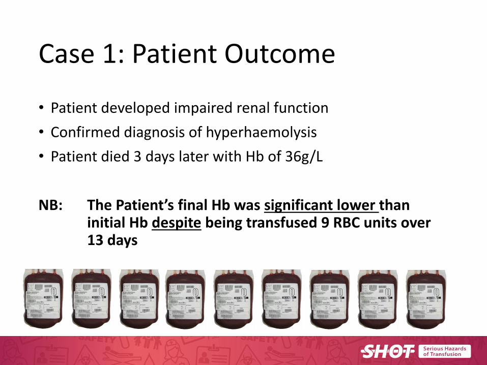
Case 1: Patient Outcome
• Patient developed impaired renal function
• Confirmed diagnosis of hyperhaemolysis
• Patient died 3 days later with Hb of 36g/L
NB: The Patient’s final Hb was significant lower than initial Hb despite being transfused 9 RBC units over 13 days
Case 1: Learning Points
Hyperhaemolysis remains a cause of transfusion-related mortality and major morbidity
Hyperhaemolysis is NOT only associated with Sickle Cell Disease patients
Key message from SHOT report: Important that ALL clinicians involved in the transfusion process have an awareness of the SIGNS and SYMPTOMS of HYPERHAEMOLYSIS so that ANY suspected cases are investigated thoroughly.
Case 2: Patient History & Clinical Presentation
• Female, 19 years old
• Admitted with Ulcerative colitis
• Transfused one unit RBC on 15/09/18
• No previous transfusions
Case 2: Laboratory Findings
• 18/09/18: Initial investigation = negative Ab screen
• Eligible for Electronic Issue crossmatch (EI XM)
• Patient reviewed by Haem. Reg, Consultant and Transfusion Practitioner
• Increased temperature >2oC, 24 hrs post transfusion
Case 2: Question 1
Q: What is a likely cause of fever following a transfusion?
A:
1. Febrile non-haemolytic transfusion reaction
2. Acute/delayed haemolytic reaction
3. Bacterial contamination
4. Underlying condition
5. Any of the above
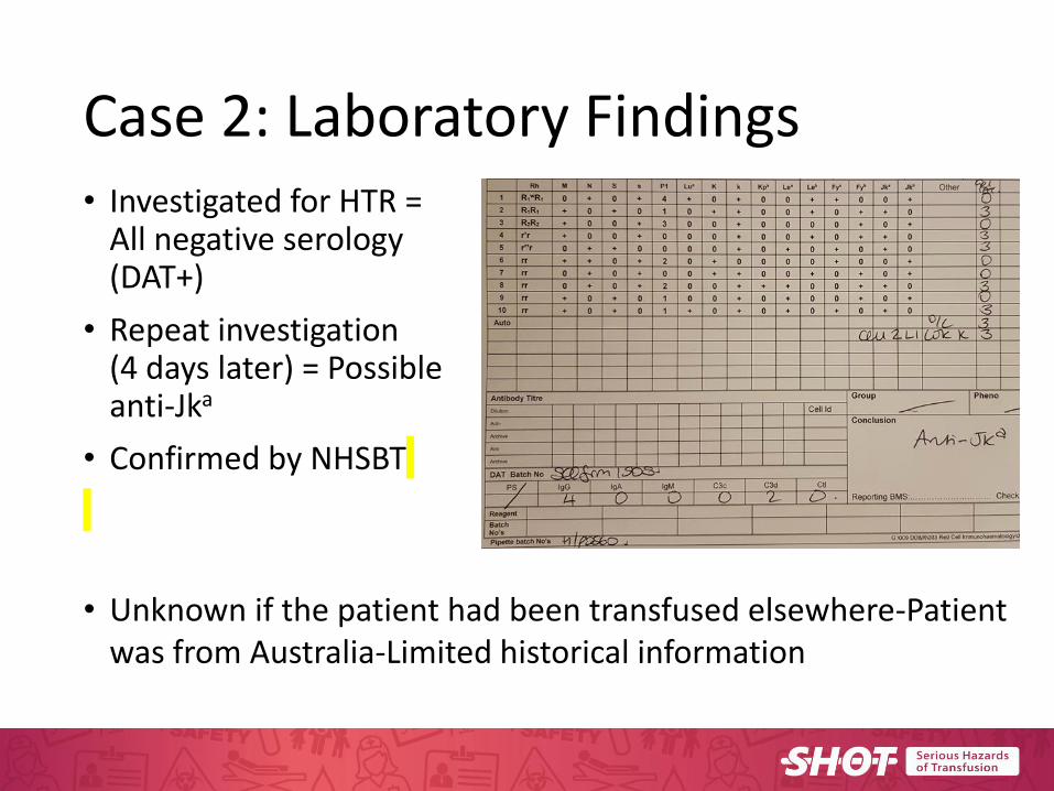
Case 2: Laboratory Findings • Investigated for HTR =
All negative serology (DAT+)
• Repeat investigation (4 days later) = Possible anti-Jka
• Confirmed by NHSBT
• Unknown if the patient had been transfused elsewhere-Patient was from Australia-Limited historical information
Case 2: Question 2
Q: What additional test could you perform on the patient’s red cells?
A:
1. Antigen typing
2. Neutralisation
3. Elution
4. Titration
5. I’m not sure
Case 2: Question 3
Q: When does haemolysis occur for a DHTR?
A:
1. <24 hours post transfusion
2. >28 days post transfusion
3. >24 hours <28 days post transfusion
4. >24 hours <7 days post transfusion
5. >14 days post transfusion
Case 2: Patient Outcome
• Patient recovered and survived
• No treatment was necessary
• Diagnosis was complicated by sepsis
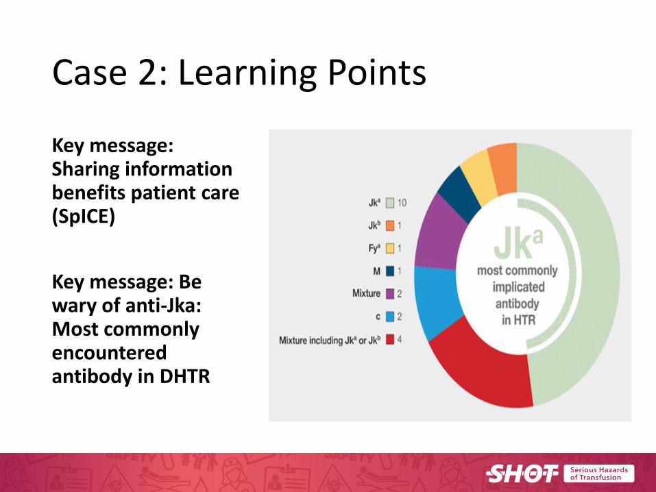
Case 2: Learning Points
Key message: Sharing information benefits patient care (SpICE)
Key message: Be wary of anti-Jka: Most commonly encountered antibody in DHTR
Case 3: Patient History and Clinical Presentation • Female, 25 years old
• Known Sickle Cell Disease (SCD) patient with multiple historical alloantibodies:
Anti-A1, Anti-S, Anti-M, Anti-Jka, Anti-Lea, Anti-Leb
• Admitted with sickle cell crisis during pregnancy (1st pregnancy)
• Multi-transfused due to SCD
Case 3: Laboratory Findings
• At time of transfusion serology only Anti-Lea, Anti-Leb and Anti-A1 were identified
• Historical Anti-S, Anti-M, Anti-Jka were currently not detected by IAT
Case 3: Question 1
Q: Should we worry about clinically insignificant antibodies?
A:
1. No, they are clinically insignificant
2. Yes, all antibodies have a potential to cause harm
3. Unsure
4. Maybe
5. BSH Guidelines state which antibodies are clinically significant
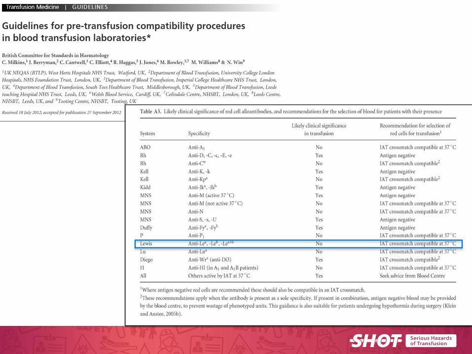
Case 3: Laboratory Findings & Clinical Information • Anti-Lea and Anti-Leb were classed by RCI as
“not clinically significant” in accordance with BSH Guidelines
• 2 RBC units were transfused over a two day period:
Group O rr, S-, M-, Jka-, Fya-, K-, HbS-
• Units transfused: 1 unit Le(a-b+) and 1 unit Le(a+b-)
• Intravascular haemolysis noted with both units
• Transfusion stopped: • Raised bilirubin • Falling Hb • Haemoglobinuria • Positive DAT
Case 3: Question 2
Q: What is the most likely type of transfusion reaction?
A:
1. TACO
2. TRALI
3. Bacterial infection
4. Hyperhaemolysis
5. Delayed haemolytic transfusion reaction
Case 3: Question 3
Q: What additional tests are useful when delayed HTR is suspected?
A: 1. Hgb/HCT 2. LDH 3. Bilirubin 4. Serum haptoglobins 5. All of the above
Case 3: Patient Outcome
• Outcome: Patient survived and recovered
• Successful pregnancy
• Subsequent Le(a-b-) units have been transfused during further sickle crisis
Case 3: Learning Points
• Multi transfused patients carry additional risk of immunisation & extra risk of HTR
• Intravascular haemolysis of transfused cells due to Anti-Lea and Anti-Leb is very rare
• A reaction reported due to Lewis antibodies which according to the text books “don’t cause reactions”
• Key message: Occasionally “clinically INSIGNIFICANT” antibodies cause haemolytic transfusion reactions too
Case 4: Patient History & Clinical Presentation
• Male, 51 years old
• Routine surgical case (day surgery)
• Proximal Left Anterior Descending (LAD) Coronary Artery Occlusion (high mortality risk)
• Patient had no apparent prior transfusion history
• Negative antibody screen (Pre-op)
• 16/03/18: XM and transfused 3 units ABO &RhD matched RBC during surgery
Case 4: Laboratory Findings
• 23/03/18: Anti-Fya, Anti-E and Anti-c were identified in antibody identification panel with +DAT
• 26/03/18: Patient had raised bilirubin level (suspecting haemolytic transfusion reaction)
Full investigation & blood films requested
• 28/03/18: Sample referred to NHSBT who confirmed Anti-Fya, Anti-E and Anti-c + additional Anti-K in patient plasma and Anti-Fya in patient eluate
Case 4: Question 1
Q: What was observed on the patient’s blood film?
A:
1. Elliptocytes
2. Normocytic RBC
3. Spherocytes
4. Fragments
5. Spherocytes, reticulocytes and fragments
Case 4: Question 2
Q: What are the most commonly responsible antibodies in DHTRs?
A:
1. Anti-Ch1
2. Anti-A1
3. Anti-K, Anti-Fya and Anti-Jka
4. Anti-S, Anti-M and Anti-s
5. I don’t know
Case 4: Question 3
Q: What is the frequency of a DHTR?
A:
1. 1:500 – 1:1,000 transfusions
2. 1:2,500- 1:11,000 transfusions
3. 1:50,000 – 1:100,000 transfusions
4. 1:5,000,000 – 1:10,000,000 transfusions
5. I wouldn’t like to hazard a guess
Case 4: Patient Outcome
• Patient transferred to ICU
• Treatment involved steroids and immunoglobulins
• Survived and recovered fully
• Was discharged 8 days later
Case 4: Learning Points
• Key message: Not all DHTRs are due to pre-formed antibodies
• Need for vigilance even in presence of negative antibody screen
• This case was initial antibody screen negative BUT had multiple antibodies in the post transfusion samples
• Key message: COMMUNICATION is essential
• Use of electronic data sharing (SpICE) improves patient outcomes
Case 5: Patient History & Clinical Presentation
• Female, 33 years old
• Admitted 05/09/18 with PPH (Post Partum Haemorrhage)
• Actively bleeding
• Patient received two units of Emergency O- RBC on 05/09/18 at 02:20hrs
• The patient had a known (allo) Anti-Jka
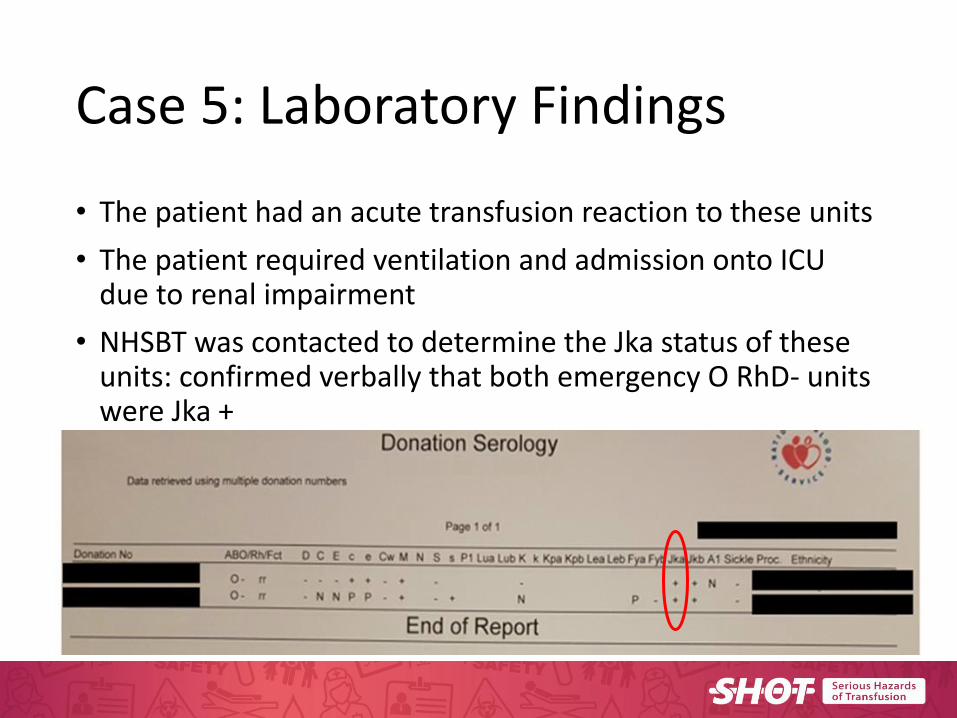
Case 5: Laboratory Findings
• The patient had an acute transfusion reaction to these units
• The patient required ventilation and admission onto ICU due to renal impairment
• NHSBT was contacted to determine the Jka status of these units: confirmed verbally that both emergency O RhD- units were Jka +
Case 5: Question 1
Q: How many cases within the latest SHOT report were due to the use of emergency O- blood?
A:
1. 10
2. 2
3. 4
4. 6
5. 1
Case 5: Question 2
Q: When is the most severe reaction likely?
A:
1. 15 mins
2. 30 mins
3. 60 mins
4. 90 mins
5. 120 mins
Case 5: Question 3
Q: Will the DAT always be positive?
A:
1. Yes
2. Yes, unless all of the donor cells have been destroyed
3. No
4. I’m not sure
5. Sometimes
Case 5: Clinical Outcome
• Patient was transferred to ICU where vital parameters (blood pressure, heart rate, respiration) and urine production were be monitored continuously
• Patient survived and recovered
• Major morbidity associated with this case
Case 5: Learning Points
Key message: Emergency units save lives
• No patient should die from lack of blood
• There is a balance between not withholding transfusion when clinically urgent and waiting to provide compatible units
Key message: Close monitoring of the patient is essential
• Reaction in a patient given antigen + blood due to clinical urgency
Thank you for listening
Dr Shruthi Narayan & Nicci Wilkes
With Thanks to Tracey Tomlinson (RCI-Colindale)
Recommended