Sciatica: Low back and Leg Pain Diagnosis and Treatment Options
Presented by
Devesh Ramnath, MD
Orthopaedic Associates Of Dallas
Baylor Spine Center
Sciatica
• Compression of the spinal nerves in the back which can lead to symptoms of leg pain, numbness and weakness along the different nerves as they travel down the leg and into the foot
• Also known as Radiculopathy

Sciatic Nerve
Sciatic Nerve
Anatomy of the Lumbar Spine
Lumbar Anatomy
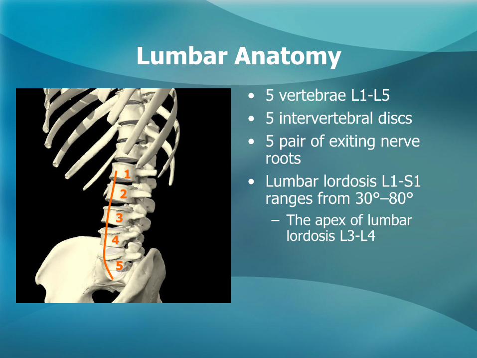
• 5 vertebrae L1-L5
• 5 intervertebral discs
• 5 pair of exiting nerve roots
• Lumbar lordosis L1-S1 ranges from 30°–80°
– The apex of lumbar lordosis L3-L4
1
2
3
4
5
Sacral Anatomy
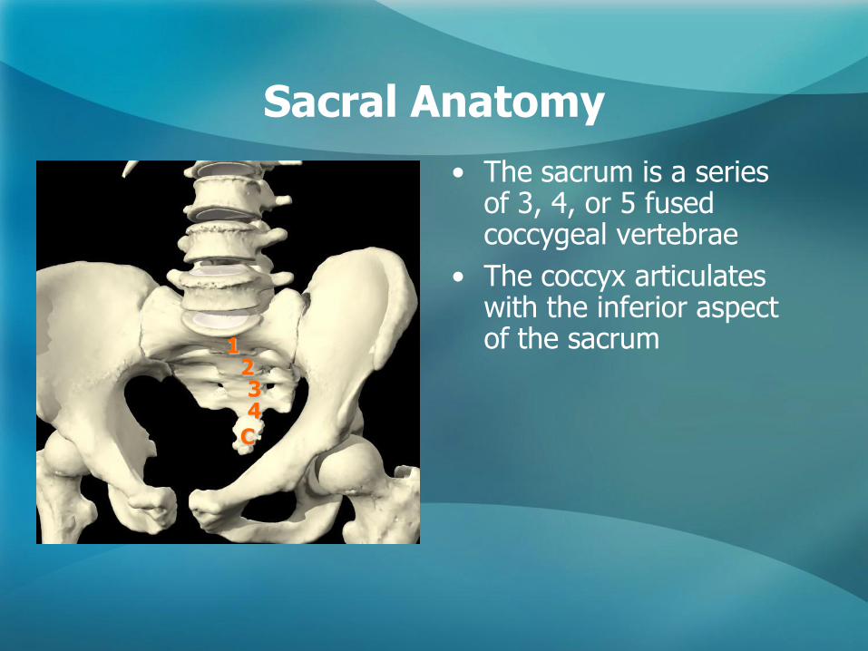
• The sacrum is a series of 3, 4, or 5 fused coccygeal vertebrae
• The coccyx articulates with the inferior aspect of the sacrum 1
2 3 4
C
Lumbar Spine Anatomy
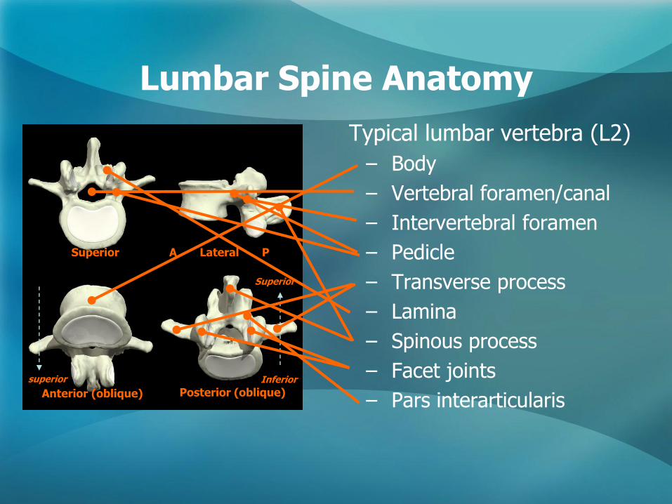
Typical lumbar vertebra (L2)
– Body
– Vertebral foramen/canal
– Intervertebral foramen
– Pedicle
– Transverse process
– Lamina
– Spinous process
– Facet joints
– Pars interarticularis
inferior
Superior
Anterior (oblique)
A Lateral P
Posterior (oblique)
Superior
Inferior superior
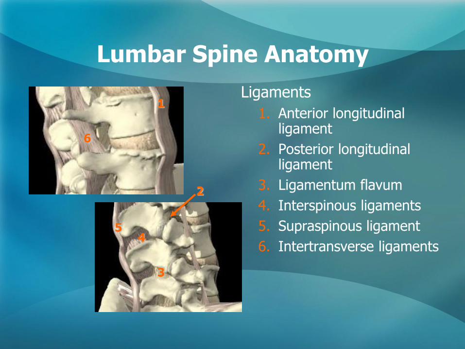
Ligaments
1. Anterior longitudinal ligament
2. Posterior longitudinal ligament
3. Ligamentum flavum
4. Interspinous ligaments
5. Supraspinous ligament
6. Intertransverse ligaments
Lumbar Spine Anatomy
1
2
3
4 5
6
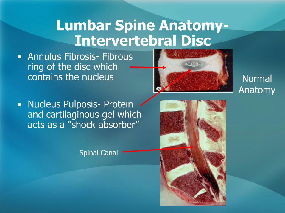
• Annulus Fibrosis- Fibrous ring of the disc which contains the nucleus
• Nucleus Pulposis- Protein and cartilaginous gel which acts as a ―shock absorber‖
Spinal Canal
Normal Anatomy
Lumbar Spine Anatomy-Intervertebral Disc
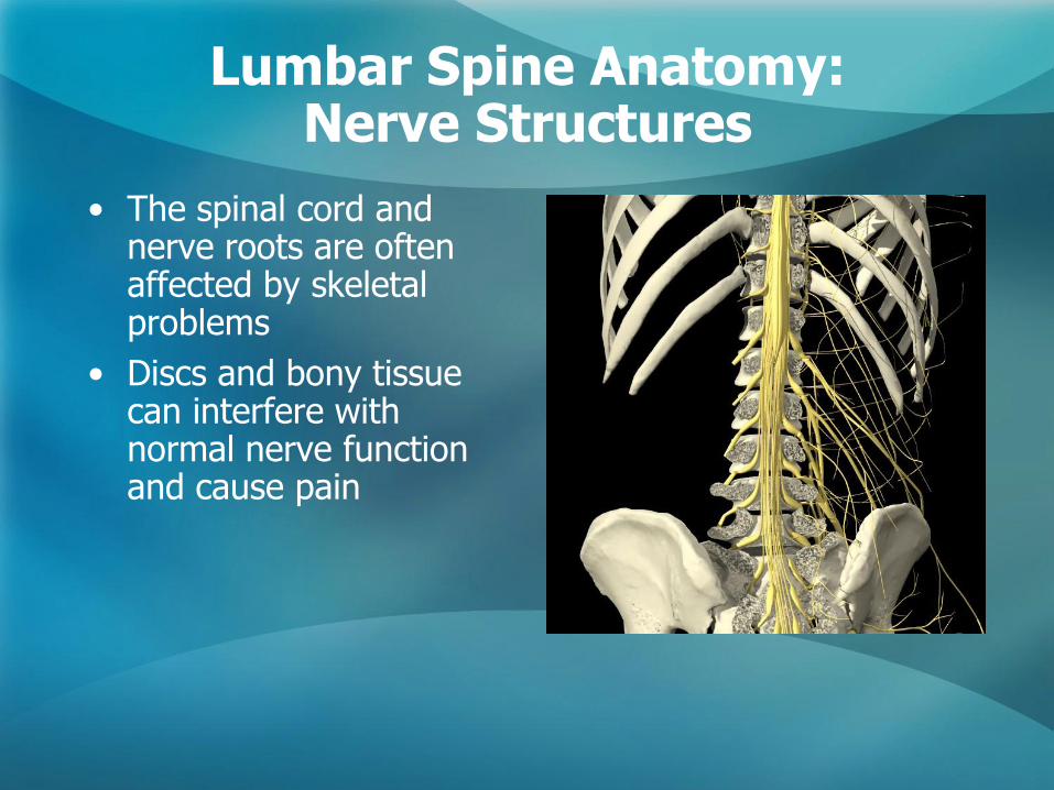
Lumbar Spine Anatomy: Nerve Structures
• The spinal cord and nerve roots are often affected by skeletal problems
• Discs and bony tissue can interfere with normal nerve function and cause pain
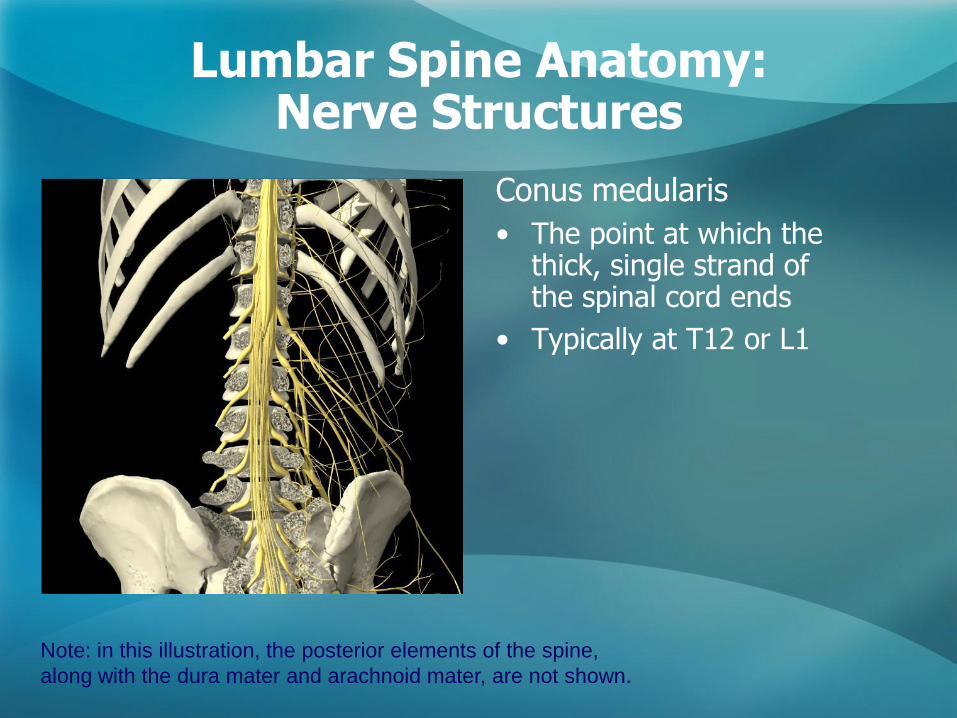
Lumbar Spine Anatomy: Nerve Structures
Conus medularis
• The point at which the thick, single strand of the spinal cord ends
• Typically at T12 or L1
Note: in this illustration, the posterior elements of the spine,
along with the dura mater and arachnoid mater, are not shown.
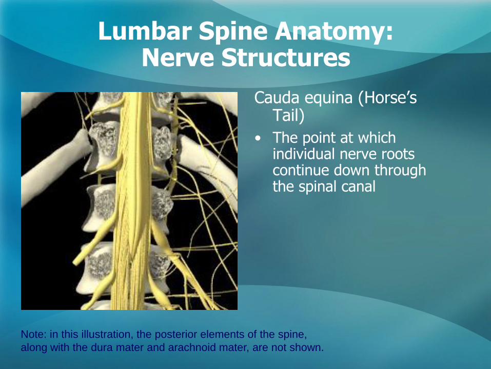
Lumbar Spine Anatomy: Nerve Structures
Cauda equina (Horse’s Tail)
• The point at which individual nerve roots continue down through the spinal canal
Note: in this illustration, the posterior elements of the spine,
along with the dura mater and arachnoid mater, are not shown.
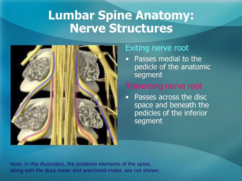
Lumbar Spine Anatomy: Nerve Structures
Exiting nerve root
• Passes medial to the pedicle of the anatomic segment
Traversing nerve root
• Passes across the disc space and beneath the pedicles of the inferior segment
Note: in this illustration, the posterior elements of the spine,
along with the dura mater and arachnoid mater, are not shown.
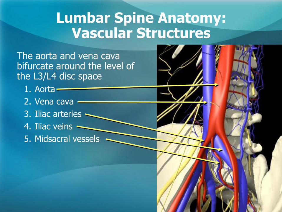
Lumbar Spine Anatomy: Vascular Structures
The aorta and vena cava bifurcate around the level of the L3/L4 disc space
1. Aorta
2. Vena cava
3. Iliac arteries
4. Iliac veins
5. Midsacral vessels
Spinal Pathologies and Treatments
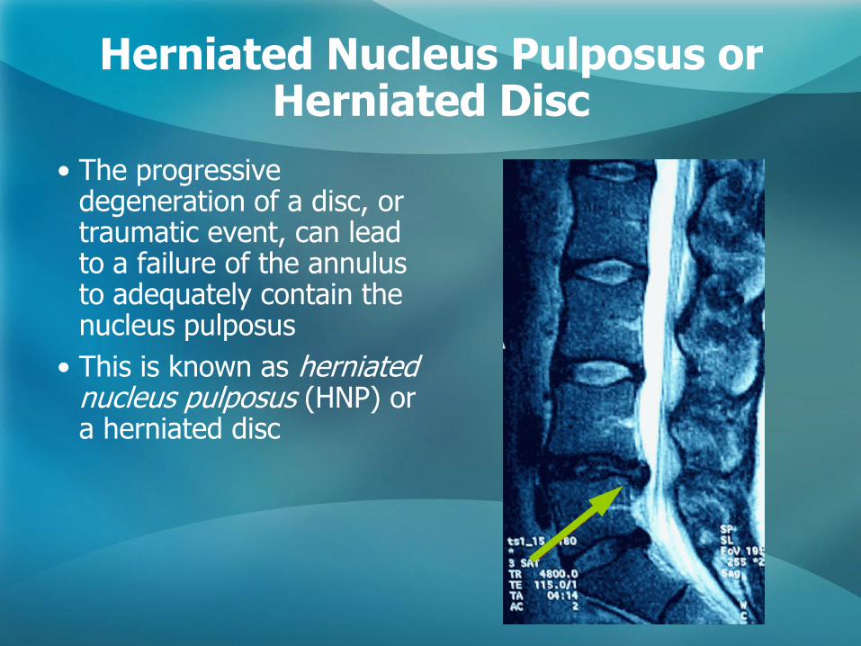
Herniated Nucleus Pulposus or Herniated Disc
• The progressive degeneration of a disc, or traumatic event, can lead to a failure of the annulus to adequately contain the nucleus pulposus
• This is known as herniated nucleus pulposus (HNP) or a herniated disc
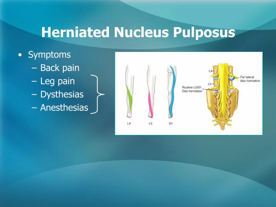
Herniated Nucleus Pulposus
• Symptoms
– Back pain
– Leg pain
– Dysthesias
– Anesthesias
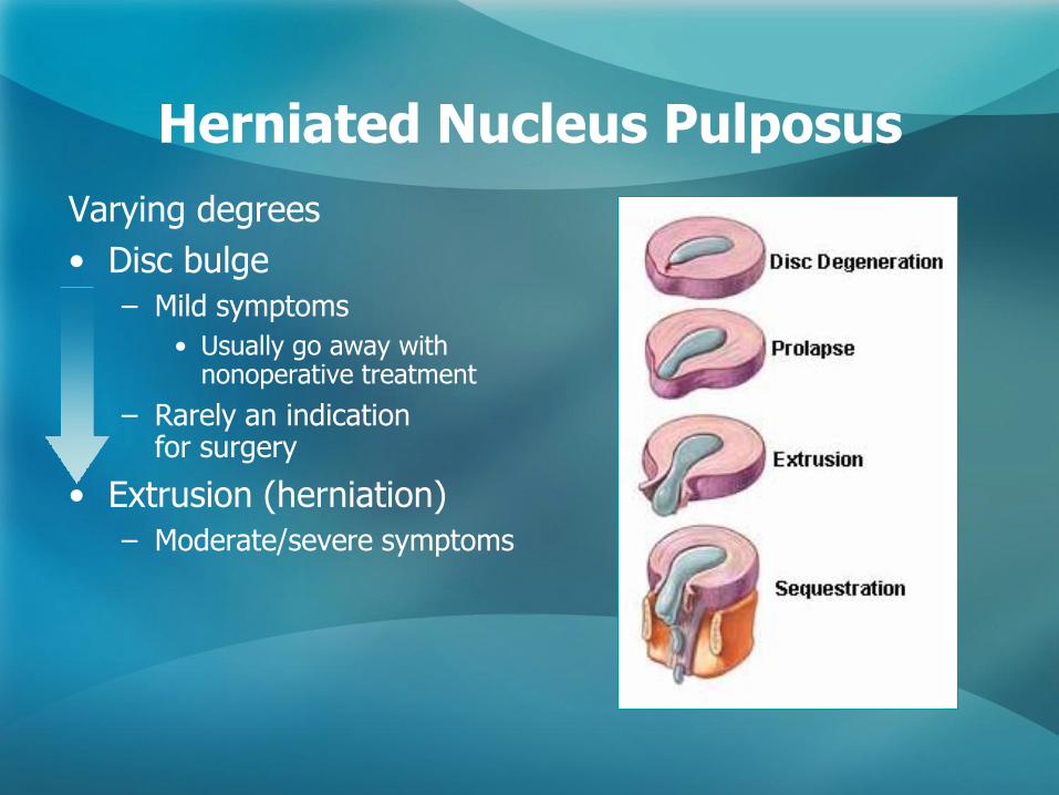
Herniated Nucleus Pulposus
Varying degrees
• Disc bulge
– Mild symptoms
• Usually go away with nonoperative treatment
– Rarely an indication for surgery
• Extrusion (herniation)
– Moderate/severe symptoms
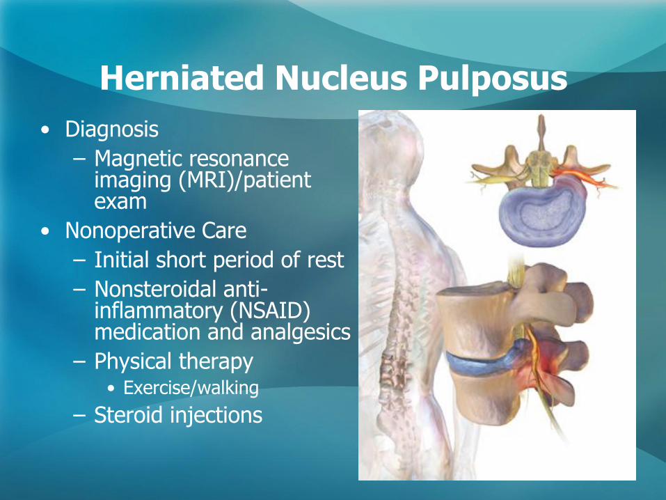
Herniated Nucleus Pulposus
• Diagnosis
– Magnetic resonance imaging (MRI)/patient exam
• Nonoperative Care
– Initial short period of rest
– Nonsteroidal anti-inflammatory (NSAID) medication and analgesics
– Physical therapy • Exercise/walking
– Steroid injections
Herniated Nucleus Pulposus
• Surgical care - Indications – Failure of nonoperative treatment
• Usually minimum of 6 weeks in duration – Can be months
– Progressive neurologic deficit – Cauda equina syndrome
• Cauda Equina Syndrome – Caused by a large central disc herniation – Symptoms include bilateral leg pain, loss of perianal
sensation, paralysis of the bladder, and weakness of the anal sphincter
– Surgical intervention in these cases is urgent or deficits can be permanent
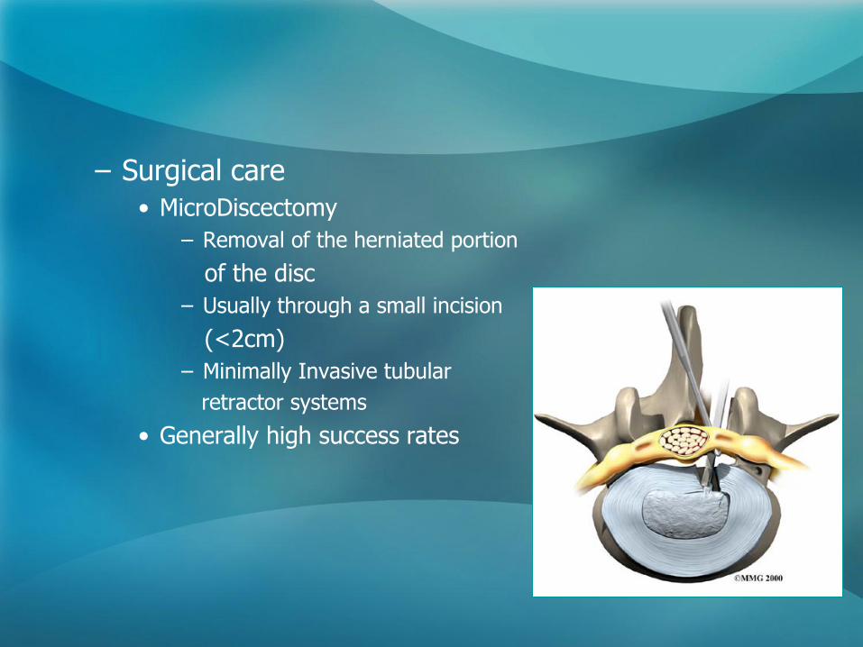
– Surgical care
• MicroDiscectomy
– Removal of the herniated portion
of the disc
– Usually through a small incision
(<2cm)
– Minimally Invasive tubular
retractor systems
• Generally high success rates
Herniated Nucleus Pulposus
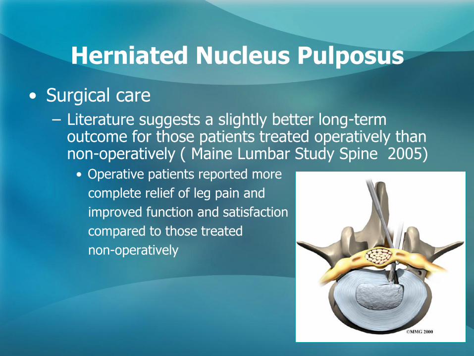
• Surgical care
– Literature suggests a slightly better long-term outcome for those patients treated operatively than non-operatively ( Maine Lumbar Study Spine 2005)
• Operative patients reported more
complete relief of leg pain and
improved function and satisfaction
compared to those treated
non-operatively
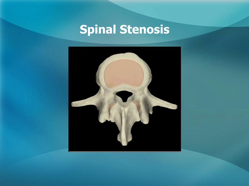
Spinal Stenosis
Spinal Stenosis
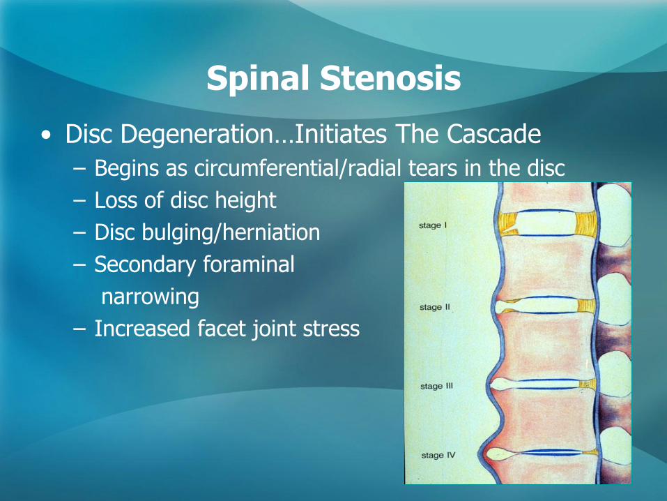
• Disc Degeneration…Initiates The Cascade
– Begins as circumferential/radial tears in the disc
– Loss of disc height
– Disc bulging/herniation
– Secondary foraminal
narrowing
– Increased facet joint stress
Spinal Stenosis
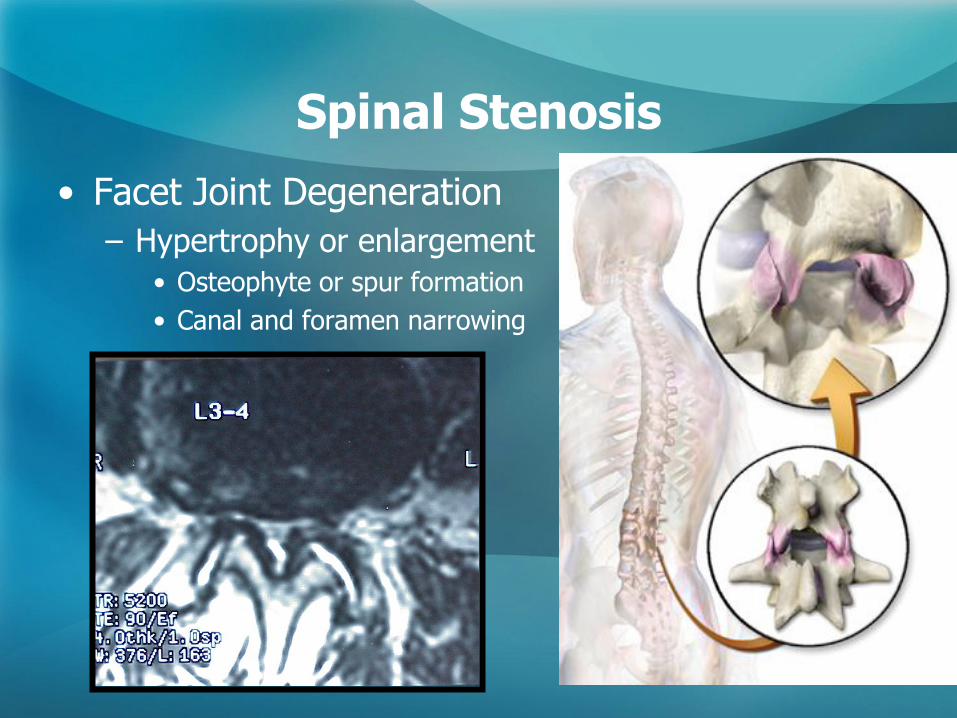
• Facet Joint Degeneration
– Hypertrophy or enlargement
• Osteophyte or spur formation
• Canal and foramen narrowing
Spinal Stenosis
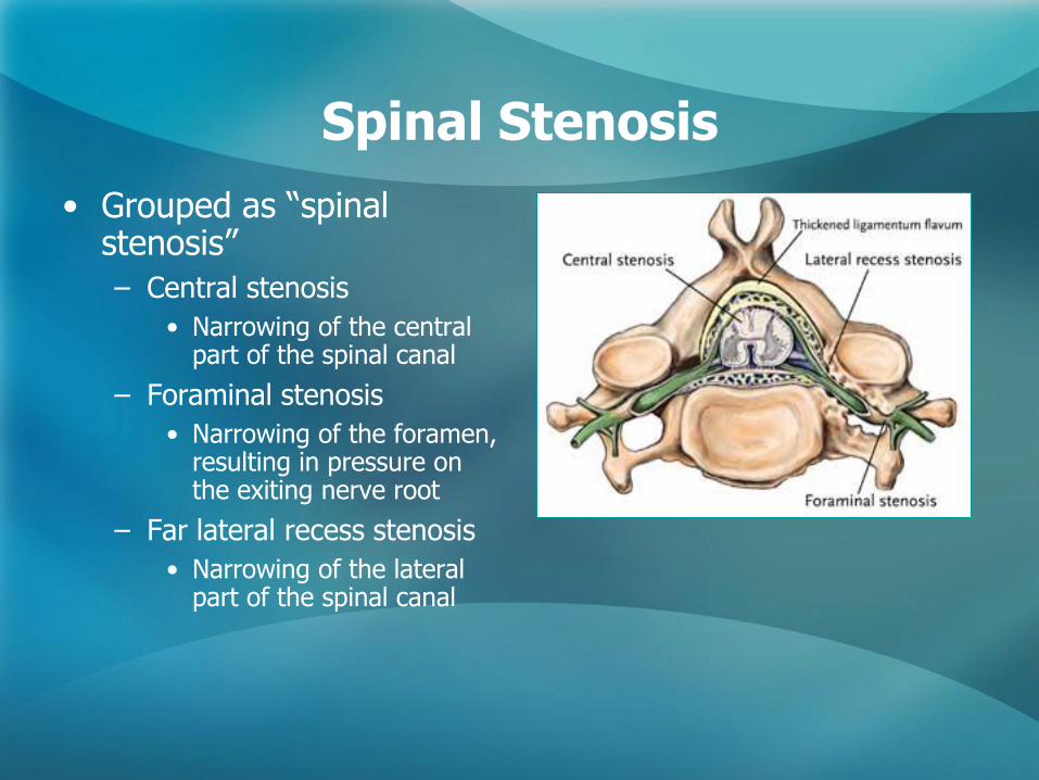
• Grouped as ―spinal stenosis‖
– Central stenosis
• Narrowing of the central part of the spinal canal
– Foraminal stenosis
• Narrowing of the foramen, resulting in pressure on the exiting nerve root
– Far lateral recess stenosis
• Narrowing of the lateral part of the spinal canal
Spinal Stenosis
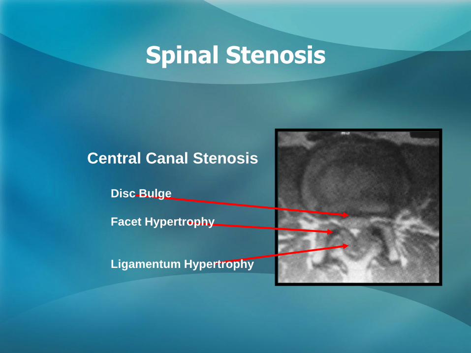
Central Canal Stenosis
Disc Bulge
Facet Hypertrophy
Ligamentum Hypertrophy
Spinal Stenosis
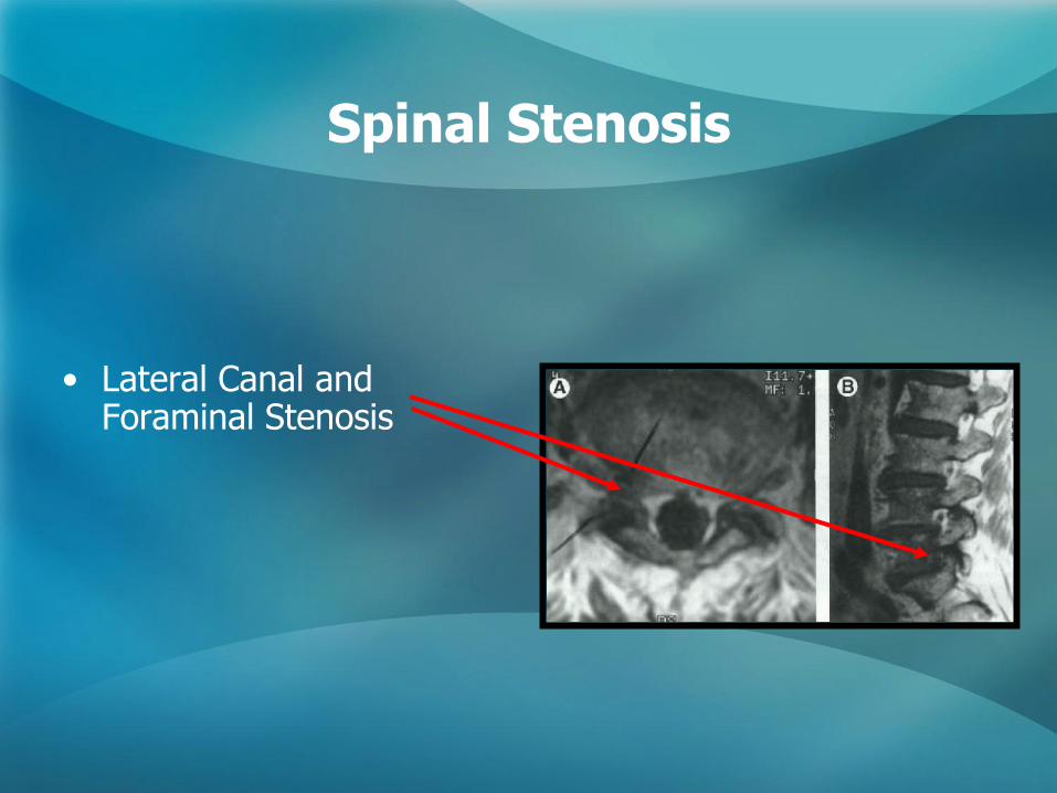
• Lateral Canal and Foraminal Stenosis
Spinal Stenosis
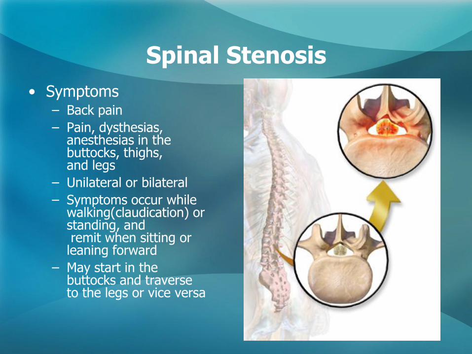
• Symptoms – Back pain
– Pain, dysthesias, anesthesias in the buttocks, thighs, and legs
– Unilateral or bilateral
– Symptoms occur while walking(claudication) or standing, and remit when sitting or leaning forward
– May start in the buttocks and traverse to the legs or vice versa
Spinal Stenosis
• Not all patients with ―stenosis‖ are clinically symptomatic
• Pathoanatomy is much better understood than pathophysiology
• What are the theories?
– Mechanical Compression
– Vascular Changes
Spinal Stenosis
• Mechanical Compression
– Minimal cross-sectional area to accommodate cauda equina is 77mm+/-13mm2
– This is approximately 45% of normal
– Small decreases in area below this level cause large increases in pressure
• Schonstrom et al Spine 1988
Spinal Stenosis
• Mechanical Compression
– Physiologic Changes
• Venous congestion
• Decrease in nutritional transport in the nerves
• Changes in nerve conduction
Spinal Stenosis
• Mechanical Compression
– Prolonged compression may lead to long-term neurologic dysfunction
Spinal Stenosis
• Vascular Changes
– Microcirculation of the nerve is affected
• Venous stasis and congestion
• Arterial Insufficiency
– Arterial supply from anterior spinal artery and segmentals is compressed causing ischemia (decreased flow) and subsequent symptoms
Spinal Stenosis
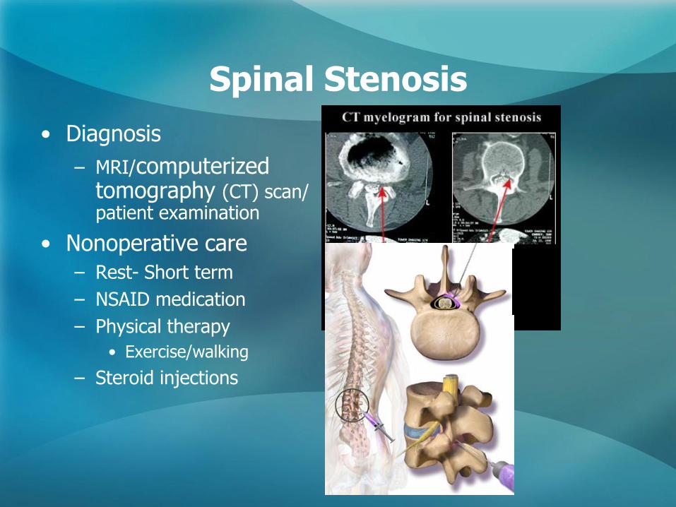
• Diagnosis
– MRI/computerized tomography (CT) scan/ patient examination
• Nonoperative care
– Rest- Short term
– NSAID medication
– Physical therapy
• Exercise/walking
– Steroid injections
Spinal Stenosis
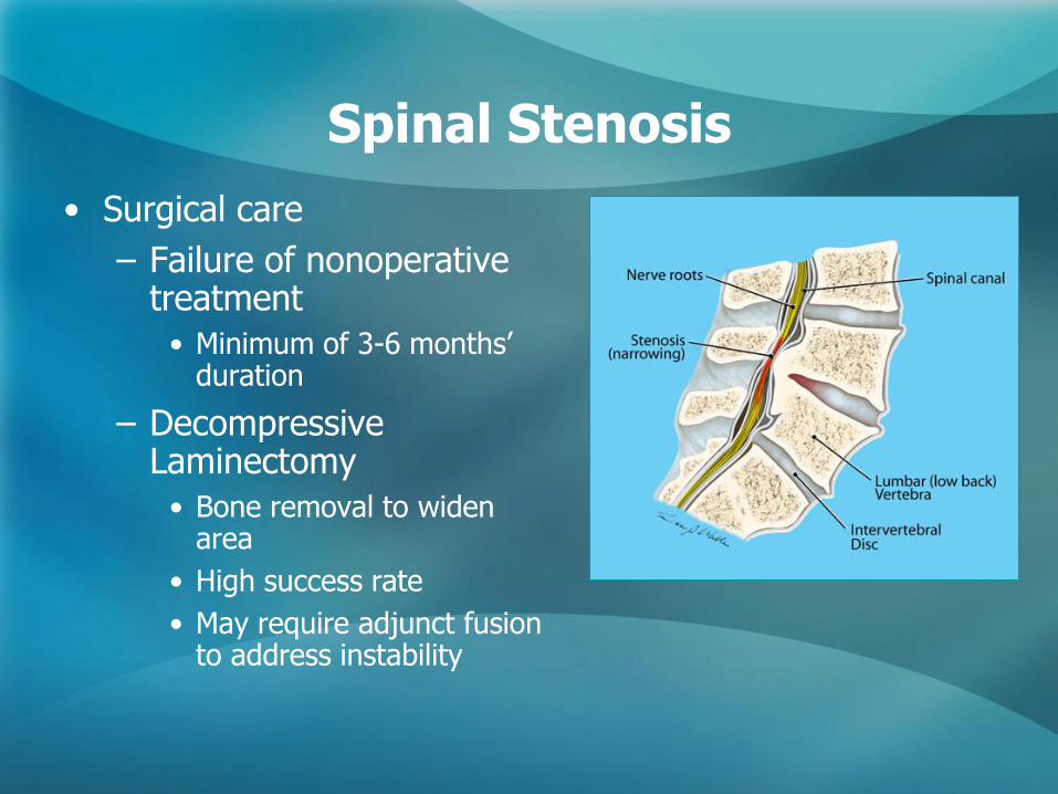
• Surgical care
– Failure of nonoperative treatment
• Minimum of 3-6 months’ duration
– Decompressive Laminectomy
• Bone removal to widen area
• High success rate
• May require adjunct fusion to address instability
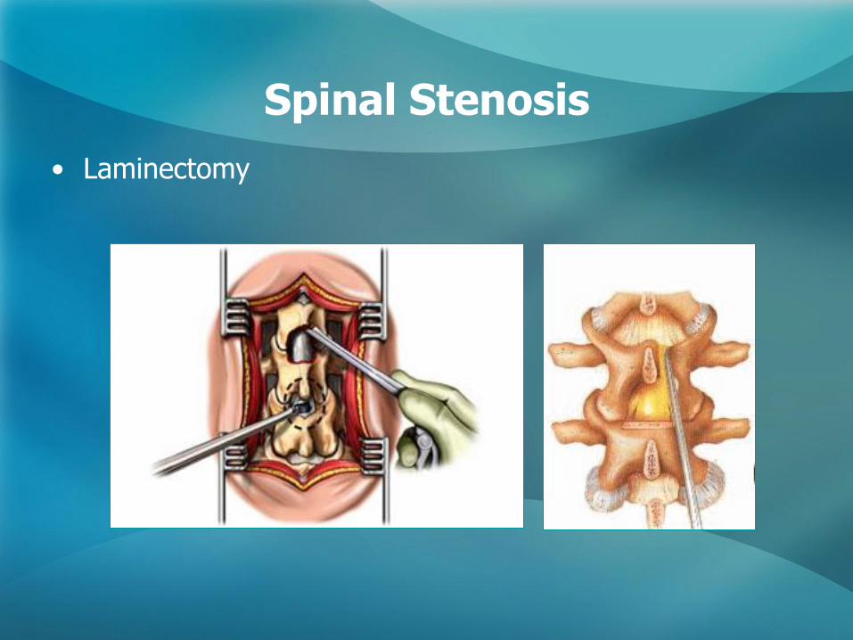
Spinal Stenosis
• Laminectomy
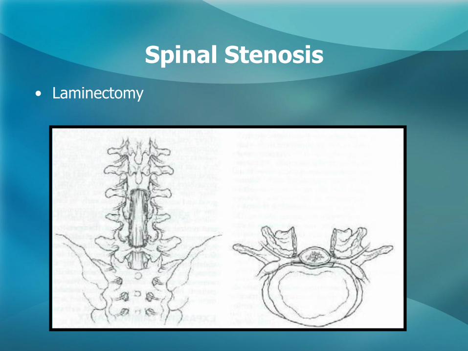
Spinal Stenosis
• Laminectomy
• Laminectomy
– Good results reported in 80-85% cases
• Results may deteriorate over time
– Recurrent stenosis, adjacent degeneration, instability
– Newer techniques
• Minimally invasive decompression using tubular retractors and microscope
• Indirect decompression with spinous process spacer devices
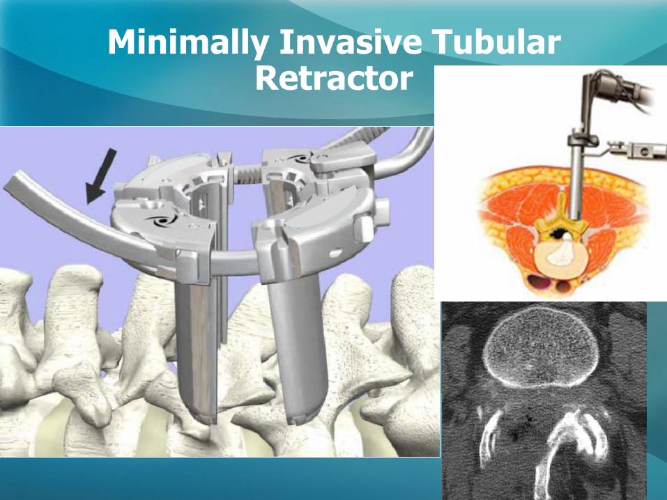
Minimally Invasive Tubular Retractor
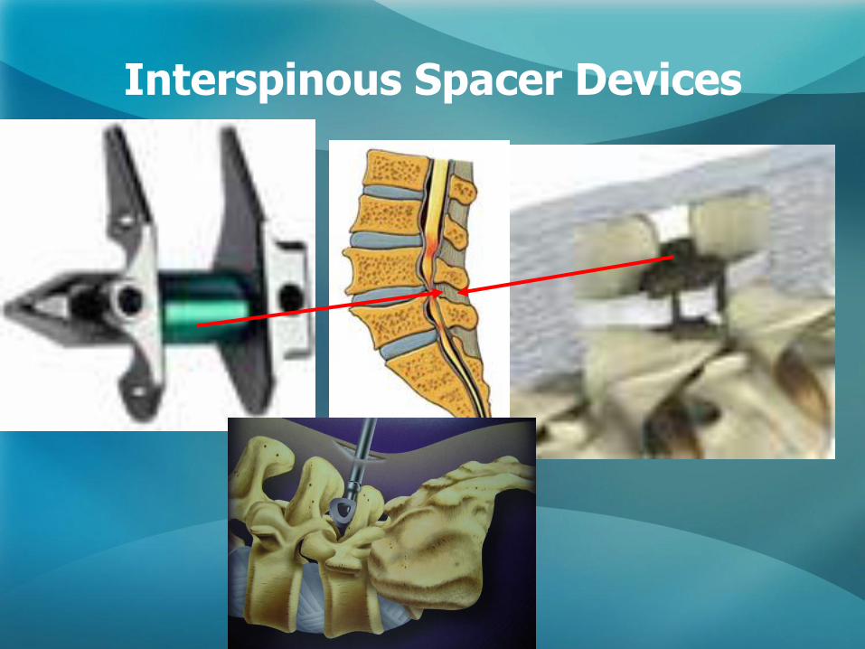
Interspinous Spacer Devices
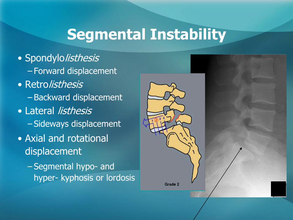
Segmental Instability
• Spondylolisthesis
– Forward displacement
• Retrolisthesis
– Backward displacement
• Lateral listhesis
– Sideways displacement
• Axial and rotational
displacement
– Segmental hypo- and
hyper- kyphosis or lordosis
Segmental Instability
• Spondylolisthesis
– A forward translation of 1 vertebral body over the adjacent vertebra
• Degenerative
– ―Adult-onset‖ progressive slip
• Lytic
– Develops in children or adolescents, but only 25% experience symptoms
• Spondylolysis
– A fracture or defect in the vertebra, usually in the posterior elements—most frequently in the pars interarticularis
• Spondyloloptosis
– Complete dislocation
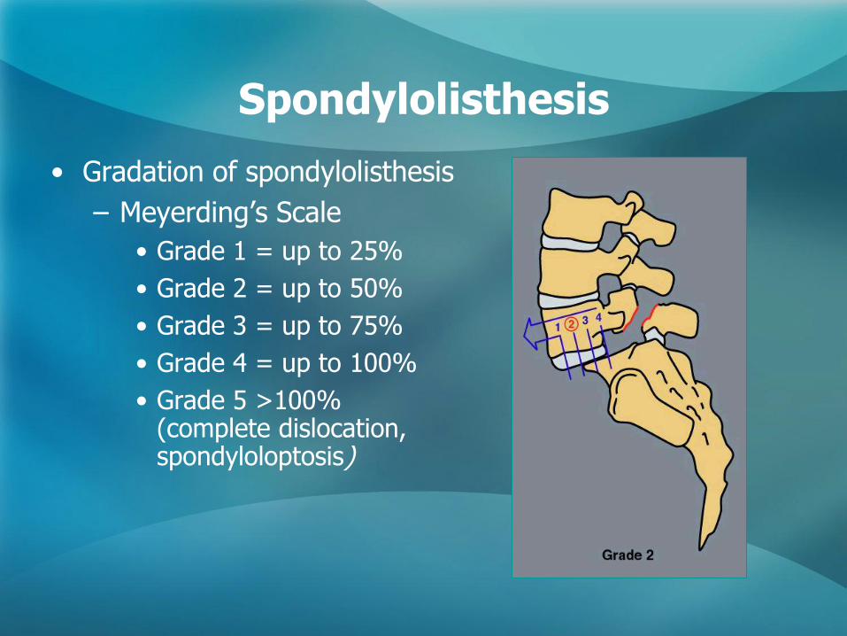
Spondylolisthesis
• Gradation of spondylolisthesis
– Meyerding’s Scale
• Grade 1 = up to 25%
• Grade 2 = up to 50%
• Grade 3 = up to 75%
• Grade 4 = up to 100%
• Grade 5 >100% (complete dislocation, spondyloloptosis)
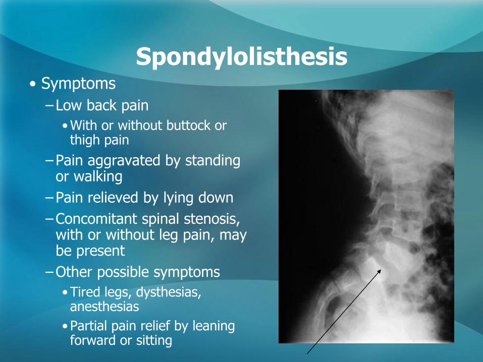
Spondylolisthesis • Symptoms
–Low back pain
•With or without buttock or thigh pain
–Pain aggravated by standing or walking
–Pain relieved by lying down
–Concomitant spinal stenosis, with or without leg pain, may be present
–Other possible symptoms
•Tired legs, dysthesias, anesthesias
•Partial pain relief by leaning forward or sitting
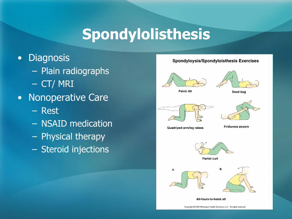
Spondylolisthesis
• Diagnosis
– Plain radiographs
– CT/ MRI
• Nonoperative Care
– Rest
– NSAID medication
– Physical therapy
– Steroid injections
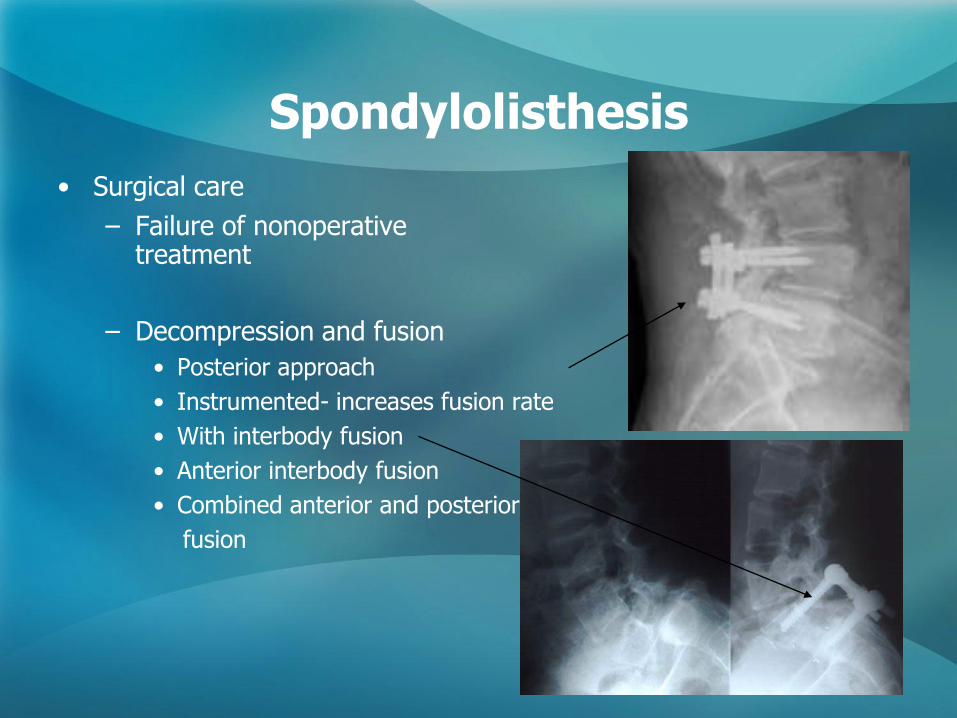
Spondylolisthesis
• Surgical care
– Failure of nonoperative treatment
– Decompression and fusion
• Posterior approach
• Instrumented- increases fusion rate
• With interbody fusion
• Anterior interbody fusion
• Combined anterior and posterior
fusion
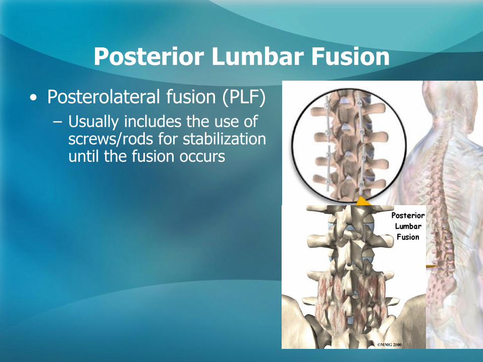
Posterior Lumbar Fusion
• Posterolateral fusion (PLF)
– Usually includes the use of screws/rods for stabilization until the fusion occurs
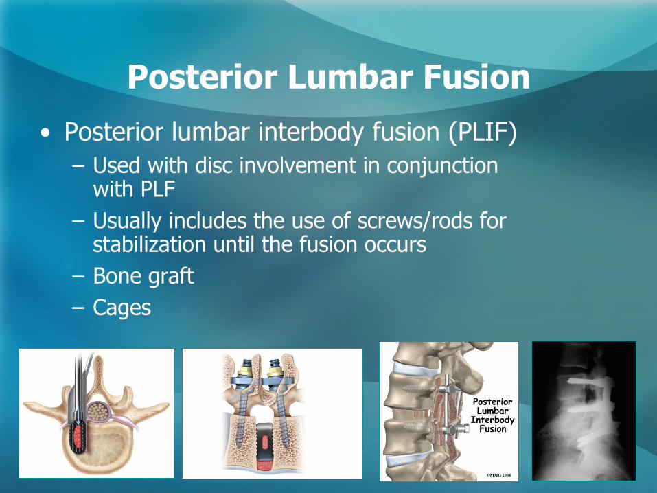
Posterior Lumbar Fusion
• Posterior lumbar interbody fusion (PLIF)
– Used with disc involvement in conjunction with PLF
– Usually includes the use of screws/rods for stabilization until the fusion occurs
– Bone graft
– Cages
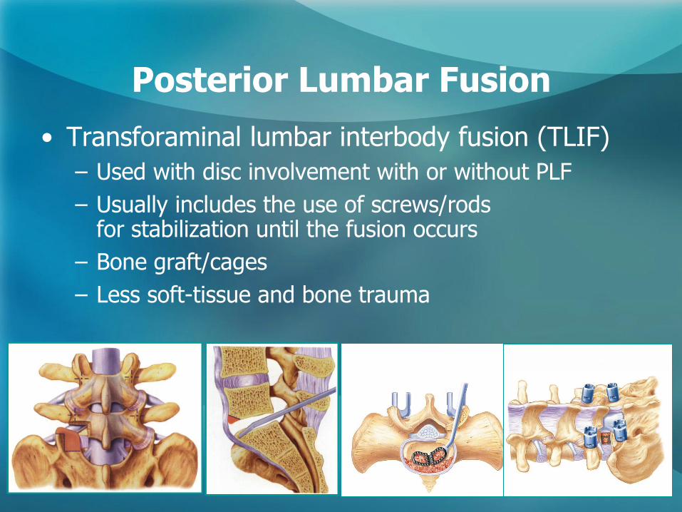
Posterior Lumbar Fusion
• Transforaminal lumbar interbody fusion (TLIF)
– Used with disc involvement with or without PLF
– Usually includes the use of screws/rods for stabilization until the fusion occurs
– Bone graft/cages
– Less soft-tissue and bone trauma
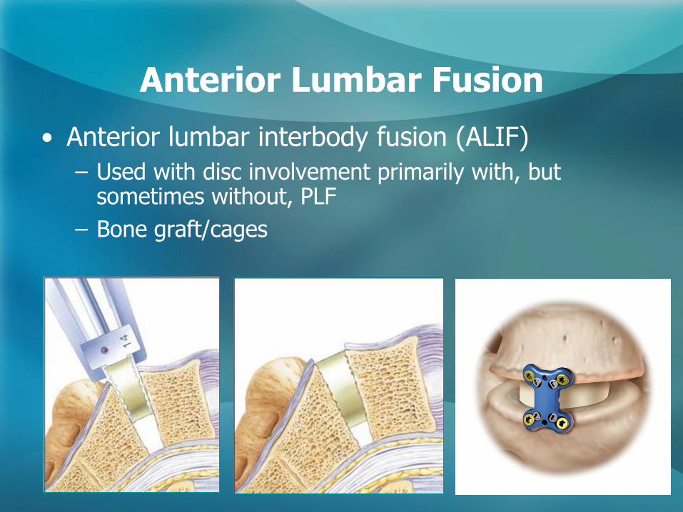
Anterior Lumbar Fusion
• Anterior lumbar interbody fusion (ALIF)
– Used with disc involvement primarily with, but sometimes without, PLF
– Bone graft/cages
Lumbar Fusion
• Newer Techniques
– Minimally invasive fusion techniques
• Tubular retractors and percutaneous pedicle screws
• XLIF- extreme lateral interbody fusion
• Advantages- Less soft tissue dissection, less blood loss, faster recovery
• Disadvantages- technically demanding, increased surgical time, limited surgical exposure
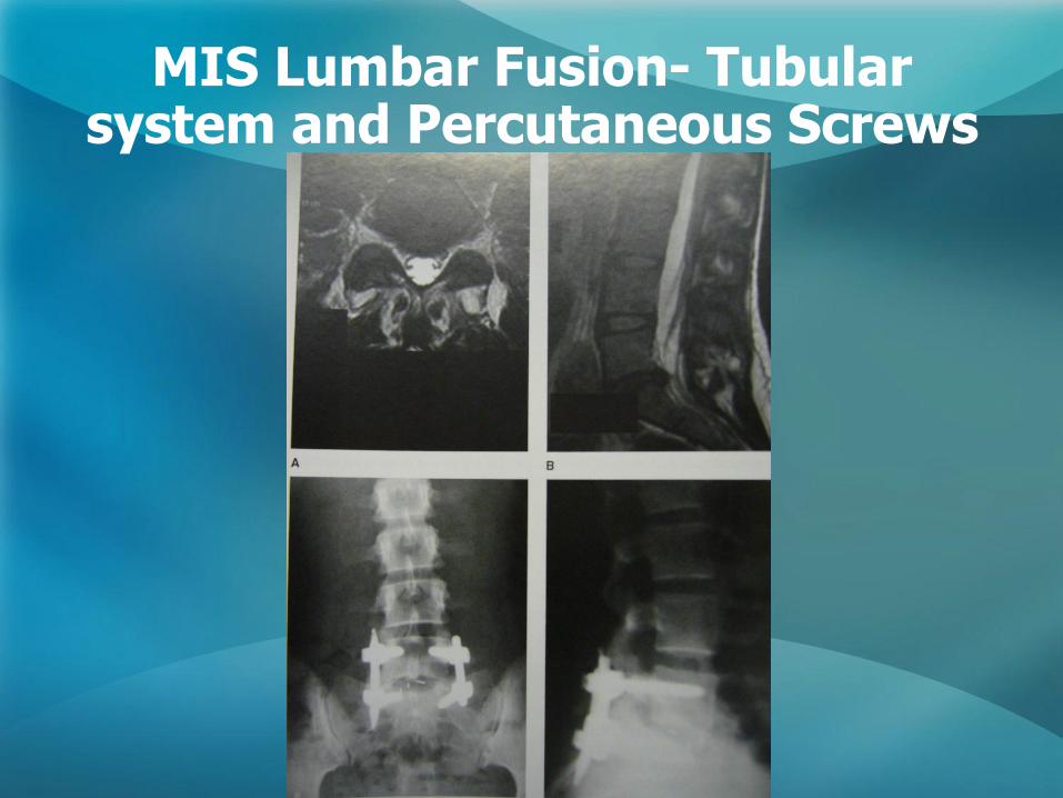
MIS Lumbar Fusion- Tubular system and Percutaneous Screws
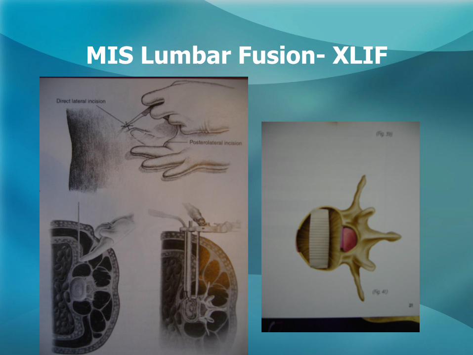
MIS Lumbar Fusion- XLIF
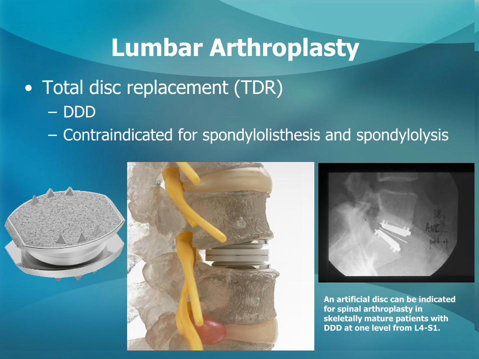
Lumbar Arthroplasty
• Total disc replacement (TDR)
– DDD
– Contraindicated for spondylolisthesis and spondylolysis
An artificial disc can be indicated for spinal arthroplasty in skeletally mature patients with DDD at one level from L4-S1.
Conclusions
• Sciatica or Radiculopathy
– Herniated Discs
– Spinal Stenosis
– Spondylolisthesis
• Treatments
– Nonoperative
– Operative
– Newer Treatments
Thank You
Recommended

![Clinical diagnostic model for sciatica developed in ... · considered most important for distinguishing sciatica from non-specific leg pain in LBLP patients [26] and (b) items used](https://img.pdfslide.us/doc/110x75/5fd415c379ff91782318c008/clinical-diagnostic-model-for-sciatica-developed-in-considered-most-important.jpg)