SACRAL INSTANT DIAGNOSIS & TREATMENT WITH LIGAMENTOUS ARTICULAR
STRAIN (LAS)
Greg Thompson, D.O., ACOFP, ABSPOMM
Assistant Dean Clinical Medicine
Chair, Osteopathic Principles and Practice
Alabama College of Osteopathic Medicine
OBJECTIVES
Understand the ten different sacral dysfunctions
Be able to diagnose sacral dysfunction based on landmarks and motion testing
in 20 seconds or less
Learn to easily treat sacral diagnoses using Ligamentous articular strain (LAS) in the other 40 seconds
First the Bad news….
LOL ROR
ROL LOR
RIGHT UNI FLEXED
RIGHT UNI EXTENDED
LEFT UNI FLEXED
LEFT UNI EXTENDED
BILATERAL FLEXED
BILATERAL EXTENDED
ROR LOR
LEFT FLEXED
LEFT EXTENDED
LOL ROL
RIGHT FLEXED
RIGHT EXTENDED
ROR
LEFT FLEXED
BILATERAL FLEXED
BILATERAL EXTENDED
BILATERAL FLEXED BILATERAL EXTENDED
LOR
LEFT EXTENDEDLOL
RIGHT FLEXED
ROL
RIGHT EXTENDED
RIGHT EXTENDED ROLLOL RIGHT FLEXEDLOR LEFT EXTENDEDROR LEFT FLEXED
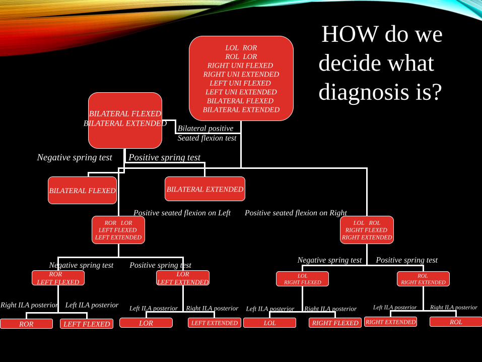
HOW do we
decide what
diagnosis is?
Bilateral positive
Seated flexion test
Negative spring test Positive spring test
Positive seated flexion on Left Positive seated flexion on Right
Negative spring test Positive spring testNegative spring test Positive spring test
Left ILA posterior Right ILA posteriorLeft ILA posterior Right ILA posteriorLeft ILA posterior Right ILA posteriorRight ILA posterior Left ILA posterior
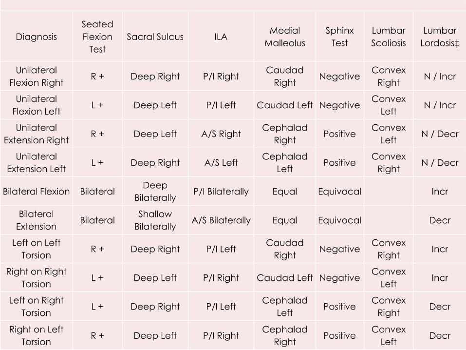
Diagnosis
Seated
Flexion
Test
Sacral Sulcus ILAMedial
Malleolus
Sphinx
Test
Lumbar
Scoliosis
Lumbar
Lordosis‡
Unilateral
Flexion RightR + Deep Right P/I Right
Caudad
RightNegative
Convex
RightN / Incr
Unilateral
Flexion LeftL + Deep Left P/I Left Caudad Left Negative
Convex
LeftN / Incr
Unilateral
Extension RightR + Deep Left A/S Right
Cephalad
RightPositive
Convex
LeftN / Decr
Unilateral
Extension LeftL + Deep Right A/S Left
Cephalad
LeftPositive
Convex
RightN / Decr
Bilateral Flexion BilateralDeep
BilaterallyP/I Bilaterally Equal Equivocal Incr
Bilateral
ExtensionBilateral
Shallow
BilaterallyA/S Bilaterally Equal Equivocal Decr
Left on Left
TorsionR + Deep Right P/I Left
Caudad
RightNegative
Convex
RightIncr
Right on Right
TorsionL + Deep Left P/I Right Caudad Left Negative
Convex
LeftIncr
Left on Right
TorsionL + Deep Right P/I Left
Cephalad
LeftPositive
Convex
RightDecr
Right on Left
TorsionR + Deep Left P/I Right
Cephalad
RightPositive
Convex
LeftDecr
And now for something almost completely different…
…the ‘Good news’
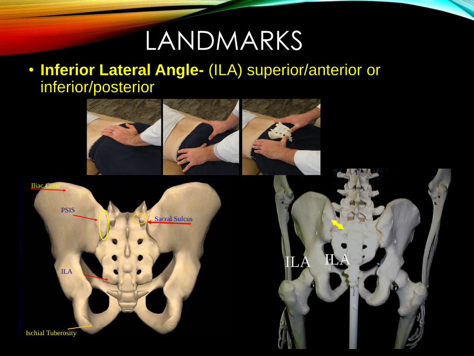
LANDMARKS• Inferior Lateral Angle- (ILA) superior/anterior or
inferior/posterior
ILA ILA
PSIS
Sacral Sulcus
Iliac Crest
Ischial Tuberosity
ILA
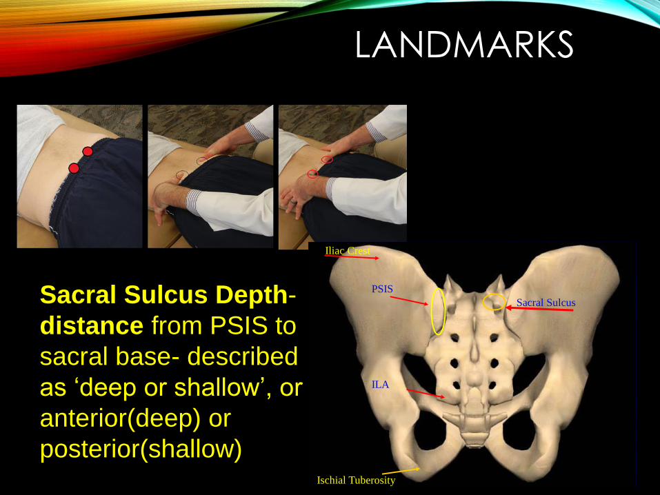
LANDMARKS
PSIS
Sacral Sulcus
Iliac Crest
Ischial Tuberosity
ILA
Sacral Sulcus Depth-
distance from PSIS to
sacral base- described
as ‘deep or shallow’, or
anterior(deep) or
posterior(shallow)
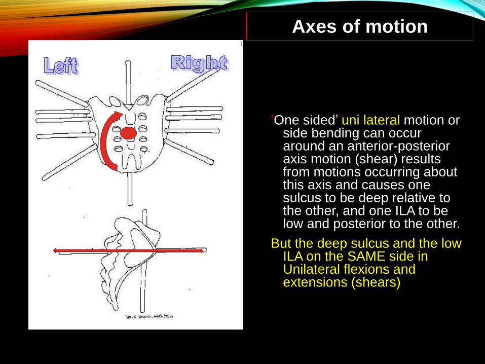
‘One sided’ uni lateral motion or side bending can occur around an anterior-posterior axis motion (shear) results from motions occurring about this axis and causes one sulcus to be deep relative to the other, and one ILA to be low and posterior to the other.
But the deep sulcus and the low ILA on the SAME side in Unilateral flexions and extensions (shears)
Axes of motion
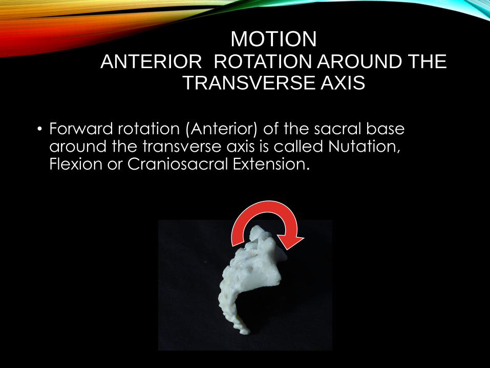
MOTIONANTERIOR ROTATION AROUND THE
TRANSVERSE AXIS
• Forward rotation (Anterior) of the sacral base around the transverse axis is called Nutation, Flexion or Craniosacral Extension.
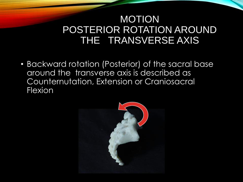
MOTIONPOSTERIOR ROTATION AROUND
THE TRANSVERSE AXIS
• Backward rotation (Posterior) of the sacral base around the transverse axis is described as Counternutation, Extension or Craniosacral Flexion
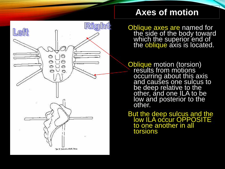
Oblique axes are named for the side of the body toward which the superior end of the oblique axis is located.
Oblique motion (torsion) results from motions occurring about this axis and causes one sulcus to be deep relative to the other, and one ILA to be low and posterior to the other.
But the deep sulcus and the low ILA occur OPPOSITE to one another in all torsions
Axes of motion
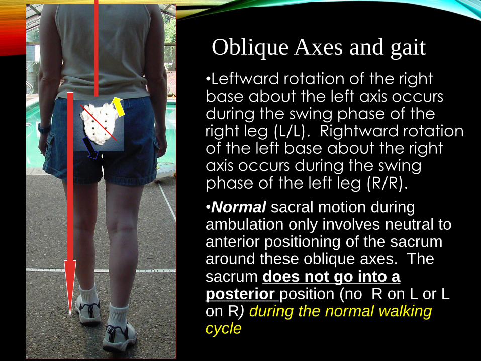
•Leftward rotation of the right base about the left axis occurs during the swing phase of the right leg (L/L). Rightward rotation of the left base about the right axis occurs during the swing phase of the left leg (R/R).
•Normal sacral motion during ambulation only involves neutral to anterior positioning of the sacrum around these oblique axes. The sacrum does not go into a posterior position (no R on L or L on R) during the normal walking cycle
Oblique Axes and gait
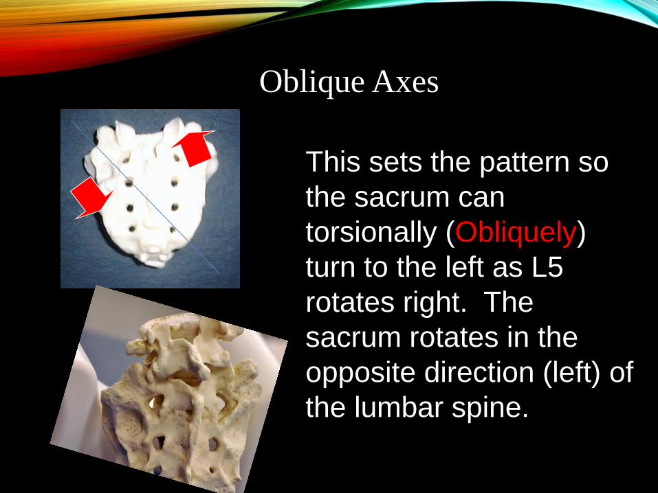
This sets the pattern so
the sacrum can
torsionally (Obliquely)
turn to the left as L5
rotates right. The
sacrum rotates in the
opposite direction (left) of
the lumbar spine.
Oblique Axes
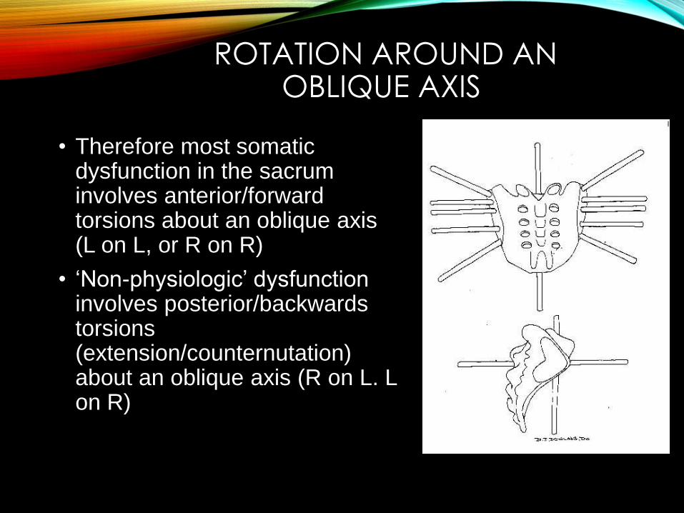
ROTATION AROUND AN OBLIQUE AXIS
• Therefore most somatic dysfunction in the sacrum involves anterior/forward torsions about an oblique axis (L on L, or R on R)
• ‘Non-physiologic’ dysfunction involves posterior/backwards torsions (extension/counternutation) about an oblique axis (R on L. L on R)
POSTERIOR MOVEMENT AROUND THE OBLIQUE AXIS
• The sacral base moves posteriorly only as a response to flexion of the lumbar spine (specifically L5)
• Somatic dysfunction involving posterior torsions (and shears) are therefore are less frequent and usually secondary to somatic dysfunction with flexion of L5 or trauma.
• This is the “tie in the fly syndrome” or the “the well man bent over and crippled stood” syndrome.. Typical to mimic diskogenic conditions
DIAGNOSING SACRAL SOMATIC DYSFUNCTION
• The sacrum can become “stuck” in any of 10 positions causing a somatic dysfunction
• The presence of a sacral somatic dysfunction will have the four classic physical findings of :
• Tenderness on palpation, *Assymetry on static palpation, *Restriction of motion, and Tissue texture changes (TART)
SACRAL SOMATIC DYSFUNCTION
• Once TART is found the specific diagnosis is distinguished by landmarks (sacral sulcus and inferior lateral angle of the sacrum and ILA) to determine the type of somatic dysfunction (Torsions, unilateral shears or bilateral dysfunction).
• Using motion tests to distinguish side of dysfunction (seated flexion test (SEFT)) or anterior or posterior position of the sacral base (spring/sphinx test) secures the complete diagnosis
PLANES OF MOTION , ANATOMIC TRAINS, AND AUTO MATICALLY MOBILE
(LANDMARKS)
• You must keep in mind the results of the landmarks, then applying the results of the
• seated flexion (side of dysfunction)
OR
• spring/sphinx test (anterior or posterior base direction)
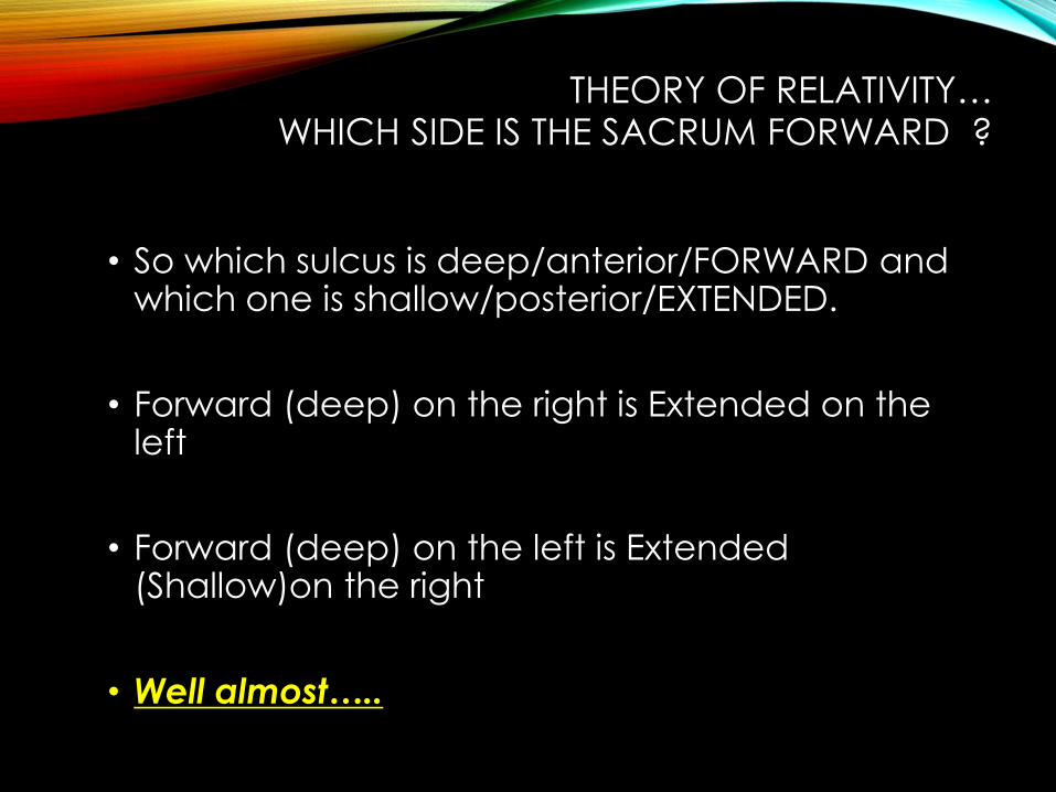
THEORY OF RELATIVITY…WHICH SIDE IS THE SACRUM FORWARD ?
• So which sulcus is deep/anterior/FORWARD and which one is shallow/posterior/EXTENDED.
• Forward (deep) on the right is Extended on the left
• Forward (deep) on the left is Extended (Shallow)on the right
• Well almost…..
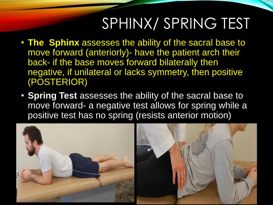
SPHINX/ SPRING TEST• The Sphinx assesses the ability of the sacral base to
move forward (anteriorly)- have the patient arch their back- if the base moves forward bilaterally then negative, if unilateral or lacks symmetry, then positive (POSTERIOR)
• Spring Test assesses the ability of the sacral base to move forward- a negative test allows for spring while a positive test has no spring (resists anterior motion)
•
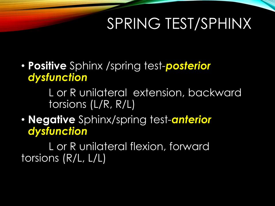
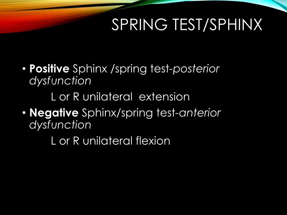
SPRING TEST/SPHINX
• Positive Sphinx /spring test-posterior dysfunction
L or R unilateral extension, backward torsions (L/R, R/L)
• Negative Sphinx/spring test-anterior dysfunction
L or R unilateral flexion, forward torsions (R/L, L/L)
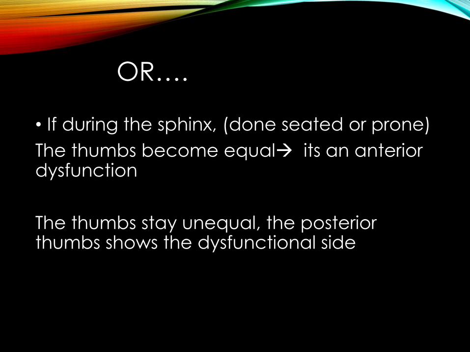
OR….
• If during the sphinx, (done seated or prone)
The thumbs become equal its an anterior dysfunction
The thumbs stay unequal, the posterior thumbs shows the dysfunctional side
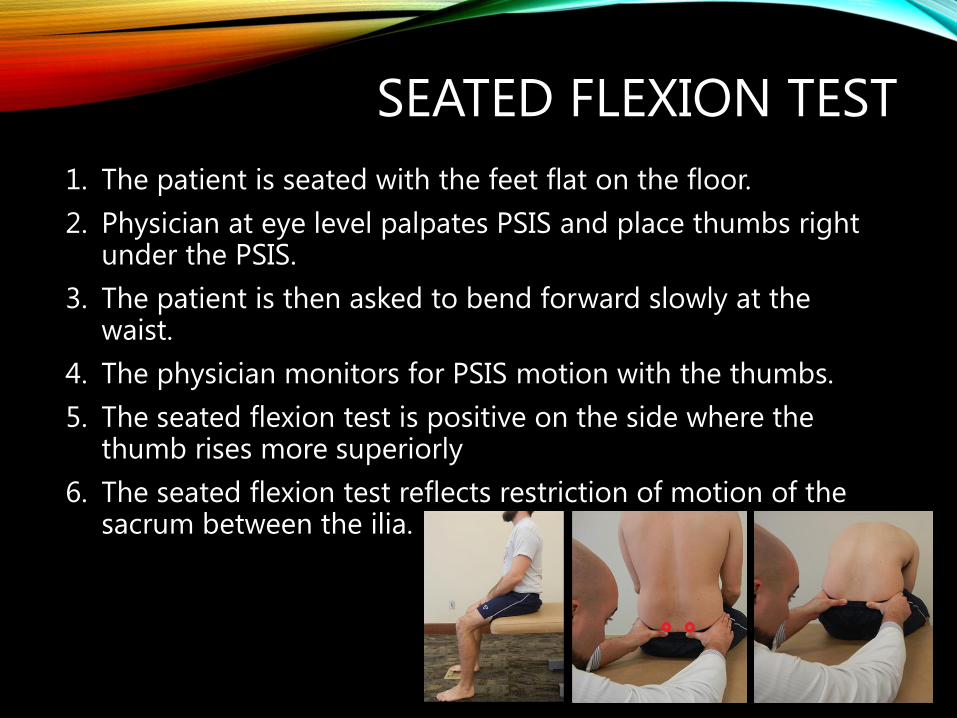
SEATED FLEXION TEST
1. The patient is seated with the feet flat on the floor.
2. Physician at eye level palpates PSIS and place thumbs right under the PSIS.
3. The patient is then asked to bend forward slowly at the waist.
4. The physician monitors for PSIS motion with the thumbs.
5. The seated flexion test is positive on the side where the thumb rises more superiorly
6. The seated flexion test reflects restriction of motion of the sacrum between the ilia.
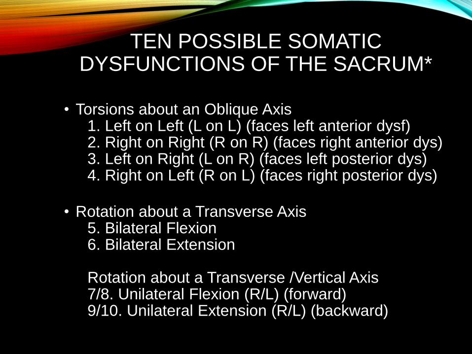
TEN POSSIBLE SOMATIC DYSFUNCTIONS OF THE SACRUM*
• Torsions about an Oblique Axis1. Left on Left (L on L) (faces left anterior dysf)2. Right on Right (R on R) (faces right anterior dys)3. Left on Right (L on R) (faces left posterior dys)4. Right on Left (R on L) (faces right posterior dys)
• Rotation about a Transverse Axis5. Bilateral Flexion6. Bilateral Extension
Rotation about a Transverse /Vertical Axis7/8. Unilateral Flexion (R/L) (forward)9/10. Unilateral Extension (R/L) (backward)
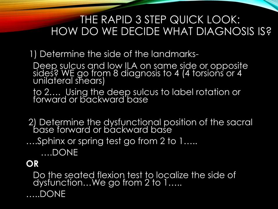
THE RAPID 3 STEP QUICK LOOK:HOW DO WE DECIDE WHAT DIAGNOSIS IS?
1) Determine the side of the landmarks-
Deep sulcus and low ILA on same side or opposite sides? WE go from 8 diagnosis to 4 (4 torsions or 4 unilateral shears)
to 2…. Using the deep sulcus to label rotation or forward or backward base
2) Determine the dysfunctional position of the sacral base forward or backward base
….Sphinx or spring test go from 2 to 1…..
….DONE
OR
Do the seated flexion test to localize the side of dysfunction…We go from 2 to 1…..
…..DONE
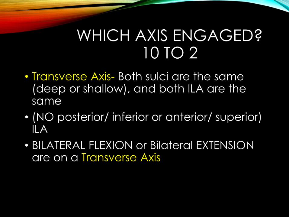
WHICH AXIS ENGAGED?10 TO 2
• Transverse Axis- Both sulci are the same (deep or shallow), and both ILA are the same
• (NO posterior/ inferior or anterior/ superior) ILA
• BILATERAL FLEXION or Bilateral EXTENSION are on a Transverse Axis
BILATERAL FLEXION OR EXTENSION
1) Determine the side of the landmarks….. equal sulci?
(and symmetric ILAs*)
Bilateral flexion or extension dysfunction… 10 dx to 2
Deep sacral sulci B/L and (~ or -) B/L sphinx, spring, and seated flexion? Bilateral sacral flexion
Shallow sacral sulci B/L and (+) sphinx, spring, and B/L seated flexion? Bilateral sacral extension
…..DONE (but FIX it)
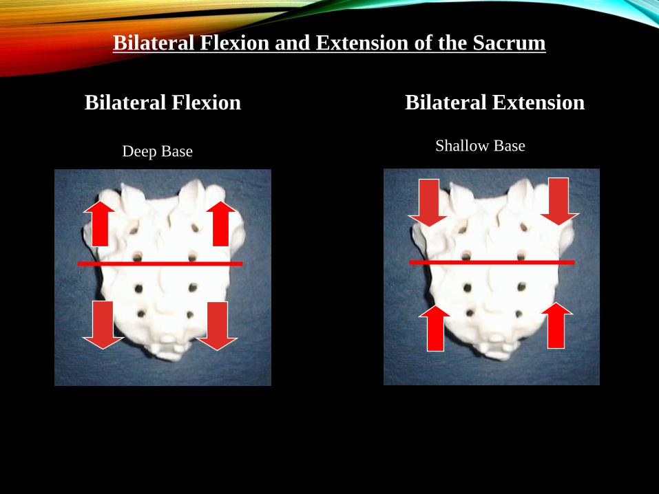
Bilateral Flexion and Extension of the Sacrum
Bilateral Flexion Bilateral Extension
Deep Base Shallow Base
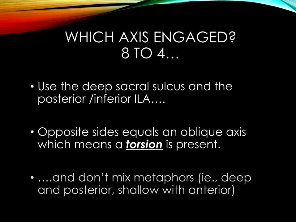
WHICH AXIS ENGAGED?8 TO 4…
• Use the deep sacral sulcus and the posterior /inferior ILA….
• Opposite sides equals an oblique axis which means a torsion is present.
• ….and don’t mix metaphors (ie., deep and posterior, shallow with anterior)
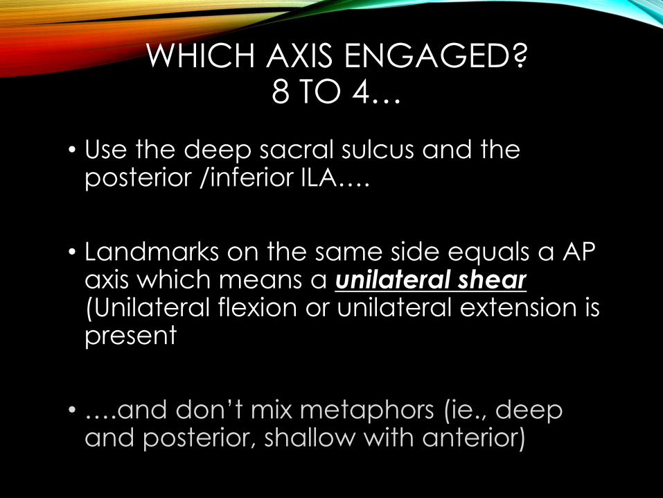
WHICH AXIS ENGAGED?8 TO 4…
• Use the deep sacral sulcus and the posterior /inferior ILA….
• Landmarks on the same side equals a AP axis which means a unilateral shear(Unilateral flexion or unilateral extension is present
• ….and don’t mix metaphors (ie., deep and posterior, shallow with anterior)
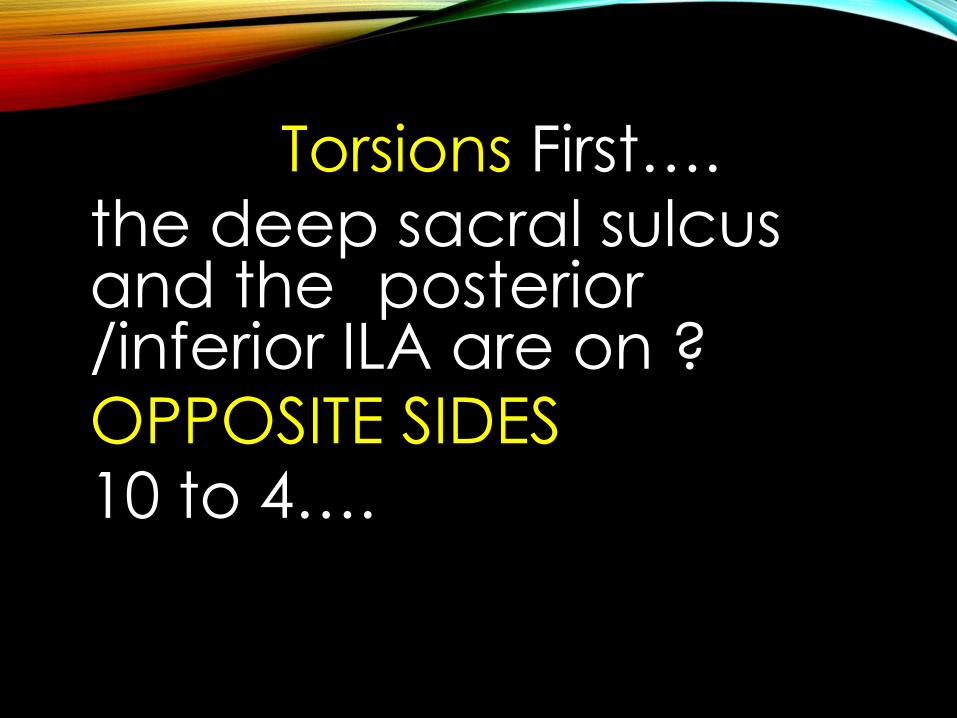
Torsions First….the deep sacral sulcus and the posterior /inferior ILA are on ?OPPOSITE SIDES10 to 4….
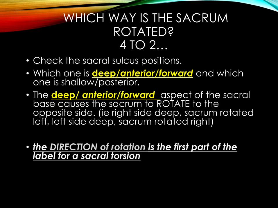
WHICH WAY IS THE SACRUM ROTATED?4 TO 2…
• Check the sacral sulcus positions.
• Which one is deep/anterior/forward and which one is shallow/posterior.
• The deep/ anterior/forward aspect of the sacral base causes the sacrum to ROTATE to the opposite side. (ie right side deep, sacrum rotated left, left side deep, sacrum rotated right)
• the DIRECTION of rotation is the first part of the label for a sacral torsion
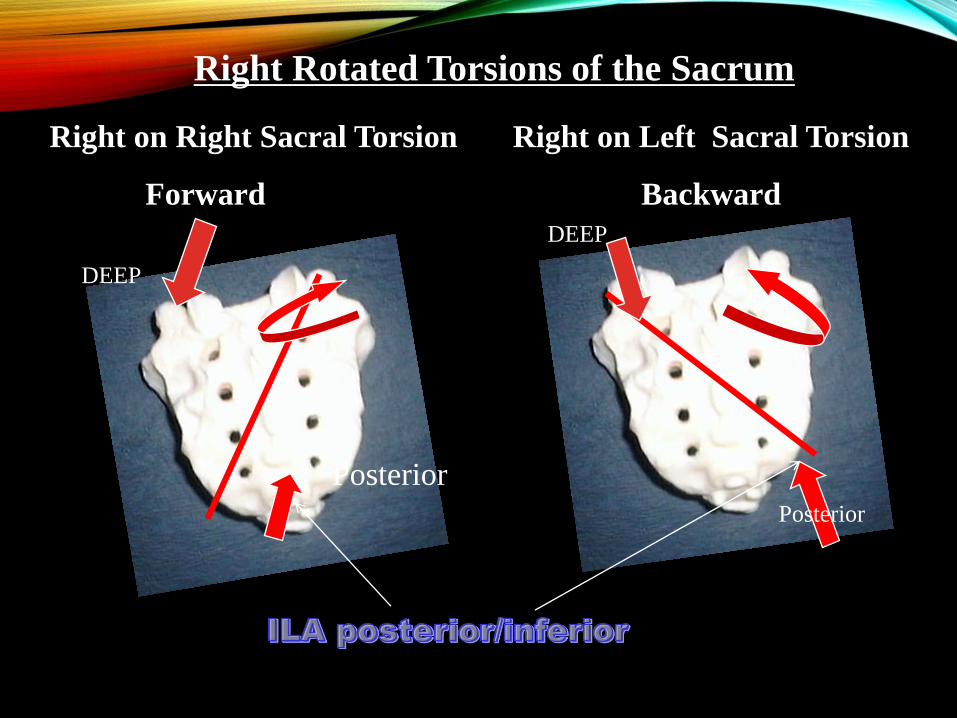
Right Rotated Torsions of the Sacrum
Right on Right Sacral Torsion
Forward
Right on Left Sacral Torsion
Backward
DEEP
DEEP
Posterior
Posterior
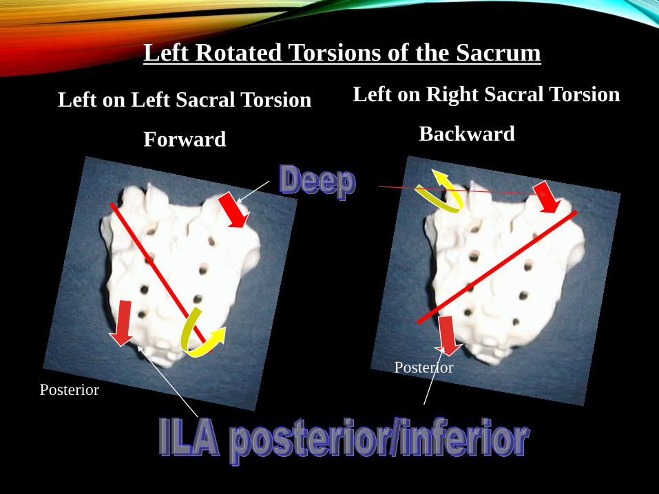
Left Rotated Torsions of the Sacrum
Left on Left Sacral Torsion
Forward
Left on Right Sacral Torsion
Backward
Posterior
Posterior
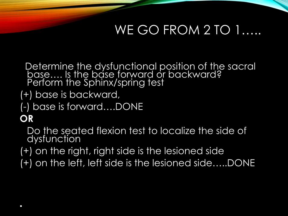
WE GO FROM 2 TO 1…..
Determine the dysfunctional position of the sacral base…. Is the base forward or backward? Perform the Sphinx/spring test
(+) base is backward,
(-) base is forward….DONE
OR
Do the seated flexion test to localize the side of dysfunction
(+) on the right, right side is the lesioned side
(+) on the left, left side is the lesioned side…..DONE
•
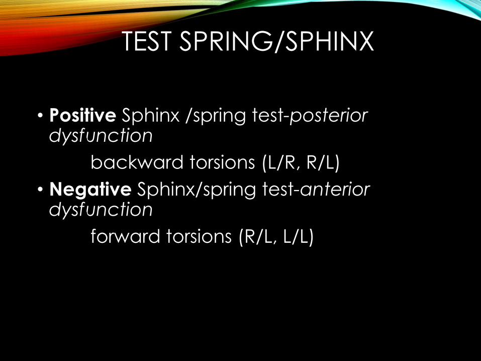
TEST SPRING/SPHINX
• Positive Sphinx /spring test-posterior dysfunction
backward torsions (L/R, R/L)
• Negative Sphinx/spring test-anterior dysfunction
forward torsions (R/L, L/L)
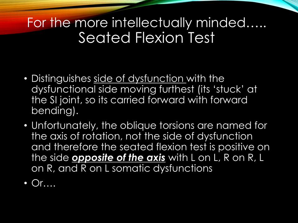
For the more intellectually minded…..Seated Flexion Test
• Distinguishes side of dysfunction with the dysfunctional side moving furthest (its ‘stuck’ at the SI joint, so its carried forward with forward bending).
• Unfortunately, the oblique torsions are named for the axis of rotation, not the side of dysfunction and therefore the seated flexion test is positive on the side opposite of the axis with L on L, R on R, L on R, and R on L somatic dysfunctions
• Or….
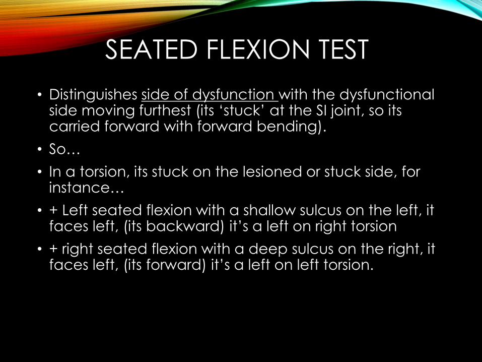
SEATED FLEXION TEST
• Distinguishes side of dysfunction with the dysfunctional side moving furthest (its ‘stuck’ at the SI joint, so its carried forward with forward bending).
• So…
• In a torsion, its stuck on the lesioned or stuck side, for instance…
• + Left seated flexion with a shallow sulcus on the left, it faces left, (its backward) it’s a left on right torsion
• + right seated flexion with a deep sulcus on the right, it faces left, (its forward) it’s a left on left torsion.
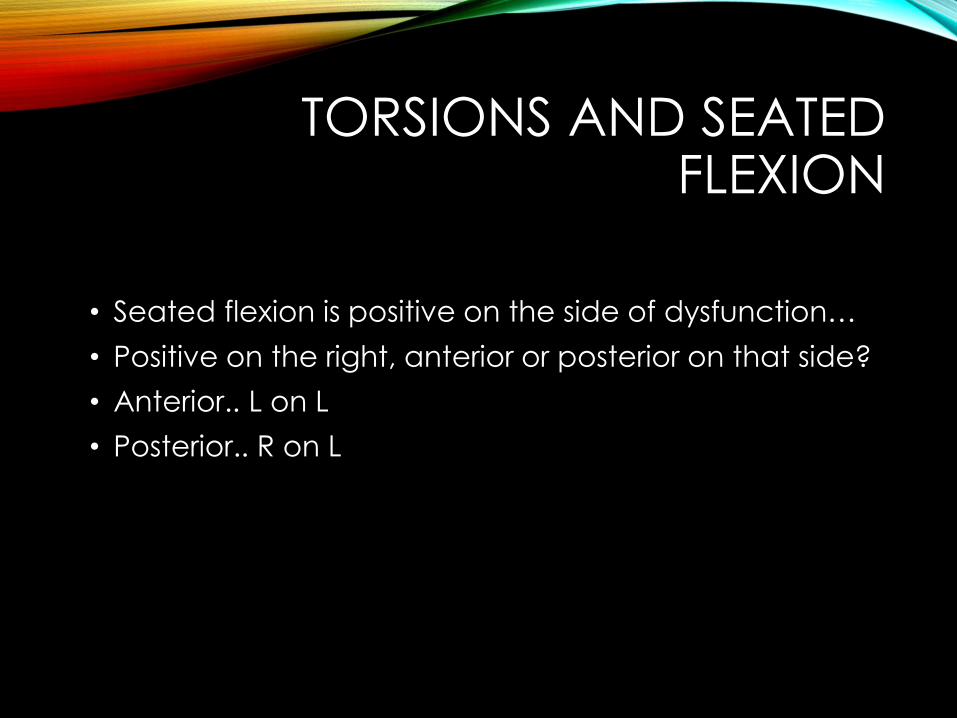
TORSIONS AND SEATED FLEXION
• Seated flexion is positive on the side of dysfunction…
• Positive on the right, anterior or posterior on that side?
• Anterior.. L on L
• Posterior.. R on L
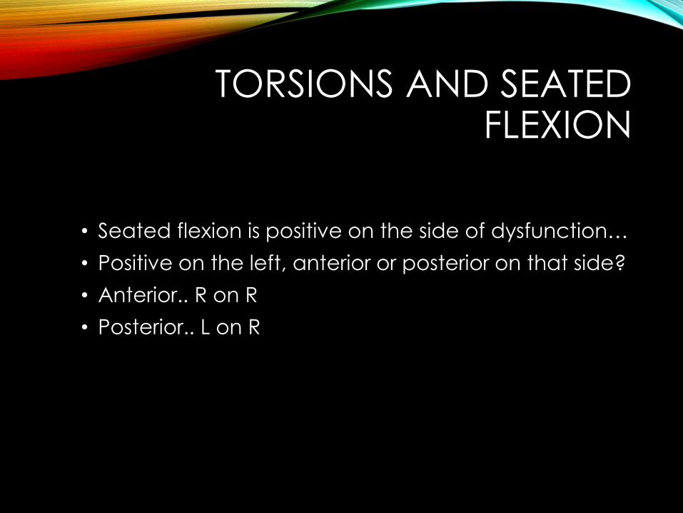
TORSIONS AND SEATED FLEXION
• Seated flexion is positive on the side of dysfunction…
• Positive on the left, anterior or posterior on that side?
• Anterior.. R on R
• Posterior.. L on R
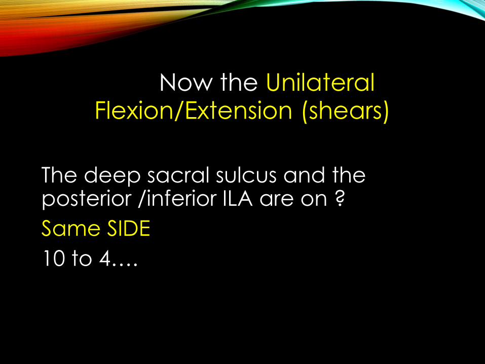
Now the Unilateral Flexion/Extension (shears)
The deep sacral sulcus and the posterior /inferior ILA are on ?
Same SIDE
10 to 4….
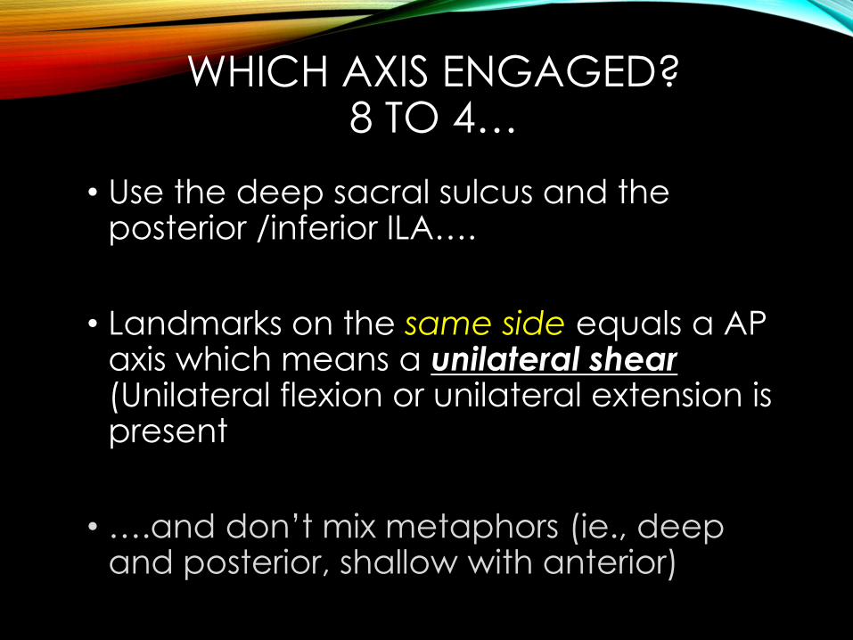
WHICH AXIS ENGAGED?8 TO 4…
• Use the deep sacral sulcus and the posterior /inferior ILA….
• Landmarks on the same side equals a AP axis which means a unilateral shear(Unilateral flexion or unilateral extension is present
• ….and don’t mix metaphors (ie., deep and posterior, shallow with anterior)
SPRING TEST/SPHINX
• Positive Sphinx /spring test-posterior dysfunction
L or R unilateral extension
• Negative Sphinx/spring test-anterior dysfunction
L or R unilateral flexion
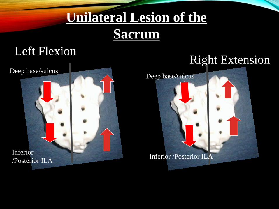
Unilateral Lesion of the
Sacrum
Right ExtensionDeep base/sulcus
Inferior
/Posterior ILA
Deep base/sulcus
Inferior /Posterior ILA
Left Flexion
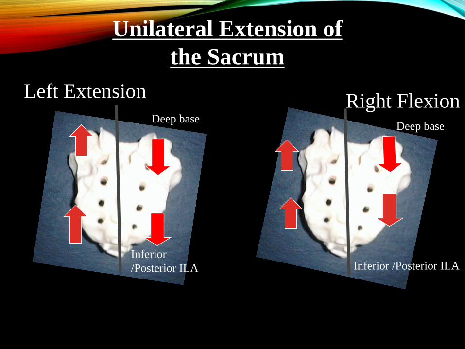
Unilateral Extension of
the Sacrum
Right FlexionDeep base
Inferior
/Posterior ILA
Deep base
Inferior /Posterior ILA
Left Extension
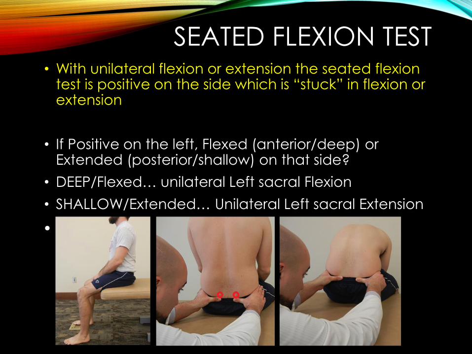
SEATED FLEXION TEST • With unilateral flexion or extension the seated flexion
test is positive on the side which is “stuck” in flexion or extension
• If Positive on the left, Flexed (anterior/deep) or Extended (posterior/shallow) on that side?
• DEEP/Flexed… unilateral Left sacral Flexion
• SHALLOW/Extended… Unilateral Left sacral Extension
•
•QUESTIONS?
TREATMENT
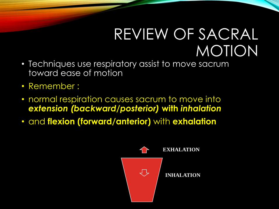
REVIEW OF SACRAL MOTION
• Techniques use respiratory assist to move sacrum toward ease of motion
• Remember :
• normal respiration causes sacrum to move into extension (backward/posterior) with inhalation
• and flexion (forward/anterior) with exhalation
EXHALATION
INHALATION
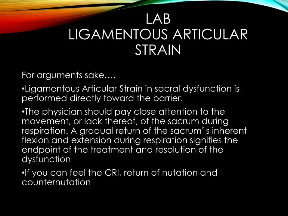
LABLIGAMENTOUS ARTICULAR
STRAIN
For arguments sake….
•Ligamentous Articular Strain in sacral dysfunction is performed directly toward the barrier.
•The physician should pay close attention to the movement, or lack thereof, of the sacrum during respiration. A gradual return of the sacrum’s inherent flexion and extension during respiration signifies the endpoint of the treatment and resolution of the dysfunction
•If you can feel the CRI, return of nutation and counternutation
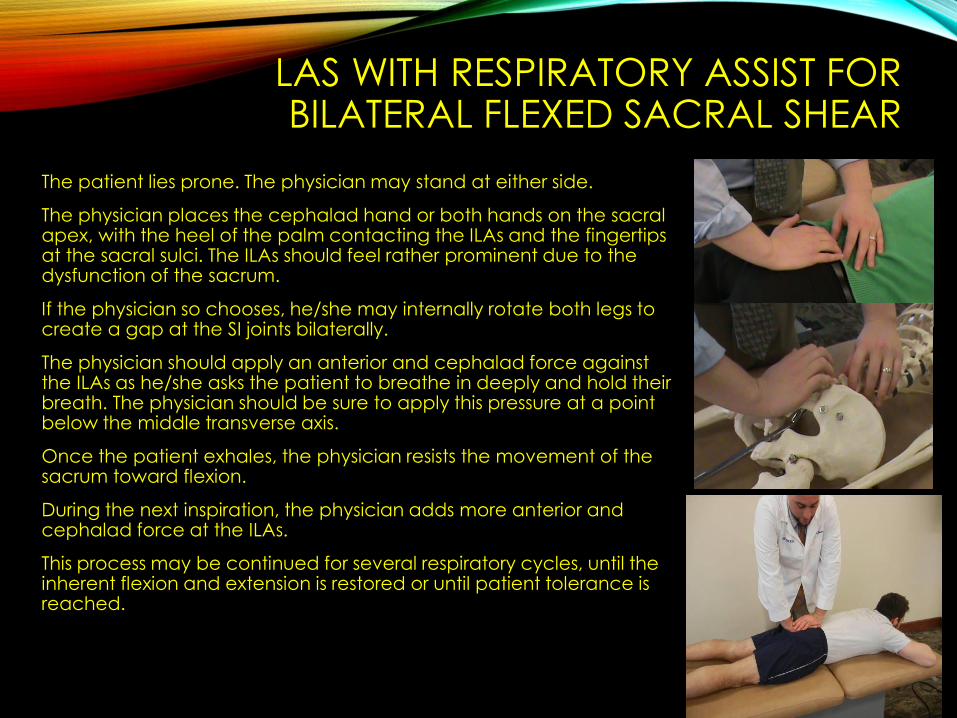
LAS WITH RESPIRATORY ASSIST FOR BILATERAL FLEXED SACRAL SHEAR
The patient lies prone. The physician may stand at either side.
The physician places the cephalad hand or both hands on the sacral apex, with the heel of the palm contacting the ILAs and the fingertips at the sacral sulci. The ILAs should feel rather prominent due to the dysfunction of the sacrum.
If the physician so chooses, he/she may internally rotate both legs to create a gap at the SI joints bilaterally.
The physician should apply an anterior and cephalad force against the ILAs as he/she asks the patient to breathe in deeply and hold their breath. The physician should be sure to apply this pressure at a point below the middle transverse axis.
Once the patient exhales, the physician resists the movement of the sacrum toward flexion.
During the next inspiration, the physician adds more anterior and cephalad force at the ILAs.
This process may be continued for several respiratory cycles, until the inherent flexion and extension is restored or until patient tolerance is reached.
LAS WITH RESPIRATORY ASSIST FOR BILATERAL EXTENDED SACRAL SHEAR
The patient lies prone. The physician may stand at either side.
The physician places the cephalad hand or both hands on the sacral base between the
PSISs, with the heel of the palm contacting the sacral sulci. The base should feel rather
prominent due to the dysfunction of the sacrum.
If the physician so chooses, he/she may internally rotate both legs to create a gap
at the SI joints bilaterally
The physician should apply an anterior and caudad force against the sacral base as
he/she asks the patient to exhale completely. The physician should be sure to apply this
pressure at a point above the middle transverse axis.
Once the patient inhales, the physician resists the movement of the sacrum toward
extension.
During the next exhalation, the physician adds more anterior and caudad force at
the sacral base.
The process may be continued for several respiratory cycles, until the inherent flexion
and extension is restored or until patient tolerance is reached.
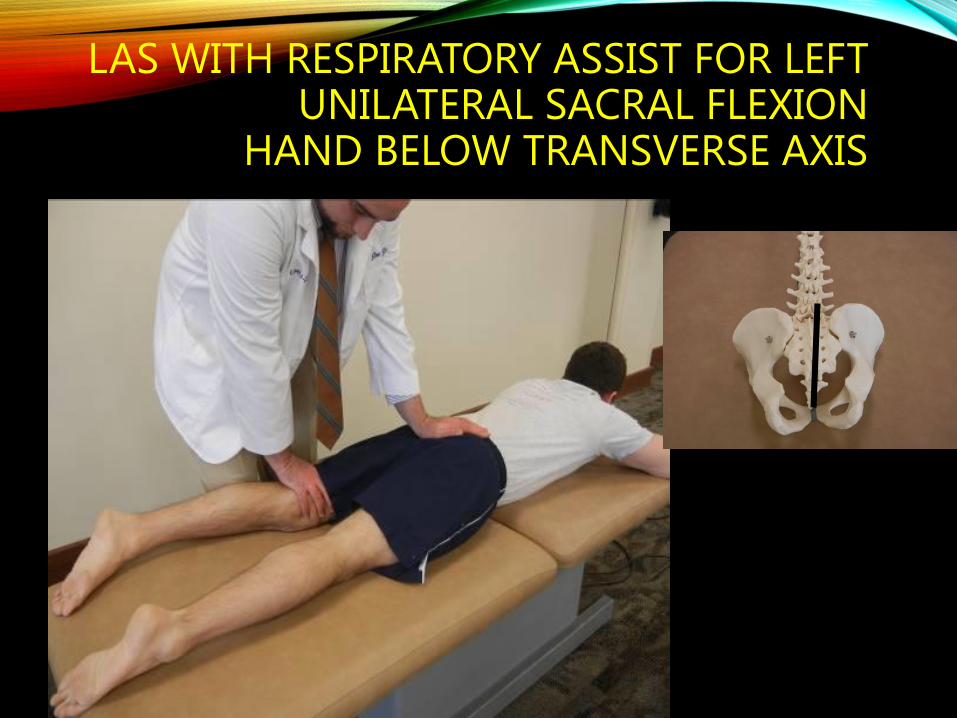
LAS WITH RESPIRATORY ASSIST FOR LEFT UNILATERAL SACRAL FLEXION
HAND BELOW TRANSVERSE AXIS
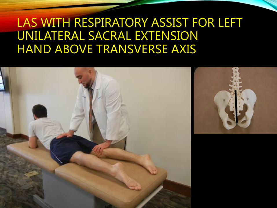
LAS WITH RESPIRATORY ASSIST FOR LEFT UNILATERAL SACRAL EXTENSIONHAND ABOVE TRANSVERSE AXIS
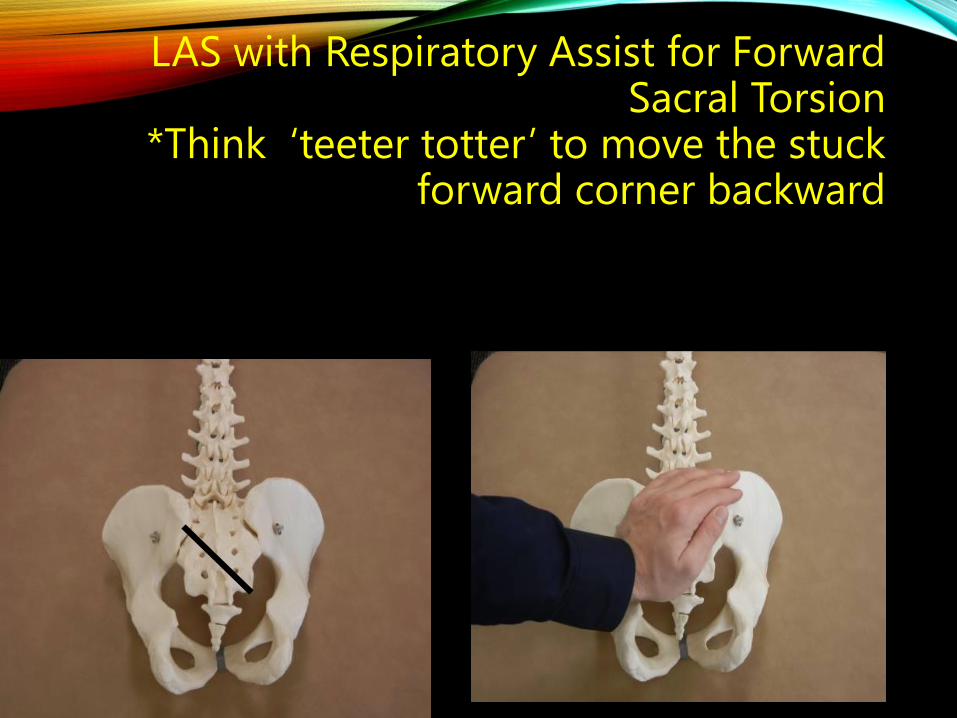
LAS with Respiratory Assist for Forward Sacral Torsion
*Think ‘teeter totter’ to move the stuck forward corner backward
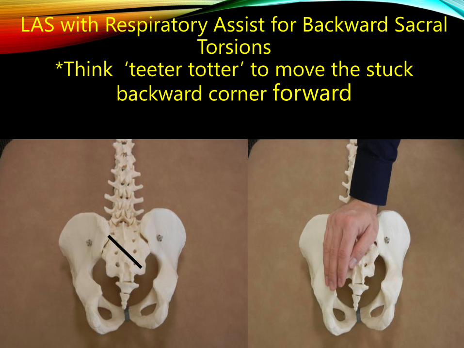
LAS with Respiratory Assist for Backward Sacral Torsions
*Think ‘teeter totter’ to move the stuck
backward corner forward
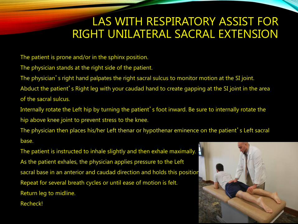
LAS WITH RESPIRATORY ASSIST FOR RIGHT UNILATERAL SACRAL EXTENSION
1. The patient is prone and/or in the sphinx position.
2. The physician stands at the right side of the patient.
3. The physician’s right hand palpates the right sacral sulcus to monitor motion at the SI joint.
4. Abduct the patient’s Right leg with your caudad hand to create gapping at the SI joint in the area
of the sacral sulcus.
5. Internally rotate the Left hip by turning the patient’s foot inward. Be sure to internally rotate the
hip above knee joint to prevent stress to the knee.
6. The physician then places his/her Left thenar or hypothenar eminence on the patient’s Left sacral
base.
7. The patient is instructed to inhale slightly and then exhale maximally.
8. As the patient exhales, the physician applies pressure to the Left
9. sacral base in an anterior and caudad direction and holds this position.
10.Repeat for several breath cycles or until ease of motion is felt.
11.Return leg to midline.
12.Recheck!
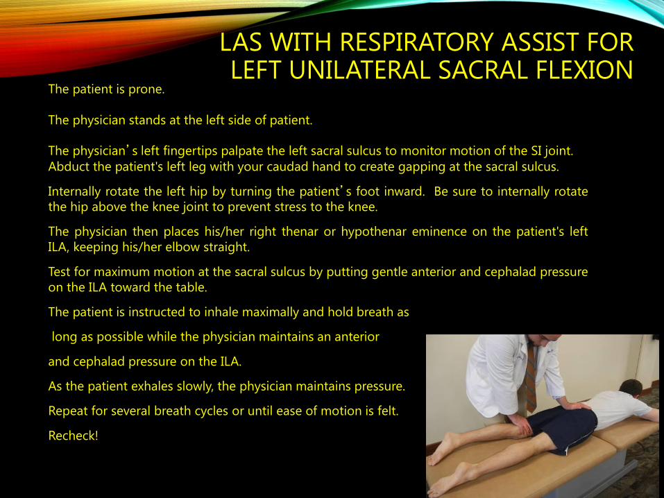
LAS WITH RESPIRATORY ASSIST FOR LEFT UNILATERAL SACRAL FLEXION
1. The patient is prone.
2. The physician stands at the left side of patient.
3. The physician’s left fingertips palpate the left sacral sulcus to monitor motion of the SI joint.
4. Abduct the patient's left leg with your caudad hand to create gapping at the sacral sulcus.
5. Internally rotate the left hip by turning the patient’s foot inward. Be sure to internally rotate
the hip above the knee joint to prevent stress to the knee.
6. The physician then places his/her right thenar or hypothenar eminence on the patient's left
ILA, keeping his/her elbow straight.
7. Test for maximum motion at the sacral sulcus by putting gentle anterior and cephalad pressure
on the ILA toward the table.
8. The patient is instructed to inhale maximally and hold breath as
9. long as possible while the physician maintains an anterior
10. and cephalad pressure on the ILA.
11. As the patient exhales slowly, the physician maintains pressure.
12. Repeat for several breath cycles or until ease of motion is felt.
13. Recheck!
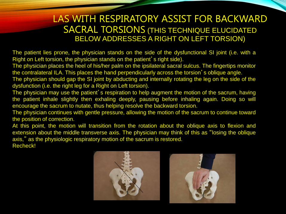
LAS WITH RESPIRATORY ASSIST FOR BACKWARD SACRAL TORSIONS (THIS TECHNIQUE ELUCIDATED
BELOW ADDRESSES A RIGHT ON LEFT TORSION)
The patient lies prone, the physician stands on the side of the dysfunctional SI joint (i.e. with a
Right on Left torsion, the physician stands on the patient’s right side).
The physician places the heel of his/her palm on the ipsilateral sacral sulcus. The fingertips monitor
the contralateral ILA. This places the hand perpendicularly across the torsion’s oblique angle.
The physician should gap the SI joint by abducting and internally rotating the leg on the side of the
dysfunction (i.e. the right leg for a Right on Left torsion).
The physician may use the patient’s respiration to help augment the motion of the sacrum, having
the patient inhale slightly then exhaling deeply, pausing before inhaling again. Doing so will
encourage the sacrum to nutate, thus helping resolve the backward torsion.
The physician continues with gentle pressure, allowing the motion of the sacrum to continue toward
the position of correction.
At this point, the motion will transition from the rotation about the oblique axis to flexion and
extension about the middle transverse axis. The physician may think of this as “losing the oblique
axis,” as the physiologic respiratory motion of the sacrum is restored.
Recheck!
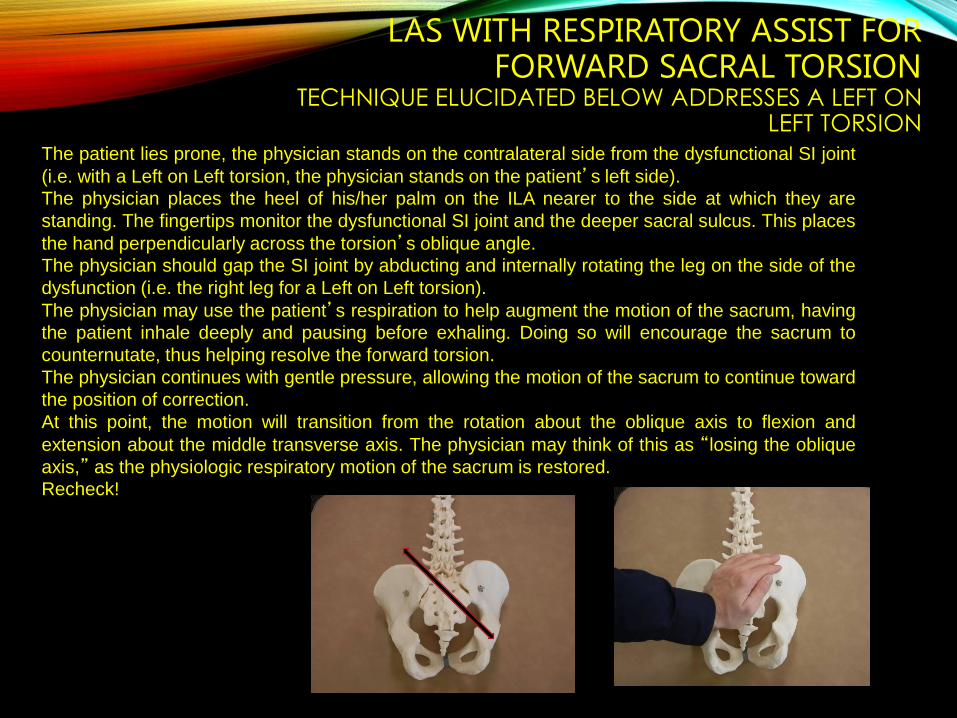
LAS WITH RESPIRATORY ASSIST FOR FORWARD SACRAL TORSION
TECHNIQUE ELUCIDATED BELOW ADDRESSES A LEFT ON LEFT TORSION
The patient lies prone, the physician stands on the contralateral side from the dysfunctional SI joint
(i.e. with a Left on Left torsion, the physician stands on the patient’s left side).
The physician places the heel of his/her palm on the ILA nearer to the side at which they are
standing. The fingertips monitor the dysfunctional SI joint and the deeper sacral sulcus. This places
the hand perpendicularly across the torsion’s oblique angle.
The physician should gap the SI joint by abducting and internally rotating the leg on the side of the
dysfunction (i.e. the right leg for a Left on Left torsion).
The physician may use the patient’s respiration to help augment the motion of the sacrum, having
the patient inhale deeply and pausing before exhaling. Doing so will encourage the sacrum to
counternutate, thus helping resolve the forward torsion.
The physician continues with gentle pressure, allowing the motion of the sacrum to continue toward
the position of correction.
At this point, the motion will transition from the rotation about the oblique axis to flexion and
extension about the middle transverse axis. The physician may think of this as “losing the oblique
axis,” as the physiologic respiratory motion of the sacrum is restored.
Recheck!

EXTRA RESOURCES
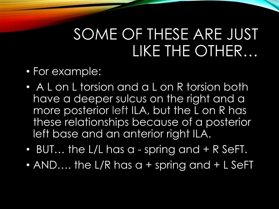
SOME OF THESE ARE JUST LIKE THE OTHER…
• For example:
• A L on L torsion and a L on R torsion both have a deeper sulcus on the right and a more posterior left ILA, but the L on R has these relationships because of a posterior left base and an anterior right ILA.
• BUT… the L/L has a - spring and + R SeFT.
• AND…. the L/R has a + spring and + L SeFT
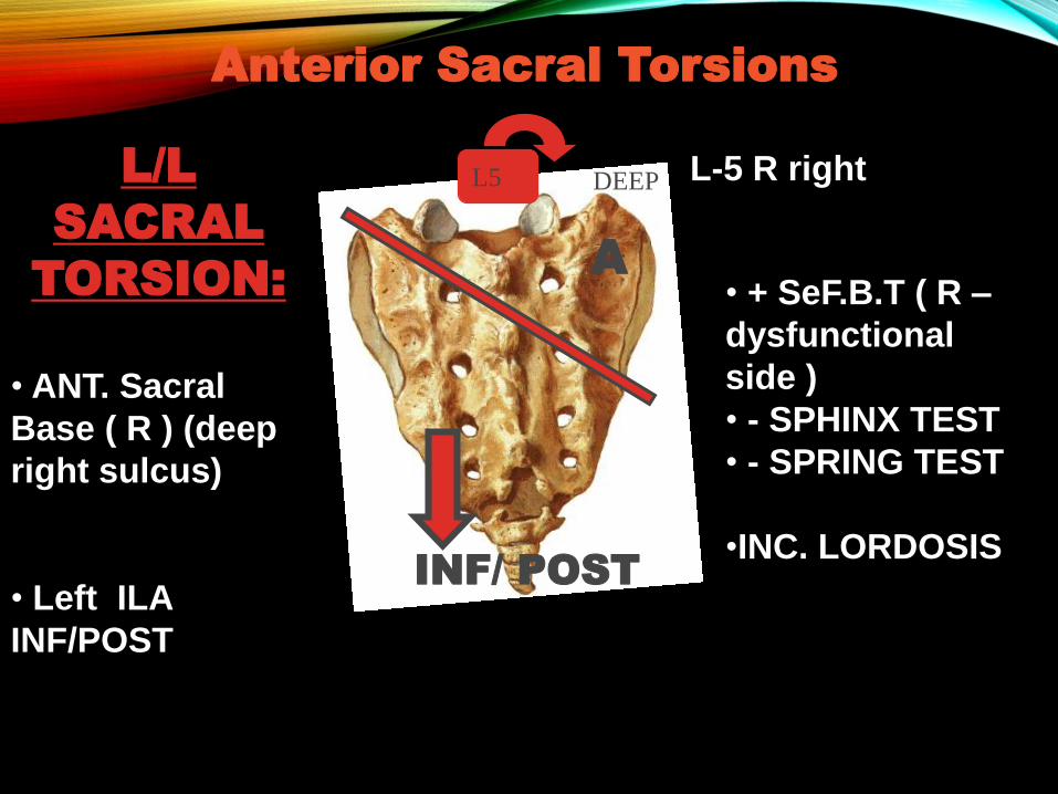
Anterior Sacral Torsions
L5
A
INF/ POST
L/L
SACRAL
TORSION:
• ANT. Sacral
Base ( R ) (deep
right sulcus)
• Left ILA
INF/POST
• + SeF.B.T ( R –
dysfunctional
side )
• - SPHINX TEST
• - SPRING TEST
•INC. LORDOSIS
L-5 R rightDEEP
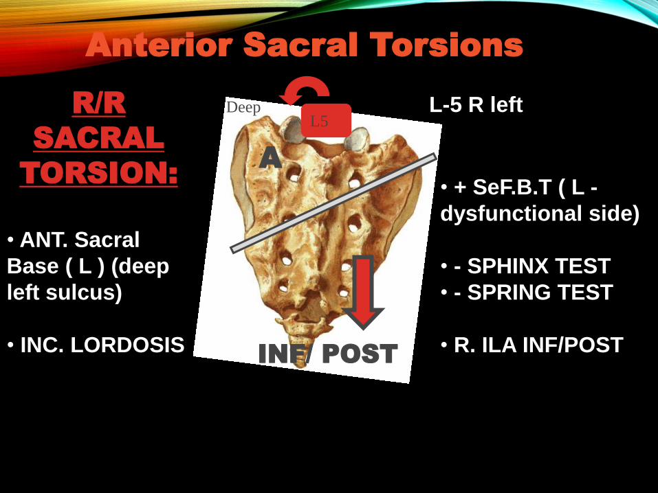
Anterior Sacral Torsions
L5
A
INF/ POST
R/R
SACRAL
TORSION:
• ANT. Sacral
Base ( L ) (deep
left sulcus)
• INC. LORDOSIS
• + SeF.B.T ( L -
dysfunctional side)
• - SPHINX TEST
• - SPRING TEST
• R. ILA INF/POST
L-5 R leftDeep
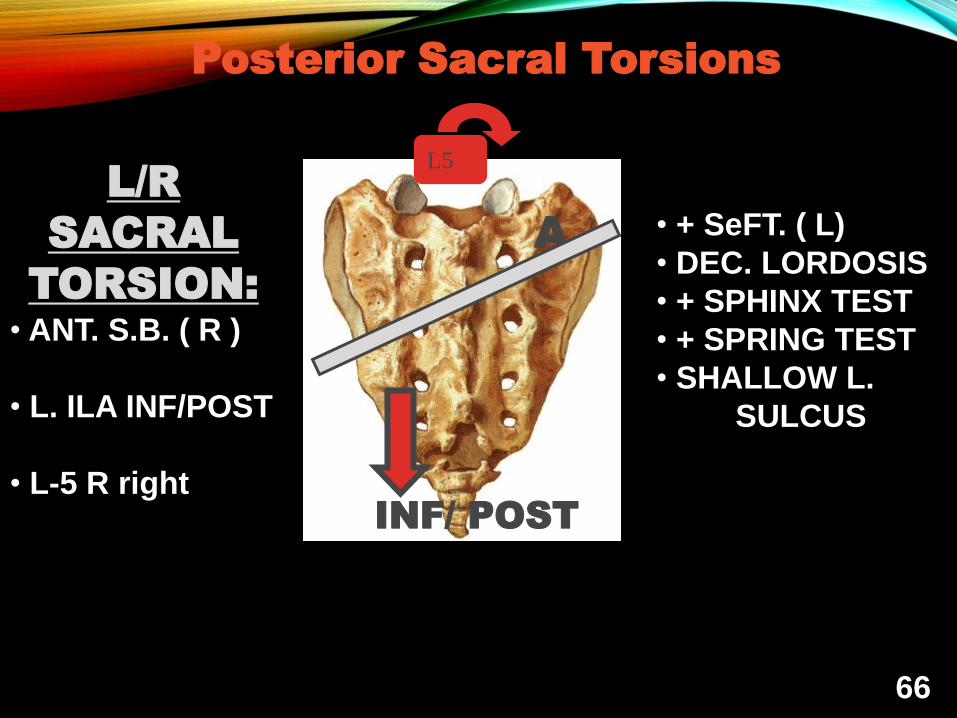
Posterior Sacral Torsions
L5
A
INF/ POST
L/R
SACRAL
TORSION:
• ANT. S.B. ( R )
• L. ILA INF/POST
• L-5 R right
• + SeFT. ( L)
• DEC. LORDOSIS
• + SPHINX TEST
• + SPRING TEST
• SHALLOW L.
SULCUS
66
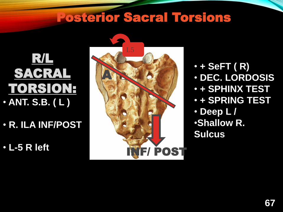
Posterior Sacral Torsions
L5
A
INF/ POST
R/L
SACRAL
TORSION:
• ANT. S.B. ( L )
• R. ILA INF/POST
• L-5 R left
• + SeFT ( R)
• DEC. LORDOSIS
• + SPHINX TEST
• + SPRING TEST
• Deep L /
•Shallow R.
Sulcus
67
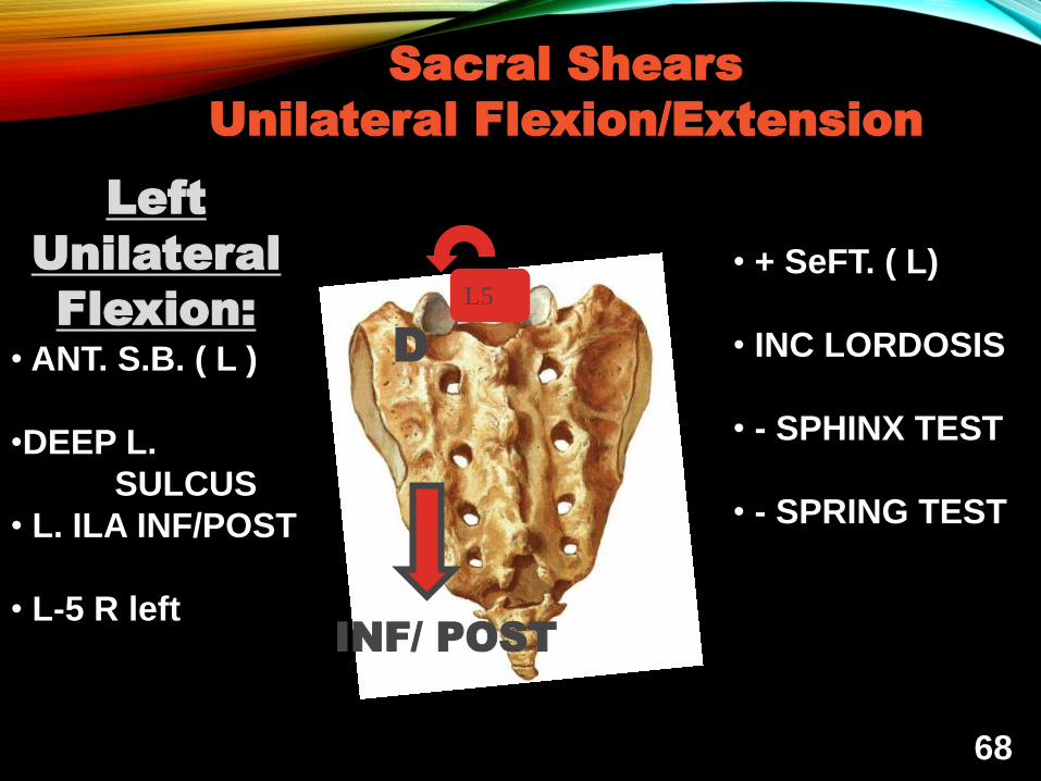
Sacral Shears
Unilateral Flexion/Extension
L5
D
INF/ POST
Left
Unilateral
Flexion:
• ANT. S.B. ( L )
•DEEP L.
SULCUS
• L. ILA INF/POST
• L-5 R left
• + SeFT. ( L)
• INC LORDOSIS
• - SPHINX TEST
• - SPRING TEST
68
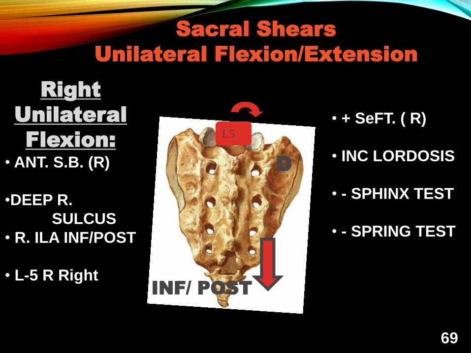
Sacral Shears
Unilateral Flexion/Extension
L5
D
INF/ POST
Right
Unilateral
Flexion:
• ANT. S.B. (R)
•DEEP R.
SULCUS
• R. ILA INF/POST
• L-5 R Right
• + SeFT. ( R)
• INC LORDOSIS
• - SPHINX TEST
• - SPRING TEST
69
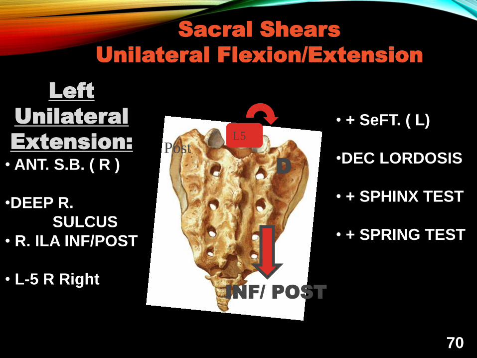
Sacral Shears
Unilateral Flexion/Extension
L5
D
INF/ POST
Left
Unilateral
Extension:
• ANT. S.B. ( R )
•DEEP R.
SULCUS
• R. ILA INF/POST
• L-5 R Right
• + SeFT. ( L)
•DEC LORDOSIS
• + SPHINX TEST
• + SPRING TEST
70
Post
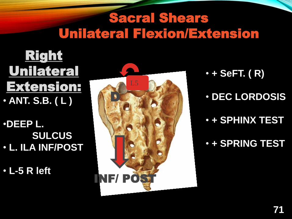
Sacral Shears
Unilateral Flexion/Extension
L5
D
INF/ POST
Right
Unilateral
Extension:
• ANT. S.B. ( L )
•DEEP L.
SULCUS
• L. ILA INF/POST
• L-5 R left
• + SeFT. ( R)
• DEC LORDOSIS
• + SPHINX TEST
• + SPRING TEST
71
REFERENCES:
•
• Foundations of Osteopathic Medicine 2nd edition, Lippincott, Williams and Wilkins, 2003. Pg 900, 941-943.
• Nicholas and Nicholas Atlas of Osteopathic Techniques. Lippincott, Williams and Wilkins 2008. pgs. 254-257, 266-268, 304-311
• An Osteopathic approach to Diagnosis and Treatment,Eileen Diovanna, DO Stanley Schiowitz, DO Second Edition. Lippincott Williams & Wilkins, 1997.
Recommended