Wayne State University
Wayne State University Dissertations
1-1-2014
Role Of The Pkna And Pknb Kinases InMycobacterium TuberculosisTripti AnandanWayne State University,
Follow this and additional works at: http://digitalcommons.wayne.edu/oa_dissertations
Part of the Biology Commons, and the Microbiology Commons
This Open Access Dissertation is brought to you for free and open access by DigitalCommons@WayneState. It has been accepted for inclusion inWayne State University Dissertations by an authorized administrator of DigitalCommons@WayneState.
Recommended CitationAnandan, Tripti, "Role Of The Pkna And Pknb Kinases In Mycobacterium Tuberculosis" (2014). Wayne State University Dissertations.Paper 1040.

1
ROLE OF THE PKNA AND PKNB KINASES IN MYCOBACTERIUM TUBERCULOSIS
by
TRIPTI ANANDAN
DISSERTATION
Submitted to the Graduate School
of Wayne State University,
Detroit, Michigan
in partial fulfillment of the requirements
for the degree of
DOCTOR OF PHILOSOPHY
2014
MAJOR: BIOLOGICAL SCIENCES
Approved by:
_______________________________ Advisor Date
_______________________________
_______________________________
_______________________________

1
DEDICATION
To everyone who made this possible
ii

4
ACKNOWLEDGEMENTS
I would like to thank my advisor, Dr. Choong-Min Kang, for giving me the
opportunity to work on such an exciting project. Initially, there were numerous
possibilities that the substrate identification project could have had, but he had the
foresight to pursue a number of basic, yet challenging questions. I appreciate his faith in
my abilities and the fact that he gave me the freedom to work on the project
independently. He taught me how to do several biochemistry and molecular biology
techniques while I was in his lab. His absolute dedication in making sure I learn my lab
skills, scientific presentation, scientific writing skills to the best of my abilities has
pushed the boundaries of my learning immensely. His positive outlook on life and
constant curiosity about science are a constant inspiration to me. Despite his many
challenges, he maintains an altruistic nature and is always willing to lend someone else a
hand.
I am extremely grateful to my committee members for their continued support and
encouragement with my project especially when times where rough and for their words
of advice that will always be appreciated. A special thanks to Dr. Gu for being generous
and kind in giving me the lab space to carry out my research in the last two years of my
PhD work. I wish to thank my past lab members, Seeta, Ming Hui, Sunny, Heather, Jae-
il, Nayeong and others for their scientific discussions, and all their support during my
time in and out of the lab. I am grateful to all the professors and teachers whose classes I
took and learnt a lot by being their student; it is them who laid a great foundation to my
scientific career and also for their efforts to help me achieve the best I can.
iii

5
I wish to thank my family for their love, encouragement and support in all these
years that has contributed a great deal to what I have achieved in my life so far. I feel
lucky to have made some of the greatest friends here, Banupriya, Vaishnavi, Janani, and
Arti for they have always been next to me whenever I needed them and will continue to
be the gifts I shall cherish for life.
I owe the Department of Biological Sciences a debt of gratitude for all the
financial and moral support during my tenure as a PhD candidate at Wayne State
University.
iv

6
TABLE OF CONTENTS
Dedication…………………………………………………………………………………. ii
Acknowledgements……………………………………………………………………….. iii
List of Figures…………………………………………………….…………………...…....vi
List of Tables…………………………………………………….…………………...…...viii
Chapter 1 General Introduction……………………………………………………...……...1
Chapter 2 In vivo search for substrates of PknA/PknB, and Role of proteasome phosphorylation in Mycobacterium tuberculosis…..………………23 Chapter 3 In vitro approach to search for substrates of PknA/PknB, and role of FbpB phosphorylation…………………………………...…….…... 50
Appendix………………………………………………………………………………….. 69
References……………………………………………………………………………...…. 80
Abstract………………………………………………………………………………...…. 97
Autobiographical Statement……………………………………………………...…..…….99
v

7
LIST OF FIGURES
Figure 1.1: Stages of M. tuberculosis infection………...……………………………….4
Figure 1.2: The lipid cell wall composition of Mycobacteria and the structure of TDM……………................……….…8 Figure 1.3: Mycobacterial proteasomal pathway..……………………………...……16
Figure 1.4: Trehalose Dimycolate synthesis pathway in M. tuberculosis………………………………………………..19 Figure 2.1: In vivo technique to identify
natural substrates of PknA and pknB………………...………………….26 Figure 2.2: PrcA phosphorylation under pknB non-overexpression in M. tuberculosis.......................................................28 Figure 2.3: In vitro phosphorylation of PrcA by PknB.………………………….…. 29
Figure2.4: PrcB is not co-eluted with PrcA under pknA overexpression..……………………………………………..…….…31 Figure 2.5: PknA affects the integrity of the proteasome core
complex in mycobacteria……………...……………………..……………32
Figure. 2.6: PknA affects proteasome integrity in E. coli. …………………….…..…33
Figure 2.7: Pre-PrcB and PrcA are substrates of PknA..……………………….……34
Figure 2.8: Confirming phosphorylation site in Pre-PrcB and PrcA……....…….…35
Figure 2.9: PknA does not affect proteasome stability..…………………...…………36
Figure 2.10: Confirming activity of PknA and PknB. …………………...……..……37
Figure 2.11: Mycobacterial resistance to H2O2 is increased by absence of proteasome……………...……………………………………39 Figure 2.12: Mycobacterial resistance to H2O2 is increased by pknA-expression....................................................................................40 Figure 2.13: H2O2 affects Pre-PrcB processing. ………………………...……………41
vi

8
Figure 2.14: H2O2 impedes the formation of holo-proteasome in M. tuberculosis……………………………..………………..………………. 42 Figure 2.15: Presence of H2O2 affects proteasomal activity in M. tuberculosis………………………………………………..44 Figure 2.16: Autophosphorylation of PknA increases in response to H2O2. ……………………………..……..……………..…45 Figure 2.17: Model for role of proteasome phosphorylation in M. tuberculosis (M. tb)................…………………………………………46 Figure 3.1: In silico search for putative substrates of PknA/PknB…...…………..…52 Figure 3.2: List of candidate proteins obtained from the in silico search for substrates of PknA/PknB. ………………………………..…53 Figure 3.3: In vitro kinase assay with synthetic peptide of substrate candidates in the presence of PknA/B and [γ-32P]ATP………….……...54 Figure 3.4: Phosphorylation of purified FbpB by PknB in the presence of [γ-32P]ATP. …………………………………………………..56 Figure 3.5: FbpB is phosphorylated in vivo……………………...……………………58
Figure 3.6: Only intracellular FbpB is phosphorylated..……………………...…..…59
Figure 3.7: Different phosphorylation pattern between intracellular and extracellular FbpB. …………………………………………………..61
Figure 3.8: FbpB is phosphorylated in vivo in M. tuberculosis..………………..……62
Figure 3.9: Mycolyl transferase assay..………………………………...…………...…64
Figure 3.10: Phosphorylation might enhance mycolyl transferase activity………....65
vii

9
LIST OF TABLES
Table 1: Strains and Plasmids used in Chapter 2………………………………….…76
Table 2: Strains and plasmids used in Chapter 3……………………………….……77
Table 3: Primers used in Chapter 2…………………………………………...………79
Table 4: Primers used in Chapter 3………………………………………………...…79
viii

1
Chapter 1
Introduction
Tuberculosis
Tuberculosis (TB) is an infectious disease caused by the pathogen Mycobacterium
tuberculosis. Scientists have been able to discover the DNA of M. tuberculosis in
Egyptian mummies as early as 2000 B.C. (1). It is identified by various names in the
historical texts, few of those terms being “consumption”, “wasting away”, and “the white
plague”, etc. (2). As tubercle bacilli can be spread through air, TB is termed a contagious
disease. The developing nations of the world are at utmost risk of being easily infected
due to their high population size and poor living conditions. Although a lot of
improvements have taken place in the therapeutic regimen for TB by improving diet,
housing, education, and sanitation conditions there seems to be bigger challenges with
increasing cost of drugs and the length of therapy (3).
Since the early 20th century Bacillus Calmette-Guérin (BCG) vaccine has been
used to prevent TB infection. BCG vaccine consists of live attenuated tubercle bacilli that
are aimed to act as a prophylactic measure to prevent TB. The duration of protection
rendered by the vaccine seems to be around 15 years. However, BCG is ineffective to
prevent adult pulmonary tuberculosis as well as reactivation of latent TB in individuals
who carry the bacteria without displaying any symptoms of the disease (4). It is estimated
that 2.2 billion people are infected worldwide annually and approximately 1.3 millions
deaths occur in HIV-negative people and approximately half a million deaths in HIV-
positive patients (5,6). Another pressing issue in the battle against TB is the increase in

2
the number of strains that are resistant to first line and second line TB drugs. These multi-
drug resistant (MDR) and extensively drug resistant (XDR) strains of M. tuberculosis
curtail the potency of chemotherapy associated with TB and thus defining a global
emergency to overcome these roadblocks in TB treatment (7).
The infection begins through the inhalation of aerosols containing M. tuberculosis
released from the lungs of an infected individual by coughing or intaking contaminated
food. The disease typically affects the lungs but can also spread to other parts of the
body. Upon inhalation of mycobacterium-laden aerosol, the microbe is engulfed by
alveolar macrophage. During this stage the majority of the bacteria is destroyed, but some
that resist the harsh intracellular condition are able to survive, multiply, and infect
neighboring macrophages (8). In general, the disease has two patterns. In the first kind,
known as the primary TB, the mycobacteria are engulfed by the host alveolar
macrophage that is then carried to hilar lymph node in order to control the infection.
During the primary infection, the initial focus of infection is identified as a tiny
granuloma (tubercle) in the lung tissue also called Ghon’s complex. The primary TB is
seen mostly in children and majority of these cases resolve as the infant begins to develop
immunity (80% of cases) whereas sometimes the bacteria enter into a state of dormancy
and remains undetectable by the host immune system. A secondary TB is a pattern of the
disease that arises as a result of reactivation of previous infection, exogenous reinfection,
or gradual progression of primary TB into chronic form mainly due to deteriorating
health status. The reactivation of latent TB infection can happen when the immune
system is weakened due to another infection, drug abuse, or immune compromise that
enable the dormant bacilli to break out of latency and enter an actively dividing state. In

3
the secondary TB the granulomatous inflammation is severe and widespread along with
extensive cavitation and lung tissue damage. The symptoms of active disease include
persistent fevers, night sweats, severe cough, sputum with blood, fatigue, and weight loss
(9).
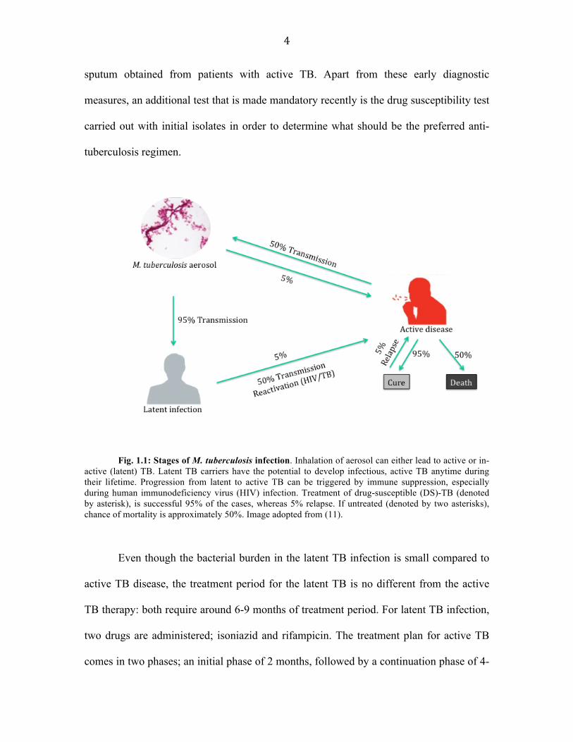
M. tuberculosis can remain latent inside the human host for decades and the
immense reservoir of infected individuals created by this microorganism has a potential
to develop active tuberculosis (TB) sometime in their life time (10). Approximately, 50%
of latent infection can lead to reactivation of TB (Fig.1.1). According to the world health
organization, it is estimated that about one-third of the world’s population is infected with
latent TB. Latency of M. tuberculosis is described as a state of the bacillus where it is
metabolically minimally active or in a non-replicating stage for undefined periods of
time. A person with latent TB is not yet ill with the disease and despite being a reservoir
for the pathogen cannot transmit the disease to other people. Therefore, latent TB
infection is marked by the presence of the tubercle bacilli in the body without displaying
any symptoms, or radiographic, or bacteriologic evidence of TB disease (11). Fig. 1.1
describes different stages of TB infection in humans.
Diagnosis of latent TB and active TB can be difficult although the general
guidelines observed for TB diagnosis entails Mantoux tuberculin skin test (TST) and
interferon-gamma release assays (IGRAs). Although TST cannot distinguish between TB
infection and TB disease, a more confirmatory screening is accomplished by chest X-ray
studies that can provide details as to the location of an infiltrate, cavities and/or fluid in
the lungs. Finally, a confirmatory laboratory test known as acid-fast bacilli smear (AFB
smear) is performed in order to microscopically analyze the presence of mycobacteria in
4
sputum obtained from patients with active TB. Apart from these early diagnostic
measures, an additional test that is made mandatory recently is the drug susceptibility test
carried out with initial isolates in order to determine what should be the preferred anti-
tuberculosis regimen.
Fig. 1.1: Stages of M. tuberculosis infection. Inhalation of aerosol can either lead to active or in-active (latent) TB. Latent TB carriers have the potential to develop infectious, active TB anytime during their lifetime. Progression from latent to active TB can be triggered by immune suppression, especially during human immunodeficiency virus (HIV) infection. Treatment of drug-susceptible (DS)-TB (denoted by asterisk), is successful 95% of the cases, whereas 5% relapse. If untreated (denoted by two asterisks), chance of mortality is approximately 50%. Image adopted from (11).
Even though the bacterial burden in the latent TB infection is small compared to
active TB disease, the treatment period for the latent TB is no different from the active
TB therapy: both require around 6-9 months of treatment period. For latent TB infection,
two drugs are administered; isoniazid and rifampicin. The treatment plan for active TB
comes in two phases; an initial phase of 2 months, followed by a continuation phase of 4-

5
7 months (total of 6 to 9 months of treatment). It is very important that people under TB
medication follow the treatment regimen without any lapses as it paves way for drug-
resistance strains of mycobacteria. TB caused by drug-resistant strain is very hard and
expensive to treat. Such long treatment plans also come with their own downsides of
which the major threat is development of drug-resistance strains of M. tuberculosis
(12,13). Therefore, better understanding of human host-M. tuberculosis interaction and
M. tuberculosis physiology is believed to be crucial to develop more efficient drugs
against TB.
Host-Pathogen interaction
As discussed earlier, tubercle bacilli are capable of both active and dormant
survival inside host macrophage cells. To establish infection M. tuberculosis possesses a
plethora of virulence factors whose expressions are regulated with progressing infection
state concluding with latent infection and/or an eventual resuscitation from dormancy.
Invading M. tuberculosis comes face to face initially with macrophages and
dendritic cells (DC). These two innate immune cells are able to recognize pathogen
components through highly conserved pattern recognition receptors (PRRs), including
Toll-like receptor (TLR) family players. This recognition initiates the activation of both
DC and macrophage, leading to phagocytosis and internalization of the bacilli in the
phagolysosome complex, where they undergo lysis. The DC and macrophages travel to
the mediastinal lymph nodes, in order to present bacterial lipids and peptide antigenic
determinants to CD4+ and CD8+ T cells via MHC-I and MHC-II, leading to T cell
activation and clonal proliferation. These T cells return to the site of primary infection

6
along with the macrophages and secrete cytokines such as IFNγ, IL-12, and TNFα (14).
There are many defense mechanisms adopted by M. tuberculosis to resist host
immune responses. One mechanism is dependent on the inhibition of phagosome-
lysosome fusion inside infected macrophages. Without successful degradation by
macrophage, the bacterium is able to adapt to intracellular conditions that can have two
outcomes. One, where the infected macrophage is utilized as a home to replicate and
releases more microbes that then infect other cells, and two, where the bacteria can shift
to a metabolically inactive stage known as latency (15). It has been shown that
mycobacterium prevents lysosomal maturation (lysosome-phagosome fusion) with the
help of the eukaryotic type Ser/Thr kinase PknG (16,17).
Despite the importance of latency in the epidemiology and pathology of
tuberculosis, it is yet not fully understood how M. tuberculosis alternates between a latent
state and active replicating state inside the host. To achieve latency, however,
mycobacterium must be able to respond to specific host signals and regulate its
metabolism and growth. For this, the M. tuberculosis genome contains a wide variety of
signaling molecules including eleven "eukaryotic-type" Ser/Thr protein kinases. Our lab
has been studying two of these kinases, PknA and PknB, and their substrate proteins. The
PknA/B signal transduction network is essential for the survival of M. tuberculosis (18).
In addition, some of their known substrates (such as Wag31 and proteasome) are also
known to be essential. Thus, understanding the roles of these kinases and their substrates
will provide important information regarding the disease process in tuberculosis and thus
can aid in the development of potent TB drugs.
7
Cell envelope of Mycobacterium tuberculosis
M. tuberculosis is a Gram-positive, aerobic, rod-shaped bacterium measuring 2 –
4 µm in length. Its cell envelope has a complex structure made up of peptidoglycan,
mycolic acid, and arabinogalactan that are highly hydrophobic (19). The success of M.
tuberculosis as one of the deadliest human pathogens can be easily attributed to its cell
envelope make-up as it is this unique protective layer in Mycobacterium that guard the
cell against the host’s immune repertoire (20). Upon entry into the host system, the
bacillus encounters several different cellular environments that it must adapt partly by
changes in the cell envelope’s physical and chemical composition. These physiological
variations in cell envelope contribute to changes in the host-pathogen immunologic
interplay that in turn plays a part in the success of the pathogen. A lot of focus has been
given to understand the cell envelope composition of M. tuberculosis in hopes to
determine the physiology and the changes that occur during host-pathogen interplay (21).
Studies have been aimed at identifying different components of cell envelope and
antigens present on the bacterial surface or secreted by the bacterium to determine
virulence factors associated with cell envelope (22).
The chemical structure and composition of the mycobacterial cell wall and
envelope have been extensively studied and reviewed (23,24). David Minnikin proposed
the definitive model of the structural composition of the mycobacterial cell envelope in
1982 (25) (Fig.1.2). In this model, the cell envelope is similar to the lipid bilayer where
mycolic acids are covalently linked to the cell wall associated arabinogalactan layer in
order to form the inner leaflet. This inner leaflet is found to have the lowest permeability
to organic chemicals in comparison to overall cell wall. The outer leaflet of cell wall is

8
believed to contain extractable phospholipids, glycolipids, peptidolipids, and mycosides
(25,26). The mycolic acids (MA) are long chain (C60-C90) fatty acids and comprise a
core structure covalently attached to an inner complex of arabinogalactan and
peptidoglycan (mAGP) (26). Mycolic acids accounts for ~ 60% of the dry weight of the
bacillus. The highly complex meshwork architecture of peptidoglycan (PG) is
accomplished by having repeating units of N-acetylglucosamine (GlcNAc) and N-
acetylmuramic acid (MurNAc) or N-acylglycolic acid (MurNGlyc) that is also cross-
linked to the arabinogalactan (AG) layer (27). The mycolic acids are also associated with
other essential virulence factors, trehalose monomycolate (TMM) and trehalose
dimycolate (TDM) (28). Apart from these elements, other lipid and glycolipid
components form the cell wall framework also play a role in pathogenesis of the bacillus
(29).

9
Fig. 1.2: The lipid cell wall composition of Mycobacteria and the structure of TDM. The
arabinogalactan is associated with mycolic acid that forms a second membrane-like structure adjacent to the peptidoglycan-linked cell wall. Above arabinoglactan layer is a thick mycolic acid layer made of complex lipid profile containing trehalose monomycolate (TMM), trehalose and trehalose dimycolate (TDM). The outermost layer containing capsule-like material consists of lipopolysaccharide and protein lies on top of mycolic acid layer. This unique cell wall composition is a crucial aspect of mycobacterial resistance to several drugs used for treating TB infection. (Image taken from (30)).
The extremely hydrophobic waxy surface of mycobacterial cells is formed by the
transfer of mycolate to the arabinogalactan chains by a group of mycolyl transferase
enzymes called fibronectin binding proteins (Fbp) that are also known as antigen 85
complex (31). In M. tuberculosis, there exist four Fbp proteins (FbpA, FbpB, FbpC1, and
FbpBC2), whereas M. smegmatis has five Fbp proteins (FbpA, FbpB, FbpC, FbpD, and
FbpE) (32). Among these various types of Fbp proteins, FbpC2 contributes the most to
the mycolic acid content of the cell wall in M. tuberculosis, and FbpA, apart from its role
as trehalose dimycolate (TDM) synthesizing enzyme, has been suggested to be required
for virulence (33,34). FbpB, which also has the mycolyl transferase activity, has been
suggested to have a role in the pathogenesis of TB by disseminating within the host
through fibronectin interactions on the mucosal surface and elevating expression of
interferon-gamma leading to a reduced phagocytosis by monocyte-derived-macrophages
(35).
Signal transduction in M. tuberculosis
The success of a pathogen depends on its capacity to quickly sense and adapt in
the host in order to subvert the constantly varying hostile host environment. To support
this aspect of an organism’s adaptive nature, proteins that are capable of sensing and
transducing external and internal cues are essential. Protein kinases and their cognate

10
phosphatases are enzymes that perform the important role in this kind of signal
transduction pathways by catalyzing reversible protein phosphorylation (36,37). Many of
those protein kinases respond to an external signal by autophosphorylation of themselves
and then relay the signal to substrate protein(s) by trans-phosphorylation. Depending on
the type of signal transduction system and the kinase involved, phospho-modification can
occur on different amino acid residues such as serine (Ser), threonine (Thr), tyrosine
(Tyr), histidine (His), or aspartate (Asp) (36,38).
In bacteria, two-component systems (TCS) and the ‘eukaryotic-type’ Ser/Thr
protein kinases (STPKs) mediate signal transduction network. M. tuberculosis has eleven
two-component systems and eleven STPKs (18). Bioinformatic analysis has revealed that
TCS and STPKs are conserved to a varying degree across the genus Mycobacterium (39).
In bacteria TCS typically comprises of a membrane-bound sensor protein kinase
(histidine kinase) that senses a specific environmental stimulus and a response regulator
protein that relays the cellular response. The sensor kinase undergoes
autophosphorylation on histidine residue and subsequently modulates the function of
response regulator by phosphorylating aspartic acid residues. TCS is commonly found in
prokaryotes with some discoveries of their existence also in eukaryotes (40). In
eukaryotes, however, STPKs and phosphatases constitute the canonical signal
transduction systems. Until recently, it was believed that STPKs are specific to
eukaryotes but sequence analyses of bacterial genomes have revealed that they are also
present in prokaryotes including M. tuberculosis (41). The first STPK discovered in
prokaryotes was in Myxococcus xanthus (42).
Protein phosphorylation by STPKs occurs on Ser, Thr, or Tyr residue. STPKs

11
have sequence homology in their kinase domain, which is used to classify STPKs into
various STPKs superfamilies (43). The kinase domain contains twelve highly conserved
subdomains that fold into a typical bi-lobed catalytic core structure that hides the
catalytic active site in a deep cleft sandwiched by the two lobes (18). The N-terminal lobe
is smaller compared to the C-terminal lobe and functions to bind and orient the phospho-
donor ATP molecule. The bigger C-terminal lobe of kinase domain is involved in
substrate binding and transfer of phosphate group (44).
Structure and function of Mycobacterial STPK
The M. tuberculosis STPKs contain nine kinases that have a transmembrane
domains and remaining two soluble kinases (PknG and PknK) (45,46). These nine
transmembrane kinases have an intracellular, N-terminal kinase domain that is linked to
an extracellular, C-terminal sensor domain through a single transmembrane helix. The
extracellular domain is predicted to interact with signaling ligands and is identified to be
a folded structure in PknB, PknD, and PknE. For example, the extracellular sensory
component of PknB has four PASTA (Penicillin-binding protein and serine/threonine
kinase associated) domains. PASTA domains have low affinity toward beta-lactam
antibiotics, possibly due to their similarity to their predicted natural ligand: stem peptides
of peptidoglycans (47). Even though the sensor domain structure of PknB and PknD is
solved, the various ligands that these kinases bind to are largely unknown (46,48). The
kinase domain structures of PknB, PknD, PknE and PknG have been well characterized
(45,49).
Hanks et al. (1988) described in their elaborate review that the kinase domain in
12
STPKs consists of approximately 250 amino acid residues spanning 12 conserved
subdomains (43,45,50). The catalytic kinase domains can be organized into evolutionary
clades based on sequence homology between STPKs of M. tuberculosis and other
mycobacterial species. PknA, PknB, and PknL belong to the first clade/clade I and are
conserved across the mycobacterial genus. Clade II contains PknH, PknE,and PknD,
whereas clade III includes PknF, PknI, and PknJ. Clade IV and V are represented by the
soluble kinases PknK and PknG (51). Apart from sharing high sequence homology, the
three kinases in clade I are suggested to share a common function in cell division and
morphology regulation (51,52). The operon containing pknA and pknB also contains the
lone Ser/Thr phosphatase gene, pstP, whose product is a substrate of both PknA and
PknB in M. tuberculosis (53). The start and stop codons in this open read frame (ORF)
overlap, suggesting that transcription and translation could be coupled, and that they
could have similar expression levels (52).
Like eukaryotic STPKs, M. tuberculosis STPKs including PknA and PknB are
activated by autophosphorylation of the activation loop (49,54). Autophosphorylation has
been identified to occur on T172 and T174 residues in PknA that has been suggested to
be important mechanism for activation of the kinase (49,54). Studies indicate, kinase
domains are dimeric, with dimerization probably occurring due to extracellular ligand
binding to the sensor domain. Additionally, it is predicted that dimerization of kinase
domains could be a regulatory mechanism to conduct trans or cis autophosphorylation of
the activation loop leading to a conformational change for the activation of the kinase
(55-57).
Phospho-proteome analysis in M. tuberculosis has led to the identification of a

13
large number of STPK substrates to be involved in cell division, morphology, and other
important regulatory pathways. Kang et al. (2005) showed that pknA and pknB are
expressed preferentially during exponential phase of growth and that overexpression of
these two kinase genes results in growth and morphological defects in M. tuberculosis.
They also showed that Wag31, a homolog of cell division protein DivIVA in other
bacteria, is phosphorylated by PknA/B in vivo and in vitro and that its phosphorylation
status determines cell shape in M. tuberculosis (52). Overexpression studies, especially
with pknB led to reduced growth rate and viability of M. smegmatis that was not seen in
the presence of kinase defective mutants. When pknA was overexpressed in M. smegmatis
or M. bovis BCG, the cells appeared elongated with branched morphology. On the other
hand, pknB overexpression in either mycobacterial species gave rise to bulging rods. Our
lab has further provided evidence to suggest PknB phosphorylates Wag31, which then
affects polar peptidoglycan synthesis and cell morphology (58). In another study
conducted by Dasgupta et al. (2006) it was shown that PknA phosphorylated Penicillin-
Binding-Protein A (PbpA) that is known to play a role in cell wall synthesis (59).
Furthermore, in vivo studies based on M. tuberculosis survival inside macrophage also
provided evidence of PknB as an important factor for growth during macrophage
infection (60). These in vitro and in vivo data implicate that PknA and PknB are key
regulators of cell shape, cell division, and growth.
Proteasome
Cellular proteins in general have a limited life span and their degradation is
controlled by several types of protein degradation units. Intracellular degradation of
protein in eukaryotes is dependent on two processes, namely the lysosomal pathway and

14
ubiquitin-dependent proteasomal pathway. Functionally, the lysosomal dependent
proteolysis is believed to be less selective, compared to the proteolysis governed by
proteasomes (61). This selectivity of proteasome is attributed to the protein called
ubiquitin that are covalently linked to proteins marked for degradation (62). The
proteasome degrades these ubiquitinated substrate proteins in the cytosol and nucleus of
eukaryotes. These ubiquitin molecules are recycled and reused for proteolysis of other
proteins (62). Proteasomes have a cylindrical multi-layered organization called 20S core
particle (CP) that is sandwiched by cap-like structures called 19S regulatory particles
(RP) (63). The active site of the proteasome is hidden in the catalytically active 20S CP
and the substrate recognition and delivery into the 20S CP is carried out by the ATPase
containing 19S RP (64).
Bacterial proteasomes were discovered only recently when the 20S proteasome
was identified in Thermoplasma acidophilum (63,65). It was determined that proteasome
is essential for this microbe’s in vitro growth under heat shock conditions (65,66).
Archaea and several eubacteria belonging to the order Actinomycetales, such as the
genera Streptomyces, Frankia, Rhodococcus, and Mycobacterium, also possess 20S
proteasomes, but they seem to lack both 26S proteasomes and ubiquitin (67).
Fig. 1.3 is a diagrammatic representation of the mycobacterial proteasome
pathway. Unlike eukaryotic proteasome subunit organization, the bacterial 20S CP is
made up of a single type of α-subunit and a single type of β-subunit (32,68). In M.
tuberculosis, proteasome structural organization is predicted to be similar to that of
Rhodococcus species (63). The β-subunits form two seven-membered ring structure
stacked on top of each other, which then form the catalytic core of the proteasome; an N-

15
terminal Thr in the β-subunit is the catalytic nucleophile (65). The β-subunits is
expressed as an unprocessed protein containing N-terminal pro-peptide that is cleaved
when half-proteasome assemble into a holo-proteasome structure. On both sides of the
double-stacked β-subunit are the seven membered α-subunit ring structures (Fig. 1.3).
The proteasome assembly begins when an α-subunit ring structure interacts with a β-
subunit ring it leads to the formation of a half-proteasome. Apposition of two half-
proteasome leads to cleavage of β-subunit pro-peptide resulting in the formation of a
catalytically active holo-proteasome complex (63). In addition to the 20S CP, M.
tuberculosis also possesses proteasomal accessory factors. Mycobacterium proteasomal
ATPase (Mpa) is similar in function to the 19S cap found in eukaryotes. Mpa functions to
recognize and unfold proteasomal substrates via its N-terminal coiled coil domain.
Recognition of proteasomal substrate occurs by tagging the target protein with
prokaryotic ubiquitin-like protein (Pup) Proteasome accessory factor A (PafA) Pup is
attached to lysine residue in proteasome target proteins with the help of the Pup ligase
(69,70).

16
In contrast to most bacteria that lack proteasome, M. tuberculosis requires
proteasome for optimal growth and long-term survival in vivo (71). When the genes
encoding the M. tuberculosis core proteasome, prcB and prcA, were depleted by
conditional silencing, it affected their growth on solid media and in vivo survival in mice
but not in vitro in liquid culture (71). Studies done using mutants depleting proteasome
and proteasome associated genes such as pafA and mpa also suggest that M. tuberculosis
proteasome plays an important role in nitric oxide resistance and in persistence during
chronic mouse infections. During nitric oxide resistance, proteasome system may be
responsible for targeted removal of nitrosylated proteins (72). On the other hand, similar
Fig. 1.3: Mycobacterial proteasomal pathway. In mycobacteria PafA is responsible for tagging the substrate protein with Pup for proteasomal degradation. Once pupylated, substrate protein is recognized by Mpa that also helps in its unfolding and entry into the catalytic core of 20S proteasome. Dop is another enzyme that can help in reversing the pupylation step so that the degradation Pup-tag is removed when deemed unnecessary.

17
proteasome depletion in M. tuberculosis increased survival in response to hydrogen
peroxide stress, suggesting that lack of proteasome is beneficial to oxidative stress
response in M. tuberculosis (71). In Chapter 2, I describe my work on how
phosphorylation of the proteasomal subunits by PknA may play a role in oxidative stress
resistance by causing a proteasome depletion effect.
Fibronectin Binding Protein5 (Fbp)
M. tuberculosis cell wall has unique characteristics that attribute to the
impermeability of most antibiotics. For example, M. tuberculosis contains a large amount
of lipids, glycolipids like mycolic acid, and arabinogalactan-lipid complex, as described
above. This robust and highly impermeable cell wall makeup is also known to play an
important role in pathogenesis (73). Additionally, the genes that are responsible for their
metabolism and transport are potential virulence factors that have been suggested as
targets for drug design (74).
Fibronectin-binding proteins (Fbp) are such enzymes responsible for the
production of some of those hydrophobic cell envelope molecules (75). Three
independent genes (fbpA, fbpB, and fbpC) encode those Fbp proteins in M. tuberculosis,
M. leprae, and M. avium. In M. tuberculosis FbpA, FbpB, FbpC2 and FbpC1 (FbpD)
comprise the Fbp complex. Except FbpC1, these proteins have dual functions in M.
tuberculosis; 1) to synthesize trehalose dimycolate and arabinogalactan-mycolate
components of the mycobacterial cell wall, and 2) to modulate the immune system during
infection by binding to fibronectin (75).

18
The Fbp proteins are also the major secreted proteins found in the culture filtrates
of all pathogenic mycobacterial species examined to date (76). These proteins have
molecular weights ranging from 30 kDa to 34 kDa with approximately 40 residues at N-
terminal region processed when secreted as matured proteins (77). It has also been found
that these proteins are not only secreted but also retained in the cell wall of M.
tuberculosis (78,79). Their role appears to be redundant in that all of the Fbp complex
members possess mycolyl transferase activity required for the biogenesis of trehalose
dimycolate (TDM), a dominant structure necessary for maintaining cell wall integrity
(Fig. 1.4). The significance of cell wall associated forms of the Fbp proteins is believed to
be in fibronectin binding and in turn for the pathogenesis of the bacilli by promoting
receptor mediated phagocytosis and adherence to mucosal surface (80). The role of
completely secreted FbpB is not yet clear but it has been suggested to be involved in
inducing cell-mediated and humoral immune responses in M. tuberculosis-infected
patients at an early stage of the infectious process (81).

19
Fig 1.4: Trehalose Dimycolate synthesis pathway in M. tuberculosis. 1. Intracellular TMM is
transported outside of cell with help of ATP and ABC transporter system. 2. Once outside, mycolic acid is transferred from the 6-OH of one molecule of TMM to the 6'-OH of another TMM molecule, forming trehalose and TDM. TMM and TDM are linked to arabinan in the arabinogalactan (AG) layer outside associated with peptidoglycan (PG) layer lying above plasma membrane (PM)(82).
The three proteins of the Fbp family are expressed at a steady-state ratio of 3:2:1
(FbpB:FbpA:FbpC). 30 kDa FbpB is the most highly expressed of the three, and is also
the most secreted protein, responsible for almost 41% of the total extracellular protein in
liquid culture (83). In addition, during macrophage infection of M. tuberculosis, FbpB is
one of the most secreted proteins observed in phagosomal space and on the bacterial cell
wall (77). Tang et al. (2008) and others have suggested the use of the 30 kDa major
secretory protein of M. tuberculosis (FbpB) as a vaccine candidate. In another study, it
was shown that guinea pigs infected with M. tuberculosis aerosols could be protected
against TB by vaccinating them with the purified FbpB (84). In Chapter, I will describe
AG#LINKED*MYCOLATES*
AG*
PG*
TMM*
ATP*+*
TMM* TMM*+*TDM* +* Trehalose*FbpABC*
TMM*
Intracellular*
Extracellular*
TDM*
PM*

20
that FbpB is phosphorylated and the phosphorylation of FbpB may play an important role
in modulating its secretion and enzymatic activity.
Mycobacterial strains in my thesis work
M. tuberculosis belongs to the family Mycobacteriaeceae and human TB is most
often caused by M. tuberculosis strains, though cases have also been reported due to M.
africanum and M. bovis infection (2). M. tuberculosis strains differ in phenotype and
virulence, such as Beijing strain being highly virulent, mostly causing extra-pulmonary
TB than other strains (85). In 1905, a H37 laboratory strain was isolated from the sputum
of a 19-year old chronic pulmonary TB patient, that later dissociated into a virulent strain
(H37Rv) and an avirulent strain (H37Ra) (86). It is only the virulent strain H37Rv that is
capable of replication inside human macrophage although both the strains have been
successfully cultured in vitro using appropriate media (87). There are genetic and
phenotypic anomalies between H37Rv and H37Ra, and the major difference lies in a
mutation in the phoP gene, which is important for survival and replication in the
intracellular environment (88). In the last two decades H37Ra strain has been routinely
used as a control in M. tuberculosis identification and investigation of its virulence and
genetic properties (89).
Apart from H37-strains, BCG and different clinical isolates are commonly used to
study the pathogenesis of mycobacteria. M. marinum is also commonly used, as it is
genetically very similar to M. tuberculosis as well as safer to handle than M. tuberculosis
(90). Another avirulent species of mycobacteria that is commonly used as a model system
is M. smegmatis. The M. smegmatis genome has at least 14 counterparts of 19 virulence
genes of M. tuberculosis (91). Additionally, M. smegmatis shares over 2000 homologs

21
with M. tuberculosis. In this study, I have used both H37Ra and M. smegmatis as model
organisms although most of the preliminary work is based on M. smegmatis data. This
species is preferred as a model organism since it is non-pathogenic, faster growing with a
doubling time of 2.5 hours and only requires biosafety level 2 laboratory.
Scope and outline of dissertation
My thesis study is aimed at understanding the function(s) of the two Ser/Thr
kinases, PknA and PknB, in M. tuberculosis. In the current scenario of TB where one in
ten latently infected person has a potential to develop active TB disease, there is an
urgent need for improving existing and discovering more effective anti-TB drugs. To
achieve this, drugs that specifically target latent or persister cells are an attractive field of
research especially due to their possibility of preventing post-exposure TB reactivation.
The prevalence of active TB is now at its peak in the world due to HIV-TB co-
infection. The bacillus with its ability to resist a variety of host lesions and evade immune
surveillance has many researchers puzzled. To overcome such harsh in vivo conditions, it
is generally believed the bacterium must rely on a variety of cellular factors such as cell
wall components, host-pathogen signaling mechanisms including proteins kinases and
their cellular substrates. In order to understand these complex cellular processes, my
research has been focused on determining the role of two key signaling molecules PknA
and PknB. The overarching hypothesis is that identification of substrates of PknA and
PknB will lead to understanding of function(s) of these two kinases and could serve as
potential drug target(s). In the following chapters, I present data to describe; (i) An in
vivo approach to identify substrates of PknA and PknB, (ii) the role of proteasome

22
phosphorylation in H2O2 resistance in mycobacteria, (iii) an in vitro technique to identify
potential substrates of PknA and PknB, and (iv) the role of FbpB phosphorylation.

23
Chapter 2
In vivo search for substrates of PknA/PknB and role of proteasome phosphorylation in Mycobacterium tuberculosis
The work in this chapter is accepted in Journal of Microbiology.
Anandan T, Han J, Baun H, Nyayapathy S, Brown JT, Dial RL, Moltalvo JA, Kim MS, Yang SH, Ronning DR, Husson RN, Suh J, Kang CM (2014) Phosphorylation regulates Mycobacterial proteasome.
Abstract
Mycobacterium tuberculosis possesses eleven STPKs contributing to its complex
and important network of signal transduction systems. Despite evidence suggesting their
crucial role in several important biological pathways, the molecular mechanism by which
they lead to the latency or the switch to actively replicating state is still unknown. In this
chapter, I demonstrate an in vivo method to hunt for potential substrates of PknA/PknB.
Previous studies based on whole proteome analysis using phospho-(S/T)Q antibody led to
identification of two in vivo substrates (Rv1422 and Wag31) of these kinases. Here, I
provide evidence for the in vivo phosphorylation of the proteasome by PknA and PknB
using a proteomic approach, Western blot with a phospho-T antibody, and mass
spectrometry analysis. Additionally, I show that PknA phosphorylation of unprocessed
proteasome β-subunit (pre-PrcB) and α-subunit of the proteasome leads to assembly
defect of the proteasome complex, which then contribute to mycobacterial resistance to
H2O2. Finally, I show that H2O2 stress diminishes the formation of the proteasome
complex in a PknA-dependent manner.

24
Introduction
Signal transduction pathways in many pathogens not only function as an adaptive
response regulator, but also have implications in controlling virulence determinants (92).
As the most ubiquitous post-translational modification, protein phosphorylation can
modulate cellular activities such as protein function or subcellular localization, target
proteins for proteolysis as well as modulate protein-protein interactions. Moreover, a
single protein could be phosphorylated by more than one kinase depending on the desired
cellular activity, thus paving the way for a range of combinatorial regulation at the post-
translational level (51). Therefore, knowing which kinases are stimulated during a
specific response and which proteins are their targets are integral to deciphering the
mechanism underlying a wide range of biological processes. To accomplish this goal, our
lab has been studying two STPKs, PknA and PknB, by identifying and studying the
function(s) of their substrates in M. tuberculosis (52). We are interested in these PknA
and PknB particularly because our previous studies had suggested that these two kinases
have regulatory function in cell wall synthesis and cell shape, thus implicating their
possible association in latency and/or active replication processes (52).
To determine the functions of PknA and PknB in M. tuberculosis, Kang et al.
(2005) took a peptide-library screening approach to screen for phosphorylation-sequence
preference, and found that PknA and PknB have a preference for phosphorylating a Thr
(T) residue followed by Gln (Q) (52). In addition, by using a phospho-proteome analysis
with anti-phospho-(S/T)Q antibody and mass spectrometry after over-expressing PknA
and PknB, they identified two in vivo substrates: Rvl422 (a hypothetical protein) and
Rv2145c (Wag31, a homolog of B. subtilis DivIVA). Purified PknA and PknB both

25
phosphorylated Rvl422 in vitro. On the other hand, PknA and PknB combined together
maximally phosphorylated Wag31. PknA alone only weakly phosphorylated Wag31 and
PknB could not phosphorylate Wag31 at all (52). Even though this initial search were
successfully able to identify those two in vivo substrates, this approach was limited to
only those proteins that had the phosphorylated TQ motif due to the phospho-(S/T)Q
antibody being used.
In this chapter, I will describe another in vivo method to screen for potential
substrate(s) of PknA and PknB. This technique is based on in vivo screening for
substrates that required overexpression of PknA and PknB and whole proteome analysis
using 2-D SDS-PAGE, western blot with a phospho-T antibody, and mass spectrometry.
Furthermore, I report the phosphorylation of the M. tuberculosis proteasomal subunits by
the PknA and PknB kinases. I determined that PknA phosphorylates unprocessed β-
subunit (pre-PrcB) and α-subunit, which then affects proteasome assembly and renders
mycobacterial cells more resistant to H2O2. My finding thus suggests that
phosphorylation of the proteasome by PknA/B contributes to the survival of M.
tuberculosis under oxidative stress conditions.
Results
1. In vivo approach to search for the substrates of PknA/PknB in M.
tuberculosis
To further understand the role of PknA and PknB kinases, we attempted to
identify their additional substrates by a similar in vivo proteomic approach, but with a

26
phospho-T antibody, instead of phospho-(S/T)Q antibody (Fig.2.1). To do this, M.
tuberculosis H37Rv cells over-expressing pknA, pknB or vector alone were cultured in
the presence of acetamide to induce kinase genes. Cells were harvested after 24 hr of
induction and the whole proteome was purified. Whole proteome was separated using
2D-PAGE, followed by partial transfer on to PVDF membrane and immunoblot with
phospho-T antibody. The residual proteins in 2D-PAGE gels after partial transfer were
subjected to a flamingo staining. Approximately 15 protein spots were found to be
stronger in pknB-overexpressing M. tuberculosis than the vector-only control (Fig. 2.1).
Vector'
pknA''overexpression'
pknB&overexpression'
Fig. 2.1: In vivo approach to identify additional substrates of PknA and pknB. Whole proteome analysis of M. tuberculosis cells overexpressing pknA or pknB by 2D-Western Blot using phospho-T antibody. Upper panel: Vector only, middle panel: pknA over-expression, bottom panel: pknB over-expression. Mass spectrometry analysis of a spot (dotted red circle) in the pknB over-expression sample suggested PrcA to be phosphorylated at T84 and T202 residues. A weak spot of PrcA phosphorylation was also visible (red circle) in vector and pknA over-expression samples.

27
Mass spectrometry analysis of a spot from pknB-overexpression cells (dotted red circle,
Fig. 2.1) revealed PrcA (α-subunit of the M. tuberculosis proteasome) as a potential
substrate of PknB with two phosphorylation residues (T84 and T202). In this report, all
the genes and proteins for kinases (PknA and PknB) and proteasome (PrcA and PrcB) are
from M. tuberculosis unless otherwise indicated.
The presence of a weak but distinct immunoblot signal at the similar location in
cells with vector alone or pknA-overexpression (red circle, Fig. 2.1) suggests that PrcA is
a physiologic target of PknB in vivo. However, as overexpression of kinase genes can
lead to non-specific targets being phosphorylated, I conducted a confirmatory experiment
to test whether PrcA is phosphorylated even without pknA-overexpression. For this, wild-
type M. tuberculosis was cultured in 7H9 liquid medium to mid-log phase where the
kinase gene expression is known to be the maximum, and total lysate protein was
subjected to 2D SDS-PAGE followed by western blot analysis, first with a phospho-T
polyclonal antibody and subsequently with a PrcA polyclonal antibody after striping off
the phospho-T antibody (Fig. 2.2). Superimposition of the two images revealed that PrcA
(pI=5.19, MW=26.9; bottom panel) from M. tuberculosis cells without pknB-
overexpression appeared to be phosphorylated (upper panel).

28
2. Phosphorylation of PrcA (α-subunit of the proteasome) by PknB in vitro
To study the role of phosphorylation of the proteasome subunit PrcA by PknB,
Seeta Nyayapathi and Jae-il Han in our lab conducted further investigation. For this they
transformed wild-type M. smegmatis with pMH94-strep-prcA (pCK206), and the
expression of strep-prcA was induced by 0.2 % acetamide for 4 hr at mid-log phase. To
examine the phosphorylation of PrcA in vitro, strep-tagged PrcA was affinity purified
using Strep-Tactin Affinity Purification Kit (Novagen). Affinity purification of strep-
PrcA afforded co-purification of PrcBMs, the β-subunit of the M. smegmatis proteasome,
in approximately equimolar amounts (bottom panel, Fig. 2.3). This complex of PrcATB
and PrcBMS, termed “chimeric proteasome” exhibited proteolytic activity against a
Phospho&T(Antibody(
PrcA(Antibody(37(KDa((25(KDa(
Fig.2.2: PrcA phosphorylation without pknB-overexpression in M. tuberculosis. Wild-type M. tuberculosis (attenuated mc26230 strain) cells was cultured and whole cell lysate was prepared for 2D-Western Blot using phospho-T antibody followed by stripping and re-probing the same blot with a polyclonal PrcA antibody. Confirming that PrcA is still phosphorylated under PknB non-overexpression conditions in vivo in M. tuberculosis.

29
fluorogenic substrate Suc-LLVY-Amc (93), indicating that PrcA forms an active
proteasome complex with PrcBMs in M. smegmatis (data not shown).
When incubated with [γ-32P]ATP, strep-PrcA was specifically phosphorylated by
PknB but not by PknA (lanes 1 and 3, Fig. 2.3). Interestingly, PrcBMs from M. smegmatis
was also phosphorylated by PknA and PknB (lanes 1 and 3, Fig. 2.3), suggesting that
PrcB in M. tuberculosis may also be phosphorylated by one or both of these kinases. To
validate the phosphorylation of PrcA in vivo, Jae-il examined the phosphorylation of
strep-PrcA in the presence of pknB-overexpression in M. smegmatis. He purified strep-
PrcA from cells with or without pknB-overexpression and analyzed by 2-D Western
blots, first with a phospho-T antibody and subsequently with a strep antibody after
PrcATB
PrcBMs
Autoradiograph Stained
PknA/PknB
PrcATB
PrcBMs
PknA PknAK42M PknB PknBK40M Strep-‐PrcA+PrcBMS
+ +
+
+
1 2 3 4 5 6
+ + + + + +
+
+ -‐ -‐ -‐ -‐
-‐
-‐
-‐
-‐ -‐
-‐
-‐
-‐ -‐
-‐
-‐ -‐ -‐ -‐
Fig. 2.3: In vitro phosphorylation of PrcA by PknB. Chimeric complex of strep-PrcA and PrcBMs was incubated with GST-PknA, GST-PknB, or kinase-inactive mutant protein (GST-PknAK42M or GST-PknBK40M) in the presence of [γ-32P]ATP followed by SDS-PAGE, Coomassie blue staining (bottom row), and autoradiography (upper row).

30
striping off the phospho-T antibody. Mass spectrometry was performed on spots that
were suspected to be phosphorylated and the results revealed PrcA to contain a third
phosphorylation site at T178 in addition to the already identified T84 and T202. Jae-il
also later demonstrated that PknB phosphorylated these three threonine residues in a
sequential manner (T84, T202, 1178), and that PrcA phosphorylation is functionally
important in regulating its proteolytic activity against a known substrate Ino1.
Note: The work described henceforth describes my results leading to the
identification of phosphorylation of Pre-PrcB and PrcA by PknA and its implications in
H2O2 resistance in M. tuberculosis.
3. PknA affects proteasome integrity in M. smegmatis
Since we found that PrcBMs from M. smegmatis was phosphorylated by PknA in
vitro (Fig. 2.3), I wanted to investigate if PknA can also phosphorylate M. tuberculosis
PrcB. To do this, I expressed prcB-prcA-strep in the ΔprcBA M. smegmatis strain over-
expressing pknA, purified PrcA-strep to co-elute PrcB and examine its phosphorylation.
To our surprise, PrcB was not co-eluted with PrcA-strep (lane 2, Fig. 2.4). In contrast,
PrcA-strep purification from the ΔprcBA M. smegmatis strain overexpressing pknB (lane
1) or containing vector alone (lane 3) co-purified PrcB (lane 1 and 3) as shown in Fig 2.4.
This result suggested that pknA-overexpression may affect the proteasome assembly or
make the proteasome complex unstable (lane 2, Fig. 2.4).

31
To further analyze the levels of processed and unprocessed PrcB in these samples,
I used 2D-PAGE and loaded more of the protein sample (15 µg) per gel in order to
reassess the presence of PrcB in the purified proteasome samples. By loading more of the
purified proteasome sample on 2D-PAGE and flamingo staining the gel, I was able to
visualize processed and unprocessed PrcB along with PrcA (Fig. 2.5). Compared to the
sample from the pknB-overexpressing cells, both pre-PrcB and processed PrcB levels
were very low when pknA is over-expressed, indicating a possible role of PknA in
processing of PrcB thereby affecting assembly or the stability of the proteasome complex
(Fig. 2.5). In addition, the total amount of PrcA-strep plus PrcB purified from M.
smegmatis expressing pknA was 5 to 10 fold lower than that of pknB-expressing or vector
alone cells (data not shown) so that I had to load more volume of purification elution
from the pknB-overexpressing cells to achieve the gel picture presented here. This result
pre-‐PrcB PrcA
pknB pknA vector
Over-‐expression
1 2 3
Fig. 2.4: PrcB is not co-eluted with PrcA under pknA overexpression. Purification of PrcA-strep and co-elution of PrcB was analyzed using a 1-D SDS-PAGE and Gel code blue staining. Approximately 5 µg of protein was used for each lane. Lane 1: purified PrcA-strep and co-eluted PrcB from cells with overexpression of pknB, lane 2: purified PrcA-strep without PrcB co-elution from cells with overexpression of pknA, lane 3: purified PrcA-strep and co-eluted PrcB from cells with vector alone.

32
suggests that, once again, PknA may affect the proteasome assembly or the stability of
the proteasome complex.
In order to investigate whether the lack of the co-eluted PrcB in the pknA-
overexpressing strain is due to direct effect of PknA on proteasome, I conducted a second
round of analysis in E. coli. E. coli was chosen for this experiment because it lacks the
proteasomal system as well as PknA/PknB kinases, and therefore can provide an
environment for a direct measure of PknA effect on proteasome integrity. As before,
recombinant PrcA-strep was purified from E. coli expressing pknA or pknB, and co-
elution of PrcB was examined in 1-D PAGE. Fig 2.6 shows that while the formation of
holo-proteasome was unaffected by pknB-expression, pknA-expression resulted in a
mixture of half-proteasome (pre-PrcB + PrcA) and holo-proteasome (PrcB + PrcA),
suggesting that PknA affects the processing of PrcB during the assembly of holo-
pre-‐PrcB
PrcB
PrcA
PrcA pre-‐PrcB
PrcB
pknB Over-‐expression pknA
Fig. 2.5: PknA affects the integrity of the proteasome core complex in mycobacteria. Proteasome purified from M. smegmatis containing pknA- or pknB-overexpressing constructs was analyzed using 2D-PAGE and flamingo staining. Top panel: purification from cells with pknB, bottom panel: purification from cells with pknA. PrcA-strep (dotted box), pre-PrcB (dotted red circle), and processed PrcB (red circle) are shown.

33
proteasome. Taken together with the results from M. smegmatis, E. coli experiments
suggest that this effect of PknA is probably via directly phosphorylating PrcB or PrcA
because E. coli lacks both a PknA homolog and the proteasomal system.
4. PknA phosphorylates both PrcA and pre-PrcB
As the E. coli experiment in Fig. 2.6 suggests a probable direct effect of PknA on
proteasome integrity, I hypothesized that PknA exerts this effect on the proteasome via
directly phosphorylating proteasome subunit(s). To test which proteasomal subunit(s)
is/are phosphorylated by PknA, the mixture of PrcA-strep and PrcB purified from E. coli
expressing pknA was analyzed using immunoblot with phospho-T and strep antibodies
(Fig. 2.7). Strong phospho-signals were found from pre-PrcB and PrcA but not from
processed PrcB (lane 1, Fig. 2.7), suggesting that PknA phosphorylates the half-
proteasome and thereby may regulate its assembly into the holo-proteasome.
Pre-‐PrcB PrcA
PrcB
1 2
pknA pknB
Over-‐expression
Fig. 2.6: PknA affects proteasome integrity in E. coli. E. coli cells expressing prcBA-strep in the presence of pknA, pknB was used and PrcA-strep was purified using strep affinity column, and co-purification of PrcB was tested. As in M. smegmatis, pknA-overexpression led to decreased level of PrcA-PrcB and therefore 5-10 times more of elution from PknA-expressing cells was loaded than those of PknB-overexpression to achieve similar intensities of PrcA.

34
To identify residues phosphorylated in pre-PrcB and PrcA by PknA, I conducted
the following experiment in M. smegmatis expressing pknA. The ΔprcBA M. smegmatis
strain that constitutively expresses prcBA-Strep and pknA was cultured with 0.2%
acetamide for 7 hours. The cells were harvested in order to purify recombinant PrcA-
strep and PrcB using Strep column. Purified sample was dialyzed and subjected to 2D-
PAGE analysis, visualized using Flamingo stain and PrcB spots were cut out and
analyzed by mass spectrometry. Mass spectrometry revealed that PrcA is phosphorylated
at T195 and PrcB at T239. To test if these phospho-sites are correct, I made PrcAT195A
and PrcBT239A mutants by site-directed mutagenesis, and tested their phosphorylation
by using western blot with a phospho-T and a strep antibody (lane 2 and 3, Fig. 2.8).
Briefly, the ΔprcBA M. smegmatis strain that constitutively expresses prcBA or prcB-
prcAT195A or prcBT23A9-prcA and contains pknA was cultured with 0.2% acetamide for
7 hours. Cells were harvested, proteasome was purified using Strep-column, and
1 2
Pre-‐PrcB PrcA
PrcB
Phospho-‐T Ab
Strep Ab
Fig. 2.7: Pre-PrcB and PrcA are phosphorylated by PknA. Purified PrcA-strep and co-purified PrcB in the presence of pknA-overexpression in E. coli BL21 (DE3) was analyzed using western blot with a phospho-T antibody (lane 1) or a strep antibody (lane 2).

35
phosphorylation of each phospho-mutant form was analyzed by immunoblot with
phospho-T antibody.
Fig. 2.8 depicts the result of the phospho-mutant analysis, where both the
phospho-ablative (T to A) mutants have lost most of phosphorylation (lanes 2 and 3, Fig.
2.8) while wild-type proteasome showed phosphorylation of both pre-PrcB and PrcA.
Protein loading was comparable in each sample as shown from the bottom western with a
strep antibody. While I still have to perform another control experiment that show the
levels of pre-PrcB in each mutant purification, it appears that mutation of one phospho-T
residue results in the abolition of phosphorylation in another residue, suggesting that
pre-‐PrcB
PrcA
PrcA
phospho-‐T Ab
Strep
1 2 3
prcB/A + pknA
prcBT239A/A + pknA
prcB/AT195A + pknA
Fig. 2.8. Confirming phosphorylation site in Pre-PrcB and PrcA. Confirmation of proteasome phosphorylation sites. M. smegmatis cells overexpressing pknA was used to purify proteasome mutant proteins PrcB/AT195A or PrcBT239A/A and analyzed by westerns with phospho-T and strep antibodies.

36
phosphorylation of these residues by PknA may be dependent on each other’s
phosphorylation.
5. PknA does not affect the proteasome stability
Since the total proteasome level was decreased by pknA-expression in both M.
smegmatis and E. coli (Fig. 2.4 and 2.5), it was also possible that PknA destabilized the
half-proteasome and/or the holo-proteasome complex after the assembly. To test this
possibility, 3 µM proteasome complex was incubated alone (lane 3, Fig. 2.9), with 1 µM
GST-PknA (lane 4), or GST-PknB (lane 5) in the presence of 1 mM ATP for 2 hr at 37
°C. Kinase alone controls (lanes 1 and 2) were also included.
Proteins in each reaction were then separated on two native PAGE gels; one was
stained with Flamingo Fluorescent stain to test the integrity of the proteasome complex
and the other was used for enzymatic assay in an in-gel proteasome assay by incubating
1 2 3 4 5
proteasome
proteasome
Native PAGE + Flamingo stain
In gel proteasome assay
PknA PknB PrcB/A
+ + -‐ -‐
-‐ -‐ -‐ -‐
+
+ -‐ +
-‐ + +
Fig. 2.9: PknA does not affect proteasome stability. To determine if PknA affect the stability of the holo-proteasome, proteasome purified from E. coli cells was incubated with PknA, PknB, or without any kinase in the presence of ATP followed by an in-gel proteasome assay. Duplicate of the same reaction was also used in a native PAGE and stained with Flamingo staining.
37
the gel with 100 µM Suc-LLVY-Amc for 30 min at 30 °C. Fluorescence from Amc
released was analyzed by the ChemiDoc XRS system and Quantity one 1-D analysis
software (Bio-Rad). Proteasome integrity or activity for the peptide substrate was not
affected by the incubation with PknA or PknB (Fig. 2.9), suggesting that PknA affects the
integrity of the proteasome before the assembly step into the holo-proteasome.
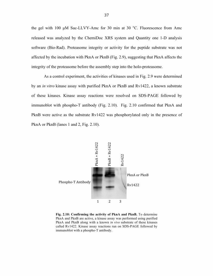
As a control experiment, the activities of kinases used in Fig. 2.9 were determined
by an in vitro kinase assay with purified PknA or PknB and Rv1422, a known substrate
of these kinases. Kinase assay reactions were resolved on SDS-PAGE followed by
immunoblot with phospho-T antibody (Fig. 2.10). Fig. 2.10 confirmed that PknA and
PknB were active as the substrate Rv1422 was phosphorylated only in the presence of
PknA or PknB (lanes 1 and 2, Fig. 2.10).
PknA%+%Rv1422!
PknB%+%Rv1422!
Rv1422!
PknA%or%PknB%%Rv1422%Phospho2T%Antibody!
%
1%%%%%%%%%%%%2%%%%%%%%%%%3%
Fig. 2.10: Confirming the activity of PknA and PknB. To determine PknA and PknB are active, a kinase assay was performed using purified PknA and PknB along with a known in vivo substrate of these kinases called Rv1422. Kinase assay reactions run on SDS-PAGE followed by immunoblot with a phospho-T antibody.
38
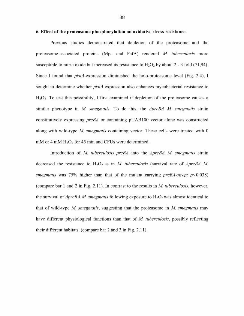
6. Effect of the proteasome phosphorylation on oxidative stress resistance
Previous studies demonstrated that depletion of the proteasome and the
proteasome-associated proteins (Mpa and PafA) rendered M. tuberculosis more
susceptible to nitric oxide but increased its resistance to H2O2 by about 2 - 3 fold (71,94).
Since I found that pknA-expression diminished the holo-proteasome level (Fig. 2.4), I
sought to determine whether pknA-expression also enhances mycobacterial resistance to
H2O2. To test this possibility, I first examined if depletion of the proteasome causes a
similar phenotype in M. smegmatis. To do this, the ΔprcBA M. smegmatis strain
constitutively expressing prcBA or containing pUAB100 vector alone was constructed
along with wild-type M. smegmatis containing vector. These cells were treated with 0
mM or 4 mM H2O2 for 45 min and CFUs were determined.
Introduction of M. tuberculosis prcBA into the ΔprcBA M. smegmatis strain
decreased the resistance to H2O2 as in M. tuberculosis (survival rate of ΔprcBA M.
smegmatis was 75% higher than that of the mutant carrying prcBA-strep; p<0.038)
(compare bar 1 and 2 in Fig. 2.11). In contrast to the results in M. tuberculosis, however,
the survival of ΔprcBA M. smegmatis following exposure to H2O2 was almost identical to
that of wild-type M. smegmatis, suggesting that the proteasome in M. smegmatis may
have different physiological functions than that of M. tuberculosis, possibly reflecting
their different habitats. (compare bar 2 and 3 in Fig. 2.11).

39
Even though the phenotype of proteasome depletion in M. smegmatis was
different from the one in M. tuberculosis, this result provided us a tool to test whether
pknA-expression can enhance resistance to H2O2 of ΔprcBA M. smegmatis carrying
prcBA. To do this, I conducted another survival rate experiment where the ΔprcBA M.
smegmatis cells expressing prcBA in the presence of pknA-, pknB-overexpression, or
vector alone was treated with 0 mM or 4 mM H2O2 for 45 min and the relative survival
rate was determined as described above. As a control, ΔprcBA M. smegmatis that
contains only pknA-overexpression cassette without prcBA was also included.
Relative Survival Rate
0
0.5
1
1.5
2
1 2 3
prcBA/
ΔprcBA
Vector/Δp
rcBA
vector/WT
Fig. 2.11: Mycobacterial resistance to H2O2 is increased by absence of proteasome. ΔprcBA or wild-type M. smegmatis cells with constitutive prcBA expression or vector alone was treated with 0 or 4 mM H2O2 for 45 min and CFUs were determined. Relative survival rate was calculated by dividing CFU from 4mM H2O2 by CFU from 0 mM H2O2 sample after 45 min.

40
Fig. 2.12 shows that pknA-expression in the presence of prcBA confers 67%
higher resistance to H2O2 than cells containing vector only (p<0.012) (compare bar 2 and
1). Though the percent change seems modest, the increased resistance to H2O2 by pknA-
expression was statistically significant and was obtained consistently over three
independent experiments. In contrast, co-expression of pknB and prcBA did not change
the resistance to H2O2 (compare 3 and 1, Fig. 2.12), consistent with the intact proteasome
complex formation under pknB-overexpressing condition shown earlier in Fig. 2.4. In
addition, pknA-overexpression alone (bar 4) did not change the bacterial survival rate in
0
0.5
1
1.5
2
1 2 3 4
vector+pknA/
ΔprcBA
prcBA+
pknA/Δ
prcBA
prcBA+
pknB/Δ
prcBA
Relative Survival
prcBA+
vector/Δp
rcBA
Fig. 2.12: Mycobacterial resistance to H2O2 is increased by pknA-expression. ΔprcBA M. smegmatis cells expressing prcBA in the presence of pknA-, pknB-overexpression or pMH94 vector alone was treated with 0 or 4 mM H2O2 for 45 min and the relative survival rate was determined as described in Fig. 2.11. As a control, ΔprcBA M. smegmatis that contains only pknA-overexpression cassette without prcBA was included. This result is a representative experiment of three replicates where each performed with triplicate plating.

41
the presence of H2O2, indicating that PknA contributes to H2O2 resistance mainly through
the proteasome.
7. H2O2 impedes the formation of holo-proteasome in a PknA-dependent manner
If PknA increases the resistance to H2O2 via the proteasome, I predicted that H2O2
stress would inhibit the formation of holo-proteasome in a PknA-dependent manner. To
test this prediction, I wanted to examine the formation of the holo-proteasome under
H2O2 treatment in ΔprcBA M. smegmatis that constitutively expresses prcBA in the
presence of pknA- or pknB-expression (Fig. 2.13). For this, levels of PrcB processing
were measured to examine the integrity of the holo-proteasome, by performing an
immunoblot with a PrcB antibody.
1 2 3 4
Pre-‐PrcB
PrcB (processed)
Pre-‐PrcB
PrcB (processed)
1.07 2.03 0.79 0.83
pknA pknB prcBA H2O2(2mM)
+ + +
+ +
-‐
+
+ +
-‐
-‐ +
-‐ +
-‐
-‐
PrcB Ab (H2O2, 1.5 hr)
PrcB Ab (H2O2, 4 hr)
Densitometric ratio of Pre-‐PrcB/Prcb
Fig. 2.13: H2O2 affects Pre-PrcB processing. The ΔprcBA M. smegmatis strain that constitutively expresses prcBA (pCK322) and contains pknA- (pCK5) or pknB-expression (pCK7) were treated with 2 mM H2O2 for 1.5 and 4 hr. 20 µg total lysate protein from each culture was analyzed in immunoblot with a polyclonal PrcB antibody to test the processing of PrcB.

42
Two-fold more pre-PrcB was accumulated in cells expressing pknA after 1.5 hr
H2O2 treatment (compare lanes 1 and 2, Fig. 2.13, upper panel), but not in cells with
pknB-expression (lanes 3 and 4). Longer treatment of H2O2 dramatically diminished the
level of both pre-PrcB and processed PrcB in pknA-expressing cells while processing of
PrcB was near complete in pknB-expressing cells (Fig. 2.13, lower panel). Taken
together, these data support that PknA plays an important role in oxidative stress response
by impeding the formation of holo-proteasome.
8. H2O2 affects holo-proteasome activity in M. tuberculosis
To further confirm the effect of H2O2 on the PrcB processing, I wanted to
investigate whether in wild-type M. tuberculosis holo-proteasome formation and its
activity is affected in the presence of H2O2. Wild-type M. tuberculosis cells were treated
with 0 mM or 2 mM H2O2 for 24 hr. Cells were harvested and total lysate protein from
each culture was analyzed by immunoblot with a polyclonal PrcB antibody. As shown in
Fig. 2.14, exposure to H2O2 affects the processing of pre-PrcB and leads to defect in
holo-proteasome formation in M. tuberculosis.
Pre-‐PrcB
PrcB (processed)
H2O2(2mM) -‐ +
PrcB Ab (H2O2, 24 hr)
Densitometric ratio of Pre-‐PrcB/PrcB
0.18 0.91
43
T
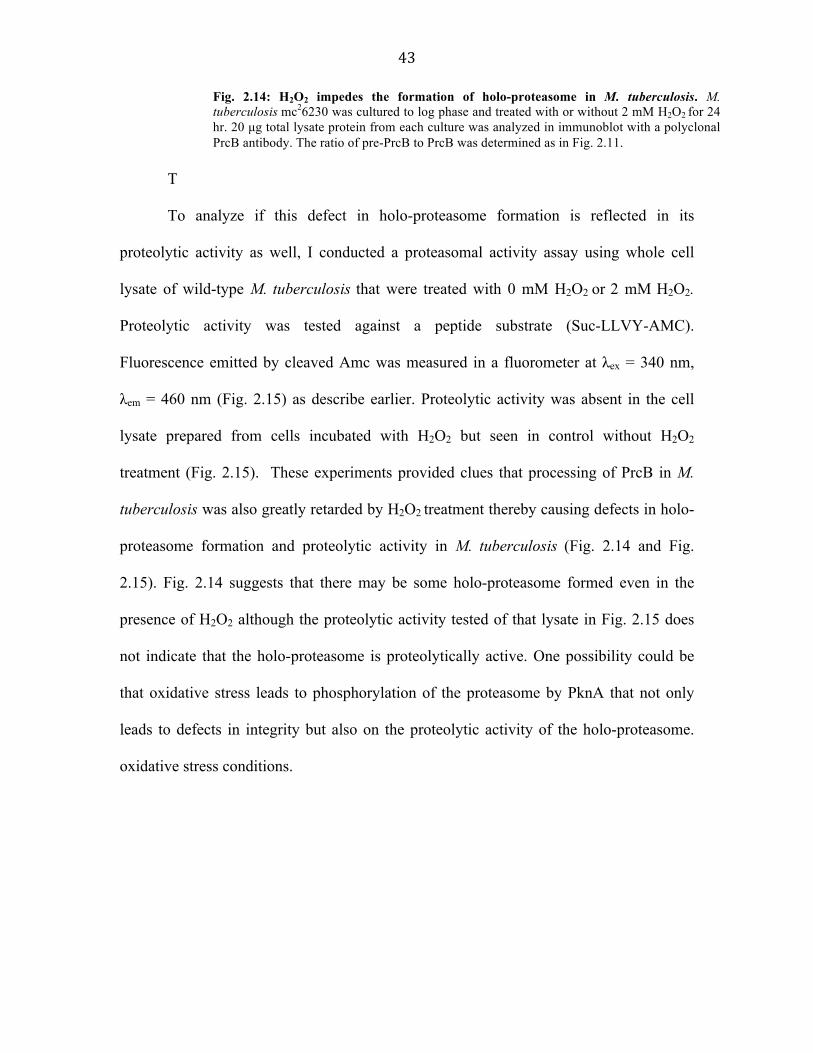
To analyze if this defect in holo-proteasome formation is reflected in its
proteolytic activity as well, I conducted a proteasomal activity assay using whole cell
lysate of wild-type M. tuberculosis that were treated with 0 mM H2O2 or 2 mM H2O2.
Proteolytic activity was tested against a peptide substrate (Suc-LLVY-AMC).
Fluorescence emitted by cleaved Amc was measured in a fluorometer at λex = 340 nm,
λem = 460 nm (Fig. 2.15) as describe earlier. Proteolytic activity was absent in the cell
lysate prepared from cells incubated with H2O2 but seen in control without H2O2
treatment (Fig. 2.15). These experiments provided clues that processing of PrcB in M.
tuberculosis was also greatly retarded by H2O2 treatment thereby causing defects in holo-
proteasome formation and proteolytic activity in M. tuberculosis (Fig. 2.14 and Fig.
2.15). Fig. 2.14 suggests that there may be some holo-proteasome formed even in the
presence of H2O2 although the proteolytic activity tested of that lysate in Fig. 2.15 does
not indicate that the holo-proteasome is proteolytically active. One possibility could be
that oxidative stress leads to phosphorylation of the proteasome by PknA that not only
leads to defects in integrity but also on the proteolytic activity of the holo-proteasome.
oxidative stress conditions.
Fig. 2.14: H2O2 impedes the formation of holo-proteasome in M. tuberculosis. M. tuberculosis mc26230 was cultured to log phase and treated with or without 2 mM H2O2 for 24 hr. 20 µg total lysate protein from each culture was analyzed in immunoblot with a polyclonal PrcB antibody. The ratio of pre-PrcB to PrcB was determined as in Fig. 2.11.

44
9. H2O2 activates PknA
So far, my data suggest an important role of PknA in H2O2 resistance via
proteasome but the mechanism underlying this process was not answered. To pursue this
goal, I wanted to know whether H2O2 could activate PknA that then could lead to defects
in proteasome integrity and H2O2 resistance. For this, E. coli BL21 (DE3) containing a
pGEX-4T-3-pknA plasmid was cultured in the presence of 0.5 mM IPTG for 6 hr, and
then exposed to 2 mM H2O2 for 0, 60, and 120 min. The levels of the PknA
phosphorylation and the GST-PknA protein were determined by immunoblot with
phospho-T (row 1, Fig. 2.16) and GST (row 2, Fig. 2.16) antibodies, respectively. Fig
2.16 shows an increase in autophosphorylation levels of PknA in cells incubated with
H2O2 with increasing incubation time, suggesting that PknA can be activated by H2O2.
0
2000
4000
6000
8000
10000
12000
0 20 40 60 80
RFU
Time (minutes)
without hydrogen peroxide
with hydrogen peroxide
Blank
Fig. 2.15: Presence of H2O2 affects proteasomal activity in M. tuberculosis. M. tuberculosis cells were harvested and lysate was used for a proteasomal activity assay. Lysates were treated with 2mM H2O2 and as controls lysate without H2O2 or blank containing no lysate were used. X-axis: Time (minutes), Y-axis: Relative Fluorescence Units (RFU).

45
This result is similar to that seen in the IkB kinase (inhibitor of NF-kB kinases) of some
eukaryotes, where H2O2 has been shown to promote autophosphorylation of IkB kinase
by phosphorylating two serine residues on activation loop thereby activating the kinase
(95). Taken together, these data suggest that PknA plays an important role in the
oxidative stress response by impeding the formation of holo-proteasome in M.
tuberculosis.
Discussion
In this work, I determined that proteasome subunits, pre-PrcB and PrcA, are
phosphorylated by PknA and that their phosphorylation enhances the resistance against
H2O2 stress in mycobacteria. Together with the holo-proteasome assembly defects, H2O2
resistance, and activation of PknA data presented here, these results indicate that PknA
plays an important role in regulating proteasome integrity, oxidative stress response, and
GST-‐PknA
GST-‐PknA
Densitometric ratio of Pre-‐PrcB/PrcB
1.3 1.8 2.4
Incubation with H2O2 (min)
0 60 120
phospho-‐T Ab
GST Ab
Fig. 2.16: Autophosphorylation of PknA increases in response to H2O2. E. coli BL21 (DE3) containing a pGEX-4T-3-pknA plasmid (pCK3) was cultured in the presence of 0.5 mM IPTG for 6 hr, and then exposed to 2 mM H2O2 for 0, 60, and 120 min. The levels of the PknA phosphorylation and the GST-PknA were determined by immunoblot with phospho-T (row 1) and GST (row 2) antibodies, respectively. Densitometric ratio of the phosphorylation level and the GST-PknA level was determined as in Fig. 2.11.

46
possibly pathogenesis in mycobacteria. Fig. 2.17 describes a model speculated based on
my findings of role of proteasome phosphorylation in increasing resistance to H2O2.
In prokaryotes, genes encoding putative 20S proteasome are found only in the
phylum Actinobacteria that mycobacteria belong to. Recently, phosphorylation of
proteasome subunits (both α- and β-subunits) have been reported in eukaryotes and
archaeal proteasomes (96,97). Phosphorylation was determined to enhance proteolytic
activity against peptide substrates in eukaryotes (98). Here, I report that phosphorylation
of mycobacterial proteasomal subunits (pre-PrcB and PrcA) by PknA affects the integrity
INACTIVE(
PO4(
PO4(
PO4(
PO4(
PknA(
EXTERIOR'
INTERIOR'
(H2O2(
(H2O2(
(H2O2((H2O2(
(H2O2( Oxida&ve)stress)
M. tb'
Holo,proteasome'
Macrophage
M.#tb#Survival'
PO4(
PO4(
ACTIVE(
PknA(
Autophosphoryla;on'
Fig. 2.17: Model for role of proteasome phosphorylation in M. tuberculosis (M. tb). Host induced defense factors such as H2O2 are made against an invading pathogen such as M. tb cells that are then available for them to activate PknA. Autophosphorylation of PknA results in activation of PknA and downstream substrate phosphorylation occurs on proteasomal subunits. This affects holo-proteasome integrity and thereby leading to a state of proteasome depletion that somehow leads to increased resistance to H2O2.

47
of the proteasome complex, via inhibition of processing of pre-PrcB and thus assembly
into the holo-proteasome. Therefore, it is suggested that M. tuberculosis is able to
modulate the levels of holo-proteasome complex through PknA whose activation is
stimulated by H2O2. Phosphorylation of proteasome by PknA mimics a state of
proteasome depletion that may then contribute to the pathogen’s higher resistance to
H2O2 by possibly saving some unknown oxidative stress response proteins from
proteolysis. I also present data to suggest that H2O2 activates PknA during H2O2 stress,
and thus activated PknA become available to phosphorylate proteasome leading to rescue
of bacilli in the presence of H2O2. Thus, these results could suggest a function of
proteasome regulation by PknA to have a probable end result of establishing dormancy or
persistence by M. tuberculosis.
The pathogenesis of TB has been widely recognized as a tug-of-war between the
pathogen and the host immune system. It is believed that once endocytosed by host
macrophage, the bacilli has to encounter a robust supply of immune system molecules to
overcome the stress and maintain dormancy or lead to active infection. One major
defense mechanism seen with M. tuberculosis is reduced growth rate as a survival
mechanism to fool the host immune system. Darwin et al. (2003) and Gandotra et al.
(2007) have independently shown that lack of proteasome aids survival of M.
tuberculosis against H2O2 stress (71,72). Consistently, I have found that PknA diminished
the proteasome assembly that in turn led to increased resistance to H2O2, and that the
H2O2 stress, via activating PknA, reduced the formation of the proteasome. What remains
to be answered is how lack of the proteasomal system results in higher resistance to
H2O2. M. tuberculosis possibly relies on PknA as a mode to sense the H2O2 stress at this

48
stage and affects the integrity of the proteasome, which may then lead to accumulation of
oxidative stress proteins such as catalase-peroxidase (KatG) (99). Thus, it would be
interesting to see if the integrity of KatG can be affected by proteasome and PknA.
Another question is how H2O2 affects the proteasome assembly through PknA. It is
known that in eukaryotes, several protein kinases (ERK [extracellular signal-regulated
kinases] and MAP [mitogen-activated protein] kinases) are activated by H2O2 that serves
as a survival mechanism against H2O2 exposure (95,100). Tabet et al (2005) showed
MAP kinase activation through phosphorylation in the presence of H2O2 to be also
increasing in a time dependent manner (95,100). Thus my results are in agreement with
the MAP kinase activation by H2O2 seen in eukaryotes, as PknA autophosrylation level
also seemed to increase when exposure time was increased in the presence of H2O2. In
eukaryotes, IkB kinases (inhibitor of NF-kB kinases) is known to be activated by H2O2
through phosphorylation of its activation loop at two serine residues (95). In this study I
suggest a similar mechanism for the PknA activation in the presence of H2O2. Although
the residue known to be target of autophosphorylation on PknA are T172 and T174; it
will be important to confirm whether H2O2 promote phosphorylation of same residues
(95). Further experiments to show whether activation of PknA can be inhibited in the
presence of KatG can provide clues to the H2O2 induced stimulation of PknA
autophosphorylation shown here.
To better understand proteasome and its role in stress response, further studies
focusing on determining how the phosphorylation of pre-PrcB and PrcA regulates the
assembly of the proteasome complex would be crucial. We have yet to identify additional
substrates of the PrcA-phosphorylated proteasome, and the molecular mechanisms by

49
which PrcA phosphorylation affect the substrate proteolysis.

50
Chapter 3
In silico approach to search for new substrates of PknA/PknB, and
role of FbpB phosphorylation Abstract
The molecular switch between latency and active replication in M. tuberculosis is
not greatly understood. It is speculated that precise regulation of cell wall biosynthesis
must be a key mechanism to achieve transition between the two drastically varied growth
stages. Therefore, to sense extracellular environment conditions protein-signaling
cascades must act as messengers to relay information from outside to the inside of the
cell. To investigate this possibility, I wanted to identify the role(s) of two eukaryotic-type
Ser/Thr kinases, PknA and PknB by identifying and characterizing their substrates.
Here, I describe an in silico homology approach to identify new substrates of
PknA/PknB using the predicted preferred substrate phosphorylation motifs of PknA/B
(ATQXIP or TQXIP, where X is any amino acid), and a synthetic peptide screening
technique. Through this approach, I identified seven candidate substrate proteins. Of
those seven candidate proteins, FbpB appeared to be of great interest because it belongs
to a complex know as antigen 85 complex involved in the synthesis of cell wall
component mycolic acid, an important virulence factor of M. tuberculosis. My work
suggests that FbpB is phosphorylated in vivo in M. smegmatis and in M. tuberculosis, and
that the 30KDa-secreted form of FbpB is dephosphorylated in M. smegmatis. Mass
spectrometry analysis of the M. tuberculosis FbpB protein purified from M. smegmatis
revealed 255T as a single phosphorylation site. The synthetic mycolyl transferase assay
employed for assessing FbpB’s enzyme activity suggested that phosphorylation may

51
regulate its enzyme activity. The kinase responsible for FbpB phosphorylation is yet to be
answered.
Introduction
Mycobacterium tuberculosis not only actively replicates during infection but also
has a dormant intracellular phase where it has reduced metabolic activity and minimal
cell division. This may account for the characteristic slow growth rate observed in M.
tuberculosis, and could thus contribute to the ability of disease-causing members of
mycobacteria to survive for long periods of time in animal hosts. While the molecular
basis of the transition between these two completely different cell cycle phases is yet to
be identified, it has been suggested that one key underlying mechanism must be in
regulation of cell signaling in order to govern cell division and cell wall synthesis
required for cell division.
Previously, our lab showed that PknA and PknB phosphorylated preferably at the
phospho-acceptor threonine (T) residue followed by glutamine (Q) at +1 position and
hydrophobic residues at the +3 and +4 positions. Subsequent identification of the in vivo
substrates (Wag31 and Rv1422) of these kinases demonstrated that those substrate
proteins have a phosphorylation site that matches very well with the previous finding of
substrate specificity (73TQAIP in Wag31 and 325TQEIP in Rv1422) (52). Further analyses
of the preferred phosphorylation sequence for PknA/B against peptide library revealed
that ATQXIP, (where X is any amino acid) is the consensus sequence preferred for the
phosphorylation by PknA and PknB (52). In this chapter, I will describe an in silico
method based on this consensus peptide sequence to screen for additional potential

52
substrate(s) of PknA and PknB, and investigate the role of phosphorylation of one such
potential substrate, FbpB.
Results
1. Genomic search using a consensus peptide motif reveals 7 putative substrates of
PknA/PknB
Based on the preferred phosphorylation motif (ATQXIP) identified for PknA and
PknB, I hypothesized that additional substrates of PknA and PknB have similar amino
acid sequences around their phosphorylation site. Homology search for additional
proteins containing the ATQXIP sequence in the whole M. tuberculosis genome resulted
in no new protein except for the already identified substrates, Wag31 and Rv1422 (Fig.
3.1).
Wag31 : GVTPQATQAIPAYE Rv1422 : EPPVAATQEIPIDG
Search for proteins containing similar sequence in M. tuberculosis genome
ATQXIP
Wag31 Rv1422
TQXIP
AceE (Probable pyruvate dehydrogenase E1 component) FbpB (Secreted Antigen 85-B) MbtB (Phenyloxazzoline synthase) Rv0940c (Possible Oxidoreductase) Rv1102c (PemK-like RNase toxin) Rv2743 (Conserved transmembrane protein) SugA (Probable sugar-transport integral membrane protein) Wag31 Rv1422

53
However, a second round of investigation done by reducing the stringency of the
consensus using TQXIP sequence revealed seven new candidate proteins that contain one
or more of the potentially phosphorylated TQ motif. The list of candidate substrate
protein is described in Fig. 3.2.All the peptides generated had MA residues at the amino
terminus as a way to tag the peptides while verifying their sequence. The poly-lysine
(KKK) tail at the carboxyl-terminus adds a net positive charge to the peptides at low pH
thereby helping in their adsorption onto p81 phosphocellulose paper (101).
2. PknB maximally phosphorylates FbpB and Rv1102c in vitro
In order to determine whether PknA and/or PknB phosphorylate any of these new
substrate candidates, I synthesized a series of peptides that include the TQ motif and the
1. FbpB' ' 'MARNDPTQQIPKLVKKK ''2. AceE ' 'MAEFRDTQRIPVSDKKK'3. MbtB ' 'MASVLATQVVAGIRKKK'4. Rv0904c 'MATPAPTQPIPILVKKK'5. Rv1102c' 'MASCDNTQTIPVCDKKK'6. SugA ' 'MASAPLTQQIPSLGKKK'7. Rv2743c 'MATIAVTQAIPATLKKK'
Fig. 3.1: In silico search for putative substrates of PknA/PknB. Initial search with ATQXIP identified only Wag31 and Rv1422, however reducing stringency to TQXIP motif resulted in identifying 7 putative substrates of PknA/PknB.
Fig 3.2: List of candidate proteins obtained from the in silico search for substrates of PknA/PknB. Each of the seven putative substrate proteins of PknA/PknB containing preferred phosphorylation motif highlighted in blue (TQXIP) was custom synthesized as 17-mer peptides for initial screening by in vitro phosphorylation.

54
surrounding residues of these proteins. All the peptides were synthesized as custom oligo
peptides of 18mers with sequences spanning upstream and downstream of TQXIP motif
(Fig. 3.2). To test if these peptides can be phosphorylated by PknA/B, I also needed to
purify active PknA/B kinase proteins. The intracellular kinase domains of M. tuberculosis
PknA and PknB were expressed and purified as fusion proteins with glutathione-S-
transferase (GST) tag from E. coli. As described before, a preliminary in vitro kinase
assay was performed with Rv1422 to confirm their catalytic activity. Each synthetic
peptide (200ng) was then subjected to in vitro kinase assay with PknA or PknB (200ng)
in the presence of [γ-32P]ATP. The peptide of Wag31 was included as a positive control,
along with peptide-only and enzyme-only as negative controls. The reaction was
performed for two different incubation periods (20 and 40 min), and the incorporation of
32P into peptides was measured using a scintillation counter. Fig. 3.3 shows that PknA
highly phosphorylates the peptides of Rv0940c and SugA while PknB strongly
phosphorylates those of Rv1102c, FbpB, and SugA, suggesting that PknA and PknB
show specificity even with these short synthetic peptides.
CP
M
Peptides

55
Among these substrate candidates phosphorylated by PknA or PknB, I chose to
further study FbpB (Fibronectin binding protein B) for the following reasons; 1) It is a
member of a mycolyl transferase enzyme complex that is involved in the biosynthesis of
trehalose dimycolate, a major cell wall components in mycobacteria, which contributes to
the impermeable nature of mycobacterial cell wall and thereby their virulence, and 2) It
interacts with extracellular matrix protein fibronectin and this interaction has been
suggested to play a role in binding of tubercle bacillus cell to the mucosal surface and
thus in the phagocytosis of the bacteria by the monocytes (80).
3. FbpB is highly phosphorylated in vivo in Mycobacterium smegmatis Since PknB phosphorylated the synthetic peptide of FbpB, I decided to further
test whether PknB could also phosphorylate the FbpB protein in vitro. To do this, I first
attempted to express the 6His-tagged fbpB gene in E. coli but this was not successful,
probably due to difference in codon usage between E. coli and M. tuberculosis.
Consistent with this, Kravchenko et al. (2007) showed that altering a few amino acid
codons of FbpB resulted in high expression levels of FbpB in E. coli (102). As an
alternative approach, I decided to express and purify M. tuberculosis FbpB from M.
smegmatis. The M. tuberculosis fbpB gene was amplified by PCR with the C-terminal
strep-tag and cloned behind an acetamide-inducible promoter in an integration vector
Fig. 3.3: In vitro kinase assay with synthetic peptide of substrate candidates in the presence of PknA/B and [γ-32P]ATP. The yellow bars indicate the levels of phosphorylation in counts per minute for 200 ng of each peptide for 20 minutes of reaction incubation time and green bars indicate the same for 40 minutes of reaction incubation time. X-axis: Peptides; Y-axis: Counts per minute (CPM). Kinase assay reactions run using peptides labeled as follows; 1.FbpB, 2.AceE, 3.MbtB, 4.Rv0940c, 5.Rv1102c, 6.SugA, 7.Wag31, 8.Rv2743c, 9.Peptide only control; and 10.Kinase only control.

56
pMH94A (pMH94A-Pacet-fbpB-strep). This plasmid was then transformed into M.
smegmatis and the recombinant FbpB-strep was expressed by adding 0.2% acetamide,
and purified by using the Strep-Tactin affinity purification system (Novagen). Activity of
PknB was tested by an in vitro kinase assay with the Rv1422 protein, a known substrate
of PknB, where Rv1422 showed a high level of phosphorylation, suggesting that this
PknB was an active enzyme (In the work presented in this dissertation, activity of both
PknA and PknB has been confirmed for every in vitro kinase assay by this technique,
data not shown). When this FbpB protein was incubated with PknB and [γ-32P]ATP, it
showed very weak signal for phosphorylation (Fig. 3.4). The presence of other strong
signals is a result of long exposure time for this autoradiograph.
37kDa 25kDa
Stained
gel In vitro kinase
assay
PknB FbpB
Fig. 3.4: Weak phosphorylation of FbpB purified from M. smegmatis by PknB in the presence of [γ-32P]ATP. Left panel shows Gel Code stained gel. Right panel is the autoradiograph for in vitro kinase assay.

57
To obtain the signal intensity from FbpB shown in Fig. 3.4 (right column), I had
to expose the gel for a long time and the signal from FbpB was even weaker than
nonspecifically phosphorylated proteins (Left panel, Fig 3.4). One possible reason for this
low level of radioactivity (phosphorylation) from FbpB was that FbpB was already
phosphorylated in M. smegmatis before the purification. To test this possibility, I
measured the phosphorylation of the FbpB protein without incubating with PknB by a
Western blot analysis with a phospho-[S/T]Q antibody (Fig. 3.5). The FbpB protein
revealed a strong Western signal, suggesting that FbpB had already been highly
phosphorylated in M. smegmatis. Since there exists only one TQ motif in FbpB (T234),
the synthetic peptide of FbpB was highly phosphorylated by PknB, and phospho-[S/T]Q
antibody detected high level of phosphorylation in FbpB, I hypothesized that the 234T is a
phosphorylation site.

58
4. Secreted FbpB is not phosphorylated in M. smegmatis
Since FbpB is the second major secreted protein in M. tuberculosis after ESAT-6,
I also wanted to determine if the secreted form of FbpB is also phosphorylated (103). To
test this possibility, M. smegmatis cells containing pMH94A-Pacet-fbpB-strep was
cultured in the presence of 0.2% acetamide until late log-phase, and intracellular FbpB-
strep was purified as described above. The secreted extracellular FbpB-strep was also
purified by collecting the culture supernatant and precipitated total protein with
ammonium sulfate (60%), and using the Strep-Tactin purification Kit. After dialysis,
strep Ab
P [S/T]Q Ab
FbpB
Fig. 3.5: FbpB has been phosphorylated in M. smegmatis before purification. Western blot with a phospho-[S/T]Q antibody (lane 2) followed by stripping and re-probing with a strep antibody against Strep-Tactin purified FbpB (lane 1),

59
approximately equal amount of the intracellular and extracellular FbpB-strep was
subjected to a gradient SDS-PAGE (4 - 12%, Bio-Rad) to analyze by an immunoblot
analysis with a strep antibody or a phospho-[S/T]Q antibody (Fig. 3.6).
FbpB undergoes signal peptide processing, which leads to the removal of 40
residues at the N-terminus during its secretion (77). In Fig. 3.6, we observed two major
bands for FbpB-strep. While I plan to identify these bands by Edman degradation and
mass spectrometry, predicted molecular weight of these bands are consistent with the
processed form (the lower band with ~30 kDa) and the unprocessed form of FbpB (the
upper band with ~34 kDa). Moreover, it is known that the secreted FbpB exists in two
different forms, one as cell wall associated and the other as completely secreted into the
extracellular environment (77). Consistent with this, I found both unprocessed and
processed FbpB proteins from cell pellet (Intracellular, left column, Fig. 3.6) but mainly
processed form of FbpB from supernatant (Extracellular, left column).
antibody) α*strep) Puri1ied)FbpB)
α*PTQ) Puri1ied)FbpB)
source) Intra*cellular)
Extra*)cellular)
Intra*cellular)
Extra*)cellular)
Total)protein)µg)loaded) 5) 10) 5) 10) 5) 10) 5) 10)
)))))
Unprocessed)FbpB))Processed)FbpB)
Fig. 3.6: Only intracellular FbpB is phosphorylated. Western blot with intracellular (CELL) and culture filtrate protein (CFP) of FbpB using a strep antibody followed by re-probing with a phospho-[S/T]Q antibody.

60
Interestingly, while both processed and unprocessed forms of intracellular FbpB
were highly phosphorylated, the secreted FbpB did not show any phosphorylation
(Intracellular and Extracellular, right column, Fig. 3.6), suggesting that the
phosphorylation status of FbpB may be important for determining whether FbpB is
attached to cell wall or completely secreted, or for processing and secretion processes of
FbpB.
To further confirm the phosphorylation status between the intracellular and the
extracellular FbpB, I conducted a 2D western blot with protein samples used in Fig 3.6
(Fig. 3.7). 200 µg of purified FbpB-strep was first rehydrated in a 2D strip (pH 4.7-5.9),
and subjected to isoelectric focusing and 2D SDS-PAGE. First, a western blot was
performed with a phospho-[S/T]Q antibody, and the membrane was stripped before
probing with a strep antibody. Fig. 3.7 shows that most of the purified FbpB protein in
the cell lysate is phosphorylated (Intracellular, Fig. 3.7), but after secretion there seems to
be a loss of phosphorylation (Extracellular, Fig 3.7), confirming that the extracellular
form of FbpB is not phosphorylated.

61
5. FbpB is phosphorylated in M. tuberculosis
To examine whether FbpB is phosphorylated in M. tuberculosis, the pMH94A-
Pacet-fbpB-strep construct was transformed into an attenuated M. tuberculosis mc26030
strain and cultured in the synthetic Sauton’s minimal media for 3 weeks. Cells were
further incubated for additional 3 days with 0.2% acetamide to induce the expression of
FbpB-strep. Intracellular Strep-FbpB was purified using Strep-Tactin affinity column and
subjected to western blot with a phospho-[S/T]Q and a strep antibody (Fig. 3.8). The
FbpB-strep protein purified from M. tuberculosis showed processed and unprocessed
FbpB, similar to those found in M. smegmatis. Western blot with a phospho-[S/T]Q
antibody demonstrated the in vivo phosphorylation of FbpB purified from M.
α-p[S/T]Q
pH 4.7-5.9
Extracellular Intracellular
α-Strep
Fig. 3.7. Different phosphorylation pattern between intracellular and extracellular FbpB. 2D-Western blot of intracellular and extracellular FbpB with a strep (top panel) and a phospho-[S/T]Q (bottom panel) antibody.

62
tuberculosis. This experiment also suggested that processing/secretion process for FbpB
in M. smegmatis is similar to that of M. tuberculosis.
6. FbpB is phosphorylated at 255T
With in vivo phosphorylation of FbpB in M. tuberculosis identified, I wanted to
determine the actual site(s) of phosphorylation in FbpB. To do this, FbpB-strep purified
from M. smegmatis was resolved using 2D-PAGE and desired spots were analyzed by
mass spectrometry. Mass spectrometry analysis revealed a weak phosphorylation signal
FbpB M. smegmatis
FbpB M. tuberculosis
FbpB M. smegmatis
FbpB M. tuberculosis
Strep Ab p[S/T]Q Ab
Fig. 3.8. FbpB is phosphorylated in vivo in M. tuberculosis. Western blot of intracellular FbpB from M. tuberculosis with a strep and a p[S/T]Q antibody. FbpB purified from M. smegmatis used as a positive control.

63
from 255T, but not from the predicted 234T (data not shown). This was disappointing, as
255T did not represent the predicted phosphorylation site based on the motif TQXIP that
was hypothesized to be the preferred phosphorylation motif for PknA/PknB. Upon
analyzing the trypsin digest report for FbpB, I came to conclude that it’s possible that the
peptide length spanning the 234T residue may not be the ideal ionization limit of mass
spectrometry analysis. A second round of digest with chymotrypsin or other enzyme
could be done to overcome this barrier and confirm the phosphorylation site in FbpB by
mass spectrometry.
7. Mycolyl transferase activity of FbpB is affected by phosphorylation
Next, because FbpB is a mycolyl transferase, I wanted to test whether
phosphorylation has any effect on its enzymatic activity. To measure the mycolyl
transferase activity, I collaborated with Dr. Don Ronning group (at the University of
Toledo, OH) by using an assay system they developed (104). I incubated the purified
FbpB with p-nitrophenyl 6-O-octanoyl-β-d-glucopyranoside and β-glucosidase at room
temperature and the rate of p-nitrophenolate production was measured as the absorbance
at 405nm. A negative control was a reaction without FbpB. In addition, FbpC purified in
Dr. Ronning group was included as a positive control since they used it to standardize the
assay system.
In Fig. 3.9 (A), wild-type FbpB purified from M. smegmatis shows mycolyl
transferase activity, suggesting that this assay system can be used to test whether the
phosphorylation of FbpB is involved in regulating the mycolyl transferase activity of
FbpB. The right panel (B) in Fig. 3.9 shows the positive control assay performed with the

64
standardized FbpC enzyme (blue line) along with a negative control having no
enzyme/blank reaction (red line).
Fig. 3.9. Mycolyl transferase assay. (A) Wild-type FbpB purified from M. smegmatis was incubated with the synthetic substrate and samples tested include; (blue line) WT FbpB purified from M. smegmatis and dialyzed against a buffer containing glycerol; (red line) WT FbpB purified from M. smegmatis and dialyzed against a buffer without glycerol; (yellow line) Blank; (green line) WT FbpB purified from culture filtrate protein and dialyzed against a buffer without glycerol. (B) FbpC purified from E. coli used as a positive control for enzyme assay. Samples tested include (red line) No enzyme blank; (blue line) FbpC purified from E. coli.
As I confirmed that wild-type FbpB-strep had mycolyl transferase activity in Fig.
3.9, I wanted to test whether phosphorylation has any effect on its enzymatic activity
using wild-type FbpB, phospho-ablative FbpBT234A and phospho-mimetic FbpBT234E.
by using the assay system used in Fig.3.9. Fig. 3.10 (left panel) shows that FbpBT234A
has lower mycolyl transferase activity than wild-type FbpB. However, a similar
experiment with FbpBT234E and wild-type FbpB activity on right panel suggested
almost similar activity of both these proteins, suggesting that phosphorylation could be
enhancing mycolyl transferase activity of FbpB. In summary, although 234T is yet to be
confirmed as a phosphorylation site in FbpB, it still seems to be an important residue in
determining the mycolyl transferase activity in FbpB.
! !
(A) (B)

65
Discussion
In this study, I demonstrated an in silico method to screen for potential substrates
of PknA/PknB. This approach provides an opportunity to identify potential targets of
PknA/PknB phosphorylation, and also to later characterize the substrate proteins so as to
determine their regulatory function thereby identifying the role(s) of PknA/PknB in M.
tuberculosis. The peptide library screen employs in vitro kinase assay using the STPKs-
PknA and PknB that catalyze the transfer of the γ-phosphate of adenosine triphosphate
(ATP) to substrate peptide’s thr residues.
Previously, Kang et al. (2005) have shown that PknA and PknB predominantly
phosphorylate at thr residues, with a strong phosphorylation motif preference for
ATQXIP. In this work, it is interesting to note that PknA and PknB phosphorylate
!1000$
0$
1000$
2000$
3000$
4000$
5000$
6000$
7000$
1$ 21$ 41$ 61$ 81$ 101$
RFU$
Time$(sec)$
blank&
wild&type&FbpB&
FbpBT234A&
!1000$
0$
1000$
2000$
3000$
4000$
5000$
6000$
7000$
1$ 21$ 41$ 61$ 81$ 101$ 121$
RFU$
Time$(sec)$
blank&
wild&type&FbpB&
FbpBT234E&
Fig. 3.10: Phosphorylation might enhance mycolyl transferase activity. Wild type and FbpBT234A purified from E. coli subjected to enzyme assay. Left panel: FbpBT234A mutant showing lower activity than wild type FbpB. Right panel: FbpBT234E purified from E. coli shows higher activity as compared to FbpBT234A mutant. RFU value indicated for each sample here represents average of triplicate reactions.

66
different sets of peptides thereby suggesting their slightly different substrate specificity
(52). Among the seven candidate peptide substrates, four of the peptides namely, FbpB,
Rv0940c, Rv1102c and SugA showed highest level of phosphorylation by PknA/PknB.
Rv0940c whose peptide was maximally phosphorylated by PknA, is predicted to be an
oxidoreductase. Rv1102c is an mRNA interferase, with its peptide showing highest
phosphorylation by PknB when compared to PknA. Further investigations on this protein
led us to the finding that Rv1102c is part of a toxin-antitoxin system (Rv1102c-Rv1103c
system) (105). Additionally, Dr. Jeong-Sun Han and I were able to demonstrate that 1)
Rv1102c interacts with Rv1103c, 2) Rv1103c antagonizes the mRNA interferase activity
of Rv1102c and thus acts as its cognate antitoxin, and lastly 3) the expression of Rv1102c
is coupled to that of Rv1103c. The research paper titled ‘Characterization of
chromosomal toxin-antitoxin, Rv1102c-Rv1103c system in Mycobacterium tuberculosis”
was second authored by me and published in 2010 (105). SugA is part of an ABC
transporter complex, whose role in virulence is highlighted by its specific function in
trehalose uptake.
Of the newly identified candidate substrate proteins, FbpB seemed to be of great
interest as it belongs to a complex know as antigen 85 complex involved in the synthesis
of cell wall component mycolic acid, which is one of the major virulence factors of M.
tuberculosis. I have shown that FbpB is phosphorylated in vivo in M. smegmatis as well
as in M. tuberculosis and that the 30 KDa secreted form of FbpB is not phosphorylated.
Additionally, I also determined that in M. smegmatis intracellular FbpB is phosphorylated
while the extracellular is not, suggesting a possible role of phosphorylation in its
processing and/or secretion (Fig. 3.5). Based on this analysis, I investigated the role of

67
FbpB phosphorylation in its secretion by purifying secreted form of FbpB from M.
smegmatis culture-filtrate and compared it with intracellular FbpB by western blot with
phospho-[S/T]Q antibody. Furthermore, mass spectrometry analysis of FbpB purified
from M. smegmatis revealed 255T as a single phosphorylation site in FbpB and currently
this phosphorylation site needs to be confirmed. The synthetic mycolyl transferase assay
employed for assessing FbpB’s enzyme activity suggested phosphorylation may reduce
the mycolyl transferase activity of FbpB. Even though, mass spectrometry did not
identify 234T as the phosphorylation site, phospho-mutants analysis suggests that the
residue 234T is important for mycolyl transferase activity of FbpB. This data could thus
suggest the reason for the apparent difference in level of mycolyl transferase activity seen
between FbpB and the other members of the antigen 85 family; FbpA and FbpC. FbpB’s
reduced mycolyl transferase activity could be due to the probable lack of 234T
phosphorylation upon secretion since we know that secreted form and FbpBT234A had
the least enzyme activity.
The hydrophobic nature of mycobacterial cell wall confers passive resistance
against chemicals, dehydration, and degradation by host factors, inactivation of reactive
oxygen intermediate (ROI) and reactive nitrogen intermediate (RNI), and prevents entry
of toxic anti-TB compounds and antibiotics into the cell (106,107). Careful regulation of
the STPKs enables precise cellular responses to external signals, allowing bacteria to
survive in a variety of changing environmental conditions. As discussed earlier, FbpB is a
predominantly secreted protein forming the bulk of culture filtrate protein in
mycobacteria that is predicted to play a role in internalization and adaptation of M.
tuberculosis. The kinase responsible for FbpB phosphorylation and the role of FbpB

68
phosphorylation in host-pathogen interaction, for example in fibronectin binding, and
pathogenesis are yet to be answered.
In summary, I provide an in silico peptide library screening method to fish for
PknA and PknB targets in M. tuberculosis, which could possibly be applied to screen for
substrates of other kinases. Future work focusing on other two candidate substrates;
SugA and Rv0940c could be tested in order to provide insight into the way these protein
act as molecular switches that regulate diverse cellular event such as growth,
development and pathogenesis by signaling molecules. The global urgency related to TB,
which is currently the leading infectious disease causing the highest mortality, with the
added potential of these proteins in regulating vital metabolic pathways, provide strong
rationales for investigating the functions and mechanisms of the M. tuberculosis STPK
signaling systems.

69
APPENDIX A
EXPERIMENTAL PROCEDURES A.1. Culture conditions.
H37Rv, mc26230 M. tuberculosis, and M. smegmatis MC2155 cultures were
grown at 37 °C in Middlebrook 7H9 liquid medium (Difco) supplemented with 10%
ADC [ADC = 5% (w/v) BSA fraction V powder, 2% (w/v) glucose and 0.85% (w/v)
NaCl], and 0.05% (w/v) Tween-80, or on Middlebrook 7H9-ADC agar plates
supplemented with 0.05% (w/v) glycerol. Kanamycin (50 µg/ml), hygromycin (50 µg/ml)
or apramycin (50 µg/ml) was added to culture media where required. Escherichia coli
DH5α (Invitrogen) was used as a cloning host, and E. coli BL21 (DE3) for expression of
recombinant proteins. Cultures were grown at 37 °C in LB broth or solid medium with
apramycin (50 µg/ml), kanamycin (50 µg/ml), chloramphenicol (34 µg/ml) or ampicillin
(50 µg/ml) where appropriate.
A.2. Plasmid construction
For in vitro kinase assay of the chimeric proteasome complex, strep-prcA was
PCR amplified using the genomic DNA of M. tuberculosis H37Rv and cloned into an
integrating plasmid pCK202 driven by the acetamide-inducible promoter. All the primers
used in Chapter 2 are listed in Table 2. This construct, named as pCK206 (All the
constructs made are listed in Table 1), comprises the attachment site (attP) and the
integrase gene (int) of mycobacteriophage L5 for integration (108,109). For in vivo
phosphorylation analysis of PrcA in DprcBA M. smegmatis, prcBA-strep was PCR
amplified by using primers TBprcBA pUAB100-F and TBprcBA-C-strep3', and cloned

70
behind a constitutive hsp60 promoter in a replicating plasmid, pCK315 (and named as
pCK322). For testing the phosphorylation of WT and mutant forms of PrcA in E. coli,
prcBA-strep was PCR amplified by using primers C-StrepTBprcB-5 and TBprcBA-C-
strep3', and cloned under T7 promoter in pACYCDeut-1 (Novagen) (named as pCK343).
To construct a ∆prcBA M. smegmatis strain (KMS98), 5’- and 3’-flanking regions of the
prcBAMs genes was amplified by PCR by using primers Dn-PrcA-F, Dn-PrcA-R, Up-
PrcB-F, and Up-PrcB-R, and the genomic DNA of WT M. smegmatis MC2155. The
hygromycin resistance gene was inserted between the two flanking regions in pRH1351
and named as pCK260.
For in vivo and in vitro kinase assay of the FbpB, strep-fbpB was PCR amplified
using the genomic DNA of M. tuberculosis H37Rv. All the primers used in Chapter 3 are
listed in Table 3, and cloned into an integrating plasmid pCK202 carrying an acetamide-
inducible promoter, and named as pCK206 (All the constructs are listed in Appendix A,
Table 1). Plasmids expressing fbpBT234A and fbpBT234E were generated by using the
QuickChange Site-Directed Mutagenesis Kit (Stratagene) with primers indicated in Table
3.
A.3. Protein expression, purification and analysis of recombinant proteins To purify the chimeric proteasome complex, M. smegmatis KMS65 was grown to
mid-log phase where expression of strep-prcA was induced by 0.2% acetamide for 4 hr.
Strep-PrcA was purified by using the Strep-Tactin Affinity Purification Kit (Novagen).
The M. tuberculosis proteasome complex for the in vivo phosphorylation was purified
from ΔprcBA M. smegmatis carrying pCK322 plus pCK5 (KMS109), pCK7 (KMS108),
or pMH94 vector (KMS157) by overexpressing pknB at mid-log phase with 0.2%

71
acetamide for 4 and 16 hr. A parallel flask without acetamide was also cultured for 16 hr.
PrcA-strep was purified by using the Strep-Tactin Affinity Purification Kit (Novagen) as
described above. To test the effect of PknA on proteasome complex formation in E. coli,
pCK343 was transformed into E. coli BL21 (DE3) containing pCK3 or pCK4, which
were cultured in Overnight Express Autoinduction medium at 37 °C for 10 hr, and PrcA-
strep was purified as described above. For 2-D PAGE, immunoblot, and mass
spectrometry, total protein extraction from M. tuberculosis harboring pMH94 plasmid
alone, pCK5, or pCK7 and subsequent 2-D SDS-PAGE were performed as described
previously. For the in vivo phosphorylation of PrcA, 30 µg of each purified PrcA-Strep
was similarly analyzed in 2-D SDS-PAGE with IPG strips of pH 4-7 (Bio-Rad). Then,
Western blots were performed by a phospho-T antibody, and subsequently with a strep-II
antibody (Novagen) after stripping off the first antibody by using a stripping buffer (100
mM 2-mercaptoethanol, 2% (w/v) SDS, 62.5 mM Tris-HCl, pH 6.7) at 50 °C for 30 min.
Mass spectrometry for the identification of the phosphorylation sites in PrcA was
performed as previously described. To analyze the proteasome complex in M. smegmatis
overexpressing pknA, 15 µg of PrcA-strep plus PrcB complex used in Fig. 3.5 was
analyzed in 2-D SDS-PAGE with Flamingo Stain (Bio-Rad).
For purification of FbpB-strep, and all the phosphomutants of FbpB used in
Chapter 3 cells were grown to mid-log phase where expression of FbpB-strep was
induced by 0.2% acetamide until stationary phase. FbpB-strep including all it phospho-
mutant proteins were purified by using the Strep-Tactin Affinity Purification Kit
(Novagen). To purify extracellular FbpB, culture filtrate protein was isolated using 40%
ammonium sulfate (AS) precipitation method. Mycobacterial cells expressing FbpB were

72
inoculated in modified Sauton’s medium and incubated at 370C until early stationary
phase.The cultures were centrifuged at 16,000 g for 30 min at 40C and filtered through
0.22µm Millex GV PVDF membrane (Millipore). The filtrate was later used for a 40%
AS precipitation to isolate culture filtrate protein and purification of recombinant FbpB
was carried out using strep affinity column (Profinia) as described before.
A.4. In-gel proteasome assay
3 µM proteasome complex purified from E. coli containing pCK343 was
incubated with 1 µM GST-PknA or GST-PknB in the presence of 1 mM ATP in kinase
assay buffer (50 mM HEPES, 50 mM NaCl, 10 mM MgCl2, 2 mM MnCl2) for 2 hr at 37°
C. Reaction was stopped by adding Native Sample buffer (Bio-Rad), and proteins were
separated on two native PAGE gels; one was used to visualize proteins by staining with
Flamingo Fluorescent stain (Bio-Rad) and the other was used for an in-gel proteasome
assay by incubating the gel in proteasome assay buffer (50 mM HEPES pH 7.4, 5 mM
EDTA) and 100 µM Suc-LLVY-Amc (Boston Biochem) for 30 minutes at 30° C.
Fluorescence from Amc released was analyzed by the ChemiDoc XRS system and
Quantity one 1-D analysis software (Bio-Rad). Activity of PknA and PknB was tested in
a parallel reaction that included 2 µM 6His-Rv1422, a known substrate of PknA and
PknB, and its phosphorylation was analyzed in immunoblotting with a phospho-T
antibody.
A.5. H2O2 susceptibility assays
To examine the effect of the proteasome on M. smegmatis survival in the
presence of H2O2, wild-type (KMS197) and ΔprcBA M. smegmatis strains (KMS128 and

73
KMS116) were cultured overnight in 7H9 medium (50 µg/ml hygromycin and 50 µg/ml
apramycin), diluted to OD600=0.01 in 7H9 medium, treated with 4 mM H2O2, and
incubated at 37 °C for 45 min. The cultures were serially diluted into a buffer containing
0.8% NaCl and 0.01% Tween-80 and plated on 7H9 plates for CFU determination. The
influence of pknA-expression on the survival in the presence of H2O2 was tested by using
the ΔprcBA M. smegmatis strain constitutively expressing prcBA with induction of pknA,
pknB, or vector (KMS108, KMS109, and KMS157) overnight in 7H9 containing
hygromycin, apramycin, and kanamycin. As a control, ΔprcBA M. smegmatis carrying
pknA-expression cassette alone (KMS201) was included. To induce the expression of
kinases, overnight culture was diluted to initial OD600=0.05 and incubated with 0.1%
acetamide for 5 hrs. To test for sensitivity to H2O2 cultures were further treated with 4
mM H2O2 and incubated at 37° C for 45 min, followed by serial dilution to determine
CFU.
A.6. Effect of H2O2 on proteasome assembly and PknA phosphorylation
ΔprcBA M. smegmatis that constitutively expresses prcBA and contains pknA-
(KMS109) or pknB-expression cassette (KMS108) was cultured with 0.2% acetamide for
6 hr. Cells were then treated with 2 mM H2O2 for 1.5 and 4 hr. 20 µg total lysate protein
purified with TRIzol was used for SDS-PAGE followed by Western blot with a PrcB
polyclonal antibody. Autophosphorylation of PknA in response to H2O2 was tested in E.
coli BL21 (DE3) containing a pGEX-4T-3-pknA plasmid (pCK3) cultured in the presence
of 0.5 mM IPTG for 6 hr, and then exposed to 2 mM H2O2 for 0, 60, and 120 min. The
levels of the PknA phosphorylation and the GST-PknA were determined by Western
blottings with phospho-T and GST antibodies.

74
A.7. In vitro kinase assay
To test the phosphorylation of FbpB in vitro, FpbB-strep, FbpBT234A, GST-
PknB, or Rv1422-his protein was incubated with strep-FbpB in 1X kinase buffer (50 mM
Tris-HCl at pH 7.5, 50 mM NaCl, 10 mM MgCl2, 10 mM MnCl2) along with 1 µCi of [γ-
32P]ATP, and incubated at room temperature for 1 hour. Reactions were terminated by
adding SDS gel-loading buffer and the proteins were resolved by 8% SDS-PAGE,
followed by staining with GelCode Blue (Pierce). Gels were dried under vacuum with the
help of Hoefer slab gel dryer (Model GD2000) at 65 °C for 3 hours before exposing to
CL-XPosure film (Thermo Scientific). Phosphorylation of the phosphoablative forms of
FbpB was examined by incubating of the proteasome complex containing FbpBTA with
GST-PknB and 1 mM regular ATP. The relative phosphorylation levels of each protein
were determined by immunoblot with either a phospho-[S/T]Q or phospho-T antibody
and, stripped to be re-probed with anti-strep antibody or GelCode staining of parallel
reactions.
A.8. Mycolyl transferase assay
To test the mycolyl transferase enzyme activity of FbpB and its mutant forms, a
mycolyl transferase assay system developed by Dr. Ronning group (University of Toledo,
OH) was used. For this, I collaborated with the laboratory of Dr. Donald Ronning
(University of Toledo, Ohio) that has established an assay system to measure mycolyl
transferase activity using synthetic substrates. Briefly, the system uses a synthetic
substrate (100 µm), p-nitrophenyl 6-O-octanoyl-β-d-glucopyranoside that undergoes a
nucleophillic attack on the ester linkage of the octanoyl moiety by the enzyme to release
p-nitrophenyl β-d-glucopyranoside. This product of the first catalysis then serves as a

75
substrate for β-glucosidase that releases glucose and p-nitrophenolate the chromogenic
product that is measured as absorbance at 405nm. For the mycolyl transferase assay, each
protein sample used was approximately 1 µm.

76
APPENDIX B Table 1. Strains and Plasmids used in Chapter 2
Strain
Description
Reference
E. coli E. coli Top10 E. coli host for molecular
cloning Invitrogen
E. coli BL21 (DE3) E. coli host for protein expression
Invitrogen
M. smegmatis mc2155 WT strain of M. smegmatis This study KMS65 M. smegmatis mc2155
carrying pCK206 This study
KMS98 ΔprcBAMs M. smegmatis mutant (prcBA::hyg)
This study
KMS108 KMS110 carrying pCK322 This study KMS109 KMS111 carrying pCK322 This study KMS110 KMS98 carrying pCK7 This study KMS111 KMS98 carrying pCK5 This study KMS116 KMS98 carrying pCK322 This study KMS128 KMS98 carrying pCK315 This study KMS153 KMS98 carrying pMH94 This study KMS157 KMS153 carrying pCK322 This study KMS179 M. smegmatis mc2155
carrying pCK435 and pCK5 This study
KMS183 M. smegmatis mc2155 carrying pCK435 and pMH94
This study
prcba ΔprcBAMs M. smegmatis mutant constructed independently by Barry group
Plasmids pMH94 mycobacterial integration
vector (Kanr) (52)
pUAB100 mycobacterial protein fragment complementation vector
(52)
pCK3 pGEX-4T-3 (Ampr) carrying pknA under PT7
(52)
pCK4 pGEX-4T-3 carrying pknB under PT7
(52)
pCK5 pMH94 carrying pknA under Pacet (Kanr)
(52)
pCK7 pMH94 carrying pknB under Pacet (Kanr)
(52)
pCK21 pGEX-4T-3 carrying pknBK40M under PT7
(52)
pCK22 pGEX-4T-3 carrying (52)

77
pknAK42M under PT7 pCK40 pET28a carrying 6His-Rv1422
under PT7 (52)
pCK202 pMH94A (Aprar) carrying Pacet The Kmr gene in pMH94 vector was replaced with Aprar gene from pCKACEI.
This study
pCK206 pCK202 carrying strep-prcA under Pacet
This study
pCK260 pRH1351 carrying a Hygr gene cassette in flanking regions of prcBA of M. smegmatis.
This study
pCK315 pUAB100A (Aprar). The Kmr gene in pUAB100 vector was replaced with Aprar gene from pMV261.
This study
pCK322 pUAB100A carrying prcBA-strep under PHsp60
This study
Table 2. Strains and plasmids used in Chapter 3
Strain
Description
Reference
E. coli E. coli Top10 E. coli host for molecular
cloning Invitrogen
E. coli BL21 (DE3) E. coli host for protein expression
Invitrogen
M. smegmatis mc2155 WT strain of M. smegmatis (52) KMS65 M. smegmatis mc2155
carrying pCK206 (52)
KMS98 ΔprcBAMs M. smegmatis mutant (prcBA::hyg)
(52)
KMS108 KMS110 carrying pCK322 This study KMS109 KMS111 carrying pCK322 This study KMS110 KMS98 carrying pCK7 This study KMS111 KMS98 carrying pCK5 This study KMS116 KMS98 carrying pCK322 This study KMS128 KMS98 carrying pCK315 This study KMS153 KMS98 carrying pMH94 This study KMS157 KMS153 carrying pCK322 This study KMS179 M. smegmatis mc2155
carrying pCK435 and pCK5 This study
KMS183 M. smegmatis mc2155 carrying pCK435 and pMH94
This study
Plasmids pMH94 mycobacterial integration (52)

78
vector (Kanr) pCK3 pGEX-4T-3 (Ampr) carrying
pknA under PT7 This study
pCK4 pGEX-4T-3 carrying pknB under PT7
(52)
pCK7 pMH94 carrying pknB under Pacet (Kanr)
(52)
pCK21 pGEX-4T-3 carrying pknBK40M under PT7
(52)
pCK22 pGEX-4T-3 carrying pknAK42M under PT7
(52)
pCK40 pET28a carrying 6His-Rv1422 under PT7
(52)
pCK202 pMH94A (Aprar) carrying Pacet The Kmr gene in pMH94 vector was replaced with Aprar gene from pCKACEI.
This study

79
APPENDIX C
Table 3. Primers used in Chapter 2
Primer Name
Sequence
TBprcBA pUAB100-F 5'-TGCAGATCTTTCGAACATGACCTGGCCGTTGCCCGATC-3' TBprcBA-C-strep3' 5'-AGTTTCCAGCTGTCACTTCTCGAACTGGGGGTGCGACCA
GCCCGACGATTCGCCGTC-3 Dn-PrcA-F 5'-GCGACAAACCCACCGAATAG-3’ Dn-PrcA-R 5'-TCATCTAGAATCCACGAGACGCTGGAC-3' Up-PrcB-F 5'-TCCTCTAGAGCTGCAACGCATCTACCTC-3' Up-PrcB-R 5'-CTATTCGGTGGGTTTGTCGCTTAATTAAAGTTCGGAGAAC
GAGGA3' C-StrepTBprcB-5 5'-GACTAGCATATGACCTGGCCGTTGCCCGATC-3' ∆prcBA-F 5'-CTGGAGACCACGCTCAAAC-3' ∆prcBA-R 5'-GGGAGTCAGGCAACTATGGA-3' a712g_sense (T234A) 5'-ACGACTCCGCCGCCGGCGGTCCG-3' a712g_antisense (T234A) 5'-CGGACCGCCGGCGGCGGAGTCGT-3' a583g_sense(T195A) 5’-GGCAGTGCCGACGCCTCGGGTGGTG-3' a583g_antisense (T195A) 5’-CACCACCCGAGGCGTCGGCACTGCC-3' RV1422F 5’-CCAGGATCCGATGACCGATGGCATCGTC-3’ RV1422R 5’-CCTATCGATTCACTTCTCGAACTGGGGGTGCGACCATCG
CCACGCGTCGTCAC-3’ Table 4. Primers used in Chapter 3
Primer Name
Sequence
FbpB-5’ 5’-AGTCTAGCATATGACAGACGTGAGCCGAAAG-3’ FbpB-3’ 3’-TAGTCTAGATCACTTCTCGAACTGGGGGTGCGACCAGCCGGCGC
CTAACGAACT-3’ A700gTAFbpBF 5’-GGGAGCGCAACGACCCTGCGCAGCAGA-3’ A700gTAFbpBR 5’-TCTGCTGCGCAGGGTCGTTGCGCTCCC-3’ FbpBTEF 5’-GGGAGCGCAACGACCCTGAGCAGCAGATCCC-3’ FbpBTER 5’-GGGATCTGCTGCTCAGGGTCGTTGCGCTCCC-3’ FbpBSA5 5’-CCTGCTGGACCCCGCCCAGGGGATGGGGC-3’ FbpBSA3 5’-GCCCCATCCCCTGGGCGGGGTCCAGCAGG-3’

80
REFERENCES
1. Nerlich, A. G., Haas, C. J., Zink, A., Szeimies, U., and Hagedorn, H. G. (1997)
Molecular evidence for tuberculosis in an ancient Egyptian mummy. Lancet
350, 1404
2. Ducati, R. G., Ruffino-‐Netto, A., Basso, L. A., and Santos, D. S. (2006) The
resumption of consumption -‐-‐ a review on tuberculosis. Memorias do
Instituto Oswaldo Cruz 101, 697-‐714
3. Nunn, P., Williams, B., Floyd, K., Dye, C., Elzinga, G., and Raviglione, M. (2005)
Tuberculosis control in the era of HIV. Nature reviews. Immunology 5, 819-‐
826
4. Kammerer, J. S., McNabb, S. J., Becerra, J. E., Rosenblum, L., Shang, N.,
Iademarco, M. F., and Navin, T. R. (2005) Tuberculosis transmission in
nontraditional settings: a decision-‐tree approach. American journal of
preventive medicine 28, 201-‐207
5. (2003) WHO annual report on global TB control-‐-‐summary. Releve
epidemiologique hebdomadaire / Section d'hygiene du Secretariat de la Societe
des Nations = Weekly epidemiological record / Health Section of the
Secretariat of the League of Nations 78, 122-‐128
6. Gandhi, N. R., Nunn, P., Dheda, K., Schaaf, H. S., Zignol, M., van Soolingen, D.,
Jensen, P., and Bayona, J. (2010) Multidrug-‐resistant and extensively drug-‐
resistant tuberculosis: a threat to global control of tuberculosis. Lancet 375,
1830-‐1843

81
7. Fattorini, L., Migliori, G. B., and Cassone, A. (2007) Extensively drug-‐resistant
(XDR) tuberculosis: an old and new threat. Annali dell'Istituto superiore di
sanita 43, 317-‐319
8. Nguyen, L., and Pieters, J. (2005) The Trojan horse: survival tactics of
pathogenic mycobacteria in macrophages. Trends in cell biology 15, 269-‐276
9. Avery, O. T., and Lyall, H. W. (1913) Concerning Secondary Infection in
Pulmonary Tuberculosis. The Journal of medical research 28, 111-‐139
10. (2010) WHO global tuberculosis control report 2010. Summary. Central
European journal of public health 18, 237
11. Koul, A., Arnoult, E., Lounis, N., Guillemont, J., and Andries, K. (2011) The
challenge of new drug discovery for tuberculosis. Nature 469, 483-‐490
12. Shah, N. S., Wright, A., Bai, G. H., Barrera, L., Boulahbal, F., Martin-‐Casabona,
N., Drobniewski, F., Gilpin, C., Havelkova, M., Lepe, R., Lumb, R., Metchock, B.,
Portaels, F., Rodrigues, M. F., Rusch-‐Gerdes, S., Van Deun, A., Vincent, V.,
Laserson, K., Wells, C., and Cegielski, J. P. (2007) Worldwide emergence of
extensively drug-‐resistant tuberculosis. Emerging infectious diseases 13, 380-‐
387
13. Pardini, M., Iona, E., Varaine, F., Karakozian, H., Arzumanian, H., Brunori, L.,
Orefici, G., Fattorini, L., and Group, L.-‐D. S. (2005) Mycobacterium
tuberculosis drug resistance, Abkhazia. Emerging infectious diseases 11, 501-‐
503
14. Handzel, Z. T., Barak, V., Altman, Y., Bibi, H., Lidgi, M., Iancovici-‐Kidon, M.,
Yassky, D., and Raz, M. (2007) Increased Th1 and Th2 type cytokine

82
production in patients with active tuberculosis. The Israel Medical Association
journal : IMAJ 9, 479-‐483
15. Russell, D. G. (2001) Mycobacterium tuberculosis: here today, and here
tomorrow. Nature reviews. Molecular cell biology 2, 569-‐577
16. Smith, T., Wolff, K. A., and Nguyen, L. (2013) Molecular biology of drug
resistance in Mycobacterium tuberculosis. Current topics in microbiology and
immunology 374, 53-‐80
17. Walburger, A., Koul, A., Ferrari, G., Nguyen, L., Prescianotto-‐Baschong, C.,
Huygen, K., Klebl, B., Thompson, C., Bacher, G., and Pieters, J. (2004) Protein
kinase G from pathogenic mycobacteria promotes survival within
macrophages. Science 304, 1800-‐1804
18. Av-‐Gay, Y., and Everett, M. (2000) The eukaryotic-‐like Ser/Thr protein
kinases of Mycobacterium tuberculosis. Trends in microbiology 8, 238-‐244
19. Brennan, P. J. (2003) Structure, function, and biogenesis of the cell wall of
Mycobacterium tuberculosis. Tuberculosis 83, 91-‐97
20. Ribi, E., Anacker, R. L., Brehmer, W., Goode, G., Larson, C. L., List, R. H., Milner,
K. C., and Wicht, W. C. (1966) Factors influencing protection against
experimental tuberculosis in mice by heat-‐stable cell wall vaccines. Journal of
bacteriology 92, 869-‐879
21. Stanley, S. A., and Cox, J. S. (2013) Host-‐pathogen interactions during
Mycobacterium tuberculosis infections. Current topics in microbiology and
immunology 374, 211-‐241

83
22. Barry, C. E., 3rd. (2001) Interpreting cell wall 'virulence factors' of
Mycobacterium tuberculosis. Trends in microbiology 9, 237-‐241
23. Matsuhashi, M. (1966) [Biosynthesis in the bacterial cell wall]. Tanpakushitsu
kakusan koso. Protein, nucleic acid, enzyme 11, 875-‐886
24. Mahapatra, S., Yagi, T., Belisle, J. T., Espinosa, B. J., Hill, P. J., McNeil, M. R.,
Brennan, P. J., and Crick, D. C. (2005) Mycobacterial lipid II is composed of a
complex mixture of modified muramyl and peptide moieties linked to
decaprenyl phosphate. Journal of bacteriology 187, 2747-‐2757
25. Minnikin, D. E., Minnikin, S. M., Goodfellow, M., and Stanford, J. L. (1982) The
mycolic acids of Mycobacterium chelonei. Journal of general microbiology
128, 817-‐822
26. Collins, M. D., Goodfellow, M., and Minnikin, D. E. (1982) Fatty acid
composition of some mycolic acid-‐containing coryneform bacteria. Journal of
general microbiology 128, 2503-‐2509
27. Mahapatra, S., Scherman, H., Brennan, P. J., and Crick, D. C. (2005) N
Glycolylation of the nucleotide precursors of peptidoglycan biosynthesis of
Mycobacterium spp. is altered by drug treatment. Journal of bacteriology
187, 2341-‐2347
28. Watanabe, M., Aoyagi, Y., Mitome, H., Fujita, T., Naoki, H., Ridell, M., and
Minnikin, D. E. (2002) Location of functional groups in mycobacterial
meromycolate chains; the recognition of new structural principles in mycolic
acids. Microbiology 148, 1881-‐1902
84
29. Bloch, H. (1950) Studies on the virulence of tubercle bacilli; isolation and
biological properties of a constituent of virulent organisms. The Journal of
experimental medicine 91, 197-‐218, pl
30. Tahlan, K., Wilson, R., Kastrinsky, D. B., Arora, K., Nair, V., Fischer, E., Barnes,
S. W., Walker, J. R., Alland, D., Barry, C. E., 3rd, and Boshoff, H. I. (2012) SQ109
targets MmpL3, a membrane transporter of trehalose monomycolate
involved in mycolic acid donation to the cell wall core of Mycobacterium
tuberculosis. Antimicrobial agents and chemotherapy 56, 1797-‐1809
31. Belisle, J. T., Vissa, V. D., Sievert, T., Takayama, K., Brennan, P. J., and Besra, G.
S. (1997) Role of the major antigen of Mycobacterium tuberculosis in cell
wall biogenesis. Science 276, 1420-‐1422
32. Puech, V., Bayan, N., Salim, K., Leblon, G., and Daffe, M. (2000)
Characterization of the in vivo acceptors of the mycoloyl residues transferred
by the corynebacterial PS1 and the related mycobacterial antigens 85.
Molecular microbiology 35, 1026-‐1041
33. Kremer, L., Maughan, W. N., Wilson, R. A., Dover, L. G., and Besra, G. S. (2002)
The M. tuberculosis antigen 85 complex and mycolyltransferase activity.
Letters in applied microbiology 34, 233-‐237
34. Copenhaver, R. H., Sepulveda, E., Armitige, L. Y., Actor, J. K., Wanger, A.,
Norris, S. J., Hunter, R. L., and Jagannath, C. (2004) A mutant of
Mycobacterium tuberculosis H37Rv that lacks expression of antigen 85A is
attenuated in mice but retains vaccinogenic potential. Infection and immunity
72, 7084-‐7095

85
35. Middleton, A. M., Chadwick, M. V., Nicholson, A. G., Dewar, A., Groger, R. K.,
Brown, E. J., Ratliff, T. L., and Wilson, R. (2004) Inhibition of adherence of
Mycobacterium avium complex and Mycobacterium tuberculosis to
fibronectin on the respiratory mucosa. Respiratory medicine 98, 1203-‐1206
36. Greenstein, A. E., Grundner, C., Echols, N., Gay, L. M., Lombana, T. N.,
Miecskowski, C. A., Pullen, K. E., Sung, P. Y., and Alber, T. (2005)
Structure/function studies of Ser/Thr and Tyr protein phosphorylation in
Mycobacterium tuberculosis. Journal of molecular microbiology and
biotechnology 9, 167-‐181
37. Kusebauch, U., Ortega, C., Ollodart, A., Rogers, R. S., Sherman, D. R., Moritz, R.
L., and Grundner, C. (2014) Mycobacterium tuberculosis supports protein
tyrosine phosphorylation. Proceedings of the National Academy of Sciences of
the United States of America
38. Cozzone, A. J. (1988) Protein phosphorylation in prokaryotes. Annual review
of microbiology 42, 97-‐125
39. Zahrt, T. C., and Deretic, V. (2001) Mycobacterium tuberculosis signal
transduction system required for persistent infections. Proceedings of the
National Academy of Sciences of the United States of America 98, 12706-‐
12711
40. Inouye, S., and Nariya, H. (2008) Dual regulation with Ser/Thr kinase cascade
and a His/Asp TCS in Myxococcus xanthus. Advances in experimental
medicine and biology 631, 111-‐121

86
41. Cole, S. T., and Barrell, B. G. (1998) Analysis of the genome of Mycobacterium
tuberculosis H37Rv. Novartis Foundation symposium 217, 160-‐172;
discussion 172-‐167
42. Munoz-‐Dorado, J., Inouye, S., and Inouye, M. (1991) A gene encoding a
protein serine/threonine kinase is required for normal development of M.
xanthus, a gram-‐negative bacterium. Cell 67, 995-‐1006
43. Hanks, S. K., and Hunter, T. (1995) Protein kinases 6. The eukaryotic protein
kinase superfamily: kinase (catalytic) domain structure and classification.
FASEB journal : official publication of the Federation of American Societies for
Experimental Biology 9, 576-‐596
44. Pereira, S. F., Goss, L., and Dworkin, J. (2011) Eukaryote-‐like
serine/threonine kinases and phosphatases in bacteria. Microbiology and
molecular biology reviews : MMBR 75, 192-‐212
45. Hanks, S. K., and Quinn, A. M. (1991) Protein kinase catalytic domain
sequence database: identification of conserved features of primary structure
and classification of family members. Methods in enzymology 200, 38-‐62
46. Good, M. C., Greenstein, A. E., Young, T. A., Ng, H. L., and Alber, T. (2004)
Sensor domain of the Mycobacterium tuberculosis receptor Ser/Thr protein
kinase, PknD, forms a highly symmetric beta propeller. Journal of molecular
biology 339, 459-‐469
47. Jones, G., and Dyson, P. (2006) Evolution of transmembrane protein kinases
implicated in coordinating remodeling of gram-‐positive peptidoglycan: inside
versus outside. Journal of bacteriology 188, 7470-‐7476

87
48. Barthe, P., Mukamolova, G. V., Roumestand, C., and Cohen-‐Gonsaud, M. (2010)
The structure of PknB extracellular PASTA domain from mycobacterium
tuberculosis suggests a ligand-‐dependent kinase activation. Structure 18,
606-‐615
49. Boitel, B., Ortiz-‐Lombardia, M., Duran, R., Pompeo, F., Cole, S. T., Cervenansky,
C., and Alzari, P. M. (2003) PknB kinase activity is regulated by
phosphorylation in two Thr residues and dephosphorylation by PstP, the
cognate phospho-‐Ser/Thr phosphatase, in Mycobacterium tuberculosis.
Molecular microbiology 49, 1493-‐1508
50. Hanks, S. K., Quinn, A. M., and Hunter, T. (1988) The Protein-‐Kinase Family -‐
Conserved Features and Deduced Phylogeny of the Catalytic Domains.
Science 241, 42-‐52
51. Narayan, A., Sachdeva, P., Sharma, K., Saini, A. K., Tyagi, A. K., and Singh, Y.
(2007) Serine threonine protein kinases of mycobacterial genus: phylogeny
to function. Physiological genomics 29, 66-‐75
52. Kang, C. M., Abbott, D. W., Park, S. T., Dascher, C. C., Cantley, L. C., and Husson,
R. N. (2005) The Mycobacterium tuberculosis serine/threonine kinases PknA
and PknB: substrate identification and regulation of cell shape. Genes &
development 19, 1692-‐1704
53. Sajid, A., Arora, G., Gupta, M., Upadhyay, S., Nandicoori, V. K., and Singh, Y.
(2011) Phosphorylation of Mycobacterium tuberculosis Ser/Thr
phosphatase by PknA and PknB. PloS one 6, e17871

88
54. Duran, R., Villarino, A., Bellinzoni, M., Wehenkel, A., Fernandez, P., Boitel, B.,
Cole, S. T., Alzari, P. M., and Cervenansky, C. (2005) Conserved
autophosphorylation pattern in activation loops and juxtamembrane regions
of Mycobacterium tuberculosis Ser/Thr protein kinases. Biochemical and
biophysical research communications 333, 858-‐867
55. Greenstein, A. E., Echols, N., Lombana, T. N., King, D. S., and Alber, T. (2007)
Allosteric activation by dimerization of the PknD receptor Ser/Thr protein
kinase from Mycobacterium tuberculosis. The Journal of biological chemistry
282, 11427-‐11435
56. Young, T. A., Delagoutte, B., Endrizzi, J. A., Falick, A. M., and Alber, T. (2003)
Structure of Mycobacterium tuberculosis PknB supports a universal
activation mechanism for Ser/Thr protein kinases. Nature structural biology
10, 168-‐174
57. Scherr, N., Hormappa, S., Kunz, G. R., Mueller, P., Jayachandran, R., Winkler, F.,
Pieters, J., and Steinmetz, M. O. (2007) Structural basis for the specific
inhibition of protein kinase G, a virulence factor of Mycobacterium
tuberculosis (vol 104, July, pg 12151, 2007). Proceedings of the National
Academy of Sciences of the United States of America 104, 16388-‐16388
58. Kang, C. M., Nyayapathy, S., Lee, J. Y., Suh, J. W., and Husson, R. N. (2008)
Wag31, a homologue of the cell division protein DivIVA, regulates growth,
morphology and polar cell wall synthesis in mycobacteria. Microbiology 154,
725-‐735

89
59. Dasgupta, A., Datta, P., Kundu, M., and Basu, J. (2006) The serine/threonine
kinase PknB of Mycobacterium tuberculosis phosphorylates PBPA, a
penicillin-‐binding protein required for cell division. Microbiology 152, 493-‐
504
60. Av-‐Gay, Y., Jamil, S., and Drews, S. J. (1999) Expression and characterization
of the Mycobacterium tuberculosis serine/threonine protein kinase PknB.
Infection and immunity 67, 5676-‐5682
61. Ciechanover, A. (2005) Proteolysis: from the lysosome to ubiquitin and the
proteasome. Nature reviews. Molecular cell biology 6, 79-‐87
62. Michalek, M. T., Grant, E. P., Gramm, C., Goldberg, A. L., and Rock, K. L. (1993)
A role for the ubiquitin-‐dependent proteolytic pathway in MHC class I-‐
restricted antigen presentation. Nature 363, 552-‐554
63. Tamura, T., Nagy, I., Lupas, A., Lottspeich, F., Cejka, Z., Schoofs, G., Tanaka, K.,
De Mot, R., and Baumeister, W. (1995) The first characterization of a
eubacterial proteasome: the 20S complex of Rhodococcus. Current biology :
CB 5, 766-‐774
64. Xie, Y. (2010) Structure, assembly and homeostatic regulation of the 26S
proteasome. Journal of molecular cell biology 2, 308-‐317
65. Ruepp, A., Eckerskorn, C., Bogyo, M., and Baumeister, W. (1998) Proteasome
function is dispensable under normal but not under heat shock conditions in
Thermoplasma acidophilum. FEBS letters 425, 87-‐90

90
66. Lowe, J., Stock, D., Jap, B., Zwickl, P., Baumeister, W., and Huber, R. (1995)
Crystal structure of the 20S proteasome from the archaeon T. acidophilum at
3.4 A resolution. Science 268, 533-‐539
67. Zwickl, P., and Baumeister, W. (1999) AAA-‐ATPases at the crossroads of
protein life and death. Nature cell biology 1, E97-‐98
68. Valas, R. E., and Bourne, P. E. (2008) Rethinking proteasome evolution: two
novel bacterial proteasomes. Journal of molecular evolution 66, 494-‐504
69. Festa, R. A., Pearce, M. J., and Darwin, K. H. (2007) Characterization of the
proteasome accessory factor (paf) operon in Mycobacterium tuberculosis.
Journal of bacteriology 189, 3044-‐3050
70. Burns, K. E., Pearce, M. J., and Darwin, K. H. (2010) Prokaryotic ubiquitin-‐like
protein provides a two-‐part degron to Mycobacterium proteasome
substrates. Journal of bacteriology 192, 2933-‐2935
71. Gandotra, S., Schnappinger, D., Monteleone, M., Hillen, W., and Ehrt, S. (2007)
In vivo gene silencing identifies the Mycobacterium tuberculosis proteasome
as essential for the bacteria to persist in mice. Nature medicine 13, 1515-‐
1520
72. Darwin, K. H., Ehrt, S., Gutierrez-‐Ramos, J. C., Weich, N., and Nathan, C. F.
(2003) The proteasome of Mycobacterium tuberculosis is required for
resistance to nitric oxide. Science 302, 1963-‐1966
73. Seiler, P., Aichele, P., Bandermann, S., Hauser, A. E., Lu, B., Gerard, N. P.,
Gerard, C., Ehlers, S., Mollenkopf, H. J., and Kaufmann, S. H. (2003) Early
granuloma formation after aerosol Mycobacterium tuberculosis infection is

91
regulated by neutrophils via CXCR3-‐signaling chemokines. European journal
of immunology 33, 2676-‐2686
74. Forrellad, M. A., Klepp, L. I., Gioffre, A., Sabio y Garcia, J., Morbidoni, H. R., de
la Paz Santangelo, M., Cataldi, A. A., and Bigi, F. (2013) Virulence factors of the
Mycobacterium tuberculosis complex. Virulence 4, 3-‐66
75. Armitige, L. Y., Jagannath, C., Wanger, A. R., and Norris, S. J. (2000) Disruption
of the genes encoding antigen 85A and antigen 85B of Mycobacterium
tuberculosis H37Rv: effect on growth in culture and in macrophages.
Infection and immunity 68, 767-‐778
76. Abou-‐Zeid, C., Harboe, M., and Rook, G. A. (1987) Characterization of the
secreted antigens of Mycobacterium bovis BCG: comparison of the 46-‐
kilodalton dimeric protein with proteins MPB64 and MPB70. Infection and
immunity 55, 3213-‐3214
77. Harth, G., Lee, B. Y., Wang, J., Clemens, D. L., and Horwitz, M. A. (1996) Novel
insights into the genetics, biochemistry, and immunocytochemistry of the 30-‐
kilodalton major extracellular protein of Mycobacterium tuberculosis.
Infection and immunity 64, 3038-‐3047
78. Rambukkana, A., Das, P. K., Chand, A., Baas, J. G., Groothuis, D. G., and Kolk, A.
H. (1991) Subcellular distribution of monoclonal antibody defined epitopes
on immunodominant Mycobacterium tuberculosis proteins in the 30-‐kDa
region: identification and localization of 29/33-‐kDa doublet proteins on
mycobacterial cell wall. Scandinavian journal of immunology 33, 763-‐775

92
79. Abou-‐Zeid, C., Garbe, T., Lathigra, R., Wiker, H. G., Harboe, M., Rook, G. A., and
Young, D. B. (1991) Genetic and immunological analysis of Mycobacterium
tuberculosis fibronectin-‐binding proteins. Infection and immunity 59, 2712-‐
2718
80. Peake, P., Gooley, A., and Britton, W. J. (1993) Mechanism of interaction of the
85B secreted protein of Mycobacterium bovis with fibronectin. Infection and
immunity 61, 4828-‐4834
81. Hetland, G., Wiker, H. G., Hogasen, K., Hamasur, B., Svenson, S. B., and Harboe,
M. (1998) Involvement of antilipoarabinomannan antibodies in classical
complement activation in tuberculosis. Clinical and diagnostic laboratory
immunology 5, 211-‐218
82. Takayama, K., Wang, C., and Besra, G. S. (2005) Pathway to synthesis and
processing of mycolic acids in Mycobacterium tuberculosis. Clinical
microbiology reviews 18, 81-‐101
83. Yoneda, M., and Fukui, Y. (1965) Isolation, purification, and characterization
of extracellular antigens of Mycobacterium tuberculosis. The American review
of respiratory disease 92, 9-‐18
84. Horwitz, M. A., Lee, B. W., Dillon, B. J., and Harth, G. (1995) Protective
immunity against tuberculosis induced by vaccination with major
extracellular proteins of Mycobacterium tuberculosis. Proceedings of the
National Academy of Sciences of the United States of America 92, 1530-‐1534
85. Junqueira-‐Kipnis, A. P., Basaraba, R. J., Gruppo, V., Palanisamy, G., Turner, O.
C., Hsu, T., Jacobs, W. R., Jr., Fulton, S. A., Reba, S. M., Boom, W. H., and Orme, I.

93
M. (2006) Mycobacteria lacking the RD1 region do not induce necrosis in the
lungs of mice lacking interferon-‐gamma. Immunology 119, 224-‐231
86. Steenken, W., Jr., and Gardner, L. U. (1946) History of H37 strain of tubercle
bacillus. American review of tuberculosis 54, 62-‐66
87. Zheng, H., Lu, L., Wang, B., Pu, S., Zhang, X., Zhu, G., Shi, W., Zhang, L., Wang, H.,
Wang, S., Zhao, G., and Zhang, Y. (2008) Genetic basis of virulence attenuation
revealed by comparative genomic analysis of Mycobacterium tuberculosis
strain H37Ra versus H37Rv. PloS one 3, e2375
88. Ferrer, N. L., Gomez, A. B., Soto, C. Y., Neyrolles, O., Gicquel, B., Garcia-‐Del
Portillo, F., and Martin, C. (2009) Intracellular replication of attenuated
Mycobacterium tuberculosis phoP mutant in the absence of host cell
cytotoxicity. Microbes and infection / Institut Pasteur 11, 115-‐122
89. Chen, M., Gan, H., and Remold, H. G. (2006) A mechanism of virulence:
virulent Mycobacterium tuberculosis strain H37Rv, but not attenuated
H37Ra, causes significant mitochondrial inner membrane disruption in
macrophages leading to necrosis. Journal of immunology 176, 3707-‐3716
90. Pozos, T. C., and Ramakrishnan, L. (2004) New models for the study of
Mycobacterium-‐host interactions. Current opinion in immunology 16, 499-‐
505
91. Reyrat, J. M., and Kahn, D. (2001) Mycobacterium smegmatis: an absurd
model for tuberculosis? Trends in microbiology 9, 472-‐474
92. Rappuoli, R., and Unutmaz, D. (1995) Microbial pathogenesis-‐-‐an
interdisciplinary point of view. Trends in biotechnology 13, 128-‐129

94
93. Lin, G., Hu, G., Tsu, C., Kunes, Y. Z., Li, H., Dick, L., Parsons, T., Li, P., Chen, Z.,
Zwickl, P., Weich, N., and Nathan, C. (2006) Mycobacterium tuberculosis
prcBA genes encode a gated proteasome with broad oligopeptide specificity.
Mol Microbiol 59, 1405-‐1416
94. Darwin, K. H., Ehrt, S., Gutierrez-‐Ramos, J. C., Weich, N., and Nathan, C. F.
(2003) The proteasome of Mycobacterium tuberculosis is required for
resistance to nitric oxide.[see comment]. Science 302, 1963-‐1966
95. Kamata, H., Manabe, T., Oka, S., Kamata, K., and Hirata, H. (2002) Hydrogen
peroxide activates IkappaB kinases through phosphorylation of serine
residues in the activation loops. FEBS letters 519, 231-‐237
96. Castano, J. G., Mahillo, E., Arizti, P., and Arribas, J. (1996) Phosphorylation of
C8 and C9 subunits of the multicatalytic proteinase by casein kinase II and
identification of the C8 phosphorylation sites by direct mutagenesis.
Biochemistry 35, 3782-‐3789
97. Iwafune, Y., Kawasaki, H., and Hirano, H. (2004) Identification of three
phosphorylation sites in the alpha7 subunit of the yeast 20S proteasome in
vivo using mass spectrometry. Archives of biochemistry and biophysics 431, 9-‐
15
98. Mason, G. G., Hendil, K. B., and Rivett, A. J. (1996) Phosphorylation of
proteasomes in mammalian cells. Identification of two phosphorylated
subunits and the effect of phosphorylation on activity. European journal of
biochemistry / FEBS 238, 453-‐462

95
99. Manca, C., Paul, S., Barry, C. E., 3rd, Freedman, V. H., and Kaplan, G. (1999)
Mycobacterium tuberculosis catalase and peroxidase activities and
resistance to oxidative killing in human monocytes in vitro. Infection and
immunity 67, 74-‐79
100. Sabri, A., Govindarajan, G., Griffin, T. M., Byron, K. L., Samarel, A. M., and
Lucchesi, P. A. (1998) Calcium-‐ and protein kinase C-‐dependent activation of
the tyrosine kinase PYK2 by angiotensin II in vascular smooth muscle.
Circulation research 83, 841-‐851
101. Zhou, S. Y., Blechner, S., Hoagland, N., Hoekstra, M. F., Piwnicaworms, H., and
Cantley, L. C. (1994) Use of an Oriented Peptide Library to Determine the
Optimal Substrates of Protein-‐Kinases. Current Biology 4, 973-‐982
102. Kravchenko, T. B., Platonov, M. E., Vahrameeva, G. M., Bannov, V. A.,
Kudryavtseva, T. J., Mokrievich, A. N., and Pavlov, V. M. (2007) Cloning and
expression of protective antigens of Mycobacterium tuberculosis Ag85B and
ESAT-‐6 in Francisella tularensis 15/10. Biochemistry. Biokhimiia 72, 735-‐743
103. Harboe, M., Oettinger, T., Wiker, H. G., Rosenkrands, I., and Andersen, P.
(1996) Evidence for occurrence of the ESAT-‐6 protein in Mycobacterium
tuberculosis and virulent Mycobacterium bovis and for its absence in
Mycobacterium bovis BCG. Infection and immunity 64, 16-‐22
104. Ronning, D. R., Klabunde, T., Besra, G. S., Vissa, V. D., Belisle, J. T., and
Sacchettini, J. C. (2000) Crystal structure of the secreted form of antigen 85C
reveals potential targets for mycobacterial drugs and vaccines. Nature
structural biology 7, 141-‐146

96
105. Han, J. S., Lee, J. J., Anandan, T., Zeng, M., Sripathi, S., Jahng, W. J., Lee, S. H.,
Suh, J. W., and Kang, C. M. (2010) Characterization of a chromosomal toxin-‐
antitoxin, Rv1102c-‐Rv1103c system in Mycobacterium tuberculosis.
Biochemical and biophysical research communications 400, 293-‐298
106. Firmani, M. A., and Riley, L. W. (2002) Reactive nitrogen intermediates have a
bacteriostatic effect on Mycobacterium tuberculosis in vitro. Journal of
clinical microbiology 40, 3162-‐3166
107. Daffe, M., and Draper, P. (1998) The envelope layers of mycobacteria with
reference to their pathogenicity. Advances in microbial physiology 39, 131-‐
203
108. Lee, M. H., Pascopella, L., Jacobs, W. R., Jr., and Hatfull, G. F. (1991) Site-‐
specific integration of mycobacteriophage L5: integration-‐proficient vectors
for Mycobacterium smegmatis, Mycobacterium tuberculosis, and bacille
Calmette-‐Guerin. Proc Natl Acad Sci U S A 88, 3111-‐3115
109. Parish, T., Mahenthiralingam, E., Draper, P., Davis, E. O., and Colston, M. J.
(1997) Regulation of the inducible acetamidase gene of Mycobacterium
smegmatis. Microbiology 143, 2267-‐2276

97
ABSTRACT
ROLE OF THE PKNA AND PKNB KINASES IN MYCOBACTERIUM TUBERCULOSIS
by
TRIPTI ANANDAN
December 2014
Advisor: Dr. Choong-Min Kang
Major: Biological Sciences
Degree: Doctor of Philosophy
To respond to environmental changes, M. tuberculosis possesses eleven
"eukaryotic-type" Ser/Thr protein kinases. The aim of the study described in this
dissertation was to identify role of two of these kinases; PknA and PknB that are essential
in M. tuberculosis. Two approaches are described to screen for potential in vivo
substrates of PknA/PknB. First approach is based on proteomic search by over-expressing
PknA/PknB in M. tuberculosis. Proteomic search led to identification of proteasome to be
a substrate of PknA and PknB in M. tuberculosis. Furthermore, I demonstrate that the
phosphorylation of PrcA and PrcB by PknA regulates processing of Pre-PrcB.
Additionally, I show that PknA phosphorylation of pre-PrcB and PrcA reduces the
assembly of the proteasome complex and thereby enhances mycobacterial resistance to
H2O2. Thus, H2O2 stress diminishes the formation of the proteasome complex in a PknA-
dependent manner. The second approach to identify potential in vivo substrates is by a
homology search using the preferred phosphorylation motif ATQXIP identified based on
already known in vivo substrates of PknA/PknB. This screen identified 7 new putative
substrates of PknA/PknB and I chose to investigate the candidate substrate FbpB.

98
This study has confirmed the role of PknA and PknB in regulating holo-proteasome
assembly, function, and aso highlighted the possible role of M. tuberculosis proteasome
in oxidatice stress response. The phosphorylation of proteasome is important for
increasing resistance to H2O2 has been reported for the first time in this study. This study
was also the first time that identified secreted form of FbpB has reduced mycolyl
transferase activity when compared to intracellular FbpB and that the residue 234T is
important for FbpB enzyme activity possibly due to their different phosphorylation status.

99
AUTOBIOGRAPHICAL STATEMENT
TRIPTI ANANDAN
EDUCATION 2007-2014 Ph.D. in Biology, Wayne State University, Detroit, MI, USA 2004-2006 M.S. Molecular biology, University of Madras, Chennai, India 2001-2004 B.Sc. Microbiology, University of Madras, Chennai, India
Recommended