
ABDOMINAL CT IMAGING (A JOSHI, SECTION EDITOR)
Role of MRI in Ano-rectal Fistulas
Anagha R. Joshi • Sukhvinder G. Siledar
Published online: 3 August 2014
� Springer Science+Business Media New York 2014
Abstract Perianal fistulae are a relatively common con-
dition. They are thought to be a result of anal gland
obstruction, with secondary abscess formation and external
rupture of the abscess. They have traditionally been imaged
by conventional fistulograms: transrectal ultrasound and
CT scan. However, each of these procedures had its own
disadvantages & limitations. Until the recent past, mag-
netic resonance (MR) imaging had a limited role in the
preoperative assessment of perianal fistulas, though the role
of MR imaging has been shown to demonstrate accurately
the anatomy of the perianal region. In addition to showing
the anal sphincter mechanism, MR imaging clearly shows
the relationship of fistulas to the pelvic diaphragm (levator
plate) and the ischiorectal fossae. This relationship has
important implications for surgical management.
Keywords MRI � Ano-rectal fistulas � Perianal fistulae �Surgical management
Introduction
‘Fistula’ is the Latin word for a reed, pipe, or flute. In
medicine, it implies a chronic granulating track connecting
two epithelium lined surfaces. These surfaces may be
cutaneous or mucosal. Perianal fistulas run from the anal
canal to the perianal skin or perineum. Perianal fistulas are
not uncommon, with a prevalence of 0.01–0.05 %, and are
associated with considerable discomfort and morbidity to
the patient.
The first treatise regarding the treatment of perianal
fistulas was probably written by Hippocrates of Cos (460
BC–370 BC). Although perianal fistulas have been descri-
bed throughout centuries, they began to receive special
attention in the nineteenth century. Much work on peri-
anal fistulas was done at the St. Mark’s hospital, London
by surgeons such as Salmon, Goodsall, and Parks.
Goodsall described the famous Goodsall rule [1] to judge
the position of internal opening, and Parks published the
classification of fistulas [2•] which is widely known and
used.
Although fistula seems to be a simple disease, both the
disease and its improper surgical treatment can cause sig-
nificant morbidity. Hence, it is of utmost importance that as
much information be available about the disease in a
patient as possible prior to the surgery, else treatment
failure or recurrence is inevitable. Prior to the introduction
of radiological imaging, management of patients with
perianal fistula included clinical examination of the peria-
nal region and per rectal examination to know the
approximate course of the tract and the presence or absence
of internal opening and its location. Further, adages like the
Goodsall’s rule were taken into account. However, it was
impossible to differentiate between a fistula and a sinus, to
know about exact location of internal opening of the fistula,
any branching of the fistulous tract, associated collections,
and associated inflammatory disease of the rectum if any.
Treatment included simply laying open the fistulous tract
i.e., fistulotomy. The other method of treatment was the
‘seton’ technique [3], which also has been described in the
Ayurveda by Sushruta (termed as ‘Ksharsutra’).
This article is part of Topical Collection on Abdominal CT Imaging.
A. R. Joshi
LTMMC & LTMGH, Sion, Mumbai, India
e-mail: [email protected]
S. G. Siledar (&)
Government Cancer Hospital, Aurangabad, India
e-mail: [email protected]
123
Curr Radiol Rep (2014) 2:63
DOI 10.1007/s40134-014-0063-y

However, even with meticulous clinical examination
and seemingly adequate surgery, there was significantly
high rate of recurrence. The need for revision of the
diagnostic methods and treatment planning was felt with
the increasing knowledge of inadequate treatment response
in most patients. Hence, radiological imaging was intro-
duced in the management of perianal fistulas. Few of the
causes thought for the recurrence were incomplete removal
of the fistulous tract, persistent infection/inflammation, and
associated inflammatory disease of the rectum and colon.
Thus began the use of radiology in management of perianal
fistulas, with the use of imaging modalities available at that
time i.e., fistulography and if required barium enema
examinations.
However, with time, the limitations of these modalities
were realized. Ultrasonography also was introduced as to
overcome the limitations of conventional diagnostic
methods. Still, in most cases, these modalities could not
provide all the information that were required, and there
were recurrences. The advent of MRI, with its superior
soft-tissue contrast resolution and multiplanar imaging
capabilities, brought a dramatic change in imaging of
perianal fistulas. Surgical procedures after MRI have
showed significantly better results. Still, MRI for perianal
fistulas has yet to gain wider acceptance. Numerous studies
have been done comparing the efficacy of MRI with the
other imaging modalities available. Much work has also
been done on refining the MRI protocol for evaluating
perianal fistulas from the use of endorectal coils to the use
of newer and advanced sequences. The use of MRI in
perianal fistulas is continuously evolving.
However, with more advancement, comes greater
responsibility. With the availability of advanced technol-
ogy, there are not only more choices and better perfor-
mance but also greater expectations. The availability of
these options, each with its own pros and cons, has made it
necessary to identify the best modality in general and for
each patient so as to be able to answer important questions
the presence or absence of fistula, its course and relations,
exact location of the internal opening, associated inflam-
matory conditions like abscesses or collections, and any
other relevant information that can help patient manage-
ment. It is the duty of the radiologist to choose the imaging
modality which would give the maximum relevant infor-
mation in any given case, to choose the most cost-effective
imaging modality, and to always comply with the principle
of As Low As Reasonably Achievable (ALARA) if the
workup of a patient requires exposure to radiation. Patients
should not be subjected to unnecessary investigations
which do not provide much diagnostic information. In case
of MRI, every radiologist must be familiar with its role in
evaluating perianal fistulas, and its pros and cons as com-
pared to other methods.
Relevant Anatomy
The anal canal extends from the anus to the rectal ampulla
and is 2–5 cm in length, and shorter in women than in men.
The anal canal is a cylindrical structure surrounded by two
muscular layers, the internal and external sphincters.
The Internal Sphincter
It is composed of smooth muscle, the fibers of which are
continuous with the circular smooth muscle of the rectum.
This sphincter contracts involuntarily and is responsible for
85 % of the resting tone of the anal canal [4••].
The longitudinal muscle is formed by distal termination
of rectal longitudinal smooth muscle and does not clearly
contribute to the function of the anal sphincter.
The External Sphincter
It is composed of striated muscle and has posterior
attachments to the anococcygeal ligament and anterior
attachments to the perineal body and urogenital diaphragm.
It merges proximally with the puborectalis muscle, which
then merges with the levator plate of the pelvic floor. The
puborectalis is the lowermost part of the funnel-shaped
levator ani muscles, which separate the perineum from the
pelvic cavity. The external sphincter contributes only 15 %
of the resting anal tone, although its strong voluntary
contractions prevent defecation [4••].
The internal sphincter can be divided without causing
loss of continence, but excessive division of the external
sphincter can lead to fecal incontinence.
Intersphincteric Space
The two sphincters are separated by the intersphincteric
space, which contains fat, areolar tissue, and the longitu-
dinal muscle. This space forms a natural plane of lower
resistance in which fistulas and pus can readily spread.
Anal Canal Lining
In terms of the lining of the anal canal, somatic skin should
theoretically reach the anal margin, but in fact it advances
up to a point approximately halfway along the anal canal.
Here, squamous epithelium gives way to columnar epi-
thelium, often through a transition zone. The proximal half
of the anal canal is characterized by longitudinal mucosal
folds, the anal columns of Morgagni. The distal part of
each column is linked to its neighbors by small semilunar
folds, the anal valves, which in turn form small pockets, the
crypts of Morgagni. The undulating distal limit of these
valves is known as the dentate line (pectinate line), which
63 Page 2 of 9 Curr Radiol Rep (2014) 2:63
123

marks the most distal region of the anal transition zone,
approximately 2 cm proximal to the anal verge. At the
dentate line, the epithelium becomes transitional: this anal
transition zone with modified columnar epithelium has a
high sensory enervation important for continence and
normal defecation.
The Anal Glands
First described by Chiari in 1878, they are six to 10
branched glandular structures with a stratified columnar
epithelium lining. These glands are evenly distributed
around the circumference of the anal canal, with ducts
opening into the base of the crypts of Morgagni, located
above the anal valves at the level of the dentate line. In
most of the population, these glands are subepithelial
(submucosal), but some branches may pass through the
internal sphincter to end in the areolar tissue of the inter-
sphincteric space. Branches of any gland may extend over
an area of about 1 cm2, but as a general rule, the anal
glands do not extend out into the external sphincter. Anal
glands provide a free channel facilitating the spread of
infection from the anal lumen deep into the sphincter
muscles, from where it may spread secondarily in almost
any direction (Figs. 1, 2, 3, 4, 5, 6).
Etiology and Pathophysiology
Perianal fistulas may be caused by several inflammatory
conditions and events, including Crohn’s disease, pelvic
infection, tuberculosis, diverticulitis, trauma during child-
birth, pelvic malignancy, and radiation therapy. However,
most are idiopathic and are generally thought to represent
the chronic phase of intramuscular anal gland sepsis. Per-
haps, the most widespread theory about the cause of peri-
anal fistula is the ‘‘Cryptoglandular hypothesis,’’ whereby
intersphincteric gland infection represents the initial event,
which leads to the formation of an intersphincteric fistula
track or abscess if the draining duct becomes obstructed.
Chronic infection in the primary site in the intersphincteric
plane produces a persistently discharging fistula or recur-
rent abscess.
Most of the glands are subepithelial, with some lying in
the longitudinal layer deep in the internal sphincter,
although others may terminate in the intersphincteric
space, close to the external sphincter. If an abscess devel-
ops in a superficial gland, then it is most likely to discharge
spontaneously into the anal canal. However, if the abscess
is located deep to the internal sphincter, then the sphincter
can act as a barrier. In such cases, rupture of the abscess
results in pus traveling along the path of least resistance,
Fig. 1 Diagrammatic representation to demonstrate normal anatomy
in a coronal section [4••]
Fig. 2 Diagrammatic representation demonstrating normal male
perianal anatomy in axial plane at the level of ischiorectal fossae [4••]
Fig. 3 Diagrammatic representation demonstrating normal female
perineal anatomy in axial plane at the level of ischiorectal fossae [4••]
Curr Radiol Rep (2014) 2:63 Page 3 of 9 63
123
the intersphincteric space, and an intersphincteric fistula
will form when it reaches the skin. Alternatively, infection
may pass through both layers of the external sphincter,
forming a transsphincteric fistula, and enter the ischiorectal
fossa, causing inflammatory changes and abscesses.
However, the cryptoglandular hypothesis cannot explain
the formation of fistulas in inflammatory processes such as
Crohn’s disease and diverticulitis, which result in the
development of extrasphincteric fistulas, with a direct
communication between the perineum and rectum or other
visceral structures such as the vagina, with no involvement
of the anal canal.
Fistula Anatomy and Classification
A thorough knowledge of the anatomy of the anal canal and
rectum, with the different muscle layers of sphincters and
pelvic floor and the associated surrounding spaces, is
essential for the classification and understanding of anal
fistulas and subsequent decisions on treatment. The pre-
dominant classification system is that described by Parks
et al. [2•], which classifies the fistula according to the primary
tract’s relation to the external and internal sphincters and the
levator ani muscle. The following are the four categories:
Parks’ Classification
I. Intersphincteric
II. Transsphincteric
III. Suprasphincteric
IV. Extrasphincteric
Superficial fistulas were not included in the original
classification as they were considered to have a different
etiology.
Intersphincteric fistulas accounted for 45 % of cases in
the study of Parks et al. [2•] and represented the most
common of the four categories. These fistulas ramify only
in the intersphincteric space and do not traverse the
external sphincter, which forms a relative barrier to the
spread of infection. The track runs along the longitudinal
muscle layer between the internal and external sphincters
and may reach the perianal skin through or medial to the
subcutaneous external sphincter.
In transsphincteric fistulas (30 % of cases in the study),
the track passes from the intersphincteric space through the
external sphincter into the ischiorectal fossa.
In suprasphincteric fistulas (20 % of cases in the study),
the track progresses upward into the intersphincteric space,
passes over the top of the puborectalis muscle, and then
descends through the levator plate to the ischiorectal fossa
and finally to the skin.
The extrasphincteric fistula (5 % of cases in the study)
is the only type of fistula whose etiology cannot be
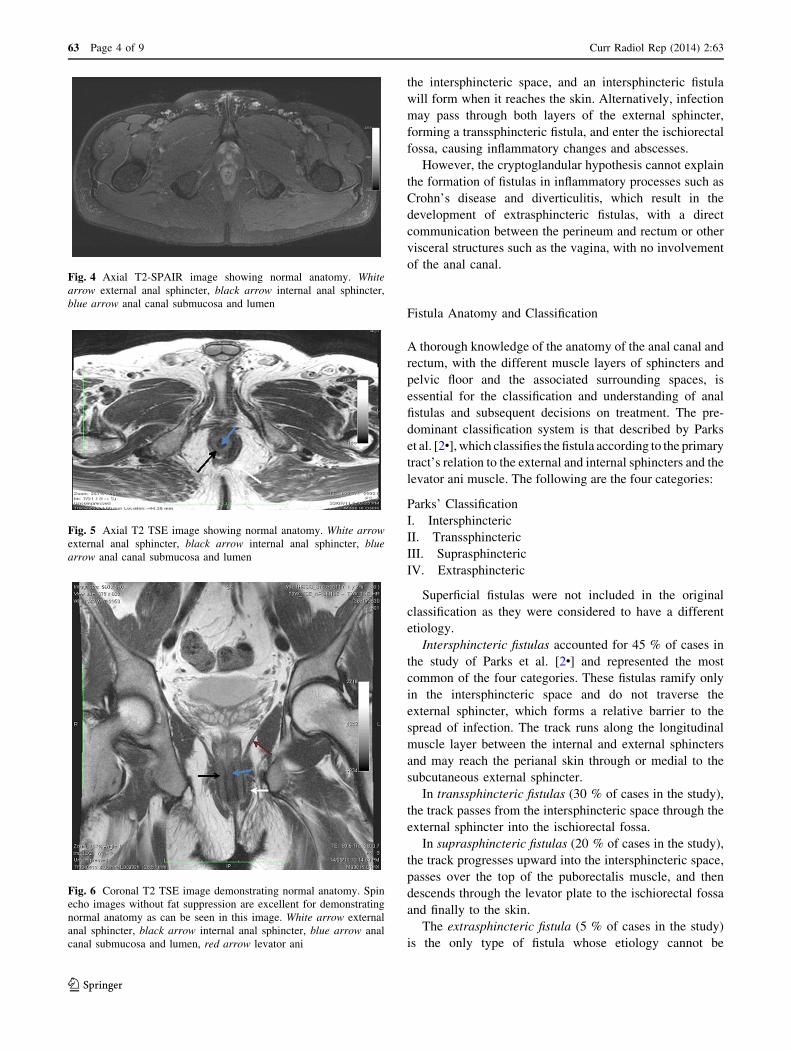
Fig. 4 Axial T2-SPAIR image showing normal anatomy. White
arrow external anal sphincter, black arrow internal anal sphincter,
blue arrow anal canal submucosa and lumen
Fig. 5 Axial T2 TSE image showing normal anatomy. White arrow
external anal sphincter, black arrow internal anal sphincter, blue
arrow anal canal submucosa and lumen
Fig. 6 Coronal T2 TSE image demonstrating normal anatomy. Spin
echo images without fat suppression are excellent for demonstrating
normal anatomy as can be seen in this image. White arrow external
anal sphincter, black arrow internal anal sphincter, blue arrow anal
canal submucosa and lumen, red arrow levator ani
63 Page 4 of 9 Curr Radiol Rep (2014) 2:63
123

explained by the cryptoglandular hypothesis. In extra-
sphincteric fistulas, the track passes from the perineal skin
through the ischiorectal fossa and levator muscles and then
into the rectum. Thus, this fistula lies completely outside
the external sphincter complex. No infection is found in the
intersphincteric space, and the anal canal is not involved.
When diagnosing this type of fistula, it is important to
exclude primary rectal or pelvic diseases, such as Crohn
disease, diverticular disease, or carcinoma.
Another widely used classification system is the St.
James University Hospital Classification [5••] which clas-
sifies the perianal fistulas as follows.
• Grade 1: simple linear intersphincteric fistula
• Grade 2: intersphincteric with abscess or secondary
track
• Grade 3: transsphincteric
• Grade 4: transsphincteric with abscess or secondary
track in ischiorectal or ischioanal fossa
• Grade 5: supralevator and translevator.
Apart from the primary tract, a fistula can also have
secondary tracts or extensions, both vertically and hori-
zontally. A special type of secondary tract is the horseshoe
tract, which extends horizontally in the intersphincteric,
ischiorectal, or supralevator space, in most cases dorsal to
the anal canal.
The level where the primary tract transverses the
external sphincter or levator muscle is another important
aspect in classifying fistulas into high and low types, as it
determines how much of the sphincter mechanism is
encompassed by the fistula: if this level is above the level
of the dentate line, then the fistula is considered high. The
internal opening of an anal fistula is usually situated at the
level of the dentate line. When the external opening(s) is
situated in the posterior half of the perianal area, the
internal opening is usually located in the posterior midline,
but when the external opening is anterior, the internal
opening is usually situated radially in the same direction
(Goodsall’s rule) (Figs. 7, 8, 9).
Role of Magnetic Resonance Imaging
Magnetic resonance imaging (MRI) is a test that uses a
magnetic field and pulses of radio-wave energy to image
body tissues. MRI machines make use of the fact that body
tissue contains lots of water, and hence protons (1H nuclei),
which get aligned in a large magnetic field. It uses mag-
netic fields of high strengths and radiofrequency waves to
obtain data which are then analyzed by complex algo-
rithms, and finally images are produced which can be used
to make a diagnosis. The main advantages of MRI are its
superior soft-tissue contrast resolution even without use of
intravenous contrast and its multiplanar imaging capabili-
ties. The value of MRI in assessing anal fistula was first
demonstrated for perianal Crohn’s disease [6]. The first
studies on cryptoglandular fistula were performed with
Fig. 7 Diagrammatic representation demonstrating the various types
of perianal fistulas as described in the Parks’ classification. A Inter-
sphincteric. B Transsphincteric. C Suprasphincteric. D Extrasphinc-
teric. The external sphincter is the keystone of the Parks classification
[4••]
Fig. 8 Figure demonstrating the Goodsall rule—when the external
opening(s) is situated in the posterior half of the perianal area, the
internal opening is usually located in the posterior midline, but when
the external opening is anterior, the internal opening is usually
situated radially in the same direction. An exception to the rule is
anterior fistulas lying more than 3 cm from the anus, which may have
a curved track (similar to posterior fistulas) that opens into the
posterior midline of the anal canal
Curr Radiol Rep (2014) 2:63 Page 5 of 9 63
123

body coil MRI [7, 8]. Later comparisons have in some
cases demonstrated improved results with an endoanal coil
[9, 10]. Many studies have demonstrated superiority of
MRI in the management of perianal fistulas as compared to
other imaging modalities (Tables 1, 2, 3; Figs. 10, 11, 12,
13, 14, 15, 16, 17, 18).
The advantages of MRI are
(1) No radiation exposure.
(2) Excellent soft-tissue contrast resolution.
(3) Can easily provide all the required information.
(4) Both functional and anatomical assessment can be
carried out at once if required.
(5) Better patient compliance as compared to conven-
tional fistulography and endoanal ultrasound.
Fig. 9 Anal clock Axial T2-weighted MR image of the male
perineum shows which is the surgeon’s view of the perianal region
when the patient is in the lithotomy position. This schema exactly
corresponds to the orientation of axial MR images of the perianal
region and can be used to correctly locate anal fistulas with respect to
the anal canal [5••]
Table 1 MR imaging features of perianal fistulas and abscesses
Condition Pulse sequence Signal intensity appearance
(fistula/edema)
Fistula/
edema
T1-weighted Low/low
T2-weighted High/high
STIR High/high
T1-weighted contrast-
enhanced
Enhancing/low
Abscess T1-weighted Low
T2-weighted High
STIR High
T1-weighted contrast-
enhanced
Low, with peripheral
enhancement
Table 2 Comparison of various modalities
Conventional
fistulography
Endoanal USG MRI
Use of
radiation
? - -
Ability to
demonstrate
secondary
tracts
± ± ?
Ease of
performing
- - ?
Cost Cheaper Cheaper Costly
investigation,
but cost-
effective in
terms of
information
provided
Sensitivity 40–60 % 70–90 %
Lower for deep
seated
abscesses and
complex
branching
fistulae due to
limited field of
view
90–100 %
Table 3 Optimal sequence for evaluation of perianal fistulas as per
our study
Sequence Slice thickness/slice gap (mm/mm)
Survey/localizer sequence –
T1TSE axial 4/0.4
T2 TSE axial 4/0.4
T2 SPAIR-axial 4/0.4
Sagittal 3/0.3
Coronal 3/0.3
Fig. 10 Axial T2 SPAIR image demonstrating transsphincteric
fistula with the fistulous tract seen traversing through both the
internal and external sphincters to open at 6 o’clock position
63 Page 6 of 9 Curr Radiol Rep (2014) 2:63
123

Fig. 11 Sagittal T2 SPAIR image showing a branching fistulous tract
Fig. 12 Axial T2 SPAIR image showing multiple collections in
bilateral ischiorectal fossae appearing hyperintense
Fig. 13 Axial T2 SPAIR image showing a collection in right gluteal
region tracking along the gluteus maximus muscle. It is impossible
with conventional imaging to identify findings such as these
Fig. 14 Axial T2 SPAIR image showing numerous inflammatory
collections—located in both the ischiorectal fossae and in inter-
sphincteric plane as well. The external anal sphincter also appears
buly with T2 hyperintense signal s/o inflammatory changes (edema)
Fig. 15 Axial T2 SPAIR image showing small collections inter-
sphincteric plane and extrasphincteric plane posteriorly
Fig. 16 Sagittal T2 SPAIR image showing a posterior transsphinc-
teric fistula which continues as another tract till just below the levator
ani muscles
Curr Radiol Rep (2014) 2:63 Page 7 of 9 63
123

The disadvantages are
(1) Takes a longer time
(2) Costly
(3) Lesser availability
(4) Some patients may experience claustrophobia and
may not be able to co-operate in the study.
Conclusions
1. Accuracy of magnetic resonance (MR) imaging in
imaging perianal fistulas:
MRI is highly accurate in identifying the fistulous
tract, its course, branches, and associated inflammatory
collections if any. In a study performed at our centre, it
accurately identified the fistulous tract in all the cases
which is better than the previously reported sensitivity
of 85 % by Maier et al. [11], 91 % by Buchanan et al.
[12•], and 94 % by Lunniss et al. [7].
It can accurately demonstrate the relationship of fis-
tulous tract with the external and internal anal
sphincter and can, thus, accurately classify the type of
fistula.
2. Additional clinical value of preoperative MR imaging:
MRI noninvasively offers important information that
can reduce surgical complications and postoperative
recurrence of the disease. The exact disease burden,
deep seated and occult inflammation, tract ramifica-
tions can only be accurately demonstrated by MRI. No
other imaging modality can as accurately demonstrate
these important pathologies as an MRI examination. It
must be noted that failure to identify these is the most
important cause of persistence of disease and recur-
rence [13]. The importance of associated findings, their
identification by MRI and implications for surgical
management, was also highlighted in articles by
Spencer et al. [14] and Beets-Tan et al. [15].
As mentioned previously, MRI also accurately clas-
sifies the type of perianal fistula by assessing its rela-
tionship with the internal and external anal sphincters.
This information is particularly valuable in deciding
the surgical management and estimating the risk of
postoperative incontinence.
Operating a perianal fistula without an MRI would be
doing a ‘blind’ and partial management of the disease.
This clearly highlights that the important role of MRI
has in the management of perianal inflammatory dis-
ease.
In our experience, T2-weighted fat-saturated sequen-
ces (i.e., T2 SPAIR) are excellent in the demonstration
of perianal fistulas and associated pathology. Thinner
sections with minimum inter-slice gap must be used
for better spatial resolution. Spin echo sequences, i.e.,
T1 & T2 TSE are useful for demonstrating normal
anatomy.
The study protocol for optimal imaging on a 3T MRI
scanner can be fairly short and time saving without any
compromise of the usefulness of the study. In our experi-
ence, the following protocol is optimum (Table 3).
The time required for the scan with these sequences is
approximately 14–15 min only. The protocol is well suited
both for demonstrating the pathology and for reducing scan
time, which in turn improves patient co-operation as well
improves patient throughput in a busy department.
Fig. 17 Coronal T2 SPAIR image showing a transsphincteric fistula
coursing just lateral to the external anal sphincter
Fig. 18 Sagittal T2 SPAIR image showing an extrasphincteric fistula
63 Page 8 of 9 Curr Radiol Rep (2014) 2:63
123

To summarize, MRI exquisitely depicts the perianal
anatomy and shows the fistulous tracks and their associated
ramifications and abscesses. It thus provides an excellent
preoperative understanding of the disease, enabling selec-
tion of the most appropriate surgical treatment and, there-
fore, minimizing all chances of recurrence. It should be
considered the investigation of choice for evaluation of
perianal fistulas.
Compliance with Ethics Guidelines
Conflict of Interest Anagha R. Joshi is a section editor for Current
Radiology Reports. Sukhvinder G. Siledar declares no potential
conflicts of interest.
Human and Animal Rights and Informed Consent This article
does not contain any studies with human or animal subjects
performed by any of the authors.
References
References of particular interest have been highlighted as:• Of importance•• Of major importance
1. Goodsall DH, Ernest Miles W. Classic articles in colonic and
rectal surgery. Diseases of the anus and rectum. Dis Colon
Rectum. 1982;25(3):262–78.
2. • Parks AG, Gordon PH, Hardcastle JD. A classification of fis-
tula-in-ano. Br J Surg. 1976;63(1):1–12. Article describing the
widely used Parks classification of Perianal fistulas.
3. Kuijpers JH, Schulpen T, Buyck B. The Seton method for the
treatment of perianal fistula located outside of the sphincter. Ned
Tijdschr Geneeskd. 1985;129(20):945–7.
4. •• de Miguel Criado J, del Salto LG, Rivas PF, del Hoyo LF,
Velasco LG, de las Vacas MI, et al. MR imaging evaluation of
perianal fistulas: spectrum of imaging features. Radiographics.
2012;32(1):175–94. Excellent article describing pathology, MR
imaging protocol & role of MRI in management of perianal
fistulas.
5. •• Morris J, Spencer JA, Ambrose NS. MR imaging classification
of perianal fistulas and its implications for patient management.
Radiographics. 2000;20(3):623–35; discussion 35–7. Article
evaluating the importance of preoperative MRI imaging of per-
ianal fistulas.
6. Koelbel G, Schmiedl U, Majer MC, Weber P, Jenss H, Kueper K,
et al. Diagnosis of fistulae and sinus tracts in patients with Crohn
disease: value of MR imaging. AJR. 1989;152(5):999–1003.
7. Lunniss PJ, Barker PG, Sultan AH, Armstrong P, Reznek RH,
Bartram CI, et al. Magnetic resonance imaging of fistula-in-ano.
Dis Colon Rectum. 1994;37(7):708–18.
8. Myhr GE, Myrvold HE, Nilsen G, Thoresen JE, Rinck PA. Per-
ianal fistulas: use of MR imaging for diagnosis. Radiology.
1994;191(2):545–9.
9. Hussain SM, Stoker J, Schouten WR, Hop WC, Lameris JS.
Fistula in ano: endoanal sonography versus endoanal MR imag-
ing in classification. Radiology. 1996;200(2):475–81.
10. Zbar AP, de Souza NM, Puni R, Kmiot WA. Comparison of
endoanal magnetic resonance imaging with surgical findings in
perirectal sepsis. Br J Surg. 1998;85(1):111–4.
11. Maier AG, Funovics MA, Kreuzer SH, Herbst F, Wunderlich M,
Teleky BK, et al. Evaluation of perianal sepsis: comparison of
anal endosonography and magnetic resonance imaging. JMRI.
2001;14(3):254–60.
12. • Buchanan GN, Halligan S, Bartram CI, Williams AB, Tarroni
D, Cohen CR. Clinical examination, endosonography, and MR
imaging in preoperative assessment of fistula in ano: comparison
with outcome-based reference standard. Radiology. 2004;233(3):
674–81. A study of 104 patients comparing Endoanal USG with
MRI.
13. Lilius HG. Fistula-in-ano, an investigation of human foetal anal
ducts and intramuscular glands and a clinical study of 150
patients. Acta Chir Scand Suppl. 1968;383:7–88.
14. Spencer JA, Chapple K, Wilson D, Ward J, Windsor AC,
Ambrose NS. Outcome after surgery for perianal fistula: predic-
tive value of MR imaging. AJR. 1998;171(2):403–6.
15. Beets-Tan RG, Beets GL, van der Hoop AG, Kessels AG, Vlie-
gen RF, Baeten CG, et al. Preoperative MR imaging of anal
fistulas: does it really help the surgeon? Radiology. 2001;
218(1):75–84.
Curr Radiol Rep (2014) 2:63 Page 9 of 9 63
123
Recommended


![Received: Magnetic Resonance Imaging (MRI ......rectal diseases. Perianal fistulas occur in approximately 10 out of 10,000 people [4]. They usually occur in adult men with a maxi-mum](https://img.pdfslide.us/doc/110x75/5f5924e988689c5e020ce9c4/received-magnetic-resonance-imaging-mri-rectal-diseases-perianal-fistulas.jpg)