Role of Actin-mediated Motility of Peripheral
Astrocytic Processes in Synaptic Function
Dmitry Molotkov
Neuroscience Center and
Division of Physiology Department of Biosciences
Faculty of Biological and Environmental Sciences and
Finnish Graduate School in Neuroscience
Academic Dissertation
To be presented for public examination with the permission of the Faculty of Biological and Environmental Sciences of the University of Helsinki in Telkänpönttö
(lecture hall 2402), Biocenter 3, Viikinkaari 1, Helsinki, on Thursday the 12th June 2014, at 12 o’clock noon.
Helsinki 2014
Supervised by:
Docent Leonard Khirug, PhD
Neuroscience Center
University of Helsinki, Finland
and
Professor Eero Castren, MD, PhD
Neuroscience Center
University of Helsinki, Finland
Reviewed by:
Docent Sarah Coleman, PhD
Department of Biological and Environmental Sciences
University of Helsinki, Finland
and
Nathalie Rouach, PhD
Center for Interdisciplinary Research in Biology
College de France, Paris, France
Opponent:
Professor Dmitri Rusakov, PhD
Institute of Neurology
University College London, United Kingdom
Custos:
Professor Juha Voipio, PhD
Department of Biosciences
University of Helsinki, Finland
ISBN 978-952-10-9963-2 (paperback) ISBN 978-952-10-9964-9 (PDF)
Unigrafia Oy, Helsinki 2014
CONTENTS
LIST OF ORIGINAL PUBLICATIONS
ABSTRACT
ABBREVIATIONS
INTRODUCTION ................................................................................................... 1
REVIEW OF THE LITERATURE ....................................................................... 1
1. Different glial cell types and their role in the mammalian brain .......................... 1
1.1 Radial glia and glial progenitor cells ............................................................ 2
1.2 Oligodendrocytes and Schwann cells ........................................................... 3
1.3 Microglial cells ............................................................................................. 3
1.4 Astroglial cells (a.k.a. astrocytes) ................................................................. 4
2. Astroglia functions in the brain ............................................................................ 5
2.1 Metabolic support of neurons by astrocytes ................................................. 5
2.2 Blood-brain barrier (BBB) formation and regulation by astrocytes ............. 7
2.3 Astrocytic function in maintenance and regulation
of extracellular matrix (ECM) .............................................................................. 9
2.4 Astrocytic coupling through gap junctions ................................................. 12
2.5 Ion channels, transporter and receptors expressed in
astrocytic plasma membrane .............................................................................. 14
2.5.1. Glutamate transporters ........................................................................... 14
2.5.2. Connexin and pannexin hemichannels .................................................. 16
2.5.3. Glutamate receptors ............................................................................... 17
2.5.4. GABA receptors and transporters.......................................................... 17
2.5.5. Channels and transporters involved in D-serine release
from astrocytes ................................................................................................. 18
2.5.6. Water and potassium channels .............................................................. 18
2.5.7. Purinoreceptors ...................................................................................... 18
2.5.8. Ephrin mediated reception ..................................................................... 19
2.6 Chemical excitability and calcium signaling in astrocytes ......................... 19
2.7 Modulation of synaptic transmission by astrocytes:
the concept of multipartite synapse .................................................................... 21
2.7.1. Presynaptic mechanisms of astrocyte action ......................................... 23
2.7.1.1. Presynaptic action of purines released from astrocytes .................... 23
2.7.1.2. Modulation of synaptic events by presynaptic glutamate ................ 24
2.7.2. Postsynaptic mechanisms of astrocyte action ........................................ 25
2.7.2.1. D-serine release from astrocytes ...................................................... 25
2.7.2.2. Postsynaptic action of Glycine ......................................................... 26
2.7.2.3. Postsynaptic action of astrocyte derived ATP .................................. 26
2.7.2.4. Neurotransmitter and ion clearance by astrocytic
transporters and regulation of spillover …….…………………….……..... 27
3. Peripheral astrocytic process as a functional microdomain ................................ 27
3.1 Ultrastructure of PAPs ................................................................................ 28
3.2 Receptors and transporters in PAPs ............................................................ 29
3.3 Morphological changes of PAPs: two different mechanisms proposed ..... 30
3.3.1. Aquaporin mediated morphological changes ........................................ 30
3.3.2. Actin-dependent morphological changes .............................................. 30
4. Methodological approaches to study neuro-glial interactions ............................ 32
4.1 Hypothalamus as a classical model of PAPs retraction .............................. 32
4.2 Transgenic mouse models to study astrocytes ............................................ 32
4.3 Acute brain slices and organotypic cultures
to study neuro-glial interactions ......................................................................... 34
4.4 In vivo methods to probe astrocytic morphological
and functional changes ....................................................................................... 35
4.4.1. Two-photon microscopy on living mouse brain .................................... 35
4.4.1.1. Probes and dyes to label astroglia in vivo for TPEM ....................... 36
4.4.1.2. In vivo microscopy on anaesthetized animals .................................. 37
4.4.1.3. In vivo microscopy on awake animals .............................................. 38
4.5 Gene delivery to astrocytes ......................................................................... 39
4.5.1. Specificity and efficiency of gene delivery to astrocytes ...................... 39
4.5.2. Gene-gun mediated gene delivery (biolistics) ....................................... 40
4.5.3. In vivo electroporation ........................................................................... 41
4.5.4. Viral gene delivery methods to get astrocyte specificity in vivo ........... 42
4.5.4.1. Adeno-associated viral vectors for astrocytes .................................. 42
4.5.4.2. Lentiviral vectors for astrocytes ....................................................... 43
4.5.4.3. Adenoviral vectors for astrocytes ..................................................... 44
AIMS ....................................................................................................................... 46
METHODS ............................................................................................................. 47
Image analysis (I, II and III) ............................................................................... 47
Primary cortical astrocyte cultures (I) ................................................................ 48
Photolysis of caged calcium and live cell imaging (I)........................................ 48
Postnatal in vivo electroporation (II) .................................................................. 48
Preparation and in vivo application of adenoviral vectors (III) .......................... 49
In vivo two-photon microscopy through the cranial window (III) ..................... 49
Intrinsic BOLD signal optical imaging (III) ....................................................... 50
Experimental animals (I, II and III) .................................................................... 50
RESULTS AND DISCUSSION ............................................................................ 51
1. The role of Profilin-1 in the activity-dependent outgrowth of astrocytic
peripheral processes (study I). ................................................................................. 51
1.1 Actin turnover as the major mediator for Ca2+
-dependent
PAPs motility in vitro ......................................................................................... 51
1.2 The novel tool to suppress activity-dependent PAPs motility .................... 53
2. Gene delivery to a postnatal rodent brain (studies II and III). ............................ 53
2.1 Stereotaxic plasmid microinjection and electroporation
on neonatal rat brain (study II) ........................................................................... 53
2.2 Astrocyte-specific gene delivery using adenoviral vectors (study III). ...... 55
3. Motility of cortical astrocytes and neuronal function (study III). ...................... 57
3.1 Astroglia spontaneous morphological changes are Profilin-1 dependent. .. 57
3.2 Structure-functional interplay: the effect of suppressed astrocytic
motility on neuronal processing. ........................................................................ 58
CONCLUSIONS .................................................................................................... 61
ACKNOWLEDGEMENTS .................................................................................. 62
LIST OF REFERENCES ..................................................................................... 63
LIST OF ORIGINAL PUBLICATIONS
I. Molotkov D*., Zobova S*., Arcas JM., Khiroug L. (2013) Calcium-induced
outgrowth of astrocytic peripheral processes requires actin binding by Profilin-1.
Cell Calcium 53: 338-348.
The candidate substantially contributed to the experimental design, designed and
performed molecular biology manipulations, performed microscopic experiments
together with SZ, designed analysis approaches, analyzed the data together with SZ
and wrote the manuscript together with SZ and LK.
II. Molotkov D., Yukin A., Afzalov R., Khiroug L. (2010) Gene delivery to
postnatal rat brain by non-ventricular plasmid injection and electroporation. J of
Vis Exp 43.
The candidate designed experiments together with AY, performed experiments and
analyzed the data, prepared the video together with RA, LK and AY, wrote the
manuscript together with LK.
III. Molotkov D., Kislin M., Zobova S., Toptunov D., Castren E., Khiroug L.
(2014) Suppression of astrocytic morphological changes does not affect BOLD
signal during visual processing in anesthetized mice. Manuscript.
The candidate substantially contributed to the experimental design, planed and
performed molecular biology and viral work, performed BOLD signal acquisition
experiments, participated in in vivo microscopy experiments, designed data
analysis approaches together with DT and MK, analyzed the data and wrote the
manuscript.
*Equal contribution
ABSTRACT
Among other glial cell types such as microglia, oligodendrocytes and radial glia,
astrocytes are known to be involved in brain function; metabolically supporting
neurons, regulating blood flow dynamics, participating in the development of
pathological states, sensing and modulating synaptic activity. At the same time the
complex astrocytic morphology, with a number of highly ramified peripheral
processes located near the synaptic terminals, suggests them as a possible source
for morpho-functional plasticity in the brain. This thesis summarizes the work on
the in vitro development and further in vivo implementation, using a gene delivery
system, of a tool for suppressing activity-dependent astrocytic motility. Calcium-
induced astrocyte process outgrowth and its dependence on Profilin-1, novel in
vivo gene delivery approaches, a demonstration of astrocytic motility in vivo and
the independence of visual processing from astrocytic motility rates are the main
findings of the project. The results described in this work increase our
understanding of the interactions occurring between astrocytes and neurons as well
as the consequences for brain function.
ABBREVIATIONS
AAV –adeno-associated virus
AD – Alzheimer disease
AMPA – α-Amino-3-hydroxy-5-methyl-4-isoxazolepropionic acid
Ang1 – angiopoietin 1
AQP – aquaporin (channel)
ATP – adenosine triphosphate
BBB – blood-brain barrier
BOLD – blood oxygenation level-dependent
[Ca2+
]i – intracellular calcium concentration
CAG promoter – chicken beta actin promoter with cytomegalovirus enhancer
cAMP – cyclic adenosine monophosphate
CAR – coxsackie and adenoviral receptor
CMV promoter – cytomegalovirus early genes promoter
CNS – central nervous system
CSPG – chondroitin sulfate proteoglycan
DHK – dihydrokainic acid
EAAT – excitatory amino acid transporter
ECM – extracellular matrix
EGFP – enhanced green fluorescent protein
F-actin – filamentous actin
FGF – fibroblasts growth factor
GABA – gamma-aminobutyric acid
GAT – gamma-aminobutyric acid transporter
GDNF – glial cell-derived neurotropic factor
GFAP – glial fibrillary acidic protein
GLAST – glutamate-aspartate transporter
GLT – glutamate transporter
GLUT – glucose transporter
GlyT – glycine transporter
GPCR – G-protein coupled receptor
HIV – human immunodeficiency virus
InsP3 – inositol-3-phosphate
IP3 – inositol-3-phosphate
Kir – potassium inward rectifying (channel)
LCMV – lymphocytic choriomeningitis virus
LDH – lactate dehydrogenase
LTD – long-term depression
LTP – long-term potentiation
mGluR – metabotropic glutamate receptor
miRNA – micro ribonucleic acid (molecule)
MMP – matrix metalloproteinase
NKCC – neuronal potassium-chloride cotransporter
NMDA – N-methyl-D-aspartic acid
NMDAR – N-methyl-D-aspartic acid receptor
OAPs – orthogonal arrays of particles
P2X – purinoreceptor
PAPs – peripheral astrocytic processes
PKC – protein kinase C
PNS – peripheral nervous system
shRNA – short hairpin ribonucleic acid (molecule)
SON – supraoptic nucleus
SR101 – sulforhodamine 101
SVZ – subventricular zone
TBOA – DL-threo-beta-benzyloxyaspartate
TCA – tricarboxylic acid
TGF – tumor growth factor
TPEM – two-photon excitation microscopy
TRP – transient receptor potential (channel)
VSVG – vesicular stomatitis virus glycoprotein
1
INTRODUCTION
The general assumption that brain functionality relies exclusively on wired neurons
is far from the truth. Among the neurons there are several other cell types that play
essential structural and functional roles in the brain. Indeed, astroglia, microglia,
oligodendrocytes and radial glia cells are involved in brain development,
maintenance, re-wiring, synaptic turnover and modulation of synaptic properties
(Nicholls et al., 2001, pp. 133-46; Volterra et al., 2002). Modulation of basal
synaptic transmission (Navarrete and Araque, 2011), facilitation of long-term
synaptic potentiation (Henneberger et al., 2010), regulation of neuronal network
activity (Pannasch and Rouach, 2013) and metabolic support of neurons
(Magistretti et al., 2006) are the main points of astrocyte and neuron interactions.
On the other hand astrocytes are known to display a set of unique morphological
features: occupation of non-overlapping spatial domains (Bushong et al., 2002;
Ogata and Kosaka, 2002), forming a complex 3D network of peripheral processes
(Witcher et al., 2007; Shigetomi et al., 2013) and their strategic positioning near
the synaptic terminals (reviewed by Reichenbach et al., 2010) that allow us to
propose that astrocytes are the link between structural and functional changes
occurring in the brain. In this study we posed the questions are astrocytes in vivo
subjected to continuous morphological changes and will the suppression of these
changes somehow affect neuronal activity in a restricted brain region?
REVIEW OF THE LITERATURE
1. Different glial cell types and their role in the mammalian
brain
Glial cells were first described in 1840s by Rudolf Virchow (Virchow, 1846;
Virchow, 1858), who coined a general name for them - “neuroglia”, i.e. “nerve
glue” in 1856. Half a century later, Camillo Golgi (Golgi, 1903) in 1883 and
Santiago Ramon y Cajal (Ramon y Cajal, 1995) in 1890s predicated that glial cells
are more than “brain glue” and not merely metabolic suppliers for neurons. For
many decades since, the doctrine that promotes neurons as central players of
nervous system functionality was prevalent. Starting in the 1990’s, it became more
and more evident that some aspects of nervous system physiology as well as
pathophysiology could not be explained in light of exclusive neuronal doctrine, and
the idea emerged that some glial cells might be also involved in information
2
processing within the nervous system (Kettenmann and Ransom, 1995). Glia is a
general term for a diverse population of different non-neuronal cell types in a brain,
including microglial cells that act as immune cells in central nervous system
(CNS), oligodendrocytes and Schwann cells – myelinating cells, radial glia playing
important role in brain development, NG2 and Bergman glial cells (Ransom,
1991), and astrocytes that metabolically support neurons and are involved in
synaptic transmission and development (Ransom, 1991; Kettenmann and Ransom,
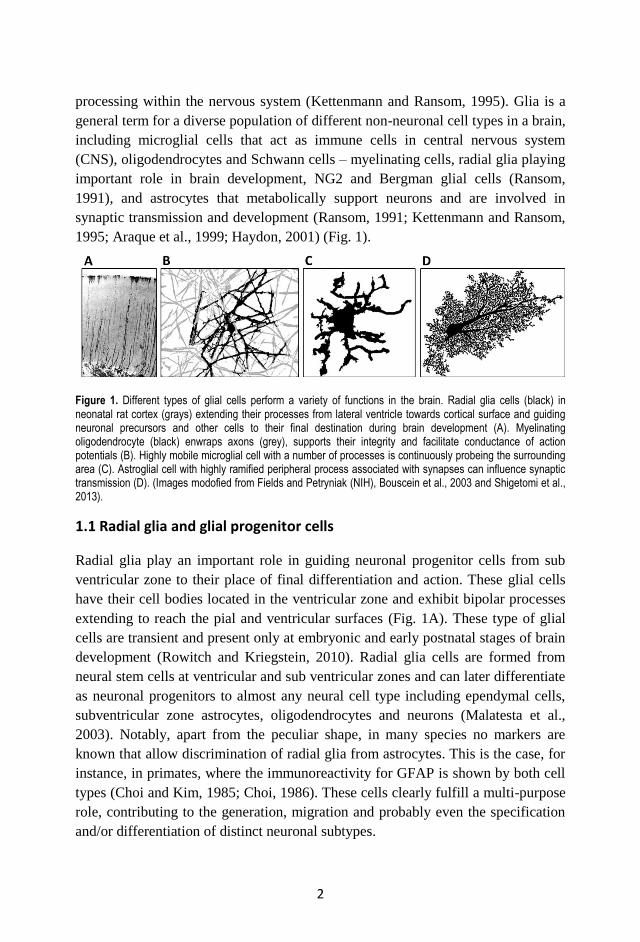
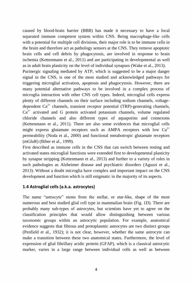
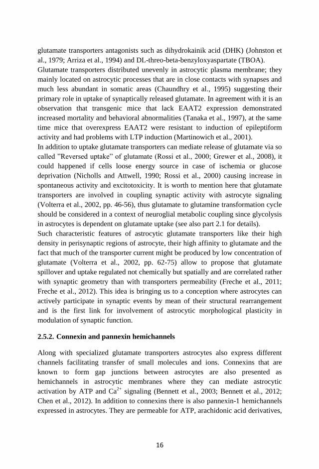
1995; Araque et al., 1999; Haydon, 2001) (Fig. 1).
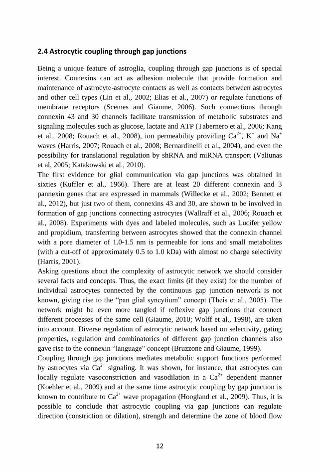
Figure 1. Different types of glial cells perform a variety of functions in the brain. Radial glia cells (black) in neonatal rat cortex (grays) extending their processes from lateral ventricle towards cortical surface and guiding neuronal precursors and other cells to their final destination during brain development (A). Myelinating oligodendrocyte (black) enwraps axons (grey), supports their integrity and facilitate conductance of action potentials (B). Highly mobile microglial cell with a number of processes is continuously probeing the surrounding area (C). Astroglial cell with highly ramified peripheral process associated with synapses can influence synaptic transmission (D). (Images modofied from Fields and Petryniak (NIH), Bouscein et al., 2003 and Shigetomi et al., 2013).
1.1 Radial glia and glial progenitor cells
Radial glia play an important role in guiding neuronal progenitor cells from sub
ventricular zone to their place of final differentiation and action. These glial cells
have their cell bodies located in the ventricular zone and exhibit bipolar processes
extending to reach the pial and ventricular surfaces (Fig. 1A). These type of glial
cells are transient and present only at embryonic and early postnatal stages of brain
development (Rowitch and Kriegstein, 2010). Radial glia cells are formed from
neural stem cells at ventricular and sub ventricular zones and can later differentiate
as neuronal progenitors to almost any neural cell type including ependymal cells,
subventricular zone astrocytes, oligodendrocytes and neurons (Malatesta et al.,
2003). Notably, apart from the peculiar shape, in many species no markers are
known that allow discrimination of radial glia from astrocytes. This is the case, for
instance, in primates, where the immunoreactivity for GFAP is shown by both cell
types (Choi and Kim, 1985; Choi, 1986). These cells clearly fulfill a multi-purpose
role, contributing to the generation, migration and probably even the specification
and/or differentiation of distinct neuronal subtypes.
3
1.2 Oligodendrocytes and Schwann cells
Both Oligodendrocytes and Schwann cells are very specialized glial cells types that
have reached their final differentiation step. They are formed at the latest stages of
the nervous system development. Oligodendrocytes are presented in a CNS where
they myelinate several axons simultaneously (Fig. 1B) whereas Schwann cells
were found in peripheral nervous system (PNS) where they can be associated with
single axon or neuro-muscular junctions. One of the most important functions of
oligodendrocytes and Schwann cells is to form a myelin sheath around axons
(Nave, 2010). This kind of axon insulation enables rapid action potential
propagation within thin and long mammalian axons that relies as a key concept of
neurophysiology that providing possibility for complex and at the same time
compact nervous system. In addition to their insulation role these cells can also
support axonal integrity and provide functions that may be independent of myelin
itself (Griffiths et al., 1998). Additionally myelinating glia are involved in several
demyelinating disorders, like multiple sclerosis, leukodystrophies and
demyelinating neuropathies (Nave, 2010).
1.3 Microglial cells
Microglia represents approximately 10% of total amount of cells in the mammalian
CNS (reviewed by Kettenmann et al., 2011). They can appear as tree-like cells with
many ramified processes (Fig. 1C) or as amoeboid cells with reduced morphology
depending on developmental stage of the CNS and on their activation and/or
migration status (Streit et al., 1988; Kettenmann et al., 2011). Common feature for
all microglial cells is their high motility rates even in a resting state when changes
occur within peripheral processes (Nimmerjahn et al., 2005). This continuous
motility proposed to be linked with probing of perineuronal space (Kettenmann et
al., 2013) or in case of activation could involve morphological remodeling of the
whole cell. Both morphological changes and migration of microglial cells is
thought to be caused by actin cytoskeleton remodeling regulated mainly via protein
kinase C (PKC) and inositol-3-phosphate (InsP3) signaling pathways (Kettenmann
et al., 2011). Migration of microglia in addition to their morphological changes in
the adult CNS can be switched on by a variety of pathophysiological actions. For
instance in laser induced brain micro lesion or thrombotic micro stroke microglial
cells are known to extend their processes rapidly towards a lesion site or
thrombotized blood vessels (Davalos et al., 2005).
Functions of microglial cells are very diverse and their portfolio has been
extensively added to during last few years. The immune status quo of the CNS
4
caused by blood-brain barrier (BBB) has made it necessary to have a local
separated immune competent system within CNS. Being macrophage-like cells
with a potential for multiple cell divisions, their major role is to be immune cells in
the brain and therefore act as pathology sensors at the CNS. They remove apoptotic
brain cells and cell debris by phagocytosis, are involved in response to brain
ischemia (Kettenmann et al., 2011) and are participating in developmental as well
as in adult brain plasticity on the level of individual synapses (Wake et al., 2013).
Purinergic signaling mediated by ATP, which is suggested to be a major danger
signal in the CNS, is one of the most studied and acknowledged pathways for
triggering microglial activation, apoptosis and phagocytosis. However, there are
many potential alternative pathways to be involved in a complex process of
microglia interaction with other CNS cell types. Indeed, microglial cells express
plenty of different channels on their surface including sodium channels, voltage-
dependent Ca2+
channels, transient receptor potential (TRP)-generating channels,
Ca2+
activated and G protein activated potassium channels, volume regulated
chloride channels and also different types of aquaporins and connexons
(Kettenmann et al., 2011). There are also some evidences that microglial cells
might express glutamate receptors such as AMPA receptors with low Ca2+
permeability (Noda et al., 2000) and functional metabotropic glutamate receptors
(mGluR) (Biber et al., 1999).
First described as immune cells in the CNS that can switch between resting and
activated states microglial functions were extended first to developmental plasticity
by synapse stripping (Kettenmann et al., 2013) and further to a variety of roles in
such pathologies as Alzheimer disease and psychiatric disorders (Aguzzi et al.,
2013). Without a doubt microglia have complex and important impact on the CNS
development and function which is still enigmatic in the majority of its aspects.
1.4 Astroglial cells (a.k.a. astrocytes)
The name “astrocyte” stems from the stellar, or star-like, shape of the most
numerous and best studied glial cell type in mammalian brain (Fig. 1D). There are
probably many sub-types of astrocytes, but scientists have yet to agree on the
classification principles that would allow distinguishing between various
taxonomic groups within an astrocytic population. For example, anatomical
evidence suggests that fibrous and protoplasmic astrocytes are two distinct groups
(Penfield et al., 1932); it is not clear, however, whether the same astrocyte can
make a transition between these two anatomical states. Furthermore, the level of
expression of glial fibrillary acidic protein (GFAP), which is a classical astrocytic
marker, varies in a large range between individual cells as well as between
5
different patho-physiological conditions (Chiu and Goldman, 1985; Baba et al.,
1997; Gomes et al., 1999; Messing and Brenner, 2003; Middeldorp and Hol, 2011).
Finally, brain region-dependent specialization of astrocytes is likely to occur, and
in some regions astrocytes even have been assigned a separate name (e.g.,
Bergmann glia in cerebellum (Bergmann, 1857)).
Functionally, astrocytes may also be divided in a number of subtypes (although
again, a consensus in the field is still lacking). Undoubtedly, though, functions of
astrocytes are numerous and vary widely from vital metabolic support of neurons
and formation of blood-brain barrier to enabling synaptogenesis and synaptic
plasticity all the way to elimination of synapses. Some of these classical and more
recently discovered functions will be discussed in detail in following chapters. It
seems important to note that, although astrocytes have been studied for decades
(anatomically since 1850s and functionally since 1990s), their role in the CNS is
still somewhat enigmatic as more questions arise from each new study.
2. Astroglia functions in the brain
2.1 Metabolic support of neurons by astrocytes
Neurons are supposed to be the main consumers of energetic substrates over the
mammalian body. The energy is utilized for maintenance of membrane potential in
resting condition as well as for action potential generation during neuronal network
electrical activity (reviewed by Magistretti, 2006; Allaman et al., 2011) and long
term memory formation (Suzuki et al., 2011). Regional blood flow changes, energy
and as a consequence oxygen and glucose consumption by neural tissue are related
to neuronal activity and are used as a basis for different brain functional imaging
techniques (Magistretti and Pellerin, 1999; Raichle and Mintun, 2006; Hyder,
2009; Bandettini, 2012). It is a well-established fact also that, in comparison to
other cell types, glucose is not the main energetic substrate for neurons and that
glycolysis is much more ineffective in neurons as compared to the tricarboxylic
acid (TCA) cycle that utilizes lactate for energy production (Pellerin et al., 2007;
Magistretti, 2006). On the other hand astrocyte metabolism demonstrates
remarkably more active glycolysis and provides a source of lactate for neurons.
This metabolic complementarity is not likely to be a fortuity but a result of
metabolic coupling between astrocytes and neurons (Magistretti, 2006). This
neuro-glial metabolic coupling includes active transport of glucose by astrocytes
from a blood flow, glycolysis in astrocytes and active lactate transfer from
astrocytes to neurons in an activity-dependent manner (Volterra et al., 2002;
Kasischke et al., 2004).
6
Glucose from blood flow is transported into astrocytes via specific glucose GLUT-
1 type transporters that are expressed in endfeet surrounding blood vessels
(Morgello et al., 1995; Yu and Ding, 1998). This glucose is used in classical
glycolysis that results in a anaerobic production of pyruvate and further to lactate
in astrocytes with a muscle form of lactate dehydrogenase 5 (LDH) (Bittar et al.,
1996). Increased glucose utilization in response to glutamate uptake (Pellerin and
Magistretti, 1994; Takahashi et al., 1995) as well as lactate production following
sensory stimulation of neuronal activity (Fellows et al., 1993) are key signs for
coupling between astrocytic metabolism which acts as a glutamate source and
neuronal activity. Astrocytes can actively evacuate glutamate from active synaptic
sites by specific excitatory amino acid transporters (EAAT) 1 and 2 (reviewed in
Volterra et al., 2002; McKenna, 2007). The glutamate transport is accompanied by
3 Na+ ions transfer for each glutamate molecule that cause remarkable sodium
current in astrocytes involved in glutamate scavenging. Both maintenance of
sodium homeostasis and glutamate conversion to glutamine are ATP-dependent
processes and thereby stimulate glycolysis and lactate production in astrocytes.
It seems that the role of astrocytes in activity-dependent neuro-glial metabolic
coupling might be even more extensive and includes such aspects as regulation of
vasoconstriction (Mulligan and MacVicar, 2004; Gordon et al., 2011) in response
to Ca2+
transients in soma and endfeet as well rapid vasodilation mediated by local
Ca2+
increase in astrocytic endfeet (Takano et al., 2006; Gordon et al., 2011).
Astrocytic regulation of local blood flow and oxygenation level of brain tissue is
not restricted to their influence on vessel diameter but also includes more
generalized mechanisms of pH-dependent control of breathing via ATP signaling
pathway (Gourine et al., 2010). Metabotropic glutamate receptor mediated Ca2+
transients in astrocytes caused by stimulation of neuronal networks activity (Wang
et al., 2006), might also be involved in fine tuning of astrocytic homeostasis and
metabolism.
It is also worth mentioning that astrocytes form an extensive metabolic network in
the brain. Their coupling through connexin 43 and 30 channels allows trafficking
of energetic substrates such as glucose (reviewed in Giaume et al., 1997) and ATP
(Kang et al., 2008) between neighboring astrocytes and influences excitatory
glutamatergic synaptic transmission within hippocampal neuronal networks
(Rouach et al., 2008; Pannasch et al., 2011). Astrocytic K+ homeostasis is mediated
by Na+,K
+-ATPase activity in astrocytic plasma membrane and thus is dependent
on ATP level and glucose utilization via glycolytic pathway in astrocytes. Ca2+
dependent uptake of extracellular K+ by astrocytes can modulate neural network
activity by transient local decrease of K+ ions leading to neuronal hyperpolarization
and synaptic suppression (Wang et al., 2012). Thus, not only are Ca2+
and Na+ ions
7
connected with maintenance of neuro-glial metabolic coupling but K+ changes
might also be involved in regulation of brain metabolism in an astrocyte-dependent
manner.
Astrocytes provide not just metabolic support for neuronal cells by lactate supply
for TCA cycle but are involved in a metabolic crosstalk with neuronal cells that
could even include such features as suppression of neuronal activity. They are also
actively participating in regulation of cerebral blood flow, breathing control and
can act as a metabolic network connected by gap junctions. Astro-neuronal
metabolic coupling and the role of astrocytes in brain metabolism is strongly
related to the fundamental role that astrocytes play as a component of multipartite
synapses.
2.2 Blood-brain barrier (BBB) formation and regulation by astrocytes
The modern concept of BBB was summarized by Hugh Davson in 1976 (Davson,
1976) and includes such features as barrier function per se, active transport and
facilitated transport options across the barrier, leading role for endothelial cells in
barrier formation and support, maintenance of brain homeostasis and ontogenetic
developmental changes in BBB as well as significant role for astrocytes in BBB
transport and homeostasis. To further develop this concept it is now an accepted
fact that even though BBB is a relatively stable structure it continuously changes
under different modulating factors among which astrocytes play not the least role
(Abbott, 2005).
BBB starts to form at embryonic developmental stages with the help of pericytes at
the time when astrocytes have not yet appeared (Daneman et al., 2010). At later
stages of BBB formation astrocytes participate in its establishment by direct
contacts with pericytes and endothelial cells by their endfeet. Astrocytes are also
supposed to play a role in BBB maturation and tight junctions formation by means
of secretion of such angiogenic compounds such as Ang1, TGFβ, GDNF and FGF2
(Quaegebeur et al., 2011). Particularly it was shown that factors derived from
astrocytes can induce BBB-like phenotypes of endothelial cells and formation of
tight junctions in vitro (Lee et al., 2003) suggesting an important role for astrocytes
in BBB formation in vivo as well.
Besides participation in BBB formation during ontogenesis, astrocytic components
are involved in transport across the barrier. While small lipophilic molecules (less
than 400 Da) can cross BBB by lipid-mediated diffusion (Pardridge, 2007), other
compounds need to be transported actively or by mean of special channels and
transporters. Strategic location of astrocytes between neurons and blood vessels
makes them major players in glucose transport through GLUT1 transporters, water
8
through AQP4 channels (Abbott, 2006) and also ions, glutamate and other amino
acids (Ohtsuki and Terasaki, 2007) across the barrier. For instance, spatial K+
buffering by astrocytes provided by Kir4.1 channels located on perivascular
astrocytic endfeet (Kofuji and Newman, 2004) as well as by other transporters such
as Na+,K
+-ATPase and NKCC1 (Abbott et al., 2006) were proposed to be one of
the major mechanisms for maintenance of K+ homeostasis in the brain and for
regulation of neuronal firing.
By cryo-electron microscopy studies it was shown that astrocytic endfeet form
orthogonal arrays of particles (OAPs) containing specialized sets of proteins
located at contact sites with endothelial cells (Fallier-Becker et al., 2011; Nico and
Ribatti, 2012). These polarized structural protein expressing micro domains are
involved in maintenance of water and K+ homeostasis since they express aquaporin
4 (AQP4) channels and Kir4.1 K+ channels segregated by agrin and α1-syntrophin
(Abbott et al., 2006). Using dystrophin deficient mice it was shown that actin
organization in astrocytic endfeet is crucial for AQP4 distribution and function
(Nico et al., 2003) suggesting the role astrocytic actin cytoskeleton for clustering of
OAPs and as a consequence its importance in regulating BBB properties.
There is more and more evidence that most CNS pathologies involve some aspects
of BBB disruption at least at some stages. Thus diabetes, alcohol, Ischemic
conditions, HIV-1 infection are factors for BBB leakage (Zlokovic, 2008; Eugenin
et al., 2011); inflammation processes can also cause opening of BBB (Huber et al.,
2001). Some of these pathological states involve astrocytes, thus astrocytic
activation due to Alzheimer disease (AD) pathology and amyotrophic lateral
sclerosis disease plays a role in BBB disruption in severe AD (Zlokovic, 2008).
One of the possible mechanisms of astrocytic regulation of BBB permeability is
based on ATP-mediated astrocytic and endothelium Ca2+
signaling. In this situation
astrocytes act as a network connected by gap junctions and thus can propagate Ca2+
and ATP mediated signals to neighboring cells. At the same time intracellular Ca2+
changes in endothelial cells may act as a trigger for phosphorylation of
cytoskeleton proteins and tight junctions opening (Abbott et al., 2006) increasing
BBB permeability.
Although BBB is mainly formed by endothelial cells and pericytes, astrocytes form
numerous connections with both endothelial cells and pericytes and, as recently
discovered, can regulate their functionality at least in terms of cerebral blood flow
adjustment (Attwell et al., 2010). It is also unclear to what extent astrocytes can
influence formation and integrity of tight junctions – one of most critical
component of BBB. Although in vitro studies show astrocytic roles in almost every
function of BBB (reviewed by Abbot et al., 2006), their role in BBB formation,
maintenance and pathophysiology in vivo seems to be under studied. Focusing on
9
distal BBB components, like astrocytic endfeet, in the natural environment of the
intact brain may shed light on novel therapeutic strategies based on selective BBB
permeability, and open the way for new diagnostic approaches for CNS pathologies
implicating unidirectional trans-BBB transport of diagnostic markers from CNS to
the blood flow.
2.3 Astrocytic function in maintenance and regulation of extracellular
matrix (ECM)
Regarding ECM we intend to take into account not just scaffold and cell-adhesion
molecules but also soluble macromolecular factors derived from astrocytes,
neurons and other cell types in the CNS. Roles of ECM can be divided in two main
categories: mechanical that includes its implications in general and local diffusion
properties of brain tissue (Sykova, 2004; Frischknecht et al., 2009; Zamecnik et al.,
2012) and as a consequence influencing synaptic as well as extrasynaptic
transmission and cell migration in physical aspects; and biochemically active
components of ECM affecting CNS properties via cell adhesion molecules,
secretion of soluble proteins in extracellular space and protein-protein interactions
in ECM (Dityatev and Schachner, 2003; Kochlamazashvili et al., 2010; Dityatev
and Rusakov, 2011). Molecular scaffolds, cell adhesion, and soluble proteins that
are synthetized and secreted by astrocytes play complex and important role in
physiological (Ehlers, 2005; Faissner et al. 2010; Wiese et al., 2012; Hawkins et
al., 2013) and pathological states of CNS (Pantazopoulos et al., 2010; Giordano et
al., 2011; Beretta, 2012).
Early in vitro co-culturing studies discovered the role of astrocytes in
synaptogenesis and showed that astrocytes are able to produce some soluble factors
that promote synaptogenesis even without direct astro-neuronal contacts (Pfrieger
and Barres, 1997; Nägler et al., 2001; Ullian et al., 2001). Later it was shown that
astrocyte-derived cholesterol (Mauch et al., 2001) and thrombospondins 1 and 2
stimulate formation of functional synapses at retinal ganglion cells (Christopherson
et al., 2005). These findings suggested that astrocytes can be involved in
synaptogenesis via secretion of ECM molecules. Additionally, it was demonstrated
later that synaptogenic action of astroglia-derived thrombospondins is facilitated
via gapapentin receptor, the α2δ1 auxiliary subunit of voltage gated Ca2+
channel in
neurons (Eroglu et al., 2009).
Recent studies have demonstrated that astrocytes in vitro secrete a number of
proteins including procollagen, enolase, protein disulfide isomerase and Ser/Cys
protease inhibitor (Schubert et al., 2009). It was also shown earlier that astrocytes
can form a fibrillar collagen matrix when cultivated in vitro (Heck et al., 2003) and
10
in case of brain injury repair (Hirano et al., 2004). Taking into account that
astrocyte-derived glioma cells also have epithelial-like phenotype (Lin et al.,
2002), it is tempting to speculate that activated astrocytes have some fibroblast
features, particularly in their ability to form primary extracellular scaffolds
containing collagen, laminin and fibronectin for cellular adhesion.
In addition to general scaffold ECM proteins astrocytes are known to express
different glycoproteins, such as chondroitin sulfate proteoglycans (CSPG)
brevican, aggrecan, versican and phosphacan that are involved in CNS
synaptogenesis regulation and can also influence Schwann cell migration (Pyka et
al., 2011; Afshari et al., 2010). ECM glycoprotein tenascin C that plays a role in
developing and mature CNS functionality including regulation of neuro-glial
interactions in synapses (Theodosis et al., 2004; Faissner et al., 2010, Geissler et
al., 2013), particularly it is able to regulate patterning genes in astrocytes during
development of spinal cord (Karus et al., 2011) and modulate activity of membrane
Ca2+
channels (Evers et al., 2002). Such glycoprotein as dystroglycan was shown to
regulate general astrocytic endfeet organization and AQP4 distribution by linking
ECM protein agrin with the cytoskeleton (Noell et al., 2011). In turn, heparan
sulfate proteoglycan agrin, whose main function is related to astrocytic
maintenance of BBB (reviewed by Abbott et al., 2006), is also known for its role in
formation and maintenance of neuromuscular junctions by Schwann cells (Burden,
1998). The story of astrocytic adhesion molecules would not been completed
without mentioning ephrine-A3 ligand that is exposed on astrocytic surface and
mediates signaling between neurons and astrocytes contributing to dendritic spines
development (Murai et al., 2003).
Integrin family proteins are also important for regulation of cell migration and
proliferation mediated by astrocytes. Thus expression of α6β4 Integrin in astrocytes
is related to their activation due to hypoxic conditions and act as a promoting factor
for proliferation of endothelial cells (Li et al., 2010). At the same time another
astrocyte-derived integrin αvβ8 is involved in sprouting of blood vessels during
retinal development (Hirota et al., 2011).
Astrocytes are known to express several types of matrix metalloproteinases (MMP)
that are involved in cell migration by selective degradation of ECM molecules. The
most studied MMP-9 whose activity is dependent on inflammation factors and on
astrocyte activation (Hantamalala et al., 2012), has a pivotal role in a formation of
glial scar after spinal cord injury (Hsu et al., 2008) and is involved in proteolysis of
many ECM molecules that can promote neuronal death by degradation of vital
laminin matrix or, on the other hand, can facilitate neurite outgrowth by degrading
CSPG inhibiting components of surrounding ECM. It is known that such pro
inflammatory agents that cause astrocyte activation as bradykinin can also induce
11
MMP-9 expression in astrocytes (Hsieh et al., 2008). Much less is known about
MMP-2, but since it utilizes different distribution and trafficking compare to
MMP-9 (Sbai et al., 2010) it is possible to assume that it might have different
function or at least different regulation mechanism. A little aside from mentioned
MMP-9 and MMP-2 there is another astrocyte-derived MMP-13 that can enhance
permeability of brain endothelium under hypoxic conditions (Lu et al., 2009).
Interestingly that astrocytes can regulate their MMPs on site by secreting specific
inhibitors (Moore et al., 2011) providing local feed-back control of their activity.
It is interesting to trace the role of astrocyte-derived ECM in different pathological
states of the CNS as well even though some pathological aspects of ECM remain
controversial. For instance, astrocytes are responsible for fibronectin production
which can form aggregates on sites of multiple sclerosis lesions that proposed to be
one of the factors that prevent remyelination and increase disease severity (Stoffels
et al., 2013). Similarly different strategies for treatment consequences of CNS
injuries are based on facilitation of neuronal process regrowth by prevention of
glial scar formation and/or decreasing the level of CSPGs that thought to suppress
neuronal regeneration (Gris et al., 2007; Cua et al., 2013). Effectiveness of such
strategies is not univocal yet.
Generally, active ECM deposition by astrocytes requires their activation by injury
or internal inflammation process. Thus, pro inflammatory cytokine interleukin 1β is
required for astrocyte activation followed by ECM production, regulation of cell
adhesion and morphological changes (Summers et al., 2010). Interestingly, that
some abnormalities in ECM, particularly related to reelin and CSPGs, were found
in case of clearly psychiatric disorders, like schizophrenia, with no signs of severe
brain damage and astrocyte activation (Pantazopoulos et al., 2010; Beretta, 2012).
It is worth to mention in this context that some other CNS pathologies, like
epilepsy, are related to changes in ECM and diffusion properties of the brain
(Sykova, 2001) that is likely involve astrocytes.
Despite there are comprehensive data about ECM molecules expressed by primary
astrocytes in vitro (Dow and Wang, 1998; Heck et al., 2003; Schubert et al., 2009),
unfortunately there is no detailed proteomic analysis for astrocytes in vivo under
different conditions. Since astrocytic expression profile can change dramatically
upon activation, we can only speculate what extracellular molecules derived from
astrocytes are related specifically to pathological states where inflammation and
astrocyte activation take place, how subset of proteins excreted by astrocytes
changes over developmental stages and what characteristically for astrocytes and
neurons in normal mature CNS.
12
2.4 Astrocytic coupling through gap junctions
Being a unique feature of astroglia, coupling through gap junctions is of special
interest. Connexins can act as adhesion molecule that provide formation and
maintenance of astrocyte-astrocyte contacts as well as contacts between astrocytes
and other cell types (Lin et al., 2002; Elias et al., 2007) or regulate functions of
membrane receptors (Scemes and Giaume, 2006). Such connections through
connexin 43 and 30 channels facilitate transmission of metabolic substrates and
signaling molecules such as glucose, lactate and ATP (Tabernero et al., 2006; Kang
et al., 2008; Rouach et al., 2008), ion permeability providing Ca2+
, K+ and Na
+
waves (Harris, 2007; Rouach et al., 2008; Bernardinelli et al., 2004), and even the
possibility for translational regulation by shRNA and miRNA transport (Valiunas
et al, 2005; Katakowski et al., 2010).
The first evidence for glial communication via gap junctions was obtained in
sixties (Kuffler et al., 1966). There are at least 20 different connexin and 3
pannexin genes that are expressed in mammals (Willecke et al., 2002; Bennett et
al., 2012), but just two of them, connexins 43 and 30, are shown to be involved in
formation of gap junctions connecting astrocytes (Wallraff et al., 2006; Rouach et
al., 2008). Experiments with dyes and labeled molecules, such as Lucifer yellow
and propidium, transferring between astrocytes showed that the connexin channel
with a pore diameter of 1.0-1.5 nm is permeable for ions and small metabolites
(with a cut-off of approximately 0.5 to 1.0 kDa) with almost no charge selectivity
(Harris, 2001).
Asking questions about the complexity of astrocytic network we should consider
several facts and concepts. Thus, the exact limits (if they exist) for the number of
individual astrocytes connected by the continuous gap junction network is not
known, giving rise to the “pan glial syncytium” concept (Theis et al., 2005). The
network might be even more tangled if reflexive gap junctions that connect
different processes of the same cell (Giaume, 2010; Wolff et al., 1998), are taken
into account. Diverse regulation of astrocytic network based on selectivity, gating
properties, regulation and combinatorics of different gap junction channels also
gave rise to the connexin “language” concept (Bruzzone and Giaume, 1999).
Coupling through gap junctions mediates metabolic support functions performed
by astrocytes via Ca2+
signaling. It was shown, for instance, that astrocytes can
locally regulate vasoconstriction and vasodilation in a Ca2+
dependent manner
(Koehler et al., 2009) and at the same time astrocytic coupling by gap junction is
known to contribute to Ca2+
wave propagation (Hoogland et al., 2009). Thus, it is
possible to conclude that astrocytic coupling via gap junctions can regulate
direction (constriction or dilation), strength and determine the zone of blood flow
13
changes. Interestingly, Ca2+
waves could propagate both by connexins
hemichannels-dependent mechanism, when Ca2+
increase generated de novo in
each neighboring cell, utilizing ATP as a mediator (Kang et al., 2008; Hoogland et
al., 2009), as well as by hemichannels-independent mechanism, when Ca2+
spreads
directly through gap junctions in adherent astrocytes (Bennett et al., 2003). These
two mechanisms presumably should differ by kinetics of Ca2+
signaling as well as
by localization and distance for which Ca2+
signals are propagating throughout the
astrocytic network.
Often astrocytes are considered as separate cells that can individually influence
neuronal functionality by different ways. Here we want to ask two major questions
about involvement of astrocyte-to-astrocyte communication in regulation of
neuronal network activity and vice versa. Does the astrocytic network regulate
neuronal activity and does neuronal activity have an impact on the properties of the
astrocytic network?
There are several recent studies that address this question indirectly. For instance,
ATP release (caused by Ca2+
mediated activation of astrocytes) from the glial
network can promote distal synaptic suppression by adenosine (Serrano et al.,
2006). Also astrocytic network is involved as a functional unit in buffering K+ ions
and thus, the modulation of general excitability of neurons (Wallraff et al., 2006).
Although indirect evidence that astrocytic networks could modulate synaptic
activity were known, a direct demonstration for this occurred only recently. It was
shown that astrocytic coupling through connexins 43 and 30 is involved in
regulation of neuronal excitability, release probability and insertion of new AMPA
receptors in hippocampal synapses (Pannasch et al., 2011). These data suggest
astrocytic coupling through gap junctions to be involved in such basic process as
formation of new synapses and LTP induction. In addition there is interesting
evidence for alignment between neuronal and astrocytic domain organizations in
different brain areas (Houades et al., 2008; Roux et al., 2011). Thus, within
somatosensory barrel cortex astrocytic coupling through gap junctions resembles
the columnar organization of neurons, indeed, astrocytes within a single column
have very strong coupling with much less intra columnar coupling (Houades et al.,
2008) suggesting the role of astrocytic communication in functional intracolumnar
neuronal organization.
One of the first studies that address directly this question was investigation of
coupling between neuronal activity and increase in intra glial permeability in frogs
(Marrero and Orkand, 1996). Later it was shown that neurons itself and neuronal
activity upregulate connexin 43 expression in astrocytes and their coupling through
gap junctions in mixed astrocyte-neuronal cultures (Rouach et al., 2000). The
mechanism for tuning of astrocytic networks by neuronal activity is possibly
14
underlined by K+ dependent astrocyte depolarization (Enkvist and McCarthy,
1994).
In addition to maintenance of normal CNS homeostasis in some cases astrocytic
coupling by gap junctions can disservice brain function and aggravate
neuropathology. Thus, in case of HIV infection there is just small fraction of
astrocytes of around of 8 % that are taking up the virus but coupling of infected
astrocytes with healthy ones via gap junctions provides severe breach in BBB
integrity (Eugenin et al., 2011). It was also shown recently that astrocytic coupling
by connexin 43 but not by connexin 30 is involved in generation of neuropathic
pain followed by spinal cord injury and consequent inflammatory reaction (Chen et
al., 2012). In addition it has been reported that deletion of connexins 43 and 30
leads to demyelination phenotype due to oligodendrocytes dysregulation (Lutz et
al., 2009). All in all, reactive gliosis that is characterized by astrocytic
morphological changes, increase in GFAP expression level and other signs of
astrocytes activation, accompanying commonly different pathological states of the
CNS and is mediated by signaling via gap junctions (Kuchibhotla et al., 2009;
Koulakoff et al., 2012; Chen et al., 2012).
Now it is accepted that gap junctional coupling of astrocytes is a subject for
modulation by extra- and intracellular signals. Astrocytic syncytium not just
summarizing properties of individual cells but acts as a new functional unit in the
brain, influencing neuronal network properties (Giaume et al., 2010; Pannasch et
al., 2011), and representing quantity to quality transition in some instances.
2.5 Ion channels, transporter and receptors expressed in astrocytic
plasma membrane
Interaction with the environment as well as reaction to different stimuli coming
from outside and inside the cell is mediated by receptors, channels and a variety of
different transporters which facilitate information transfer between different cell
types by means of signaling. Astrocytes that play an active role in communication
with other cell type in the CNS, including neurons and microglia, express a range
of transmembrane receptors, transporters and channels that provide permeability
for different ions, transport of metabolic substrates, neurotransmitters and water.
2.5.1. Glutamate transporters
One of the major functions of astrocytes is glutamate uptake from a synaptic cleft
that regulates spillover of glutamate, reduces the time for its action on postsynaptic
receptors and maintains presynaptic pool of neurotransmitter by glutamate-
glutamine cycle (Fig. 2). Astrocytes express mainly two types of glutamate
15
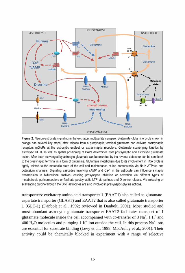
Figure 2. Neuron-astrocyte signaling in the excitatory multipartite synapse. Glutamate-glutamine cycle shown in
orange has several key steps: after release from a presynaptic terminal glutamate can activate postsynaptic
receptors mGluRs at the astrocytic endfeet or extrasynaptic receptors. Glutamate scavenging kinetics by
astrocytic GLUT as well as spatial positioning of PAPs determines both postsynaptic and astrocytic glutamate
action. After been scavenged by astrocyte glutamate can be excreted by the reverse uptake or can be sent back
to the presynaptic terminal in a form of glutamine. Glutamate metabolism due to its involvement in TCA cycle is
tightly related to the metabolic state of the cell and maintenance of ion homeostasis via Na-K-ATPase and
potassium channels. Signaling cascades involving cAMP and Ca2+ in the astrocyte can influence synaptic
transmission in bidirectional fashion, causing presynaptic inhibition or activation via different types of
metabotropic purinoreceptors or facilitate postsynaptic LTP via purines and D-serine release. Via releasing or
scavenging glycine through the GlyT astrocytes are also involved in presynaptic glycine actions.
transporters: excitatory amino acid transporter 1 (EAAT1) also called as glutamate-
aspartate transporter (GLAST) and EAAT2 that is also called glutamate transporter
1 (GLT-1) (Danbolt et al., 1992; reviewed in Danbolt, 2001). Most studied and
most abundant astrocytic glutamate transporter EAAT2 facilitates transport of 1
glutamate molecule inside the cell accompanied with co-transfer of 3 Na+, 1 H
+ and
400 H2O molecules and pumping 1 K+ ion outside the cell. In this process Na
+ ions
are essential for substrate binding (Levy et al., 1998; MacAulay et al., 2001). Their
activity could be chemically blocked in experiment with a range of selective
16
glutamate transporters antagonists such as dihydrokainik acid (DHK) (Johnston et
al., 1979; Arriza et al., 1994) and DL-threo-beta-benzyloxyaspartate (TBOA).
Glutamate transporters distributed unevenly in astrocytic plasma membrane; they
mainly located on astrocytic processes that are in close contacts with synapses and
much less abundant in somatic areas (Chaundhry et al., 1995) suggesting their
primary role in uptake of synaptically released glutamate. In agreement with it is an
observation that transgenic mice that lack EAAT2 expression demonstrated
increased mortality and behavioral abnormalities (Tanaka et al., 1997), at the same
time mice that overexpress EAAT2 were resistant to induction of epileptiform
activity and had problems with LTP induction (Martinowich et al., 2001).
In addition to uptake glutamate transporters can mediate release of glutamate via so
called ”Reversed uptake” of glutamate (Rossi et al., 2000; Grewer et al., 2008), it
could happened if cells loose energy source in case of ischemia or glucose
deprivation (Nicholls and Attwell, 1990; Rossi et al., 2000) causing increase in
spontaneous activity and excitotoxicity. It is worth to mention here that glutamate
transporters are involved in coupling synaptic activity with astrocyte signaling
(Volterra et al., 2002, pp. 46-56), thus glutamate to glutamine transformation cycle
should be considered in a context of neuroglial metabolic coupling since glycolysis
in astrocytes is dependent on glutamate uptake (see also part 2.1 for details).
Such characteristic features of astrocytic glutamate transporters like their high
density in perisynaptic regions of astrocyte, their high affinity to glutamate and the
fact that much of the transporter current might be produced by low concentration of
glutamate (Volterra et al., 2002, pp. 62-75) allow to propose that glutamate
spillover and uptake regulated not chemically but spatially and are correlated rather
with synaptic geometry than with transporters permeability (Freche et al., 2011;
Freche et al., 2012). This idea is bringing us to a conception where astrocytes can
actively participate in synaptic events by mean of their structural rearrangement
and is the first link for involvement of astrocytic morphological plasticity in
modulation of synaptic function.
2.5.2. Connexin and pannexin hemichannels
Along with specialized glutamate transporters astrocytes also express different
channels facilitating transfer of small molecules and ions. Connexins that are
known to form gap junctions between astrocytes are also presented as
hemichannels in astrocytic membranes where they can mediate astrocytic
activation by ATP and Ca2+
signaling (Bennett et al., 2003; Bennett et al., 2012;
Chen et al., 2012). In addition to connexins there is also pannexin-1 hemichannels
expressed in astrocytes. They are permeable for ATP, arachidonic acid derivatives,
17
are involved in Ca2+
waves propagation and might play a role vascular regulation
(Bennett et al., 2012; Suadicani et al., 2012; MacVicar and Thompson, 2010). It
was proposed also that hemichannels in astrocytes might mediate glutamate release
(Ye et al., 2003) and thus play a role in coupling astrocytes with synaptic activity.
2.5.3. Glutamate receptors
Presence of functional ionotropic glutamate receptors in astrocytic plasma
membrane is mysterious. Even though there were evidences for expression of
ionotropic glutamate receptors mRNA and even functional NMDA and AMPA
receptors in cortical and hippocampal astrocytes (Porter and McCarthy, 1996;
Gallo and Ghiani, 2000; Schipke et al., 2001), their functionality remains
controversial (reviewed by Volterra et al., 2002, pp .34-46). In opposite
metabotropic glutamate receptors in astrocytes are well characterized and were
shown to be involved in regulation of synaptic function (Perea and Araque, 2007;
Panatier et al., 2011). Astrocytes were demonstrated to express mRNA for several
metabotropic glutamate receptors subtypes; mGluR3 coupled with adenylate
cyclase signaling pathway and mGluR5 that are connected to IP3 and Ca2+
signaling cascade (Schools and Kimelberg, 1999).
2.5.4. GABA receptors and transporters
Besides excitatory glutamate sensing and release astrocytes are also exhibit
sensitivity to inhibitory neurotransmitter GABA. It was reported that there is a high
affinity GABA membrane transporter GAT-1 expressed in astrocytes (Minelli et
al., 1995). On the other hand astrocytes can indirectly affect GABA-mediated
synaptic transmission by influencing glutamate-glutamine cycle in presynaptic
GABAergic terminal (Liang et al., 2006). There are also some evidences regarding
metabotropic GABAB receptors which can trigger glutamate or ATP release from
astrocytes (Kang et al., 1998; Serrano et al., 2006) and ionotropic GABAA
receptors expression (Steinhäuser et al., 1994). Latter are known to be
developmentally regulated and disappears in mature astroglia (reviewed by
Volterra et al., 2002, in pp. 34-46).
2.5.5. Channels and transporters involved in D-serine release from astrocytes
Within a special interest astrocytic involvement in recently described D-serine
actions (Panatier et al., 2006; Henneberger et al., 2010; Papouin et al., 2012).
Astrocytes are proposed to express selective transporters for NMDA receptors co-
agonists such as D-serine transporter and glycine transporter 1 (GlyT1) (Papouin et
18
al., 2012). These transporters are thought to mediate glutamate stimulated release
of D-serine and control of synaptic function through neuronal NMDA receptors co-
activation (Schell et al., 1995; Panatier et al., 2006). D-serine can also be secreted
by astrocytes by less specific release mechanisms. It could be mediated by
exocytotic release as well as by non-exocytotic release via P2X7 channels or
volume regulated anion channels in case of astrocyte swelling (reviewed in
Hamilton and Attwell, 2010). Despite it was clearly shown that D-serine released
from astrocytes is a key regulator for synaptic plasticity and transmission (Yang et
al., 2003; Panatier et al., 2006; Henneberger et al., 2010; Papouin et al., 2012),
actual mechanism for D-serine release from astrocytes remains mostly elusive.
2.5.6. Water and potassium channels
Astrocytes intermediate exchange between neurons and blood vessels and thus are
the main players in water homeostasis in the CNS. Their ability to regulate water
balance is based on dense expression of aquaporin channels arrays. Aquaporins are
the family of trans-membrane water channels. Whereas these channels are
presented in different cell types, astrocytes express only AQP4 channel that at the
same time is the most abundant water channel in the brain (reviewed by
Papadopoulos and Verkman, 2013). AQP4 channels are mainly located on
astrocytic endfeet surrounding blood capillaries as well on astrocytic processes that
enwrap synaptic terminals (Nagelhus et al., 2004; Papadopoulos and Verkman,
2013). Interestingly that AQP4 channels are coexpressed with Kir4.1 K+ channels
both at astrocytic endfeet (Nagelhus et al., 2004; Abbott et al., 2006) and at
perisynaptic astrocytic processes (Nagelhus et al., 2004) suggesting their synergetic
role in maintenance of water and K+ homeostasis. Notably that astrocytic AQP4
also plays a critical role in epilepsy (Binder et al., 2012) and during formation of
traumatic brain edema (Nase et al., 2007).
2.5.7. Purinoreceptors
Purinergic signaling underlies many signaling events in astrocytes. Purine receptors
family P2X1-7 mediating exchange of K+ to Ca
2+ and N
+ in a response to
extracellular ATP, thus providing Ca2+
and Na+ influx to astrocytes. Astrocytes
express at least two types of purinoreceptors P2X1 and P2X7 that differ by their
affinity to agonist (reviewed by Illes et al., 2012) and are involved in different
signaling cascades. As a part of ATP signaling system in astroglia it is worth to
mention exocytotic mechanisms for ATP release in a response to intracellular Ca2+
elevations (Bal-Price et al., 2002; Coco et al., 2003; Pangrsic et al., 2007;
Pryazhnikov and Khiroug, 2008), suggesting exocytosis as one of the major
19
mechanism for bioactive substances release from astrocytes (reviewed in Hamilton
and Attwell, 2010).
2.5.8. Ephrin mediated reception
In order to make the story about astrocytic receptors complete we should consider
that signaling might be underlined not just by soluble factors that diffuse from one
cell to another but also by integral membrane molecules that are working while
cells have direct physical contacts. Commonly known ephrin-A3 ligands expressed
on astrocytic surface can interact with their neuronal partners ephrin-A4 receptors
located on dendrite. These interactions promote regulation of glutamate
transporters in astrocytes and also involved in AMPA receptor endocytosis and
degradation in dendritic spines as well as in Rac-mediated spine stabilization
(reviewed in Murai and Pascuale, 2011).
2.6 Chemical excitability and calcium signaling in astrocytes
Astrocytes were long thought to be non-excitable cells when studied using
electrophysiological tools applicable for neurons (reviewed in Verkhratsky et al.,
1998; Agulhon et al., 2008; Kirischuk et al., 2012). But as many other cell types
they have signaling system underlying information transfer within the cell,
astrocyte to astrocyte interactions as well as astrocyte interaction with other cell
types like neurons, microglia and endothelial cells. Along with general signaling
mechanisms astrocytes exhibit some unique signaling features that are originated
from their gap junctional coupling and their intercalating position relative to
neurons, synapses and blood vessels.
Ca2+
is the most popular second messenger in different mammalian and non-
mammalian cell types (reviewed in Akerman, 1982; Gardner, 1989; Webb and
Miller, 2003). It logically led to a general concept of Ca2+
excitability of astrocytes
(Bowman and Kimelberg, 1984; Kettenmann et al., 1984; Jensen and Chiu, 1990;
Verkhratsky and Kettenmann, 1996). Indeed, it was demonstrated that astrocytes
can exhibit Ca2+
waves induced by glutamate application (Cornell-Bell et al., 1990,
Charles et al., 1991) emerging the idea of glutamate dependent neuro-glial
signaling and supporting the concept of astrocytic Ca2+
excitability.
Even though astrocytes express channels that are permeable for Ca2+
ions, mainly
changes in [Ca2+
]i are mediated by Ca2+
mediated Ca2+
entry either from
endoplasmic reticulum or mitochondria (reviewed by Verkhratsky et al., 1998).
These astrocytic Ca2+
spikes are regulated by signaling cascades that involve G-
protein coupled receptors (GPCR) particularly mGluR5 (Panatier et al., 2011),
protein kinase C phosphorylation and IP3 mediated signaling (reviewed by
20
Verkhratsky et al., 1998). It is worth to mention also that during brain maturation
mGluR5 are replaced by mGluR3 that cause prevalence of adenylate cyclase
mediated pathway and shifting mature astroglia signaling from developing one
(Sun et al., 2013). On the other hand astroglial Ca2+
spikes mediate a range of
different processes including exocytosis of ATP (Bal-Price et al., 2002; Pangrsic et
al., 2007), D-serine (Panatier et al., 2006; Henneberger et al., 2010) and glutamate
release (Liu et al., 2011). Unique astrocytic feature – their coupling through gap
junctions and expression of hemichannels (Giaume, 2010; Bennett et al., 2003)
provides additional options for Ca2+
signaling along ensembles of astroglial cells
and can cause remarkable Ca2+
waves and synchronous astrocytic activity under
physiological (Scemes and Giaume, 2006; Hoogland et al., 2009) and pathological
conditions (Kuchibholta et al., 2009).
It is interesting that if the modulation of Ca2+
signaling in astrocytes via GPCR
activation caused by synaptic activity is doubtless (Dani et al., 1992; Porter and
McCarthy, 1996; Wang et al., 2006; Perea and Araque, 2007; Gordon et al., 2009;
Panatier et al., 2011; Min and Nevian, 2012) the feedback loop that includes
modulation of synaptic function by astrocytic Ca2+
transients and transmitters
release from astrocytes is under debate (Fiacco et al., 2007; Petravicz et al., 2008;
Agulhon et al., 2010). There are several explanations for such ambiguous role of
astrocytic Ca2+
in glia-neuronal communication. Most of them are relied on the
idea of inadequate tools used for astrocytic stimulation (Agulhon et al., 2010;
Nedergaard and Verkhratsky, 2012), possible involvement of another universal
second messenger replacing Ca2+
, for instance, Na+ ions (reviewed by Kirischuk et
al., 2012; Bhattacharjee and Kaczmarek, 2005), or developmental shift in Ca2+
signaling occurring in astrocytes (Sun et al., 2013). It is worth to remember at this
point that spatial localization of the signal, direction and rate of its propagation as
well as dose dependence should always be considered when artificial stimulation
approaches are used. For example ATP exocytosis from astrocytes triggered by
Ca2+
transients has its own dose-dependent as well as temporal patterns
(Pryazhnikov and Khiroug, 2008). Another elegant example where stimulus
localization plays a critical role is provided by studies of local blood flow
regulation by astrocytes. Exact location of Ca2+
elevations caused by local
photolysis in soma or endfeet can cause opposite effects on blood vessels
mediating either vasodilation or vasoconstriction (Takano et al., 2006; Gordon et
al., 2011).
If stimulus amplitude and timing could be easily controlled in standard
experimental paradigms, directionality, rate and precise localization should be
refined. Possibly novel genetically encodable tools that can be delivered to a subset
of astrocytes and affect defined and well predicted properties of the cell
21
(Armbruster et al., 2007; Airan et al., 2009; Molotkov et al., 2013) combined with
proper delivery and localization system will shed the light on the ambiguous role of
astrocytes in modulation of synaptic function.
Astrocyte excitability is accompanied by ion fluxes mediated by membrane
channels in case of Na+ and Ca
2+ or by internal stores mainly in case of Ca
2+ and is
coupled to neuronal activity. Moreover Na+ and Ca
2+ mediated signaling might
complement each other (Verkhratsky et al., 1998; Kirischuk et al., 2012). In fact
Na+ homeostasis that is strongly dependent on Na
+,K
+-ATPase activity and glucose
utilization (Bernardinelli et al., 2004) might connect Ca2+
signaling to metabolic
status of astroglia thus coupling indirectly internal astrocytic Ca2+
signaling with
glycolysis and neuronal activity. In addition to it the role of K+ ions also could be
considered as possible indirect messengers for neuro-glial communication
especially if astrocytic role in neuronal network activity regulation via K+ buffering
(reviewed by Simard and Nedergaard, 2004) would be taken into account.
Hypothesizing further we can propose that water homeostasis in astrocytes is a
candidate for playing a significant role both in maintenance and regulation of
general and local ion mediated signaling, coupling metabolic activity of astrocytes
with Ca2+
transients, glutamate uptake and possibly exocytosis events.
Despite application of advanced methodology the role astrocytic signaling inside
the cell, among different astrocytes and also between astrocytes and neurons
remains controversial with a prevalence of descriptive approaches to colligating
ones. Diversity of methodological approaches might be one of the most critical
factors that affect sensitive intracellular machinery. For instance, it was recently
shown for awake mouse cortex that general anesthesia can disrupt Ca2+
signaling in
astrocytes (Schummers et al., 2008; Thrane et al., 2012), but see also the work of
Hirase and colleagues (Hirase et al., 2004) where astrocytic Ca2+
signaling was
shown to be in a direct correlation with neuronal activity in anaesthetized rats. We
can further suggest that signaling in primary astrocytic cell culture would be
different from those in acute brain slice preparation that in turn would be different
from whole brain in vivo situation.
2.7 Modulation of synaptic transmission by astrocytes: the concept of
multipartite synapse
Complexity of chemical CNS synapse is often reduced to simple donor-acceptor
model where presynaptic site releases neurotransmitter which is sensed by
receptors on postsynaptic terminal. This view that pointing the synapse as one way
neurotransmitter action site does not consider different important features of
synapse such as geometry of synaptic cleft (Savtchenko and Rusakov, 2007),
activation of extrasynaptic receptors (Kullmann and Asztely, 1998; Fellin and
22
Carmignoto, 2004), electrodiffusion of neurotransmitter caused by electrical fields
generated near the synapse (Sylantyev et al., 2013), diffusion dynamics of
neurotransmitter determined by ECM and surrounding cellular components
(Sykova, 2004; Dityatev and Rusakov, 2011) and retrograde signaling that
transform unidirectional synaptic transmission into crosstalk between pre and post
synaptic terminals. This crosstalk which seemed to be a dialogue in a first
approximation actually involves other active and passive participants besides
neuronal components and generally synaptic events might be much more complex
than proposed before.
It was known for quite a long time already that glutamate uptake by glial glutamate
transporters is involved in synaptic transmission and neuronal excitability (Tanaka
et al., 1997; Bergles and Jahr, 1998; Oliet et al., 2001) moreover efficacy of
neurotransmitter clearance appeared to be affected by structural interplay between
neuronal and glial compartments of synapse (Oliet et al., 2001) that raises a
question about the role of structure-functional relationship between glia and
synaptic structures. Glutamate uptake function of astrocytes is accompanied with a
well-known fact that glial cells can produce and release neuroactive substances
(Martin, 1992) and particularly glutamate in Ca2+
dependent manner (Bezzi et al.,
2004; Marchaland et al., 2008; Santello and Volterra, 2009) and thus scale synaptic
strength (Jourdain et al., 2007). It is also known that approximately 50% of
hippocampal glutamate synapses are opposed to astrocytic processes and exhibit
cooperative dynamics (Ventura and Harris, 1999; Witcher et al., 2007; Haber et al.,
2006). These morphological changes also may underlie synaptic development and
spine stabilization (Nishida and Okabe, 2007; Haber et al., 2006; Murai et al.,
2003; Witcher et al., 2007). All mentioned above facts leaded to an idea of
tripartite synapse (Araque et al., 1999; Perea et al., 2009) where astrocytes playing
a role of active participants of synaptic events. Hypothesizing further it was
proposed that ECM components represent a part of synaptic structure and involved
in diffusion regulation, thus synapse representation could be upgraded to
quadripartite structure (Sykova, 2004). Indeed, among non-neuronal cell types not
just astroglia can actively participate in synapse functionality and structural
changes. There are accumulating evidences that microglia could be an active
synaptic partner as well (Bessis et al., 2007; Graeber, 2010; Tremblay and
Majewska, 2011; Aguzzi et al., 2013; Kettenmann et al., 2013) allowing us to
propose that the synapse represents a multipartite structure.
Although the combination of ability of astrocytes to perform bidirectional control
of synaptic strength (Navarrete and Araque, 2011) and their highly organized
territories with no spatial overlap and coverage of different dendritic trees
(Bushong et al., 2002; Volterra and Meldolesi, 2005; Halassa et al., 2007) together
with evidences for activation of astrocytic signaling pathways in a response to
sensory stimulation in vivo (Wang et al., 2006; Winship et al., 2007; Petzold et al.,
2008; Schummers et al., 2008; Nimmerjahn et al., 2009) allows to propose
astrocytes as major CNS pacemakers. The astrocytic crosstalk with neurons is
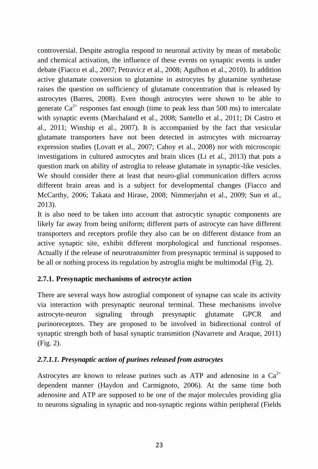
23
controversial. Despite astroglia respond to neuronal activity by mean of metabolic
and chemical activation, the influence of these events on synaptic events is under
debate (Fiacco et al., 2007; Petravicz et al., 2008; Agulhon et al., 2010). In addition
active glutamate conversion to glutamine in astrocytes by glutamine synthetase
raises the question on sufficiency of glutamate concentration that is released by
astrocytes (Barres, 2008). Even though astrocytes were shown to be able to
generate Ca2+
responses fast enough (time to peak less than 500 ms) to intercalate
with synaptic events (Marchaland et al., 2008; Santello et al., 2011; Di Castro et
al., 2011; Winship et al., 2007). It is accompanied by the fact that vesicular
glutamate transporters have not been detected in astrocytes with microarray
expression studies (Lovatt et al., 2007; Cahoy et al., 2008) nor with microscopic
investigations in cultured astrocytes and brain slices (Li et al., 2013) that puts a
question mark on ability of astroglia to release glutamate in synaptic-like vesicles.
We should consider there at least that neuro-glial communication differs across
different brain areas and is a subject for developmental changes (Fiacco and
McCarthy, 2006; Takata and Hirase, 2008; Nimmerjahn et al., 2009; Sun et al.,
2013).
It is also need to be taken into account that astrocytic synaptic components are
likely far away from being uniform; different parts of astrocyte can have different
transporters and receptors profile they also can be on different distance from an
active synaptic site, exhibit different morphological and functional responses.
Actually if the release of neurotransmitter from presynaptic terminal is supposed to
be all or nothing process its regulation by astroglia might be multimodal (Fig. 2).
2.7.1. Presynaptic mechanisms of astrocyte action
There are several ways how astroglial component of synapse can scale its activity
via interaction with presynaptic neuronal terminal. These mechanisms involve
astrocyte-neuron signaling through presynaptic glutamate GPCR and
purinoreceptors. They are proposed to be involved in bidirectional control of
synaptic strength both of basal synaptic transmition (Navarrete and Araque, 2011)
(Fig. 2).
2.7.1.1. Presynaptic action of purines released from astrocytes
Astrocytes are known to release purines such as ATP and adenosine in a Ca2+
dependent manner (Haydon and Carmignoto, 2006). At the same time both
adenosine and ATP are supposed to be one of the major molecules providing glia
to neurons signaling in synaptic and non-synaptic regions within peripheral (Fields
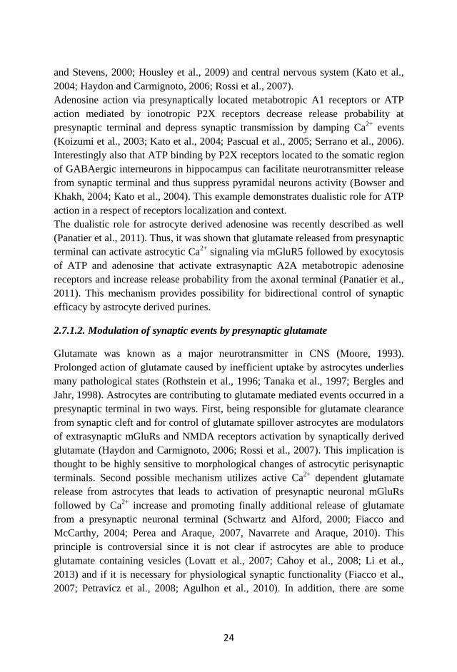
24
and Stevens, 2000; Housley et al., 2009) and central nervous system (Kato et al.,
2004; Haydon and Carmignoto, 2006; Rossi et al., 2007).
Adenosine action via presynaptically located metabotropic A1 receptors or ATP
action mediated by ionotropic P2X receptors decrease release probability at
presynaptic terminal and depress synaptic transmission by damping Ca2+
events
(Koizumi et al., 2003; Kato et al., 2004; Pascual et al., 2005; Serrano et al., 2006).
Interestingly also that ATP binding by P2X receptors located to the somatic region
of GABAergic interneurons in hippocampus can facilitate neurotransmitter release
from synaptic terminal and thus suppress pyramidal neurons activity (Bowser and
Khakh, 2004; Kato et al., 2004). This example demonstrates dualistic role for ATP
action in a respect of receptors localization and context.
The dualistic role for astrocyte derived adenosine was recently described as well
(Panatier et al., 2011). Thus, it was shown that glutamate released from presynaptic
terminal can activate astrocytic Ca2+
signaling via mGluR5 followed by exocytosis
of ATP and adenosine that activate extrasynaptic A2A metabotropic adenosine
receptors and increase release probability from the axonal terminal (Panatier et al.,
2011). This mechanism provides possibility for bidirectional control of synaptic
efficacy by astrocyte derived purines.
2.7.1.2. Modulation of synaptic events by presynaptic glutamate
Glutamate was known as a major neurotransmitter in CNS (Moore, 1993).
Prolonged action of glutamate caused by inefficient uptake by astrocytes underlies
many pathological states (Rothstein et al., 1996; Tanaka et al., 1997; Bergles and
Jahr, 1998). Astrocytes are contributing to glutamate mediated events occurred in a
presynaptic terminal in two ways. First, being responsible for glutamate clearance
from synaptic cleft and for control of glutamate spillover astrocytes are modulators
of extrasynaptic mGluRs and NMDA receptors activation by synaptically derived
glutamate (Haydon and Carmignoto, 2006; Rossi et al., 2007). This implication is
thought to be highly sensitive to morphological changes of astrocytic perisynaptic
terminals. Second possible mechanism utilizes active Ca2+
dependent glutamate
release from astrocytes that leads to activation of presynaptic neuronal mGluRs
followed by Ca2+
increase and promoting finally additional release of glutamate
from a presynaptic neuronal terminal (Schwartz and Alford, 2000; Fiacco and
McCarthy, 2004; Perea and Araque, 2007, Navarrete and Araque, 2010). This
principle is controversial since it is not clear if astrocytes are able to produce
glutamate containing vesicles (Lovatt et al., 2007; Cahoy et al., 2008; Li et al.,
2013) and if it is necessary for physiological synaptic functionality (Fiacco et al.,
2007; Petravicz et al., 2008; Agulhon et al., 2010). In addition, there are some
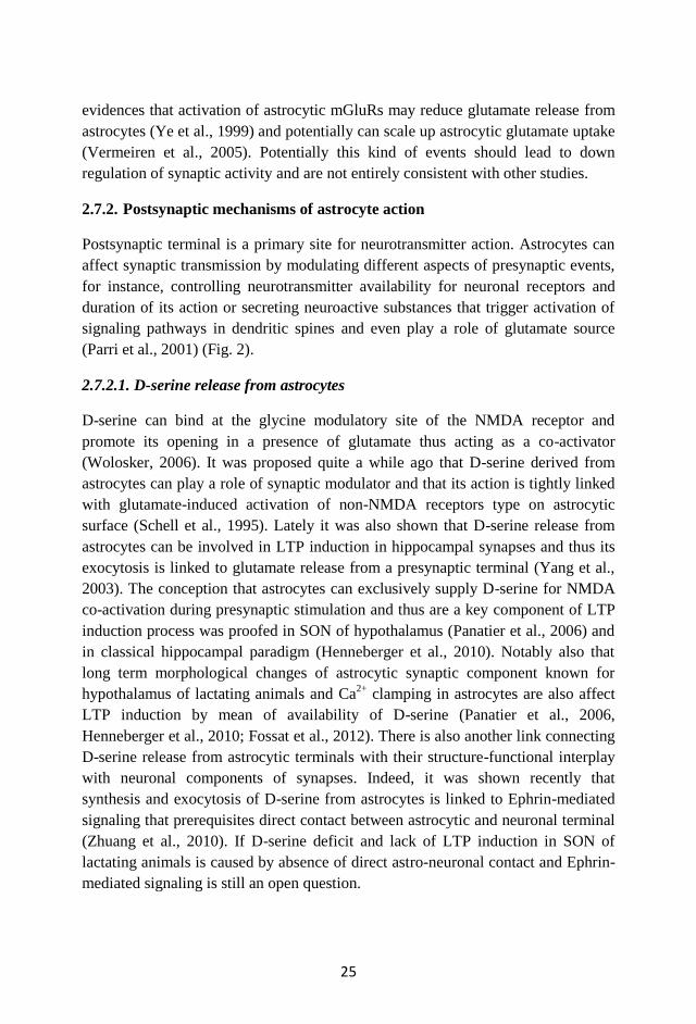
25
evidences that activation of astrocytic mGluRs may reduce glutamate release from
astrocytes (Ye et al., 1999) and potentially can scale up astrocytic glutamate uptake
(Vermeiren et al., 2005). Potentially this kind of events should lead to down
regulation of synaptic activity and are not entirely consistent with other studies.
2.7.2. Postsynaptic mechanisms of astrocyte action
Postsynaptic terminal is a primary site for neurotransmitter action. Astrocytes can
affect synaptic transmission by modulating different aspects of presynaptic events,
for instance, controlling neurotransmitter availability for neuronal receptors and
duration of its action or secreting neuroactive substances that trigger activation of
signaling pathways in dendritic spines and even play a role of glutamate source
(Parri et al., 2001) (Fig. 2).
2.7.2.1. D-serine release from astrocytes
D-serine can bind at the glycine modulatory site of the NMDA receptor and
promote its opening in a presence of glutamate thus acting as a co-activator
(Wolosker, 2006). It was proposed quite a while ago that D-serine derived from
astrocytes can play a role of synaptic modulator and that its action is tightly linked
with glutamate-induced activation of non-NMDA receptors type on astrocytic
surface (Schell et al., 1995). Lately it was also shown that D-serine release from
astrocytes can be involved in LTP induction in hippocampal synapses and thus its
exocytosis is linked to glutamate release from a presynaptic terminal (Yang et al.,
2003). The conception that astrocytes can exclusively supply D-serine for NMDA
co-activation during presynaptic stimulation and thus are a key component of LTP
induction process was proofed in SON of hypothalamus (Panatier et al., 2006) and
in classical hippocampal paradigm (Henneberger et al., 2010). Notably also that
long term morphological changes of astrocytic synaptic component known for
hypothalamus of lactating animals and Ca2+
clamping in astrocytes are also affect
LTP induction by mean of availability of D-serine (Panatier et al., 2006,
Henneberger et al., 2010; Fossat et al., 2012). There is also another link connecting
D-serine release from astrocytic terminals with their structure-functional interplay
with neuronal components of synapses. Indeed, it was shown recently that
synthesis and exocytosis of D-serine from astrocytes is linked to Ephrin-mediated
signaling that prerequisites direct contact between astrocytic and neuronal terminal
(Zhuang et al., 2010). If D-serine deficit and lack of LTP induction in SON of
lactating animals is caused by absence of direct astro-neuronal contact and Ephrin-
mediated signaling is still an open question.
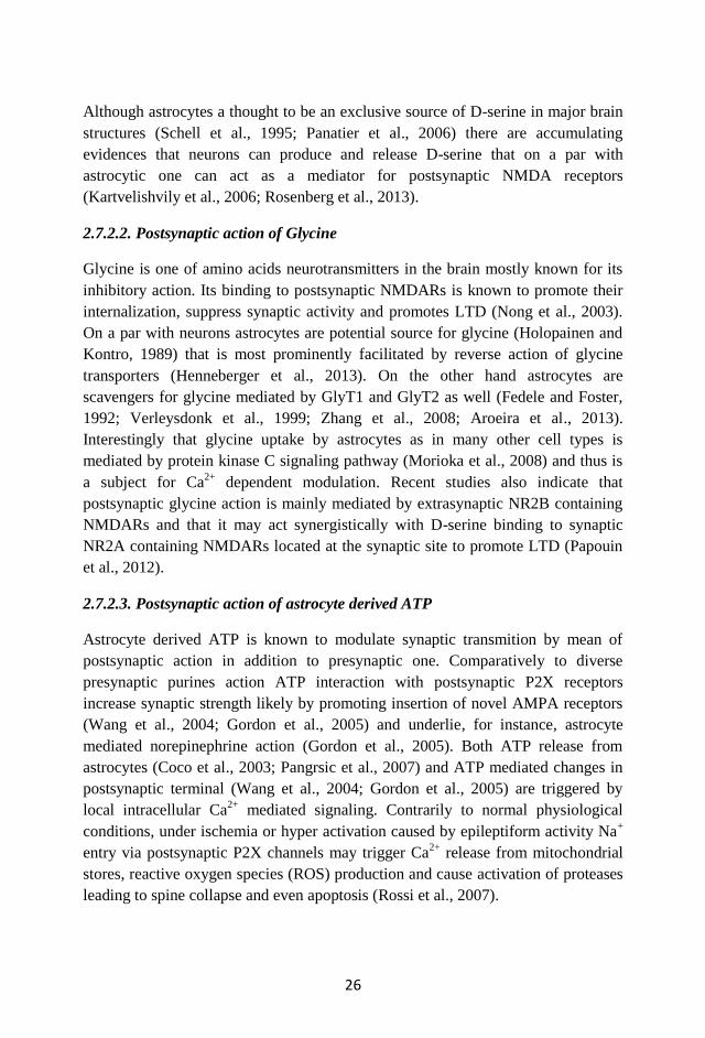
26
Although astrocytes a thought to be an exclusive source of D-serine in major brain
structures (Schell et al., 1995; Panatier et al., 2006) there are accumulating
evidences that neurons can produce and release D-serine that on a par with
astrocytic one can act as a mediator for postsynaptic NMDA receptors
(Kartvelishvily et al., 2006; Rosenberg et al., 2013).
2.7.2.2. Postsynaptic action of Glycine
Glycine is one of amino acids neurotransmitters in the brain mostly known for its
inhibitory action. Its binding to postsynaptic NMDARs is known to promote their
internalization, suppress synaptic activity and promotes LTD (Nong et al., 2003).
On a par with neurons astrocytes are potential source for glycine (Holopainen and
Kontro, 1989) that is most prominently facilitated by reverse action of glycine
transporters (Henneberger et al., 2013). On the other hand astrocytes are
scavengers for glycine mediated by GlyT1 and GlyT2 as well (Fedele and Foster,
1992; Verleysdonk et al., 1999; Zhang et al., 2008; Aroeira et al., 2013).
Interestingly that glycine uptake by astrocytes as in many other cell types is
mediated by protein kinase C signaling pathway (Morioka et al., 2008) and thus is
a subject for Ca2+
dependent modulation. Recent studies also indicate that
postsynaptic glycine action is mainly mediated by extrasynaptic NR2B containing
NMDARs and that it may act synergistically with D-serine binding to synaptic
NR2A containing NMDARs located at the synaptic site to promote LTD (Papouin
et al., 2012).
2.7.2.3. Postsynaptic action of astrocyte derived ATP
Astrocyte derived ATP is known to modulate synaptic transmition by mean of
postsynaptic action in addition to presynaptic one. Comparatively to diverse
presynaptic purines action ATP interaction with postsynaptic P2X receptors
increase synaptic strength likely by promoting insertion of novel AMPA receptors
(Wang et al., 2004; Gordon et al., 2005) and underlie, for instance, astrocyte
mediated norepinephrine action (Gordon et al., 2005). Both ATP release from
astrocytes (Coco et al., 2003; Pangrsic et al., 2007) and ATP mediated changes in
postsynaptic terminal (Wang et al., 2004; Gordon et al., 2005) are triggered by
local intracellular Ca2+
mediated signaling. Contrarily to normal physiological
conditions, under ischemia or hyper activation caused by epileptiform activity Na+
entry via postsynaptic P2X channels may trigger Ca2+
release from mitochondrial
stores, reactive oxygen species (ROS) production and cause activation of proteases
leading to spine collapse and even apoptosis (Rossi et al., 2007).
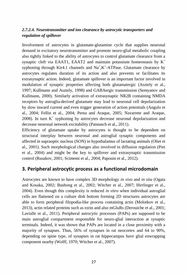
27
2.7.2.4. Neurotransmitter and ion clearance by astrocytic transporters and
regulation of spillover
Involvement of astrocytes in glutamate-glutamine cycle that supplies neuronal
demand in excitatory neurotransmitter and promote neuro-glial metabolic coupling
also tightly linked to the ability of astrocytes to control glutamate clearance from a
synaptic cleft via EAAT1, EAAT2 and maintain potassium homeostasis by K+
syphoning through Kir4.1 channels and Na+,K
+-ATPase. Glutamate clearance by
astrocytes regulates duration of its action and also prevents or facilitates its
extrasynaptic action. Indeed, glutamate spillover is an important factor involved in
modulation of synaptic properties affecting both glutamatergic (Asztely et al.,
1997; Kullmann and Asztely, 1998) and GABAergic transmission (Semyanov and
Kullmann, 2000). Similarly activation of extrasynaptic NR2B containing NMDA
receptors by astroglia-derived glutamate may lead to neuronal cell depolarization
by slow inward current and even trigger generation of action potentials (Angulo et
al., 2004; Fellin et al., 2004; Perea and Araque, 2005; Navarrete and Araque,
2008). In turn K+ syphoning by astrocytes decrease neuronal depolarization and
decrease neuronal network excitability (Pannasch et al., 2011).
Efficiency of glutamate uptake by astrocytes is thought to be dependent on
structural interplay between neuronal and astroglial synaptic components and
affected in supraoptic nucleus (SON) in hypothalamus of lactating animals (Oliet et
al., 2001). Such morphological changes also involved in diffusion regulation (Piet
et al., 2004) and might be the key to spillover and extrasynaptic transmission
control (Rusakov, 2001; Scimemi et al., 2004; Papouin et al., 2012).
3. Peripheral astrocytic process as a functional microdomain
Astrocytes are known to have complex 3D morphology in vivo and in situ (Ogata
and Kosaka, 2002; Bushong et al., 2002; Witcher et al., 2007; Hirrlinger et al.,
2004). Even though this complexity is reduced in vitro when individual astroglial
cells are flattened on a culture dish bottom forming 2D structures astrocytes are
able to form peripheral filopodia-like process containing actin (Molotkov et al.,
2013), actin related proteins such as ezrin and also mGluRs (Derouiche et al., 2001;
Lavialle et al., 2011). Peripheral astrocytic processes (PAPs) are supposed to be
main astroglial compartment responsible for neuro-glial interaction at synaptic
terminals. Indeed, it was shown that PAPs are located in a close proximity with a
majority of synapses. Thus, 56% of synapses in rat neocortex and 64 to 90%,
depending on spine type, of synapses in rat hippocampus have glial enwrapping
component nearby (Wolff, 1970; Witcher et al., 2007).
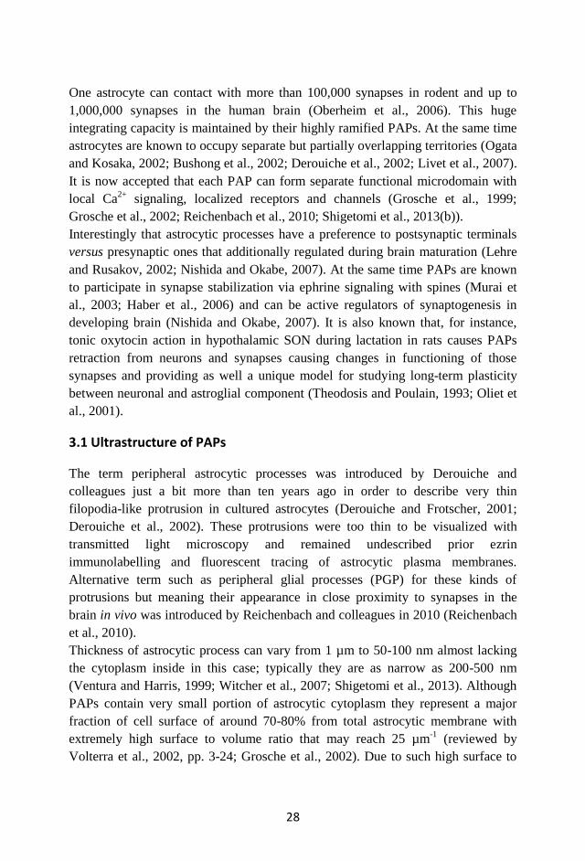
28
One astrocyte can contact with more than 100,000 synapses in rodent and up to
1,000,000 synapses in the human brain (Oberheim et al., 2006). This huge
integrating capacity is maintained by their highly ramified PAPs. At the same time
astrocytes are known to occupy separate but partially overlapping territories (Ogata
and Kosaka, 2002; Bushong et al., 2002; Derouiche et al., 2002; Livet et al., 2007).
It is now accepted that each PAP can form separate functional microdomain with
local Ca2+
signaling, localized receptors and channels (Grosche et al., 1999;
Grosche et al., 2002; Reichenbach et al., 2010; Shigetomi et al., 2013(b)).
Interestingly that astrocytic processes have a preference to postsynaptic terminals
versus presynaptic ones that additionally regulated during brain maturation (Lehre
and Rusakov, 2002; Nishida and Okabe, 2007). At the same time PAPs are known
to participate in synapse stabilization via ephrine signaling with spines (Murai et
al., 2003; Haber et al., 2006) and can be active regulators of synaptogenesis in
developing brain (Nishida and Okabe, 2007). It is also known that, for instance,
tonic oxytocin action in hypothalamic SON during lactation in rats causes PAPs
retraction from neurons and synapses causing changes in functioning of those
synapses and providing as well a unique model for studying long-term plasticity
between neuronal and astroglial component (Theodosis and Poulain, 1993; Oliet et
al., 2001).
3.1 Ultrastructure of PAPs
The term peripheral astrocytic processes was introduced by Derouiche and
colleagues just a bit more than ten years ago in order to describe very thin
filopodia-like protrusion in cultured astrocytes (Derouiche and Frotscher, 2001;
Derouiche et al., 2002). These protrusions were too thin to be visualized with
transmitted light microscopy and remained undescribed prior ezrin
immunolabelling and fluorescent tracing of astrocytic plasma membranes.
Alternative term such as peripheral glial processes (PGP) for these kinds of
protrusions but meaning their appearance in close proximity to synapses in the
brain in vivo was introduced by Reichenbach and colleagues in 2010 (Reichenbach
et al., 2010).
Thickness of astrocytic process can vary from 1 µm to 50-100 nm almost lacking
the cytoplasm inside in this case; typically they are as narrow as 200-500 nm
(Ventura and Harris, 1999; Witcher et al., 2007; Shigetomi et al., 2013). Although
PAPs contain very small portion of astrocytic cytoplasm they represent a major
fraction of cell surface of around 70-80% from total astrocytic membrane with
extremely high surface to volume ratio that may reach 25 µm-1
(reviewed by
Volterra et al., 2002, pp. 3-24; Grosche et al., 2002). Due to such high surface to
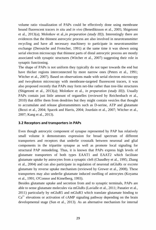
29
volume ratio visualization of PAPs could be effectively done using membrane
bound fluorescent tracers in situ and in vivo (Benediktsson et al., 2005; Shigetomi
et al., 2013(a); Molotkov et al.,in preparation (study III)). Interestingly there are
evidences that the thinnest astrocytic process are also involved in neurotransmitter
recycling and have all necessary machinery to participate in neurotransmitter
exchange (Derouiche and Frotscher, 1991) at the same time it was shown using
serial electron microscopy that thinnest parts of distal astrocytic process are mostly
associated with synaptic structures (Witcher et al., 2007) suggesting their role in
synaptic functioning.
The shape of PAPs is not uniform they typically do not taper towards the end but
have thicker regions interconnected by more narrow ones (Peters et al., 1991;
Witcher et al., 2007). Based on observations made with serial electron microscopy
and two-photon microscopy with membrane-targeted fluorescent tracers, it was
also proposed recently that PAPs may form net-like rather than tree-like structures
(Shigetomi et al., 2013(a); Molotkov et al., in preparation (study III)). Usually
PAPs contain just little amount of organelles (reviewed by Reichenbach et al.,
2010) that differ them from dendrites but they might contain vesicles that thought
to accumulate and release gliotransmitters such as D-serine, ATP and glutamate
(Bezzi et al., 2004; Spacek and Harris, 2004; Jourdain et al., 2007; Witcher et al.,
2007; Kang et al., 2013).
3.2 Receptors and transporters in PAPs
Even though astrocytic component of synapse represented by PAP has relatively
small volume it demonstrates expression for broad spectrum of different
transporters and receptors that underlie crosstalk between neuronal and glial
components in the tripartite synapse as well as promote local signaling for
structural PAP remodeling. Thus, it is known that PAPs express high levels of
glutamate transporters of both types EAAT1 and EAAT2 which facilitate
glutamate uptake by astrocytes from a synaptic cleft (Chaudhry et al., 1995; Zhang
et al., 2004) and can also participate in regulation of neuronal mGluRs or excrete
glutamate by reverse uptake mechanism (reviewed by Grewer et al., 2008). These
transporters may also underlie glutamate induced swelling of astrocytes (Koyama
et al., 1991; O'Connor and Kimelberg, 1993).
Besides glutamate uptake and secretion from and to synaptic terminals, PAPs are
able to sense glutamate molecules via mGluRs (Lavialle et al., 2011; Panatier et al.,
2011) particularly by mGluR5 and mGluR3 which translate glutamate binding to
Ca2+
elevations or activation of cAMP signaling pathway depending on the brain
developmental stage (Sun et al., 2013). As an alternative mechanism for internal
30
store-independent Ca2+
signaling in PAPs the Ca2+
entry into astrocytes through
TRPA1 channels was proposed (Shigetomi et al., 2013(b)). This mechanism does
not require activation of IP3 signaling pathways and may operate in mGluRs
independent way.
Another group of channels expressed in PAPs is tightly linked to morphological
changes of astroglial cells due to regulation of chloride, potassium and water
homeostasis. Thus, chloride channels presented in PAPs were shown to mediate
glial reshaping and swelling (Ransom and Sontheimer, 2001). On the other hand
one of the theories for astroglial reshaping is based on water entry through AQP4
which is enriched in PAPs where it is often colocalized with Kir4.1 potassium
channels suggesting their mutual role in maintenance of water and K+ homeostasis
in PAPs (Nagelhus et al., 2004; Holen, 2011). Another trans membrane protein
presented on PAPs surface Ephrin-A3 ligand is involved in contact glia-neuronal
interactions and was shown to regulate dendritic spine development and
stabilization in a bidirectional fashion (Murai et al., 2003, Haber et al., 2006;
Nishida and Okabe, 2007). Additionally Eprin-A3 and A4 interactions are involved
in expression regulation of glutamate transporters in perisynaptically localized
PAPs and thus can influence synaptic functional properties via modulation of
glutamate uptake and spillover (Filosa et al., 2009).
3.3 Morphological changes of PAPs: two different mechanisms proposed
3.3.1. Aquaporin mediated morphological changes
Water channel AQP4 is supposed to be potential regulator of cells shape (Nicchia
et al., 2003; Nagelhus et al., 2004). Water uptake by astrocytes can cause swelling
which, in turn, changes their contacting surface with neurons. Astrocytic swelling
also affects activity of neuronal networks and is typical for several pathological
states (Scharfman and Binder, 2013). Similarly rise of extracellular potassium
might be one of keys for switching morphological changes of PAPs. Indeed, there
is an evidence that increase of extracellular potassium ions can stimulate formation
of new branches in cultured Muller cells (Reichelt et al., 1989).
3.3.2. Actin-dependent morphological changes
Actin cytoskeleton is the most common driver for different types of cellular
motility including migration, rapid morphological changes of cell edge (Pollard
and Borisy, 2003), lamellipodia and filopodia formation and extension (Faix and
Rottner, 2006; Mattila and Lappalainen, 2008), exocytosis and endocytosis events
(Muallem et al., 1995; Engqvist-Goldstein and Drubin, 2003). Generally actin
31
cytoskeleton is linking intracellular signaling to membrane dynamics and underlies
as well receptor trafficking on the cell surface (Malinow and Malenka, 2002;
Collingridge et al., 2004; Cingolani and Goda, 2008). Actin driving force is
underlined by constant treadmilling cycle between monomeric actin pool and
filamentous actin (F-actin) strands and can be regulated by many co-factors that
acts as primers, exchangers and catalyzers for actin monomers (Pollard, 2003).
The evidence that PAPs motility can be reduced by cytochalasin treatment (Haber
et al., 2006) suggesting the active role of actin cytoskeleton remodeling for
morphological plasticity of PAPs. This observation is also confirmed by the fact
that morphological changes of PAPs are diminished by overexpression of
dominant-negative form of small GTPase Rac that is known to be involved in
cytoskeleton regulation (Nishida and Okabe, 2007). Besides functional evidences
for actin cytoskeleton based motility of PAPs there are several structural supports
for this idea. It is based on the evidence for localization of actin-related proteins
such as ezrin (Derouiche and Frotscher, 2001) and α-adducin (Seidel et al., 1995)
inside PAPs. Indeed, direct demonstration for the presence of F-actin inside
astrocytic filopodia in culture was recently shown as well (Molotkov et al., 2013).
PAPs outgrowth could be switched both by rise of [Ca2+
]i (Molotkov et al., 2013)
or by application of extracellular glutamate (Cornell-Bell et al., 1990; Hirrlinger et
al., 2004). In both cases they demonstrate relatively high outgrowth rates of around
1 µm·min-1
for brain slices with glutamate-induced outgrowth (Hirrlinger et al.,
2004) and approximately 3 µm·min-1
for cultured astrocytes stimulated by
photolysis of caged Ca2+
(Molotkov et al., 2013). Most likely PAPs outgrowth is
triggered by similar mechanisms as filopodia outgrowth in other cell types, for
instance, fibroblasts or keratinocytes. The role of plasma membrane-to-
cytoskeleton linker Ezrin is within a special interest if focusing on reshaping of
neuron glial connections. Regulated by phosphorylation/dephosphorylation it can
potentially represent a key to rapid bidirectional activity-mediated morphological
plasticity of PAPs (Gautreau et al., 2002; Lavialle et al., 2011). Other possible
players are chloride channels that are known to have actin-dependent activity
(Lascola et al., 1998; Ahmed et al., 2000) and bridging together “passive”
astrocytic swelling and “active” actin-based astroglial motility.
Finally it is interesting to address the question whether mGluR3 mediated
activation of adenylate cyclase pathway can promote rapid morphological changes
in PAPs similar to those that could be obtained by local Ca2+
uncaging or local
glutamate application in rat neonatal astrocytes that supposed to mimic store-
operated Ca2+
entry mediated by mGluR5 receptors.
32
4. Methodological approaches to study neuro-glial interactions
4.1 Hypothalamus as a classical model of PAPs retraction
Morphology of the synapse is one of the critical aspects that underlies amount of
postsynaptic receptors being activated and time course for neurotransmitter action
as well as possible transactivation of neighboring synapses and extrasynaptic
receptors (Freche et al., 2011; Allam et al., 2012).
It was known a while ago that oxytocin can induce morphological changes in SON
of hypothalamus in lactating rodents (Theodosis et al., 1986). These changes
represent evidence for mature brain structural plasticity and was a unique case until
recent studies of antidepressant-induced plasticity (Maya Vetencourt et al., 2008;
Karpova et al., 2011). Lactation and increased oxytocin level induced long term
retraction of astrocytic process from synapses and neuronal somata facilitating
direct contact between neighboring neurons and blood vessels (Theodosis and
Poulain, 1989; Theodosis and Poulain, 1993). This also affected glutamatergic
synaptic transmission (Oliet et al., 2001; Panatier et al., 2006). Morphological
changes in SON could also be driven by dehydration and changes in vasopressin
level which also caused astrocyte retraction (Hatton, 1997; Theodosis et al., 2008).
Interestingly, the model of astrocyte retraction in SON in a response to oxytocin
works in vitro as well. Thus, bath application of oxytocin on brain slices containing
SON caused similar rapid (within hours) and reversible morphological changes in
astro-neuronal contacts as observed in vivo in lactating animals (Langle et al.,
2003). In addition dehydration and rehydration could be also used as a reverse
switcher for GFAP level in astrocytes and may reversibly affect astrocytic
morphology and coverage of neurons in SON (Hawrylak et al., 1998). If this model
could be simplified for in vitro studies, by application of hyperosmotic solutions on
an acute brain slice is questionable.
4.2 Transgenic mouse models to study astrocytes
Genetic manipulation in mice is a powerful tool to understand CNS physiology and
pathology. It allows the study of particular gene function by their ablation or
overexpression in a subset of cells in the brain using cell-type specific promoters.
There are a number of different genetic strategies to manipulate and study
astrocytes in a rodent brain (reviewed by Pfrieger and Slezak, 2012). Some of these
approaches are based on available transgenic mice models.
Glial fibrillary acidic protein (GFAP) is an intermediate filament component that is
preferentially expressed in astrocytes and glial progenitors. Recombinant GFAP
33
promoter is the most studied and the most popular tool for transgene expression in
astrocytes both in vitro and in vivo and is often used for generation of transgenic
mouse lines.
GFAP promoter as a tool for transgene expression in astroglial and neuronal cells
has been known for more than 20 years (Mucke et al., 1991; Su et al., 2004).
Besides endogenous promoter elements, its expression efficiency and specificity
also depends on protein coding sequence for the gene of interest, its genomic and
temporal context, methylation and acetylation status and genetic background of the
animal (Su et al., 2004; Bai et al., 2013). Although, it is known that the 2.2 kb
human GFAP (hGFAP 2.2 kb) promoter facilitates astrocyte-specific expression
(Brenner et al., 1994); the same promoter variant was also used in several studies to
direct expression to astrocytes, Muller cells and Schwann cells in the peripheral
nervous system in transgenic mice (Zhuo et al., 1997; Nolte et al., 2001).
Particularly intriguing is that when the 1.7 kb mouse GFAP promoter is delivered
to neonatal rat brain in the form of a bacterial plasmid by in vivo electroporation, it
has an expression preference for neurons rather than astrocytes (Molotkov et al.,
2011).
Novel variants of engineered transgenic mice provide more flexibility in exogenous
gene expression in astrocytes and allow combining, for instance, specificity and
efficiency of a well characterized promoter with flexibility of viral gene delivery
approaches. Thus, transgenic mice expressing Cre recombinase under the control of
hGFAP 2.2 kb facilitates homologous recombination and stable expression of the
gene of interest in astrocytes (Zhuo et al., 2001; Hirrlinger et al., 2006). This
system could have additional temporal control facilitated by inducible Cre
(Hirrlinger et al., 2006). However, it was shown that some neurons also express
Cre in this model (Zhuo et al., 2001), thus additional specificity filtering by
addressed gene delivery done by viral vector has a good potential in this model, if
an absolute specificity to astroglia is needed.
Time-lapse microscopy on thin structures such as PAPs was always a challenge
due to poor signal to noise ratio and a size that is sometimes less than the
diffraction limit for light microscopy. However, at least two transgenic mouse
models expressing cytoplasmic EGFP were shown to be effective for studying
PAPs dynamics. The first one is based on the mouse 2.6 kb GFAP promoter
(Suzuki et al., 2003), and another one utilizes the 2.2 kb hGFAP promoter to direct
EGFP expression to astroglia (Hirrlinger et al., 2004). Both models demonstrated
that PAPs dynamics could be studied with cytoplasmic fluorescent reporters, and
that the expression level of GFAP promoter is sufficient to get a good signal to
noise level. Nevertheless both the resolution and expression efficiency in these
studies were subject to future improvement.

34
The promoter is not a uniform sequence, it might contain parts responsible for
transcription itself (core promoter), as well as parts that can modulate promoter
efficiency in a bidirectional fashion. Thus, a large 2.2 kb segment of human GFAP
promoter contains not only the elements that enhance transcription of a
downstream gene, but also elements that can decrease promoter efficiency.
Interestingly, the minimal 447 bp long gfa28 promoter directs expression only in a
subset of astroglia and its expression efficiency is not altered by injury as classical
2.2 kb hGFAP promoter; moreover, it was shown that its restricted activity is
dependent on chromatin remodeling by histone deacetylation during development
(Lee et al., 2006). These finding led to an idea that some enhancer elements of
GFAP promoter can increase transgene expression without affecting astrocyte
specificity (Lee et al., 2006) and to the creation of improved astrocyte-specific
promoter. The novel gfaABC1D promoter, which is only 681 bp in length, and
which was obtained from the 2.2 hGFAP promoter, contains all necessary elements
for strong and astrocyte-specific expression and lacks all the elements that suppress
expression. It demonstrated higher efficiency and specificity when it was used for
creation of transgenic mouse strain (Lee et al., 2008).
Another prospective strategy to improve expression specificity in transgenic mice
utilizes the so called “Split-Cre” system that overcomes expression specificity
problems, since simultaneous expression from two cell-type specific promoters is
needed to activate recombination (Hirrlinger et al., 2009). This model has also
demonstrated its usefulness for studying cell differentiation in vivo (Hirrlinger et
al., 2009).
4.3 Acute brain slices and organotypic cultures to study neuro-glial
interactions
The brain is a complex organ where structure and intracellular interactions are of
the same importance as its individual components. Brain slices provide an easy-to-
use and reliable alternative for in vivo approaches to study properties of individual
cells as well as interactions between different cell types. This feature is the most
valuable when such property of the brain as connectivity is investigated.
There are many examples of the use of acute brain slices obtained from different
brain areas for studying neuro-glial and interglial interactions which demonstrate
the potential of this model (Hirrlinger et al., 2004; Panatier et al., 2006; Perea and
Araque, 2008; Rouach et al., 2008; Henneberger et al., 2010; Panatier et al., 2011).
Ease of accessibility to the slice has made it possible to perform advanced
electrophysiological recordings and dye infusion via patch pipette (Perea and
Araque, 2008; Rouach et al., 2008; Henneberger et al., 2010; Panatier et al., 2011;
35
Pannasch et al., 2012). Combination of slicing procedure with the use of transgenic
mouse models or application of viral vectors expressing fluorescent reporters
allows performing high resolution imaging of entire astrocytes and their peripheral
processes (Hirrlinger et al., 2004; Shigetomi et al., 2013(a)). Despite their many
advantages, acute brain slices were shown to be different when compared to an
intact brain. Thus, it was shown that in acute hippocampal slices dendrites form
additional synapses that are not characteristic for hippocampus in situ (Kirov et al.,
1999). Another issue that should be addressed is the post-slicing recovery
conditions and timing that significantly affect the morphology of glia and neurons
in acute brain slices (Fiala et al., 2003; Kirov et al., 2004).
As an alternative to acute brain slices, the procedure for preparation of organotypic
brain cultures has been developed (reviewed by Gähwiler et al., 1997; Gähwiler et
al., 2001). Originally invented as a roller-tube culturing method (Gähwiler, 1981;
Gähwiler, 1984), it results in obtaining a 1-2 cell layer of brain tissue that
resembles the entire architecture of the brain region from which it was dissected
(Gähwiler et al., 1997). The method was further simplified and improved to
stationary interphase culturing method using porous membrane as a substrate for
the slice (Stoppini et al., 1991). Cultivation results in 1-4 cell layer thick culture in
which organotypic morphological and functional organization is relatively well
preserved. The well-defined procedures for preparation of such slice cultures (De
Simoni et al., 2006), their applicability for long term imaging experiments (up to 6-
8 weeks) (Gogolla et al., 2006), good optical transparency and their convenience
for different transfection procedures (Wirth and Wahle, 2003) combined with
preserved cell morphology and connectivity, have made organotypic brain slices a
popular tool to study astrocytic morphological changes and neuro-glial interactions
(Benediktsson et al., 2005; Haber et al., 2006; Nishida and Okabe, 2007;
Bernardinelli et al., 2011).
4.4 In vivo methods to probe astrocytic morphological and functional
changes
4.4.1. Two-photon microscopy on living mouse brain
Since its invention more than twenty years ago (Denk et al., 1990), two-photon
excitation microscopy (TPEM) became a powerful tool for understanding
processes in the brain with cellular and subcellular resolution due to its good
penetration depth and high resolution. TPEM is applicable for functional imaging
in individual neuronal and glial cells as well as in their distal processes, using
synthetic or genetically encoded Ca2+
indicators enabling longitudinal studies of
36
development of brain pathological states and tracing morphological changes
underlying memory (reviewed by Bacskai et al., 2002; Helmchen and Denk, 2005;
Svoboda and Yasuda, 2006). Recently, it became also possible to perform TPEM
on awake rodents involved in virtual navigation tasks (Dombeck et al., 2007;
Dombeck et al., 2010).Additionally, two-photon microscope is not just a tool for
observation but also a tool for manipulation. Thus, it is possible to produce a local
stroke by thrombotizing a single penetration arteriole (Nishimura et al., 2006; Shih
et al., 2013), to use a laser beam for photolysis of caged compounds (Noguchi et
al., 2011), photoactivate fluorescent markers (Piatkevich et al., 2013), optogenetic
manipulation (Figueiredo et al., 2011; Prakash et al., 2012) or for direct activation
of astrocytes (Choi et al., 2011).
In vivo microscopy on the brain requires an optically transparent skull. Currently,
there are two distinct techniques available to prepare cranial optical window with
their advances and limitations (Xu et al., 2007). The craniotomy method, in which
a part of the bone is removed and replaced with glass (Holtmaat et al., 2009); has
an advantage of higher transparency and as a consequence better penetration depth
for light; on the other hand, post-surgical recovery period for such preparation is
around 2-3 weeks. In addition, the glass transplant does not allow injecting any
substances under the cranial window during experiment. As an alternative for
craniotomy, there is a method for thinned polished skull preparation (Xu et al.,
2007; Drew et al., 2010). This preparation induces much less inflammatory
changes in the brain since it is almost non-invasive, but lacks perfect optical
transparency and long-term stability featured by craniotomy.
Recent development of in vivo microscopic imaging techniques provides a good
opportunity to manipulate and to observe neuro-glial interactions in the intact
rodent brain and to study rapid changes in glial morphology as well as long term
neuro-glial plasticity.
4.4.1.1. Probes and dyes to label astroglia in vivo for TPEM
In vivo patching techniques for unlabeled neuronal cells (Kitamura et al., 2008;
Witte et al., 2011) are difficult to apply for astrocytes due to their small soma
dimensions. This obstacle makes it problematic to load individual astroglial cells
with tracers in vivo. The dye that can overcome these limitation should have
intrinsic tropism to astroglial cells, thus being applied topically to the brain surface
or injected to the brain parenchyma should and be able to enter specifically
astrocytes. A synthetic water-soluble dye sulforodamine 101 (SR101) was shown
to solve the problem of astrocyte-specific labeling via bulk loading in rodent
neocortex in vivo and trace astrocytic morphology when used in combination with
37
two-photon excitation microscopy (Nimmerjahn et al., 2004). Most likely the dye
enters cells via active uptake (Schnell et al., 2012). Later this dye was shown to be
effective also for in vivo labeling of Bergman glia cells in the cerebellum of freely
moving mice (Nimmerjahn et al., 2009) and for astrocyte loading in hippocampus
in acute brain slices (Kafitz et al., 2008). Despite SR101 benefits, it has also some
limitations (Nimmerjahn and Helmchen, 2012). Particularly it was shown that,
when applied to hippocampus, SR101 can enhances activation of NMDARs,
trigger spontaneous neuronal firing and may cause in vivo epileptic seizures (Kang
et al., 2010) and, thus may interfere with results obtained from tracing astrocytic
morphology. It was also shown that SR101 uptake to astrocytes changes during
developmental stages (Kafitz et al., 2008).
In comparison to neurons, astrocytes are known to lack electrical activity but they
can interact with each other by Ca2+
waves (van den Pol et al., 1992). Intracellular
Ca2+
is also a key player for astro-neuronal signaling (Fiacco and McCarthy, 2004)
and for induction of morphological changes in astrocytes (Molotkov et al., 2013).
Monitoring Ca2+
events in vivo in astrocytes and their processes constitutes a
special interest. Two prospective strategies are currently available for highly
localized Ca2+
imaging. The first one utilizes newly developed fast and sensitive
Ca2+
sensor GCaMP6 for detection of highly compartmentalized neuronal Ca2+
domains in vivo (Chen et al., 2013). Another approach that was tested on acute
brain slices is based on targeting GCaMP sensor to astrocytic plasma membrane
which improves signal to noise ratio and improves resolution when Ca2+
signals in
PAPs are monitored (Shigetomi et al., 2013(a)). Combination of these two
approaches may lead to a creation of Ca2+
sensor optimized for in vivo TPEM on
PAPs.
4.4.1.2. In vivo microscopy on anaesthetized animals
An intact entire brain provides much more appropriate preparation for studying its
function than organotypic cultures or acute brain slices. Moreover, some aspects of
brain functionality cannot be studied in those simplified preparations. These
aspects include cerebral blood flow and brain pH regulation by glial cells (Attwell
et al., 2010; Gourine et al., 2010), long term structural changes related to
experience (Hofer et al., 2009), network activity (Svoboda et al., 1997;
Kuchibhotla et al., 2009) and pathology development (Bacskai et al., 2002). All
these questions could be addressed in anaesthetized animals. To make complex
things simpler, it was shown recently that use of conventional laser scanning
microscope for in vivo imaging is also possible (Perez-Alvarez et al., 2013). We
can conclude that the main advantage of in vivo microscopy performed on
38
anaesthetized animals is a combination of its relatively high relevance with relative
simplicity, although its results should be interpreted with caution since the brain is
the main site of action for all general anesthetics.
4.4.1.3. In vivo microscopy on awake animals
General anesthetics cause disruption of Ca2+
signaling in astrocytes and may have a
strong impact on data obtained on anesthetized animals (Schummers et al., 2008;
Thrane et al., 2012). Moreover some experimental paradigm such as different
cognitive and behavioral tasks or coupling between cerebellar neuronal activity and
Bergman glia Ca2+
signaling during exploratory behavior (Nimmerjahn et al., 2009)
is impossible to perform on unconscious animals.
The first approach for imaging in freely moving animals came from the idea of
miniaturized microscopic device that can be mounted on animal’s head without
restricting its movements (Helmchen et al., 2001; Flusberg et al., 2008; Ghosh et
al., 2011). These microscopes can be placed in cortical as well as in hippocampal
regions of the brain and allow imaging of individual cells, blood vessels and
measure a blood flow dynamics. The main limitation for these systems is their
narrow field of view, stationary positioning, high level of customization and
relatively low numerical aperture restricted by limited optics dimensions.
As an alternative, commercially available upright microscopes can be used in
combination with systems that compensates for an animal’s movements. The first
prototype of a spherical treadmill was shown to be effective for microscopy on
awake mice (Dombeck et al., 2007) as well as for complex behavioral paradigms
utilizing virtual reality (Harvey et al., 2009). The main advantages for this strategy
are the use of commercially available stock optics, higher resolution and better
signal to noise ratio due to the possibility to use high numerical aperture objectives.
On the other hand this approach is difficult to apply for hippocampus or deeper
brain regions without affecting sensitivity.
Another obstacle that concerns both approaches are artifacts caused by animal
movements that always appear when in vivo microscopy on awake animal is
performed. Indeed, there are several software modules available that help to
overcome this problem.
The possibility to perform automated short term repetitive imaging sessions was
reported recently on behaving rats (Scott et al., 2013). Short image acquisition time
(up to 8 seconds), firm attachment of the head plate to microscope stage for the
moment of image acquisition and animal comfort can provide better image quality
than in long term sessions when the animal is forced by unnatural stimuli to be
head restrained.
39
In vivo microscopy on non-anaesthetized animals is the most natural microscopic
approach that allows combining micrometer spatial resolution, millisecond
temporal resolution and lack of artifacts caused by general anesthetics. The set of
these methodological applications might help us to understand how commonly
used anesthetics affect an entire brain and each of its components, and what is
happening in the brain during behavior.
4.5 Gene delivery to astrocytes
Even though transgenic animals provide a wide range of ready-to-use solutions or
can be generated for particular purposes, they lack flexibility – the characteristic
feature of acute gene delivery approaches. For instance, it is very difficult to create
a transgenic mouse line that will express different genes in two or more similar
brain regions. In contrast, such an option is feasible with focal multiple point gene
delivery by means of viral vectors. Another issue we will address here is astroglia
as a prospective target for gene therapy approaches in clinical cases. In this part we
will discuss some methodological approaches allowing transient or stable transgene
expression in astrocytes in vitro and in vivo, especially focusing on their expression
efficiency and specificity.
4.5.1. Specificity and efficiency of gene delivery to astrocytes
Both specificity and efficiency of expression are always an issue for in vivo gene
delivery, since there are many different cell types in the brain that sometimes are
difficult to distinguish and there are many chemical and mechanical barriers that
are difficult to overcome. For in vitro applications, when the dissociated cell
culture primarily contains cells of interest or cells which can be distinguished
easily by their morphology or other linage markers, the efficiency of transfection
maybe the major problem. Cell-type specificity and expression efficiency in vitro
and in vivo is determined by factors which only partially overlap. Thus, use of
astrocyte specific promoters such as hGFAP, gfaABC1D (Lee et al., 2006; Lee et
al., 2008) or ubiquitous cytomegalovirus (CMV) promoter (Thomsen et al., 1984;
Jeang et al., 1984) in combination with standard transfection protocols is usually
the best and simplest choice for dissociated astrocytic cultures. Both CMV and
enhanced versions of hGFAP promoter, such as gfaABC1D, result in relatively
high expression levels in astrocytes in vitro.
The situation is changed if there is a need to transfect astrocytes in organotypic
cultures that contain other cell types in addition to astrocytes, as well as
extracellular matrix proteins. Moreover, presence of several cellular layers may
prevent gene delivery to the lower parts of the slice. These obstacles can be
40
effectively overcome with ballistic gene delivery using different types of available
gene guns (O’Brien et al., 2001; Benediktsson et al., 2005; Shefi et al., 2006) or by
using electroporation combined with electro diffusion (Murphy and Messer, 2000;
Kawabata et al., 2004).
The most demanding case appears to be the in vivo applications when specificity of
promoters is not same as in the in vitro situation and may be determined by several
factors including the age of the animal, particular brain region, source of
exogenous DNA encoding the transgene and promoter. Such factors as microglial
and astroglial activation caused by application of exogenous biomaterial should be
also considered. Side by side with efficient viral vector strategies (reviewed by
Merienne et al., 2013) there are non-viral alternatives based on postnatal (Barnabe-
Heider et al., 2008; Molotkov et al., 2010; De la Rossa et al., 2013) and prenatal in
utero electroporation (Saito, 2006; Dixit et al., 2011). In the case of viral vector
mediated expression the specificity and efficiency is determined by nature of the
virus and the promoter; while in the case of electroporation it is determined by the
promoter and to a lesser degree by the source of DNA, its methylation and
conformation. Further we will go through some approaches that were shown to be
more or less effective for targeting transgene expression to astrocytes.
4.5.2. Gene-gun mediated gene delivery (biolistics)
The principle of the gene-gun or ballistic mediated delivery system is relatively
simple. It utilizes gas pressure to accelerate small (around 1 µm in diameter), heavy
(made of gold or tungsten), dispersed particles – “bullets”. Accelerated particles
work as carriers for different molecules that can be attached to their surface such as
DNA, proteins or small molecules like dyes. The relatively high speed and density
of particles allow them to penetrate biological tissues to a depth of up to 100
microns, depending on preparation type. This penetration depth is ideal for
organotypic brain slices, for instance, in which the normal thickness rarely exceeds
100 microns (Biewenga et al., 1997; O’Brien and Lummis, 2004).
There are two approaches available for particle acceleration. In the classical gene
gun that is used for gene delivery and immunization both accelerated particles and
the accelerating gas stream reach the transducing tissue (reviewed by Johnston and
Tang 1994). The blast wave of accelerated gas may cause tissue damage, thus
several approaches were developed to protect biological sample from outflowing
compressed gas (O’Brien et al., 2001). Alternative method solving this problem
was developed by Shefi and colleagues and is based on the idea that the bullets do
not need accelerating gas flow after they left the accelerating channel of the gene
gun. Their system is designed in way that the exhausting gas is scavenged before it
41
reaches the tissue (Shefi et al., 2006). This method minimizes tissue damage
caused by gas pressure and improves penetration depth for particles by decreasing
the distance between the gene gun accelerating channel and tissue sample.
4.5.3. In vivo electroporation
Electroporation is based on a principle that a charged DNA molecule in a form of
highly compacted circular plasmid can migrate fast in electrical field created by
application of rectangular electrical pulses and cross cellular plasma membrane.
The fate of the DNA that entered the cell is unclear; it is also unclear how plasmid
is able to cross nuclear envelope to be transcribed (Nishikawa and Huang, 2001;
Wells, 2010). It was shown, indeed, that compounds improving the DNA migration
into the nucleus also improve efficiency of its expression in non-dividing cell types
after in vivo electroporation (De la Rossa et al., 2013).
There are two main subtypes of electroporation technique: the one that is done
prenatally on rodent uterus on prenatal days 14 – 16 targets efficiently dividing and
migrating neuronal precursors and was not reported to target astroglial cells. The
plasmid solution in this case is injected into the embryo brain ventricles and
targeting of brain region is determined by positioning of electrodes (Saito, 2006;
Dixit et al., 2011). Postnatal electroporation that can be done on the first postnatal
week or even on adult animals (Boutin et al., 2008; Molotkov et al., 2010;
Barnabe-Heider et al., 2008; De la Rossa et al., 2013) allows delivering the DNA to
the site of interest by mean of stereotaxic injection and, thus facilitates targeting
non-migrating cell types such as cortical astrocytes.
Choosing the correct promoter is a critical step for successful in vivo
electroporation both for in utero and postnatal variants. Thus, CMV
enhancer/chicken β actin (CAG) promoter was shown to be the most effective to
target variety of cell types and cause high and stable expression for several weeks
(Matsuda and Cepko, 2007). Expression in astrocytes and radial glia in neonatal rat
cortex can be also targeted by CAG promoter (Molotkov et al., 2010). On the other
hand, mouse GFAP 1.7 kb promoter facilitates expression in neuronal cells of
neonatal rat cortex when delivered by postnatal electroporation (Molotkov et al.,
2011). Some common promoters such as CMV do not cause efficient gene
expression when delivered by in utero or postnatal electroporation for unknown
reason. The plasmid quality and purification method are very critical for the
efficiency of transgene expression as well for minimizing inflammation response in
the brain that can be caused by bacterial polysaccharides that are common
contaminants for bacteria-derived plasmid DNA. Thus, individual testing for each
42
genetic construct is recommended to be done in vivo in order to determine its
expression efficiency and specificity.
4.5.4. Viral gene delivery methods to get astrocyte specificity in vivo
Specificity or tropism of the virus is determined by interaction between the virus
envelope or capsid proteins with receptors on cell surface. Additionally, each viral
vector type has its characteristic features determining the mechanism of their entry,
intracellular transport and expression (Davidson and Breakefield, 2003).
Pseudotyping for lentiviral vectors (Page et al., 1990) or different combinations of
capsid proteins for Adeno and Adeno-associated viruses (AAV) (Shevtsova et al.,
2005) as well directed molecular engineering of AAVs (Gray et al., 2011) allows
changing the tropism to desirable cell types and creation of novel highly
specialized vectors. Unfortunately the tropism of a particular viral vector cannot be
predicted from results obtained in vitro on primary dissociated cells, it requires
testing and titration of viral vector by in vivo applications (reviewed in Merienne et
al., 2013).
The viral vector for in vivo application should match several minimal terms: being
able to transduce both dividing and non-dividing cells, have ability to cause little or
no immune response and be non-cytotoxic, additionally it should have good
spreading properties determined by viral particles size and their diffusion in the
brain. Currently there are several viral vector systems available that in a grater or in
a lesser extent facilitate astrocyte-specific gene delivery in vivo (reviewed in
Merienne et al., 2013).
4.5.4.1. Adeno-associated viral vectors for astrocytes
Small, 20 nm in diameter, AAV particles have several advantages and limitations:
they spread easily by diffusion in the brain and cause small or no immune
response. On the other hand, their vector capacity is limited to 4.5-5 kb of genetic
material (Concalves, 2005); the space that can accommodate a small promoter,
couple of genes and a regulatory element (for instance internal ribosome entry
site). AAV vectors are derived from naturally replication deficient viruses which
can reproduce themselves only in a presence of adenoviral infection. Transgene
expression delivered by AAV is mainly originated from episomal viral DNA
(Nakai et al., 2001).
The broad spectrum of natural AAV serotypes, chimeric vectors that combine
features of different serotypes, and new AAV-based vectors generated by random
mutagenesis with consequent tropism screening, underlie the flexibility of AAV
based vector systems and make them promising vectors for clinical gene therapy.
43
Indeed, some AAVs have natural tropism to astroglial cells but transduction
conditions and vector purification method should always be considered. Thus,
AAV serotype 2 is able to efficiently target astrocytes in vitro but lack this ability
in vivo (Gong et al., 2004). Systemic injection of AAV9 with CAG promoter
results in neuronal and astrocytic expression in an age-dependent manner; in
neonatal mice it has neuronal expression pattern (Klein et al., 2008; Foust et al.,
2009), but when administered to an adult mice expression was detected mainly in
astrocytes (Foust et al., 2009). AAV4 and AAVrh43 in combination with CAG
promoter can target astrocytes and ependymal cells in sub ventricular zone in vivo
(Liu et al., 2005) that is very similar to results obtained by in vivo electroporation
on neonatal rats with expression of CAG-EGFP cassette (Molotkov et al.,
unpublished observation). Chimeric AAVs, carrying the serotype 5 capsid proteins
in a combination with hGFAP 2.2 kb and gfaABC1D promoters, were shown to be
efficient for astrocyte transduction in vivo (Drinkut et al., 2012; Xie et al., 2010).
Due to absence of immune response and their small size facilitating diffusion,
sometimes even penetration through BBB in case of serotype 9, the AAV based
vectors have a good potential to become carriers for future gene therapy
approaches.
4.5.4.2. Lentiviral vectors for astrocytes
These relatively big enveloped viral particles of around 60-80 nm in diameter are
derived from the human immunodeficiency virus 1 (HIV-1), they facilitate
integration of transgene into the host cell genome and are able to escape immune
response. The first packaging and efficient expression of the lentiviral vector in a
rat brain was done by Naldini and colleagues who have demonstrated efficiency of
lentiviral vector in transduction of neuronal cells (Naldini et al., 1996), that made
this vector a popular tool for gene delivery into the brain. The vector integration
into genome has a potential risk of insertional mutagenesis. In order to decrease
risks and made these vectors suitable for gene therapy applications, integration-
deficient lentiviral vectors with an integrase mutation were developed (Wanisch
and Yanez-Munoz, 2009).
Ability of the lentiviral vectors to be pseudotyped with different viral glycoproteins
provides additional flexibility in adjusting vector tropism that should also be
considered with in vivo virus application. Thus, transduction of primary neuronal
and glial co-cultures by vesicular stomatitis virus glycoprotein (VSVG)
pseudotyped lentiviral vectors showed preferential expression in astrocytes
(Englund et al., 2000). At the same time, application of the same VSVG-
pseudotyped vectors demonstrated their preference to infect neuronal cell rather
44
than astroglia (Naldini et al., 1996; Deglon et al., 2000). Pseudotyping lentiviral
vector with Mokola virus or lymhocytic choriomeningitis virus (LCMV) envelope
proteins results in preferable transduction of astrocytes in vivo (Colin et al., 2009;
Cannon et al., 2011). However switching specificity by pseudotyping may result
also in lowering virus stability and titer. Thus, both LCMV and Mokola
pseudotyped lentiviral particles demonstrate decreased infectivity and lower titer
when vector was produced and purified (Miletic et al., 2004; Colin et al., 2009).
An interesting model that took an advantage of GFAP gene up-regulation in
reactive astrocytes was developed. Using lentiviral vector with the GFAP promoter
it was shown that expression can be up to 8 times higher under pathological
conditions when astrocytes are activated compare to undamaged brain (Jakobsson
et al., 2004). This model provides evidence that the promoter introduced by mean
of lentiviral vector is regulated similarly to the endogenous promoter. It also opens
a prospective strategy for using astrocytes as pathology-activated gene therapy
targets.
It is worth mentioning an elegant detargeting approach used by Colin and
colleagues. In order to obtain nearly absolute astrocyte specificity they introduced
an RNA sequence in the vector that suppresses gene expression in neurons by
interaction with the neuron-specific micro RNA miR124 (Colin et al., 2009). This
approach made possible astrocyte-specific expression from the lentiviral vector.
4.5.4.3. Adenoviral vectors for astrocytes
The surprisingly unpopular for in vivo CNS applications adenoviral vector has
natural astroglial tropism, is an emerging tool to study astroglial cells and has
variety of promoters determining its expression efficiency rather than specificity.
Natural specificity of the adenoviral vector serotype 5 is caused by its ability to
bind the coxsackie and adenoviral vector receptor (CAR). Those cells that do not
express CAR on their surface cannot be targeted by the virus. This explains ability
of astrocytes and also some dopaminergic neurons that express CAR to be
transduced with adenoviral vectors (Lewis et al., 2010).
The relatively big size (80-100 nm in diameter) of adenoviral particles and an
absence of membrane envelope are limiting factors for diffusion of adenoviral
particles when injected into the brain in vivo (Merienne et al., 2013). This problem
of the vector is compensated by the fact that it can be applied locally to transduce a
subset of cells within the well-defined spatial territory. Genetic material delivered
by the vector is always episomal thus has no risk of insertional mutagenesis and
provides transient expression of which productivity can be manipulated by the
promoter. In that sense adenoviral vectors represent the most predictable delivery
45
system in which specificity is determined by natural tropism and in which
efficiency can be adjusted by mean of different genetic insulating elements
(promoters, enhancers, suppressors etc.) in the transgene.
For twenty years adenoviral vectors have been used to transduce brain cells in vivo
(Le Gal La Salle et al., 1993; Horellou et al., 1994). A novel packaging system
(AdEasy system) that simplified vector production and improved its safety was
developed by He and colleagues (He et al., 1998). The system allows rapid
generation of replication deficient adenoviral vectors, which made them a popular
tool for gene delivery to a variety of cell types. Recently, there have been several
studies where successful in vivo application of adenoviral vectors, both in the
neonatal and in the adult rodent brain, for astrocyte targeting in different brain
regions was demonstrated (Gourine et al., 2010; Molotkov et al., in preparation
(study III)).
Adenoviral vectors are quite immunogenic when applied systemically in a blood
flow. Thus, a strong immune response induced by adenoviral vectors was reported
in humans when the effect of adenovirus based vaccine was studied (White et al.,
2011). On the other hand, such immunogenic properties could make adenovirus a
promising vector for glioma therapy (Candolfi et al., 2006).
46
AIMS
The major aim of the Thesis was to find out whether astrocytes in vivo are
subjected to morphological changes and investigate if these changes are involved
in the functionality of the neuronal network.
The specific aims were to:
1. Develop and validate in vitro a molecular genetically encodable tool for
suppression of activity-dependent astroglial motility and fine morphological tracing
(study I).
2. Design and implement the gene delivery system that can facilitate astrocyte-
specific gene expression in rodent brain cortex in vivo (studies II and III).
3. Find out if there are spontaneous morphological changes in astrocytes in vivo in
the mouse brain and check the effect of their suppression on neuronal activity
(study III).
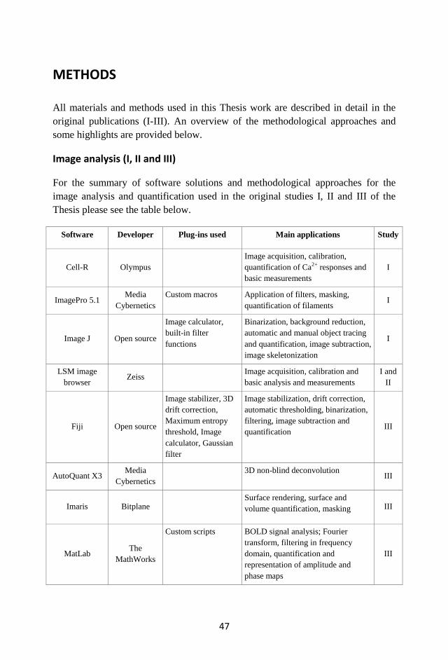
47
METHODS
All materials and methods used in this Thesis work are described in detail in the
original publications (I-III). An overview of the methodological approaches and
some highlights are provided below.
Image analysis (I, II and III)
For the summary of software solutions and methodological approaches for the
image analysis and quantification used in the original studies I, II and III of the
Thesis please see the table below.
Software Developer Plug-ins used Main applications Study
Cell-R Olympus
Image acquisition, calibration,
quantification of Ca2+ responses and
basic measurements
I
ImagePro 5.1 Media
Cybernetics
Custom macros Application of filters, masking,
quantification of filaments I
Image J Open source
Image calculator,
built-in filter
functions
Binarization, background reduction,
automatic and manual object tracing
and quantification, image subtraction,
image skeletonization
I
LSM image
browser Zeiss
Image acquisition, calibration and
basic analysis and measurements
I and
II
Fiji Open source
Image stabilizer, 3D
drift correction,
Maximum entropy
threshold, Image
calculator, Gaussian
filter
Image stabilization, drift correction,
automatic thresholding, binarization,
filtering, image subtraction and
quantification III
AutoQuant X3 Media
Cybernetics
3D non-blind deconvolution III
Imaris Bitplane Surface rendering, surface and
volume quantification, masking III
MatLab The
MathWorks
Custom scripts BOLD signal analysis; Fourier
transform, filtering in frequency
domain, quantification and
representation of amplitude and
phase maps
III
48
Primary cortical astrocyte cultures (I)
Cortical astrocytes were obtained from 2-3-day-old Wistar rats. After dissection
and cell dissociation they were plated on glass bottom MatTek dishes precoated
with poly-L-lysine or bovine plasma fibronectin at a density of 5x10⁴ cells per
square centimeter. Cells were maintained in culture with high-glucose DMEM
supplemented with 10% fetal bovine serum and antibiotics at +37 oC, 5% CO2.
Plasmids were transfected into cultured astrocytes 2-3 days after plating using
Lipofectamine reagent according to manufacturer recommendations.
Photolysis of caged calcium and live cell imaging (I)
Prior to the Ca2+
uncaging experiments cultured cortical astrocytes were loaded
with caged 10 μM Ca2+
compound o-nitrophenyl (NP)-EGTA AM ester with 4 μM
Fura-2 AM and 0.025% Pluronic F-127. The ultraviolet light beam (350-400 nm)
flash (200 ms) from 150 W xenon lamp focused by Olympus PlanApo 60x/NA
1.45 oil objective was used for the photolysis of NP-EGTA compound. Single
wavelength (380 nm) Fura-2 signal and generated ΔF/F ratio (Grynkiewicz et al.,
1985) were used to evaluate effectiveness and the slope of Ca2+
responses and to
perform calibration for estimation of peak intracellular concentrations of Ca2+
caused by uncaging.
For live cell imaging of astrocytes in culture the inverted Olympus Cell-R
microscope equipped with Olympus PlanApo 60x/NA 1.45 oil objective and CCD
camera was used. For fine morphological tracing the maximum camera resolution
1344x1024 pixels was used. Frames with an exposure time of 500 ms were
collected at a frequency of 1 to 0.2 Hz depending on particular experimental
procedure. During imaging cells were maintained at +34 oC, 5% CO2 humidified
atmosphere.
Postnatal in vivo electroporation (II)
Gene delivery using postnatal electroporation technique was done on newborn
Wistar rat pups (1-2 days old). 50 to 100 nl of Plasmid solution with a high
concentration of endotoxin-free DNA (1.5-3.0 mg/ml) was injected into the brain
cortex through the pooled glass needle using stereotaxic manipulator and automatic
microinjector. Plasmid injection was followed by the train of 5 rectangular
electrical pulses (100-150 V/cm, 50 ms, 1 Hz). All surgical procedures were done
under the isoflurane anesthesia (4% for induction, 2% for maintenance). Operated
pups were recovered from anesthesia in warm chamber and returned to their
mother within 15-20 minutes after operation was completed.
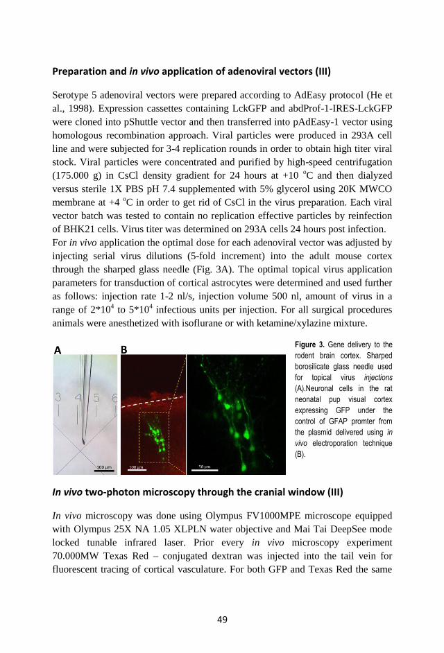
49
Preparation and in vivo application of adenoviral vectors (III)
Serotype 5 adenoviral vectors were prepared according to AdEasy protocol (He et
al., 1998). Expression cassettes containing LckGFP and abdProf-1-IRES-LckGFP
were cloned into pShuttle vector and then transferred into pAdEasy-1 vector using
homologous recombination approach. Viral particles were produced in 293A cell
line and were subjected for 3-4 replication rounds in order to obtain high titer viral
stock. Viral particles were concentrated and purified by high-speed centrifugation
(175.000 g) in CsCl density gradient for 24 hours at +10 oC and then dialyzed
versus sterile 1X PBS pH 7.4 supplemented with 5% glycerol using 20K MWCO
membrane at +4 oC in order to get rid of CsCl in the virus preparation. Each viral
vector batch was tested to contain no replication effective particles by reinfection
of BHK21 cells. Virus titer was determined on 293A cells 24 hours post infection.
For in vivo application the optimal dose for each adenoviral vector was adjusted by
injecting serial virus dilutions (5-fold increment) into the adult mouse cortex
through the sharped glass needle (Fig. 3A). The optimal topical virus application
parameters for transduction of cortical astrocytes were determined and used further
as follows: injection rate 1-2 nl/s, injection volume 500 nl, amount of virus in a
range of 2*104 to 5*10
4 infectious units per injection. For all surgical procedures
animals were anesthetized with isoflurane or with ketamine/xylazine mixture.
Figure 3. Gene delivery to the
rodent brain cortex. Sharped
borosilicate glass needle used
for topical virus injections
(A).Neuronal cells in the rat
neonatal pup visual cortex
expressing GFP under the
control of GFAP promter from
the plasmid delivered using in
vivo electroporation technique
(B).
In vivo two-photon microscopy through the cranial window (III)
In vivo microscopy was done using Olympus FV1000MPE microscope equipped
with Olympus 25X NA 1.05 XLPLN water objective and Mai Tai DeepSee mode
locked tunable infrared laser. Prior every in vivo microscopy experiment
70.000MW Texas Red – conjugated dextran was injected into the tail vein for
fluorescent tracing of cortical vasculature. For both GFP and Texas Red the same
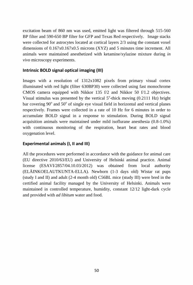
50
excitation beam of 860 nm was used, emitted light was filtered through 515-560
BP filter and 590-650 BP filter for GFP and Texas Red respectively. Image stacks
were collected for astrocytes located at cortical layers 2/3 using the constant voxel
dimensions of 0.167x0.167x0.5 microns (XYZ) and 5 minutes time increment. All
animals were maintained anesthetized with ketamine/xylazine mixture during in
vivo microscopy experiments.
Intrinsic BOLD signal optical imaging (III)
Images with a resolution of 1312x1082 pixels from primary visual cortex
illuminated with red light (filter 630BP30) were collected using fast monochrome
CMOS camera equipped with Nikkor 135 f/2 and Nikkor 50 f/1.2 objectives.
Visual stimulus was presented by the vertical 5o-thick moving (0.2111 Hz) bright
bar covering 90o and 50
o of single eye visual field in horizontal and vertical planes
respectively. Frames were collected in a rate of 10 Hz for 6 minutes in order to
accumulate BOLD signal in a response to stimulation. During BOLD signal
acquisition animals were maintained under mild isoflurane anesthesia (0.8-1.0%)
with continuous monitoring of the respiration, heart beat rates and blood
oxygenation level.
Experimental animals (I, II and III)
All the procedures were performed in accordance with the guidance for animal care
(EU directive 2010/63/EU) and University of Helsinki animal practice. Animal
license (ESAVI/2857/04.10.03/2012) was obtained from local authority
(ELÄINKOELAUTKUNTA-ELLA). Newborn (1-3 days old) Wistar rat pups
(study I and II) and adult (2-4 month old) C56BL mice (study III) were bred in the
certified animal facility managed by the University of Helsinki. Animals were
maintained in controlled temperature, humidity, constant 12/12 light-dark cycle
and provided with ad libitum water and food.
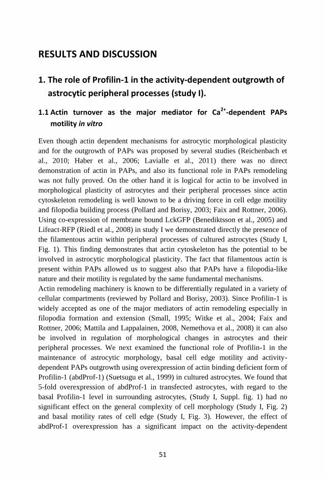
51
RESULTS AND DISCUSSION
1. The role of Profilin-1 in the activity-dependent outgrowth of
astrocytic peripheral processes (study I).
1.1 Actin turnover as the major mediator for Ca2+-dependent PAPs
motility in vitro
Even though actin dependent mechanisms for astrocytic morphological plasticity
and for the outgrowth of PAPs was proposed by several studies (Reichenbach et
al., 2010; Haber et al., 2006; Lavialle et al., 2011) there was no direct
demonstration of actin in PAPs, and also its functional role in PAPs remodeling
was not fully proved. On the other hand it is logical for actin to be involved in
morphological plasticity of astrocytes and their peripheral processes since actin
cytoskeleton remodeling is well known to be a driving force in cell edge motility
and filopodia building process (Pollard and Borisy, 2003; Faix and Rottner, 2006).
Using co-expression of membrane bound LckGFP (Benediktsson et al., 2005) and
Lifeact-RFP (Riedl et al., 2008) in study I we demonstrated directly the presence of
the filamentous actin within peripheral processes of cultured astrocytes (Study I,
Fig. 1). This finding demonstrates that actin cytoskeleton has the potential to be
involved in astrocytic morphological plasticity. The fact that filamentous actin is
present within PAPs allowed us to suggest also that PAPs have a filopodia-like
nature and their motility is regulated by the same fundamental mechanisms.
Actin remodeling machinery is known to be differentially regulated in a variety of
cellular compartments (reviewed by Pollard and Borisy, 2003). Since Profilin-1 is
widely accepted as one of the major mediators of actin remodeling especially in
filopodia formation and extension (Small, 1995; Witke et al., 2004; Faix and
Rottner, 2006; Mattila and Lappalainen, 2008, Nemethova et al., 2008) it can also
be involved in regulation of morphological changes in astrocytes and their
peripheral processes. We next examined the functional role of Profilin-1 in the
maintenance of astrocytic morphology, basal cell edge motility and activity-
dependent PAPs outgrowth using overexpression of actin binding deficient form of
Profilin-1 (abdProf-1) (Suetsugu et al., 1999) in cultured astrocytes. We found that
5-fold overexpression of abdProf-1 in transfected astrocytes, with regard to the
basal Profilin-1 level in surrounding astrocytes, (Study I, Suppl. fig. 1) had no
significant effect on the general complexity of cell morphology (Study I, Fig. 2)
and basal motility rates of cell edge (Study I, Fig. 3). However, the effect of
abdProf-1 overexpression has a significant impact on the activity-dependent
52
morphological plasticity of PAPs that was induced by Ca2+
uncaging in cultured
astrocytes (Study I, Fig. 4). Control astrocytes did respond to the Ca2+
increase by
the extensive PAPs outgrowth that resulted in doubling the PAPs density from 1 to
2 PAPs per 10 µm of cell perimeter 400 second after the Ca2+
uncaging was
performed. However, astrocytes that express abdProf-1 were not able to increase
the PAPs density in a response to Ca2+
uncaging; despite they had the same basal
level density of PAPs (Study I, Fig. 4G). Since the density of processes in the cell
at the certain time point reflects the dynamic equilibrium between filopodia
extension and retraction events we can assume that overexpression of abdProf-1
prevented the shift of equilibrium towards the prevalence of PAPs formation over
the PAPs retraction or in the other words damped the PAPs outgrowth rate to the
basal level silencing the effect of Ca2+
stimulation. These findings demonstrate that
first – astrocytes are able to form new filopodia-like processes in a response to the
increase of intracellular Ca2+
and second – that Profilin-1 interaction with actin
monomers is critical for the rapid extensive formation of astrocytic processes
caused by the increase of intracellular Ca2+
but not for the maintenance of static
morphology and base level motility of the cell edge.
Profilin-1 binds to actin monomers, catalyzes the ADP-to-ATP exchange and
facilitates shuttling of G-actin to the filamentous actin barbed ends promoting its
polymerization (Pollard and Borisy, 2003). At the same time both actin and
Profilin-1, as participants of the cyclic process of actin treadmilling, are subjected
to feedback inhibition caused by monomer deficiency in case of extensive
polymerization (Yarmola et al., 2009). Once we had found the damping effect of
abdProf-1 overexpression on extensive de novo PAPs formation in Ca2+
stimulated
astrocytes, we asked the question if there is any difference in properties of
individual PAPs. Using time-lapse microscopy on astrocytes in culture we
measured the kinetics for the appearance and extension of individual PAPs induced
by Ca2+
uncaging. In addition to quantitative differences described above we found
also qualitative differences between PAPs formed by control and abdProf-1
expressing astrocytes. Indeed, the average outgrowth rate for PAPs formed by
astrocytes expressing abdProf-1 was almost 2 times lower than in control
astrocytes that expressed LckGFP only (1.89 µm/min in abdProf-1 versus 3.26
µm/min in control). In addition to decreased outgrowth rates PAPs from astrocytes
expressing abdProf-1 had on average significantly decreased maximum length
(2.66 µm in abdProf-1 versus 4.61 µm in control) (Study I, Fig. 4H, I and J). These
observations pointed out the Profilin-1 interaction with actin as a factor affecting
the dynamics of activity-dependent PAPs outgrowth, which together with the
presence of F-actin in PAPs suggests an actin-dependent mechanism underlying
PAPs remodeling.
53
1.2 The novel tool to suppress activity-dependent PAPs motility
In several recent studies cellular motility has been manipulated by affecting
cytoskeleton-regulating signaling cascades on the level of small GTPases. Indeed,
it was implemented as a photactivatable form of Rac that allowed control of cell
migration (Wu et al., 2009) also as an overexpression of dominant negative form of
Rac that was shown to suppress PAPs motility rates in organotypic slices (Nishida
and Okabe, 2007). The main drawback for the GTPase-based approaches is caused
by divergence of the Rac signaling and the side effects on other motility types like
lamellipodia formation and general ability of the cell to migrate rather than to local
activity-dependent morphological changes (Pollard and Borisy, 2003). In order to
overcome these limitations and affect specifically astrocytic filopodia-like
processes in activity-dependent manner, rather than cell migration in general, we
utilized the Profilin-1 as a direct effector of actin polymerization which is well
known for its role in filopodia building process (Witke, 2004; Faix and Rottner,
2006; Mattila and Lappalainen, 2009) and was demonstrated to be critical
especially for microspikes formation (Suetsugu et al., 1998; 1999).
We propose that overexpression of abdProf-1 that was shown to suppress activity
dependent outgrowth of PAPs in vitro (Study I, Fig. 4) can also be used as a
genetically encodable tool to manipulate the morphological plasticity of astrocytes
in vivo and study the effect of its suppression on neuronal function. However, in
order to use the abdProf-1 in vivo the proper gene delivery system for astrocyte-
specific targeting should be implemented.
2. Gene delivery to a postnatal rodent brain (studies II and III).
2.1 Stereotaxic plasmid microinjection and electroporation on neonatal
rat brain (study II)
Gene delivery using electro-mediated nucleic acid transfer such as electroporation
or iontophoresis is a powerful alternative to time consuming creation of transgenic
animals or approaches based on viral vectors. Although the exact mechanism for
nucleic acid entering the cell is not still revealed, electro-mediated nucleic acid
transfer is widely used in CNS research (reviewed by De Vry et al., 2010(a)). The
principal idea of the method is to inject genetic material such as DNA, RNA or
shRNA into the brain and then facilitate its uptake by applying several electrical
pulses that create directed electrical field and electro-driving force for charged
nucleic acid molecules and also influence properties of cellular membranes. There
are two main modifications for this technique; the one that is performed prenatally
54
by mean of surgical removal of the embryos from the uterus, plasmid injection into
the lateral ventricles followed by electroporation (Saito and Nakatsuji, 2001;
Tabata and Nakajima, 2001; Saito, 2006; Dixit et al., 2011; Sato et al., 2013) and
another one that is performed on the different postnatal stages with a help of
stereotaxic manipulator (Barnabe-Heider et al., 2008; Boutin et al., 2008; Borrell,
2010; Molotkov et al., 2010 (study II); 2011; De la Rossa et al., 2013) or without it
(Feliciano et al., 2013). Indeed, postnatal electroporation has such advantages as
possibility to target spatially restricted region of interest regarding to the
stereotaxic coordinates and overcome developmental compensatory effects of
exogenous gene transfer. However, it also was limited mainly to the transduction of
cells in a sub ventricular zone (SVZ). There are now several recent modifications
of the postnatal electroporation technique allowing targeting of cells in brain
regions outside the SVZ (Barnabe-Heider et al., 2008; Molotkov et al., 2010 (study
II); De al Rossa et al., 2013).
Using plasmid with ubiquitously expressed CAG promoter and EGFP as a reporter
gene (Matsuda and Cepko, 2004; 2007) we modified the postnatal electroporation
technique in order to apply it to non-SVZ brain regions such as cortical areas. We
found that rapid injection (within 10 seconds) of the small volume (50 nl) of the
highly concentrated plasmid (3 µg/µl) though the thin glass capillary (outlet
diameter 20-30 µm) that was immediately followed by the application of 5
rectangular (50 ms, 1 Hz) electrical pulses in a voltage controlled mode (120 V/cm)
via forceps-like electrodes placed directly on the skull surface (Study II, movie)
produces a well-defined pattern of EGFP expression in cortex and striatum (Study
II, Fig. 1). We also found that the age between 1 and 3 postnatal days is the most
suitable for the performance of the described method likely due to relatively low
myelin content and not completely formed cranial bones which provide lower
electrical resistance and thus the same applied voltage results in generation of
higher current that improve transduction efficiency. Developed protocol also
implemented application of the electro conductive gel for the best electrical contact
between electrodes and skull bones and for the uniform distribution of the electrical
field between the electrodes that was shown before to be one of the critical factors
that can affect electroporation efficiency (Ivorra et al., 2008).
The method described in study II facilitates spatially restricted expression of genes
delivered in a form of plasmid DNA into neonatal brain parenchyma in vivo by
mean of stereotaxic injection and subsequent electroporation. There was one
previous study describing non-ventricular plasmid delivery in postnatal animals,
where the targeting of the hippocampal dentate gyrus neuronal stem cells was
shown on adult mice (Barnabe-Heider et al., 2008). In study II as well as in our
following experiments (Molotkov et al., 2011) we met the problem caused by low
55
transduction efficiency in cortex and hippocampus that is likely a characteristic
feature of non-dividing cells in the postnatal brain. The problem could be overcame
by use of new-born pups as described in study II or facilitation of DNA uptake by
application of complex-forming agents such as trans-cyclodextrane-1,2-diol that
provides DNA entry to the nucleus in the non-dividing cell (De la Rossa et al.,
2013). It is also possible that recently proposed improvements for the
electroporation procedure replacing voltage controlled pulses by current controlled
pulses (De Vry et al., 2010(b)) and use of local needle-like electrodes generating
higher electrical field density compared to plate electrodes (reviewed by De Vry et
al., 2010(a)) may improve postnatal electroporation efficiency.
The spatio-temporal restriction of the transgene expression is one of the main
issues considered in genetic manipulation in vivo. It provides cell type specificity
of expression, minimizing severe compensatory changes caused by prolonged
expression during early developmental stages and allows expressing the transgene
only in the defined region of interest within the brain. There are several approaches
that could be implemented with electroporation techniques in order to get spatio-
temporal expression selectivity (reviewed by LoTurco et al., 2009; Huh et al.,
2010). The use of cell-type specific promoters is the easiest way to get expression
in a certain subpopulations of cells in vitro or in case of creation of the transgenic
animals, but can meet some limitations when naked DNA is applied in vivo by
electroporation. Thus, if the expression of EGFP driven by ubiquitous CAG
promoter appeared in different cell types (study II, Fig. 1) use of astroglia-specific
GFAP promoter delivered into postnatal rat cortex by electroporation resulted in
EGFP expression in neuronal cells rather than in astrocytes despite its expression
specificity in vitro (Molotkov et al., 2011) (Fig. 3B). It is demonstrating the
flexibility of GFAP promoter expression pattern which is likely caused by bacterial
rather than eukaryotic methylation pattern of the plasmid DNA used for
electroporation. This observation is a hinderance to validate transgene expression
specificity in vivo when it is delivered in a form of naked bacterial plasmid DNA,
whilst at the same time it promotes in vivo electroporation technique for studying
regulatory elements influencing gene expression and effects of their epigenetic
modifications such as methylation using synthetic constructs in vivo.
2.2 Astrocyte-specific gene delivery using adenoviral vectors (study III).
An intrinsic feature of adenoviruses is their ability to interact with CAR on the cell
surface that determines a wide tropism of adenovirus-derived vectors (reviewed by
Merienne et al., 2013). In the brain the CAR expression is restricted to astroglial
56
cells and dopaminergic neurons (Lewis et al., 2010) that proposes adenoviral
vectors to be efficient and specific astrocyte transducers when they are applied
topically in the brain areas lacking dopaminergic neurons.
Using stereotaxic manipulator we applied topically to the adult mouse visual cortex
the adenoviral vector of serotype 5 that expresses the membrane form of GFP as a
fluorescent reporter under the control of CAG promoter. We checked ex vivo on
fixed brain slices and in vivo through the cranial window distribution of GFP-
expressing cells, expression selectivity, transduction efficiency and expression
timeline (Study III, Fig. 1). We found that although adenoviral particles are
relatively big (80-100 nm in diameter) they can diffuse 200-500 nm away from the
injection site causing efficient transduction in the total volume of 0.3-0.4 mm3 in
mouse cortex. Also single injection of 2*104 to 5*10
4 viral particles produces an
expression pattern occupying all cortical layers with densely packed GFP
expressing cells suggesting high transfection rate. Well defined borders of the
transduced zone are also suggests that effective expression could be achieved by
few viral particle per cell and proving the high infectivity known for adenoviral
vectors. Astrocyte labelling using immunostaining with anti-GFAP antibody on
frozen brain sections derived from transduced animals showed that all GFP
expressing cells were GFAP positive and that there are just a few non-transduced
GFAP positive cells in the region. This demonstrates astrocyte specificity and
further shows high transduction efficiency for the virus. Finally, we found that the
virus can facilitate long term expression which can be used in a period from 1 week
up to at least 6 weeks after the injection was done, providing an excellent timeline
for most experimental paradigms.
Although adenoviral vectors have been used to induce transgene expression in
astrocytes both in organotypic slices (Nishida and Okabe, 2007) and in vivo for
transduction of astroglia in the brainstem (Gourine et al., 2010) their effectiveness
and specificity is under debate (Merienne et al., 2013). Relatively high
immunogenic properties of adenovirus in comparison to other widely used vectors
such as lenti and adeno-associated viruses as well as assumptions regarding their
non-selectivity based on systemic adenovirus application (reviewed by Wirth et al.,
2013) stimulated the development of alternative viral vectors for astrocyte-specific
genetic manipulations. Indeed, there are at least two viral vectors used on a par
with adenoviruses that were shown to have high astrocytic expression specificity in
vivo. It was shown that pseudotyping of lentiviral vectors with MOKOLA envelope
protein can enhance their astrocytic tropism that could be further improved by
insertion of mir124T neuronal silencer sequence in the vector genome (Colin et al.,
2009). Another alternative to facilitate astrocyte-specific targeting is based on the
application of the AAV2/5 chimeric vector in combination with astroglia-specific
57
gfaABC1D promoter (Xie et al., 2010). Since AAV5 is known to have a wide
tropism in the brain (Burger et al., 2004; Aschauer et al., 2013) which also varies
depending on the developmental stage (Chakrabarty et al., 2013) it is possible to
assume that astroglial expression of AAV2/5 is likely caused by the promoter
rather than by virus transduction selectivity.
Based on the data we have obtained for adenovirus-mediated astrocyte transduction
and transgene expression we can conclude that the unfairly under evaluated
adenoviral vectors can be used in basic research as well is in some clinical
applications (reviewed by Wirth et al., 2013) as a safe episomal vector causing
high levels of transient expression. Naturally, limited virus tropism in the brain
allows fine tuning of the expression by use of different promoters without losing
cell-type specificity.
3. Motility of cortical astrocytes and neuronal function
(study III).
3.1 Astroglia spontaneous morphological changes are Profilin-1
dependent.
In primary cultures cortical astrocytes display both spontaneous and activity-
dependent morphological changes (see the study I). PAPs were also demonstrated
to be motile at active synaptic terminals in acute brain slices (Hirrlinger et al.,
2004) and to be involved in morphological interplay with dendritic spines in
organotypic brain slices (Haber et al., 2006; Nishida and Okabe, 2007). At the
same time spontaneous as well as activity-dependent morphological plasticity in
astrocytes in vivo has not been described yet.
Using the adenoviral vector expressing a membrane form of GFP that allows high
contrast labeling of PAPs we traced and quantified the rate of astrocytic motility at
basal conditions, after induction of microstroke and with the overexpression of
abdProf-1 (Study III, Fig. 3). We quantified the net changes of astrocyte peripheral
processes and found that under resting conditions they are subjected to continuous
relatively uniform morphological changes. This spontaneous motility results in 1.3-
1.5 % of astrocyte peripheral volume shift during 5 minutes interval. Our
measurements consider the overall motility rates which unfortunately difficult to
compare with the data obtained for the linear outgrowth rates or volume changes of
individual astrocytic processes near the active synaptic terminals (Hirrlinger et al.,
2004) or during synaptogenesis (Haber et al., 2006). Interestingly, we found that
overexpression of abdProf-1 caused suppression of spontaneous astrocyte motility
58
rates by 35-40% with respect to astrocytes expressing LckGFP only and used as a
control. This finding demonstrates that similarly to astrocytes in culture (Study I)
motility of PAPs in vivo is likely driven by actin dependent mechanism that is
dependent on actin binding by Profilin-1. Although in vitro Profilin-1
overexpression affects exclusively activity-dependent morphological changes of
astrocytes and, particularly, rapid PAPs outgrowth induced by Ca2+
uncaging, in
vivo it affects spontaneous motility of astrocytes. There are several key features of
in vivo astrocyte physiology that can cause different sensitivity to the abdProf-1
overexpression. First, astrocytes in vivo have remarkably increased
compartmentalization due to their highly ramified structure, small internal PAPs
volume limiting diffusion inside the PAP and thus, preventing local compensation
effects. Second, increased external signal inputs caused by complex environment
and also point-like localization of incoming signals sources, such as active synaptic
terminals, that create highly localized gradients of neurotransmitters. Third, it
should be considered that astrocytes in vivo are under the continuous baseline
stimulation caused by background neuronal activity even in the anesthetized brain.
Indeed, it would be extremely interesting to check whether spontaneous astrocytic
motility differs in awake and anesthetized brain and what are the key mechanisms
regulating motility of astrocytes in vivo. As a future development and improvement
for the current study we propose the use of dual membrane and cytoplasmic
fluorescent reporters that would allow more precise tracing of the surface of
astrocyte together with its internal volume that would further increase signal to
noise ratio for the microscopic investigation of PAPs in vivo.
3.2 Structure-functional interplay: the effect of suppressed astrocytic
motility on neuronal processing.
Astrocytes were recently shown to affect neuronal processing in several ways.
They can control the synaptic activity and basal synaptic transmission (Araque et
al., 1999; Perea et al., 2009; Fellin et al., 2009; Navarette and Araque, 2011), affect
metabolic coupling between neurons and astroglia (Magistretti, 2006; Pellerin et
al., 2007) and participate in local control of blood flow in an activity-dependent
manner (Zonta et al., 2003; Mulligan and McVicar, 2004; Takano et al., 2006). At
the same time some major aspects of glia-neuronal signaling are still controversial
(Fiacco et al., 2007; Petravicz et al., 2008; Agulhon et al., 2008; Agulhon et al.,
2010). There are several potential sources for the controversy in the field of neuro-
glial research. First, it became evident recently that signaling linking astroglia and
neurons is not as simple as was assumed before and can include some alternative
59
pathways as well as subjected for developmental changes (Sun et al., 2013;
Shigetomi et al., 2013(b)) and thus, have to be revised. Second, different results
were obtained under different experimental conditions (reviewed by Nedergaard
and Verkhratsky, 2012). Additionally some experimental tools cannot be assumed
as fully physiological relevant. Thus, Ca2+
uncaging differs from receptor mediated
Ca2+
spike by its amplitude, kinetics and localization providing a good solution for
general astrocyte activation but incomparable in physiological sense with glia-
neuronal signaling occurring in vivo (Wang et al., 2013). In this situation shading
the light on the role of astrocytes in neuronal function using physiologically
relevant tools in vivo is an extremely important issue.
Existing in vitro and ex vivo data demonstrate that astrocytes are highly motile cells
and can respond to a variety of external and internal stimuli by changing their
morphology in a short term manner (reviewed by Reichenbach et al., 2010).
Particularly it was shown in ultrastructural studies that PAPs and synapses are
closely associated (Witcher et al., 2007), and there is a correlation between long-
term functional changes in synapses and their coverage by PAPs (Lushnikova et
al., 2009). Using computational approaches to simulate changes in synaptic
geometry it was predicted that PAP-synapse morphological interplay as well as
GluT density in astrocytic compartment of the synapse could be the main
determinants for synaptic efficacy and kinetics of postsynaptic neurotransmitter
action (Freche et al., 2011) allowing us to assume that astroglial morphological
changes could lead to functional changes in synapses. Several studies also
demonstrated other possible molecular players involved in astrocytic
morphological changes such as Ezrin, mGluRs, AQP4 and Kir4.1 (Lavialle et al.,
2011; Nagelhus et al., 2004). Indeed, there is accumulating evidence that PAPs
structural changes are driven by actin dependent mechanism (Haber et al., 2006,
Nishida and Okabe, 2007; Lavialle et al., 2011).
We used abdProf-1 overexpression from the adenoviral vector, a molecular tool
previously demonstrated to moderately suppress spontaneous PAPs motility in vivo
(see part 3.1 and Study III, Fig. 3). By measuring BOLD signal propagation in
response to visual stimulation as the readout of net neuronal activity in the primary
visual cortex of adult anesthetized mice we tested whether suppressed astrocytic
motility can affect neuronal processing. We found no signs of any distortions in
activated cortical areas, magnitude maps or temporal phase maps as recorded from
the visual cortices of animals transduced with control adenovirus expressing
LckGFP or adenovirus expressing abdProf-1 (Study III, Fig. 4). In order to test
whether optical fBOLD signal can be used for detecting functional changes in this
experimental paradigm we quantified changes in fBOLD signal caused by laser
induced thrombosis of single blood vessels and found that our acquisition and
60
analysis protocol is suitable for detecting functional changes caused by the
microstroke (Study III, Fig. 5). These sets of data indicate that although
microstroke caused functional changes in the cortex, the suppression of astrocytic
motility by 35-40% (via abdProf-1) had no effect on net neuronal activity and
corresponding oxygen metabolism in primary visual cortex of the adult
anesthetized mouse.
Despite that there are many reasons for astrocytic motility to be closely related
with neuronal activity and metabolism, there are also aspects which can dissociate
the link between morphological plasticity of astrocytes and synaptic function.
Astrocytes are the first line to react on anesthesia since they have higher sensitivity
for general anesthetics and lower threshold level to block their Ca2+
signalization
(Schummers et al., 2008; Thrane et al., 2012). Silencing of astroglial Ca2+
signaling
could also result in increased neuronal firing rates in the anesthetized brain (Thrane
et al., 2012) which should cause metabolic changes and affect BOLD signal.
Moreover developmental changes in the brain were shown to affect neuro-astrocyte
crosstalk (Sun et al., 2013) and particularly caused decreased astrocytic sensitivity
to general anesthetics in the developing brain (Hirase et al., 2004). Thus,
investigating effect of suppressed astrocytic motility on net neuronal activity and
synaptic function in the awake brain could be proposed as the next step to continue
this study. It would also be useful to compare the obtained results with results
gathered via more drastic PAPs motility suppression and also probe the functional
effect of local astrocytic GluT blockade in awake and anesthetized brain. We also
propose that the BOLD signal imaging could be accompanied with macro scale
Ca2+
or voltage sensitive dye imaging in the cortex in order to separate neuronal
activity and hemodynamic signal. Further, use of plasticity induction paradigms
might reveal some changes caused by the suppression of astrocytic motility in the
brain.
61
CONCLUSIONS
We developed and validated on primary astroglial culture a molecular genetically
encodable tool for suppression of activity-dependent PAPs motility and fine
morphological tracing of astrocytic morphology (study I).
We developed postnatal in vivo electroporation technique applicable for the rodent
brain cortical areas (Study II) but found it to be unreliable for cases when absolute
cell-type specificity is a prerequisite. We found out the gene delivery based on
adenovirus serotype 5 with the CAG promoter to be a reliable astrocyte-specific
gene delivery tool in vivo in the rodent brain cortex (study III).
Results obtained in this work demonstrate that although astrocytes in vivo are
subjected for spontaneous morphological changes, suppression of the astroglial
motility rates by 35% does not cause detectable changes in functionality of the
neuronal network in mouse visual cortex under light isoflurane anesthesia. We did
not find the direct link between astrocytic motility and functional performance of
the brain under particular conditions (Study III).
62
ACKNOWLEDGEMENTS
Five years of PhD studies was a saturated period of my life. There were asperities as well as
a lot of very bright moments. It would be merely impossible for me to complete my thesis
project without practical help, mental support and friendship from people around.
I want to thank my supervisor Dr. Leonard Khirug for giving me freedom in project design
and selection of experimental approaches. I have learnt a lot while working with you, Leo.
Special thanks to my co-supervisor Prof. Eero Castren for giving me possibility to complete
the project and write the thesis. It was also a great experience to work in your lab.
Special thanks to members of the thesis advisory board; Prof. Kai Kaila and Prof. Pekka
Lappalainen. I appreciate your guidance and your advices during all five years of my PhD.
Even though, I did not follow all of your comments and advices, with no doubt they
influenced my current outlook.
I am grateful to Dr. Nathalie Rouach and Dr. Sarah Coleman for reviewing my thesis
manuscript and for their professional and useful comments on my dissertation.
Special thanks to FGSN coordinator Dr. Katri Wegelius, an excellent organizer and person
who care about students, for guiding me on my way to PhD.
I want to thank Maria Anastasina, Svetlana Zobova, Mikhail Kislin, Evgeny Pryazhnikov,
Mikhail Paveliev, Stanislav Khirug, Elena Vaschinkina, Julia Kolikova, Pavel Uvarov,
Alexey Adamov, Anastasia Ludwig, Mikhail Yuriev, Ramil Afzalov, Aleksandr Surin,
Dmytro Toptunov, Eva Zusinaite, Lev Levanov, Alesya Romanovskaya, Andrey
Golubtsov, Rashid Giniatullin, Ilya Kirilkin, Natalia Kulesskaya, Ekaterina Mugantseva,
Evgeny Kulessky, Vera Shirokova, Pedro Pereira, Ilya Lesnichy and Roman Brulgart.
These people helped me in performing scientific work, supported me by their friendship
and gave me the will to win.
I want also to thank current and former members of Prof. Kai Kaila and Prof. Eero
Castren’s labs for their practical help, useful advices and friendship, especially Martin
Pushkarjov, Else Tolner, Alexey Yukin, Peter Blaesse, Britta Haas, Merle Kampura, Faraz
Ahmad, Nina Karpova and Ramon Guirado.
There are several funding sources which I am very grateful for supporting my research
financially: Finnish Graduate School in Neuroscience, Center for International Mobility of
Finland, Research Foundation of the University of Helsinki, Sigrid Juselius Foundation and
Academy of Finland.
Completion of this work would not be possible without my family members, actually
without them I would absolutely another person, and thus this thesis has their significant
input as well. I am really thankful to my wife Karina and my daughter Marina for being
with me, supporting me and tolerating my not ideal nature. Special thanks to my mom, my
dad and my brother for their patience and for making me what I am.
63
LIST OF REFERENCES
Abbott, N.J., 2005. Dynamics of CNS barriers: evolution, differentiation, and modulation.
Cell. Mol. Neurobiol. 25, 5–23.
Abbott, N.J., Rönnbäck, L., Hansson, E., 2006. Astrocyte-endothelial interactions at the
blood-brain barrier. Nat. Rev. Neurosci. 7, 41–53.
Afshari, F.T., Kwok, J.C., White, L., Fawcett, J.W., 2010. Schwann cell migration is
integrin-dependent and inhibited by astrocyte-produced aggrecan. Glia 58, 857–69.
Agulhon, C., Fiacco, T., McCarthy, K.D., 2010. Hippocampal short- and long-term
plasticity are not modulated by astrocyte Ca2+ signaling. Science 327, 1250–4.
Agulhon, C., Petravicz, J., McMullen, A.B., Sweger, E.J., Minton, S.K., Taves, S.R.,
Casper, K.B., Fiacco, T. a, McCarthy, K.D., 2008. What is the role of astrocyte calcium
in neurophysiology? Neuron 59, 932–46.
Aguzzi, A., Barres, B., Bennett, M.L., 2013. Microglia: scapegoat, saboteur, or something
else? Science 339, 156–61.
Ahmed, N., Ramjeesingh, M., Wong, S., Varga, A., Garami, E., Bear, C.E., 2000. Chloride
channel activity of ClC-2 is modified by the actin cytoskeleton. Biochem. J. 352 Pt 3,
789–94.
Airan, R.D., Thompson, K.R., Fenno, L.E., Bernstein, H., Deisseroth, K., 2009. Temporally
precise in vivo control of intracellular signalling. Nature 458, 1025–9.
Akerman, K.E., 1982. Ca2+ transport and cell activation. Med. Biol. 60, 168–82.
Allam, S.L., Ghaderi, V.S., Bouteiller, J.-M.C., Legendre, A., Ambert, N., Greget, R.,
Bischoff, S., Baudry, M., Berger, T.W., 2012. A computational model to investigate
astrocytic glutamate uptake influence on synaptic transmission and neuronal spiking.
Front. Comput. Neurosci. 6, 70.
Allaman, I., Bélanger, M., Magistretti, P.J., 2011. Astrocyte-neuron metabolic
relationships: for better and for worse. Trends Neurosci. 34, 76–87.
Angulo, M.C., Kozlov, A.S., Charpak, S., Audinat, E., 2004. Glutamate released from glial
cells synchronizes neuronal activity in the hippocampus. J. Neurosci. 24, 6920–7.
Araque, A, Parpura, V., Sanzgiri, R.P., Haydon, P.G., 1999. Tripartite synapses: glia, the
unacknowledged partner. Trends Neurosci. 22, 208–15.
Armbruster, B.N., Li, X., Pausch, M.H., Herlitze, S., Roth, B.L., 2007. Evolving the lock to
fit the key to create a family of G protein-coupled receptors potently activated by an
inert ligand. Proc. Natl. Acad. Sci. U. S. A. 104, 5163–8.
Aroeira, R.I., Sebastião, A.M., Valente, C., 2013. GlyT1 and GlyT2 in brain astrocytes:
expression, distribution and function. Brain Struct. Funct.
Arriza, J.L., Fairman, W.A., Amara, S.G., Kavanaugh, P., Wadiche, I., Murdoch, H., 1994.
Functional Comparisons of Three Glutamate Cloned from Human Motor Cortex
Transporter 14, 5559–5569.
64
Aschauer, D.F., Kreuz, S., Rumpel, S., 2013. Analysis of transduction efficiency, tropism
and axonal transport of AAV serotypes 1, 2, 5, 6, 8 and 9 in the mouse brain. PLoS One
8, e76310.
Asztely, F., Erdemli, G., Kullmann, D.M., 1997. Extrasynaptic glutamate spillover in the
hippocampus: dependence on temperature and the role of active glutamate uptake.
Neuron 18, 281–93.
Attwell, D., Buchan, A.M., Charpak, S., Lauritzen, M., Macvicar, B., Newman, E., 2010.
Glial and neuronal control of brain blood flow. Nature 468, 232–43.
Baba, H., Nakahira, K., Morita, N., Tanaka, F., Akita, H., Ikenaka, K., 1997. GFAP gene
expression during development of astrocyte. Dev. Neurosci. 19, 49–57.
Bacskai, B.J., Klunk, W.E., Mathis, C., Hyman, B.T., 2002. Imaging amyloid-beta deposits
in vivo. J. Cereb. Blood Flow Metab. 22, 1035–41.
Bal-Price, A., Moneer, Z., Brown, G.C., 2002. Nitric oxide induces rapid, calcium-
dependent release of vesicular glutamate and ATP from cultured rat astrocytes. Glia 40,
312–23.
Bandettini, P., 2012. Twenty years of functional MRI: the science and the stories.
Neuroimage 62, 575–88.
Barnabé-Heider, F., Meletis, K., Eriksson, M., Bergmann, O., Sabelström, H., Harvey,
M.A., Mikkers, H., Frisén, J., 2008. Genetic manipulation of adult mouse neurogenic
niches by in vivo electroporation. Nat. Methods 5, 189–96.
Barres, B., 2008. The mystery and magic of glia: a perspective on their roles in health and
disease. Neuron 60, 430–40.
Benediktsson, A.M., Schachtele, S.J., Green, S.H., Dailey, M.E., 2005. Ballistic labeling
and dynamic imaging of astrocytes in organotypic hippocampal slice cultures 141, 41–
53.
Bennett, M.V., Contreras, J.E., Bukauskas, F.F., Sáez, J.C., 2003. New roles for astrocytes:
gap junction hemichannels have something to communicate. Trends Neurosci. 26, 610–
7.
Bennett, M.V.L., Garré, J.M., Orellana, J., Bukauskas, F.F., Nedergaard, M., Sáez, J.C.,
2012. Connexin and pannexin hemichannels in inflammatory responses of glia and
neurons. Brain Res. 1487, 3–15.
Bergles, D.E., Jahr, C.E., 1998. Glial contribution to glutamate uptake at Schaffer
collateral-commissural synapses in the hippocampus. J. Neurosci. 18, 7709–16.
Bergmann, K., 1857. Notiz über einige Structurverhältnisse des Cerebellum und
Rückenmarks. Zeitschrift für rationelle Medizin.
Bernardinelli, Y., Magistretti, P.J., Chatton, J.-Y., 2004. Astrocytes generate Na+-mediated
metabolic waves. Proc. Natl. Acad. Sci. U. S. A. 101, 14937–42.
Bernardinelli, Y., Salmon, C., Jones, E. V, Farmer, W.T., Stellwagen, D., Murai, K.K.,
2011. Astrocytes display complex and localized calcium responses to single-neuron
stimulation in the hippocampus. J. Neurosci. 31, 8905–19.
65
Berretta, S., 2012. Extracellular matrix abnormalities in schizophrenia. Neuropharmacology
62, 1584–97.
Bessis, A., Echade, C.B., Bernard, D., Roumier, A., 2007. Microglial Control of Neuronal
Death and Synaptic Properties 238, 233–238.
Bezzi, P., Gundersen, V., Galbete, J.L., Seifert, G., Steinhäuser, C., Pilati, E., Volterra, A.,
2004. Astrocytes contain a vesicular compartment that is competent for regulated
exocytosis of glutamate. Nat. Neurosci. 7, 613–20.
Bhattacharjee, A., Kaczmarek, L.K., 2005. For K+ channels, Na+ is the new Ca2+. Trends
Neurosci. 28, 422–8.
Biber, K., Laurie, D.J., Berthele, A., Sommer, B., Tölle, T.R., Gebicke-Härter, P.J., van
Calker, D., Boddeke, H.W., 1999. Expression and signaling of group I metabotropic
glutamate receptors in astrocytes and microglia. J. Neurochem. 72, 1671–80.
Biewenga, J.E., Destrée, O.H., Schrama, L.H., 1997. Plasmid-mediated gene transfer in
neurons using the biolistics technique. J. Neurosci. Methods 71, 67–75.
Binder, D.K., Nagelhus, E., Ottersen, O.P., 2012. Aquaporin-4 and epilepsy. Glia 60, 1203–
14.
Bittar, P.G., Charnay, Y., Pellerin, L., Bouras, C., Magistretti, P.J., 1996. Selective
distribution of lactate dehydrogenase isoenzymes in neurons and astrocytes of human
brain. J. Cereb. Blood Flow Metab. 16, 1079–89.
Borrell, V., 2010. In vivo gene delivery to the postnatal ferret cerebral cortex by DNA
electroporation. J. Neurosci. Methods 186, 186–95.
Boucsein, C., Zacharias, R., Färber, K., Pavlovic, S., Hanisch, U.-K., Kettenmann, H.,
2003. Purinergic receptors on microglial cells: functional expression in acute brain
slices and modulation of microglial activation in vitro. Eur. J. Neurosci. 17, 2267–76.
Boutin, C., Diestel, S., Desoeuvre, A., Tiveron, M.-C., Cremer, H., 2008. Efficient in vivo
electroporation of the postnatal rodent forebrain. PLoS One 3, e1883.
Bowman, C.L., Kimelberg, H.K., 1984. Excitatory amino acids directly depolarize rat brain
astrocytes in primary culture. Nature 311, 656–9.
Bowser, D.N., Khakh, B.S., 2004. ATP excites interneurons and astrocytes to increase
synaptic inhibition in neuronal networks. J. Neurosci. 24, 8606–20.
Brenner, M., Kisseberth, W.C., Su, Y., Besnard, F., Messing, A., 1994. GFAP promoter
directs astrocyte-specific expression in transgenic mice. J. Neurosci. 14, 1030–7.
Bruzzone, R., Giaume, C., 1999. Connexins and information transfer through glia. Adv.
Exp. Med. Biol. 468, 321–37.
Burden, S.J., 1998. The formation of neuromuscular synapses. Genes Dev. 12, 133–48.
Burger, C., Gorbatyuk, O.S., Velardo, M.J., Peden, C.S., Williams, P., Zolotukhin, S.,
Reier, P.J., Mandel, R.J., Muzyczka, N., 2004. Recombinant AAV viral vectors
pseudotyped with viral capsids from serotypes 1, 2, and 5 display differential efficiency
and cell tropism after delivery to different regions of the central nervous system. Mol.
Ther. 10, 302–17.
66
Bushong, E., Martone, M.E., Jones, Y.Z., Ellisman, M.H., 2002. Protoplasmic astrocytes in
CA1 stratum radiatum occupy separate anatomical domains. J. Neurosci. 22, 183–92.
Cahoy, J.D., Emery, B., Kaushal, A., Foo, L.C., Zamanian, J.L., Christopherson, K.S.,
Xing, Y., Lubischer, J.L., Krieg, P., Krupenko, S., Thompson, W.J., Barres, B., 2008. A
transcriptome database for astrocytes, neurons, and oligodendrocytes: a new resource
for understanding brain development and function. J. Neurosci. 28, 264–78.
Candolfi, M., Curtin, J.F., Xiong, W.-D., Kroeger, K.M., Liu, C., Rentsendorj, A.,
Agadjanian, H., Medina-Kauwe, L., Palmer, D., Ng, P., Lowenstein, P.R., Castro, M.G.,
2006. Effective high-capacity gutless adenoviral vectors mediate transgene expression
in human glioma cells. Mol. Ther. 14, 371–81.
Cannon, J.R., Sew, T., Montero, L., Burton, E.A., Greenamyre, J.T., 2011. Pseudotype-
dependent lentiviral transduction of astrocytes or neurons in the rat substantia nigra.
Exp. Neurol. 228, 41–52.
Chakrabarty, P., Rosario, A., Cruz, P., Siemienski, Z., Ceballos-Diaz, C., Crosby, K.,
Jansen, K., Borchelt, D.R., Kim, J.-Y., Jankowsky, J.L., Golde, T.E., Levites, Y., 2013.
Capsid serotype and timing of injection determines AAV transduction in the neonatal
mice brain. PLoS One 8, e67680.
Charles, A.C., Merrill, J.E., Dirksen, E.R., Sanderson, M.J., 1991. Intercellular signaling in
glial cells: calcium waves and oscillations in response to mechanical stimulation and
glutamate. Neuron 6, 983–92.
Chaudhry, F., Lehre, K.P., van Lookeren Campagne, M., Ottersen, O.P., Danbolt, N.C.,
Storm-Mathisen, J., 1995. Glutamate transporters in glial plasma membranes: highly
differentiated localizations revealed by quantitative ultrastructural
immunocytochemistry. Neuron 15, 711–20.
Chen, M.J., Kress, B., Han, X., Moll, K., Peng, W., Ji, R.-R., Nedergaard, M., 2012.
Astrocytic CX43 hemichannels and gap junctions play a crucial role in development of
chronic neuropathic pain following spinal cord injury. Glia 60, 1660–70.
Chen, T.-W., Wardill, T.J., Sun, Y., Pulver, S.R., Renninger, S.L., Baohan, A., Schreiter,
E.R., Kerr, R.A., Orger, M.B., Jayaraman, V., Looger, L.L., Svoboda, K., Kim, D.S.,
2013. Ultrasensitive fluorescent proteins for imaging neuronal activity. Nature 499,
295–300.
Chiu, F.C., Goldman, J.E., 1985. Regulation of glial fibrillary acidic protein (GFAP)
expression in CNS development and in pathological states. J. Neuroimmunol. 8, 283–
92.
Choi, B.H., 1986. Glial fibrillary acidic protein in radial glia of early human fetal cerebrum:
a light and electron microscopic immunoperoxidase study. J. Neuropathol. Exp. Neurol.
45, 408–18.
Choi, B.H., Kim, R.C., 1985. Expression of glial fibrillary acidic protein by immature
oligodendroglia and its implications. J. Neuroimmunol. 8, 215–35.
Choi, M., Yoon, J., Ku, T., Choi, K., Choi, C., 2011. Label-free optical activation of
astrocyte in vivo. J. Biomed. Opt. 16, 075003.
67
Christopherson, K.S., Ullian, E.M., Stokes, C.C., Mullowney, C.E., Hell, J.W., Agah, A.,
Lawler, J., Mosher, D.F., Bornstein, P., Barres, B. a, 2005. Thrombospondins are
astrocyte-secreted proteins that promote CNS synaptogenesis. Cell 120, 421–33.
Cingolani, L., Goda, Y., 2008. Actin in action: the interplay between the actin cytoskeleton
and synaptic efficacy. Nat. Rev. Neurosci. 9, 344–56.
Coco, S., Calegari, F., Pravettoni, E., Pozzi, D., Taverna, E., Rosa, P., Matteoli, M.,
Verderio, C., 2003. Storage and release of ATP from astrocytes in culture. J. Biol.
Chem. 278, 1354–62.
Colin, A., Faideau, M., Dufour, N., Auregan, G., Hassig, R., Andrieu, T., Brouillet, E.,
Hantraye, P., Bonvento, G., Déglon, N., 2009. Engineered lentiviral vector targeting
astrocytes in vivo. Glia 57, 667–79.
Collingridge, G.L., Isaac, J.T.R., Wang, Y.T., 2004. Receptor trafficking and synaptic
plasticity. Nat. Rev. Neurosci. 5, 952–62.
Connor, I.O., Kimelberg, K., 1993. Role of Calcium in Astrocyte of Ions and Amino Acids
Volume Regulation Release 13.
Cornell-Bell, A.H., Finkbeiner, S.M., Cooper, M.S., Smith, S.J., 1990. Glutamate induces
calcium waves in cultured astrocytes: long-range glial signaling. Science 247, 470–3.
Cua, R.C., Lau, L.W., Keough, M.B., Midha, R., Apte, S.S., Yong, V.W., 2013.
Overcoming neurite-inhibitory chondroitin sulfate proteoglycans in the astrocyte matrix.
Glia 61, 972–84.
Danbolt, N.C., 2001. Glutamate uptake. Prog. Neurobiol. 65, 1–105.
Danbolt, N.C., Storm-Mathisen, J., Kanner, B.I., 1992. An [Na++ K+]coupled L-glutamate
transporter purified from rat brain is located in glial cell processes. Neuroscience 51,
295–310.
Daneman, R., Zhou, L., Kebede, A. a, Barres, B., 2010. Pericytes are required for blood-
brain barrier integrity during embryogenesis. Nature 468, 562–6.
Dani, J.W., Chernjavsky, A., Smith, S.J., 1992. Neuronal activity triggers calcium waves in
hippocampal astrocyte networks. Neuron 8, 429–40.
Davalos, D., Grutzendler, J., Yang, G., Kim, J. V, Zuo, Y., Jung, S., Littman, D.R., Dustin,
M.L., Gan, W.-B., 2005. ATP mediates rapid microglial response to local brain injury in
vivo. Nat. Neurosci. 8, 752–8.
Davidson, B.L., Breakefield, X.O., 2003. Viral vectors for gene delivery to the nervous
system. Nat. Rev. Neurosci. 4, 353–64.
Davson, H., 1976. Review lecture. The blood-brain barrier. J. Physiol. 255, 1–28.
De la Rossa, A., Bellone, C., Golding, B., Vitali, I., Moss, J., Toni, N., Lüscher, C.,
Jabaudon, D., 2013. In vivo reprogramming of circuit connectivity in postmitotic
neocortical neurons. Nat. Neurosci. 16, 193–200.
De Vry, J., Martínez-Martínez, P., Losen, M., Temel, Y., Steckler, T., Steinbusch, H.W.M.,
De Baets, M.H., Prickaerts, J., 2010a. In vivo electroporation of the central nervous
system: a non-viral approach for targeted gene delivery. Prog. Neurobiol. 92, 227–44.
68
De Vry, J., Martínez-Martínez, P., Losen, M., Bode, G.H., Temel, Y., Steckler, T.,
Steinbusch, H.W., Baets, M. De, Prickaerts, J., 2010b. Low Current-driven Micro-
electroporation Allows Efficient In Vivo Delivery of Nonviral DNA into the Adult
Mouse Brain. Mol. Ther.
Déglon, N., Tseng, J.L., Bensadoun, J.C., Zurn, A.D., Arsenijevic, Y., Pereira de Almeida,
L., Zufferey, R., Trono, D., Aebischer, P., 2000. Self-inactivating lentiviral vectors with
enhanced transgene expression as potential gene transfer system in Parkinson’s disease.
Hum. Gene Ther. 11, 179–90.
Denk, W., Strickler, J.H., Webb, W.W., 1990. Two-photon laser scanning fluorescence
microscopy. Science 248, 73–6.
Derouiche, A, Anlauf, E., Aumann, G., Mühlstädt, B., Lavialle, M., 2002. Anatomical
aspects of glia-synapse interaction: the perisynaptic glial sheath consists of a specialized
astrocyte compartment. J. Physiol. Paris 96, 177–82.
Derouiche, A, Frotscher, M., 2001. Peripheral astrocyte processes: monitoring by selective
immunostaining for the actin-binding ERM proteins. Glia 36, 330–41.
Derouiche, A., Frotscher, M., 1991. Astroglial processes around identified glutamatergic
synapses contain glutamine synthetase: evidence for transmitter degradation. Brain Res.
552, 346–50.
Di Castro, M.A., Chuquet, J., Liaudet, N., Bhaukaurally, K., Santello, M., Bouvier, D.,
Tiret, P., Volterra, A., 2011. Local Ca2+ detection and modulation of synaptic release
by astrocytes. Nat. Neurosci. 14, 1276–84.
Dityatev, A., Rusakov, D., 2011. Molecular signals of plasticity at the tetrapartite synapse.
Curr. Opin. Neurobiol. 21, 353–9.
Dityatev, A., Schachner, M., 2003. Extracellular matrix molecules and synaptic plasticity.
Nat. Rev. Neurosci. 4, 456–68.
Dixit, R., Lu, F., Cantrup, R., Gruenig, N., Langevin, L.M., Kurrasch, D.M., Schuurmans,
C., 2011. Efficient gene delivery into multiple CNS territories using in utero
electroporation. J. Vis. Exp. 3–9.
Dombeck, D. a, Harvey, C.D., Tian, L., Looger, L.L., Tank, D.W., 2010. Functional
imaging of hippocampal place cells at cellular resolution during virtual navigation. Nat.
Neurosci. 13, 1433–40.
Dombeck, D. a, Khabbaz, A.N., Collman, F., Adelman, T.L., Tank, D.W., 2007. Imaging
large-scale neural activity with cellular resolution in awake, mobile mice. Neuron 56,
43–57.
Dow, K.E., Wang, W., 1998. Cell biology of astrocyte proteoglycans. Cell. Mol. Life Sci.
54, 567–81.
Drew, P.J., Shih, A.Y., Driscoll, J.D., Knutsen, P.M., Blinder, P., Davalos, D., Akassoglou,
K., Tsai, P.S., Kleinfeld, D., 2010. Chronic optical access through a polished and
reinforced thinned skull. Nat. Methods 7, 981–4.
Drinkut, A., Tereshchenko, Y., Schulz, J.B., Bähr, M., Kügler, S., 2012. Efficient gene
therapy for Parkinson’s disease using astrocytes as hosts for localized neurotrophic
factor delivery. Mol. Ther. 20, 534–43.
69
Ehlers, M.D., 2005. Synapse formation: astrocytes spout off. Curr. Biol. 15, R134–7.
Elias, L. B., Wang, D.D., Kriegstein, A.R., 2007. Gap junction adhesion is necessary for
radial migration in the neocortex. Nature 448, 901–7.
Englund, U., Ericson, C., Rosenblad, C., Mandel, R.J., Trono, D., Wictorin, K., Lundberg,
C., 2000. The use of a recombinant lentiviral vector for ex vivo gene transfer into the rat
CNS. Neuroreport 11, 3973–7.
Engqvist-Goldstein, A.E.Y., Drubin, D.G., 2003. Actin assembly and endocytosis: from
yeast to mammals. Annu. Rev. Cell Dev. Biol. 19, 287–332.
Enkvist, M.O., McCarthy, K.D., 1994. Astroglial gap junction communication is increased
by treatment with either glutamate or high K+ concentration. J. Neurochem. 62, 489–95.
Eroglu, C., Allen, N.J., Susman, M.W., O’Rourke, N. a, Park, C.Y., Ozkan, E.,
Chakraborty, C., Mulinyawe, S.B., Annis, D.S., Huberman, A.D., Green, E.M., Lawler,
J., Dolmetsch, R., Garcia, K.C., Smith, S.J., Luo, Z.D., Rosenthal, A., Mosher, D.F.,
Barres, B., 2009. Gabapentin receptor alpha2delta-1 is a neuronal thrombospondin
receptor responsible for excitatory CNS synaptogenesis. Cell 139, 380–92.
Eugenin, E. a, Clements, J.E., Zink, M.C., Berman, J.W., 2011. Human immunodeficiency
virus infection of human astrocytes disrupts blood-brain barrier integrity by a gap
junction-dependent mechanism. J. Neurosci. 31, 9456–65.
Evers, M.R., Salmen, B., Bukalo, O., Rollenhagen, A., Bösl, M.R., Morellini, F., Bartsch,
U., Dityatev, A., Schachner, M., 2002. Impairment of L-type Ca2+ channel-dependent
forms of hippocampal synaptic plasticity in mice deficient in the extracellular matrix
glycoprotein tenascin-C. J. Neurosci. 22, 7177–94.
Faissner, A., Pyka, M., Geissler, M., Sobik, T., Frischknecht, R., Gundelfinger, E.D.,
Seidenbecher, C., 2010. Contributions of astrocytes to synapse formation and
maturation - Potential functions of the perisynaptic extracellular matrix. Brain Res. Rev.
63, 26–38.
Faix, J., Rottner, K., 2006. The making of filopodia. Curr. Opin. Cell Biol. 18, 18–25.
Fallier-Becker, P., Sperveslage, J., Wolburg, H., Noell, S., 2011. The impact of agrin on the
formation of orthogonal arrays of particles in cultured astrocytes from wild-type and
agrin-null mice. Brain Res. 1367, 2–12.
Fedele, E., Foster, A.C., 1992. [3H]glycine uptake in rat hippocampus: kinetic analysis and
autoradiographic localization. Brain Res. 572, 154–63.
Feliciano, D.M., Lafourcade, C.A., Bordey, A., 2013. Neonatal subventricular zone
electroporation. J. Vis. Exp.
Fellin, T., Carmignoto, G., 2004. Neurone-to-astrocyte signalling in the brain represents a
distinct multifunctional unit. J. Physiol. 559, 3–15.
Fellin, T., Halassa, M.M., Terunuma, M., Succol, F., Takano, H., Frank, M., Moss, S.J.,
Haydon, P.G., 2009. Endogenous nonneuronal modulators of synaptic transmission
control cortical slow oscillations in vivo. Proc. Natl. Acad. Sci. U. S. A. 106, 15037–42.
70
Fellin, T., Pascual, O., Gobbo, S., Pozzan, T., Haydon, P.G., Carmignoto, G., 2004.
Neuronal synchrony mediated by astrocytic glutamate through activation of
extrasynaptic NMDA receptors. Neuron 43, 729–43.
Fellows, L.K., Boutelle, M.G., Fillenz, M., 1993. Physiological stimulation increases
nonoxidative glucose metabolism in the brain of the freely moving rat. J. Neurochem.
60, 1258–63.
Fiacco, T., McCarthy, K., 2004. Intracellular astrocyte calcium waves in situ increase the
frequency of spontaneous AMPA receptor currents in CA1 pyramidal neurons. J.
Neurosci. 24, 722–32.
Fiacco, T., Agulhon, C., Taves, S.R., Petravicz, J., Casper, K.B., Dong, X., Chen, J.,
McCarthy, K.D., 2007. Selective stimulation of astrocyte calcium in situ does not affect
neuronal excitatory synaptic activity. Neuron 54, 611–26.
Fiala, J.C., Kirov, S., Feinberg, M.D., Petrak, L.J., George, P., Goddard, C.A., Harris,
K.M., 2003. Timing of neuronal and glial ultrastructure disruption during brain slice
preparation and recovery in vitro. J. Comp. Neurol. 465, 90–103.
Fields, R.D., Stevens, B., 2000. ATP: an extracellular signaling molecule between neurons
and glia. Trends Neurosci. 23, 625–33.
Figueiredo, M., Lane, S., Tang, F., Liu, B.H., Hewinson, J., Marina, N., Kasymov, V.,
Souslova, E., Chudakov, D.M., Gourine, A.V., Teschemacher, A.G., Kasparov, S.,
2011. Optogenetic experimentation on astrocytes. Exp. Physiol. 96, 40–50.
Filosa, A., Paixão, S., Honsek, S.D., Carmona, M., Becker, L., Feddersen, B., Gaitanos, L.,
Rudhard, Y., Schoepfer, R., Klopstock, T., Kullander, K., Rose, C.R., Pasquale, E.B.,
Klein, R., 2009. Neuron-glia communication via EphA4/ephrin-A3 modulates LTP
through glial glutamate transport. Nat. Neurosci. 12, 1285–92.
Flusberg, B.A., Nimmerjahn, A., Cocker, E.D., Mukamel, E.A., Barretto, R.P.J., Ko, T.H.,
Burns, L.D., Jung, J.C., Schnitzer, M.J., 2008. High-speed, miniaturized fluorescence
microscopy in freely moving mice. Nat. Methods 5, 935–8.
Fossat, P., Turpin, F.R., Sacchi, S., Dulong, J., Shi, T., Rivet, J.-M., Sweedler, J. V,
Pollegioni, L., Millan, M.J., Oliet, S.H.R., Mothet, J.-P., 2012. Glial D-serine gates
NMDA receptors at excitatory synapses in prefrontal cortex. Cereb. Cortex 22, 595–
606.
Foust, K.D., Nurre, E., Montgomery, C.L., Hernandez, A., Chan, C.M., Kaspar, B.K., 2009.
Intravascular AAV9 preferentially targets neonatal neurons and adult astrocytes. Nat.
Biotechnol. 27, 59–65.
Freche, D., Lee, C.-Y., Rouach, N., Holcman, D., 2012. Synaptic transmission in
neurological disorders dissected by a quantitative approach. Commun. Integr. Biol. 5,
448–52.
Freche, D., Pannasch, U., Rouach, N., Holcman, D., 2011. Synapse geometry and receptor
dynamics modulate synaptic strength. PLoS One 6, e25122.
Frischknecht, R., Heine, M., Perrais, D., Seidenbecher, C.I., Choquet, D., Gundelfinger,
E.D., 2009. Brain extracellular matrix affects AMPA receptor lateral mobility and short-
term synaptic plasticity. Nat. Neurosci. 12, 897–904.
71
Gähwiler, B.H., 1981. Organotypic monolayer cultures of nervous tissue. J. Neurosci.
Methods 4, 329–42.
Gähwiler, B.H., 1984. Slice cultures of cerebellar, hippocampal and hypothalamic tissue.
Experientia 40, 235–43.
Gähwiler, B.H., Capogna, M., Debanne, D., McKinney, R., Thompson, S.M., 1997.
Organotypic slice cultures: a technique has come of age. Trends Neurosci. 20, 471–7.
Gähwiler, B.H., Thompson, S.M., Muller, D., 2001. Preparation and maintenance of
organotypic slice cultures of CNS tissue. Curr. Protoc. Neurosci. Chapter 6, Unit 6.11.
Gallo, V., Ghiani, C., 2000. Glutamate receptors in glia: new cells, new inputs and new
functions. Trends Pharmacol. Sci. 21, 252–8.
Gardner, P., 1989. Calcium and T lymphocyte activation. Cell 59, 15–20.
Gautreau, A., Louvard, D., Arpin, M., 2002. ERM proteins and NF2 tumor suppressor: the
Yin and Yang of cortical actin organization and cell growth signaling. Curr. Opin. Cell
Biol. 14, 104–9.
Geissler, M., Gottschling, C., Aguado, A., Rauch, U., Wetzel, C.H., Hatt, H., Faissner, A.,
2013. Primary hippocampal neurons, which lack four crucial extracellular matrix
molecules, display abnormalities of synaptic structure and function and severe deficits
in perineuronal net formation. J. Neurosci. 33, 7742–55.
Ghosh, K.K., Burns, L.D., Cocker, E.D., Nimmerjahn, A., Ziv, Y., Gamal, A. El, Schnitzer,
M.J., 2011. Miniaturized integration of a fluorescence microscope. Nat. Methods 8,
871–8.
Giaume, C., 2010. Astroglial Wiring is Adding Complexity to Neuroglial Networking.
Front. Neuroenergetics 2, 1–7.
Giaume, C., Tabernero, A., Medina, J.M., 1997. Metabolic trafficking through astrocytic
gap junctions. Glia 21, 114–23.
Giordano, G., Guizzetti, M., Dao, K., Mattison, H., Costa, L.G., 2011. Ethanol impairs
muscarinic receptor-induced neuritogenesis in rat hippocampal slices: Role of astrocytes
and extracellular matrix proteins. Biochem. Pharmacol. 82, 1792–9.
Gogolla, N., Galimberti, I., DePaola, V., Caroni, P., 2006. Long-term live imaging of
neuronal circuits in organotypic hippocampal slice cultures. Nat. Protoc. 1, 1223–6.
Golgi C., 1903. Opera Omnia. Ulrico Hoepli, Milan
Gomes, F.C., Garcia-Abreu, J., Galou, M., Paulin, D., Moura Neto, V., 1999. Neurons
induce GFAP gene promoter of cultured astrocytes from transgenic mice. Glia 26, 97–
108.
Gonçalves, M., 2005. Adeno-associated virus: from defective virus to effective vector.
Virol. J. 2, 43.
Gong, Y., Chen, S., Sonntag, C.F., Sumners, C., Klein, R.L., King, M.A., Hughes, J.A.,
Meyer, E.M., 2004. Recombinant adeno-associated virus serotype 2 effectively
transduces primary rat brain astrocytes and microglia. Brain Res. Brain Res. Protoc. 14,
18–24.
72
Gordon, G.R.J., Baimoukhametova, D. V, Hewitt, S., Rajapaksha, W., Fisher, T.E., Bains,
J.S., 2005. Norepinephrine triggers release of glial ATP to increase postsynaptic
efficacy. Nat. Neurosci. 8, 1078–86.
Gordon, G.R.J., Howarth, C., MacVicar, B., 2011. Bidirectional control of arteriole
diameter by astrocytes. Exp. Physiol. 96, 393–9.
Gordon, G.R.J., Iremonger, K.J., Kantevari, S., Ellis-Davies, G.C.R., MacVicar, B., Bains,
J.S., 2009. Astrocyte-mediated distributed plasticity at hypothalamic glutamate
synapses. Neuron 64, 391–403.
Gourine, A. V, Kasymov, V., Marina, N., Tang, F., Figueiredo, M.F., Lane, S.,
Teschemacher, A.G., Spyer, K.M., Deisseroth, K., Kasparov, S., 2010. Astrocytes
control breathing through pH-dependent release of ATP. Science 329, 571–5.
Graeber, M.B., 2010. Changing face of microglia. Science 330, 783–8.
Gray, S.J., Foti, S.B., Schwartz, J.W., Bachaboina, L., Taylor-Blake, B., Coleman, J.,
Ehlers, M.D., Zylka, M.J., McCown, T.J., Samulski, R.J., 2011. Optimizing promoters
for recombinant adeno-associated virus-mediated gene expression in the peripheral and
central nervous system using self-complementary vectors. Hum. Gene Ther. 22, 1143–
53.
Grewer, C., Gameiro, A., Zhang, Z., Tao, Z., Braams, S., Rauen, T., 2008. Glutamate
forward and reverse transport: from molecular mechanism to transporter-mediated
release after ischemia. IUBMB Life 60, 609–19.
Griffiths, I., Klugmann, M., Anderson, T., Thomson, C., Vouyiouklis, D., Nave, K., 1998.
Current concepts of PLP and its role in the nervous system. Microsc. Res. Tech. 41,
344–58.
Gris, P., Tighe, A., Levin, D., Sharma, R., Brown, A., 2007. Transcriptional Regulation of
Scar Gene Expression in Primary Astrocytes 1155, 1145–1155.
Grosche, J., Kettenmann, H., Reichenbach, A., 2002. Bergmann glial cells form distinct
morphological structures to interact with cerebellar neurons. J. Neurosci. Res. 68, 138–
49.
Grosche, J., Matyash, V., Möller, T., Verkhratsky, A., Reichenbach, A., Kettenmann, H.,
1999. Microdomains for neuron-glia interaction: parallel fiber signaling to Bergmann
glial cells. Nat. Neurosci. 2, 139–43.
Grynkiewicz, G., Poenie, M., Tsien, R.Y., 1985. A new generation of Ca2+ indicators with
greatly improved fluorescence properties. J. Biol. Chem. 260, 3440–50.
Haber, M., Zhou, L., Murai, K.K., 2006. Cooperative astrocyte and dendritic spine
dynamics at hippocampal excitatory synapses. J. Neurosci. 26, 8881–91.
Halassa, M.M., Fellin, T., Takano, H., Dong, J.-H., Haydon, P.G., 2007. Synaptic islands
defined by the territory of a single astrocyte. J. Neurosci. 27, 6473–7.
Hamilton, N.B., Attwell, D., 2010. Do astrocytes really exocytose neurotransmitters? Nat.
Rev. Neurosci. 11, 227–38.
Harris, A.L., 2001. Emerging issues of connexin channels: biophysics fills the gap. Q. Rev.
Biophys. 34, 325–472.
73
Harris, A.L., 2007. Connexin channel permeability to cytoplasmic molecules. Prog.
Biophys. Mol. Biol. 94, 120–43.
Harvey, C.D., Collman, F., Dombeck, D.A., Tank, D.W., 2009. Intracellular dynamics of
hippocampal place cells during virtual navigation. Nature 461, 941–6.
Hatton, G.I., 1997. Function-related plasticity in hypothalamus. Annu. Rev. Neurosci. 20,
375–97.
Hawkins, B.T., Gu, Y.-H., Izawa, Y., Del Zoppo, G.J., 2013. Disruption of dystroglycan-
laminin interactions modulates water uptake by astrocytes. Brain Res. 1503, 89–96.
Hawrylak, N., Fleming, J.C., Salm, A. K., 1998. Dehydration and rehydration selectively
and reversibly alter glial fibrillary acidic protein immunoreactivity in the rat supraoptic
nucleus and subjacent glial limitans. Glia 22, 260–71.
Haydon, P.G., 2001. GLIA: listening and talking to the synapse. Nat. Rev. Neurosci. 2,
185–93.
Haydon, P.G., Carmignoto, G., 2006. Astrocyte control of synaptic transmission and
neurovascular coupling. Physiol. Rev. 86, 1009–31.
He, T.C., Zhou, S., da Costa, L.T., Yu, J., Kinzler, K.W., Vogelstein, B., 1998. A
simplified system for generating recombinant adenoviruses. Proc. Natl. Acad. Sci. U. S.
A. 95, 2509–14.
Heck, N., Garwood, J., Schütte, K., Fawcett, J., Faissner, A., 2003. Astrocytes in culture
express fibrillar collagen. Glia 41, 382–92.
Helmchen, F., Denk, W., 2005. Deep tissue two-photon microscopy. Nat. Methods 2, 932–
40.
Helmchen, F., Fee, M.S., Tank, D.W., Denk, W., 2001. A miniature head-mounted two-
photon microscope. high-resolution brain imaging in freely moving animals. Neuron 31,
903–12.
Henneberger, C., Bard, L., King, C., Jennings, A., Rusakov, D., 2013. NMDA receptor
activation: two targets for two co-agonists. Neurochem. Res. 38, 1156–62.
Henneberger, C., Papouin, T., Oliet, S.H.R., Rusakov, D., 2010. Long-term potentiation
depends on release of D-serine from astrocytes. Nature 463, 232–6.
Hirano, S., Yonezawa, T., Hasegawa, H., Hattori, S., Greenhill, N.S., Davis, P.F., Sage,
E.H., Ninomiya, Y., 2004. Astrocytes express type VIII collagen during the repair
process of brain cold injury. Biochem. Biophys. Res. Commun. 317, 437–43.
Hirase, H., Qian, L., Barthó, P., Buzsáki, G., 2004. Calcium dynamics of cortical astrocytic
networks in vivo. PLoS Biol. 2, E96.
Hirota, S., Liu, Q., Lee, H.S., Hossain, M.G., Lacy-Hulbert, A., McCarty, J.H., 2011. The
astrocyte-expressed integrin αvβ8 governs blood vessel sprouting in the developing
retina. Development 138, 5157–66.
Hirrlinger, J., Hülsmann, S., Kirchhoff, F., 2004. Astroglial processes show spontaneous
motility at active synaptic terminals in situ. Eur. J. Neurosci. 20, 2235–9.
74
Hirrlinger, J., Scheller, A., Hirrlinger, P.G., Kellert, B., Tang, W., Wehr, M.C., Goebbels,
S., Reichenbach, A., Sprengel, R., Rossner, M.J., Kirchhoff, F., 2009. Split-cre
complementation indicates coincident activity of different genes in vivo. PLoS One 4,
e4286.
Hirrlinger, P.G., Scheller, A., Braun, C., Hirrlinger, J., Kirchhoff, F., 2006. Temporal
control of gene recombination in astrocytes by transgenic expression of the tamoxifen-
inducible DNA recombinase variant CreERT2. Glia 54, 11–20.
Hofer, S.B., Mrsic-Flogel, T.D., Bonhoeffer, T., Hübener, M., 2009. Experience leaves a
lasting structural trace in cortical circuits. Nature 457, 313–7.
Holen, T., 2011. The ultrastructure of lamellar stack astrocytes. Glia 59, 1075–83.
Holopainen, I., Kontro, P., 1989. Uptake and release of glycine in cerebellar granule cells
and astrocytes in primary culture: potassium-stimulated release from granule cells is
calcium-dependent. J. Neurosci. Res. 24, 374–83.
Holtmaat, A., Bonhoeffer, T., Chow, D.K., Chuckowree, J., De Paola, V., Hofer, S.B.,
Hübener, M., Keck, T., Knott, G., Lee, W.-C. a, Mostany, R., Mrsic-Flogel, T.D.,
Nedivi, E., Portera-Cailliau, C., Svoboda, K., Trachtenberg, J.T., Wilbrecht, L., 2009.
Long-term, high-resolution imaging in the mouse neocortex through a chronic cranial
window. Nat. Protoc. 4, 1128–44.
Hoogland, T.M., Kuhn, B., Göbel, W., Huang, W., Nakai, J., Helmchen, F., Flint, J., Wang,
S.S.-H., 2009. Radially expanding transglial calcium waves in the intact cerebellum.
Proc. Natl. Acad. Sci. U. S. A. 106, 3496–501.
Horellou, P., Vigne, E., Castel, M.N., Barnéoud, P., Colin, P., Perricaudet, M., Delaère, P.,
Mallet, J., 1994. Direct intracerebral gene transfer of an adenoviral vector expressing
tyrosine hydroxylase in a rat model of Parkinson’s disease. Neuroreport 6, 49–53.
Houades, V., Koulakoff, A., Ezan, P., Seif, I., Giaume, C., 2008. Gap junction-mediated
astrocytic networks in the mouse barrel cortex. J. Neurosci. 28, 5207–17.
Housley, G.D., Bringmann, A., Reichenbach, A., 2009. Purinergic signaling in special
senses. Trends Neurosci. 32, 128–41.
Hsieh, H.-L., Wu, C.-Y., Yang, C.-M., 2008. Bradykinin induces matrix metalloproteinase-
9 expression and cell migration through a PKC-delta-dependent ERK/Elk-1 pathway in
astrocytes. Glia 56, 619–32.
Hsu, J.-Y.C., Bourguignon, L.Y.W., Adams, C.M., Peyrollier, K., Zhang, H., Fandel, T.,
Cun, C.L., Werb, Z., Noble-Haeusslein, L.J., 2008. Matrix metalloproteinase-9
facilitates glial scar formation in the injured spinal cord. J. Neurosci. 28, 13467–77.
Huber, J.D., Egleton, R.D., Davis, T.P., 2001. Molecular physiology and pathophysiology
of tight junctions in the blood-brain barrier. Trends Neurosci. 24, 719–25.
Huh, Y., Oh, M.S., Leblanc, P., Kim, K.-S., 2010. Gene transfer in the nervous system and
implications for transsynaptic neuronal tracing. Expert Opin. Biol. Ther. 10, 763–72.
Hyder, F., 2009. Dynamic imaging of brain function. Methods Mol. Biol. 489, 3–21.
Illes, P., Verkhratsky, A., Burnstock, G., Franke, H., 2012. P2X receptors and their roles in
astroglia in the central and peripheral nervous system. Neuroscientist 18, 422–38.
75
Ivorra, A., Al-Sakere, B., Rubinsky, B., Mir, L.M., 2008. Use of conductive gels for
electric field homogenization increases the antitumor efficacy of electroporation
therapies. Phys. Med. Biol. 53, 6605–18.
Jakobsson, J., Georgievska, B., Ericson, C., Lundberg, C., 2004. Lesion-dependent
regulation of transgene expression in the rat brain using a human glial fibrillary acidic
protein-lentiviral vector. Eur. J. Neurosci. 19, 761–5.
Jeang, K.T., Cho, M.S., Hayward, G.S., 1984. Abundant constitutive expression of the
immediate-early 94K protein from cytomegalovirus (Colburn) in a DNA-transfected
mouse cell line. Mol. Cell. Biol. 4, 2214–23.
Jensen, A.M., Chiu, S.Y., 1990. Fluorescence measurement of changes in intracellular
calcium induced by excitatory amino acids in cultured cortical astrocytes. J. Neurosci.
10, 1165–75.
Johnston, G.A., Kennedy, S.M., Twitchin, B., 1979. Action of the neurotoxin kainic acid on
high affinity uptake of L-glutamic acid in rat brain slices. J. Neurochem. 32, 121–7.
Johnston, S.A., Tang, D.C., 1994. Gene gun transfection of animal cells and genetic
immunization. Methods Cell Biol. 43 Pt A, 353–65.
Jourdain, P., Bergersen, L.H., Bhaukaurally, K., Bezzi, P., Santello, M., Domercq, M.,
Matute, C., Tonello, F., Gundersen, V., Volterra, A., 2007. Glutamate exocytosis from
astrocytes controls synaptic strength. Nat. Neurosci. 10, 331–9.
Kafitz, K.W., Meier, S.D., Stephan, J., Rose, C.R., 2008. Developmental profile and
properties of sulforhodamine 101--Labeled glial cells in acute brain slices of rat
hippocampus. J. Neurosci. Methods 169, 84–92.
Kang, J., Jiang, L., Goldman, S., Nedergaard, M., 1998. Astrocyte-mediated potentiation of
inhibitory synaptic transmission. Nat. Neurosci. 1, 683–92.
Kang, J., Kang, N., Lovatt, D., Torres, A., Zhao, Z., Lin, J., Nedergaard, M., 2008.
Connexin 43 hemichannels are permeable to ATP. J. Neurosci. 28, 4702–11.
Kang, J., Kang, N., Yu, Y., Zhang, J., Petersen, N., Tian, G.F., Nedergaard, M., 2010.
Sulforhodamine 101 induces long-term potentiation of intrinsic excitability and synaptic
efficacy in hippocampal CA1 pyramidal neurons. Neuroscience 169, 1601–9.
Kang, N., Peng, H., Yu, Y., Stanton, P.K., Guilarte, T.R., Kang, J., 2013. Astrocytes release
D-serine by a large vesicle. Neuroscience 240, 243–57.
Karpova, N.N., Pickenhagen, A., Lindholm, J., Tiraboschi, E., Kulesskaya, N.,
Agústsdóttir, A., Antila, H., Popova, D., Akamine, Y., Bahi, A., Sullivan, R., Hen, R.,
Drew, L.J., Castrén, E., 2011. Fear erasure in mice requires synergy between
antidepressant drugs and extinction training. Science 334, 1731–4.
Kartvelishvily, E., Shleper, M., Balan, L., Dumin, E., Wolosker, H., 2006. Neuron-derived
D-serine release provides a novel means to activate N-methyl-D-aspartate receptors. J.
Biol. Chem. 281, 14151–62.
Karus, M., Denecke, B., ffrench-Constant, C., Wiese, S., Faissner, A., 2011. The
extracellular matrix molecule tenascin C modulates expression levels and territories of
key patterning genes during spinal cord astrocyte specification. Development 138,
5321–31.
76
Kasischke, K., Vishwasrao, H.D., Fisher, P.J., Zipfel, W.R., Webb, W.W., 2004. Neural
activity triggers neuronal oxidative metabolism followed by astrocytic glycolysis.
Science 305, 99–103.
Katakowski, M., Buller, B., Wang, X., Rogers, T., Chopp, M., 2010. Functional microRNA
is transferred between glioma cells. Cancer Res. 70, 8259–63.
Kato, F., Kawamura, M., Shigetomi, E., Tanaka, J., Inoue, K., 2004. Forum Mini review
ATP- and Adenosine-Mediated Signaling in the Central Nervous System : Synaptic
Purinoceptors : the Stage for ATP to Play Its “ Dual-Role ” 111, 107–111.
Kawabata, I., Umeda, T., Yamamoto, K., Okabe, S., 2004. Electroporation-mediated gene
transfer system applied to cultured CNS neurons. Neuroreport 15, 971–5.
Kettenmann, H., Backus, K.H., Schachner, M., 1984. Aspartate, glutamate and gamma-
aminobutyric acid depolarize cultured astrocytes. Neurosci. Lett. 52, 25–9.
Kettenmann, H., Hanisch, U., Noda, M., Verkhratsky, A., 2011. Physiology of Microglia
461–553.
Kettenmann, H., Kirchhoff, F., Verkhratsky, A., 2013. Microglia: new roles for the synaptic
stripper. Neuron 77, 10–8.
Kettenmann, H., Ransom, B., 1995. Neuroglia. Oxford University Press.
Kirischuk, S., Parpura, V., Verkhratsky, A., 2012. Sodium dynamics: another key to
astroglial excitability? Trends Neurosci. 35, 497–506.
Kirov, S., Petrak, L.J., Fiala, J.C., Harris, K.M., 2004. Dendritic spines disappear with
chilling but proliferate excessively upon rewarming of mature hippocampus.
Neuroscience 127, 69–80.
Kirov, S., Sorra, K.E., Harris, K.M., 1999. Slices have more synapses than perfusion-fixed
hippocampus from both young and mature rats. J. Neurosci. 19, 2876–86.
Kitamura, K., Judkewitz, B., Kano, M., Denk, W., Häusser, M., 2008. Targeted patch-
clamp recordings and single-cell electroporation of unlabeled neurons in vivo. Nat.
Methods 5, 61–7.
Klein, R.L., Dayton, R.D., Tatom, J.B., Henderson, K.M., Henning, P.P., 2008. AAV8, 9,
Rh10, Rh43 vector gene transfer in the rat brain: effects of serotype, promoter and
purification method. Mol. Ther. 16, 89–96.
Kochlamazashvili, G., Henneberger, C., Bukalo, O., Dvoretskova, E., Senkov, O., Lievens,
P.M.-J., Westenbroek, R., Engel, A.K., Catterall, W., Rusakov, D., Schachner, M.,
Dityatev, A., 2010. The extracellular matrix molecule hyaluronic acid regulates
hippocampal synaptic plasticity by modulating postsynaptic L-type Ca(2+) channels.
Neuron 67, 116–28.
Koehler, R.C., Roman, R.J., Harder, D.R., 2009. Astrocytes and the regulation of cerebral
blood flow. Trends Neurosci. 32, 160–9.
Kofuji, P., Newman, E., 2004. Potassium buffering in the central nervous system.
Neuroscience 129, 1045–56.
77
Koizumi, S., Fujishita, K., Tsuda, M., Shigemoto-Mogami, Y., Inoue, K., 2003. Dynamic
inhibition of excitatory synaptic transmission by astrocyte-derived ATP in hippocampal
cultures. Proc. Natl. Acad. Sci. U. S. A. 100, 11023–8.
Koulakoff, A., Mei, X., Orellana, J., Sáez, J.C., Giaume, C., 2012. Glial connexin
expression and function in the context of Alzheimer’s disease. Biochim. Biophys. Acta
1818, 2048–57.
Koyama, Y., Baba, A., Iwata, H., 1991. L-glutamate-induced swelling of cultured
astrocytes is dependent on extracellular Ca2+. Neurosci. Lett. 122, 210–2.
Kuchibhotla, K., Lattarulo, C.R., Hyman, B.T., Bacskai, B.J., 2009. Synchronous
hyperactivity and intercellular calcium waves in astrocytes in Alzheimer mice. Science
323, 1211–5.
Kuffler, S.W., Nicholls, J.G., Orkand, R.K., 1966. Physiological properties of glial cells in
the central nervous system of amphibia. J. Neurophysiol. 29, 768–87.
Kullmann, D.M., Asztely, F., 1998. Extrasynaptic glutamate spillover in the hippocampus:
evidence and implications. Trends Neurosci. 21, 8–14.
Langle, S.L., Poulain, D., Theodosis, D.T., 2003. Induction of rapid, activity-dependent
neuronal-glial remodelling in the adult rat hypothalamus in vitro. Eur. J. Neurosci. 18,
206–214.
Lascola, C.D., Nelson, D.J., Kraig, R.P., 1998. Cytoskeletal actin gates a Cl- channel in
neocortical astrocytes. J. Neurosci. 18, 1679–92.
Lavialle, M., Aumann, G., Anlauf, E., Pröls, F., Arpin, M., Derouiche, A., 2011. Structural
plasticity of perisynaptic astrocyte processes involves ezrin and metabotropic glutamate
receptors. Proc. Natl. Acad. Sci. U. S. A. 108, 12915–9.
Le Gal La Salle, G., Robert, J.J., Berrard, S., Ridoux, V., Stratford-Perricaudet, L.D.,
Perricaudet, M., Mallet, J., 1993. An adenovirus vector for gene transfer into neurons
and glia in the brain. Science 259, 988–90.
Lee, S.-W., Kim, W.J., Choi, Y.K., Song, H.S., Son, M.J., Gelman, I.H., Kim, Y.-J., Kim,
K.-W., 2003. SSeCKS regulates angiogenesis and tight junction formation in blood-
brain barrier. Nat. Med. 9, 900–6.
Lee, Y., Su, M., Messing, A., Brenner, M., 2006. Astrocyte heterogeneity revealed by
expression of a GFAP-LacZ transgene. Glia 53, 677–87.
Lehre, K.P., Rusakov, D., 2002. Asymmetry of glia near central synapses favors
presynaptically directed glutamate escape. Biophys. J. 83, 125–34.
Levy, L.M., Warr, O., Attwell, D., 1998. Stoichiometry of the glial glutamate transporter
GLT-1 expressed inducibly in a Chinese hamster ovary cell line selected for low
endogenous Na+-dependent glutamate uptake. J. Neurosci. 18, 9620–8.
Lewis, T.B., Glasgow, J.N., Glandon, A.M., Curiel, D.T., Standaert, D.G., 2010.
Transduction of brain dopamine neurons by adenoviral vectors is modulated by CAR
expression: rationale for tropism modified vectors in PD gene therapy. PLoS One 5.
78
Li, D., Hérault, K., Silm, K., Evrard, A., Wojcik, S., Oheim, M., Herzog, E., Ropert, N.,
2013. Lack of evidence for vesicular glutamate transporter expression in mouse
astrocytes. J. Neurosci. 33, 4434–55.
Li, L., Welser, J. V, Dore-Duffy, P., del Zoppo, G.J., Lamanna, J.C., Milner, R., 2010. In
the hypoxic central nervous system, endothelial cell proliferation is followed by
astrocyte activation, proliferation, and increased expression of the alpha 6 beta 4
integrin and dystroglycan. Glia 58, 1157–67.
Liang, S.-L., Carlson, G.C., Coulter, D., 2006. Dynamic regulation of synaptic GABA
release by the glutamate-glutamine cycle in hippocampal area CA1. J. Neurosci. 26,
8537–48.
Lin, J.H.C., Takano, T., Cotrina, M.L., Arcuino, G., Kang, J., Liu, S., Gao, Q., Jiang, L., Li,
F., Lichtenberg-Frate, H., Haubrich, S., Willecke, K., Goldman, S., Nedergaard, M.,
2002. Connexin 43 enhances the adhesivity and mediates the invasion of malignant
glioma cells. J. Neurosci. 22, 4302–11.
Liu, G., Martins, I., Wemmie, J.A., Chiorini, J.A., Davidson, B.L., 2005. Functional
correction of CNS phenotypes in a lysosomal storage disease model using adeno-
associated virus type 4 vectors. J. Neurosci. 25, 9321–7.
Liu, T., Sun, L., Xiong, Y., Shang, S., Guo, N., Teng, S., Wang, Y., Liu, B., Wang, C.,
Wang, L., Zheng, L., Zhang, C.X., Han, W., Zhou, Z., 2011. Calcium triggers
exocytosis from two types of organelles in a single astrocyte. J. Neurosci. 31, 10593–
601.
Livet, J., Weissman, T., Kang, H., Draft, R.W., Lu, J., Bennis, R., Sanes, J.R., Lichtman,
J.W., 2007. Transgenic strategies for combinatorial expression of fluorescent proteins in
the nervous system. Nature 450, 56–62.
LoTurco, J., Manent, J.-B., Sidiqi, F., 2009. New and improved tools for in utero
electroporation studies of developing cerebral cortex. Cereb. Cortex 19 Suppl 1, i120–5.
Lovatt, D., Sonnewald, U., Waagepetersen, H.S., Schousboe, A., He, W., Lin, J.H.-C., Han,
X., Takano, T., Wang, S., Sim, F.J., Goldman, S., Nedergaard, M., 2007. The
transcriptome and metabolic gene signature of protoplasmic astrocytes in the adult
murine cortex. J. Neurosci. 27, 12255–66.
Lu, D.-Y., Yu, W.-H., Yeh, W.-L., Tang, C.-H., Leung, Y.-M., Wong, K.-L., Chen, Y.-F.,
Lai, C.-H., Fu, W.-M., 2009. Hypoxia-induced matrix metalloproteinase-13 expression
in astrocytes enhances permeability of brain endothelial cells. J. Cell. Physiol. 220,
163–73.
Lushnikova, I., Skibo, G., Muller, D., Nikonenko, I., 2009. Synaptic potentiation induces
increased glial coverage of excitatory synapses in CA1 hippocampus. Hippocampus 19,
753–62.
Lutz, S.E., Zhao, Y., Gulinello, M., Lee, S.C., Raine, C.S., Brosnan, C.F., 2009. Deletion of
astrocyte connexins 43 and 30 leads to a dysmyelinating phenotype and hippocampal
CA1 vacuolation. J. Neurosci. 29, 7743–52.
MacAulay, N., Gether, U., Klaerke, D.A., Zeuthen, T., 2001. Water transport by the human
Na+-coupled glutamate cotransporter expressed in Xenopus oocytes. J. Physiol. 530,
367–78.
79
MacVicar, B., Thompson, R.J., 2010. Non-junction functions of pannexin-1 channels.
Trends Neurosci. 33, 93–102.
Magistretti, P.J., 2006. Neuron-glia metabolic coupling and plasticity. J. Exp. Biol. 209,
2304–11.
Magistretti, P.J., Pellerin, L., 1999. Cellular mechanisms of brain energy metabolism and
their relevance to functional brain imaging. Philos. Trans. R. Soc. Lond. B. Biol. Sci.
354, 1155–63.
Malatesta, P., Hack, M., Hartfuss, E., Kettenmann, H., Klinkert, W., Kirchhoff, F., Götz,
M., 2003. Neuronal or glial progeny: regional differences in radial glia fate. Neuron 37,
751–64.
Malinow, R., Malenka, R.C., 2002. AMPA receptor trafficking and synaptic plasticity.
Annu. Rev. Neurosci. 25, 103–26.
Marchaland, J., Calì, C., Voglmaier, S.M., Li, H., Regazzi, R., Edwards, R.H., Bezzi, P.,
2008. Fast subplasma membrane Ca2+ transients control exo-endocytosis of synaptic-
like microvesicles in astrocytes. J. Neurosci. 28, 9122–32.
Marrero, H., Orkand, R.K., 1996. Nerve impulses increase glial intercellular permeability.
Glia 16, 285–9.
Martin, D.L., 1992. Synthesis and release of neuroactive substances by glial cells. Glia 5,
81–94.
Martinowich, K., Mendelowitz, D., Williams, S., Sutherland M., 2001. EAAT2
overexpression plays a neuroprotective role in models of temporal lobe epilepsy. SfN
Abstracts. 27(2): 1584.
Matsuda, T., Cepko, C.L., 2004. Electroporation and RNA interference in the rodent retina
in vivo and in vitro. Proc. Natl. Acad. Sci. U. S. A. 101, 16–22.
Matsuda, T., Cepko, C.L., 2007. Controlled expression of transgenes introduced by in vivo
electroporation. Proc. Natl. Acad. Sci. U. S. A. 104, 1027–32.
Mattila, P.K., Lappalainen, P., 2008. Filopodia: molecular architecture and cellular
functions. Nat. Rev. Mol. Cell Biol. 9, 446–54.
Mauch, D.H., Nägler, K., Schumacher, S., Göritz, C., Müller, E.C., Otto, A., Pfrieger, F.W.,
2001. CNS synaptogenesis promoted by glia-derived cholesterol. Science 294, 1354–7.
Maya Vetencourt, J.F., Sale, A., Viegi, A., Baroncelli, L., De Pasquale, R., O’Leary, O.F.,
Castrén, E., Maffei, L., 2008. The antidepressant fluoxetine restores plasticity in the
adult visual cortex. Science 320, 385–8.
Mckenna, M.C., 2007. Review The Glutamate-Glutamine Cycle Is Not Stoichiometric :
Fates of Glutamate in Brain 3358, 3347–3358.
Merienne, N., Le Douce, J., Faivre, E., Déglon, N., Bonvento, G., 2013. Efficient gene
delivery and selective transduction of astrocytes in the mammalian brain using viral
vectors. Front. Cell. Neurosci. 7, 106.
Messing, A., Brenner, M., 2003. GFAP: functional implications gleaned from studies of
genetically engineered mice. Glia 43, 87–90.
80
Middeldorp, J., Hol, E.M., 2011. GFAP in health and disease. Prog. Neurobiol. 93, 421–43.
Miletic, H., Fischer, Y.H., Neumann, H., Hans, V., Stenzel, W., Giroglou, T., Hermann, M.,
Deckert, M., Von Laer, D., 2004. Selective transduction of malignant glioma by
lentiviral vectors pseudotyped with lymphocytic choriomeningitis virus glycoproteins.
Hum. Gene Ther. 15, 1091–100.
Min, R., Nevian, T., 2012. Astrocyte signaling controls spike timing-dependent depression
at neocortical synapses. Nat. Neurosci. 15, 746–53.
Minelli, A., Brecha, N.C., Karschin, C., DeBiasi, S., Conti, F., 1995. GAT-1, a high-
affinity GABA plasma membrane transporter, is localized to neurons and astroglia in
the cerebral cortex. J. Neurosci. 15, 7734–46.
Molotkov, D., Zobova, S., Arcas, J.M., Khiroug, L., 2013. Calcium-induced outgrowth of
astrocytic peripheral processes requires actin binding by Profilin-1. Cell Calcium 53,
338–48.
Molotkov, D., Zobova, S., Khiroug, L., 2011. Glia-specific mGFAP promoter directs
expression preferentially to neurons in rat visual cortex in vivo. Glia 59(S1), S55.
Molotkov, D.A., Yukin, A.Y., Afzalov, R.A., Khiroug, L.S., 2010. Gene delivery to
postnatal rat brain by non-ventricular plasmid injection and electroporation. J. Vis. Exp.
Moore, C.S., Milner, R., Nishiyama, A., Frausto, R.F., Serwanski, D.R., Pagarigan, R.R.,
Whitton, J.L., Miller, R.H., Crocker, S.J., 2011. Astrocytic tissue inhibitor of
metalloproteinase-1 (TIMP-1) promotes oligodendrocyte differentiation and enhances
CNS myelination. J. Neurosci. 31, 6247–54.
Moore, R.Y., 1993. Principles of synaptic transmission. Ann. N. Y. Acad. Sci. 695, 1–9.
Morgello, S., Uson, R.R., Schwartz, E.J., Haber, R.S., 1995. The human blood-brain barrier
glucose transporter (GLUT1) is a glucose transporter of gray matter astrocytes. Glia 14,
43–54.
Morioka, N., Abdin, J.M., Morita, K., Kitayama, T., Nakata, Y., Dohi, T., 2008. The
regulation of glycine transporter GLYT1 is mainly mediated by protein kinase Calpha
in C6 glioma cells. Neurochem. Int. 53, 248–54.
Muallem, S., Kwiatkowska, K., Xu, X., Yin, H.L., 1995. Actin filament disassembly is a
sufficient final trigger for exocytosis in nonexcitable cells. J. Cell Biol. 128, 589–98.
Mucke, L., Oldstone, M.B., Morris, J.C., Nerenberg, M.I., 1991. Rapid activation of
astrocyte-specific expression of GFAP-lacZ transgene by focal injury. New Biol. 3,
465–74.
Mulligan, S.J., Macvicar, B.A., 2004. Calcium transients in astrocyte endfeet cause
cerebrovascular constrictions 431, 195–199.
Murai, K.K., Nguyen, L.N., Irie, F., Yamaguchi, Y., Pasquale, E.B., 2003. Control of
hippocampal dendritic spine morphology through ephrin-A3/EphA4 signaling. Nat.
Neurosci. 6, 153–60.
Murai, K.K., Pasquale, E.B., 2011. Eph receptors and ephrins in neuron-astrocyte
communication at synapses. Glia 59, 1567–78.
81
Murphy, R.C., Messer, A., 2001. Gene transfer methods for CNS organotypic cultures: a
comparison of three nonviral methods. Mol. Ther. 3, 113–21.
Nagelhus, E., Mathiisen, T.M., Ottersen, O.P., 2004. Aquaporin-4 in the central nervous
system: cellular and subcellular distribution and coexpression with KIR4.1.
Neuroscience 129, 905–13.
Nägler, K., Mauch, D.H., Pfrieger, F.W., 2001. Glia-derived signals induce synapse
formation in neurones of the rat central nervous system. J. Physiol. 533, 665–79.
Nakai, H., Yant, S.R., Storm, T.A., Fuess, S., Meuse, L., Kay, M.A., 2001.
Extrachromosomal recombinant adeno-associated virus vector genomes are primarily
responsible for stable liver transduction in vivo. J. Virol. 75, 6969–76.
Naldini, L., Blömer, U., Gage, F.H., Trono, D., Verma, I.M., 1996. Efficient transfer,
integration, and sustained long-term expression of the transgene in adult rat brains
injected with a lentiviral vector. Proc. Natl. Acad. Sci. U. S. A. 93, 11382–8.
Nase, G., Helm, P.J., Enger, R., Ottersen, O.P., 2008. Water entry into astrocytes during
brain edema formation. Glia 56, 895–902.
Navarrete, M., Araque, A., 2008. Endocannabinoids mediate neuron-astrocyte
communication. Neuron 57, 883–93.
Navarrete, M., Araque, A., 2011. Basal synaptic transmission: astrocytes rule! Cell 146,
675–7.
Nave, K.-A., 2010. Myelination and support of axonal integrity by glia. Nature 468, 244–
52.
Nedergaard, M., Verkhratsky, A., 2012. Artifact versus reality--how astrocytes contribute
to synaptic events. Glia 60, 1013–23.
Nemethova, M., Auinger, S., Small, J.V., 2008. Building the actin cytoskeleton: filopodia
contribute to the construction of contractile bundles in the lamella. J. Cell Biol. 180,
1233–44.
Nicchia, G.P., Frigeri, A., Liuzzi, G.M., Svelto, M., 2003. Inhibition of aquaporin-4
expression in astrocytes by RNAi determines alteration in cell morphology, growth, and
water transport and induces changes in ischemia-related genes. FASEB J. 17, 1508–10.
Nicholls, D., Attwell, D., 1990. The release and uptake of excitatory amino acids. Trends
Pharmacol. Sci. 11, 462–8.
Nicholls, J., Martin, R., Wallace, B., Fuchs, P., 2001. From neuron to brain, 4th edition.
Sinauer Associates, Inc. Publishing.
Nico, B., Frigeri, A., Nicchia, G.P., Corsi, P., Ribatti, D., Quondamatteo, F., Herken, R.,
Girolamo, F., Marzullo, A., Svelto, M., Roncali, L., 2003. Severe alterations of
endothelial and glial cells in the blood-brain barrier of dystrophic mdx mice. Glia 42,
235–51.
Nico, B., Ribatti, D., 2012. Morphofunctional aspects of the blood-brain barrier. Curr. Drug
Metab. 13, 50–60.
82
Nimmerjahn, A., Helmchen, F., 2012. In vivo labeling of cortical astrocytes with
sulforhodamine 101 (SR101). Cold Spring Harb. Protoc. 2012, 326–34.
Nimmerjahn, A., Kirchhoff, F., Helmchen, F., 2005. Resting microglial cells are highly
dynamic surveillants of brain parenchyma in vivo. Science 308, 1314–8.
Nimmerjahn, A., Kirchhoff, F., Kerr, J.N.D., Helmchen, F., 2004. Sulforhodamine 101 as a
specific marker of astroglia in the neocortex in vivo. Nat. Methods 1, 31–7.
Nimmerjahn, A., Mukamel, E., Schnitzer, M.J., 2009. Motor behavior activates Bergmann
glial networks. Neuron 62, 400–12.
Nishida, H., Okabe, S., 2007. Direct astrocytic contacts regulate local maturation of
dendritic spines. J. Neurosci. 27, 331–40.
Nishikawa, M., Huang, L., 2001. Nonviral vectors in the new millennium: delivery barriers
in gene transfer. Hum. Gene Ther. 12, 861–70.
Nishimura, N., Schaffer, C.B., Friedman, B., Tsai, P.S., Lyden, P.D., Kleinfeld, D., 2006.
Targeted insult to subsurface cortical blood vessels using ultrashort laser pulses: three
models of stroke. Nat. Methods 3, 99–108.
Noda, M., Nakanishi, H., Nabekura, J., Akaike, N., 2000. AMPA-kainate subtypes of
glutamate receptor in rat cerebral microglia. J. Neurosci. 20, 251–8.
Noell, S., Wolburg-Buchholz, K., Mack, A.F., Beedle, A.M., Satz, J.S., Campbell, K.P.,
Wolburg, H., Fallier-Becker, P., 2011. Evidence for a role of dystroglycan regulating
the membrane architecture of astroglial endfeet. Eur. J. Neurosci. 33, 2179–86.
Noguchi, J., Nagaoka, A., Watanabe, S., Ellis-Davies, G.C.R., Kitamura, K., Kano, M.,
Matsuzaki, M., Kasai, H., 2011. In vivo two-photon uncaging of glutamate revealing the
structure-function relationships of dendritic spines in the neocortex of adult mice. J.
Physiol. 589, 2447–57.
Nong, Y., Huang, Y.-Q., Ju, W., Kalia, L. V, Ahmadian, G., Wang, Y.T., Salter, M.W.,
2003. Glycine binding primes NMDA receptor internalization. Nature 422, 302–7.
O’Brien, J., Holt, M., Whiteside, G., Lummis, S.C., Hastings, M.H., 2001. Modifications to
the hand-held Gene Gun: improvements for in vitro biolistic transfection of organotypic
neuronal tissue. J. Neurosci. Methods 112, 57–64.
O’Brien, J., Lummis, S.C.R., 2004. Biolistic and diolistic transfection: using the gene gun
to deliver DNA and lipophilic dyes into mammalian cells. Methods 33, 121–5.
Oberheim, N.A., Wang, X., Goldman, S., Nedergaard, M., 2006. Astrocytic complexity
distinguishes the human brain. Trends Neurosci. 29, 547–53.
Ogata, K., Kosaka, T., 2002. Structural and quantitative analysis of astrocytes in the mouse
hippocampus. Neuroscience 113, 221–33.
Ohtsuki, S., Terasaki, T., 2007. Contribution of carrier-mediated transport systems to the
blood-brain barrier as a supporting and protecting interface for the brain; importance for
CNS drug discovery and development. Pharm. Res. 24, 1745–58.
Oliet, S.H., Piet, R., Poulain, D., 2001. Control of glutamate clearance and synaptic
efficacy by glial coverage of neurons. Science 292, 923–6.
83
Page, K.A., Landau, N.R., Littman, D.R., 1990. Construction and use of a human
immunodeficiency virus vector for analysis of virus infectivity. J. Virol. 64, 5270–6.
Panatier, A., Theodosis, D.T., Mothet, J.-P., Touquet, B., Pollegioni, L., Poulain, D., Oliet,
S.H.R., 2006. Glia-derived D-serine controls NMDA receptor activity and synaptic
memory. Cell 125, 775–84.
Panatier, A., Vallée, J., Haber, M., Murai, K.K., Lacaille, J.-C., Robitaille, R., 2011.
Astrocytes are endogenous regulators of basal transmission at central synapses. Cell
146, 785–98.
Pangrsic, T., Potokar, M., Stenovec, M., Kreft, M., Fabbretti, E., Nistri, A., Pryazhnikov,
E., Khiroug, L., Giniatullin, R., Zorec, R., 2007. Exocytotic release of ATP from
cultured astrocytes. J. Biol. Chem. 282, 28749–58.
Pannasch, U., Rouach, N., 2013. Emerging role for astroglial networks in information
processing: from synapse to behavior. Trends Neurosci. 36, 405–17.
Pannasch, U., Vargová, L., Reingruber, J., Ezan, P., Holcman, D., Giaume, C., Syková, E.,
Rouach, N., 2011. Astroglial networks scale synaptic activity and plasticity. Proc. Natl.
Acad. Sci. U. S. A. 108, 8467–72.
Pantazopoulos, H., Woo, T.-U.W., Lim, M.P., Lange, N., Berretta, S., 2010. Extracellular
matrix-glial abnormalities in the amygdala and entorhinal cortex of subjects diagnosed
with schizophrenia. Arch. Gen. Psychiatry 67, 155–66.
Papadopoulos, M.C., Verkman, A.S., 2013. Aquaporin water channels in the nervous
system. Nat. Rev. Neurosci. 14, 265–77.
Papouin, T., Ladépêche, L., Ruel, J., Sacchi, S., Labasque, M., Hanini, M., Groc, L.,
Pollegioni, L., Mothet, J.-P., Oliet, S.H.R., 2012. Synaptic and extrasynaptic NMDA
receptors are gated by different endogenous coagonists. Cell 150, 633–46.
Pardridge, W.M., 2007. Drug targeting to the brain. Pharm. Res. 24, 1733–44.
Parri, H.R., Gould, T.M., Crunelli, V., 2001. Spontaneous astrocytic Ca2+ oscillations in
situ drive NMDAR-mediated neuronal excitation. Nat. Neurosci. 4, 803–12.
Pascual, O., Casper, K.B., Kubera, C., Zhang, J., Revilla-Sanchez, R., Sul, J.-Y., Takano,
H., Moss, S.J., McCarthy, K., Haydon, P.G., 2005. Astrocytic purinergic signaling
coordinates synaptic networks. Science 310, 113–6.
Pellerin, L., Bouzier-Sore, A.-K., Aubert, A., Serres, S., Merle, M., Costalat, R.,
Magistretti, P.J., 2007. Activity-dependent regulation of energy metabolism by
astrocytes: an update. Glia 55, 1251–62.
Pellerin, L., Magistretti, P.J., 1994. Glutamate uptake into astrocytes stimulates aerobic
glycolysis: a mechanism coupling neuronal activity to glucose utilization. Proc. Natl.
Acad. Sci. U. S. A. 91, 10625–9.
Penfield et al., 1932. Cytology and cellular pathology of the nervous system. New York:
Hoeber.
Perea, G., Araque, A., 2005. Properties of synaptically evoked astrocyte calcium signal
reveal synaptic information processing by astrocytes. J. Neurosci. 25, 2192–203.
84
Perea, G., Araque, A., 2007. Astrocytes potentiate transmitter release at single hippocampal
synapses. Science 317, 1083–6.
Perea, G., Navarrete, M., Araque, A., 2009. Tripartite synapses: astrocytes process and
control synaptic information. Trends Neurosci. 32, 421–31.
Pérez-Alvarez, A., Araque, A., Martín, E.D., 2013. Confocal microscopy for astrocyte in
vivo imaging: Recycle and reuse in microscopy. Front. Cell. Neurosci. 7, 51.
Peters, A., Palay, S., Webster, H., 1991. The fine structure of the nervous system. Oxford
University Press.
Petravicz, J., Fiacco, T., McCarthy, K.D., 2008. Loss of IP3 receptor-dependent Ca2+
increases in hippocampal astrocytes does not affect baseline CA1 pyramidal neuron
synaptic activity. J. Neurosci. 28, 4967–73.
Petzold, G.C., Albeanu, D.F., Sato, T.F., Murthy, V.N., 2008. Coupling of neural activity to
blood flow in olfactory glomeruli is mediated by astrocytic pathways. Neuron 58, 897–
910.
Pfrieger, F.W., Barres, B.A., 1997. Synaptic efficacy enhanced by glial cells in vitro.
Science 277, 1684–7.
Pfrieger, F.W., Slezak, M., 2012. Genetic approaches to study glial cells in the rodent brain.
Glia 60, 681–701.
Piatkevich, K.D., Subach, F. V, Verkhusha, V. V, 2013. Far-red light photoactivatable near-
infrared fluorescent proteins engineered from a bacterial phytochrome. Nat. Commun.
4, 2153.
Piet, R., Vargová, L., Syková, E., Poulain, D., Oliet, S.H.R., 2004. Physiological
contribution of the astrocytic environment of neurons to intersynaptic crosstalk. Proc.
Natl. Acad. Sci. U. S. A. 101, 2151–5.
Pollard, T.D., 2003. The cytoskeleton, cellular motility and the reductionist agenda. Nature
422, 741–5.
Pollard, T.D., Borisy, G.G., 2003. Cellular motility driven by assembly and disassembly of
actin filaments. Cell 112, 453–65.
Porter, J.T., McCarthy, K.D., 1996. Hippocampal astrocytes in situ respond to glutamate
released from synaptic terminals. J. Neurosci. 16, 5073–81.
Prakash, R., Yizhar, O., Grewe, B., Ramakrishnan, C., Wang, N., Goshen, I., Packer, A.M.,
Peterka, D.S., Yuste, R., Schnitzer, M.J., Deisseroth, K., 2012. Two-photon optogenetic
toolbox for fast inhibition, excitation and bistable modulation. Nat. Methods 9, 1171–9.
Pryazhnikov, E., Khiroug, L., 2008. Sub-Micromolar Increase in [Ca2+]i Triggers Delayed
Exocytosis of ATP in Cultured Astrocytes 49, 38–49.
Pyka, M., Wetzel, C., Aguado, A., Geissler, M., Hatt, H., Faissner, A., 2011. Chondroitin
sulfate proteoglycans regulate astrocyte-dependent synaptogenesis and modulate
synaptic activity in primary embryonic hippocampal neurons. Eur. J. Neurosci. 33,
2187–202.
85
Quaegebeur, A., Lange, C., Carmeliet, P., 2011. The neurovascular link in health and
disease: molecular mechanisms and therapeutic implications. Neuron 71, 406–24.
Raichle, M.E., Mintun, M. A., 2006. Brain work and brain imaging. Annu. Rev. Neurosci.
29, 449–76.
Ralay Ranaivo, H., Hodge, J.N., Choi, N., Wainwright, M.S., 2012. Albumin induces
upregulation of matrix metalloproteinase-9 in astrocytes via MAPK and reactive oxygen
species-dependent pathways. J. Neuroinflammation 9, 68.
Ramon y Cajal S., 1995. Histology of the nervous system of man and vertebrates. Oxford
University Press.
Ransom, B.R., 1991. Vertebrate glial classification, lineage, and heterogeneity. Ann. N. Y.
Acad. Sci. 633, 19–26.
Ransom, C.B., Sontheimer, H., 2001. BK channels in human glioma cells. J. Neurophysiol.
85, 790–803.
Reichelt, W., Dettmer, D., Brückner, G., Brust, P., Eberhardt, W., Reichenbach, A., 1989.
Potassium as a signal for both proliferation and differentiation of rabbit retinal (Müller)
glia growing in cell culture. Cell. Signal. 1, 187–94.
Reichenbach, A., Derouiche, A., Kirchhoff, F., 2010. Morphology and dynamics of
perisynaptic glia. Brain Res. Rev. 63, 11–25.
Riedl, J., Crevenna, A.H., Kessenbrock, K., Yu, J.H., Neukirchen, D., Bista, M., Bradke, F.,
Jenne, D., Holak, T.A., Werb, Z., Sixt, M., Wedlich-Soldner, R., 2008. Lifeact: a
versatile marker to visualize F-actin. Nat. Methods 5, 605–7.
Rosenberg, D., Artoul, S., Segal, A.C., Kolodney, G., Radzishevsky, I., Dikopoltsev, E.,
Foltyn, V.N., Inoue, R., Mori, H., Billard, J.-M., Wolosker, H., 2013. Neuronal D-serine
and glycine release via the Asc-1 transporter regulates NMDA receptor-dependent
synaptic activity. J. Neurosci. 33, 3533–44.
Rossi, D.J., Brady, J.D., Mohr, C., 2007. Astrocyte metabolism and signaling during brain
ischemia. Nat. Neurosci. 10, 1377–86.
Rossi, D.J., Oshima, T., Attwell, D., 2000. Glutamate release in severe brain ischaemia is
mainly by reversed uptake. Nature 403, 316–21.
Rothstein, J.D., Dykes-Hoberg, M., Pardo, C., Bristol, L., Jin, L., Kuncl, R.W., Kanai, Y.,
Hediger, M., Wang, Y., Schielke, J.P., Welty, D.F., 1996. Knockout of glutamate
transporters reveals a major role for astroglial transport in excitotoxicity and clearance
of glutamate. Neuron 16, 675–86.
Rouach, N., Glowinski, J., Giaume, C., 2000. Activity-dependent neuronal control of gap-
junctional communication in astrocytes. J. Cell Biol. 149, 1513–26.
Rouach, N., Koulakoff, A., Abudara, V., Willecke, K., Giaume, C., 2008. Astroglial
metabolic networks sustain hippocampal synaptic transmission. Science 322, 1551–5.
Roux, L., Benchenane, K., Rothstein, J.D., Bonvento, G., Giaume, C., 2011. Plasticity of
astroglial networks in olfactory glomeruli. Proc. Natl. Acad. Sci. U. S. A.
86
Rowitch, D.H., Kriegstein, A.R., 2010. Developmental genetics of vertebrate glial-cell
specification. Nature 468, 214–22.
Rusakov, D., 2001. The role of perisynaptic glial sheaths in glutamate spillover and
extracellular Ca(2+) depletion. Biophys. J. 81, 1947–59.
Saito, T., 2006. In vivo electroporation in the embryonic mouse central nervous system.
Nat. Protoc. 1, 1552–8.
Saito, T., Nakatsuji, N., 2001. Efficient gene transfer into the embryonic mouse brain using
in vivo electroporation. Dev. Biol. 240, 237–46.
Santello, M., Bezzi, P., Volterra, A., 2011. TNFα controls glutamatergic gliotransmission in
the hippocampal dentate gyrus. Neuron 69, 988–1001.
Santello, M., Volterra, A., 2009. Synaptic modulation by astrocytes via Ca2+-dependent
glutamate release. Neuroscience 158, 253–9.
Sato, T., Muroyama, Y., Saito, T., 2013. Inducible gene expression in postmitotic neurons
by an in vivo electroporation-based tetracycline system. J. Neurosci. Methods 214, 170–
6.
Savtchenko, L.P., Rusakov, D., 2007. The optimal height of the synaptic cleft. Proc. Natl.
Acad. Sci. U. S. A. 104, 1823–8.
Sbai, O., Ould-Yahoui, A., Ferhat, L., Gueye, Y., Bernard, A., Charrat, E., Mehanna, A.,
Risso, J.-J., Chauvin, J.-P., Fenouillet, E., Rivera, S., Khrestchatisky, M., 2010.
Differential vesicular distribution and trafficking of MMP-2, MMP-9, and their
inhibitors in astrocytes. Glia 58, 344–66.
Scemes, E., Giaume, C., 2006. Astrocyte Calcium Waves : What They Are and What They
Do 725, 716–725.
Scharfman, H.E., Binder, D.K., 2013. Aquaporin-4 water channels and synaptic plasticity
in the hippocampus. Neurochem. Int. 63, 702–11.
Schell, M.J., Molliver, M.E., Snyder, S.H., 1995. D-serine, an endogenous synaptic
modulator: localization to astrocytes and glutamate-stimulated release. Proc. Natl. Acad.
Sci. U. S. A. 92, 3948–52.
Schipke, C.G., Ohlemeyer, C., Matyash, M., Nolte, C., Kettenmann, H., Kirchhoff, F.,
2001. Astrocytes of the mouse neocortex express functional N-methyl-D-aspartate
receptors. FASEB J. 15, 1270–2.
Schnell, C., Hagos, Y., Hülsmann, S., 2012. Active sulforhodamine 101 uptake into
hippocampal astrocytes. PLoS One 7, e49398.
Schools, G.P., Kimelberg, H.K., 1999. mGluR3 and mGluR5 are the predominant
metabotropic glutamate receptor mRNAs expressed in hippocampal astrocytes acutely
isolated from young rats. J. Neurosci. Res. 58, 533–43.
Schubert, D., Herrera, F., Cumming, R., Read, J., Low, W., Maher, P., Fischer, W.H., 2009.
Neural cells secrete a unique repertoire of proteins. J. Neurochem. 109, 427–35.
Schummers, J., Yu, H., Sur, M., 2008. Tuned responses of astrocytes and their influence on
hemodynamic signals in the visual cortex. Science 320, 1638–43.
87
Schwartz, N.E., Alford, S., 2000. Physiological activation of presynaptic metabotropic
glutamate receptors increases intracellular calcium and glutamate release. J.
Neurophysiol. 84, 415–27.
Scimemi, A., Fine, A., Kullmann, D.M., Rusakov, D., 2004. NR2B-containing receptors
mediate cross talk among hippocampal synapses. J. Neurosci. 24, 4767–77.
Scott, B.B., Brody, C.D., Tank, D.W., 2013. Cellular resolution functional imaging in
behaving rats using voluntary head restraint. Neuron 80, 371–84.
Seidel, B., Zuschratter, W., Wex, H., Garner, C.C., Gundelfinger, E.D., 1995. Spatial and
sub-cellular localization of the membrane cytoskeleton-associated protein alpha-adducin
in the rat brain. Brain Res. 700, 13–24.
Semyanov, A., Kullmann, D.M., 2000. Modulation of GABAergic signaling among
interneurons by metabotropic glutamate receptors. Neuron 25, 663–72.
Serrano, A., Haddjeri, N., Lacaille, J.-C., Robitaille, R., 2006. GABAergic network
activation of glial cells underlies hippocampal heterosynaptic depression. J. Neurosci.
26, 5370–82.
Shefi, O., Simonnet, C., Baker, M.W., Glass, J.R., Macagno, E.R., Groisman, A., 2006.
Microtargeted gene silencing and ectopic expression in live embryos using biolistic
delivery with a pneumatic capillary gun. J. Neurosci. 26, 6119–23.
Shevtsova, Z., Malik, J.M.I., Michel, U., Bähr, M., Kügler, S., 2005. Promoters and
serotypes: targeting of adeno-associated virus vectors for gene transfer in the rat central
nervous system in vitro and in vivo. Exp. Physiol. 90, 53–9.
Shigetomi, E., Bushong, E. a, Haustein, M.D., Tong, X., Jackson-Weaver, O., Kracun, S.,
Xu, J., Sofroniew, M. V, Ellisman, M.H., Khakh, B.S., 2013a. Imaging calcium
microdomains within entire astrocyte territories and endfeet with GCaMPs expressed
using adeno-associated viruses. J. Gen. Physiol. 141, 633–47.
Shigetomi, E., Jackson-Weaver, O., Huckstepp, R.T., O’Dell, T.J., Khakh, B.S., 2013b.
TRPA1 channels are regulators of astrocyte basal calcium levels and long-term
potentiation via constitutive D-serine release. J. Neurosci. 33, 10143–53.
Shih, A.Y., Nishimura, N., Nguyen, J., Friedman, B., Lyden, P.D., Schaffer, C.B.,
Kleinfeld, D., 2013. Optically induced occlusion of single blood vessels in rodent
neocortex. Cold Spring Harb. Protoc. 2013, 1153–60.
Simard, M., Nedergaard, M., 2004. The neurobiology of glia in the context of water and ion
homeostasis. Neuroscience 129, 877–96.
Small, J. V, 1995. Getting the actin filaments straight: nucleation-release or treadmilling?
Trends Cell Biol. 5, 52–5.
Spacek, J., Harris, K.M., 2004. Trans-endocytosis via spinules in adult rat hippocampus. J.
Neurosci. 24, 4233–41.
Steinhäuser, C., Jabs, R., Kettenmann, H., 1994. Properties of GABA and glutamate
responses in identified glial cells of the mouse hippocampal slice. Hippocampus 4, 19–
35.
88
Stoffels, J.M.J., de Jonge, J.C., Stancic, M., Nomden, A., van Strien, M.E., Ma, D.,
Sisková, Z., Maier, O., Ffrench-Constant, C., Franklin, R.J.M., Hoekstra, D., Zhao, C.,
Baron, W., 2013. Fibronectin aggregation in multiple sclerosis lesions impairs
remyelination. Brain 136, 116–31.
Stoppini, L., Buchs, P.A., Muller, D., 1991. A simple method for organotypic cultures of
nervous tissue. J. Neurosci. Methods 37, 173–82.
Streit, W.J., Graeber, M.B., Kreutzberg, G.W., 1988. Functional plasticity of microglia: a
review. Glia 1, 301–7.
Suadicani, S.O., Iglesias, R., Wang, J., Dahl, G., Spray, D.C., Scemes, E., 2012. ATP
signaling is deficient in cultured Pannexin1-null mouse astrocytes. Glia 60, 1106–16.
Suetsugu, S., Miki, H., Takenawa, T., 1998. The essential role of profilin in the assembly of
actin for microspike formation. EMBO J. 17, 6516–26.
Suetsugu, S., Miki, H., Takenawa, T., 1999. Distinct roles of profilin in cell morphological
changes: microspikes, membrane ruffles, stress fibers, and cytokinesis. FEBS Lett. 457,
470–4.
Summers, L., Kangwantas, K., Nguyen, L., Kielty, C., Pinteaux, E., 2010. Adhesion to the
extracellular matrix is required for interleukin-1 beta actions leading to reactive
phenotype in rat astrocytes. Mol. Cell. Neurosci. 44, 272–81.
Sun, W., McConnell, E., Pare, J.-F., Xu, Q., Chen, M., Peng, W., Lovatt, D., Han, X.,
Smith, Y., Nedergaard, M., 2013. Glutamate-dependent neuroglial calcium signaling
differs between young and adult brain. Science 339, 197–200.
Suzuki, A., Stern, S. a, Bozdagi, O., Huntley, G.W., Walker, R.H., Magistretti, P.J.,
Alberini, C.M., 2011. Astrocyte-neuron lactate transport is required for long-term
memory formation. Cell 144, 810–23.
Suzuki, R., Watanabe, J., Arata, S., Funahashi, H., Kikuyama, S., Shioda, S., 2003. A
transgenic mouse model for the detailed morphological study of astrocytes. Neurosci.
Res. 47, 451–454.
Svoboda, K., Denk, W., Kleinfeld, D., Tank, D.W., 1997. In vivo dendritic calcium
dynamics in neocortical pyramidal neurons. Nature 385, 161–5.
Svoboda, K., Yasuda, R., 2006. Principles of two-photon excitation microscopy and its
applications to neuroscience. Neuron 50, 823–39.
Syková, E., 2001. Glial diffusion barriers during aging and pathological states. Prog. Brain
Res. 132, 339–63.
Syková, E., 2004. Extrasynaptic volume transmission and diffusion parameters of the
extracellular space. Neuroscience 129, 861–76.
Sylantyev, S., Savtchenko, L.P., Ermolyuk, Y., Michaluk, P., Rusakov, D., 2013. Spike-
driven glutamate electrodiffusion triggers synaptic potentiation via a homer-dependent
mGluR-NMDAR link. Neuron 77, 528–41.
Tabata, H., Nakajima, K., 2001. Efficient in utero gene transfer system to the developing
mouse brain using electroporation: visualization of neuronal migration in the
developing cortex. Neuroscience 103, 865–72.
89
Tabernero, A., Medina, J.M., Giaume, C., 2006. Glucose metabolism and proliferation in
glia: role of astrocytic gap junctions. J. Neurochem. 99, 1049–61.
Takahashi, S., Driscoll, B.F., Law, M.J., Sokoloff, L., 1995. Role of sodium and potassium
ions in regulation of glucose metabolism in cultured astroglia. Proc. Natl. Acad. Sci. U.
S. A. 92, 4616–20.
Takano, T., Tian, G.-F., Peng, W., Lou, N., Libionka, W., Han, X., Nedergaard, M., 2006.
Astrocyte-mediated control of cerebral blood flow. Nat. Neurosci. 9, 260–7.
Takata, N., Hirase, H., 2008. Cortical layer 1 and layer 2/3 astrocytes exhibit distinct
calcium dynamics in vivo. PLoS One 3, e2525.
Tanaka, K., Watase, K., Manabe, T., Yamada, K., Watanabe, M., Takahashi, K., Iwama,
H., Nishikawa, T., Ichihara, N., Kikuchi, T., Okuyama, S., Kawashima, N., Hori, S.,
Takimoto, M., Wada, K., 1997. Epilepsy and exacerbation of brain injury in mice
lacking the glutamate transporter GLT-1. Science 276, 1699–702.
Theis, M., Söhl, G., Eiberger, J., Willecke, K., 2005. Emerging complexities in identity and
function of glial connexins. Trends Neurosci. 28, 188–95.
Theodosis, D.T., Montagnese, C., Rodriguez, F., Vincent, J.D., Poulain, D.A., 1986.
Oxytocin induces morphological plasticity in the adult hypothalamo-neurohypophysial
system. Nature 322, 738–40.
Theodosis, D.T., Piet, R., Poulain, D., Oliet, S.H.., 2004. Neuronal, glial and synaptic
remodeling in the adult hypothalamus: functional consequences and role of cell surface
and extracellular matrix adhesion molecules. Neurochem. Int. 45, 491–501.
Theodosis, D.T., Poulain, D., Oliet, S.H.R., 2008. Activity-dependent structural and
functional plasticity of astrocyte-neuron interactions. Physiol. Rev. 88, 983–1008.
Theodosis, D.T., Poulain, D.A., 1989. Neuronal-glial and synaptic plasticity in the adult rat
paraventricular nucleus. Brain Res. 484, 361–6.
Theodosis, D.T., Poulain, D.A., 1993. Activity-dependent neuronal-glial and synaptic
plasticity in the adult mammalian hypothalamus. Neuroscience 57, 501–35.
Thomsen, D.R., Stenberg, R.M., Goins, W.F., Stinski, M.F., 1984. Promoter-regulatory
region of the major immediate early gene of human cytomegalovirus. Proc. Natl. Acad.
Sci. U. S. A. 81, 659–63.
Thrane, A.S., Rangroo Thrane, V., Zeppenfeld, D., Lou, N., Xu, Q., Nagelhus, E.A.,
Nedergaard, M., 2012. General anesthesia selectively disrupts astrocyte calcium
signaling in the awake mouse cortex. Proc. Natl. Acad. Sci. U. S. A. 109, 18974–9.
Tremblay, M.-È., Majewska, A.K., 2011. A role for microglia in synaptic plasticity?
Commun. Integr. Biol. 4, 220–2.
Ullian, E.M., Sapperstein, S.K., Christopherson, K.S., Barres, B.A., 2001. Control of
synapse number by glia. Science 291, 657–61.
Valiunas, V., Polosina, Y.Y., Miller, H., Potapova, I. a, Valiuniene, L., Doronin, S.,
Mathias, R.T., Robinson, R.B., Rosen, M.R., Cohen, I.S., Brink, P.R., 2005. Connexin-
specific cell-to-cell transfer of short interfering RNA by gap junctions. J. Physiol. 568,
459–68.
90
Van den Pol, A.N., Finkbeiner, S.M., Cornell-Bell, A.H., 1992. Calcium excitability and
oscillations in suprachiasmatic nucleus neurons and glia in vitro. J. Neurosci. 12, 2648–
64.
Ventura, R., Harris, K.M., 1999. Three-dimensional relationships between hippocampal
synapses and astrocytes. J. Neurosci. 19, 6897–906.
Verkhratsky, A, Orkand, R.K., Kettenmann, H., 1998. Glial calcium: homeostasis and
signaling function. Physiol. Rev. 78, 99–141.
Verkhratsky, A., Kettenmann, H., 1996. Calcium signaling in glial cells. Trends Neurosci.
19, 346–52.
Verleysdonk, S., Martin, H., Willker, W., Leibfritz, D., Hamprecht, B., 1999. Rapid uptake
and degradation of glycine by astroglial cells in culture: synthesis and release of serine
and lactate. Glia 27, 239–48.
Vermeiren, C., Najimi, M., Vanhoutte, N., Tilleux, S., de Hemptinne, I., Maloteaux, J.-M.,
Hermans, E., 2005. Acute up-regulation of glutamate uptake mediated by mGluR5a in
reactive astrocytes. J. Neurochem. 94, 405–16.
Virchow, R., 1846. Über das granulierte Aussehen der Wandungen des Gehirnvenrtikels,
Zeitschrift für Psychiatrie, Heft 2, 242ff.
Virchow, R., 1858. "Cellularpathologie", Hirschwald.
Volterra, A., Magistretti, P., Haydon, P., 2002. The tripartite synapse glia in synaptic
transmission. Oxford University Press.
Volterra, A., Meldolesi, J., 2005. Astrocytes, from brain glue to communication elements:
the revolution continues. Nat. Rev. Neurosci. 6, 626–40.
Wake, H., Moorhouse, A.J., Miyamoto, A., Nabekura, J., 2013. Microglia: actively
surveying and shaping neuronal circuit structure and function. Trends Neurosci. 36,
209–17.
Wallraff, A., Köhling, R., Heinemann, U., Theis, M., Willecke, K., Steinhäuser, C., 2006.
The impact of astrocytic gap junctional coupling on potassium buffering in the
hippocampus. J. Neurosci. 26, 5438–47.
Wang, F., Smith, N., Xu, Q., Fujita, T., Baba, A., Matsuda, T., Takano, T., Bekar, L.,
Nedergaard, M., 2012. Astrocytes modulate neural network activity by Ca2+-dependent
uptake of extracellular K+. Sci. Signal. 5, ra26 1-15.
Wang, F., Smith, N.A., Xu, Q., Goldman, S., Peng, W., Huang, J.H., Takano, T.,
Nedergaard, M., 2013. Photolysis of caged Ca2+ but not receptor-mediated Ca2+
signaling triggers astrocytic glutamate release. J. Neurosci. 33, 17404–12.
Wang, X., Lou, N., Xu, Q., Tian, G.-F., Peng, W.G., Han, X., Kang, J., Takano, T.,
Nedergaard, M., 2006. Astrocytic Ca2+ signaling evoked by sensory stimulation in
vivo. Nat. Neurosci. 9, 816–23.
Wang, Y., Haughey, N.J., Mattson, M.P., Furukawa, K., 2004. Dual effects of ATP on rat
hippocampal synaptic plasticity. Neuroreport 15, 633–6.
91
Wanisch, K., Yáñez-Muñoz, R.J., 2009. Integration-deficient lentiviral vectors: a slow
coming of age. Mol. Ther. 17, 1316–32.
Webb, S.E., Miller, A.L., 2003. Calcium signalling during embryonic development. Nat.
Rev. Mol. Cell Biol. 4, 539–51.
Wells, D.J., 2010. Electroporation and ultrasound enhanced non-viral gene delivery in vitro
and in vivo. Cell Biol. Toxicol. 26, 21–8.
White, E., Bienemann, A., Taylor, H., Castrique, E., Bunnun, C., Wyatt, M., Gill, S., 2011.
An evaluation of site-specific immune responses directed against first-generation
adenoviral vectors administered by convection-enhanced delivery. J. Gene Med. 13,
269–82.
Wiese, S., Karus, M., Faissner, A., 2012. Astrocytes as a source for extracellular matrix
molecules and cytokines. Front. Pharmacol. 3, 120.
Willecke, K., Eiberger, J., Degen, J., Eckardt, D., Romualdi, A., Güldenagel, M., Deutsch,
U., Söhl, G., 2002. Structural and functional diversity of connexin genes in the mouse
and human genome. Biol. Chem. 383, 725–37.
Winship, I.R., Plaa, N., Murphy, T.H., 2007. Rapid astrocyte calcium signals correlate with
neuronal activity and onset of the hemodynamic response in vivo. J. Neurosci. 27,
6268–72.
Wirth, M.J., Wahle, P., 2003. Biolistic transfection of organotypic cultures of rat visual
cortex using a handheld device. J. Neurosci. Methods 125, 45–54.
Wirth, T., Parker, N., Ylä-Herttuala, S., 2013. History of gene therapy. Gene 525, 162–9.
Witcher, M.R., Kirov, S.A., Harris, K.M., 2007. Plasticity of Perisynaptic Astroglia During
Synaptogenesis in the Mature Rat Hippocampus 23, 13–23.
Witke, W., 2004. The role of profilin complexes in cell motility and other cellular
processes. Trends Cell Biol. 14, 461–9.
Witte, S., Negrean, A., Lodder, J.C., de Kock, C.P.J., Testa Silva, G., Mansvelder, H.D.,
Louise Groot, M., 2011. Label-free live brain imaging and targeted patching with third-
harmonic generation microscopy. Proc. Natl. Acad. Sci. U. S. A. 108, 5970–5.
Wolff, J., 1970. Quantitative aspects of astroglia. In Proceedings of the 7th
international
congress of neuropathology pp. 324-52. Mason and Cie., Paris.
Wolff, J.R., Stuke, K., Missler, M., Tytko, H., Schwarz, P., Rohlmann, A., Chao, T.I.,
1998. Autocellular coupling by gap junctions in cultured astrocytes: a new view on
cellular autoregulation during process formation. Glia 24, 121–40.
Wolosker, H., 2006. D-serine regulation of NMDA receptor activity. Sci. STKE 2006,
pe41.
Wu, Y.I., Frey, D., Lungu, O.I., Jaehrig, A., Schlichting, I., Kuhlman, B., Hahn, K.M.,
2009. A genetically encoded photoactivatable Rac controls the motility of living cells.
Nature 461, 104–8.
92
Xie, Y., Wang, T., Sun, G.Y., Ding, S., 2010. Specific disruption of astrocytic Ca2+
signaling pathway in vivo by adeno-associated viral transduction. Neuroscience 170,
992–1003.
Xu, H.-T., Pan, F., Yang, G., Gan, W.-B., 2007. Choice of cranial window type for in vivo
imaging affects dendritic spine turnover in the cortex. Nat. Neurosci. 10, 549–51.
Yang, Y., Ge, W., Chen, Y., Zhang, Z., Shen, W., Wu, C., Poo, M., Duan, S., 2003.
Contribution of astrocytes to hippocampal long-term potentiation through release of D-
serine. Proc. Natl. Acad. Sci. U. S. A. 100, 15194–9.
Yarmola, E.G., Bubb, M.R., 2009. How depolymerization can promote polymerization: the
case of actin and profilin. Bioessays 31, 1150–60.
Ye, Z., Sontheimer, H., 1999. Agonists Reduce Glutamate Release From Cultured
Astrocytes 281, 270–281.
Ye, Z.-C., Wyeth, M.S., Baltan-Tekkok, S., Ransom, B.R., 2003. Functional hemichannels
in astrocytes: a novel mechanism of glutamate release. J. Neurosci. 23, 3588–96.
Yu, S., Ding, W.G., 1998. The 45 kDa form of glucose transporter 1 (GLUT1) is localized
in oligodendrocyte and astrocyte but not in microglia in the rat brain. Brain Res. 797,
65–72.
Zamecnik, J., Homola, A., Cicanic, M., Kuncova, K., Marusic, P., Krsek, P., Sykova, E.,
Vargova, L., 2012. The extracellular matrix and diffusion barriers in focal cortical
dysplasias. Eur. J. Neurosci. 36, 2017–24.
Zhang, L.-H., Gong, N., Fei, D., Xu, L., Xu, T.-L., 2008. Glycine uptake regulates
hippocampal network activity via glycine receptor-mediated tonic inhibition.
Neuropsychopharmacology 33, 701–11.
Zhang, Q., Pangrsic, T., Kreft, M., Krzan, M., Li, N., Sul, J.-Y., Halassa, M., Van
Bockstaele, E., Zorec, R., Haydon, P.G., 2004. Fusion-related release of glutamate from
astrocytes. J. Biol. Chem. 279, 12724–33.
Zhuang, Z., Yang, B., Theus, M.H., Sick, J.T., Bethea, J.R., Sick, T.J., Liebl, D.J., 2010.
EphrinBs regulate D-serine synthesis and release in astrocytes. J. Neurosci. 30, 16015–
24.
Zhuo, L., Theis, M., Alvarez-Maya, I., Brenner, M., Willecke, K., Messing, A., 2001.
hGFAP-cre transgenic mice for manipulation of glial and neuronal function in vivo.
Genesis 31, 85–94.
Zlokovic, B. V, 2008. The blood-brain barrier in health and chronic neurodegenerative
disorders. Neuron 57, 178–201.
Zonta, M., Angulo, M.C., Gobbo, S., Rosengarten, B., Hossmann, K.-A., Pozzan, T.,
Carmignoto, G., 2003. Neuron-to-astrocyte signaling is central to the dynamic control
of brain microcirculation. Nat. Neurosci. 6, 43–50.
Recommended

![CYTOSKELETON NEWS - fnkprddata.blob.core.windows.net · Dynamic remodeling of the actin cytoskeleton [i.e., rapid cycling between filamentous actin (F-actin) and monomer actin (G-actin)]](https://img.pdfslide.us/doc/110x75/609edd2b88630103265d18ee/cytoskeleton-news-dynamic-remodeling-of-the-actin-cytoskeleton-ie-rapid-cycling.jpg)

![Actin cytoskeleton and cell motility - Indico [Home]indico.ictp.it/event/a10138/session/33/contribution/22/material/0/... · Actin cytoskeleton and cell motility Julie Plastino, UMR](https://img.pdfslide.us/doc/110x75/5bcc339f09d3f232618dcbfd/actin-cytoskeleton-and-cell-motility-indico-home-actin-cytoskeleton-and.jpg)