Johannes W RohenChihiro YokochiElke Lritjen-Drecoll
db LtpptNco..,-r \(tLLrAMs a vru<tNs-?- A w"tt"* K,*.c".p-),
Philadelohia . Baltimorc. NewYork . LondonBuenosAires . HongKong . Sydney . Tokyo
A Photographic Studyof the Human Body
Sixth Edition
with 1258 Figures,ll47 in Color,and 111 Radiographs,CT and MRI Scans
@ Schattauer i:Ter.T

J.WRohenC.YokochiE.Liitjen-Drecoll
ColorAtlasofAnatomy
A Photographic Studyof the Human Body
Sixth Edition
Coeditionsin 17 Languages
IV
Prof. em. Dr. med. Dr. med. h. c. Johannes W. RohenAnatomisches Institut II der Universitdt Erlangen-NiirnbergUniversitiitsstr. I 9, D-9 I 054 Erlangen, Germany
ChihiroYokochi, M.D.Professor Emerifu s, Department of AnatomyKanagawa Dental College, Yokosuka, Kanagawa, JapanCorrespondence to:Prof. Chihiro Yokochi, c/o Igaku-Shoin Ltd., 5-24-3 Hongo,Bunkyo-ku, Tokyo I l3-8719, Japan
Prof. Dr. med. Elke Liitjen-DrecollHead of the Department ofAnatomyAnatomisches Institut II der Universitiit Erlangen-NiirnbergUniversitiitsstr. I 9, D-9 1 054 Erlangen, Germany
Copyright @First Edition, 1983Second Edition, 1988Third Edition, 1993 byF. K Schattauer Verlagsgesellschaft mbH,Lenzhalde 3,D-70192 Stuttgart, Germany, andIGAKU-SHOIN Medical Publishers. Inc.. NewYork. USA
Fouth Edition, 1998Fifth Edition, 2002Sixth Edition, 2006bySchattauer GmbH,Htilderlinstr. 3, D-7 017 4 Stuttgart, Germany, andLippincott Williams & Wilkrns351 West Camden StreetBaltimore, Maryland, 21201 -2436 USA227 East Washington SquarePhiladelphia, Pennsylvania I 9 106 USA
All rights reserved. This book is protected by copyright. No part ofthisbook may be reproduced in any form or by any means, including photo-copying, or utilized by any information storage and retrieval systemwithout written permission from the copyright owner.To purchase additional copies ofthis book, call our customer service de-partment at (800) 638-3030 or fax orders to (301) 824-7390, Interna-tional customers should call (301) 714-2324.
Acknorvledgements
We would like to express our great gratitude to all coworkerswho helped to make the Atlas a success. We are particularly in-debted to those who dissected new specimens with great skill andknowledge, particularly to JeffBryant (member of our staff) andDr. Martin Rexer (now Klinikum Fiirth), who prepared most ofthe new specimens of the fifth and sixth edition. We would alsolike to thank Dr. K. Okamoto (now Nagasaki, Japan), who dis-sected many excellent specimens of the fourth edition, also in-cluded in the fifth edition. Furthermore, we are greatly indebtedto Prof. Winfried Neuhuber and his coworkers for their greatefforts in supporting our work.
The specimens ofthe previous editions also depicted in this vol-ume were dissected with great skill and enthusiasm by Prof. Dr. S.Nagashima (now Nagasaki, Japan), Dr. Mutsuko Takahashi (nowTokyo), Dr. Gabriele Lindner-Funk (Erlangen), Dr. P Landgraf (Er-langen), and Miss Rachel M. McDonnell (now Dallas, Texas, USA).
We would also like to express our many thanks to Prof. W.Bautz (Radiologisches Institut, Universitiit Erlangen-Niirnberg)and Prof. A. Heuck (Radiologisches Zentrum, Miinchen-Pasing),who provided the newly included excellent CT and MRI scans.
We are also greatly indebted to Mr. Hans Sommer (SOMSOCo., Coburg), who kindly provided a number of excellent bone spe-clmens.
Finally, we would like to express oru great gratitude to ourphotographer, Mr. Marco G<iBwein, who contributed the very ex-cellent macrophotos. Excellent and untiring work was done byour secretaries, Mrs. Lis Kohler and Elisabeth Glas, as well byour artists, Mr. Jcirg Pekarsky and Mrs. Annette Gack, who notonly performed excellent new drawings but revised effectivelythe layout ofthe new edition.
Last but not least, we would like to express our sincere thanksto all scientists, students, and other coworkers, particularly to theones at the publishing companies themselves.
J. Wi RohenC. YokochiE. Liitjen-Drecoll
Visit Lippincott ll'illiams & Wilkins on the Internet:http://www.LllWcom, Lippincott Williams & Wilkins customer ser-vice representatives are available from 8:30 am to 6:00 pm, EST.
Library of Congress Cataloging-in-Publication Data has been ap-plied for.
Composing, printing, and binding:Mayr Miesbach, Druckerei und Verlag GmbH,Am Windfeld 1 5, D-837 14 Miesbach, GermanyPrinted in Germany
1 2 3 4 5 6 7 8 9 1 0
ISBN 0-7817-9013-1
V
Twenty-three years after its first edition lhe Atlas was again tho-roughly revised and modernized. Numerous new figures were in-corporated. Nearly 30 new photographs taken from newly dis-sected specimens, many new drawings, an4 for the first time,photographs ofthe surface anatomy ofthe human body were ad-ded. To avoid an undesirable increase in volume size we omittedall figures of minor quality from the previous editions and revi-sed thoroughly the layout ofthe book. To provide a more detailedoutline on cross sectional and regional anatomy which becomesincreasingly important to clinical work, we added a great numberof CT and MRI scans taken with latest modern techniques.
Each chapter of this edition consists of two parts. The firstpart describes the anatomical struchrre of the organs in a sys-temic manner, e.g., in the case of an extremity: bones, joints,ligaments, muscles, blood vessels, and nerves. In the second part,the regional anatomy is depicted, so that the description of thesuperficial layers is followed by the deeper and deepest layers;thus the student in the lab can find the orientation needed for thedissection of the cadaver. When viewing the photographs, the useof a magnifier is strongly recommended in order to identifomore precisely the three-dimensional structure of the tissues andorgans depicted.
Preface to the Sixth Edition
While preparing this new edition, the authors were remindedof how precisely, beautifully, and admirably the human body isconstructed. If this book helps the student or medical doctor toappreciate the overwhelming beauty of the anatomical architec-ture of tissues and organs in the human, then it greatly fulfills itstask. Deep interest and admiration of the anatomical structuresmay create the "love for man", which alone can be considered ofprimary importance for daily medical work.
We would like to express our great gratitude to all coworkersfor their skilled work. Without their help the improvement of theAtlas would not have been possible. We would also like to ex-press our sincere thanks to those at Schattauer GmbH, Stuttgart,Germany, Lippincott, Williams & Wilkins, Baltimore, Maryland,USA, and lgaku-Shoin, Tokyo, Japan, who always listened to oursuggestions and invested a gain a great deal oftheir effort into im-proving this book.
March 2006 J. W. RohenC. YokochiE. Ltitjen-Drecoll
VI
Preface to the First Edition
Today there exist any number of good anatomic atlases. Conse-quently, the advent of a new work requires justification. Wefound three main reasons to undertake the publication of such abook. First of all, most of the previous atlases contain mainlyschematic or semischematic drawings which often reflect realityonly in a limited way; the third dimension, i.e., the spatial effect,is lacking. In contrast, the photo of the actual anatomic specimenhas the advantage of conveying the reality of the object with itsproportions and spatial dimensions in a more exact and realisticmanner than the "idealized", colored "nice" drawings of mostprevious atlases. Furthermore, the photo of the human specimencorresponds to the student's observations and needs in the dis-section courses. Thus he has the advantage of immediate orienta-tion by photographic specimens while working with the cadaver.Secondly, some of the existing atlases are classified by systemicrather than regional aspects. As a result, the student needs severalbooks each supplying the necessary facts for a certain region ofthe body. The present atlas, however, tries to portray macroscopicanatomy with regard to the regional and stratigraphic aspects ofthe object itself as realistically as possible. Hence it is animmediate help during the dissection courses in the study of me-dical and dental anatomy.Another intention of the authors was to limit the subject to the es-sential and to offer it didactically in a way that is self-explana-tory. To all regions of the body we added schematic drawings ofthe main tributaries of nerves and vessels, of the course andmechanism of the muscles, of the nomenclature of the variousregions, etc. This will enhance the understanding of the detailsseen in the photographs. The complicated architecture of theskull bones, for example, was not presented in a descriptive way,
but rather through a series of figures revealing the mosaic ofbones by adding one bone to another, so that ultimately thecomposition of skull bones can be more easily understood.Finally, the authors also considered the present situation in medi-cal education. On one hand there is a universal lack ofcadaversin many departments of anatomy, while on the other hand therehas been a considerable increase in the number of students al-most everywhere. As a consequence, students do not have accessto sufficient illustrative material for their anatomic studies. Ofcourse, photos can never replace the immediate observation, butwe think the use of a macroscopic photo instead of a paintedmostly idealized picture is more appropriate and is an improve-ment in anatomic sfudy over drawings alone.The majority of the specimes depicted in the atlas were preparedby the authors either in the Dept. of Anatomy in Erlangen, Ger-many, or in the Dept. of Anatomy, Kanagawa Dental College,Yokosuka, Japan. The specimens ofthe chapter on the neck andthose ofthe spinal cord demonstrating the dorsal branches ofthespinal nerves were prepared by Dr. K. Schmidt with great skilland enthusiasm. The specimens of the ligaments of the vertebralcolumn were prepared by Dr. Th. Mokrusch, and a great numberof specimens in the chapter of the upper and lower limb was verycarefully prepared by Dr. S. Nagashima, Kurume, Japan.Once again, our warmest thanks go out to all of our co-workersfor their unselfish, devoted and highly qualified work.
Erlangen. Spring 1983 J.W RohenC. Yokochi
VII
Contents
Head and Neck
Bones ofthe SkullDisarticulated Skull
Sphenoidal and Occipital BonesTemporal BoneFrontal Bone
CalvariaBase of the SkullSkull of the NewbornMedian Section through the SkullDisarticulated Skull
Ethmoidal BoneEthmoidal and Palatine BonesPalatine Bone and MaxillaMaxilla. Zygomatrc Bone, and Bony PalateOrbit, and Nasal and Lacrimal Bones
Bones of the Nasal Cavity
Septum and Cartilages of the NoseMaxilla and Mandible with TeethDeciduous and Permanent TeethMandible and Dental ArchLigaments of the Temporomandibular JointTemporomandibular JointMasticatory MusclesFacial MusclesSuora- and Infrahvoid MusclesSection throueh the Cavities of the HeadMaxillary ArteryCranial Nerves
Nerves of the OrbitSections through the HeadTriseminal NerveFacial Nerve
Glossopharyngeal, Vagus, and Hypoglossal Nerves -Resions of the Head
Superficial Region ofthe FaceRetromandibular RegionPara- and Retropharyngeal Regions
Scalp and MeningesMeninges
Dura Mater and Dural Venous SinusesDura MaterPia Mater and Arachnoid
BrainMedian SectionsArteries and VeinsArteriesArteries and the Arterial Circle of WillisLobes of the CerebrumLobes of the CerebellumDissections
1 9
Organizalion of the Human BodySkeleton of the Human Body
Ossification of the BonesBone Structure
2468
l 01 3t 4l 6l 8
Joints
Muscles and Tendon AttachmentsOrganization of the Nervous SystemOrganization of the Circulatory SystemOrganization of the Lymphatic System
2024242628293035363 8383940+)A 1
4849505 152535456586062636468707274757676808384868688899090929398
100t04r06r09Limbic System
VIII Contents
HypothalamusSubcortical NucleiSubcortical Nuclei and Internal CapsuleVentricular SystemBrain StemCoronal and Cross SectionsHorizontal Sections
t 1 0n 1t12tl41 1 5l 1 61 1 8t22r26t28r29t 3 lt32r33t 3+
1 3 5t J I
r42t43143144146148150150152153r541561 5 81581601 6 1162163t64166168168170171172174174178186r87
Tiunk 188
SkeletonVertebrae
189190t92195t96t9'7198
Auditory and Vestibular ApparatusMiddle Ear
Vertebral Column and ThoraxVertebral Joints
Joints Connecting to the HeadVertebral Column of the NeckSurface Anatomy of the Anterior Body
Auditory Ossicles Costovertebral Joints and Intercostal MusclesCostovertebral JointsLigaments
Internal EarAuditorv Pathwav and Areas
Visual Apparatus and OrbitEyeball
Oral Cavity
Extra-ocular MusclesVessels ofthe Eye
Layers of the Orbit
Female
2002032042042052062102142 1 62162 1 722022r22r226230z5+
MaleVisual Pathway and Areas140 Thoracic Wall
Lacrimal Apparatus and Lids Thoracic and Abdominal WallVessels and NervesNasal Cavity
Nasal Septum Abdominal WallParanasal SinusesNerves and Arteries
Nasal and Oral Cavity
Vessels and NervesInguinal Region in the Malelnguinal Region in the FemaleBack
MusclesSubmandibular Triangle
MusclesNerves
Salivary Glands Vertebral CanalNuchal Region
and Spinal CordOrganization of the NeckMuscles of the NeckLarynx
Cartilages and Hyoid BoneMusclesVocal LisamentNerves
Larynx and Oral CavityPharynx
MusclesVessels of the Head and Neck
Arteries
Arteries and VeinsVeinsLymph Vessels and Nodes
Regions of the NeckAnterior RegionLateral Region
Cervical and Brachial PlexusSections through the Neck
Contents x
Thoracic Organs Abdominal Organs
Position of the Thoracic OrsansRespiratory System
Projections oflungs and PleuraLungsBronchopulmonary Segments
HeartMvocardiumValvesFunctionConducting SystemArteries and Veins
Regional Anatomy of the Thoracic OrgansThvrnus
PericardiumEpicardium
Posterior MediastinumMediastinal Organs
Posterior and Superior MediastinumMediastinal Organs
DiaphragmCoronal Sections through the ThoraxHorizontal Sections throueh the ThoraxFetal Circulatory SystemMammarv Gland
Position of the Abdominal OrgansAnterior Abdominal WallStomachPancreas and Bile DuctsLiverSpleenPancreas and SpleenVessels of the Abdominal Organs
Superior Mesenteric VesselsPortal CirculationSuperior Mesenteric ArteryInferior Mesenteric Artery
Dissection of the Abdominal OrgansMesenteric ArteriesMesenteryUpperAbdominal Organs
Posterior Abdominal WallPancreas and Bile DuctsPancreas and Spleen
Root of the Mesentery and Peritoneal RecessesHorizontal Sections through the Abdominal Cavity -Midsagittal Sections through the Abdominal Cavtty -
2922932942962983003013023023033043053063083 1 03 1 13163163r73 1 8320322
24424624824925025225725826026r26226426626827227327427428128r282284286288290
X Contents
6 Retroperitonealorgans J Z ) Upper Limb 368
Position of the Urinary OrgansSections through the Retroperitoneal RegionKidney
ArtenesArteries and Veins
Retroperitoneal RegionUrinary SystemLymph Vessels and NodesVessels and NervesAutonomic Nervous System
Male Urogenital SystemMale Genital Organs
PenisTestis and EpididvmisAccessory Glands
Coronal Sections through the Pelvic CavityVessels of the Pelvic Organs
Arteries and Lymph VesselsFemale External Genital OrsansUrogenital Diaphragm in the Female
ofthe Shoulder Girdle and Thorax 369J t l
J Z +
325) zo
328 3733745 / J
376378319380
SkeletonScapulaSkeleton of the Shoulder Girdle and Humerus 372Humerus
329 Skeleton olthe Forearm330 Skeleton ofthe Forearm and Hand330 Skeleton ofthe Hand) J Z
J J J
33433633834034134234334434634734835035435535836036r362364367
Joints and Lisaments of the ShoulderLigaments of the Elbow JointLigaments of the Hand and Wrist
Vessels and Nerves of the Pelvic OrsansAbdominal AortaMale External Genital Organs VeinsUrogenital and Pelvic Diaphragms in the Male NervesFemale Urogenital SystemFemale Internal Genital Orsans
Uterus and Related OrgansArteries
Surface Anatomy of the Upper LimbDorsal and Lateral AsoectsAnterior Aspect
Dorsal RegionShoulder and ArmNeck and ShoulderShoulder
3823843863883883903943963983994014014024034034054064084084104t34t44144r64164174 1 8419A a a
422424426426
Sections through the Pelvic Cavity of the Female Anterior ResionShoulder
Axillary RegionBrachial PlexusAnterior Region
Shoulder and ArmDorsal Region
Forearm and Hand
Forearm
Hand (Dorsum of Hand)
Cubital ReeionAnterior Region
Forearm and HandSections through the Upper LimbAnterior Region
Muscles of the Shoulder andArm 382Dorsal MusclesPectoral Muscles
Muscles of the ArmMuscles of the Forearm and Hand
Flexor MusclesExtensor Muscles
Muscles of the HandArteries
Hand (Palm of Hand)
Contents )o
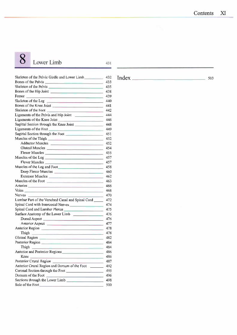
Skeleton of the Pelvic Girdle and Lower LimbBones of the PelvisSkeleton ofthe PelvisBones of the Hip JointFemurSkeleton ofthe LegBones ofthe Knee JointSkeleton ofthe FootLigaments of the Pelvis and Hip JointLigaments of the Knee JointSagittal Section through the Knee JointLigaments of the FootSagittal Section through the FootMuscles of the Thigh
Adductor MusclesGluteal MusclesFlexor Muscles
Muscles of the LegFlexor Muscles
Muscles of the Lee and FootDeep Flexor MusclesExtensor Muscles
Muscles of the FootArteriesVeinsNervesLumbar Part of the Vertebral Canal and Spinal Cord _Spinal Cord with Intercostal NervesSpinal Cord and Lumbar PlexusSurfaceAnatomy of the Lower Limb
Dorsal AspectAnterior Aspect
Anterior RegionThigh
Gluteal RegionPosterior Region
ThighAnterior and Posterior Resrons
KneePosterior Crural ReeionAnterior Crural Region and Dorsum of the FootCoronal Section through the FootDorsum of the FootSections through the Lower Limb
432 Index4334354384394404414424444464484494514s2452454455457457
503
458460462463466468470472474475476476477478478482484484486486487492495496498500Sole ofthe Foot
)ilI
List of Figures
The schematic drawings have been performed by the following graphic Jcirg Pekarsky, figures on pages: 15, 31, 55, 58, 69,71, 85, 93, 95, 96,designers: l l3, 158, 177,190,195, 199, 201,205,241,248,253,280,301, 302,
316,345,400, 445, 457 ,476,477 .Bruno Bradt, figure onpage l.
Heinz Troegero figures on pages: 18, 164, 169,193,274.Gunther Felmerer, figures on pages: 3, 62, 147 , l7 l, 202.
Christiane Wittek, figures on pages: 2, 8, 21,22,85,197,210,211,223,Eve$n Ott-Freiberger, f igures on pages: 20,46,49,65,70,73,74,76, 227,235,236,238,244,278,298,328,331,341,342,364,365,367,92,105,106, 109, 115,117,130, 131, 150,152,154,155,160, 181, 368,369,373,414,430,433,436.214, 237 , 244, 261, 271, 272, 281, 282, 284, 287, 29t, 294, 296, 303,309,314,325,375,376,377 ,378,382,399,444,447,449. Annette Gack-Buley, figures on pages: 9, 17, l7 o 23, 59, 91, ll0, 127 ,
149, 163, 166, 270, 297, 390.Sonja Moldenhauer, figures on pages: 19, 94, | 43.
1 GeneralAnatomy
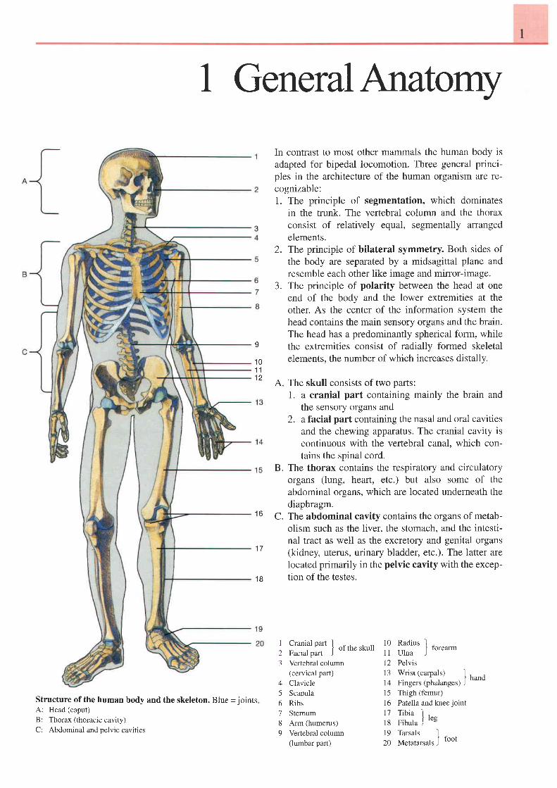
In contrast to most other mammals the human body isadapted for bipedal locomotion. Three general princi-ples in the architecture of the human organism are re-cognizable:1. The principle of segmentation, which dominates
in the trunk. The vertebral column and the thoraxconsist of relatively equal, segmentally arrangedelements.
2. The principle of bilateral symmetry. Both sides ofthe body are separated by a midsagittal plane andresemble each other like image and mirror-image.
3. The principle of polarity between the head at oneend of the body and the lower extremities at theother. As the center of the information system thehead contains the main sensory organs and the brain.The head has a predominantly spherical form, whilethe extremities consist of radially formed skeletalelements, the number of which increases distally.
9
1 01 1
A. The skull consists of two parts:1. a cranial part containing mainly the brain and
the sensory organs and2. afacial part containing the nasal and oral cavities
and the chewing apparatus. The cranial cavity iscontinuous with the vertebral canal, which con-tains the spinal cord.
The thorax contains the respiratory and circulatoryorgans (lung, heart, etc.) but also some of theabdominal organs, which are located underneath thediaphragm.The abdominal cavity contains the organs of metab-olism such as the liver. the stomach. and the intesti-nal tract as well as the excretory and genital organs(kidney, uterus, urinary bladder, etc.). The latter arelocated primarily in the pelvic cavity with the excep-tion of the testes.
B.
1 6 c .
1 7
1 8
Structure of the human body and the skeleton. Blue - jointsA: Head (caput)
B: Thorax (thoracic cavity)
C: Abdominal and pelvic cavities
I
23
A
56189
Cranialoarr I ^.I ot the skull
facral part )Vertebral column(cervical part)ClavicleScapulaRibsStemumArm (humerus)Vertebral column(lumbar part)
10 Radius I "11 ulna J
rorearm
12 Pelvisl3 Wrist (carpals) lio oi"t.rt,o'nui-g.r, I hund
15 Thigh (femur)16 Patella and kneejoint17 Tibia I18 Fibula J
re8
19 Tarsais I ^2o Metatarsals J toot
2 I General Anatomv Organization of the Human Body
1 5
1 61 7
1 81 9
32e e
q
7
1 1
1 21 3
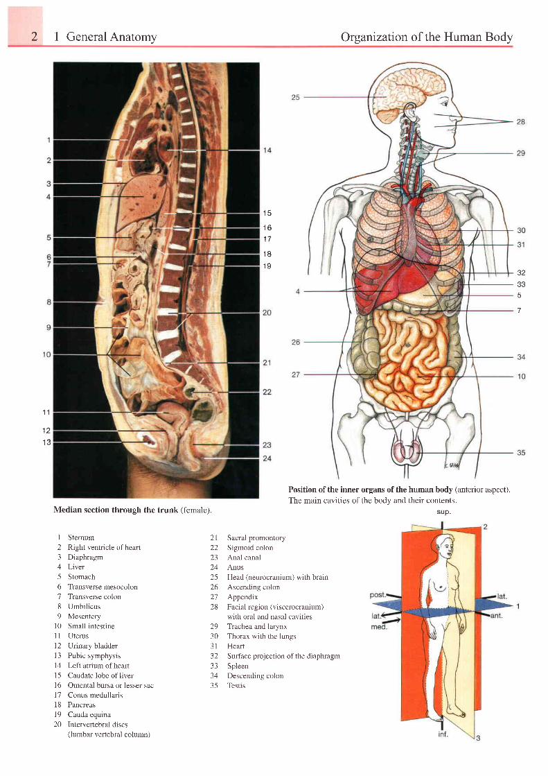
Position of the inner organs of the human body (anterior aspect).The main cavities of the body and their contents.
suD.Median section through the trunk (female).
1 Sternum2 Right ventricle ofheart3 Diaphragm4 Liver5 Stomach6 Transverse mesocolon7 Transverse colon8 Umbilicus9 Mesentery
10 Small intestine11 Uterus12 Urinary bladder13 Pubic symphysis14 Left atrium ofheart15 Caudate lobe ofliver16 Omental bursa or lesser sac17 Conus medullaris18 Pancreas19 Cauda equina20 Intervertebraldiscs
(lumbar vertebral column)
Sacral promontory
Sigmoid colon
Anal canal
Anus
Head (neurocranium) with brain
Ascending colon
Appendix
Facial region (viscerocranium)
with oral and nasal cavities
Trachea and larynx
Thorax with the lungs
Heart
Surface projection of the diaphragm
Spleen
Descending colon
Testis
2 l22LJ
2425262728
29303 132333435
Organization of the Human Body
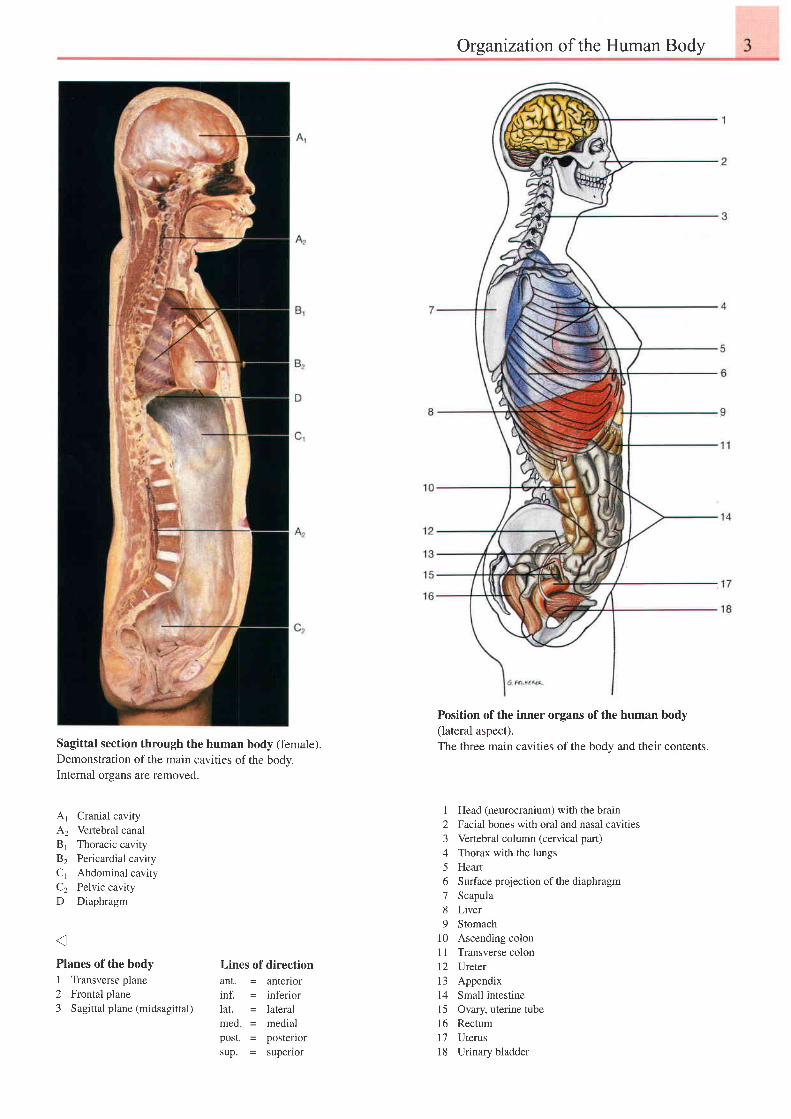
Sagittal section through the human body (female).Demonstration of the main cavities of the body.Internal organs are removed.
,A.1 Cranial cavityA2 Verlebral canalBt Thoracic cavity82 Pericardial cavityC1 Abdominal cavityC2 Pelvic cavityD Diaphragm
Position of the inner organs of the human body(lateral aspect).The three main cavities of the bodv and their contents.
1 Head (neurocranium) with the brain2 Facial bones with oral and nasal cavities3 Vertebral column (cervical part)4 Thorax with the lungs5 Heart6 Surface projection ofthe diaphragm7 Scapula8 Liver9 Stomach
10 Ascending colonI 1 Transverse colon12 Ureter13 Appendix14 Small intestine15 Ovary, uterine tube16 Rectuml7 Uterusl8 Urinary bladder
Planes of the body1 Transverse plane2 Frontal plane3 Sagittal plane (midsagittal)
Lines of directionant. = anteriorinf. = inferiorlat. = lateralmed = medialpost. - posteriorsup. = supenor
I General Anatomv Skeleton of the Human Body
t 423
22
23 221 3
2924
25
26
c l
1 l
1 2
30
35
36
37
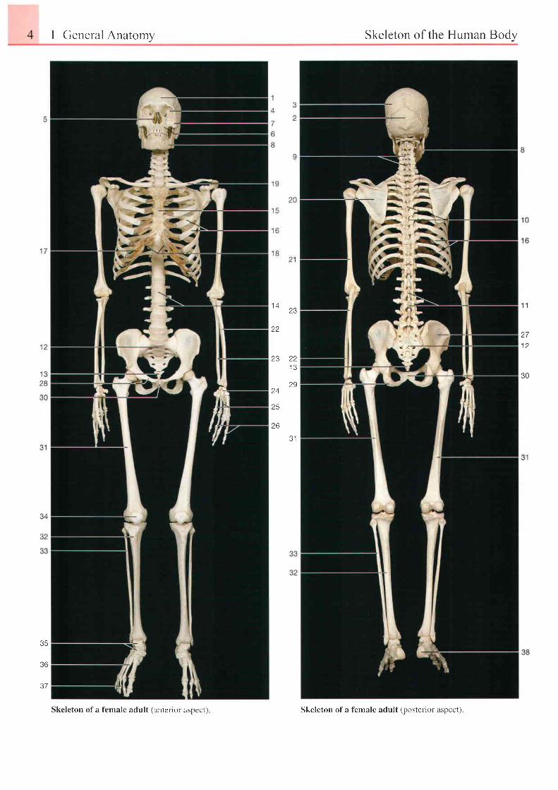
Skeleton of a female adult (anterior aspect) Skeleton of a f'emale adult (posterior aspect).
Skeleton of the Human Body 5
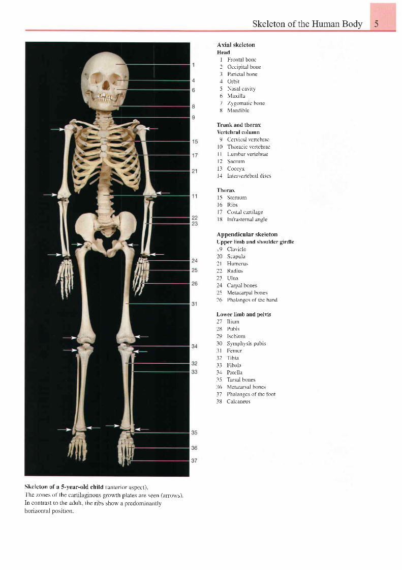
Axial skeletonHead
I Fronfal bone2 Occipital bone3 Parietal bone4 Orbit5 Nasal cavity6 Maxilla7 Zygomatic bone8 Mandible
Trunk and thoraxVertebral column9 Cervical vertebrae
10 Thoracic vertebraeI I Lumbar vertebrael2 Sacrum13 Coccyxl4 lntervertebral discs
Thorax15 Stemuml6 R ibsl7 Costal cartilage18 Infrasternal angle
Appendicular skeletonUpper limb and shoulder girdle19 Clavicle20 Scapula21 Humerus22 Radius23 Ulna24 Carpal bones25 Metacarpal bones26 Phalanges ofthe hand
Lower limb and pelvis27 Ilium28 Pubis29 Ischium30 Symphysis pubis31 Femur32 Tibia33 Fibula34 Patella35 Tarsal bones36 Metatarsal bones37 Phalanges of the fbot38 Calcaneus
Skeleton of a S-year-old child (anterior aspecr).The zones of the cartilaginous growth plates are seen (arrows).In contrast to the adult, the ribs show a predominantlyhorizontal oosition.
6 1 General Anatomy Ossification of the Bones
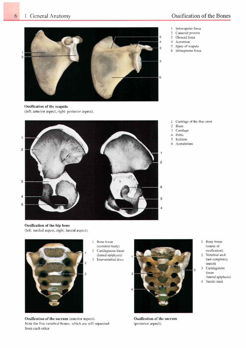
I Subscapular fossa2 Coracoid process3 Glenoid fossa4 Acromion5 Spine of scapula6 Infraspinous fossa
I23A
5f)
Cartilage ofthe iliac crestIliumCartilagePubisIschiumAcetabulum
Ossification of the scapula(left: anterior aspect, right: posterior aspect).
Ossification of the hip bone(left: medial aspect, right: lateral aspect).
Bone tissue(vertebral body)Cartilaginous tissue(lateral epiphysis)Intervertebral discs
Bony tissue(center ofossification)Vertebral arch(not completelyunited)Cartilaginoushssue(lateral epiphysis)Sacral canal
Ossification of the sacrum (anterior aspect).Note the hve vertebral bones. which are still seoarated
from each other.
Ossification of the sacrum(posterior aspect).
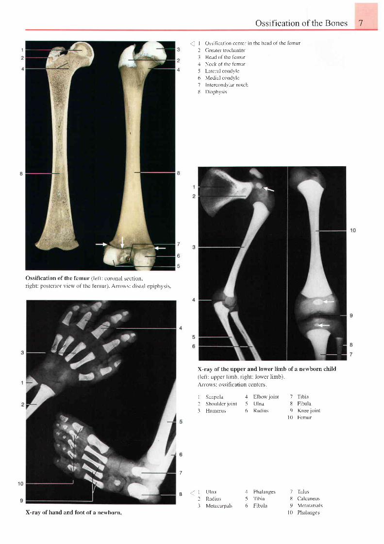
Ossification of the femur (left: coronal section,r ight: posterior view of the femur). Arows: distal epiphysis.
Ossification of the Bones 7
Ossiflcation center in the head of the femur
Greater trochanter
Head of the 1-emur
Neck of the femur
Lateral condyle
Medial condyle
Intercondylar notch
Diaphysis
X-ray of the upper and lower limb of a newborn child(left: upper limb, right: lower limb).
Arrows: ossifi cation centers.
I Scapula 4 Elbowjoint 7 Tibia2 Shoulderjoint 5 Ulna 8 Fibula3 Humerus 6 Radius 9 Knee joint
l0 Femur
'. I
23456'7
d
7
I < r23
Ulna
Radius
Metacarpals
I
5t)
Phalanges 7 Talus
Tibia 8 Calcaneus
Fibula 9 Metatarsals
l0 PhalangesX-ray ofhand and foot ofa newborn.
8 1 General Anatomy Bone Structure
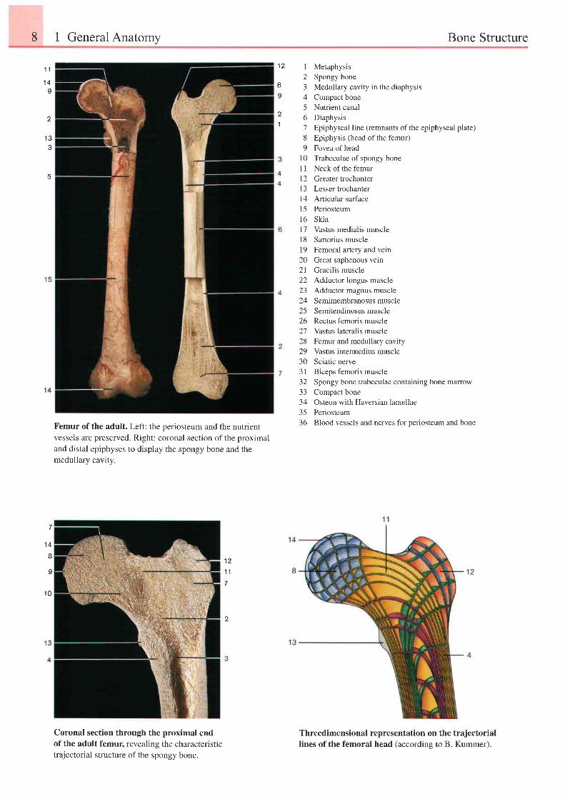
Femur of the adult. Left: the periosteum and the nutrientvessels are preserved. Right: coronal section ofthe proximaland distal epiphyses to display the spongy bone and themedullary cavity.
1 21 1
7
2
Coronal section through the proximal endof the adult femur, revealing the characteristictrajectorial structure of the spongy bone.
1 Metaphysis2 Spongy bone3 Medullary cavity in the diaphysis4 Compactbone5 Nutrient canal6 Diaphysis7 Epiphyseal line (remnants ofthe epiphyseal plate)8 Epiphysis (head of the femur)9 Fovea ofhead
l0 Trabeculae of spongy bone11 Neck of the femur12 Greatertrochanterl3 Lesser fochanterl4 Articular surfacel5 Periosteuml6 Skinl7 Vastus medialis muscle18 Sartorius musclel9 Femoral artery and veir20 Great saphenous vein21 Gracilis muscle22 Adductor longus muscle23 Adductor magnus muscle24 Semimembranosusmuscle25 Semitendinosus muscle26 Rectus femoris muscle27 Vastus lateralis muscle28 Femur and medullary cavity29 Vastus intermedius muscle30 Sciatic nerve31 Biceps femoris muscle32 Spongy bone trabeculae containing bone marrow33 Compact bone34 Osteon with Haversian lamellae35 Periosteum36 Blood vessels and nerves for periosteum and bone
Threedimensional representation on the trajectoriallines of the femoral head (according to B. Kummer).
1 21 l
1 4I
z
11
1 3e
5
1 4
a
I
1 0
Bone Structure 9
1 6
1 B1 7
20
1 92122
23
24
25
t o
1 7
2 8 1 81 q
29 zo
2221
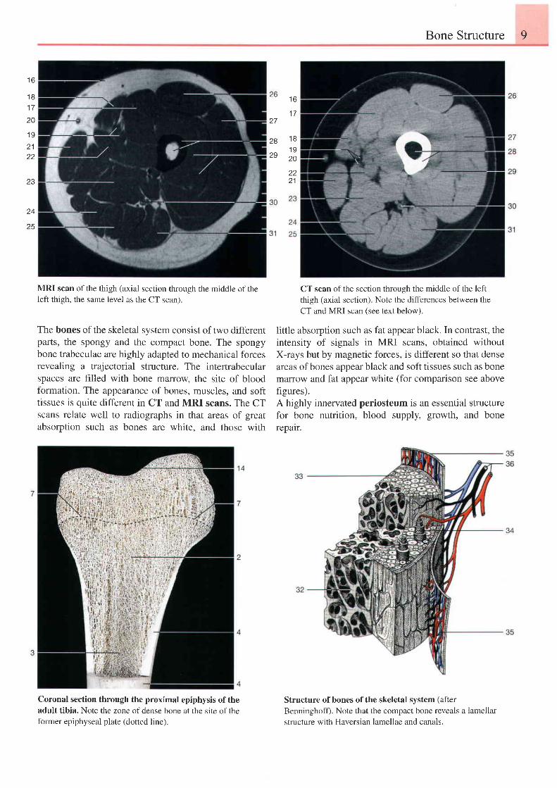
MRI scan of the thigh (axial section through the middle of theleft thigh, the same level as the CT scan).
The bones of the skeletal system consist of two differentparts, the spongy and the compact bone. The spongybone trabeculae are highly adapted to mechanical forcesrevealing a trajectorial structure. The intertrabecularspaces are filled with bone malrow, the site of bloodformation. The appearance of bones, muscles, and softtissues is quite different in CT and MRI scans. The CTscans relate well to radiographs in that areas of greatabsorption such as bones are white, and those with
CT scan of the section through the middle of the left
thigh (axial section). Note the differences between the
CT and MRI scan (see text below).
little absorption such as fat appear black. In contrast, theintensity of signals in MRI scans, obtained withoutX-rays but by magnetic forces, is different so that denseareas ofbones appear black and soft tissues such as bonemzrrow and fat appear white (for comparison see abovefigures).A highly innervated periosteum is an essential structurefor bone nutrition, blood supply, growth, and bonerepalr.
Coronal section through the proximal epiphysis of theadult tibia. Note the zone of dense bone at the site of theformer epiphyseal plate (dotted line).
Structure of bones of the skeletal system (afterBenninghoff). Note that the compact bone reveals a lamellarstructure with Haversian lamellae and canals.
10 1 General Anatomy Joints
1 0
1 920
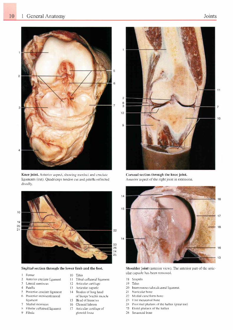
Knee joint. Anterior aspect, showing menisci and cruciateIigaments (cut). Quadriceps tendon cut and patella reflecteddistally.
Sagittal section through the lower limb and the foot.
Coronal section through the knee joint.Anterior aspect of the rightjoint in extension.
Shoulderjoint (anterior view). The anterior part ofthe artic-
ular capsule has been removed.
I 8 Scapula
19 Talus
20 Interosseous talocalcaneal ligament
2l Navicular bone
22 Medial cuneiform bone
23 First metatarsal bone
24 Proximal phalanx ofthe hallux (great toe)
25 Distal phalanx of the hallux
26 Sesamoid bone
Z J
26
1 C
I Femur
2 Anterior cruciate ligament
3 Lateral meniscus
4 Patella
5 Posterior cruciate ligament
6 Posterior meniscofemoral
ligament
7 Medial meniscus
8 Fibular collateral ligament
9 Fibula
l0 Tibia
| | T ib i r l col lateral l igament
l2 Articular cartilage
l3 Articular capsule
l4 Tendon of long head
of biceps brachii muscle
l5 Head of humerus
l6 Glenoid labrum
I 7 Articular cartilage of
glenoid fossa
Joints 1 l
1 2 4
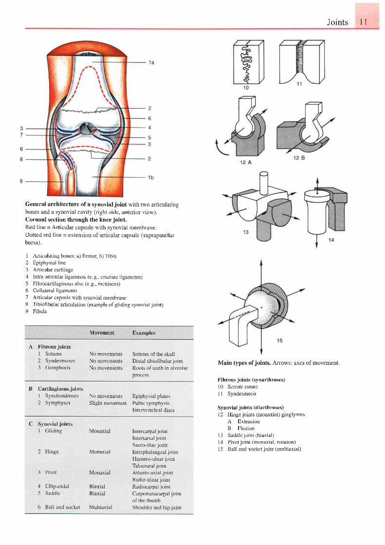
General architecture of a synovial joint with two articulatingbones and a synovial cavity (right side, anterior view).Coronal section through the knee joint.
Red line = Articular capsule with synovial membrane.Dotted red line = extension of articular capsule (suprapatellar
bursa).
1 Articulating bones: a) Femur, b) Tibia2 Epiphysial line3 Articular cartilage4 Intra-articular ligaments (e.g., cruciate ligaments)5 Fibrocartilaginous disc (e.g., meniscus)6 Collateral ligaments7 Anicular capsule with synovial membrane8 Tibiofibular articulation (example of gliding synovial joint)9 Fibula
Movement Examples
A Fibrous joints
I Sutures2 Syndesmoses3 Gomphosis
No movements Sutures of the skullNo movements Distal tibiohbular jointNo movements Roots of teeth in alveolar
process
B Cartilaginous jointsi Synchondroses No movements2 Symphyses Slight movement
Epiphysial platesPubic symphysislntervertebral discs
Main types of joints. Arrows: axes of movement.
Fibrous joints (synarthroses)10 Serrate suture11 Syndesmosis
Synovial joints (diarthroses)12 Hinge joints (monaxial) ginglymus
A ExtensionB Flexion
13 Saddlejoint (biaxial)14 Pivot joint (monaxial, rotation)15 Ball-and-socket joint (multiaxial)
C Synovialjoints1 Gliding
2 Hinge
3 Pivot
4 Ellipsoidal5 Saddle
6 Ball-and-socket
lntercarpal jointIntertarsal jointSacro-iliac jointInterphalangeal jointHumero-ulnar jointTalocrural jointAtlanto-axial jointRadio-ulnar jointRadiocarpal jointCarpometacarpal jointof the thumbShoulder and hip joint
Monaxial
Monaxial
Monaxial
BiaxiaiBiaxial
Multiaxial
1 General Anatomv Joints
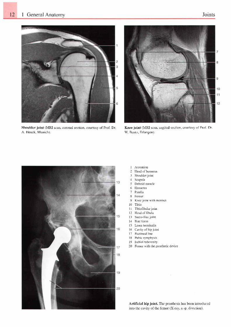
Shoulder joint (MRI scan, coronal section, coufiesy of Prof. Dr.A. Heuck. Munich).
Knee joint (MRI scan, sagittal section, courtesy of Prof. Dr.W Bautz, Erlangen).
I Acromion2 Headofhumerus3 Shoulderjoint4 Scapula5 Deltoid muscle6 Humerus7 Patella8 Femur9 Knee joint with menisci
10 Tibia11 Tibiofibularjointl2 Head of fibulal3 Sacro-iliacjoint14 Iliac fossa15 Linea terminalis16 Cavity of hip joint17 Pectineal line18 Pubic symphysis19 Ischial tuberosity20 Femur with the prosthetic device
Artificial hip joint. The prosthesis has been introducedinto the cavity of the femur (X-ray, a.-p. direction).
Muscles andTendonAttachments 13
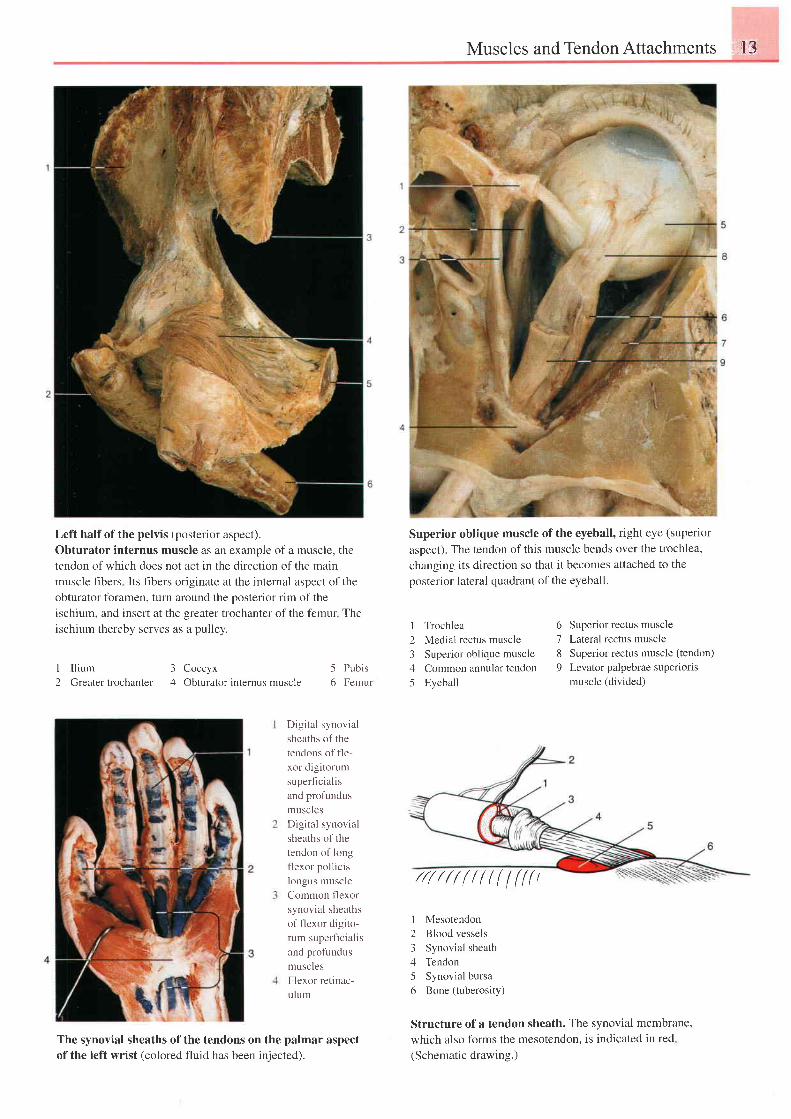
I I l ium 3 Coccyx2 Greatet trochanter 4 Obturator internus muscle
Left half of the pelvis (posterior aspect).
Obturator internus muscle as an example of a muscle, the
tendon of which does not act in the direction of the main
muscle fibers Its fibers originate at the internal aspect of the
obturator foramen, turn around the posterior rim of the
ischium, and insert at the greater trochanter of the femur The
ischium thereby serves as a pulley.
Superior oblique muscle of the eyeball, right eye (superior
aspect). The tendon of this muscle bends over the trochlea,
changing its direction so that it becomes attached to the
posterior lateral quadrant of the eyeball.
I Trochlea 6 Superior rectus muscle
2 Medial rectus muscle '/ Lareral rectus muscle
3 Superior oblique muscle 8 Superior rectus muscle (tendon)
4 Common annular tendon 9 Levator palpebrae superioris
5 Eyeball muscle (divided)
I Mesotendon
2 Blood vessels
3 Synovial sheath
4 Tendon
5 Synovial bursa
6 Bone (tuberosity)
Structure of a tendon sheath. The synovial membrane,
which also forms the mesotendon, is indicated in red
(Schematic drawing.)
5 Pubis6 Femur
Digital synovial
sheaths of the
tendons of fle-
xor digitorum
superficialis
and profundus
muscles
Digital synovial
sheaths of the
tendon of long
flexor pollicis
longus muscle
Common flexor
synovial sheaths
of flexor digito-
rum superficialis
and profundus
muscles
Flexor retinac-
ulum
The synovial sheaths of the tendons on the palmar aspect
ofthe left wrist (colored fluid has been iniected).
//r/////l((l(((t
t4 1 General Anatomy Organization of the Nervous System
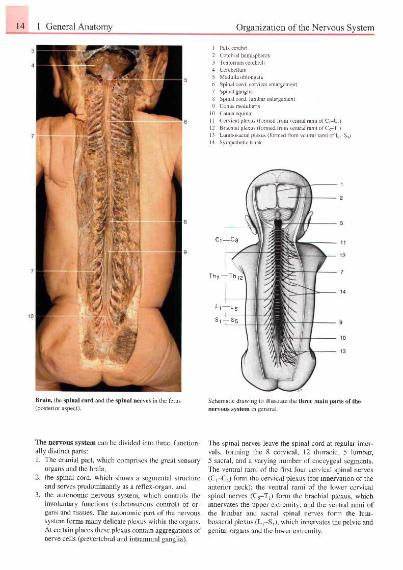
Brain, the spinal cord and the spinal nerves in the fetus(posterior aspect).
The nervous system can be divided into three, function-ally distinct parts:1. The cranial part, which comprises the great sensory
organs and the brain,the spinal cord, which shows a segmental structureand serves predominantly as a reflex-organ, andthe autonomic neryous system, which controls theinvoluntary functions (subconscious control) of or-gans and tissues. The autonomic part of the nervoussystem forms many delicate plexus within the organs.At certain places these plexus contain aggregations ofnerve cells (prevertebral and intramural ganglia).
I Falx cerebri
2 Cerebral hemispheres
3 Tentorium cerebelli
4 Cerebellum
5 Medulla oblongata
6 Spinal cord, cervical enlargement
7 Spinal ganglia
8 Spinal cord, lumbar enlargement
9 Conus medullaris
10 Cauda equina
I I Cervical plexus (formed from ventral rami of C,-Co)l2 Brachial plexus (formed from ventral rami of C5-T1)
13 Lumbosacral plexus (formed from ventral rami of L,-Sa)
14 Sympathetic trunk
cr -cs
T h 1 - T h
1 2
L r - L s
St -:- ss
Schematic drawing to illustrate the three main parts of thenervous system in general.
The spinal nerves leave the spinal cord at regular inter-vals, forming the 8 cervical, 72 thoracic, 5 lumbar,5 sacral, and a varying number of coccygeal segments.The ventral rami of the first four cervical spinal nerves(Cr-C+) form the cervical plexus (for innervation of theanterior neck); the ventral rami of the lower cervicalspinal nerves (C5-T1) form the brachial plexus, whichinnervates the upper extremity; and the ventral rami ofthe lumbar and sacral spinal nerves form the lum-bosacral plexus (Lr-S+), which innervates the pelvic andgenital organs and the lower extremity.
2.
3 .
Organization of the Nervous System
I
1 01 1
c
1 0
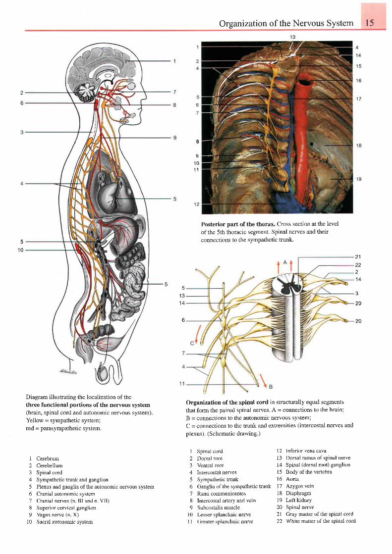
Diagram illustrating the localization of thethree functional portions of the nervous system(brain, spinal cord and autonomic nervous system).Yellow = sympathetic system;red = parasympathetic system.
I Cerebrum2 Cerebellum3 Spinal cord4 Sympathetic trunk and ganglion5 Plexus and ganglia ofthe autonomic nervous system6 Cranial autonomic system7 Cranial nerves (n. III and n. VII)8 Superior cervical ganglion9 Vagus nerve (n. X)
l0 Sacral autonomic system
Posterior part ofthe thorax. Cross section at the levelof the 5th thoracic segment. Spinal nerves and theirconnections to the sympathetic trunk.
Organization of the spinal cord in structurally equal segmentsthat form the paired spinal nerves. A = connections to the brain;B = connections to the autonomic nervous system;C = connections to the trunk and extremities (intercostal nerves andplexus). (Schematic drawing.)
1 31 4
b
212221 4
3
20
20
I Spinal cord2 Dorsal root3 Ventral root4 Intercostal nerves5 Sympathetic trunk6 Ganglia of the sympathetic trunk7 Rami communicantes8 Intercostal anery and vein9 Subcostalis muscle
10 Lesser splanchnic nerve11 Greater splanchnic nerve
12 Inferior vena cava13 Dorsal ramus of spinal nerve14 Spinal (dorsal root) ganglion15 Body ofthe vertebra16 Aorta17 Azygos vein18 Diaphragm19 Leftkidney20 Spinal nerve27 Gray matter of the spinal cord22 Whtte matter of the spinal cord
t6 1 General Anatomv Organtzation of the Circulatory System
1 21 3
1 4
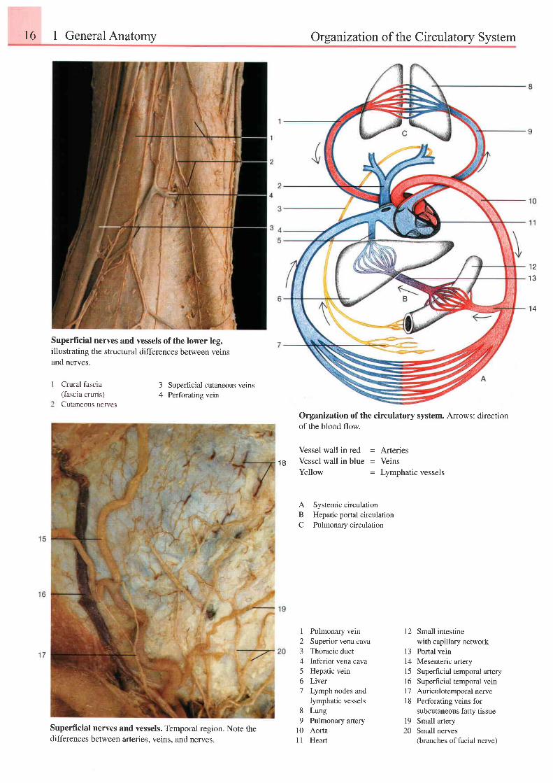
Superflrcial nerves and vessels of the lower leg,illustrating the structural differences between veinsand nerves.
Crural fascia(fascia cruris)Cutaneous nerves
3 Superhcial cutaneous veins4 Perforatins vein
1 8
Organization of the circulatory system. Arrows: directionof the blood flow.
Vessel wall in red = ArteriesVessel wall in blue = VeinsYellow = Lymphatic vessels
A Systemic circulationB Hepatic portal circulationC Pulmonarycirculation
Superficial nerves and vessels. Temporal region. Note thedifferences between arteries. veins, and nerves.
I
234
567
89
l0l l
I 2
1 3141516l7l 8
Pulmonary veinSuperior vena cavaThoracic ductInferior vena cavaHepatic veinLiverLymph nodes andlymphatic vesselsLungPulmonary arteryAortaHeart
Small intestinewith capillary networkPortal veinMesenteric arterySuperfi cial temporal arterySuperficial temporal veinAuriculotemporal nervePerforating veins forsubcutaneous fatty tissue
19 Small artery20 Small nerves
(branches of facial nerve)
Organtzation of the Circulatory System
e
1 3l l
1 2I J
1 4
15
16 2728
17 29
1 8
1 9
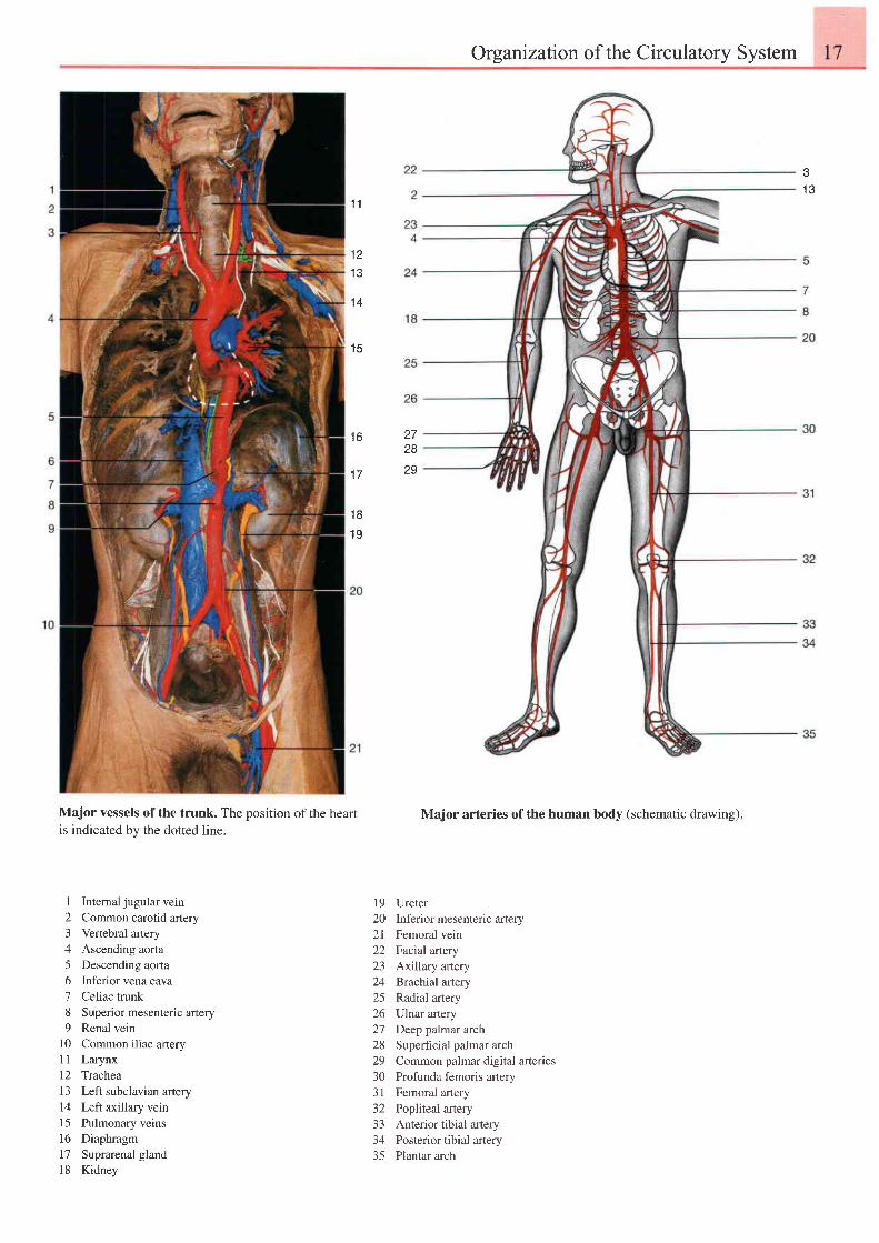
Major vessels of the trunk. The position of the heartis indicated by the dotted line.
I Intemal jugular vein2 Common carotid artery3 Vertebral artery4 Ascending aorta5 Descending aorla6 Inferior vena cava7 Celiac trunk8 Superior mesenteric artery9 Renal vein
10 Common iliac artery11 Larynx12 Trachea13 Left subclavian artery14 Left axillary vein15 Pulmonary veins16 Diaphragm17 Suprarenal gland18 Kidney
Major arteries of the human body (schematic drawing).
19 Ureter20 Inferior mesenteric artery2l Femoral vein22 Factalartery23 Axillary artery24 Brachialartery25 Radial artery26 Ulnar artery27 Deep palmar arch28 Superficial palmar arch29 Common palmar digital arteries30 Profunda femoris artery31 Femoral artery32 Popliteal artery33 Anterior tibial artery34 Posterior tibial artery35 Plantar arch
18 I General Anatomy Organization of the Lymphatic System
t c2425
2o4 1
1 6 31 7 4
1 2
1 01 1
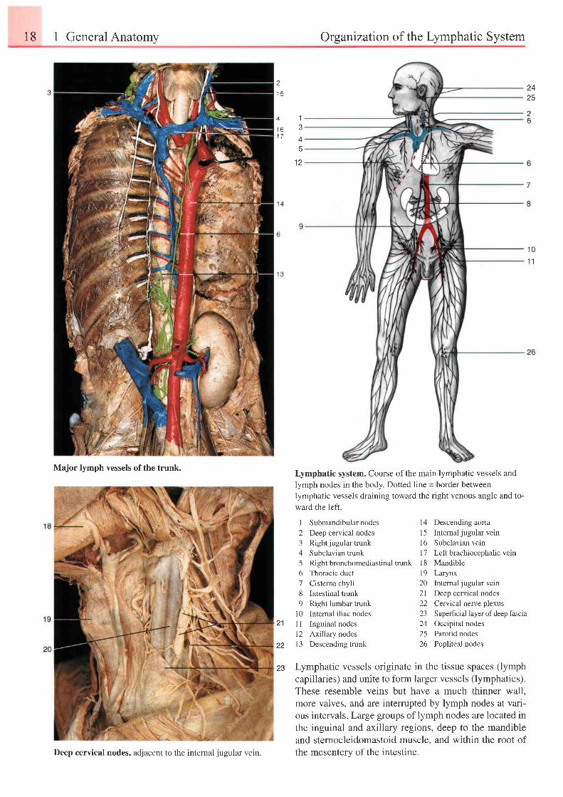
Major lymph vessels of the trunk.Lymphatic system. Course of the main lymphatic vessels and
lymph nodes in the body. Dotted line = border between
lymphatic vessels draining toward the right venous angle and to-
ward the left.
21
22
I Submandibular nodes2 Deep cervical nodes3 Rightjugular trunk4 Subclavian trunk5 Right bronchomediastinal trunk6 Thoracic duct7 Cisterna chyli8 lntestinal trunk9 Right lumbar trunk
l0 Internal iliac nodesI I Inguinal nodesl2 Axillary nodesl3 Descending trunk
l4 Descending aofia
15 Internaljugularvein
16 Subclavian vein
l1 Left brachiocephalic vern
18 Mandible
19 Larynx
20 Intemal jugular vein
2l Deep cervical nodes
22 Cervical nerve plexus
23 Superficial layer ofdeep fascia
24 Occipital nodes
25 Parotid nodes
26 Popliteal nodes
Lymphatic vessels originate in the tissue spaces (lymphcapillaries) and unite to form larger vessels (lymphatics).These resemble veins but have a much thinner wall,more valves, and are interrupted by lymph nodes at vari-ous intervals . Large groups of lymph nodes are located inthe inguinal and axillary regions, deep to the mandibleand sternocleidomastoid muscle, and within the root ofthe mesentery of the intestine.Deep cervical nodes, adjacent to the intemal jugular vetn.
23
I9
2 HeadandNeck
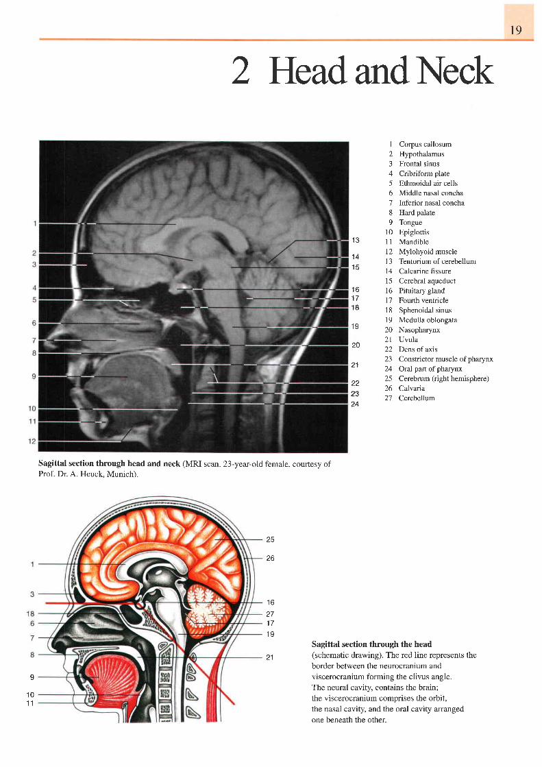
1 Corpus callosum2 Hypothalamus3 Frontal sinus4 Cribriform plate5 Ethmoidal air cells6 Middle nasal concha7 Inferior nasal concha8 Hard palate9 Tongue
l0 Epiglottis11 Mandible12 Mylohyoid muscle13 Tentoriumof cerebellum14 Calcarine fissure15 Cerebral aqueduct16 Pituitary gland17 Fourth ventricle18 Sphenoidal sinusl9 Medulla oblongata20 Nasopharynx21 Uvula22 Dens ofaxis23 Constrictor muscle of pharynx24 Oral part ofpharynx25 Cerebrum (righthemisphere)26 Calvaria27 Cerebellum
{ J
1 41 5
1 6n a
1 8
1 9
2A
21
222324
Sagittal section through head and neck (MRI scan. 23-year-old female, courtesy ofProf. Dr. A. Heuck, Munich).
25
zo
t o
271 71 9
21
I
1 01 1
Sagittal section through the head(schematic drawing). The red line represents theborder between the neurocranium andviscerocranium forming the clivus angle.The neural cavity, contains the brain;the viscerocranium comprises the orbit,the nasal cavity, and the oral cavity arrangedone beneath the other.
20 2 Head and Neck Bones of the Skull
1 9
20
21
232526272829
30
31
32
J
4
6
II
1 0
t <
1 11 3
1 4
I E
1 6
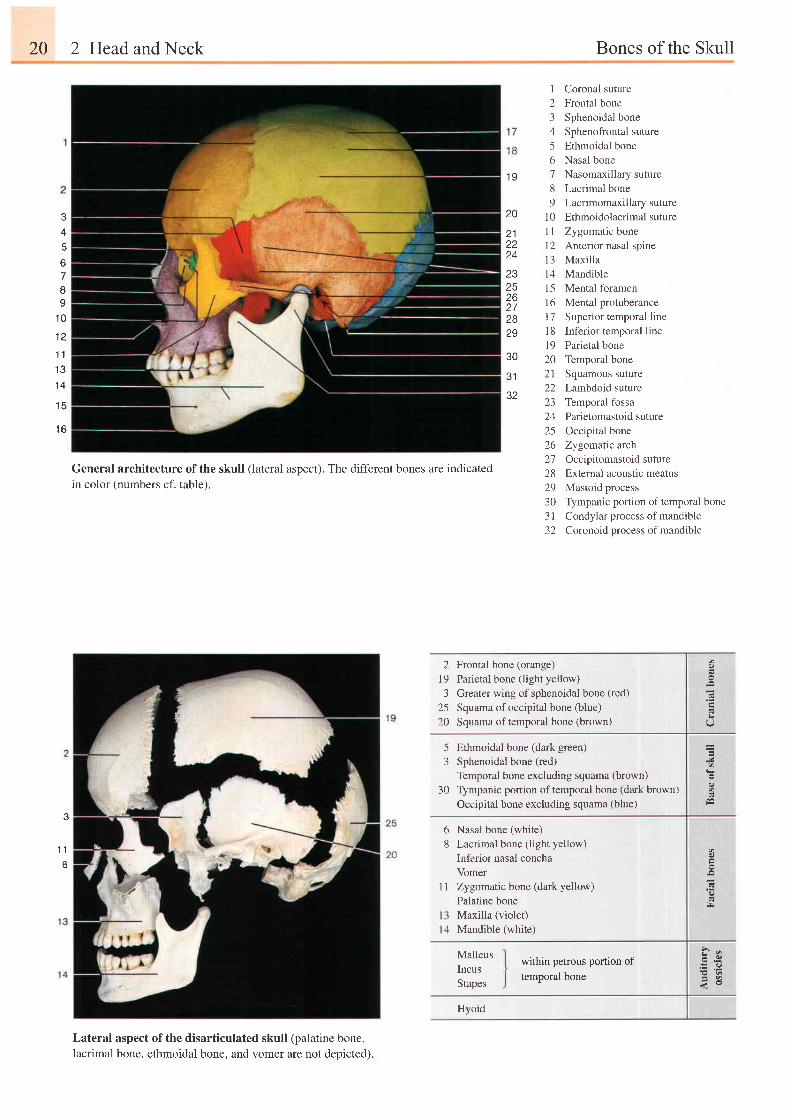
I Coronal suture2 Frontal bone3 Sphenoidal bone4 Sphenofrontal suture5 Ethmoidal bone6 Nasal bone7 Nasomaxillary suture8 Lacrimal bone9 Lacrimomaxillarysuture
10 Ethmoidolacrimal suture11 Zygomatic bonel2 Anterior nasal spine13 Maxil la14 Mandible15 Mental foramen16 Mental protuberance17 Superior temporal linel8 Inferior temporal line19 Parietal bone20 Temporal bone21 Squamous suture22 Lambdoid suture23 Temporal fossa24 Parietomastoidsuture25 Occipital bone26 Zygomatic arch27 Occipitomastoidsuture28 External acoustic meatus29 Mastoid process30 Tympanic portion of temporal bone3 I Condylar process of mandible32 Coronoid process ofmandible
General architecture of the skull (lateral aspect). The different bones are indicated
in color (numbers cf. table).
2 Frontal bone (orange)l9 Parietal bone (light yellow)3 Greater wing ofsphenoidal bone (red)
25 Squama of occipital bone (blue)20 Squama of temporal bone (brown)
5 Ethmoidal bone (dark green)3 Sphenoidal bone (red)
Temporal bone excluding squama (brown)
30 Tympanic portion oftemporal bone (dark brown)Occipital bone excluding squama (blue)
6 Nasal bone (white)
8 Lacrimal bone (light yellow)
Inferior nasal concha
Vomer
1 1 Zygomatic bone (dark yellow)
Palatine bone
Maxilla (violet)
Mandible (white)
,Q
b
MalleusIncusStapes
w:ithin petrous portion oftemporal bone
?*.: .9E q
Lateral aspect of the disarticulated skull (palatine bone,lacrimal bone, ethmoidal bone, and vomer are not depicted).
Hyoid
23
1 2a a
I D
1 7I O
a 1
6
1 8
J U
1 9
24
20
Bones of the Skull 21
I
a q
7
1 1
1 4
28
13
? q
1 0
2627
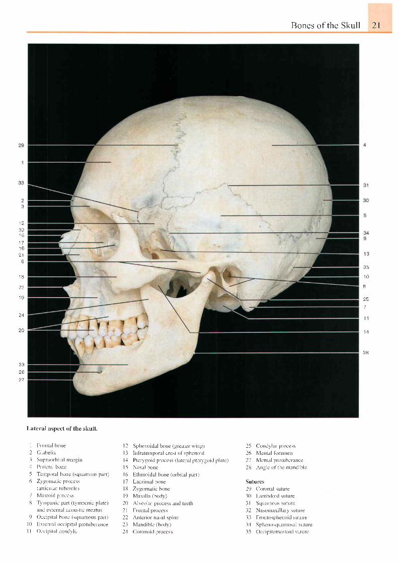
Lateral aspect of the skull.
Frontal bone
C l rbc l l a
Supraorbi ta l margin
Par icta l bone
Terlporal bone (squarnor.rs part)
Zygornat ic proccss(a l t i cu l a r t ubc rc l e )
Mastoic l process
Tymprnic part ( tympanic p late)
u t t J e r l e t t t ; r l u ( ( ) u \ l i L I n ( ' i l l u \
Occip i ta l bone (squanious part )
Hxternal occip i ta l p lotubcrance
Occipi ta l condyJe
l2 Sphenoidal bone (greatcr wing)
I 3 Infiaternporal crest of sphenoicl
l4 Pterygoid proccss ( lateral ptervgoid platc)
l5 Nasal bone
l6 Ethmoidal bone (orbr ta l part )
l7 Lacr imal bone
l8 Zygomat ic bonc
l9 Max i l l a ( body )
20 Alvcolar process and tccth
2l Frontal proccss
22 Antcr ior nasal spine
23 Mandible (body)
2,1 Coronoid process
25 Condylar process
26 Mcntal ltrramen
27 Mental protubelance
28 Angle ol thc nrandiblc
Sutures
29 Coronal suture
30 I -arnbdoid suture
3 I Scluanrous suturc
32 Nasomaxi l larysutule
33 Frontosphcnoidsutule
34 Sphenoscluanrosal suturc
3-5 Occip i tonrastoid suture
I
23I56
1
t 0l l
22 2 Head and Neck
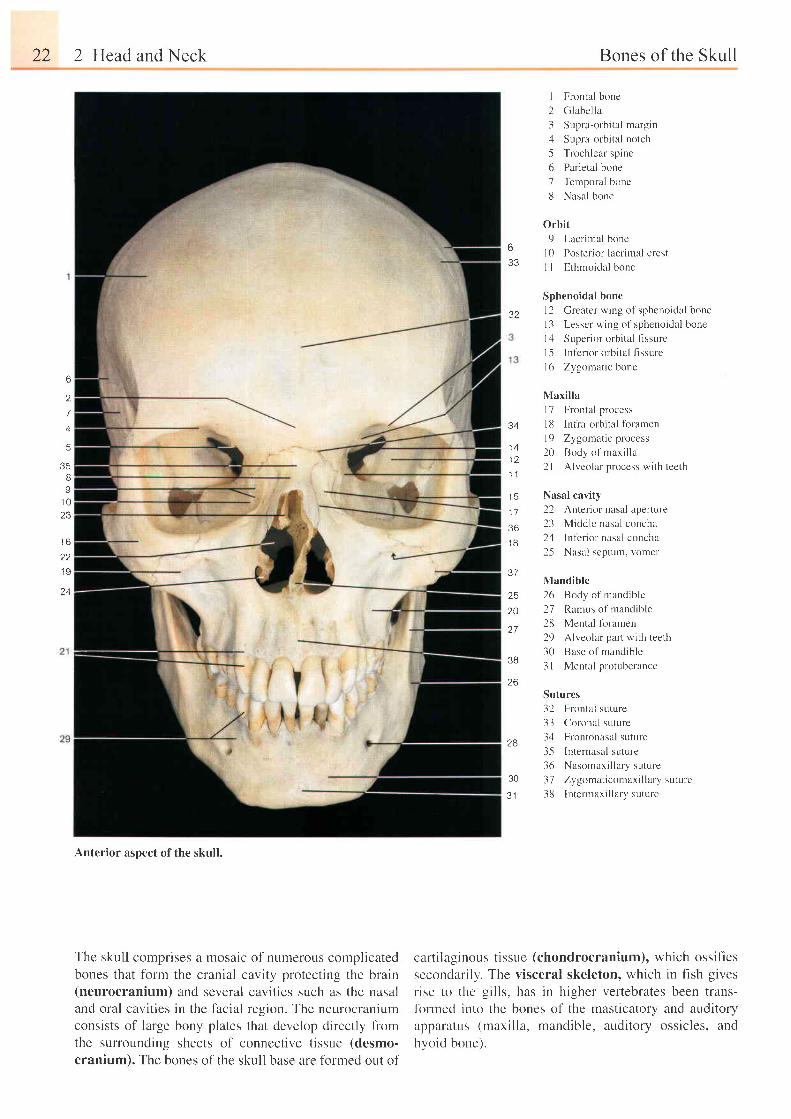
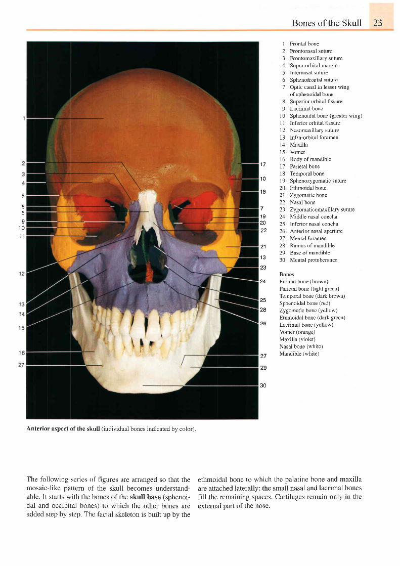
Anterior aspect of the skull.
The skull comprises a mosaic of numerous complicatedbones that form the cranial cavity protecting the brain(neurocranium) and several cavities such as the nasaland oral cavities in the facial region. The neurocraniumconsists of large bony plates that develop directly fiomthe surrounding sheets of connective tissue (desmo-cranium). The bones of the skull base are formed out of
633
Bones of the Skull
I Frontal bone
2 Glabella
3 Supra-orbi ta l margin
4 Supra-orbital notch
5 Trochlear spine
6 Parietal bone
7 Temporal bone
8 Nasal bone
0rbit
9 Lacr imal bone
l0 Posterior lacrimal crest
I I Ethrnoidal bone
Sphenoidal bone
l2 Greater wing of sphenoidal bone
I 3 Lesser wing of sphenoidal bone
l4 Super ior orbi ta l f lssurc
I -5 Inl'crior orbital fissure
16 Zygourat ic bone
3?
6
2
7
4
5
35II
1 023
Maxilla
l7 Frontal process
34 l8 Infia-orbital fbramen
1 A l9 Zygomat ic process
, ; 2 0 B t ' d ) o l m r r i l l r
- 2 l Alveolar nrocess wi th teeth
1 1
15 Nasal cavity
17 22 Anterior nasal aperture
36 23 Middle nasal concha
j g 24 Inf'erior nasal concha
25 Nasal septum, vomer
37Mandible
25 26 Body of mandible
zo 27 Ramus of mandible
27 28 Mental fbramen
29 Alveolar part wiLh teeth
30 Base of nrandibrc38
3l Mental protuberance
26Sutures
28
30
J I
32 Frontal suture
33 CoKrnal suture
34 Frontonasal suture
35 [nternasal suture
36 Nasonraxi l lary suture
37 Zygomaticomaxillary suture
38 Intermaxi l lary suture
t o
1 9
24
cartilaginous tissLle (chondrocranium), which ossif-iessecondarily. The visceral skeleton, which in fish givesrise to the gills, has in higher vertebrates been trans-fbrmed into the bones of the masticatory and auditoryapparertus (maxilla, mandible. auditory ossicles, andhvoid bone).
Bones of the Skull 23
l 7
1 0
I Frontal bone2 Frontonasal suture3 Frontomaxillarysuture4 Supra-orbital margin5 Internasal suture6 Sphenofrontal suture7 Optic canal in lesser wing
of sphenoidal bone8 Superior orbital fissure9 Lacrimal bone
10 Sphenoidal bone (greater wing)1 I Inferior orbital fissure12 Nasomaxillary suture13 Infra-orbital foramen14 Maxilla15 Vomer16 Body of mandible17 Parietal bone18 Temporalbone19 Sphenozygomatic suture20 Ethmoidal bone21 Zygomatic bone22 Nasal bone23 Zy gomaticomaxillary suture24 Middle nasal concha25 Inferior nasal concha26 Anterior nasal aperture27 Mental foramen28 Ramus of mandible29 Base of mandible30 Mental protuberance
BonesFrontal bone (brown)Parietal bone (light green)Temporal bone (dark brown)Sphenoidal bone (red)Zy gomatic bone (yellow)Ethmoidal bone (dark green)Lacrimal bone (yellow)Vomer (orange)Maxilla (violet)Nasal bone (white)Mandible (white)
o
1 01 1
1 8
71 92022
21
13
23
24
25
28
26
27
29
30
Anterior aspect of the skull (individual bones indicated by color).
The following series of figures are arranged so that themosaic-like pattern of the skull becomes understand-able. It stafis with the bones of the skull base (sphenoi-dal and occipital bones) to which the other bones areadded step by step. The facial skeleton is built up by the
ethmoidal bone to which the palatine bone and maxillaare attached laterally; the small nasal and lacrimal bonesfill the remaining spaces. Cartilages remain only in theexternal pafi of the nose.
o
8E
24 2 Hc ld and Ncck
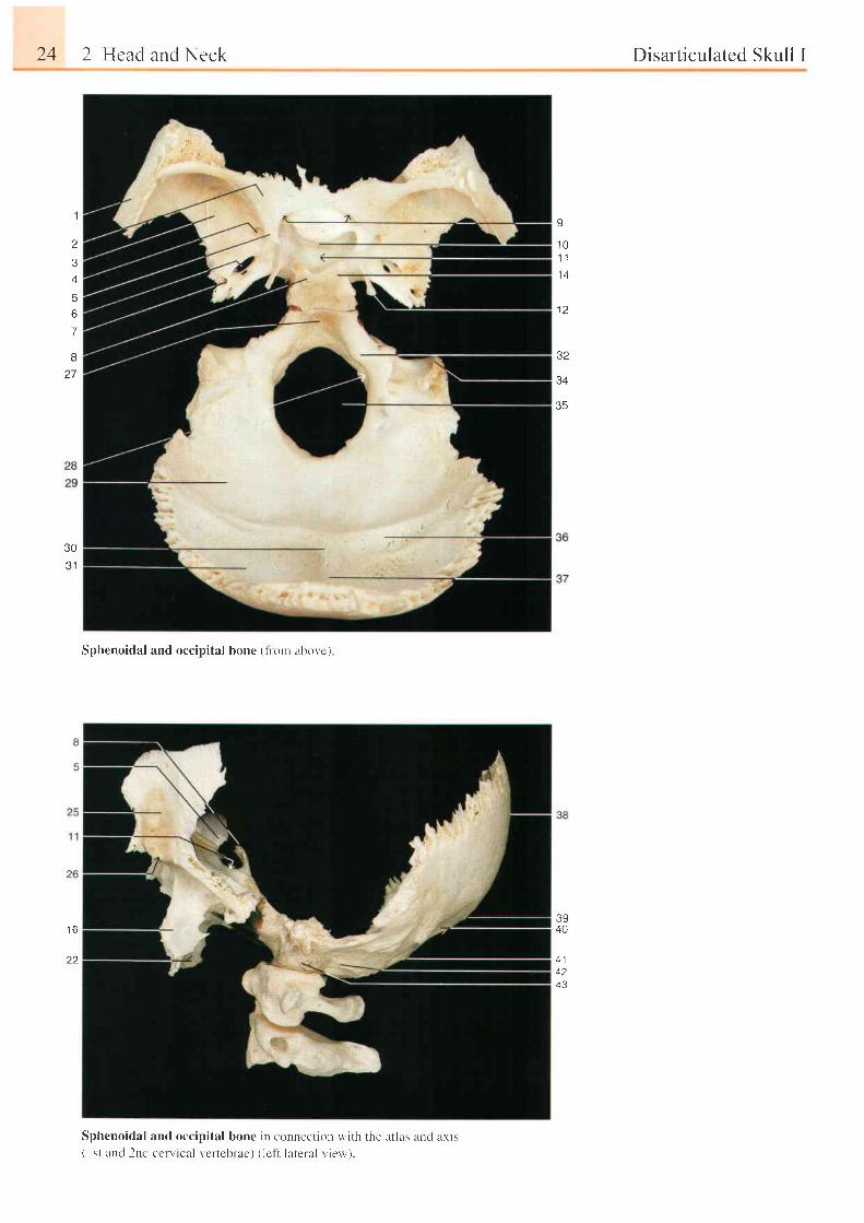
Sphenoidal and occipital bone (1r 'orr abovc)
Disarticulated Skull I
,l
3
6
7
8
U
1 0'1 .l
l a
t z
32
Q A
35
3031
3940
4 142n a
t o
22
Sphenoidal and occipital bone in connecl ion with the at las ancl axis( I st and 2nd ccrvical verteblae) ( lcft lateral view ).
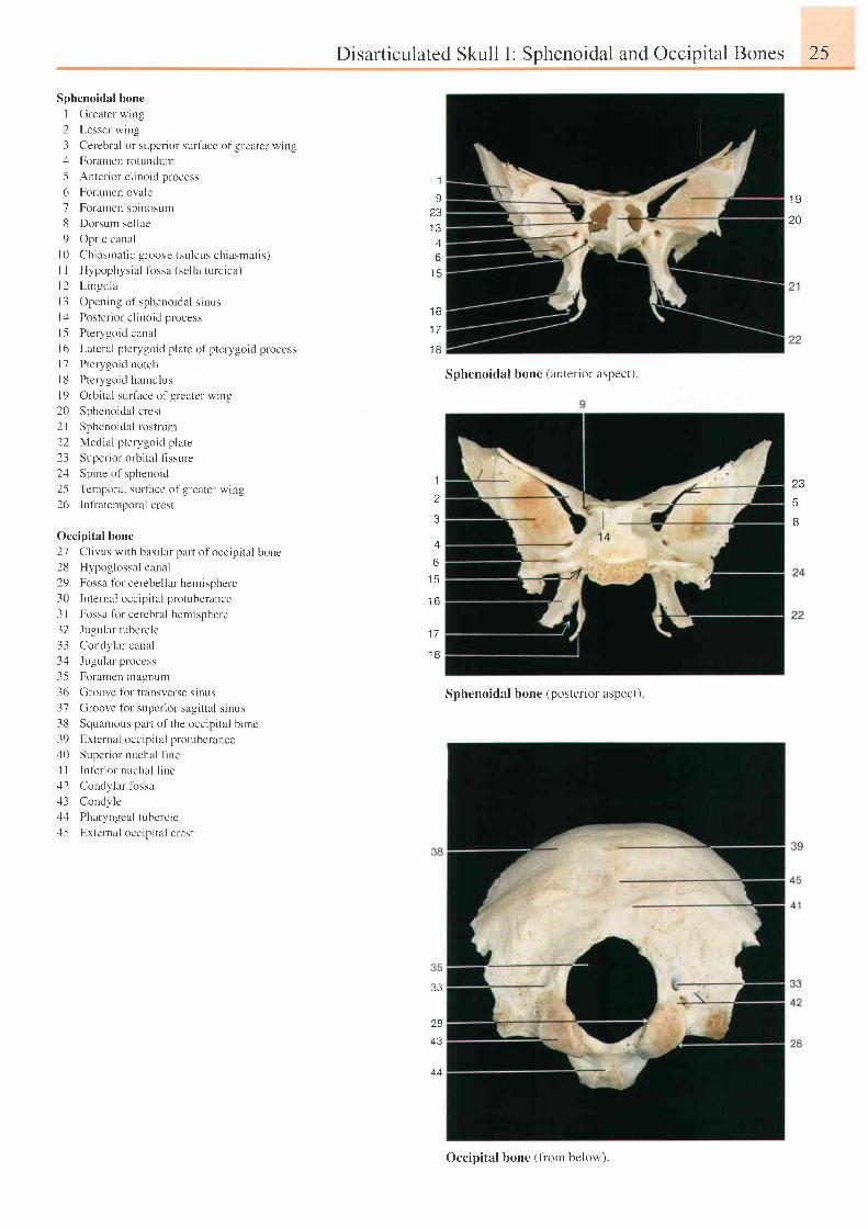
Sphenoidal bone
I Greater wing
2 Lesser wing
3 Cerebral or superior surface of greater wing4 Foramen rotundum
5 Anter ior c l inoid process
6 Foramen ovale
7 Foramen spinosum
8 Dorsum sellae
9 Optic canal
l0 Chiasmat ic groove (sulcus chiasmat is)
I I Hypophysia l f i rssa (sel la turc ica)
l 2 L i ngu la
l 3 Open ing o l ' spheno ida l r i nus
l4 Poster ior c l inoid process
15 Pterygoid canal
l6 Lateral pterygoid platc of pterygoidprocess
l7 Pteryto id notch
l8 Pterygoid hamulus
l 9 O rb i t a l su r l l ce o l g rea te r w ing
20 Sphenoidal crest
2l Sphenoidal rostrum
22 Medial pterygoid plate
23 Superior orbital flssure
2zl Spine of sphenoid
2-5 Temporal surface of greater wing
26 Infraternporalcrest
Occipital bone
27 Cl ivus wi th basi lar part of occip i ta l borre
28 Hypoglossal canal
29 Fossa fbr cerebellar hemisphere
30 Internal occipital protuberance
3l Fossa lbr cerebral hemisphere
32 Jugular tubercle
33 Condylar canal
34 Ju-tular process
35 Foramen magnum
-16 Croore l i r r l ran:rerse s inrrs
37 Groove fbr superior sagittal sinus
38 Squamous part of the occipital bone
39 Extcrnal occipital protubcrance,10 Super ior nuchal l ine
4l Inter ior nuchal l ine
42 Condylar fossazl3 Condyle
44 Pharyngeal tubercle;l-5 External occipital crest
Disarticulated Skull I: Sphenoidal and Occipital Bones 25
1
I23
A
D
t f ,
I D
1 7
t 6
Sphcnoidal bone { i lntcr ior aspect )
1
z
3
6I f ,
1 6
1 7
1 8
Sphenoidal bone (posterior aspect)
33
28
43
44
' 19
20
23
5
B
Occipital bone (fiom betow).
26 2 Head and Neck Disarticulated Skull lI
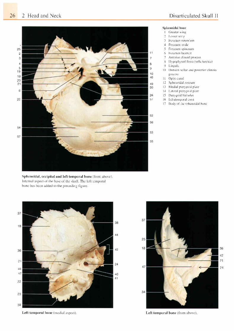
Sphenoidal bone
I Grcater wing
2 Lesser wing
3 Foramen rotllndum'l Foramen ovale
5 Foramen spinosum
6 Foramen lacerum
7 Anter ior c l inoid process
8 Hypophysia l lbssa (sel la turc ica)
9 Lingula
l0 Dorsum sel lae and poster ior c l inoid
process
I I Optic canal
12 Sphenoidal rostrum
l3 Medial pterygoid plate
l,l Lateral pterygoid plate
l5 Pterygoid harnulus
l6 Inf iatemporal crest
17 Body o1 thc sphenoidal bone
125
z
3
45
1 9
t o
20D 4
22
1 1
7
I
9
1 048
4950
24
5 1
Sphenoidal, occipital and left temporal bone (tiom aoove.;lnternal aspect of the base of the skull The left ternporalboue has been added to the preceding f igure.
36
42z l2 1
46
47
22
1 J
39
40
Lelt temporal bone (medial aspect). Lef't temporal bone (frorn above)
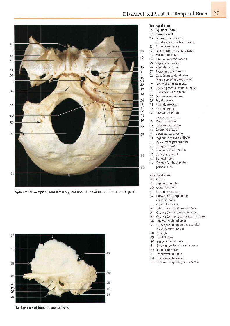
Disarticulated Skull II: Temporal Bone 27
1 3
I I
65
6
64
5B
62
50
51
1 5
254
A q
282627I Y
2e30e l
J I
34
33
59
Temporal bone18 Squamous partl9 Carotid canal20 Hiatus oftacial canal
2 122232425262128
29303 l3233343536
- ) l
3839404 l424344
464'7
(for the greater petrosal nerve)
Arcuate eminence
Groove for the s igmoid s inus
Mastoid foramen
Intemal acoustic meatus
Zygomatic process
Mandibular fossa
Petrotympanic fissure
Canalis musculotubarius
(bony part of auditory tube)
External acoustic meatus
Styloid process (remnant only)
Stylomastoid foramen
Mastoid canaliculus
Jugular fossa
Mastoid process
Mastoid notch
Groove for middle
meningeal vessels
Parietal margin
Sphenoidal margin
Occipital margin
Cochlear canaliculus
Aqueduct of the vestibule
Apex of the petrous part
Tympanic part
Trigeminal impression
Articular tubercle
Parietal notch
Groove fbr the superior
petrosal sinus
Sphenoidal, occipital, and left temporal bone. Base ofthe skull (external aspect).
Occipital bone
48 Cl ivus
49 Jugular tubercle
50 Condylar canal
5l Foramen magnum
52 Lower part of squamous
occipital bone
(cerebellar fossa)
53 Internal occipital protuberance
54 Croove for the transverse sinus
55 Groove for the superior sagittal sinus
56 Internal occipital crest
57 Upper part of squamous occipital
bone (cerebral fossa)
58 Condyle
59 Nuchal plane
60 Superior nuchal line
61 Extemal occipital protuberance
62 Jugular foramen
63 Inferior nuchal line
64 Pharyngeal tubercle
65 Spheno-occipital synchondrosis
452627
Left temporal bone (lateral aspect).
28 2 Head and Neck Disarticulated Skull III
222124
23
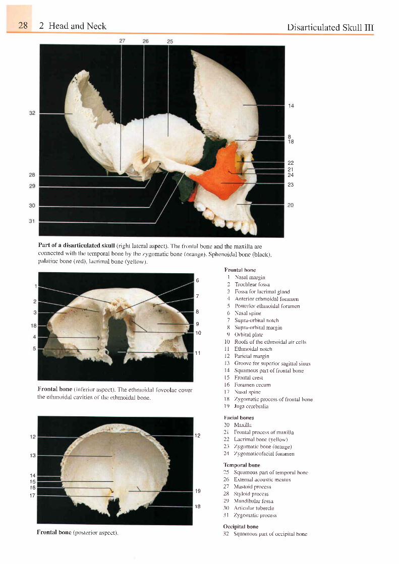
Part of a disarticulated skull (right lateral aspect). The frontal bone and the maxilla areconnected with the temporal bone by the zygomatic bone (orange). Sphenoidal bone (black),palatine bone (red), lacrimal bone (yellow).
1 3
1 41 q
t o
1 7
Frontal bone (inferior aspect). The ethmoidal foveolae coverthe ethmoidal cavities of the ethmoidal bone.
Frontal bone
o I Nasal marginb "
2 Trochlear fossa
3 Fossa for lacrimal gland7 4 Anterior ethmoidal foramen
5 Posrerior ethmoidal foramen8 6 Nasal spine
o 7 Supra-orbi ta l notch
- 8 Supra-orbi ta l margin
10 9 Orbital plate
10 Roofs of the ethmoidal air cells1l Ethmoidal notch'|
1 12 Parietal margrn
13 Groove fbr superior sagittal sinusl4 Squamous part offrontal bonel5 Frontal crest
16 Foramen cecum
l7 Nasal spine
18 Zygomatic process ofliontal bone19 Juga cerebralia
Facial bones
20 Maxilla
12 2 l F ron ta l p rocess o f max i l l a
22 Lacr imal bone (yel low r23 Zygomattc bone (orange)
24 Zygomalicofacial foramen
Temporal bone
25 Squamous part of temporal bone26 External acoustic meatus
27 Mastoid process1 0
28 Styloid process29 Mandibular fossa
18 30 Articular tubercle31 Zygomatic process
Occipital bone32 Squamous part ofoccipital boneFrontal bone (posterior aspect).
Calvana 29
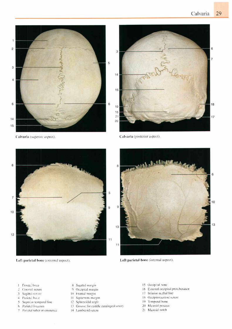
( 'a l ra r ia {supcr io I r \peet }
Lef't parietal bone (external aspect)
Fr-ontal bone( l r ronal suture
Sagi t ta l suture
Par icta l bone
Super ior tcmporal l inc
Parietal firrarnen
Par ieta l tuber or eminence
8 Sagi t ta l rnargin
9 Occipi ta l margin
l0 Frontal margin
I I Squamous margrn
l2 Sphenoidal an-ule
l3 Groovc l i l mic ld le
l,l Lau-rbdoid suture
1 9
I D
t l
20
Calvaria (posterior aspect).
Left parietal bone t internal a\pcct)
1
-l
+-')61
l5 Occip i ta l bone
l6 E,xternal occip i ta l protubcrance
17 Inf 'er iornuchal l ine
113 Occipi tomastoid suture
| 9 Temporal bone
20 Mastoid process
2l Mastoid norchmenirrgeal ar tcr \
30 2 Head and Neck
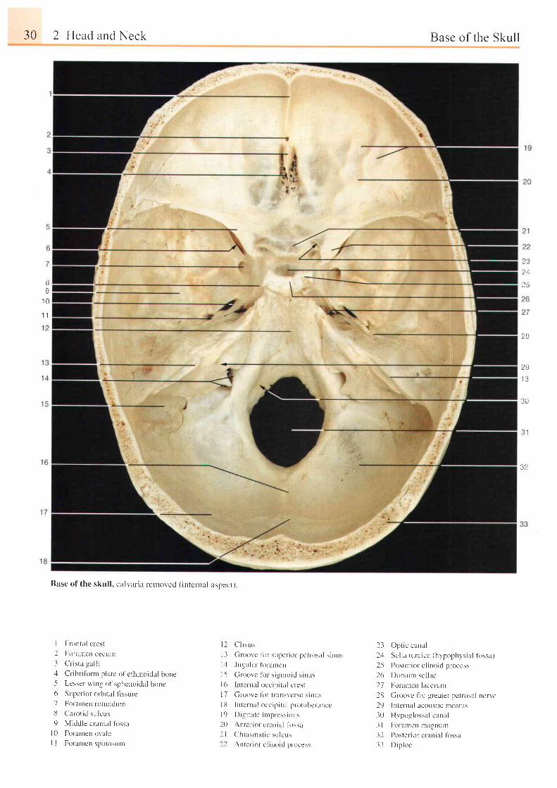
Base of the skul l , calvaria removed ( internal aspect)
I Frontai crest
2 For-alnen cecurlr
3 C r i s t a ga l l i;l Cr ibrifbrnt plate ol'ethrnoidal bone5 Lesser wing of spheuoid:r l bone6 Supcr ior orbi ta l t lssure
7 Foramen rotunclurn
8 Clarot id sulcr , rs
9 Middle crania l l i rssal0 Foranterr ot'ale
I I Foramcn spinosum
l 2 C l i v u s
I 3 Groove l i r r sr . rper io l pet losul s inus
1 4 Jugr,rlar firranrcn
l5 [ i rooi 'e f i l s igntoid s inusl6 lnte lnal occip i ta l crest
l 7 C ro0 r e l i ) f l t : l n \ \ r . f \C \ i i l u \I 8 lnternal occip i ta l protuber-ance
l9 D ig i t a t e i n rp r css i ons
20 Anter ior c lania l fossa
2 l Ch iasma t i c su l cus
2 l A t t t e r i u r r ' j i t t r t i t l I r l i r . c . .
2- j Opt ic canal
2.1 Sel la turc ica (hypophysia l f i rssa)
.2-{ Po: tcr ior e l inoi t l p locc ' .
26 Dorsunr sel lae
27 Foramen lacerum
28 Groovc lor greater pctrosal uerve29 lnte lnal acoust ic nteatus
30 Hypoglossal canal
3l Foranren magnum
32 Poster ior-crania l lbssa
33 D ip l oe
Base of the Skull
?.324
?5BI
1 0
l . l
28
2S'13
30
3 1
32
Base of the Skul l 31
23A
5
6
1 1
1 2
I J
1 41 5
1 6
1 8
1 9
8
9t 0t l
I 2t 3
1
234
o
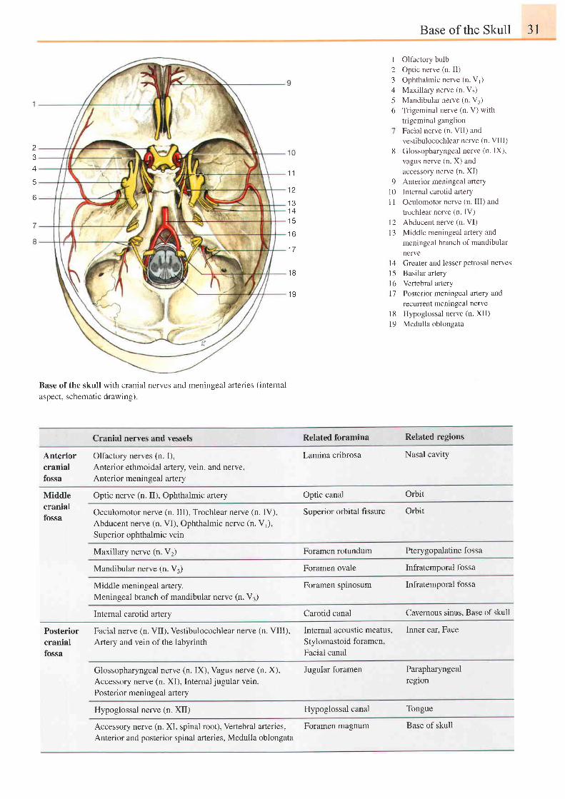
Olfactory bulb
Optic nerve (n. II)
Ophthalmic nerve (n. V1)
Maxillary nerve (n. V2)
Mandibular nerve (n V,)
Tr igeminal nerve (n. V) wi th
trigeminal ganglion
Facial nerve (n VII) and
vestibulocochlear nerve (n. VIII)
Glossopharyngeal nerve (n. IX),
vagus nerve (n. X) and
accessory nerve (n. XI)
Anterior meningeal artery
Internal carotid artery
Oculomotor nerve (n. III) and
trochlear nerve (n. IV)
Abducent nerve (n. VI)
Middle meningeal artery and
meningeal branch of mandibular
nerve
l4 Greater and lesser petrosal nerves
l5 Basilar artery
l6 vertebral artery
17 Posterior meningeal artery and
recunent mentngeal nerve
18 Hypoglossal nerve (n. XII)
19 Medulla oblongata
Base of the skull with cranial nerves and meningeal arteries (intemal
aspect, schematic drawing)
Cranial nerves and vessels Related foramina Related regions
Anterior Olfactory nerves (n. I),cranial Anterior ethmoidal artery, vein, and nerve,fossa Anterior meninqeal arterv
Lamina cribrosa Nasal cavity
Optic nerve (n. II), Ophthalmic artery Optic canal OrbitMiddlecranialfossa
Occulomotor nerve (n. III), Trochlear nerve (n. IV),Abducent nerve (n. VI), Ophthalmic nerve (n.Vr),Superior ophthalmic vein
Superior orbital fissure Orbit
Maxillary nerve (n. V2) Foramen rotundum Pterygopalatine fossa
Mandibular nerve (n. V3) Foramen ovale Infratemooral fossa
Middle meningeal artery,Meningeal branch of mandibular nerve (n. V3)
Foramen spinosum Infratemporal fossa
Internal carotid artery Carotid canal Cavernous sinus, Base of skull
Posterior Facial nerve (n. VID, Vestibulocochlear nerve (n. VIII),cranial Artery and vein of the labyrinthfossa
Internal acoustic meatus, Inner ear, Face
Stylomastoid foramen,
Facial canal
Glossopharyngeal nerve (n. IX), Vagus nerve (n. X),Accessory nerve (n. XI), Internal jugular vein,Posterior meningeal artery
Jugular foramen Parapharyngealreglon
Hypoglossal nerve (n. XII) Hypoglossal canal Tongue
Accessory nerve (n. XI, spinal root), Vertebral arteries,Anterior and posterior spinal arteries, Medulla oblongata
Foramen magnum Base of skull
1 a_ ) L I l i . ' . r , i , i r ) t i \ ee 1 , ,
1 U1 , 7
1 3
1 4
1 6
1 7
1 8
l Jasc o f t l r c \ l i u i l
2 1
22
a a
Base of the Skull 33
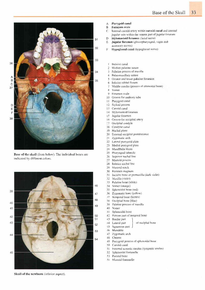
BC
Pterygoid canalForamen ovaleInternal carotid artery within carotid canal and internaljugular vein within the venous part ofjugular foramen
Stylomastoid foramen (facial nerve)
Jugular foramen (glossopharyngeal, vagus andaccessory nerves)Hypoglossal canal (hypoglossal nerve)
31
32
DE
ABU
6 t
n
EF
3B
Base of the skull (from below). The individual bones are
indicated by different colors.
I Incisive canal2 Median palatine suture3 Palatine process of maxilla4 Palatomaxillarysuture5 Greater and lesser palatine foramina6 Inferior orbital fissure7 Middle concha (process of ethmoidal bone)8 Vomer9 Foramen ovale
10 Groove for auditory tube.l
I Pterygoid canall2 Styloid process13 Carotid canall4 Stylomastoid foramen15 Jugular foramen16 Groove for occipital arteryl7 Occipital condyle18 Condylarcanal19 Nuchal plane20 Externaloccipitalprotuberance21 Zygomatic arch22 Lateral pterygoid plate23 Medial pterygoid plate24 Mandibular fossa25 Pharyngeal tubercle26 Superior nuchal line27 Mastoid process28 Inferior nuchal line29 Mastoid notch30 Foramen magnum3l Incisive bone or premaxilla (dark violet)32 Maxilla (violet)33 Palatine bone (white)34 Vomer (orange)35 Sphenoidal bone (red)
36 Zygomaric bone (yellow)
37 Temporal bone (brown)
38 Occipital bone (blue)
39 Palatine process of maxilla40 Vomer4l Sphenoidal bone42 Petrous part oftemporal bone43 Basilar part 144 Lateral part
I of occipital bone
45 Squamous part J46 Mandible41 Zygomatic arch48 Choana49 Pterygoid process of sphenoidal bone50 Carotid canal51 External acoustic meatus (tympanic anulus)52 Sphenoidal fontanelle53 Parietal bone54 Mastoid fontanelle
47
48
49
50c t
52
53
Skull of the newborn (inferior aspect).
54
34 2 Head and Neck
t o
1 B'I
1 9
2
6
Base of the Skull
1 7
t 3
'I
234
5
7
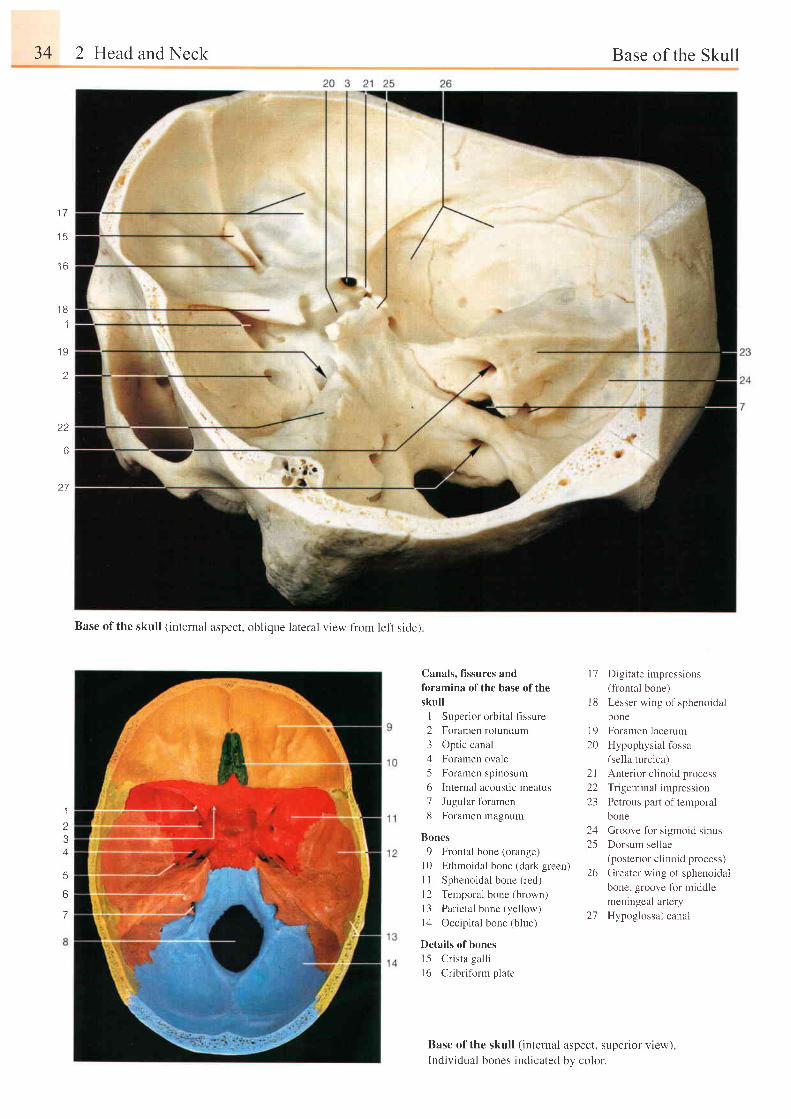
Canals, fissures and
foramina of the base of the
skull
I Superior orbital fissure
2 Foramen rotundum
3 Optic canalz[ Foramen ovale
5 Foramen spinosum
6 Internal acoustic meatus
7 Jugular fbramen
8 Foramen magnum
Bones
9 Frontal bone (orange)
l 0 E thmo ida l bone l d r r k g reen r
I I Sphenoidal bone (red)
l2 Temporal bone (brown)
l3 Par ieta l bone (yel low)
l4 Occipital bone (blue)
Details of bones
l5 Cr ista gal l i
l6 Cribriform plate
l7 Digi tate impressions(frontal bone)
I 8 Lesser wing of sphenoidal
bone
19 Foramen lacerum
20 Hypophysial fossa(sella turcica)
2 l An te r i o r c l i no i d p rocess
22 Trigeminal impression
2.1 Petrous part of tempora.
bone
24 Groove for sigmoid sinus
25 Dorsum sellae(posterior clinoid process)
26 Greater wing of sphenoidal
bone. groove for middle
meningeal artery
27 Hypoglossal canal
Base of the skull (internai aspect, superior view)Individual bones indicated by color.
Base of the skull (internal aspect, oblique lateral view from left side)
Skull of the Newborn 35
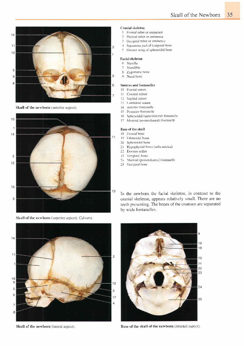
Skull of the newborn (anterior aspect).
Cranial skeleton
I Frontal tuber or eminence
2 Parietal tuber or eminence
3 Occipital tuber or eminence
4 Squamous part of temporal bone
5 Greater wing of sphenoidal bone
Facial skeleton
6 Maxi l la
7 Mandible
8 Zygomatic bone
9 Nasal bone
Sutures and fontanelles
l0 Frontal suture
I I Coronal suture
l2 Sagittal suture
l3 Lambdoid suture
14 Anterior fontanelle
| 5 Posterior fontanelle
l6 Sphenoidal (anterolateral) fontanelle
l7 Mastoid (posterolateral) fbntanelle
Base of the skull
l8 Frontal bone
l9 Ethmoidal bone
20 Sphenoidal bone
2l Hypophysial fossa (sella turcica)
22 Dorsum sellae
23 Temporal bone
24 Mastoid (posterolateral) fontanelle
25 Occipital bone
In the newborn the facial skeleton, in contrast to thecranial skeleton, appears relatively small. There are noteeth presenting. The bones of the cranium are separatedby wide fontanelles.
I
1 81 9
20
212223
25
1 6I
Skull of the newborn (superior aspect). Calvaria.
Skull ofthe newborn (lateral aspect). Base ofthe skull ofthe newborn (internal aspect)
36 2 Hcad and Neck Median Section throush the Skull
20
2 1
2?
, \
24
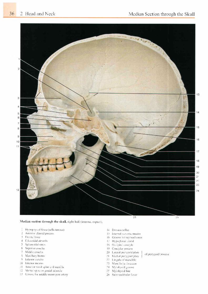
\Iedian sect ion through the skul l , r ' ight hal f ( inLernrLl lspccr)
J Hvpop l r vs i a l l i r s sa ( sc l l a t u r c i cx )2 Antcr- ior c l inoid process
- l I r rontal bonc.1 F. t l r r r ro idal arr cc l ls
5 Sphcno i c l a l s i nus
6 Supcr ior concl ta
7 \ { idr i lc concha
3 N , l a r i l l a r l , h i a tus
9 I r f e l i o r - conc l l r
I 0 I n l c r i o r n r c t r t us
I I A r r t c r i o r n t r sa l sp i ne und n ta r i l l a1 l N l cn ta l sp i r r c o r -gen iu l t L rbc l - c l cI i Croor c f or nt iddlc rneniuqcal ar terrr
26
l - 1 [ ) o l sL rn r sc l l ac
I5 I n t cn l r l l i coL l s t i c n l a i r t u r ,
l ( r ( i r - oo rc l i r r s i sn ro i c l s i nus
I 7 H r pou lossa l cuna l
J l l Occ ip i t a l con r l v l c
l 9 C r l t r L l l l r - p r - occss
l { } L i r l r ' r ' i l l l l l . ' r ' \ = - { ' i d p l i r l r II | \ l e , l r : r l 1 ' t . 1 r - r ) t \ l l r l i l t . i
t ) l | l t \ ' r l l t ) l ( l l ' 1 1 r ( e ' \
l l I - i ng r r l l r o l mu r rd i b l e
l . l Nl l rn i l ibrr lu- l i r r - l r l r . rcn
l -1 N' l r lo l r r o ic l l toor c
l - 5 N l r l o l t r o i c l l i r t c
l ( r SL rbn lL l t l r bu l r i r I o r ca
25
Median Section throush the Skull 37
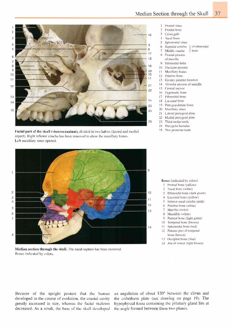
----------:- 16
I
2345678
Frontal sinus
Frontal bone
Crista galli
Nasal bone
Sphenoidal sinus
Superiorconcha ]
ofethmoidal
Middle concha J bone
Frontal process
of maxilla
Ethmoidal bulla
Uncinate process
Maxillary hiatus
Palatine bone
Greater palatine fbramen
Alveolar process of maxilla
Central incisor
Zygomatic bone
Ethmoidal bone
Lacrimal bone
Pterygopalatine fossa
Maxillary sinus
Lateral pterygoid plate
Medial pterygoid plate
Third molar tooth
Pterygoid hamulus
Two premolar teeth
4
6
7BI
1 0.1 1
1 2
1 3
1 4
1 A
Facial part of the skull (viscerocranium), divided in two halves (lateral and medial
aspect). Right inferior concha has been removed to show the maxillary hiatus.Left maxillary sinus opened.
Median section through the skull. The nasal septum has been removed.Bones indicaled bv colors.
4
l l
RJ
1 9201 01 1
21
A A
Z J
25
Bones (indicated by colors)
I Frontal bone (yellow)
2 Nasal bone (white)
3 Ethmoidal bone (dark green)
4 Lacrimal bone (yellow)
5 Inferior nasal concha (pink)
6 Palatine bone (white)
7 Maxilla (violet)
8 Mandible (white)
9 Parietal bone (light green)
10 Temporai bone (brown)
I I Sphenoidal bone (red)
l2 Petrous part of temporal
bone (brown)
l3 Occipital bone (blue)
14 Ala of vomer (light brown)
an angulation of about 120' between the clivus andthe cribriform plate (see drawing on page 19). Thehypophysial fossa containing the pituitary gland lies atthe angle formed between these two planes.
91 0l 1l 2l - l
1 41 51 6l '7t 81 9202 l22L )
. A
25
Because of the upright posture that the humandeveloped in the course of evolution, the cranial cavitygreatly increased in size, whereas the facial skeletondecreased. As a result. the base of the skull develooed
38 2 Head and Neck Disarticulated Skull JV
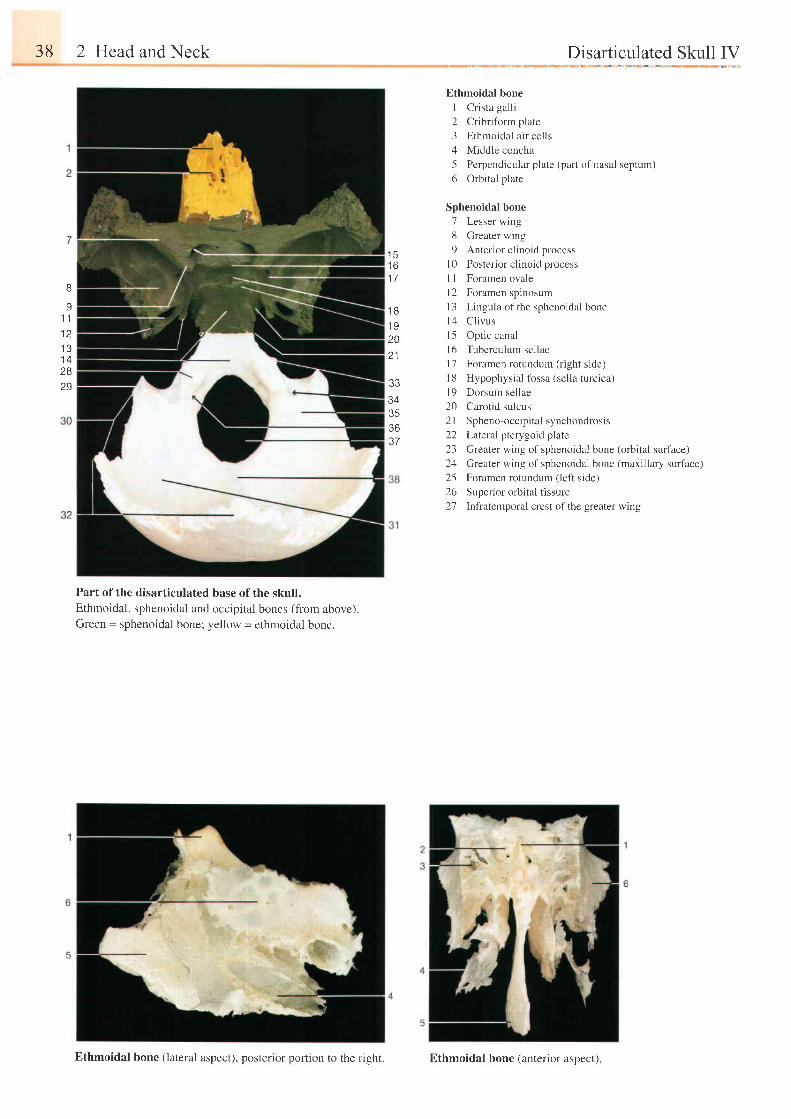
Ethmoidal bone
I Cr ista gal l i
2 Cr ibr i fbrm plate
3 Ethmoidal a i r cel ls
4 Middle concha
5 Perpendicular plate (part of nasal septum)
6 Orbital plate
Sphenoidal bone
7 Lesser wing
8 Greater wing
9 Anterior clinoid process
l0 Posterior clinoid process
I I Foramen ovale
l2 Foramen spinosum
l3 Lingula of the sphenoidal bone
1,1 Cl ivus
l5 Opt ic canal
l6 Tuberculum sellae
l7 Foramen rotundum (right side)
l8 Hypophysial fossa (sella turcica)
l9 Dorsum sel lae
20 Carotid sulcus
2l Spheno-occipital synchondrosis
22 Lareral pterygoid plate
23 Greater wing of sphenoidal bone (orbital surface)
24 Greater wing of sphenoidal bone (maxillary surface)
25 Foramen rotundunr (left side)
26 Superior orbital fissure
27 lnfratemporal crest of the greater wing
t )t o
1 7R
Y
1 11 21 31 42829
1 B1 920
3334353637
Part of the disarticulated base of the skull.Ethmoidal, sphenoidal and occipital bones (from above).Green = sphenoidal bone; yellow = ethmoidal bone.
Ethmoidal bone (lateral aspect), posterior portion ro rhe right. Ethmoidal bone (anterior aspect)
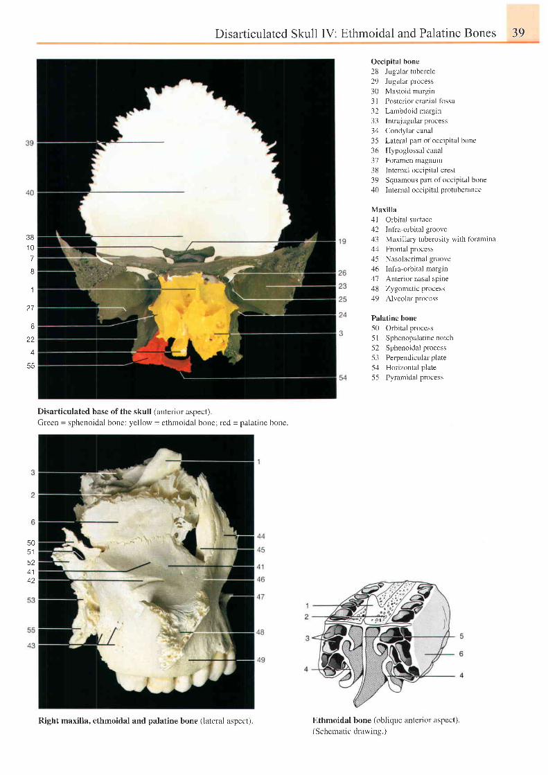
Disarticulated Skull IV: Ethmoidal andPalatine Bones 39
3B1 07R
1
2 7
6
22
4 4
Occipital bone28 Jugular tubercle29 Jugular process30 Mastoid margin3l Posterior cranial fossa32 Lambdoid margin33 Intrajugularprocess34 Condylar canal35 Lateral part ofoccipital bone36 Hypoglossal canal37 Foramen magnum38 Internal occipital crest39 Squamous part of occipital bone40 Internaloccipital protuberance
Maxilla41 Orbital surface42 Infra-orbital groove43 Maxillary tuberosity with fbramina44 Frontal process45 Nasolacrimal groove46 Iniia-orbital margin47 Anterior nasal spine48 Zygomatic process49 Alveolar process
Palatine bone50 Orbital process5l Sphenopalat inenotch52 Sphenoidal process53 Perpendicuiar plate54 Horizontal plate55 Pyramidal process
Disarticulated base of the skullGreen = sphenoidal bone; yellow
(anterior aspect).= ethmoidal bone; red = palatine bone.
50c l
524142
Ethmoidal bone (oblique anterior aspect).
(Schematic drawing.)Right maxilla, ethmoidal and palatine bone (lateral aspect).
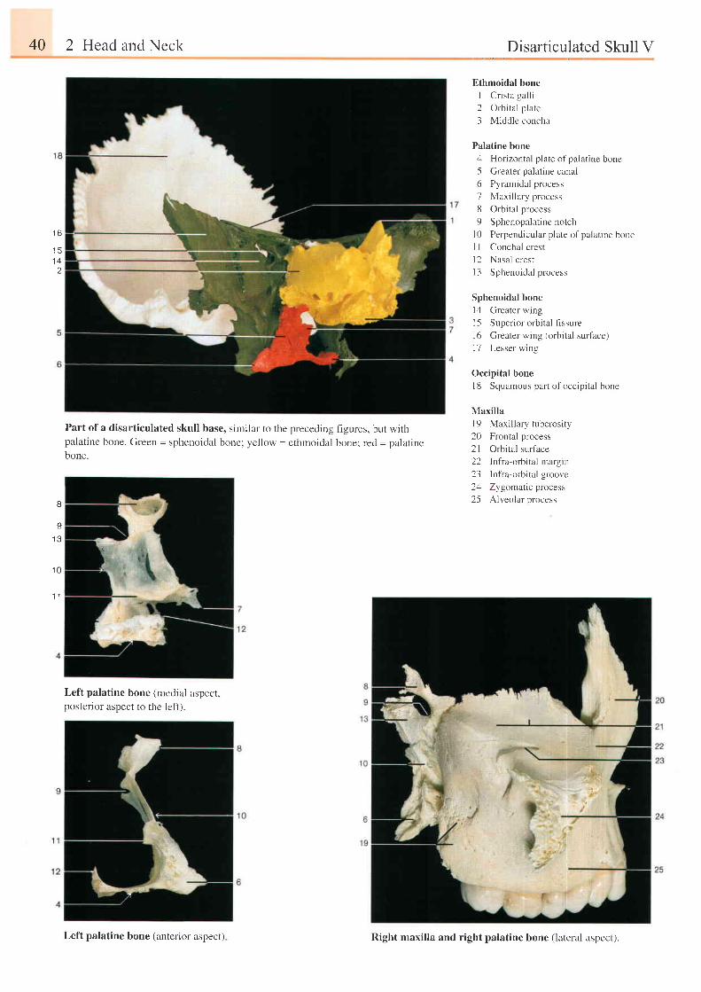
40 2 Head and Neck Disarticulated Skull V
Ethmoidal bone
I Cr ista gal l i
2 Orbital plare
3 Middle concha
Palatine bone
4 Horizontal plate of palatine bone
5 Greater palatine canal
6 Pyramidal process
7 Maxillary process
8 Orbital process
9 Sphenopalatine notch
l0 Perpendicular plate ofpalatine bone
I I Conchal crest
l2 Nasal crest
I 3 Sphenoidal process
Sphenoidal bone
l4 Greater wing
l5 Superior orbital fissure
l6 Greater wing (orbital surface)
l7 Lesser wing
Occipital bone
l8 Squamous part ofoccip i ta l bone
Maxilla
l9 Maxillary tuberosity
20 Frontal process
2l Orbital surface
22 Infra-orbitalmargin
23 Infra-orbi ta lgroove
2zl Zygomatic process
25 Alveolar process
t o
' 15
1 4
Part of a disarticulated skull base, similar to the precedingpalatine bone. Green = sphenoidal bone; yellow = ethmoidalbone.
f igures, but with
bone; red = palat ine
I
Y
I J
1 0
1 1
Left palatine bone (medial aspect,posterior aspect to the left).
Left palatine bone (anterior aspect) Right maxilla and right palatine bone (lateral aspect).
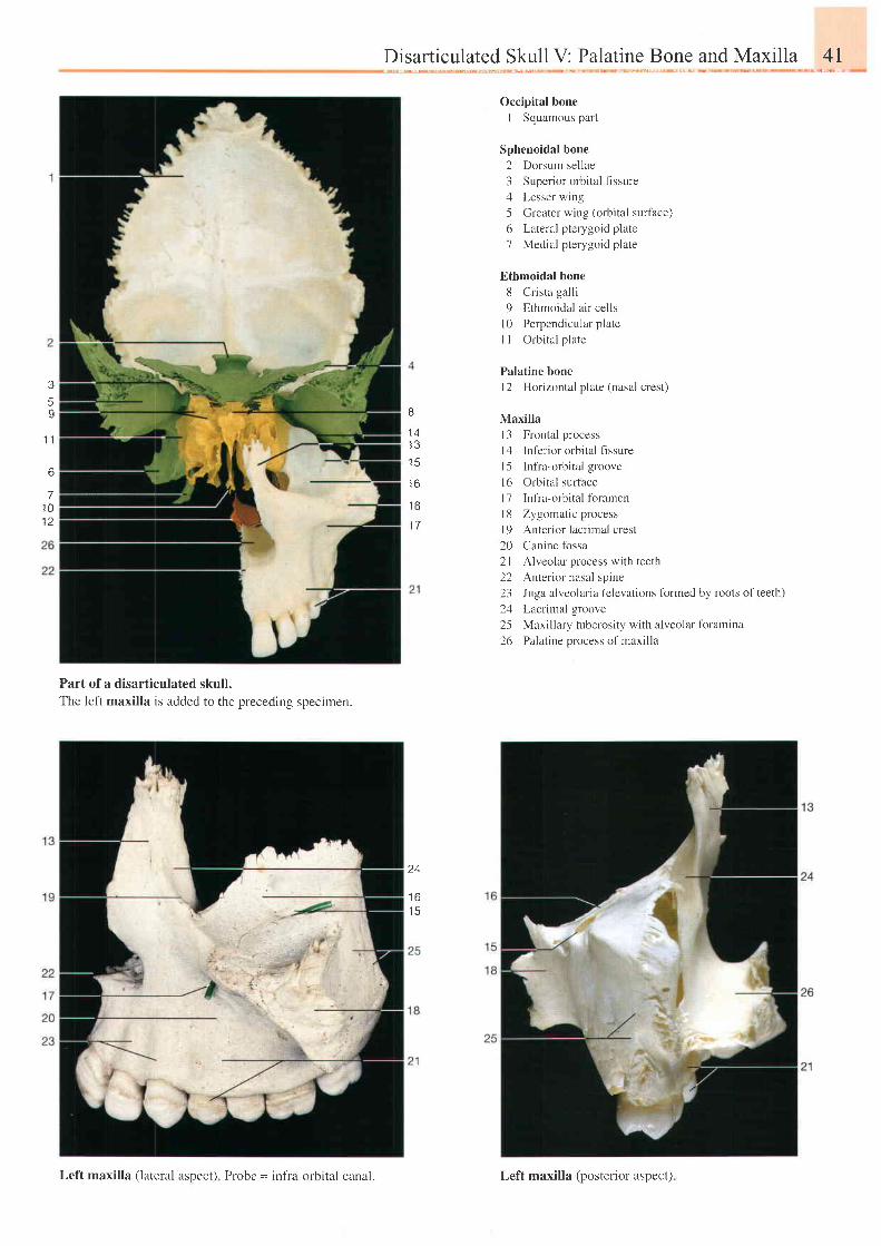
Disarticulated Skull V: Palatine Bone and Maxilla 4I
o
1 41 3' t 4
t o
1 B
36
I
t ,
6
71 0
Occipital bone
I Squamous part
Sphenoidal bone
2 Dorsum sellae
3 Superior orbital flssure
4 Lesser wing
5 Creater wing (orbital surface)
6 Lateral pterygoid plate
7 Medial pterygoid plate
Ethmoidal bone
8 Crista galli
9 Ethmoidal air cells
10 Perpendicular plate
I I Orbital plate
Palatine bone
l2 Horizontal plate (nasal crest)
Maxilla
I 3 Frontal process
I 4 Inf'erior orbital frssure
l5 Infia-orbital groove
l6 Orbital surface
l7 Infia-orbital foramen
18 Zygomat ic process
l9 Anterior lacrimal crest
20 Canine fossa
2l Alveolar process with teeth
22 Anter ior nasal spine
23 Juga alveolaria (elevations formed by roots of teeth)
24 Lacrimal groove
25 Maxillary tuberosity with alveolar fbramina
26 Palatine process of maxilla
Part of a disarticulated skull.The left maxilla is added to the preceding specimen.
24
1 6t 3
Left maxilla (lateral aspect) Probe = infia-orbital canal. Left maxilla (posterior aspect)
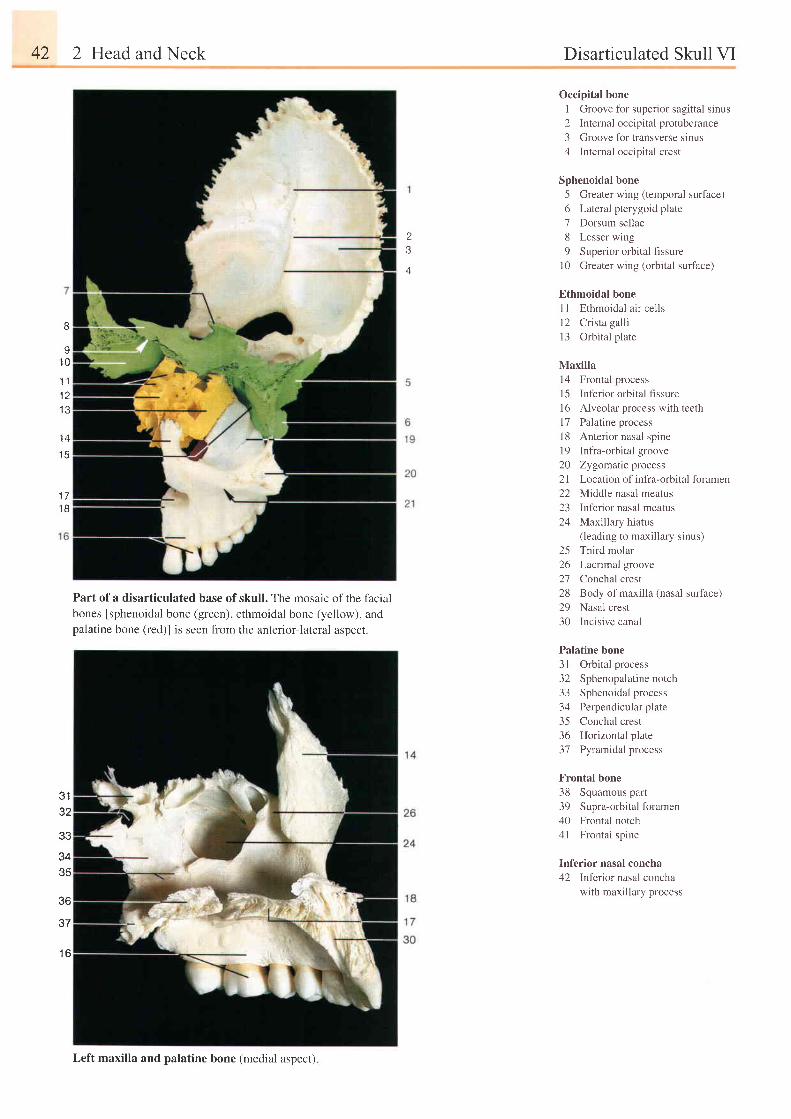
42 2 Head and Neck Disarticulated Skull VI
n
,)A
Occipital bone
1 Groove for superior sagittal sinus
2 Internal occipital protuberance
3 Groove fbr transverse sinus
4 Internal occipital crest
Sphenoidal bone
5 Greater wing (temporal surface)
6 Lateral pterygoid plate
7 Dorsum sellae
8 Lesser wing
9 Superior orbital fissure
10 Greater wing (orbital surface)
Ethmoidal bone
I I Ethmoidal a i r cel ls
l2 Cr ista gal l i
l3 Orbital plate
Maxilla
l4 Frontal process
l5 lnferior orbital fissure
l6 Alveolar process with teeth
l7 Palatine process
l8 Anterior nasal spine
19 Infia-orbital groove
20 Zygomatic process
21 Location of infia-orbital foramen
22 Middle nasal meatus
23 Inferior nasal meatus
24 Maxillary hiatus( leading to maxi l lary s inus)
25 Third molar
26 Lacrimal groove
27 Conchal crest
28 Body ofmaxilla (nasal surface)
29 Nasal crest
30 Incisive canal
Palatine bone
3l Orbital process
32 Sphenopalatinenotch
33 Sphenoidal process
34 Perpendicular plate
35 Conchal crest
36 Horizontal plate
37 Pyramidal process
Frontal bone
38 Squamous part
39 Supra-orbital foramen
40 Frontal notch
4l Frontal spine
Inferior nasal concha
42 Inferior nasal concha
with maxillary process
B
vl 0
1 1t t
t 3
1 4
l 5
1 7t 6
Part of a disarticulated base of skull. The mosaic of the facialbones [sphenoidal bone (green), ethmoidal bone (yellow), andpalatine bone (red)l is seen from the anterior-lateral aspect.
3 132
33
3435
36
37
Left maxilla and palatine bone (medial aspect)
1 6
Drsarticulated Skull VI: Palatine Bone and Maxilla
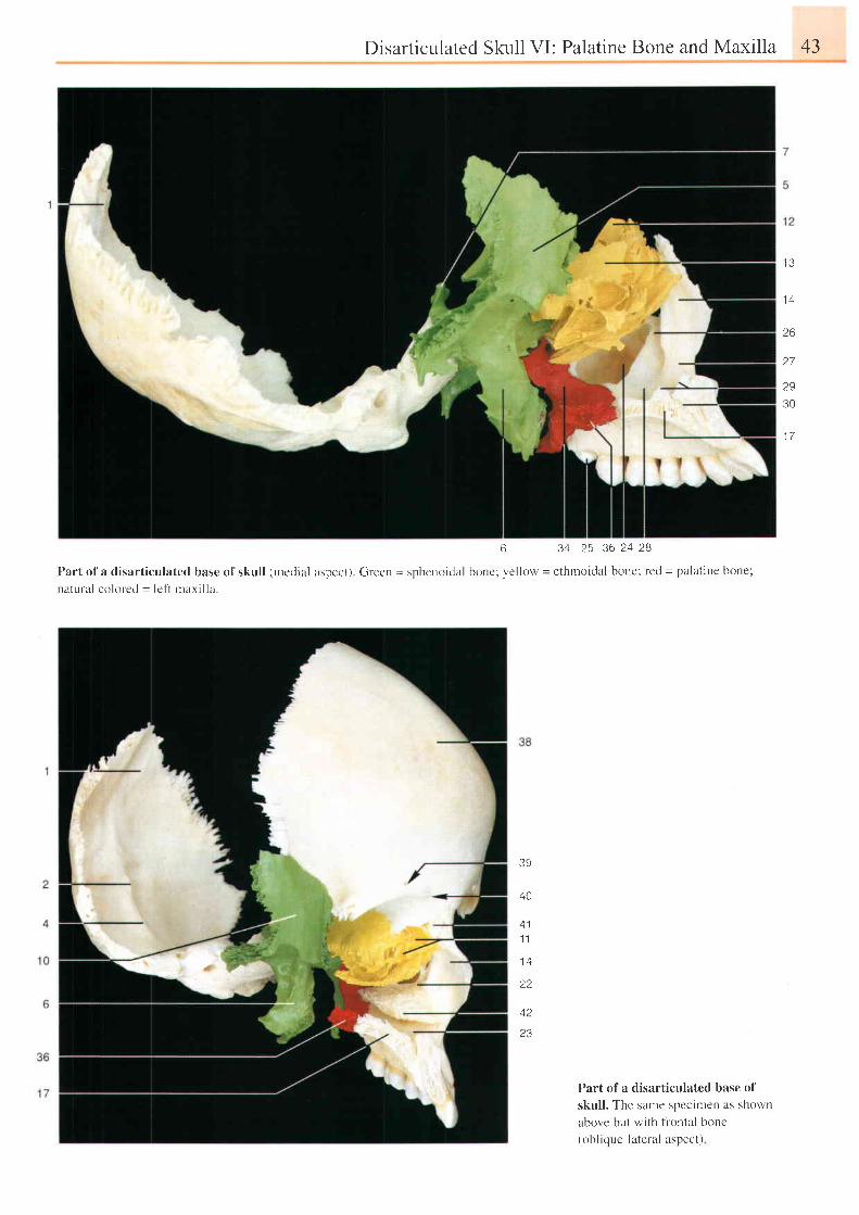
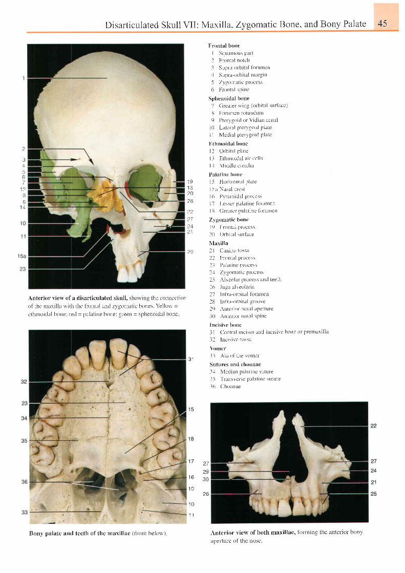
6 3'l 2.5 36 ?.4 2B
Part ofa disart iculated base ofskul l (mcdial aspect). Green = sphcnoidal bonc: yel low = ethmoidal bone, red = palat ine bone;
natural cokrred = let i maxi l la.
Part of a disarticulated base of
skul l . The same specimen as shown
above but with f lontal bone(obl ique-latefal aspect)
43
1 3
l a
26
27
29
30
1 7
3E
40
1 1
1 4
t 2
i a
23
44 2 Head and Neck Disarticulated Skull VII
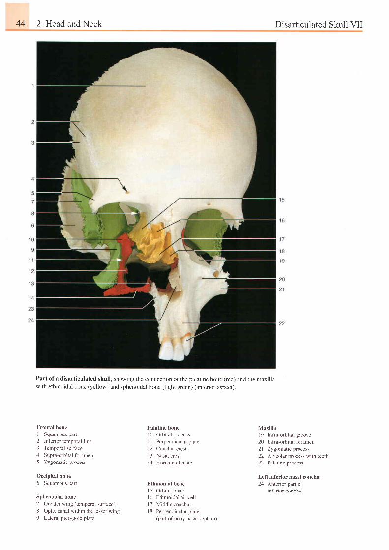
Part of a disarticulated skull, showing the connection of the palatine bone (red) and the maxillawith ethmoidal bone (yellow) and sphenoidal bone (light green) (anterior aspect).
Frontal bone
1 Squamous part
2 Inferior temporal line3 Temporal surface
4 Supra-orbital foramen
5 Zygomatic process
Occipital bone
6 Squamous part
Sphenoidal bone
7 Greater wing (temporal surface)8 Op t i c cana l w i t h i n t he l esse r w ing
9 Lateral pterygoid plate
Palatine bone
l0 Orbital process
I 1 Perpendicular plate
12 Conchal crest
I 3 Nasal crest
14 Horizontal plate
Ethmoidal bone
15 Orbital plate
16 Ethmoidal air cell
17 Middle concha
18 Perpendicular plate
(part of bony nasal septum)
Maxillal9 Infra-orbital groove20 Infra-orbitalforamen21 Zygomatic process22 Alveolar process with teeth23 Palatine process
Left inferior nasal concha24 Anterior part of
inferior concha
2
3,x567
1 2BI
1 n
Disarticulated Skull VII :
Anterior vielv of a disarticulated skull, showing the connectiort
of the rnaxilla with the fiontal and zi,gornatic bones Yelkrw =
ethrnoidal bonel red = palatine bone; green = sphenoidal bone
Bony palate and teeth of the maxillae (fiom below)
Maxilla, Zygomattc Bone, and Bony Palate 45
F-rontal bone
I Squanrous part
2 Frontal notch
3 Supra-orbital fbramen.1 SLrpra-orbital margin
5 Zygouralic process
6 Flonla l spine
Sphenoidal bone
7 Grcater wing (orbi ta l sur tace)
8 Foramcn rotllndLlm
9 Ptcrygoid or Vidian caual
l0 Lateral pterygoid plate
I I Medial pterygoid platc
Flthmoidal bone
l2 O rb i t a l p l a t c
l l Ethnroidal r i l cel ls
1,1 Middlc concha
Palatine bonc
1.5 Hor izontnl p late
l 5a Nasa l c res t
l(r P-vrarridal proccss
l7 Lcsser palat ine fbramett
l [3 Greatcr palat inc foranrcn
Zygomatic bone
l9 Frontal process
f0 Orbi ta l sur lace
Nlaxilla
2 | (lanine lirssa
22 Florrtal pt'ocess
23 Palat ine process
2,1 Zygornatic pntcess
25 Alvcolar process and teeth
2(r Juga alveolar ia
27 lnfra-orbital fbr amen
2u ln l ia-orbi ta l groove
29 Anter ior nasal apcrture
J0 Anter ior r rasal spine
Incisive bone
3 I Central inc isor and incis ive bone or prernaxi l la
32 lncis ive tbssa
Vomer
33 Ala of the vomer
Sutures and choanae
3,1 Median palat inc suture
35 ' l ' ransverse palat iue suture
J6 Choanae
1 91 320t 6
?227242 1
26
J I
t o
1 0
1 0
27to
30
26
Anterior view of both maxillae, fbrming the anterior bony
aperturc of the nose.
'1 1
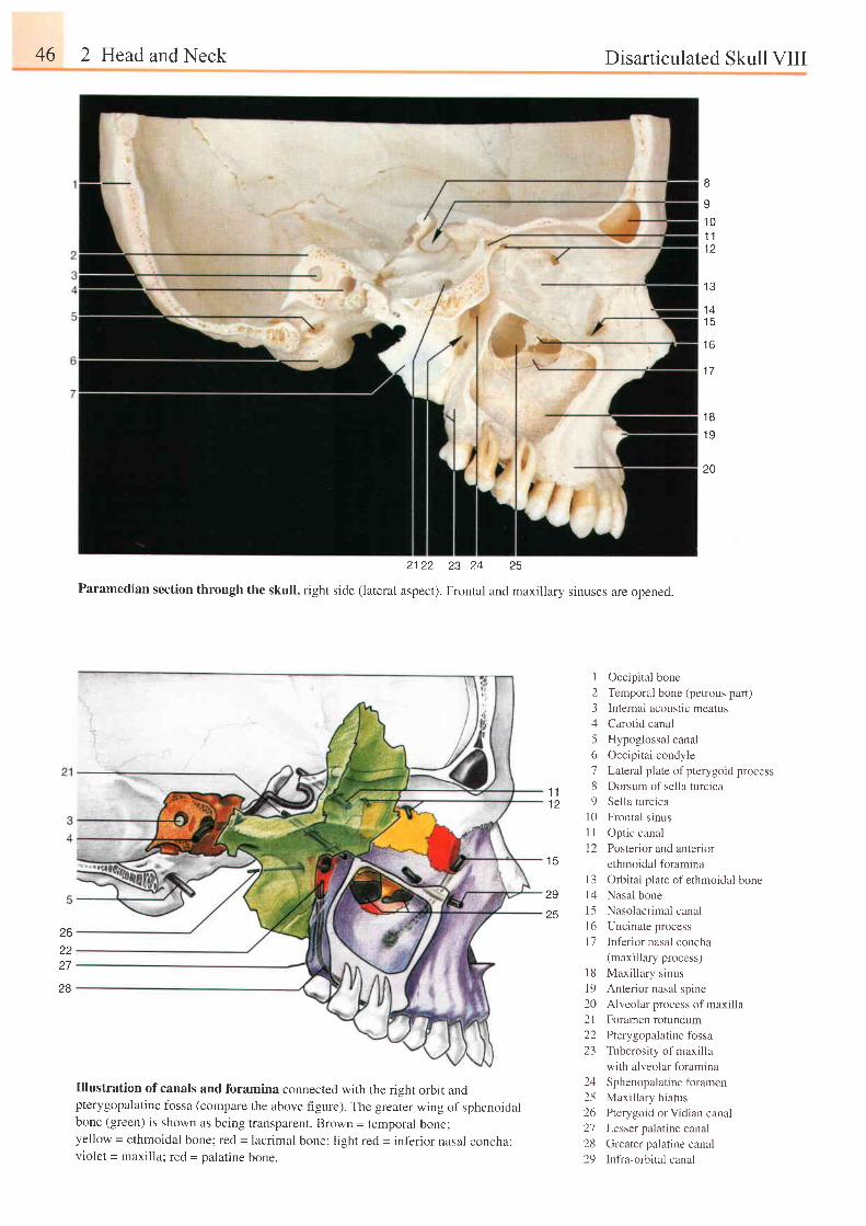
46 2 Head and Neck Disarticulated Skull VIII
B
1 01 11 2
1 7
1 B
1 9
10
I Occipital bone
2 Temporal bone (petrous part)
3 Internal acoustic meatus
4 Carotid canal
5 Hypoglossal canal
6 Occipital condyle
J Lateral plate ofpterygoid process
8 Dorsum of sella turcica
9 Sella turcica
l0 Frontal sinus
I I Optic canal
[2 Posterior and anterior
ethmoidal foramina
1 3
1 4t 0
1 6
2122 23 ?4 25
Paramedian section through the skull, right side (lateral aspect). Frontal and maxillary sinuses are opened.
1 11 2
29
25
1 5
262227
28
l 3t 41 5I 6t '7
l 81 9202 l2223
2425zo
212829
Orbital plate of ethmoidal boneNasal bone
Nasolacrimal canal
Uncinate process
Inf'erior nasal concha(maxillary process)
Maxillary sinus
Anterior nasal spine
Alveolar process of maxilla
Foramen rotundum
Pterygopalatine fossa
Tuberosity of maxilla
with alveolar foramina
Sphenopalatine foramen
Maxilltuy hiatus
Pterygoid or Vidian canalLesser palatine canal
Greater palatine canal
Infra-orbital canal
Illustration of canals and foramina connected with the right orbit andpterygopalatine fossa (compare the above figure). The greater wing of sphenoidalbone (green) is shown as being transparent. Brown = temporal bone;yellow - ethmoidal bone; red = lacrimal bone; light red = inf-erior nasal concha;violet = maxilla; red = palatine bone.
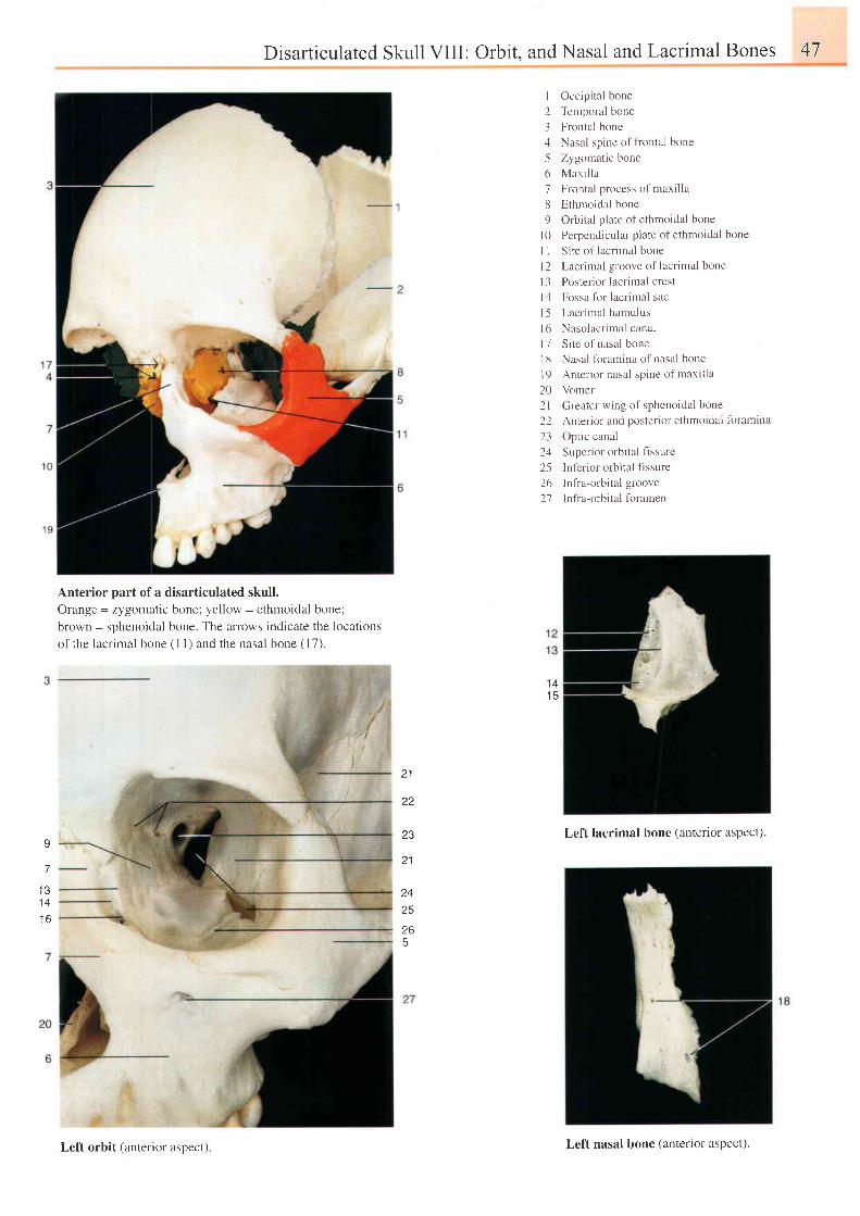
Disarticulated Skull VIII: Orbit. and Nasal and Lacrimal Bones 47
I Occipital bone
2 Temporal bone
3 Frontal bone
4 Nasal spine offiontal bone
5 Zygomatic bone
6 Max i l l a
7 Frontal process of maxilla
8 Ethmoidal bone
9 Orbital plate of ethmoidal bone
| 0 Perpendicular plate of ethmoidal bone
I I Site o1 lacrirnal bone
l2 Lacrinal groove of lacrimal bone
13 Posterior lacrimal crest
l4 Fossa for lacrimal sac
l5 Lacr imal hatnulus
l6 Nasolacr imal canal
I 7 Sitc of nasal bone
l8 Nasal fbrarnina o1'nasal bone
l9 Anter ior nasal spine of maxi l la
20 Vomer
2l Greater wing of sphenoidal bone
22 Anterior and poslerior ethmoidal fbramina
23 Optic canal
24 Superior orbital flssure
25 Inferior orbital fissure
26 Infra-orbital groove
27 Infra-orbital fbramen
Anterior part of a disarticulated skull.
Orange = zygomatic bone; yel low = ethmoidal bone,
brown = sphenoidal bone. The arrows indicate the locations
of the lacrimal bone ( l l ) and the nasal bone (17).
1 41 5
21
22
23
24
I
7
I J
1 4
l o
Left lacrimal bone (anterior aspect).
1 6
5
Left orbit (anterior aspect) Left nasal bone (anterior aspect).
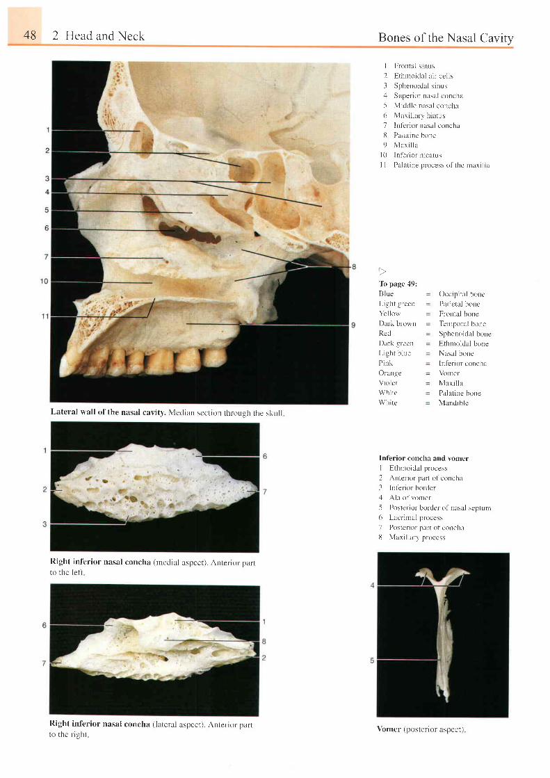
48 2 Head and Neck
Lateral wall of the nasal cavity. Median scction throLrgh the skul l
Right inferior nasal concha (rnedial aspect). Anterior.parlto the lel't
Right inferior nasal concha (lateral aspect). Anterior partto the r ight
Bones of the Nasal Cavity
I Frontal s inr :s
2 E th rno ida l a i r - ce l l s
3 Sphenoidal s inus;1 Superior nasnl concha
5 Middle nasr l concha
6 Maxi l lary h iatus
7 In lcr ior nasal concha
8 Palat ine bone
9 Mar i l l a
|0 Inf'elior mcal-r.rs
I I Palat ine process of the maxi l la
lb page 49:
B lue
Light green
Yel low
Dark blown
Rcd
Dark grecn
L igh t b l ue
P ink
Orange
V io l e t
Wh i t c
Wh i t e
Occipi ta l bone
Parietal bonc
Frontal bone
Ternporal bone
Sphenoidal bone
Ethmoidal bone
Nasal bonc
lnfcr ior concha
Vller
Max i l l a
Palat ine bone
Mandible
Inl'erior concha and vomer
I Ethmoidal process
2 Antel ior part o l 'concha
3 lnt'erior borderz l Ala of vomer
5 Poster-ior border of nasal septurn
6 Lacr imal process
7 Posterior part of concha
8 Maxi l lary process
Vomer (posterior aspect)
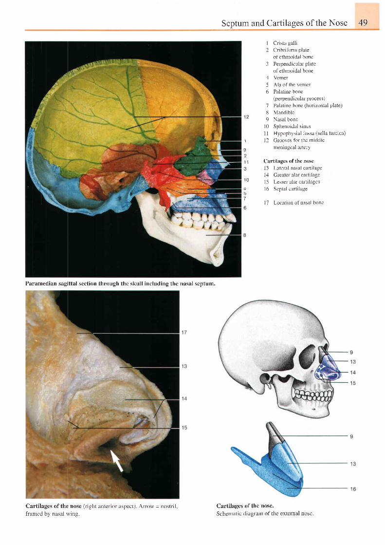
Septumand Cartilaqes of the Nose 49
3
456
189
1 0l l1 2
Crista galli
Cribrifbrm plate
of ethmoidal bone
Perpendicular plate
of ethmoidal bone
Vomer
Ala of the vomer
Palatine bone
(perpendicular process)
Palatine bone (horizontal plate)
Mandible
Nasal bone
Sphenoidal sinus
Hypophysia l forsa tsel la turc ica)
Grooves for the middle
meningeal arteryI
v
.l 1
o
1 0
457
Cartilages of the nose
13 Lateral nasal cartilage
l4 Greater alar cartilage
15 Lesser alar cartilages
16 Septal cartilage
17 Location ofnasal bone
Paramedian sagittal section through the skull including the nasal septum.
Cartilages of the nose (right anterior aspect). Arrow = nostril,framed by nasal wing.
Cartilages of the nose.Schematic diaeram of the extemal nose
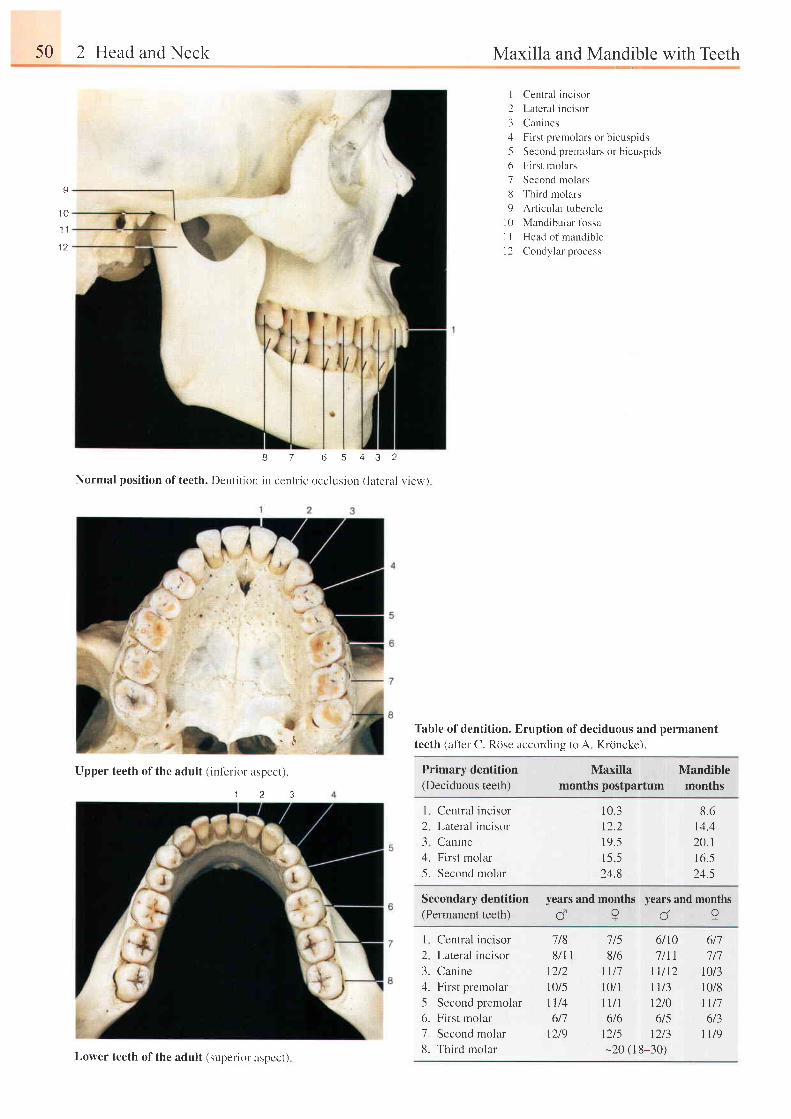
50 2 Head and Neck
8 7 6 5 4 3 2
Normal posi t ion of teeth. Dent i t ion in cent l ic occlusion ( lateral v iew)
Maxilla and Mandible with Teeth
I Central inc isor
2 Lateral incisor
3 Canines:1 First premolars or b icuspids
5 Second premolars or bicuspids
6 First rnolars
7 Second molars
ti Third molars
9 Art icular tubercle
10 Mandibular fbssa
I I Head of mandible
I 2 Condylar process
Table of dentition. Eruption of deciduous and permanentteeth (after C. Rose accordins to A. Krcincke).
I
1 0
1 1
1 a
Primary dentition(Deciduous teeth)
Maxilla Mandiblemonthspostpartum months
I Central incisor
2 Lateral incisor
3 Canine4. First molar
5. Second molar
10.312.219.51 5 524.8
8 .614.120.116 .524.5
Secondary dentition(Permanent teeth)
years and months years and monthsc?c?
I Central incisor
2 Lateral incisor
3. Canine4. First premolar
-5. Second premolar
6. First molar
7. Second molar
8. Third molar
'7 t5 6/10 6t78/6 7ill 7 t7
Ll l '1 l t /12 l0 t3tJ/t n/3 10/8Il/r 1210 |t76/6 6t5 6/3
t2t5 l2t3 |t9-20 (r 8-30)
7t88/1 r
12/2l0/5t1/4617
t2t9
, " c
Upper teeth of the adult (inf-erior aspect).
1 2 3
Lower teeth of the adult (superior aspecr)
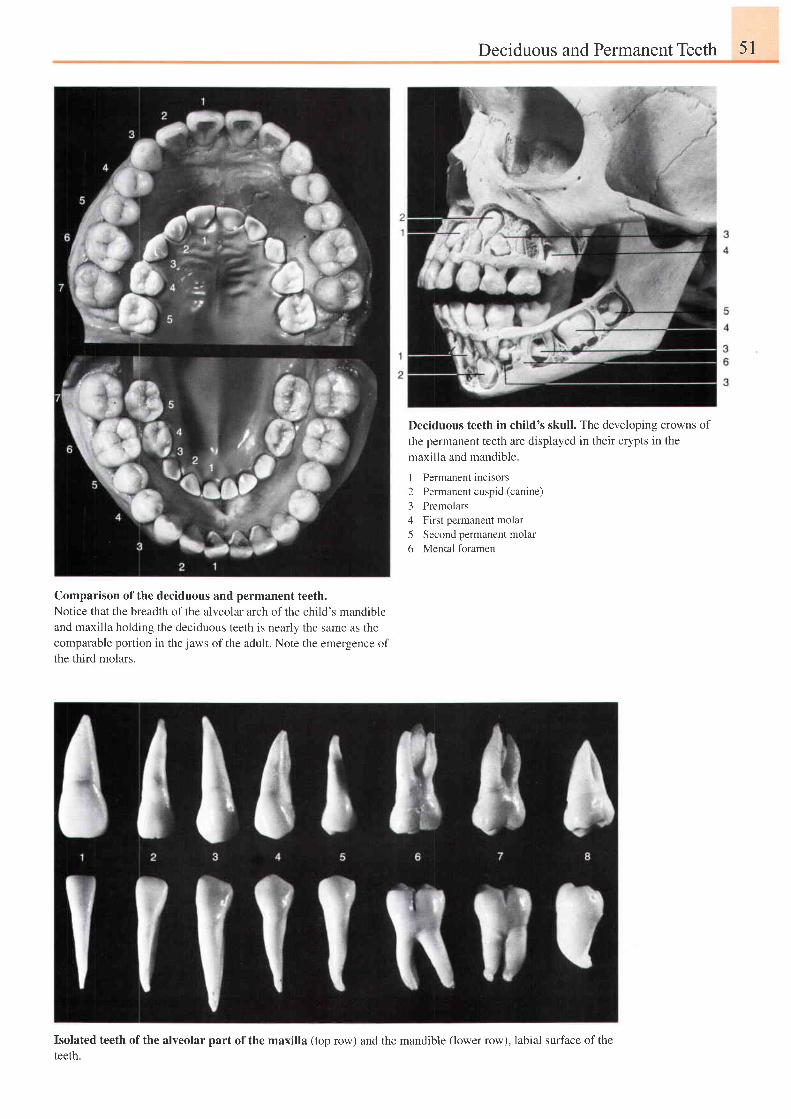
Deciduous and Permanent Teeth 51
Deciduous teeth in child's skull. The developing crowns of
the permanent teeth are displayed in their crypts in the
maxilla and mandible.
I Permanent incisors2 Permanent cuspid (canine)3 Premolars4 First permanent molar5 Second permanent molar6 Mental foramen
Comparison of the deciduous and permanent teeth.Notice that the breadth of the alveolar arch of the child's mandibleand maxilla holding the deciduous teeth is nearly the same as thecomparable portion in the jaws of the adult. Note the emergence ofthe third molars.
Isolated teeth of the alveolar part of the maxilla (top row) and the mandible (lower row), labial surface of the
teeth.
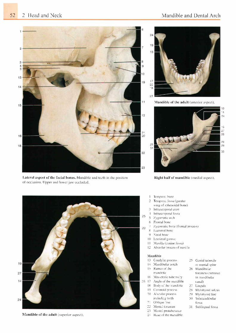
52 2 Head and Neck
Lateral aspect of the facial bones. Mandible and teeth in thc position
of occlusion. Uooer and lowcr iaw occluded
Mandible and Dental Arch
Right half of mandible (medial aspect)
Temporal bone
Temporal fbssa (greater
wing of sphenoidal bone)
Inf ia lcmporal crest
Infiatemporal fossa
Zygomat ic arch
Frontal bonc
Zygornatic bone (frontal process)
Lacr i rnal bone
Nasal bone
Lacrimll groove
Maxi l la (canine fbssa)
Al veolar process o{ ' mr i r i l la
1 7221 8
4 J
20
1 4
J 3
2726
2829
30
i!it
t0
203 1
l
2
31-561d
9t 0l l1 2
Mandible
I 3 Condylar proccss
1,1 Mancl ibr . r lar notch
l5 Rarnus of the
mand ib l c
l6 Masseter ic tuberosi ty
l7 Angle of the mandible
l 8 Body o f t he mand ib l e
l 9 Coronoid process
20 Alveolar process
including teeth
2 l Ob l i q r . r e l i ne
22 Mental tbramen
23 Mental protuberance
2,1 Head of the mandible
Gcnial tubercle
or nrental spine
Mandibular
fbramen (entrance
to mandibular
canal)
L ingula
My lohvo id su l cus
My lohyo id l i ne
Submandibr.rlar
tossa
Subl ingr. ra l lbssa
25
zo
21282930
l l
Mandible of the adult (anterior aspect)
Mandible of the adult (superior aspect)
:t8
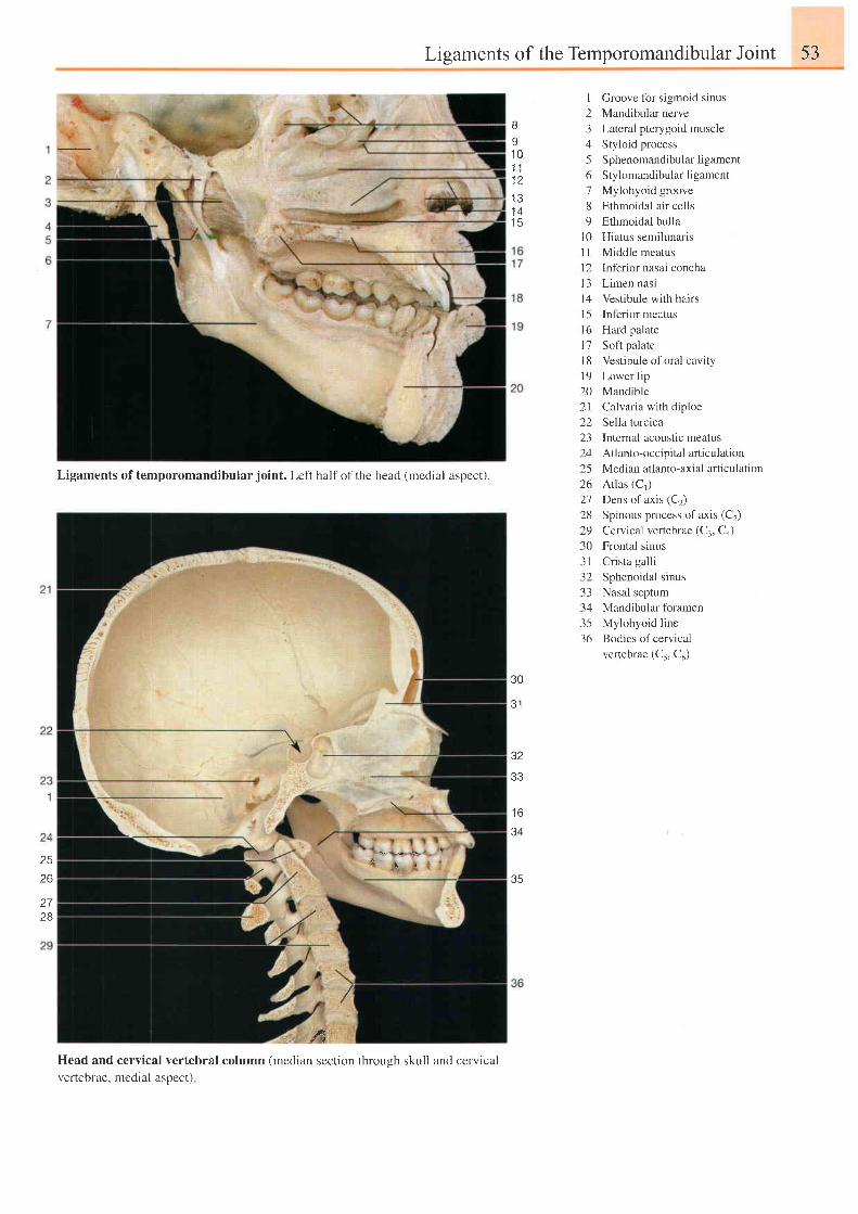
Ligaments of the Temporomandibular Joint 53
u , r r r
' { i
Head and cervical vertebral column (median section through skull and cervicalvertebrae. rnedial aspect)
s1 01 11 21 31 41 5
J Z
33
t o
34
35
I Groove tbr sigmoid sinus
2 Mandibular nerve
3 Lateral pterygoid muscle
4 Styloid process
5 Sphenomandibularligament
6 Stylomandibular ligament
7 Mylohyoid groove
8 Ethmoidal air cells
9 Ethmoidal bulla
l0 Hiatus semilunaris
I I Middle meatus
l2 lnferior nasal concha
l3 Limen nasi
l4 Vestibule with hairs
l5 Inferior meatus
l6 Hard palate
17 Soft palate
1 8 Vestibule of oral cavity
l9 Lower l ip
20 Mandible
2l Calvaria with diploe
22 Sella turcica
23 Internal acoustic meatus
24 Atlanto-occipital articulation
25 Median atlanto-axial articulation
26 Atlas (C,)
27 Dens of axis (C2)
28 Spinous process of axis (C2)
29 Cervical vertebrae (C:, Co)
30 Frontal sinus
31 Cr ista gal l i
32 Sphenoidal sinus
33 Nasal septum
34 Mandibular foramen
35 Mylohyoid line
36 Bodies ofcervical
vertebrae (Cs, Co)
Ligaments of temporomandibular joint. Left half of the head (medial aspect).
30
e l
2526
2728
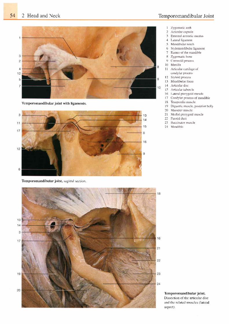
54 2 Head and Neck Temporomandibular Joint
41 2
E
7
I23456l89
1 0l l
1 21 3t 41 51 6t 7l 8l 9202 122a-)
24
Zygomatic archArticular capsuleExternal acoustic meatusLateral ligamentMandibular notchStylomandibular ligamentRamus of the mandibleZygomatic boneCoronoid processMaxillaArticular cartilage ofcondylar processStyloid processMandibular fossaArticular discArticuiar tubercleLateral pterygoid muscleCondylar process of mandibleTemporalis muscleDigastric muscle, posterior bellyMasseter muscleMedial pterygoid muscleParotid ductBuccinator muscleMandible
Temporomandibular joint with ligaments.
Temporomandibular joint, sagittal section.
I J
1 4
1 5
1 3
1 4
Temporomandibular joint.
Dissection of the afticular discand the related muscles (lateral
aspect).
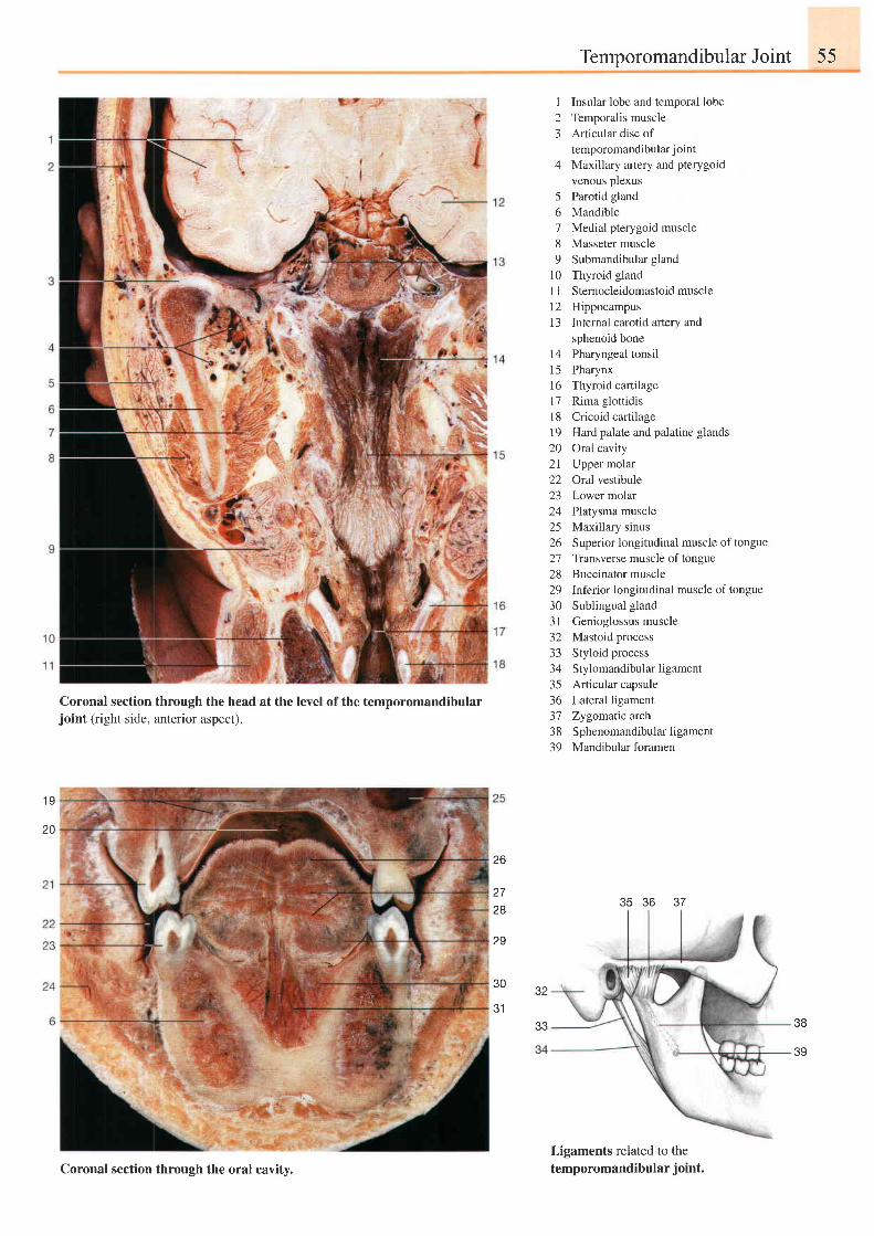
TemporomandibularJoint 55
I Insular lobe and temporal lobe2 Temporalis muscle3 Articular disc of
temporomandibular j oint4 Maxillary artery and pterygoid
venous plexus5 Parotid gland6 Mandible7 Medial pterygoid muscle8 Masseter muscle9 Submandibular gland
l0 Thyroid gland11 Sternocleidomastoidmuscle12 Hippocampus13 Internal carotid artery and
1 41 51 61 11 8l 9202 l22z3
24252627282930J I
J Z
-l -t
3435363'73839
sphenoid bonePharyngeal tonsilPharynxThyroid cartilageRima glottidisCricoid carlilageHard palate and palatine glandsOral cavityUpper molarOral vestibuleLower molarPlatysma muscleMaxillary sinusSuperior longitudinal muscle of tongueTransverse muscle of tongueBuccinator muscleInferior longitudinal muscle of tongueSublingual glandGenioglossus muscleMastoid processStyloid processStylomandibular ligamentArticular capsuleLateral ligamentZygomatic archSphenomandibular ligamentMandibular foramen
1 9
20
Coronal section through the head at the level of the temporomandibularjoint (right side, anterior aspect).
zo
2728
29
30
31
37J O
32
eq
Ligaments related to thetemporomandibular j oint.Coronal section through the oral cavity.
ea
39
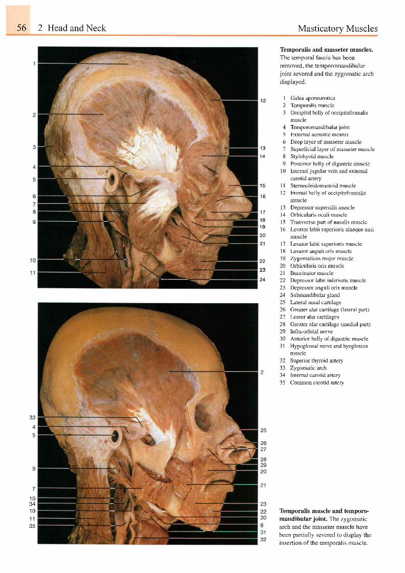
56 2 Head and Neck Masticatory Muscles
1 2
Temporalis and masseter muscles.The temporal fascia has been
removed, the temporomandibularjoint severed and the zygomatic arch
displayed.
I Galea aponeurotica2 Temporalis muscle3 Occipital belly of occipitofrontalis
muscle4 Temporomandibularjoint5 Extemal acoustic meatus6 Deep layer of masseter muscle7 Superficial layer of masseter muscle8 Stylohyoid muscle9 Posterior belly of digastric muscle
10 Internal jugular vein and extemalcarotid artery
I I Sternocleidomastoid muscle12 Frontal belly of occipitofrontalis
muscle13 Depressor supercilii muscle14 Orbicularis oculi musclel5 Transverse part ofnasalis muscle16 Levator labii superioris alaeque nasi
muscle17 Levator labii superioris muscle18 Levator anguli oris muscle19 Zygomaticus major muscle20 Orbicularis oris muscle2l Buccinator muscle22 Depressor labii inferioris muscle23 Depressor anguli oris muscle24 Submandibular gland25 Lateral nasal cartilage26 Greater alar cartilage (lateral part)27 Lesser alar cartilages28 Greater alar cartilage (medial part)29 Infra-orbital nerve30 Anterior belly of digastric muscle3 1 Hypoglossal nerve and hyoglossus
muscle32 Superior thyroid artery33 Zygornatic arch34 lntemal carotid artery35 Common carotid artery
Temporalis muscle and temporo-mandibular joint. The zygomaticarch and the masseter muscle havebeen partially severed to display theinsertion of the temporalis muscle.
o
8
o
1 0341 01 135
1 3
1 4
I t
1 6
1 81 9
20
21
22
23
24
33
4 25
26
282920
232230IQ 1
32
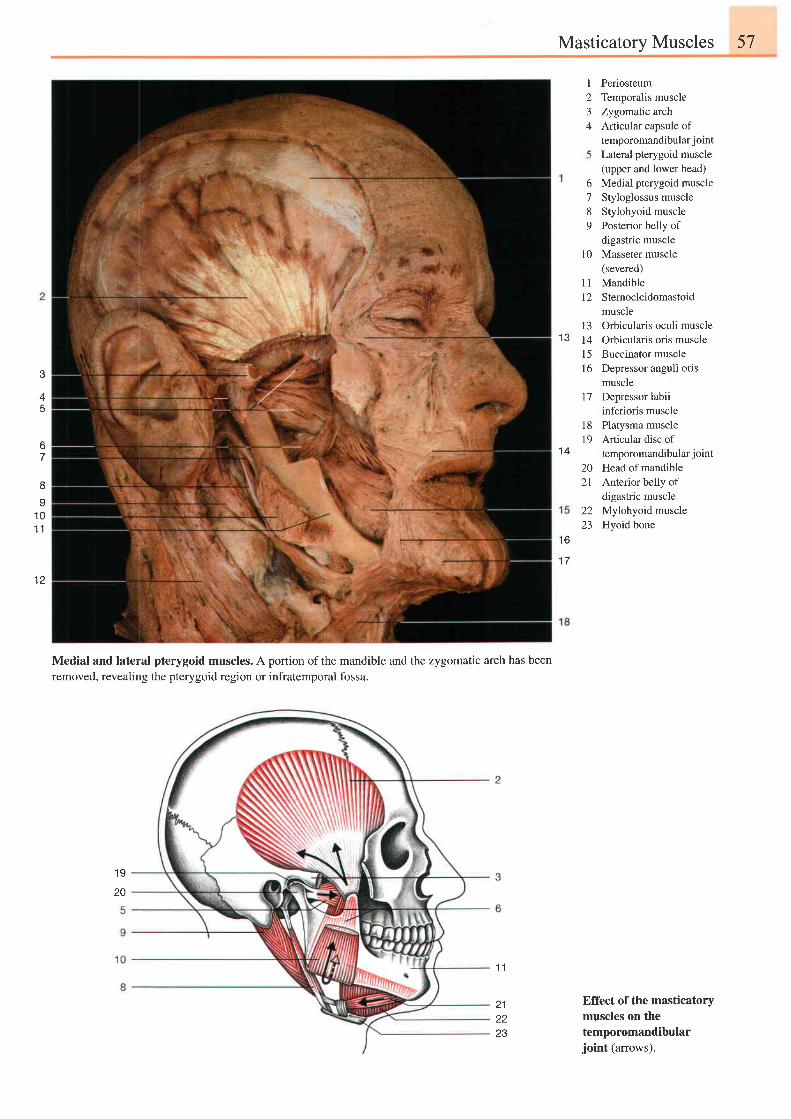
Masticatorv Muscles 57
1 3
t23+
PeriosteumTemporalis muscleZygomatic archArticular capsule oftemporomandibular j ointLateral pterygoid muscle(upper and lower head)Medial pterygoid muscleStyloglossus muscleStylohyoid musclePosterior belly ofdigastric muscleMasseter muscle(severed)MandibleSternocleidomastoidmuscle
6'7
89
1 0
1 1l 2
13 Orbicularis oculi muscle14 Orbicularis oris musclel5 Buccinator muscle16 Depressor anguli oris
muscle17 Depressor labii
inferioris muscle18 Platysma muscle19 Articulardiscof
temporomandibular joint
20 Head of mandible21 Anterior belly of
digastric muscle22 Mylohyoid muscle23 Hyoid bone
Effect of the masticatorymuscles on thetemporomandibularjoint (arrows).
3
45
o
71 4
I
I1 01 l
t z
t o
1 7
Medial and lateral pterygoid muscles. A portion of the mandible and the zygomatic arch has beenremoved, revealing the pterygoid region or infratemporal fossa.
1 9
20
1 1
2122z 5
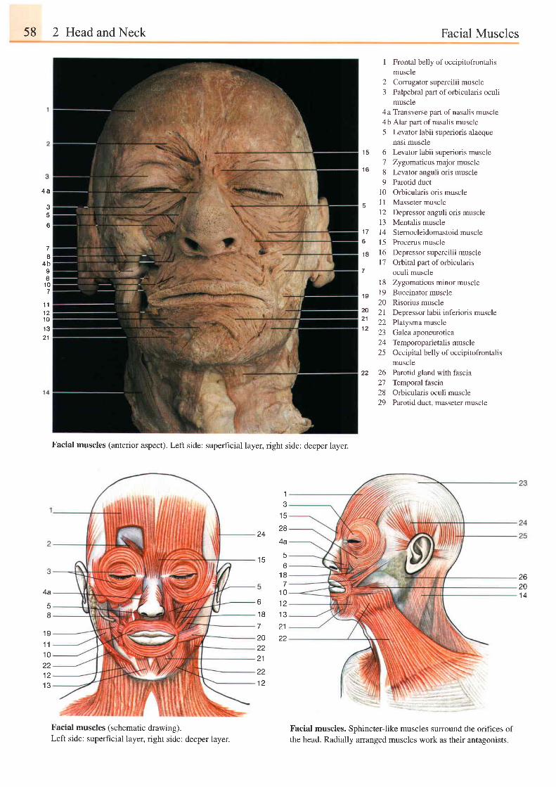
58 2 Head and Neck Facial Muscles
4 a
a
c
Facial muscles (anterior aspect). Left side: superhcial layer, right side: deeper layer.
1e
t c
28
4a
I Frontal belly of occipitofrontalismuscle
2 Comrgator supercilii muscle3 Palpebral part oforbicularis oculi
muscle4 a Transverse part of nasalis muscle4b Alar part of nasalis muscle5 Levator labii superioris alaeque
nasi muscle15 6 Levator labii superioris muscle
7 Zygomaticns major muscle16 8 Levator anguli oris muscle
9 Parotid duct10 Orbicularis oris muscle
u 1l Masseter muscle12 Depressor anguli oris muscle13 Mentalis muscle
17 14 Sternocleidomastoid muscle6 15 Procerus muscle
1B 16 Depressor supercilii musclel7 Orbital part oforbicularis
7 oculi muscle18 Zygomaticus minor muscle
19 19 Buccinator muscle20 Risorius muscle
20 2l Depressor labii inferioris muscle21 22 Platysma muscle1 2
zJ ualea aDoneurotrca24 Temporoparietalis muscle25 Occipitalbelly ofoccipitofrontalis
muscle22 26 Parotid gland with fascia
27 Temporal fascia28 Orbicularis oculi muscle29 Parotrd duct, masseter muscle
Facial muscles. Sphincter-like muscles surround the orihces ofthe head. Radially arranged muscles work as their antagonists.
d
4 bIU
1 0
1 11 21 0
1 321
24
1 F4
o
1 87
1 01 2t \ t
26201 44a
58
1 91 11 0221 21 3
1 87202221
221 2
21zz
Facial muscles (schematic drawing).Left side: superficial layeq right side: deeper layer.
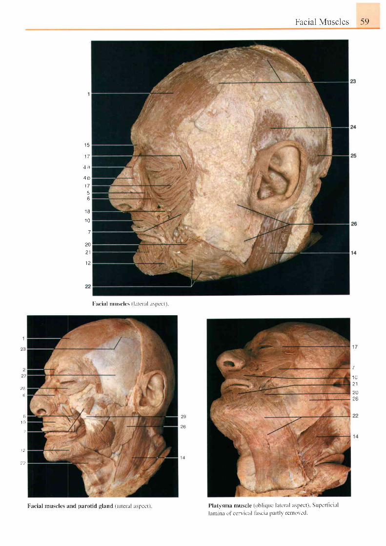
Facial Muscles 59
1 5
1 7
4 b
6
F-acial muscles ( lateral aspect)
I ' latysma muscle (obl ique lateral aspect) Supett icial
l lnr ina ol ccrvicr i l f l rscia part ly rctnoved.
1 8
1 0
7
20
z l
7
1 02 1
2026
2 8
6
81 0
7
1 2
2 2
Facial musclcs and parotid gland ( latclal rspcct)
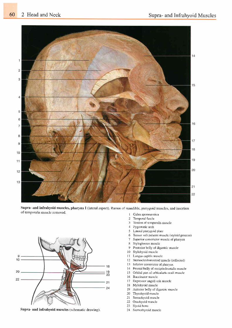
60 2 Head and Neck Supra- and Infrahyoid Muscles
Supra- and infrahyoid muscles, pharynx I (lateral aspect). Ramus of mandible, pterygoid muscles, and insertionof temporalis muscle removed.
91 0
I
2J
45678o
1 0l l1 21 3t4l 5l 6
Galea aponeuroticaTemporal fasciaTendon of temporalis muscleZygomatic archLateral pterygoid plateTensor veli palatini muscle (styloid process)Superior constrictor muscle of pharynxStyloglossus musclePosterior belly of digastric muscleStylohyoid muscleLongus capitis muscleStemocleidomastoid muscle (reflected)
Inferior constrictor of pharynxFrontal belly of occipitofrontalis muscleOrbital part of orbicularis oculi muscleBuccinator muscleDepressor anguli oris muscleMylohyoid muscleAnterior belly of digastric muscle
1 8
1 923
20
21 l 71 8
24 1920 Thyrohyoid muscle2l Stemohyoid muscle22 Omohyoid muscle23 Hyoidbone24 Stemothyroid muscleSupra- and infrahyoid muscles (schematic drawing).
22
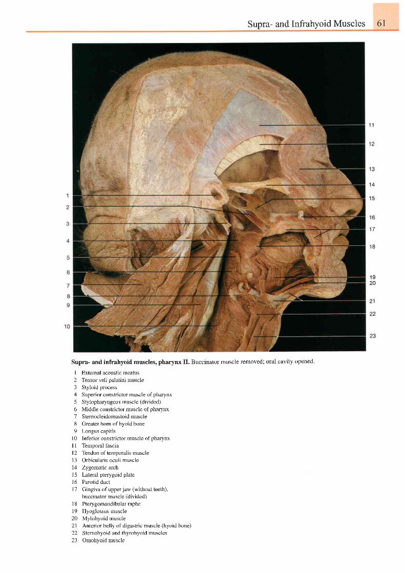
Supra- and Infrahyoid Muscles 6l
1 920
Supra- and infrahyoid muscles, pharynx II. Buccinator muscle removed; oral cavity opened.
I
23+
5o789
1 0l l1 21 3I 41 5t 6l 7
1 8l 9)n
2 l22z-)
Extemal acoustic meatusTensor veli palatini muscleStyloid processSuperior constrictor muscle of pharynxStylopharyngeus muscle (divided)Middle constrictor muscle of pharynxSternocleidomastoid muscleGreater horn of hyoid boneLongus capitisInferior constrictor muscle of oharvnxTemporal fasciaTendon of temporalis muscleOrbicularis oculi muscleZygomatic archLateral pterygoid plateParotid ductGingiva of upper jaw (without teeth),buccinator muscle (divided)Pterygomandibular rapheHyoglossus muscleMylohyoid muscleAnterior belly of digastric muscle (hyoid bone)Sternohyoid and thyrohyoid musclesOmohyoid muscle
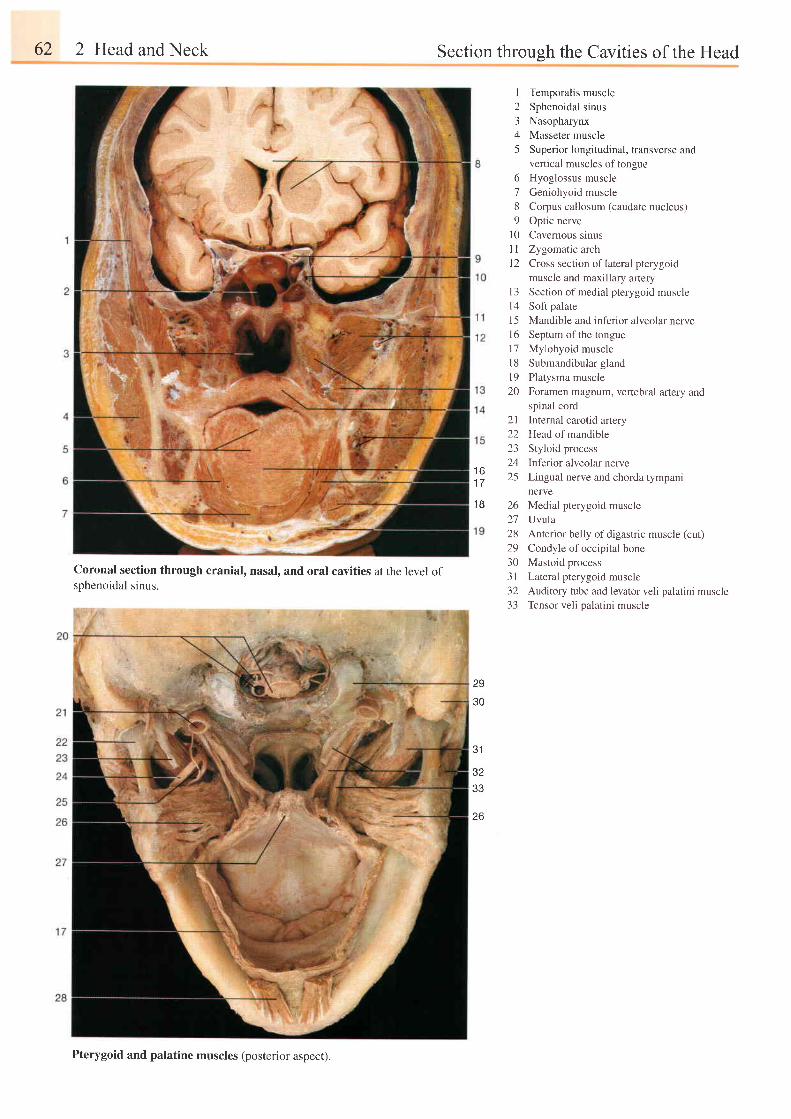
62 2 Head and Neck Section through the Cavities of the Head
6189
l 01 lt 2
I Temporalis muscle2 Sphenoidal sinus3 Nasopharynx4 Masseter muscle5 Superior longitudinal, transverse and
vertical muscles of tongueHyoglossus muscleGeniohyoid muscleCorpus callosum (caudate nucleus)Optic nerveCavernous sinusZygomatic archCross section of lateral pterygoidmuscle and maxillary artery
13 Section of medial pterygoid muscle
l4 Sofi palate
15 Mandible and inferior alveolar nervel6 Septum ofthe tongue
17 Mylohyoid muscle
l8 Submandibular gland
l9 Platysma muscle
20 Foramen magnum, vertebral artery and
spinal cord
Internal carotid artery
Head of mandible
Styloid process
Inferior alveolar nerve
Lingual nerve and chorda tympani
nerve
Medial pterygoid muscle
Uvula
Anterior belly of digastric muscle (cut)
Condyle of occipital bone
Mastoid process
Lateral pterygoid muscle
Auditory tube and levator veli palatini muscleTensor veli palatini muscle
2 l22232425t o
1 7
1 8 262'72829303 1) z
33
Coronal section through cranial, nasal, and oral cavities at the level ofsphenoidal sinus.
29
30
31
J I
33
26
Pterygoid and palatine muscles (posterior aspect).
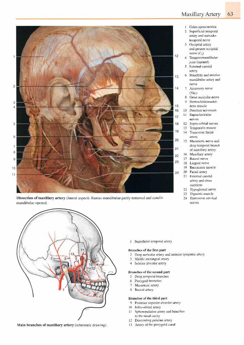
Maxillarv Arterv 63
1 2
1 Galea aponeurotica2 Superficial temporal
artery and auriculo-temporal nerve
3 Occipital arteryand greater occipitalnerve (C2)
4 Temporomandibularjoint (opened)
5 External carotidartery
6 Mandible and inferiormandibular artery andnewe
7 Accessory nerve(Var.)
8 Great auricular nerve9 Stemocleidomastoi-
deus muscle10 Punctum nervosumI 1 Supraclavicular
nerves12 Supra-orbital nerves13 Temporalis muscle14 Transverse facial
afiery15 Masseteric nerve and
deep temporal branchof maxillary artery
16 Maxillary artery17 Buccal nerve18 Lingual nerve19 Buccinator muscle20 Facial artery21 External carotid
artery and sinuscaroticus
22 Hypoglossal nerve23 Digastric muscle24 Transverse cervical
nerves
1 e
1 4
Dissection of maxillary artery (lateral aspect). Ramus mandibulae partly removed and canalismandibulae opened.
1 51 6
1 7
1 8
1 9
20
21
22
23
24
I Superhcial temporal artery
Branches of the first part
2 Deep auricular artery and anterior tympanic artery
3 Middle meningeal artery4 Inferior alveolar artery
Branches of the second part
5 Deep temporal branches6 Pterygoid branches7 Masseteric artery8 Buccal artery
Branches of the third part
9 Posterior superior alveolar arteryl0 Infra-orbital arteryI I Sphenopalatine aftery and branches
to the nasal cavityl2 Descending palatine artery13 Artery ofthe pterygoid canalMain branches of maxillary artery (schematic drawing).
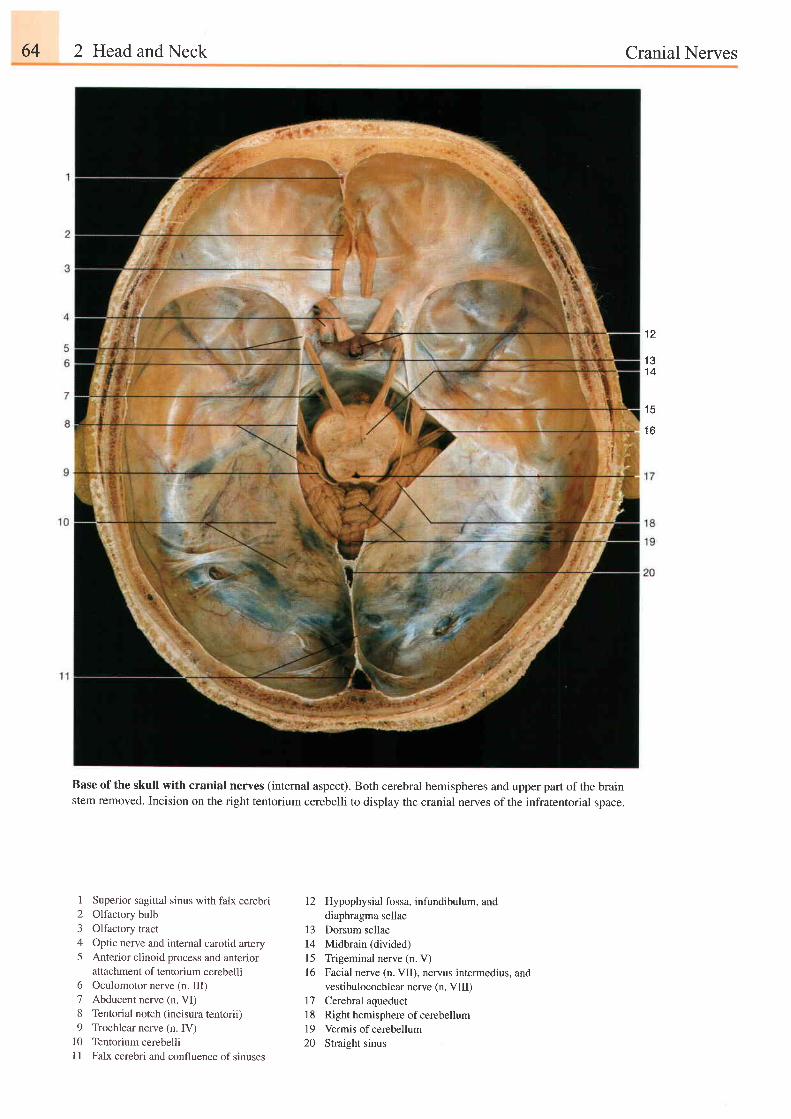
64 2 Head and Neck Cranial Nerves
l a
1 31 4
1 5
1 A
Base ofthe skull with cranial nerves (internal aspect). Both cerebral hemispheres and upper part ofthe brainstem removed. Incision on the right tentorium cerebelli to display the cranial nerves ofthe infratentorial space.
1 Superior sagittal sinus with falx cerebri2 Olfactory bulb3 Olfactory tract4 Optic nerve and internal carotid artery5 Anterior clinoid process and anterior
attachment of tentorium cerebelli6 Oculomotor nerve (n. III)7 Abducent nerve (n. VI)8 Tentorial notch (incisura tentorii)9 Trochlear nerve (n. IV)
10 Tentorium cerebelliI I Falx cerebri and confluence of sinuses
12 Hypophysial fossa, infundibulum, anddiaphragma sellae
13 Dorsum sellae14 Midbrain (divided)
15 Trigeminal nerve (n. V)16 Facial nerve (n. VII), nervus intermedius, and
vestibulocochlear nerve (n. VIII)17 Cerebral aqueduct18 Righthemisphere of cerebellum19 Vermis of cerebellum20 Straisht sinus
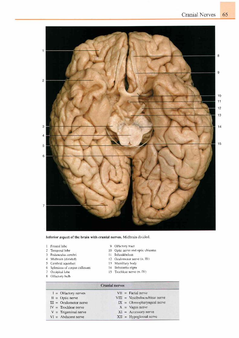
Cranial Nerves 65
1 0
1 1
1 2
1 3
Inferior aspect of the brain with cranial nerves. Midbrain divided
I Frontal lobe2 Temporal lobe3 Pedunculus cerebri4 Midbrain (divided)5 Cerebral aqueduct6 Splenium ofcorpus callosum7 Occipital lobe8 Olfactory bulb
9 Olfactory tractl0 Optic nerve and optic chtasma11 Infundibulum12 Oculomotor nerve (n III)13 Mamillary body14 Substantia nigra15 Trochlear nerve (n. IV)
Cranial nerves
I = Olfactory nervesII = Optic nerve
III = Oculomotor nerveIV = Trochlear nerveV = Trigeminal nerve
VI = Abducentnerve
Facial nerve
Vestibulocochlear nerve
Glossopharyngeal nerve
Vagus nerve
Accessory nerve
Hypoglossal nerve
VII =
VIII =I X =
Y _
X I =XII =
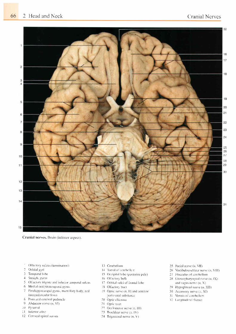
66 2 Head and Neck
Cranial nerves. Brain (inl'erior aspect).
Oltactory sulcus ( t -er t i inat ion)
Ortrital -ryriTemporal lohc
S t ra i gh t t y rus
Oltactory t r igone and int 'er ior tcmporal sulcusMedial occip i totcntporal gvrus
Parahippocarrpal gyrus. ntami l lary body. andintcrpecluncular fossaPons and cerebral peduncleAbdr-rccrr t nerve (n VI)Pyramid
Inter ior o l ive
Cer i ' ical spinal nerves
l3 Ce rebe l l un r
l 4 Tons i l o f ce r cbc l l um
|5 Occipi ta l lobe (poster ior pole)
l 6 O l l ac to r y bu lb
|7 Orbi ta l sulc i of l iontal looe
l8 Ol factor l ' t ract
l9 Opt ic nen'e (n I I ) and anter iorpcr lorated substancc
20 Opt ic chiasnra
2l Opt ic r ract
22 Oculornotor ncn'e (n I l l )
23 Trochlear ncrvc (n IV)
2.1 Tr igeminal nerve (n V)
Cranial Nerves
25 Facia l nervc (n. VI I )
26 Vest ibulocochlcar nerve (n VI I I )
27 Flocculus of cerebel lunr
213 Glossopharyngeal nervc (n. IX)
and vagus nerve (n X)
29 Hypoglossal nerve (n XI I )
30 Accessory nervc (n Xl)
3 l Vermis of cerebel lurn
32 Longi tudinal f lssure
24
252627
2829
30
I231561
IJ
1 0l ll 2
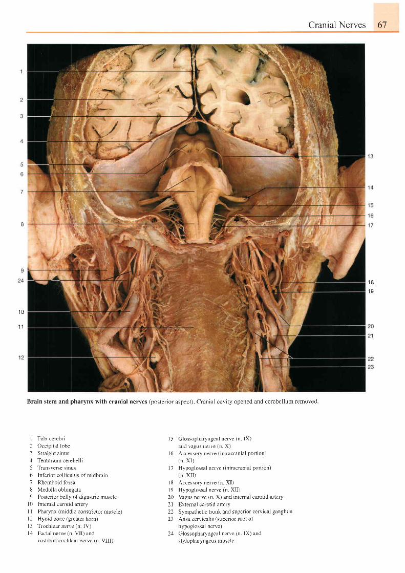
Cranial Nerves 67
1 81 0
1 0
1 1
Brain stem and pharynx with cranial nerves (posterior aspect). Cranial cavity opened and cerebellum removed.
I Falx cerebri2 Occipital lobe3 Straight sinus4 Tentorium cerebelli5 Transverse sinus6 Inferior colliculus of midbrain7 Rhomboid fossa8 Medulla oblongata9 Posterior belly of digastric muscle
l0 Intemal carotid arteryI I Pharynx (middle constrictor muscle)l2 Hyoid bone (greater horn)13 Trochlearnerve (n IV)l4 Facial nerve (n. VII) and
vestibulocochlear nerve (n. VIII)
l5 Glossopharyngealnerve (n lX)
and vagus nerve (n. X)
l6 Accessory nerve (intracranial portion)
(n. XI)
l7 Hypoglossal nerve (intracranial portion)
(n. XI I )
l8 Accessory nerve (n. XI)
l9 Hypoglossal nerve (n. XII)
20 Vagus nerve (n. X) and internal carotid artery
21 External carotid aftery
22 Sympathetic trunk and superior cervical ganglion
23 Ansa cervicalis (superior root of
hypoglossal nerve)
24 Glossopharyngeal nerve (n. IX) and
stylopharyngeus muscle
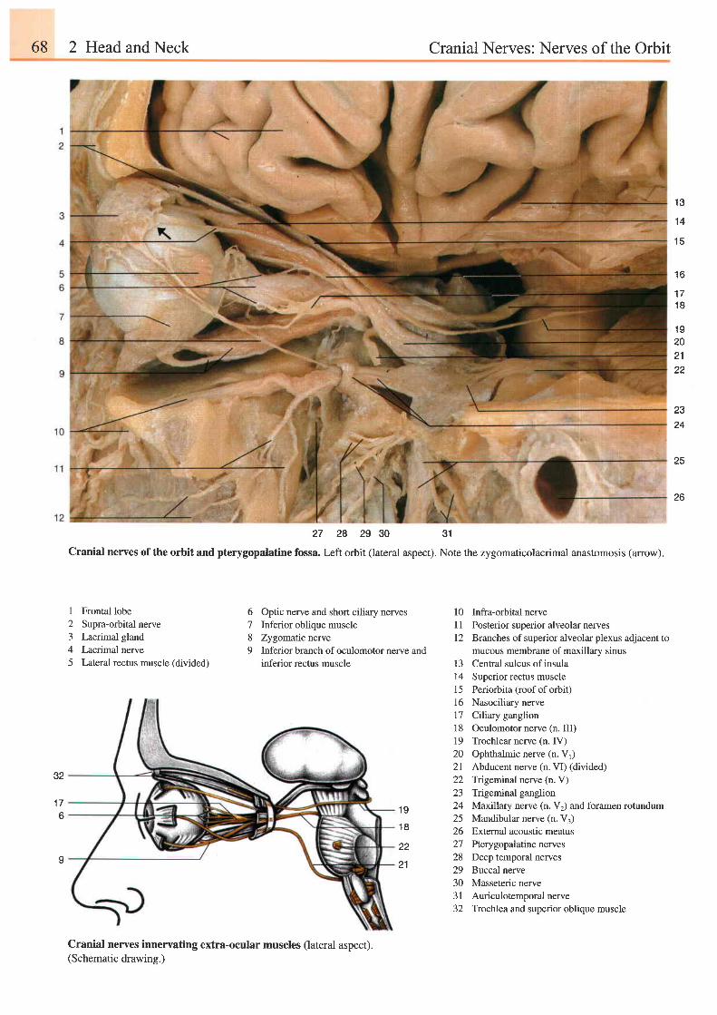
68 2 Head and Neck Cranial Nerves: Nerves of the Orbit
1 3
1 4
1 5
1 6
t t
1 8
1 9202122
2324
25
26
27 28 29 30 31
Cranial nerves of the orbit and pterygopalatine fossa. Left orbit (lateral aspect). Note the zygomaticolacrimal anastomosis (arrow).
32
1 76
I
I Frontal lobe2 Supra-orbital nerve3 Lacrimal gland4 Lacrimal nerve5 Lateral rectus muscle (divided)
6 Optic nerve and short ciliary nerves7 Inferior oblique muscle8 Zygomatic nerve9 Inferior branch of oculomotor nerve and
inferior rectus muscle
l0 Infra-orbital nerveI 1 Posterior superior alveolar nerves12 Branches of superior alveolar plexus adjacent to
mucous membrane of maxillarv sinus13 Central sulcus ofinsula14 Superior rectus muscle15 Periorbita (roof of orbit)16 Nasociliary nerve17 Clliary ganglion18 Oculomotor nerve (n. III)19 Trochlear nerve (n. IV)20 Ophthalmic nerve (n. V1)2l Abducentnerve (n.VI) (divided)22 Tigeminal nerve (n. V)23 Trigeminal ganglion24 MaxilIary nerve (n. V2) and foramen rotundum25 Mandibular nerve (n. V3)26 Extemal acoustic meatus27 Pterygopaladne neryes28 Deep temporal nerves29 Buccal nerve30 Masseteric nerve31 Auriculotemporal nerve32 Trochlea and superior oblique muscle
1 9
1 8
22
Cranial nerves innervating extra-ocular muscles (lateral aspect).(Schematic drawing.)
21
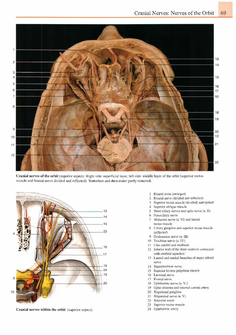
CranialNerves: Nerves of the Orbit 69
1 3
1 4
1 5
1 61 7
1 0
1 8
1 9
201 0
21
Cranial nerves of the orbit (superior aspect). Right side: superfrcial layer, left side: middle layer of the orbit (superior rectusmuscle and frontal nerve divided and reflected). Tentorium and dura mater partly removed.
1 3
1 4
1 5
23
1 Frontal sinus (enlarged)2 Frontal nerve (divided and reflected)3 Superior rectus muscle (divided) and eyeball4 Superior oblique muscle5 Short ciliary nerves and optic nerve (n. II)6 Nasociliary nerve7 Abducent nerve (n. VI) and lateral
rectus muscle8 Ciliary ganglion and superior rectus muscle
(reflected)9 Oculomotor nerve (n. III)
10 Trochlear nerve (n. IV)I I Crus cerebri and midbrain1 2 Inferior wa1l of the third ventricle connected
with cerebral aqueduct13 Lateral and medial branches of supra-orbital
nerve14 SupratrochlearnerveI 5 Superior levator palpebrae muscle16 Lacrimal nervel7 Frontal nervel8 Ophthalmic nerve (n. V1)l9 Optic chiasma and internal carotid aftery20 Trigeminal ganglion21 Trigeminal nerve (n. V)22 Tentorial notch23 Superior rectus muscle24 Ophthalmic artery
1 6
1 7
1 8241 9
Z U
Cranial nerves within the orbit (superior aspect).
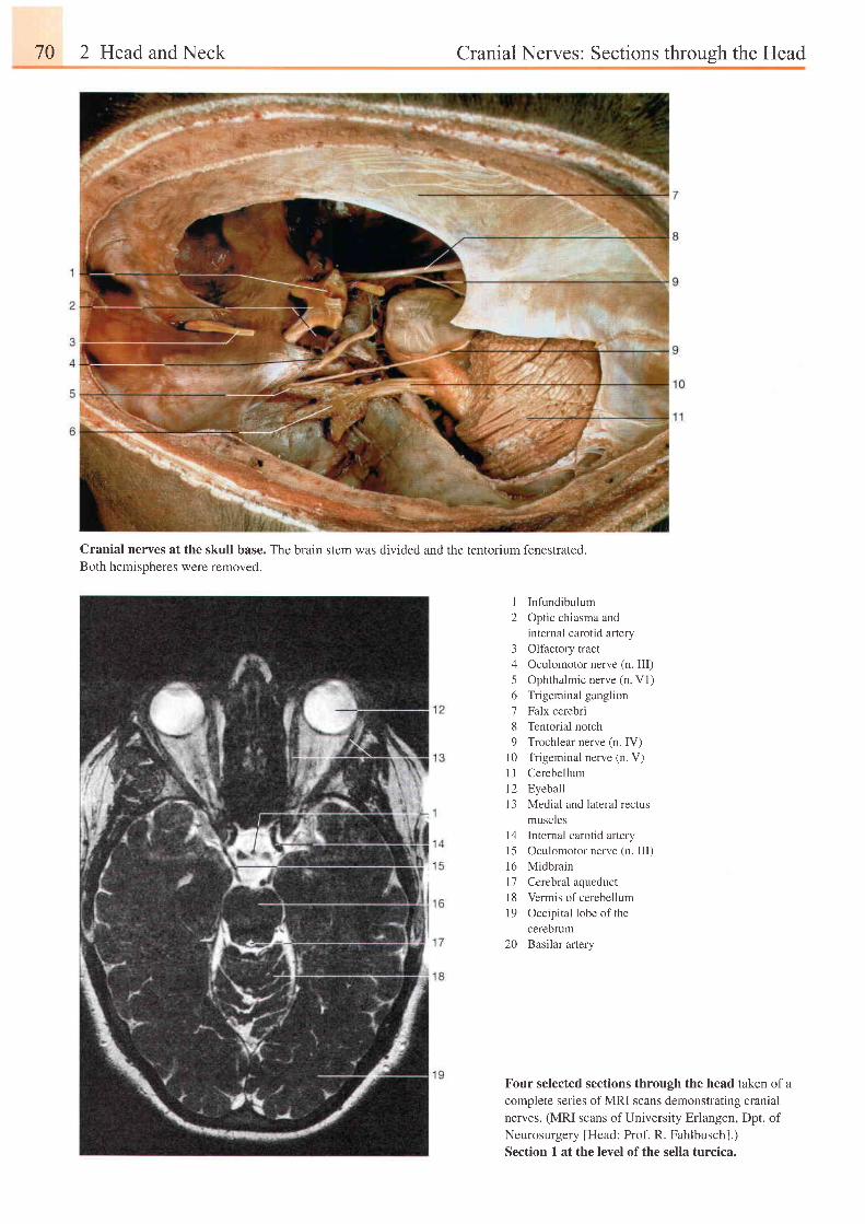
70 2 Head and Neck Cranial Nerves: Sections throuqh the Head
Cranial nerves at the skull base. TheBoth hemispheres were removed.
brain stem was divided and the tentorium fenestrated.
I2
3456789
l 0l tl 2l 3
InfundibulumOptic chiasma andintemal carotid arteryOlfactory tractOculomotor nerve (n. III)Ophthalmic nerve (n. Vl)Trigeminal ganglionFalx cerebriTentorial notchTrochlear nerve (n. IV)Trigeminal nerve (n. V)CerebellumEyeballMedial and lateral rectusmuscles
l4 Internal carotid artery
15 Oculomotor nerve (n. III)
16 Midbrain
l7 Cerebral aqueduct
l8 Vermis of cerebellum
19 Occipital lobe of the
cerebrum
20 Basilar artery
Four selected sections through the head taken of a
complete series of MRI scans demonstrating cranial
nerves. (MRI scans of University Erlangen, Dpt. of
Neurosurgery [Head: Prof. R. Fahlbusch].)
Section 1 at the level ofthe sella turcica.
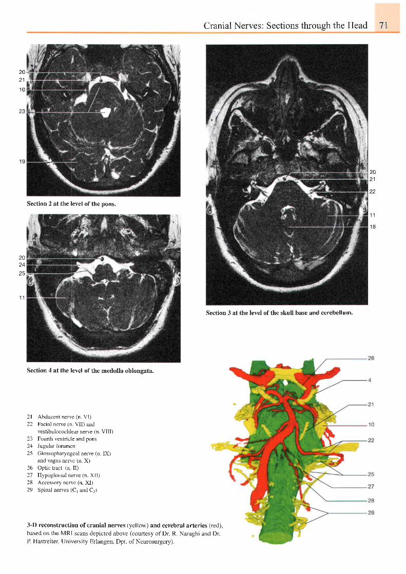
Cranial Nerves: Sections through the Head 7'',1
2021
1 0
20z+
25
Section 4 at the level of the medulla oblongata.
2 l22
L-)
2425
26272829
Abducent nerve (n. VI)Facial nerve (n. VII) andvestibulocochlear nerve (n. VIII)Fourth ventricle and ponsJugular foramenGlossopharyngeal nerve (n. IX)and vagus nerve (n. X)Optic tract (n. II)Hypoglossal nerve (n. XII)Accessory nerve (n. XI)Spinal nerves (C1 and Cr)
3-D reconstruction ofcranial nerves (yellow) and cerebral arteries (red),based on the MRI scans depicted above (courtesy of Dr. R. Naraghi and Dr.P. Hastreiter, University Erlangen, Dpt. of Neurosurgery).
2021
22
Section 2 at the level of the pons.
Section 3 at the level of the skull base and cerebellum.
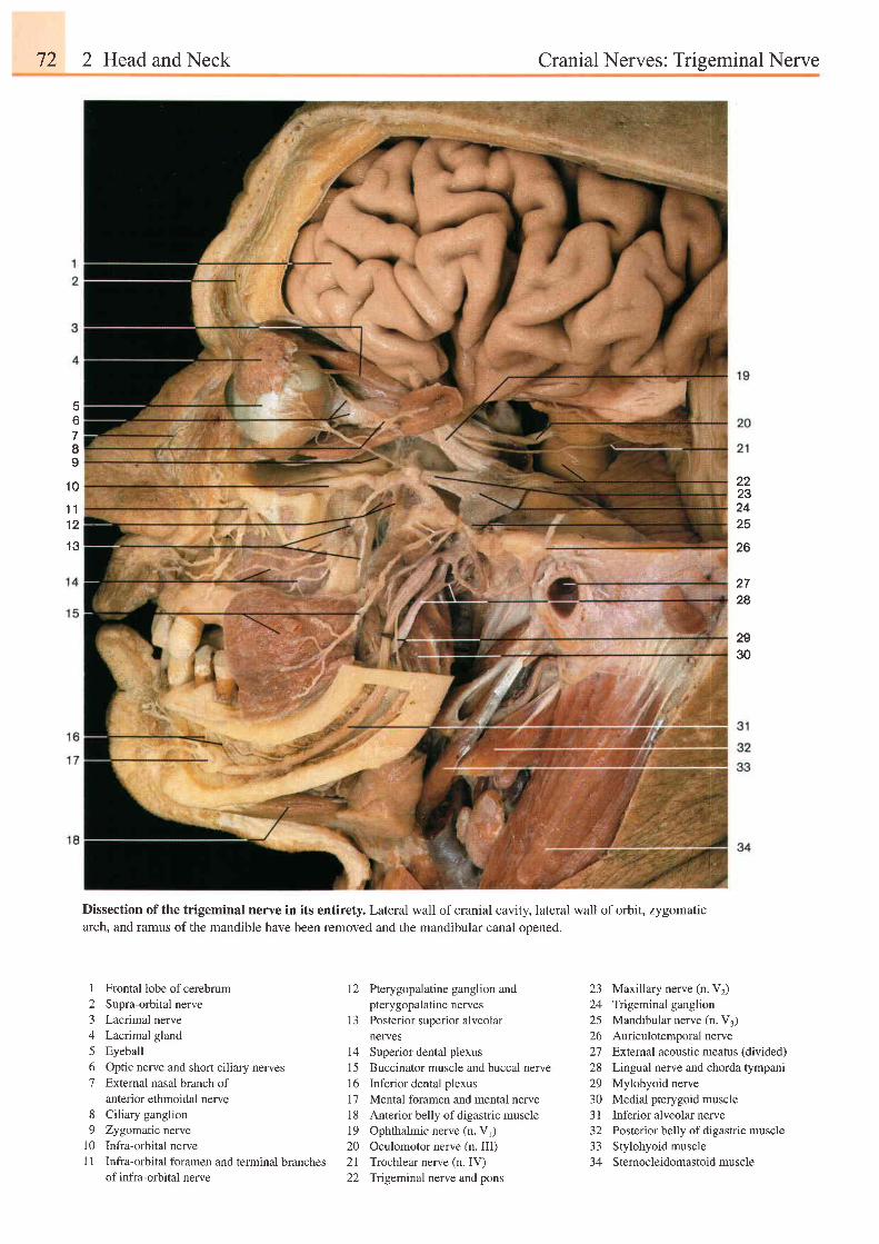
72 2 Head and Neck Cranial Nerves: Trigeminal Nerve
E
b
7I9
1 0
t 11 2
1 3
22232425
26
272A
2930
Dissection of the trigeminal nerve in its entirety. Lateral wall of cranial cavity, lateral wall of orbit, zygomaticarch, and ramus of the mandible have been removed and the mandibular canal opened.
1 Frontal lobe of cerebrum2 Supra-orbital nerve3 Lacrimal nerve4 Lacrimal gland5 Eyeball6 Optic nerve and short ciliary nerves7 External nasal branch of
anterior ethmoidal nerve8 Ciliary ganglion9 Zygomaticnewe
10 Infra-orbital nerve11 Infra-orbital foramen and terminal branches
of infra-orbital nerve
12 Pterygopalatine ganglion andpterygopalatine nerves
13 Posterior superior alveolarneryes
14 Superior dental plexus1 5 Buccinator muscle and buccal nerve16 Inferior dental plexus17 Mental foramen and mental nerve18 Anterior belly of digastric muscle19 Ophthalmic nerve (n. V1)20 Oculomotor nerve (n. III)2l Trochlear nerve (n. IV)22 Trigeminalnerve and pons
23 Maxillary nerve (n. V2)24 Trigeminal ganglion25 Mandibular nerve (n. V3)26 Auriculotemporalnerve27 Exlemal acoustic meatus (divided)
28 Lingual nerve and chorda tympani29 Mylohyoid nerve30 Medial pterygoid muscle3l Inferior alveolar nerve32 Posterior belly of digastric muscle33 Stylohyoid muscle34 Sternocleidomastoid muscle
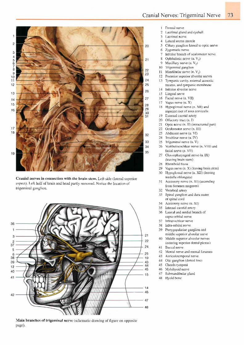
Cranial Nerves: Triseminal Nerve 73
34
6
a
I1 01 11 2
2223
2425
I Frontal nerve2 Lacrrmal gland and eyeball3 Lacrimal nerve4 Lateral rectus muscle5 Ciliary ganglion lateral to optic nerve6 Zygomatic nerve7 Inferior branch of oculomotor nerve8 Ophthalmic nerve (n. V1)9 Maxillary nerve (n. V2)
10 Trigeminal ganglionl1 Mandibular nerve (n. V3)l2 Posterior superior alveolar nerves13 Tympanic cavity, extemal acoustic
meatus, and tympanic membrane14 Inferior alveolar nerve15 Lingual nerve16 Facial nerve (n. VII)17 Vagus nerve (n. X)18 Hypoglossal nerve (n. XII) and
superior root of ansa cervicalis19 External carotid artery20 Olfactory tract (n I)21 Optic nerve (n. II) (intracranial part)22 Oculomotor nerve (n. III)23 Abducent nerve (n. VI)24 Trochlear nerve (n. IV)25 Trigeminal nerve (n. V)26 Vestibulocochlear nerve (n. VIII) and
facial nerve (n. VII)27 Glossopharyngeal nerve (n. IX)
(leaving brain stem)28 Rhomboid fossa29 Vagus nerve (n. X) (leaving brain stem)30 Hypoglossal nerve (n. XII) (leaving
medulla oblongata)31 Accessory nerve (n. XI) (ascending
from foramen magnum)32 Venebral artery33 Spinal ganglion and dura mater
of spinal cordAccessory nerve (n. XI)Intemal carotid arteryLateral and medial branch ofsupra-orbital nerveInfratrochlear nerveInfra-orbital nervePterygopalatine ganglion andmiddle superior alveolar nerveMiddle superior alveolar nerves(entering superior dental plexus)Buccal nerveMental nerve and mental foramenAuriculotemporal nerveOtic ganglion (dotted line)Chorda tympaniMylohyoid nerveSubmandibular glandHyoid bone
1 3
1 41 5't6
282S3031
1 71 8
!t
2
o38391 240
Cranial nerves in connection with the brain stem. Left side (lateral superioraspect). Left halfofbrain and head partly removed. Notice the location oftrigeminal ganglion.
Main branches of trigeminal nerve (schematic drawing of figure on oppositepage).
24
251 04J
4445t c
1 440
47
48
343536
3'l3839
40
4142434445464'748
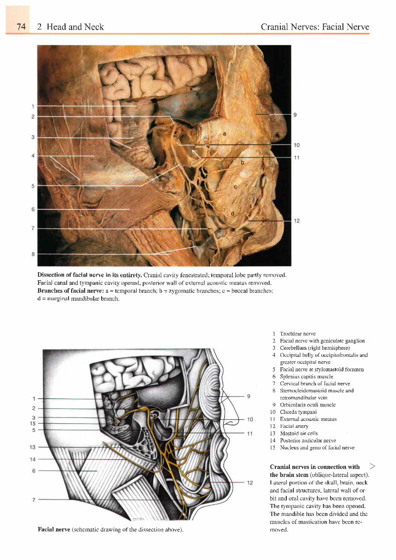
74 2 Head and Neck Cranial Nerves: Facial Nerve
J
t c
Dissection of facial nerve in its entirety. Cranial cavity fenestrated; temporal lobe partly removed.
Facial canal and tympanic cavity opened, posterior wall of external acoustic meatus removed.Branches offacial nerve: a = temporal branch; b = zygomatic branches; c = buccal branches;d = marginal mandibular branch.
1 Trochlear nerve2 Facial nerve with geniculate ganglion3 Cerebellum (right hemisphere)4 Occipital belly ofoccipitofrontalis and
greater occipital neile5 Facial nerve at stylomastoid foramen6 Splenius capitis muscle7 Cervical branch offacial nerve8 Sternocleidomastoid muscle and
retromandibular vein9 Orbicularis oculi muscle
10 Chorda tympaniI I External acoustic meatus12 Facial artery13 Mastoid air cells14 Posteriorauricularnerve15 Nucleus and genu offacial nerve
Cranial nerves in connection with
the brain stem (obliquelateral aspect).
Lateral portion of the skull, brain, neck
and facial structures, lateral wall of or-
bit and oral cavity have been removed.
The tympanic cavity has been opened.
The mandible has been divided and the
muscles of mastication have been re-
moved.Facial nerve (schematic drawing of the dissection above).
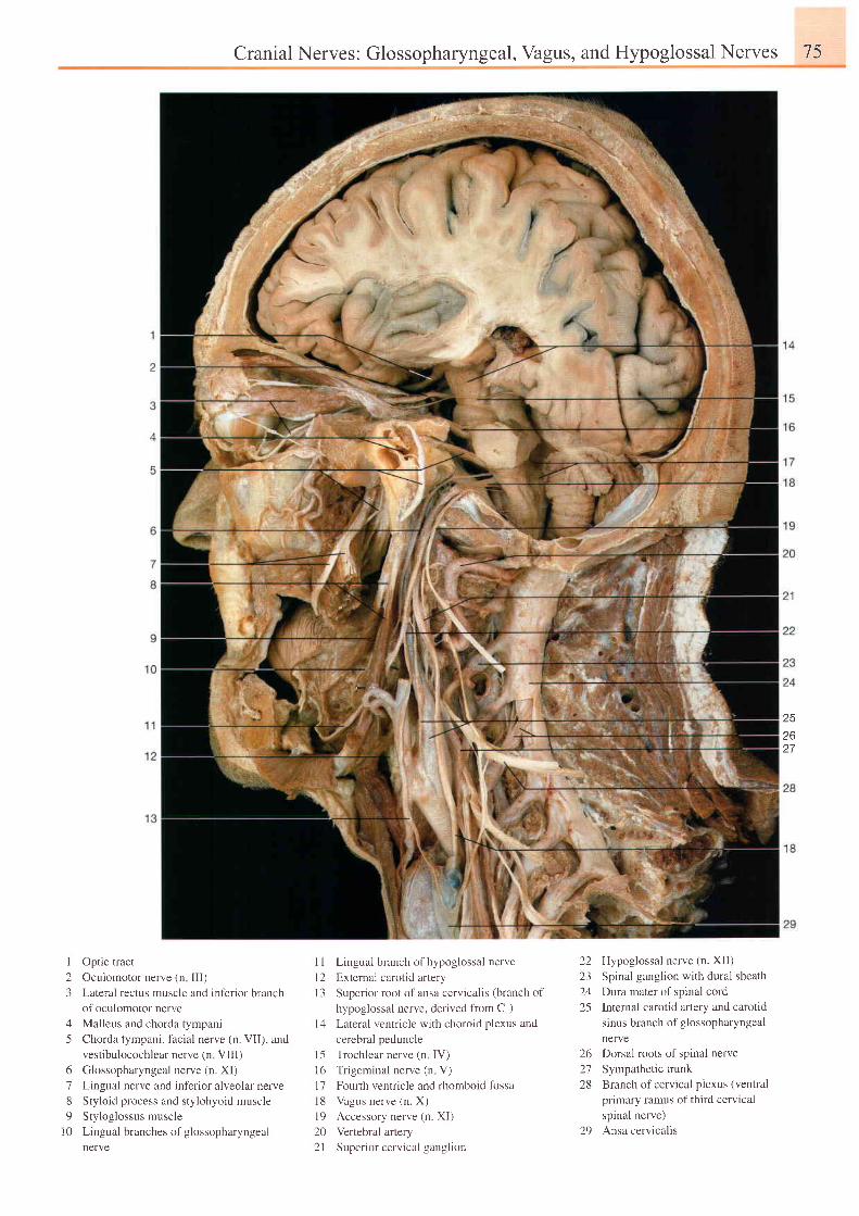
Cranial Nerves: Glossopharyngeal,Vagus, and Hypoglossal Nerves 75
252627
I
23
45
6789
l 0
Optic tract
Oculomotor nerve (n lll)
Lateral rectus muscle and inferior branch
of oculomotor nerve
Malleus and chorda tympani
Chorda tympani, facial nerve (n. VII), and
vestibulocochlear nerve (n Vlll)
Glossopharyngeal nerve (n. XI)
Lingual nerve and inferior alveolar nerve
Styloid process and stylohyoid muscle
Styloglossus muscle
Lingual branches of glossopharyngeal
nerve
1 I Lingual branch ofhypoglossal nerve
12 Extemal carotid artery
I 3 Superior root of ansa cervicalis (branch of
hypoglossal nerve, derived fiom Cr)
l4 Lateral ventricle with choroid plexus and
cerebral peduncle
15 Trochlear nerve (n IV)
16 Trigeminal nerve (n V)
l7 Fourth ventricle and rhomboid fossa
l8 Vagus nerve (n. X)
l9 Accessory nerve (n. XI)
20 Vertebral artery
21 Superior cervical ganglion
22 Hypoglossal nerve (n. Xil)
23 Spinal ganglion with dural sheath
24 Dwa mater of spinal cord
25 Internal carotid artery and carotid
sinus branch of glossopharyngeal
nerve
26 Dorsal roots of spinal nerve
27 Sympathetic trunk
28 Branch of cervical plexus (ventral
primary ramus of third cervical
spinal nerve)
29 Ansa cervicalis
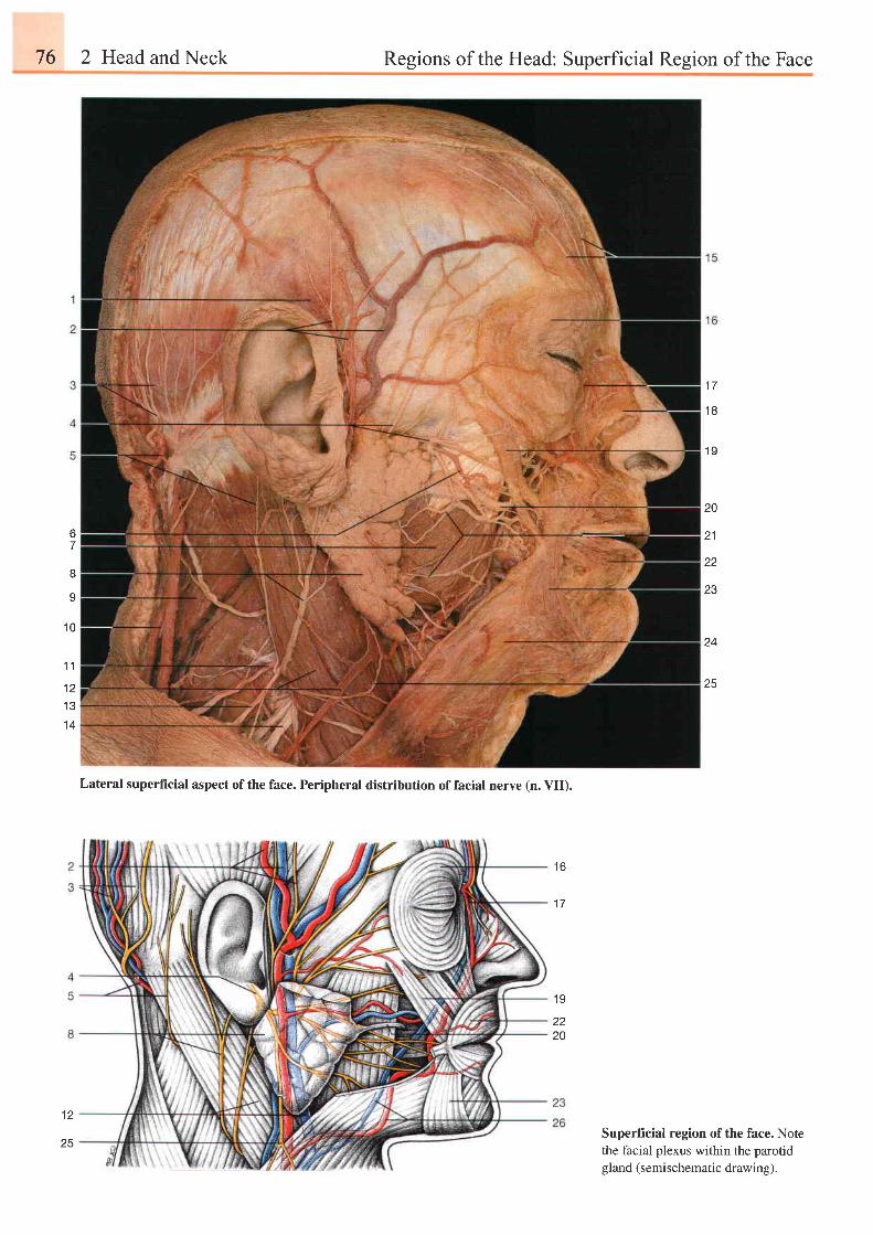
76 2 Head and Neck Regions of the Head: Superficial Region of the Face
1 7
1 8
6
8
I
1 0
1 9
20
21
22
23
z+
25
Superficial region of the face. Notethe facial plexus within the parotidgland (semischematic drawing).
1 1
1 2I J
1 4
t o
1 7
1 9
zz
20
Lateral superficial aspect ofthe face. Peripheral distribution offacial nerve (n. VI!.
1 2
25
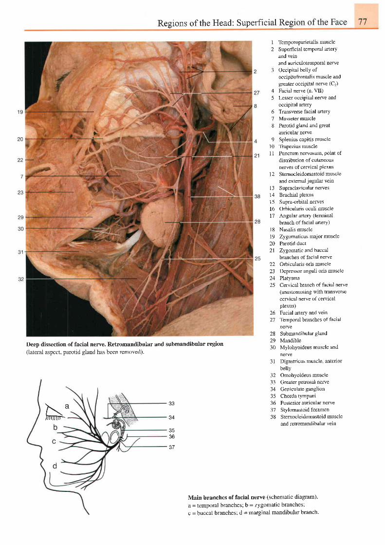
Regions of the Head: Superficial Reg
45
678
91 0l 1
t 2
t 3141 5l 61 7
l 819202 l
22ZJ
2425
12
26 Facial artery and vein27 Temporal branches of facial
nerye28 Submandibulargland
MandibleMylohyoideus muscle andnerveDigastricus muscle, anteriorbellyOmohyoideus muscleGreater petrosal nerveGeniculate ganglionChorda tympaniPosterior auricular nerveStylomastoid foramenSternocleidomastoid muscleand retromandibular vein
Temporoparietalis muscleSuperficial temporal arteryand veinand auriculotemporal nerveoccipital belly ofoccipitofrontalis muscle andgreater occipital nerve (C2)
Facial nerve (n. VII)Lesser occipital nerve andoccipital aneryTransverse facial arteryMasseter muscleParotid gland and greatauricular nerveSplenius capitis muscleTrapezius musclePunctum nervosum, point ofdistribution of cutaneousnerves of cervical plexus
Sternocleidomastoid muscleand external jugular veinSupraclavicular nervesBrachial plexusSupra-orbital nervesOrbicularis oculi muscleAngular artery (terminal
branch of facial artery)Nasalis muscleZygomaticus major muscleParotid ductZygomatic and buccalbranches of facial nerveOrbicularis oris muscleDepressor anguli oris musclePlatysmaCervical branch of facial nerve(anastomosing with transversecervical nerve of cervicalplexus)
Deep dissection of facial nerve. Retromandibular and submandibular region(lateral aspect, parotid gland has been removed).
2930
31
3233J +
35365 l
38
33
34
JO
37
Main branch€s of facial nerve (schematic diagram).
a = temporal branches; b = zygomatic branches;
c = buccal branches; d = marginal mandibular branch'
N:
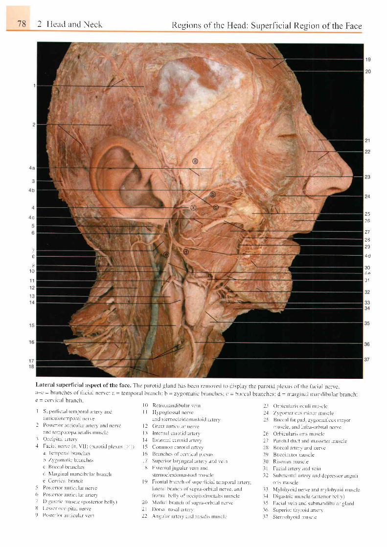
78 2 Hcad and Neck Regions of the Head: Superficial Region of the Face
ta",{:i.", ,.r
-8)
Lateral superf icial aspect of the face. The parotid glancl has bccn rentovecl to cl isplal ' thc pargtid plcrus of the f ircial ner vea e=branchcso l fac ia l l te rve : lL= tcn tpora l b ranch: b=zygor ra t i c b ranchcs : c= br . rcca l b ranches ; d = rnarg ina l tnand ibu la rbr -anch:e = ccrl ' ical branch
2526
9 a
2829
7B
s1 0
304 e
o l
I Super- l ic ia l tcrnpolal ar terv undl lur icu lote l rpot t l ncr t 'e
2 Poster ior aur icular l r r tcr l , ancl nervelncl tcr lpor opal ic ta l is nruscle
3 Occipi ta l ar- tcr v- l Fac ia l uc r ve (u V I I ) ( pa ro t i d p l c rus l X l )
a TcmponLl bral tchcs
b Zt ' l lourat ic bnLnchesc l luccal b lunches
r l N, larginal ntancl ibular branchc Ct ' r r ical brarrch
5 Posler ior nul icular ncr ve6 P i l \ t c I i r r l i rU t iC l t l i l r : l t l \ . r )7 Distrst r ic ntusclc (postcr ior . bcl ly)8 Lcsser occrpi ta l nervct) Poster ior- aur icular I 'c in
l0 RetromarrdibLr lur veinI I Hypog lossa l ncne
and sternoclc i ( lorrrastoid lLr tery| 2 G rca t au r i cu l a r ne r re
l3 lnternal carot i t l ar tcrv
l : l Extelnul car-ot id ur- tcr vI5 Comnrorr cafol id l l r tc f \| 6 Br anches ol cer t , ical p lcrusl7 Sr-rper ior laryngcal ar terv and vcinl8 Extcrnal juer-r lar le in ancl
sternocle idonrastoi t l r lLrsclc
C)rbrcular is ocul i nruscle
Z1'gonrat icus nr inor r .nusclc
Buccal lat pad. zygomat icus ntajormuscle. lnd infra-orbi ta l nerve
Or b i cu l a l i s o l i s n rusc l e
Palot ic l duct aud massctef ln l lsc lcBuccal ar tcrv lLncl nervc
Buccinator nrusclc
Risor i i rs muscle
Frcial artcry ancl I'ein
Sub rnen ta l a r t c r v and dep rcsso r angu l ior is rnuscle
33 Mvlohvord ren'e und rnylohyoic l r rusclel :1 Di-sastr ic uruscle (auter ior bel ly)35 Fac ia l ve i n and sub rnund ibu la l g l and
36 Super ior thyr-o id lLr tery37 Stenrohvoid nruscle
2l2125
2627282r)30.|32
t 9
202 l22.
Frontal branch ol superf ic i lJ tcr l lpr) ru l i l r te l ' rl a t c ra l b ranch o f sup ra o rb i t a l ue r r c . andt t r rnLal bel lv o l ucci l . r i to l r -ontr l i s t lusclcMcdial b l rnch o1 str l . r r -u or-b i ta l neneDorsal r rasl l ar tcr t
Angr,r lar ar tcr-v ar t r l nt rstL l is r ruscle
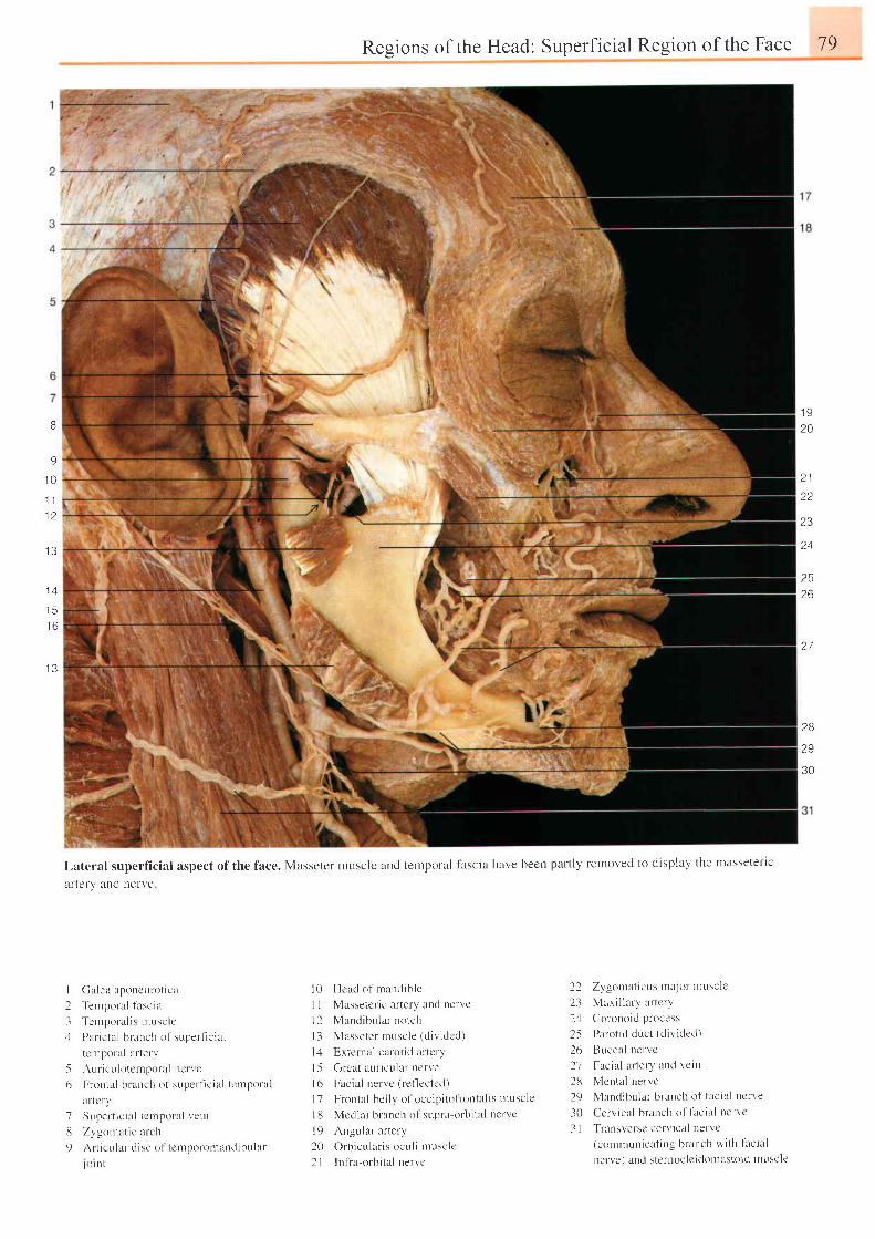
8
9
1 0.l
l
1 2
1 3
.15
I D
1 3
t!;
;l
l,ateral superficial aspect of the face. Masseter nlLlsclc
afteIV and lrerve
Regions of the Head: Superficial Region of the Face 79
I J
20
2 1o a
23
a t
2526
27
and ternporal lascia have becn part ly removed to display the mltssetet ' lc
28
29
30
I ( l l l ea l Lponcu ro t i ca
2 ' lenrporal
l i tsc ia
3 Tcn rpo ra l i s n r t t s c l c-1 Par ieta l br-ancl t of sr"rpert ic ia l
terrporal r r terv
5 Au r i cu l o ten tpo r i t l n c t r c
6 Frontal hrr anch of superf ic ia l tcrnponl l
Ir-tcr\l
7 Supertrc ia l tc lnpol l i l vein
i3 Zvgomat ic urc l r
9 Art icLr lar c l isc o l tcrnporor.nancl ibular
- l o | n t
l0 Hcad of mancl ib lc
I I Massetcr ic ar tery and l lefvc
l 2 Mand ibu la r no t ch
l3 Masse te r musc le ( d i v i dcd )
l4 Bxternal carot id ar tcry
l 5 Grcat aur icul r r ncrve
l6 Facia l nervc (ref lectcd)
l7 Frontal bel ly o1 occip i to l r -ontal is rnusclc
l8 Mcdial branch o1'supta orbi ta l t lervc
l9 Angular ar tery
20 Orh ie t r l l r r i . oe t t l i t nu . r ' l r '
2 l ln f ia orbi ta l nervc
22 Zygornat icus t ra jot ' ntuscle
23 Maxi l lary arLcr l "
24 Corouoid process
2-5 Parot id dLrct (d iv ided)
26 Buccr l nervc
/ I r n e l r l i . I t L - l \ i l l l L l V e l l l
28 Mental nervc
29 Mancl ibular branch of lac ia l ncrve
30 Cervicel branch of t i rc ia l t rer-r 'c
3 I Transversc cerv ical nervc(coumuuicat i r rg branch wi th l lc ia l
nerve ) and stertloc lc icltl urasttlicl tn uscle
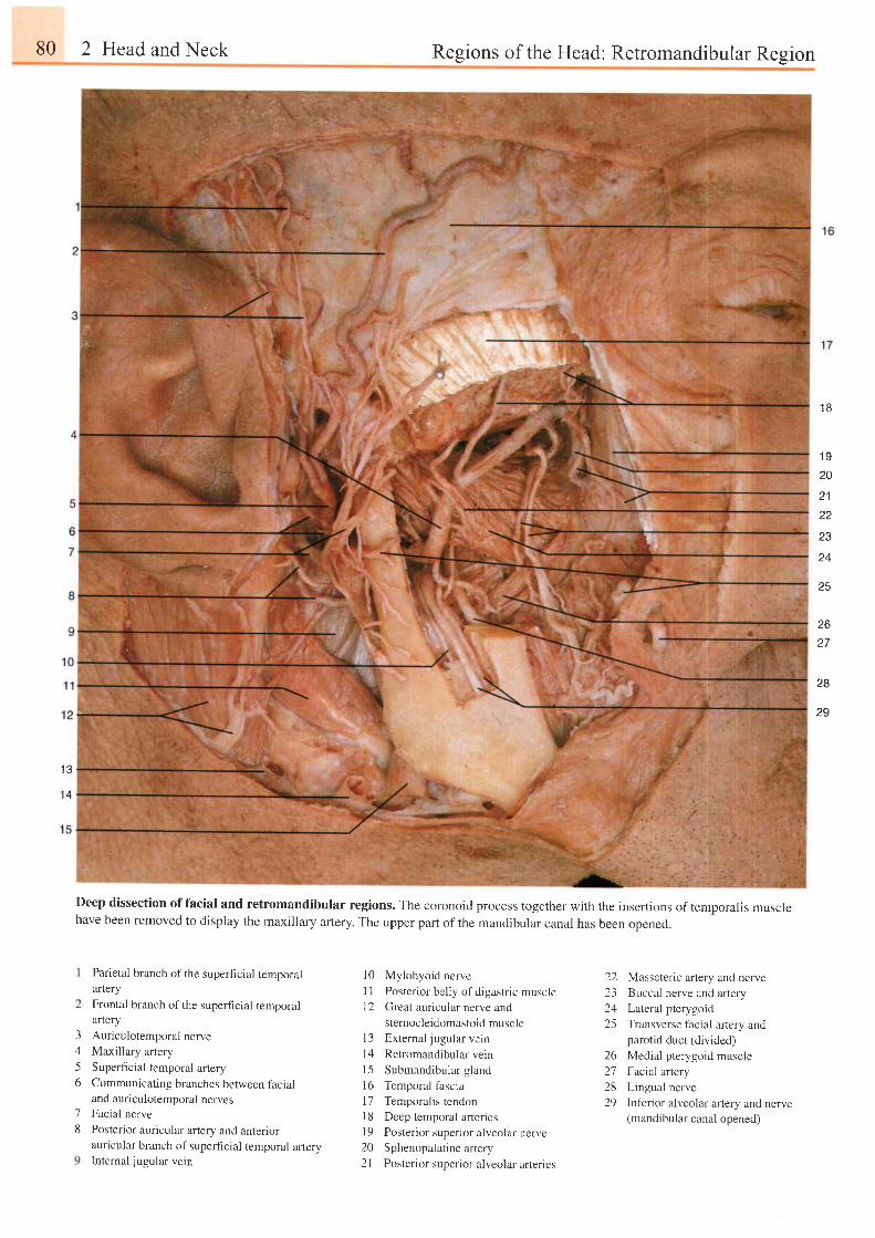
80 2 Head and Neck Regions of the Head: Retromandibular Region
Deep dissection of facial and retromandibular regions. The coronoid process together with the inseftions of temporalis musclehave been removed to display the maxillary artery. The upper part of the mandibular canal has been opened.
1 B
1 S
20
I t
tt
23
25
2627
2B
29
1 e
t 4
3A
56
18
Parietal branch of the superficial temporalarlery
Frontal branch of the superficial temporalartery
Auriculotemporal nerveMaxillary artery
Superficial temporal arteryCommunicating branches between facialand aur iculotemporal nervesFacial nerve
Posterior auricular artery and anteriorauricular branch of superficial temporal arteryInternal jugular vein
Mylohyoid nerve
Posterior belly of digastric muscleGreat auricular nerve and
stemocleidomastoid muscleExternal jugular vein
Retromandibular vein
Submandibular gland
Temporal fascia
Temporalis tendonDeep temporal arteriesPosterior superior alveolar nerveSphenopalatine artery
Posterior superior alveolar arteries
Masseteric artery and nerveBuccal nerve and artery
Lateral pterygoid
Transverse facial artery andparotid duct (divided)
Medial pterygoid muscle
Facial artery
Lingual nerve
Inferior alveolar artery and nerve(mandibular canal opened)
1 0l 11 2
t 3t 41 5l 61 7l 8l 9202 1
2223a /
25
262'72829
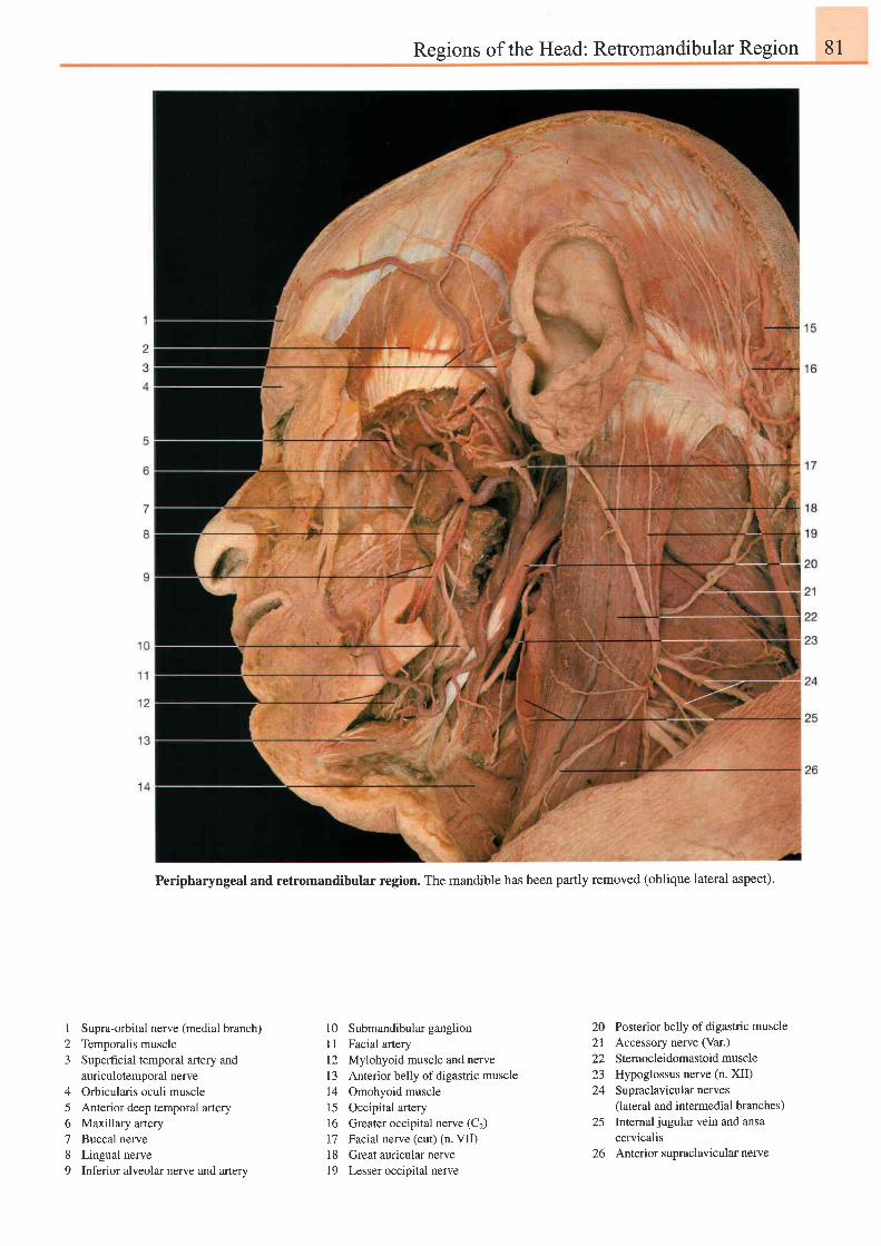
Regions of the Head: Retromandibular Region 81
Peripharyngeal and retromandibular region. The mandible has been partly removed (oblique lateral aspect).
I
23
4
56789
Supra-orbital nerve (medial branch)Temporalis rnuscleSuperfrcial temporal artery andauriculotemporal nerveOrbicularis oculi muscleAnterior deep temporal arteryMaxillary arteryBuccal nerveLingual nerveInferior alveolar nerve and artery
10 Submandibular ganglion11 Facial anery12 Mylohyoid muscle and nerve13 Anterior belly of digastric muscle14 Omohyoid muscle15 Occipital atery16 Greater occipital nerve (C2)17 Facial nerve (cut) (n. VII)18 Greatauricularnerve19 Lesser occipital nerve
20 Posterior belly of digastric muscle21 Accessory nerve (Var.)
22 Stemocleidomastoidmuscle23 Hypoglossus nerve (n. XII)24 Supraclavicularnerves
(lateral and intermedial branches)25 Internaljugularveinand ansa
cervicalis26 Anterior supraclavicular nerve
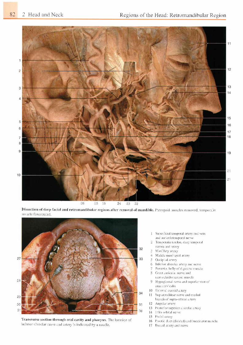
82 I F le l t i . rnt i \ccl . ,
€ ' . . : ,i . 4
l . r a t r s r c r sc \ ( ' c l t ( ) n t l r r ough o ra l ca r , i t r and pha r t n r . ' [ ' h c j o t l L i r , , n L r t
l l l l c r - r o r . L i r e i ! l i I t t e r " r , l r r r i l i t t - l e t - \ ' l \ t r t t l r c e l r : d h r l t t r e c i r r t
'qt
Regions of the Head: Retrornandibular Rcgion
\ * ,
,3.9b
q . J - r
l f . ; .1,
a* J20
2 iI
' : ' i : ' - : : ' : '
l l l L l \ ! l r ' l a l l r ' \ l t , i l ! i l
I
\
r i
Supe r l i c i a l t cn rpo l i r l a r t cn l r n t l l c i r r
i u t r i l r r r r i c r r l o l en r l l o r l t l ne r r c
Tc t t r Io l a l i ' tet t t l t t t t . t ler ' | t tc t r Pt t t ' . t ln c r I e s l t n d l i t l t t t
\ l x \ r l l l l r \ i l r t c f \
\ l i r l t l l c r r r r r t r r t g c l l i r r t c r r
O c c r p r t a l u r t c r r
I n f c t r o r l r l r e o l l r r t r r t c r r l l c l l c r r c
I ) o s L c r r o f b c l l r o l r l i g l r s t r i e n r r r s c l t '
( i r c i L t l L r r r - i c r r l a r n e r r c l t n t l
s l c r n O c l c i t l t r r t t l r s t O i c l n t r r : e l c
I I r p i r L l o s s l t l t t c t r c l t n d s u l ) c f l o l I ( ) o t ( ) l
l r l : l i c e t r t t l t l t s
[ ] \ t c r n l l c l r r0 t rd l r r - t r ' r r
S L r p r - i r t r o c h l c a r r i c r \ c r r r ( l n l c r i i i r l
hnrnc i r o l s t rpu t -o t [ r i l i r l l t r - t c r r
A n g u l l i f l l f t e r \
P o s t c u r i r s u p c r i o r t r i t c o l t r r a r r c n
l n l r - l i o r b i l t r l n e r r c
t - i i f l l r l l l t l c f \
I ) l t r o t i t i t l r r c t t r l i r i t i c r l i t L r r i l h r r c c i r r i r t o r n t r r s c l r
[ : ] r rcc lL l lL r - tc r r lL r r r l nc r rc
. ''i:.;.
_ l.-t:-- I-ff.i1uri i /q.rif \
{t
,!,:
al-.
? r i
ii,r
1 1
._t il
,Jir
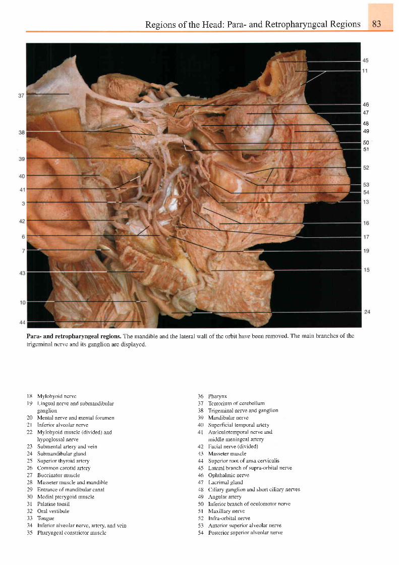
Regions of the Head: Para- and Retropharyngeal Regions
46A 7
4849
5051
Para- and retropharyngeal regions. The mandible and the lateral wall of the orbit have been removed. The main branches of thetrigeminal nerve and its ganglion are displayed.
18 Mylohyoid nerve19 Lingual nerve and submandibular
ganglion20 Mental nerve and mental foramen2l Inferior alveolar nerve22 Mylohyoid muscle (divided) and
hypoglossal nerve23 Submental artery and vein24 Submandibular gland25 Superior thyroid artery26 Common carotid artery27 Buccinator muscle28 Masseter muscle and mandible29 Enlranceof mandibularcanal30 Medial pterygoid muscle31 Palatine tonsil32 Oral vestibule33 Tongue34 Inferior alveolar nerve, artery, and vein35 Pharyngeal constrictor muscle
363738394041
4243444546
4849505 l5253< ^
PharynxTentorium of cerebellumTrigeminal nerve and ganglionMandibular nerveSuperficial temporal arteryAuriculotemporal nerve andmiddle meningeal arteryFacial nerve (divided)Masseter muscleSuperior root of ansa cervicalisLateral branch of supra-orbital nerveOphthalmic nerveLacrimal glandCiliary ganglion and short ciliary nervesAngular arteryInferior branch of oculomotor nerveMaxillary nerveInfra-orbital nerveAnterior superior alveolar nervePosterior superior alveolar nerve
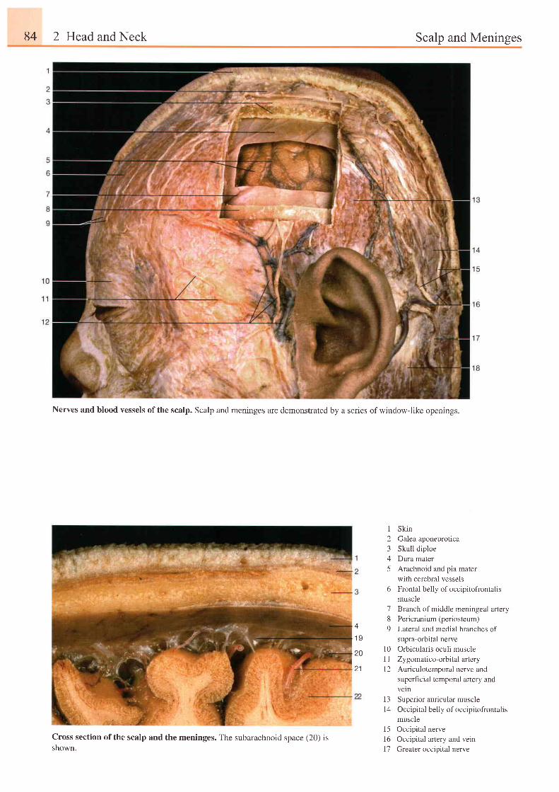
84 2 Head and Neck Scalp and Meninges
Nerves and blood vessels of the scalp. Scalp and meninges are demonstrated by a series of window-like openings.
4
1 9
20
2 1
I Skin2 Galeaaponeurotica3 Skull diploe4 Dura mater5 Arachnoid and pia mater
with cerebral vessels6 Frontal belly of occipitofrontali s
muscle7 Branch of middle meningeal artery8 Pericranium (periosteum)9 Lateral and medial branches of
supra-orbital nervel0 Orbicularis oculi muscle1 1 Zy gomatico-orbital artery12 Auriculotemporal nerve and
superficial temporal artery andveln
13 Superior auricular muscle14 Occipital belly of occipitofrontalis
muscle15 Occipital nerve16 Occipital artery and vein17 Greater occipital nerve
Cross section of the scalp and the meninges. The subarachnoid space (20) isshown.

Scalp and Meninges 85
18 Sternocleidomastoid muscle19 Arachnoid mater20 Subarachnoid space2l Pta mater with cerebral vessels22 Cerebral cortex23 Arachnoidgranulations24 Confluens sinuum25 Cerebellum26 Superior sagittal sinus27 lnfeior sagittal sinus28 Straight sinus (Sinus rectus)29 Subdural space30 Falx cerebri31 Frontal lobe32 Chiasmatic cistem33 Interpeduncularcistem34 Corpus callosum35 Cerebellomedullarcistern
A coronal section through the vertex of the skull,showing the arrangement of the meninges.
Cross section through the vertex of the skull showing the dura, pia,and arachnoid mater. The subarachnoid space (20) appears enlarged.
Confluens sinuum containing arachnoid granulations of Pacchioni.
3132
Subarachnoid cisterns of the brain (midsagittal section).
Green = cistems; blue = dural sinus and ventricles;
red = choroid plexus of third and fourth ventricles; alrows = flow
of cerebrospinal fluid.
4
1 920
I
I
20
zo
21
291 923
zz
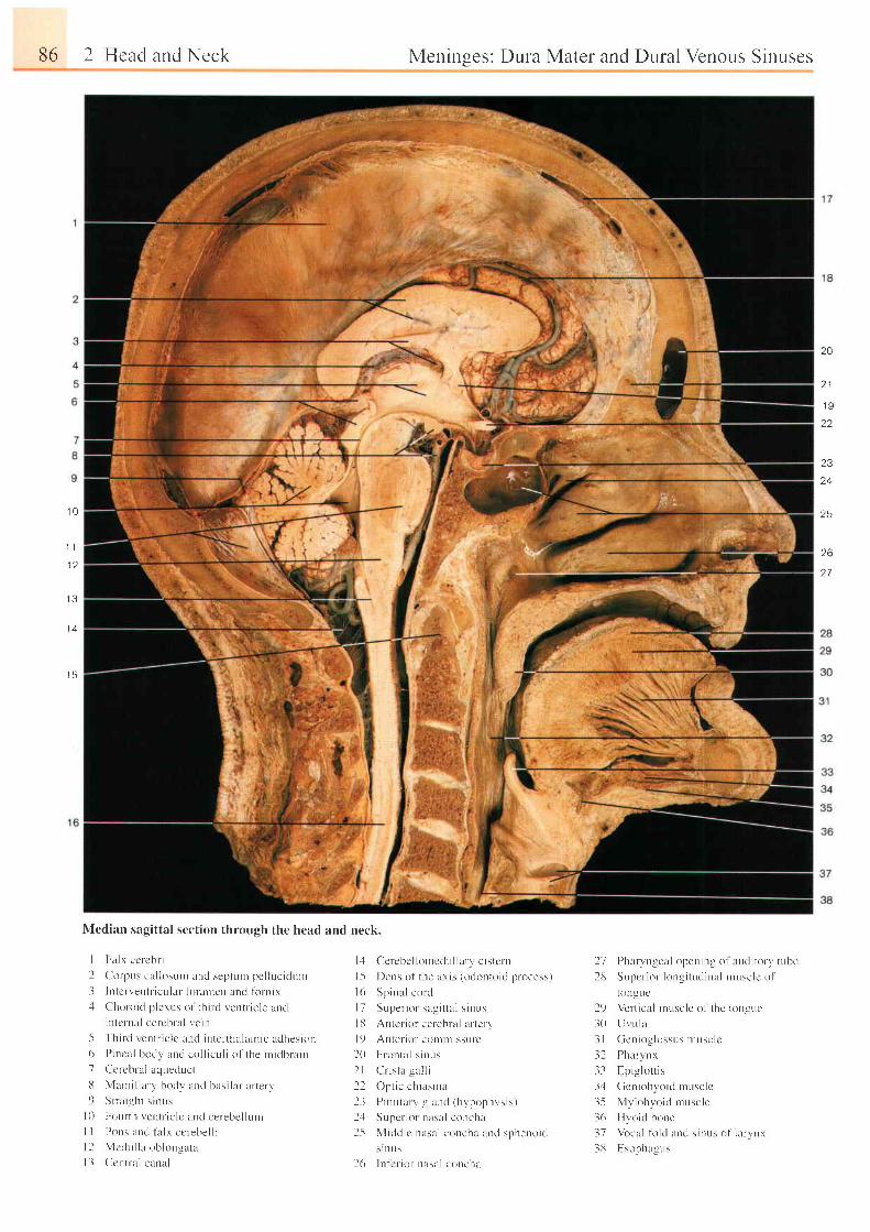
86 2 Hcad and Neck
Nledian sagittal section through the head and neck.
I Falx ccr-cbr- i
I Cor-pus cal losunt and septurn pel luc i tLurn
3 Intcn 'ent t ' icu lar f i r ramen and l i r r r r ix.1 Clhoroic l p lerus o1' th i rc l r , 'entr ic le ancl
intcrnnl cer-cbr-al vein
5 ' fh i rd
\ ent l ic le and inter thala ln ic aclhesion6 Pincal bodv and col l icul i of the nt idbrain7 Cereblal aqueduct
13 Marr i l lury body lLrrd hasi l l r ar terv9 S t ra i gh t s i nus
I 0 Fourt l r ventr ic lc and cerebel lunt
l l Pons unt l f i r lx cc lebel l il 2 Mcc lu l l a ob longa ta
I I Ccntra l canal
Meninges: Dura Mater and DuralVenor-rs Sinuses
20
o t
1 9
23
24
?5
26
27
1 0
1 1
1 2
I J
1 4
1 5
1,1 Cerebel lonrcdul larv c istcr-n
l -5 Dens of the ar is (odontoid process)
l 6 Sp ina l co r r l
l 7 Supe r i o r sa r . i t t a l s i nL r s
l13 Antcr ror celehral ur ter l ,
l 9 An te r i o r con r r r r i s su re
20 l r t on ta l s i nL r s
2 l C r i s t l ga l l i
22 Opt ic chirsr lu
l l P i t r r i t : r r r , . . : l : r r r . l r h r l r o n l r \ \ i \ r
2r1 Supcr ior nusal concha
15 Middlc nasul couchr ani l sphenoic l
s l n U S
26 In lcr ior- nasul corc lur
27 Pharyngcal opcnirrg o1 audi tory t r " rbe
213 Supc r i o r - l ong i t uc l i na l r nusc i co l
tonguel
2t) Ver- t ical muscle of the tongue
30 Lir u lu
-3 I Gcnioglossus nrusclc
32 Phartnx
33 Epi-u lot t is
3,1 Gcrr iohvoid mr,rsc lc
35 My lohyo id musc le
36 [ {yoid bone
37 Vrcal f i r ld and s inr ts of r l r r , r r r
38 Esopha- lus
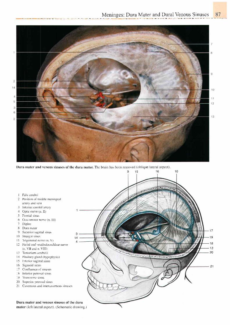
Mgnlnges; Dula Mat91 and ou1a,] Vel9..y1 p"inuggt ..,97_,,.".
Dura mater and venous sinuses of the dura mater. The brain has been removed (oblique lateral aspect).o 1 6 1 6
1 Falx cerebri
2 Position of middle meningeal
artery and vein
3 Intemal carotid artery
4 Optic nerve (n. II)
5 Frontal sinus
6 Oculomotor nerve (n. III)
7 Diploe
8 Dura mater
9 Superior sagittal sinus
l0 Stra ight s inus
11 Trigeminal nerve (n V)
l2 Facial and vestibulocochlear nerve(n VI I and n. VI I I )
l3 Tentorium cerebelli
14 Pituitary gland (hypophysis)
I 5 Inferior sagittal sinus
l6 Sigmoid s inus
17 Confluence of sinuses
I 8 Inf'erior petrosal sinus
l9 Transverse sinus
20 Superior petrosal sinus
2l Cavernous and intercavernous slnuses
Dura mater andmater (left lateral
venous sinuses of the duraaspect) . (Schemat ic drawing. )
1 7e
1 44
1 9
1 8
1 32A
21
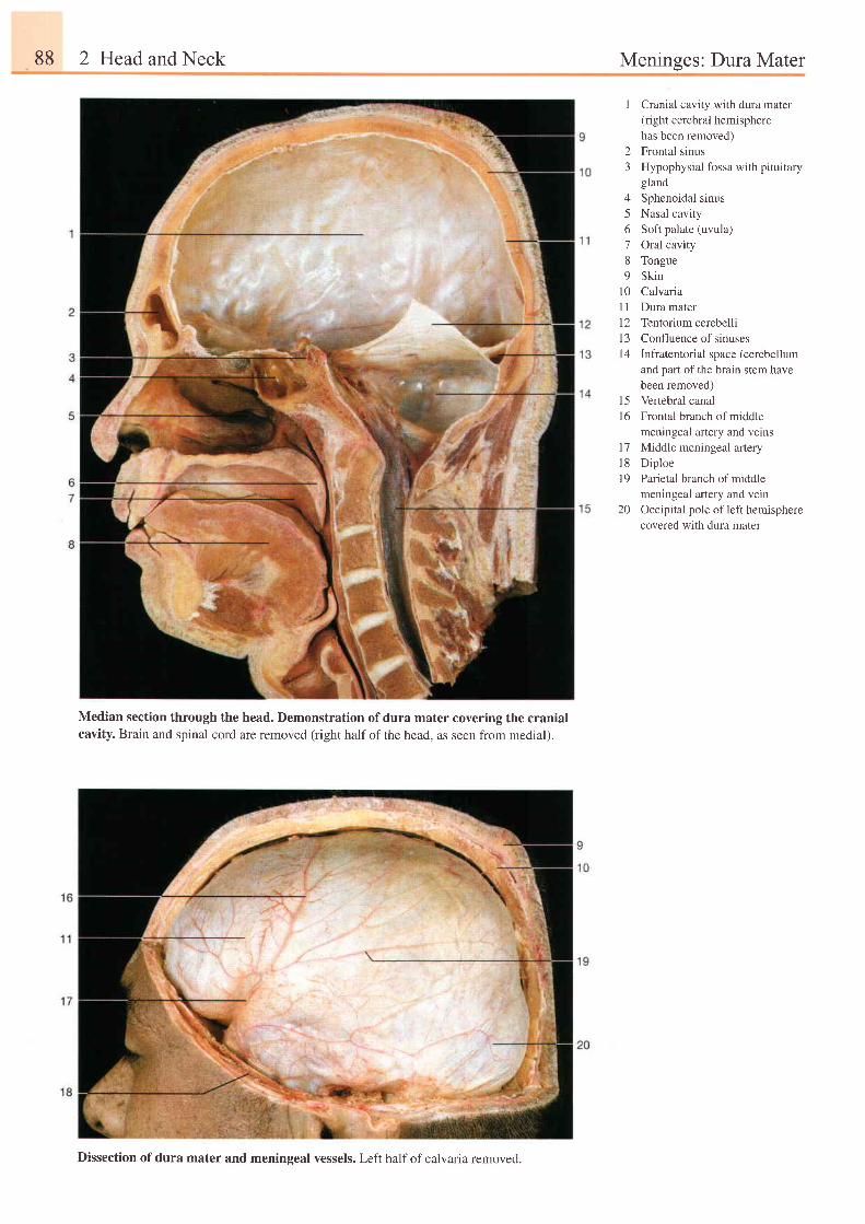
88 2 Head and Neck Meninees: Dura Mater
1 Cranial cavity with dura mater(right cerebral hemispherehas been removed)
2 Frontal sinus3 Hypophysial fossa with pituitar1,
gland4 Sphenoidal sinus5 Nasal cavity6 Soft palate (uvula)7 Oral cavity8 Tongue9 Skin
10 Calvaria11 Dura mater12 Tentorium cerebelli13 Confluence of sinusesl4 Infratentorial space (cerebellum
and part of the brain stem havebeen removed)Vertebral canalFrontal branch of middlemeningeal artery and veinsMiddle meningeal arteryDiploeParietal branch of middlemeningeal artery and veinOccipital pole of left hemispherecovered with dura mater
15t6
1 71 8t 9
20
Median section through the head. Demonstration of dura mater covering the cranialcavity. Brain and spinal cord are removed (right half of the head, as seen from medial).
Dissection of dura mater and meningeal vessels. Left half of calvaria removed.
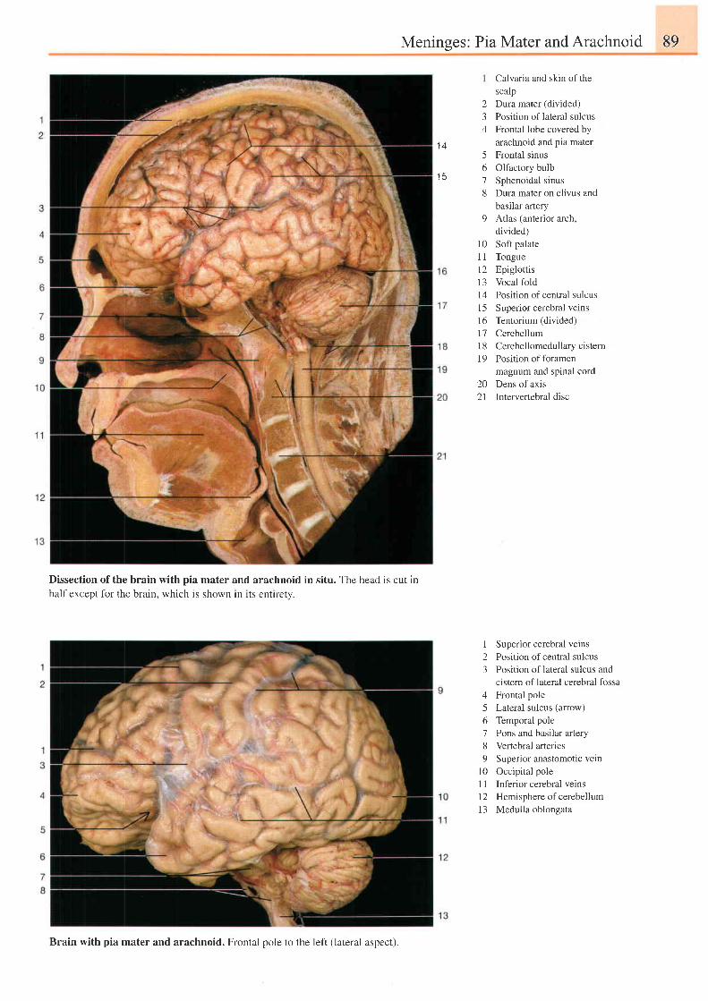
Meninses: Pia Mater and Arachnoid 89
1 4
t c
23/l
5678
o
1 0ul 2t 3t 41 51 61'71 8t 9
202 l
Calvaria and skin of thescalpDura mater (divided)Position of lateral sulcusFrontal lobe covered byarachnoid and pia materFrontal sinusOlfactory bulbSphenoidal sinusDura mater on clivus andbasilar arteryAtlas (anterior arch,divided)Soft palateTongueEpiglottisVocal foldPosition of central sulcusSuperior cerebral veinsTentorium (divided)CerebellumCerebellomedull ary cisternPosition of foramenmagnum and spinal cordDens of axisIntervertebral disc
Dissection of the brain with pia mater and arachnoid in situ. The head is cut inhalf except for the brain, which is shown rn 1ts entlrety.
1 Superior cerebral veins2 Position ofcentral sulcus3 Position oflateral sulcus and
cistem of lateral cerebral fossa4 Frontal pole5 Lateral sulcus (anow)6 Temporal pole7 Pons and basilar artery8 Vertebral arteries9 Superior anastomotic vetn
l0 Occipital pole1 1 Inferior cerebral veins12 Hemisphere of cerebellum13 Medulla oblongata
Brain with pia mater and arachnoid. Frontal pole to the left (lateral aspect).
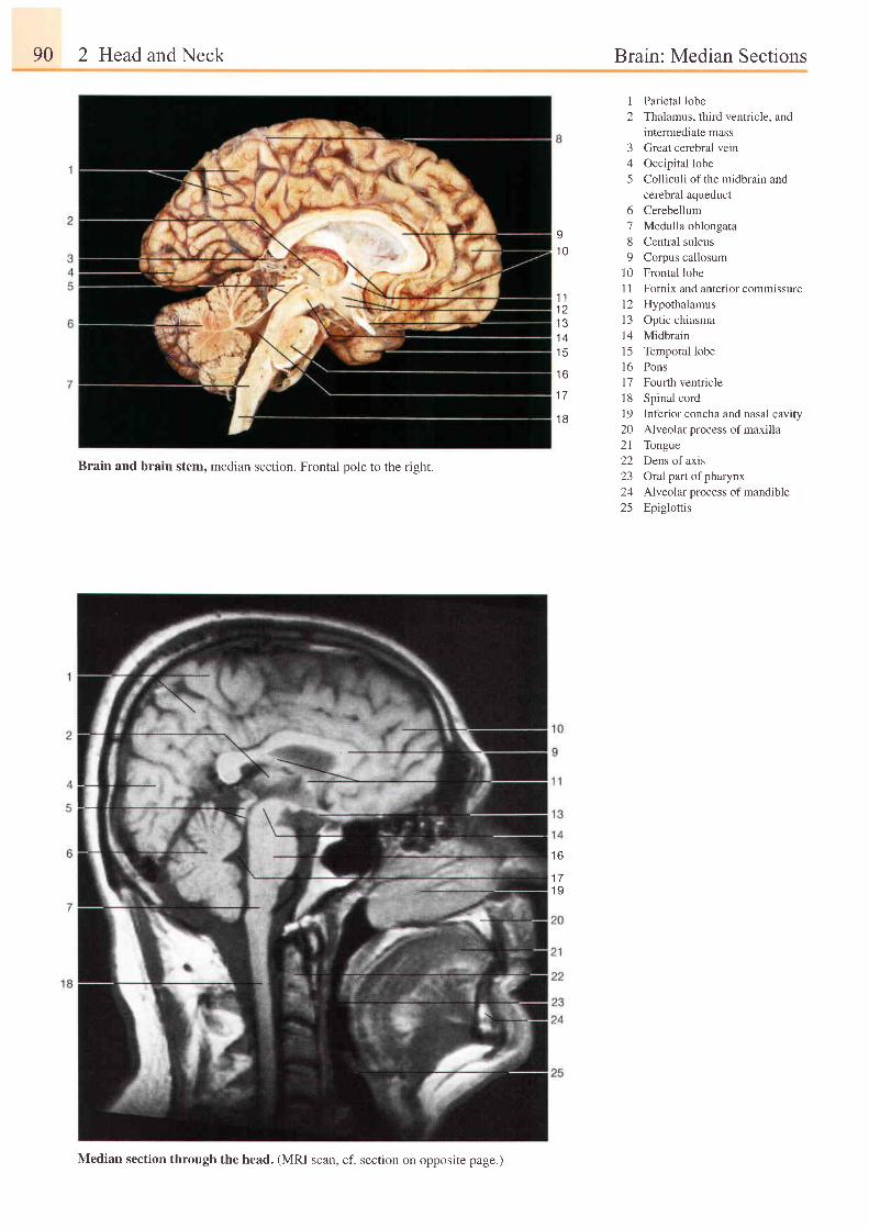
90 2 Head and Neck Brain: Median Sections
I.10
1 11 21 31 41 5
t o
I
2
345
6
789
1 0l l
1 2l 31 41 51 61 7l 81 9202 122L-)
2425
Parietal lobeThalamus, third ventricle, andintermediate massGreat cerebral veinOccipital lobeColliculi of the midbrain andcerebral aqueductCerebellumMedulla oblongataCentral sulcusCorpus callosumFrontal lobeFornix and anterior commissureHypothalamusOptic chiasmaMidbrainTemporal lobePonsFourth ventricleSpinal cordInferior concha and nasal cavityAlveolar process of maxillaTongueDens of axisOral part of pharynxAlveolar process of mandibleEpiglottis
Brain and brain stem, median section. Frontal pole to the right.
1 7
1 B
t b
t t
1 9
Median section through the head. (MRI scan, cf. section on opposite page.)
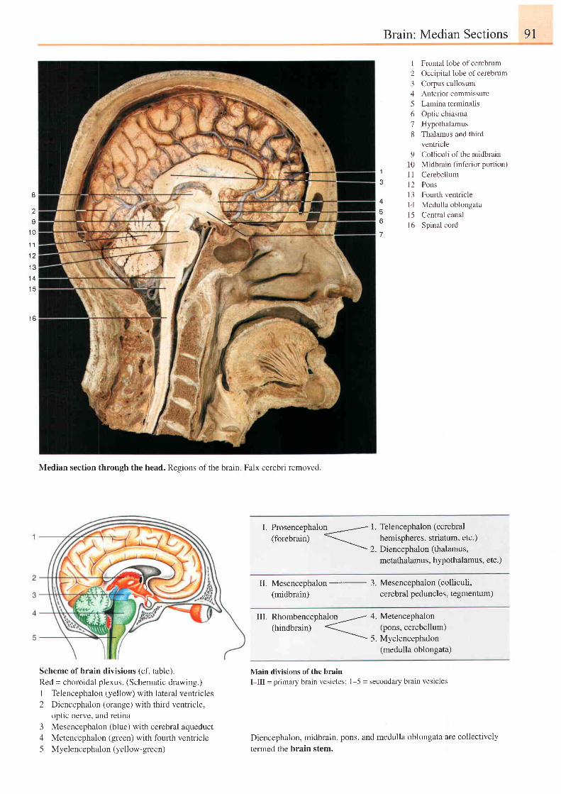
Brain: Median Sections 91
1
1 Frontal lobe ofcerebrum2 Occipital lobe of cerebrum3 Corpus callosum4 Anterior commissure5 Lamina terminalis6 Optic chiasma7 Hypothalamus8 Thalamus and third
ventricle9 ColUculi of the midbrain
10 Midbrain (inferior portion)1 1 Cerebellum12 Pons13 Fourth ventricle14 Medulla oblongatal5 Central canal16 Spinal cord
I
2
I
1 0
1 1
1 2
1 3
1 4
1 5
I O
4
o
7
Median section through the head. Regions of the brain. Falx cerebri removed.
I. Prosencephalon L Telencephalon (cerebral(forebrain) hemispheres, striatum, etc.)
\ 2. Diencephalon (thalamus,metathalamus, hypothalamus, etc.)
III. Rhombencephalon ---/ 4. Metencephalon(hindbrain) "-'' (pons, cerebellum)
-- S. Myelencephalon
(medulla oblongata)
Main divisions of the brainI-III = primary brain vesicles; 1 5 = secondary brain vesicles
Diencephalon, midbrain, pons, and medulla oblongata are collectively
termed the brain stem.
II. Mesencephalon - 3
(midbrain)
Mesencephalon (colliculi,
cerebral peduncles, tegmentum)
Scheme of brain divisions (cf. table).Red = choroidal plexus. (Schematic drawing.)
I Telencephalon (yellow) with lateral ventricles
2 Diencephalon (orange) with third ventricle,optic nerve, and retina
3 Mesencephalon (blue) with cerebral aqueduct
4 Metencephalon (green) with fourth ventricle
5 Myelencephalon(yellow-green)
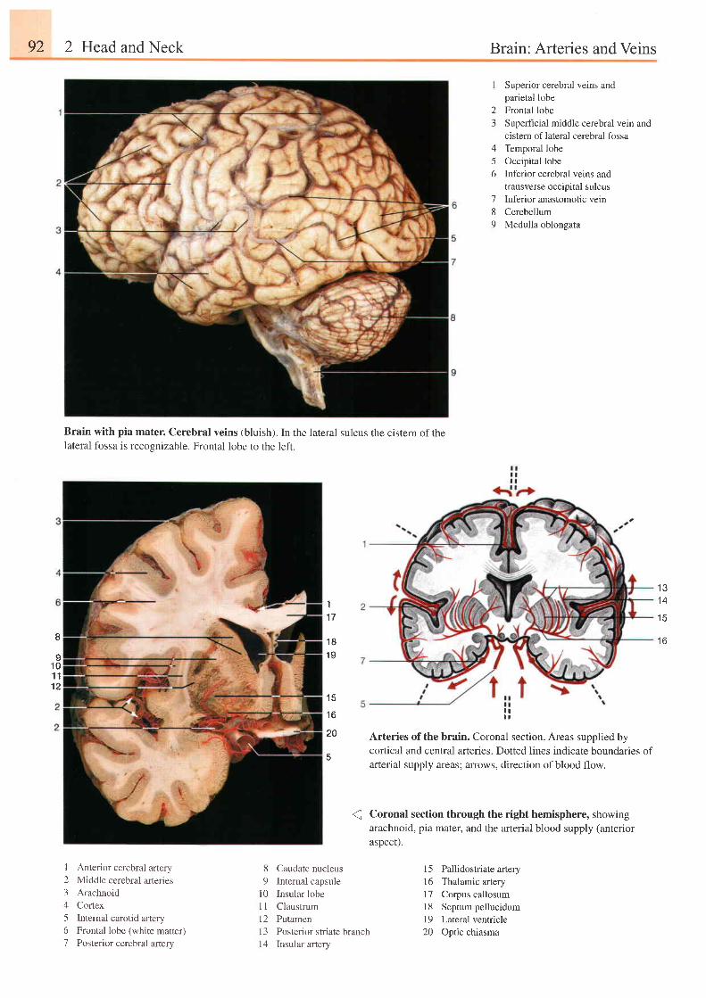
92 2 Head and Neck Brain: Arteries and Veins
Brain with pia mater. Cerebral veins (bluish). In the lateral sulcus the cistern of thelateral fossa is recognizable. Frontal lobe to the 1eft.
11 7
1 81 9
1 5
1 6
20
23
Superior cerebral veins andparietal lobeFrontal lobeSuperficial middle cerebral vein andcistern of lateral cerebral fossaTemporal lobeOccipital lobeInferior cerebral veins andtransverse occipital sulcusInferior anastomotic veinCerebellumMedulla oblongata
456
78o
! :
Arteries of the brain. Coronal section. Areas supplied bycortical and central arteries. Dotted lines indicate boundaries ofarterial supply areas; arrows, direction ofblood flow.
( Coronal section through the right hemisphere, showingarachnoid, pia mater, and the arterial blood supply (anterioraspect).
15 Pallidostriate artery16 Thalamic artery17 Corpus callosum18 Septumpellucidum19 Lateral ventricle20 Optic chiasma
1 31 4
1 5
1 6I
I1 01 11 2
I Anterior cerebral artery2 Middle cerebral arteries3 Arachnoid4 Cortex5 Intemal carotid artery6 Frontal lobe (white matter)7 Posterior cerebral artery
8 Caudate nucleus9 Internal capsule
l0 Insular lobel1 Claustruml2 PutamenI 3 Posterior striate branchl4 Insular artery
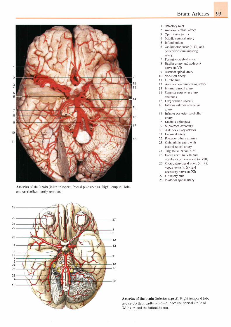
Brain: Arteries 93
2
^
6
A
6t +78
z+
25
z o
9
1 0
Arteries ofthe brain (inferior aspect, frontal pole above). Right temporal lobe
and cerebellum partly removed.
I Olfactory tract2 Anterior cerebral artery3 Optic nerve (n. II)4 Middle cerebral artery5 Infundibulum6 Oculomotor nerve (n. III) and
posterior communicatingartery
7 Posterior cerebral artery8 Basilar artery and abducent
nerve (n. VI)9 Anterior spinal artery
l0 Vertebral arteryl1 Cerebellum12 Anterior communicating artery13 Internal carotid artery14 Superior cerebellar aftery
and pons15 Labyrinthine arteries16 Inferior anterior cerebellar
arteryl7 Inferior posterior cerebellar
arteryMedulla oblongataSupratrochlear arteryAnterior ciliary arteriesLacrimal arteryPosterior ciliary arteriesOphthalmic artery withcentral retinal arteryTrigeminal nerve (n. V)Facial nerve (n. VII) andvestibulocochlear nerve (n. VIII)Glossopharyngeal nerve (n. IX),vagus nerve (n. X), andaccessory nerve (n. XI)Olfactory bulbPosterior spinal arterY
Arteries of the brain (inferior aspect). Right temporal lobeand cerebellum pafily removed. Note the arterial circle ofWillis around the infundibulum.
1 21 3
1 4
1 5
t o
1 71 81 9202122za
2425
2728
20
2 122
z
1 2
1 3
t o
I T
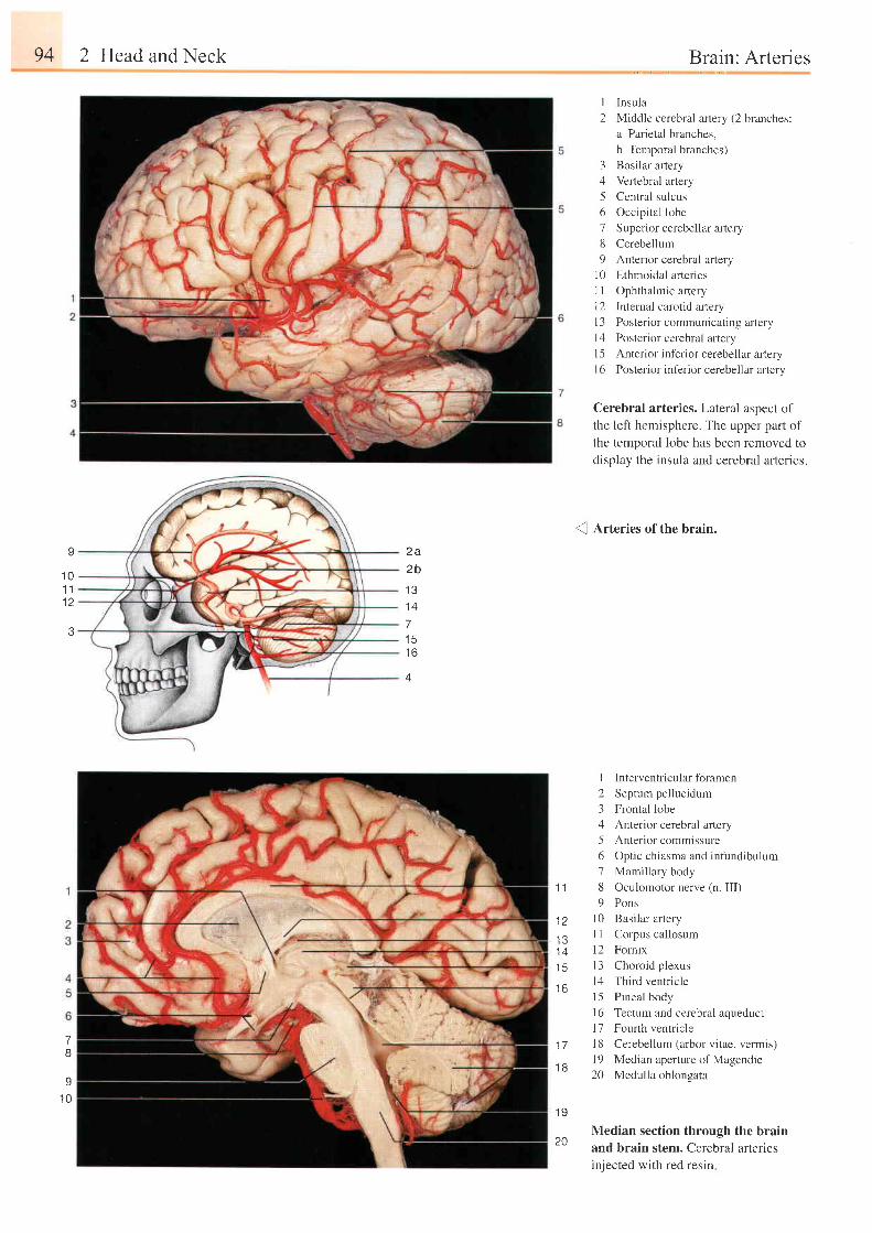
94 2 Head and Neck
v
1 01 l1 2
3
7B
I1 0
Brain: Arteries
I Insula
2 Middle cerebral artery (2 branches:
a Parietal branches,
b Temporal branches)
3 Basilar artery
4 Vertebral artery
5 Central sulcus
6 Occipital lobe
7 Superior cerebellar artery
8 Cerebellum
9 Anterior cerebral artery
l0 Ethmoidal arteries
1l Ophthalmic ar tery
12 Intemal carotid artery
l3 Posterior communicating artery
l r l Posler ior cerebrr l ar ter l
I 5 Anterior inferior cerebellar artery
I 6 Posterior inferior cerebellar arlery
Cerebral arteries. Lateral aspect of
the left hemisphere. The upper part of
the temporal lobe has been removed to
display the insula and cerebral arteries.
2 a2 b
I J
1 A
7
1 6
4
(l Arteries of the brain.
I Interventricularforamen
2 Septum pel lucidum
3 Frontal lobe
4 Anterior cerebral artery
5 Anter ior commissure
6 Optic chiasma and infundibulum
7 Mamil lary body
1 1 8 Oculomotor nerve (n. III)
9 Pons
j2 l0 Basi lar ar tery
-13 l l Corpus oal losum
14 12 Fornix
1 b l3 Choroid p lexus
14 Third ventr ic lero
15 Pineal body
16 Tectum and cerebral aqueduct
17 Fourth ventricle
1Z l8 Cerebellum (arbor vitae, vermis)
. , , q l9 Median aperture of Magendie
20 Medu l l a ob longa ta
1 9
Median section through the brainand brain stem. Cerebral arteriesinjected with red resin
20
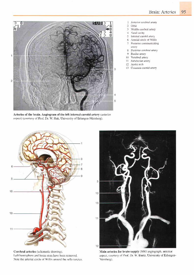
Brain: Arteries 95
Arteries of the brain. Angiogram of the left internal carotid artery (antenoraspect) (courtesy of Prof. Dr. W. Huk, University of Erlangen-Niirnberg).
Cerebral arteries (schematic drawing).Left hemisphere and brain stem have been removed.Note the arterial circle of Willis around the sella turclca.
I
23456'7
Anterior cerebral arteryOrbitMiddle cerebral arteryNasal cavityInternal carotid arteryArterial circle of WillisPosterior communicatingartery
8 Posterior cerebral artery9 Basilar arlery
l0 Vertebral arteryI I Subclavian artery12 Aortic arch13 Common carotid artery
Main arteries for brain supply (MRI angiograph, anterioraspect, courtesy ofProf. Dr. W Bautz, University ofErlangen-Ntirnberg).
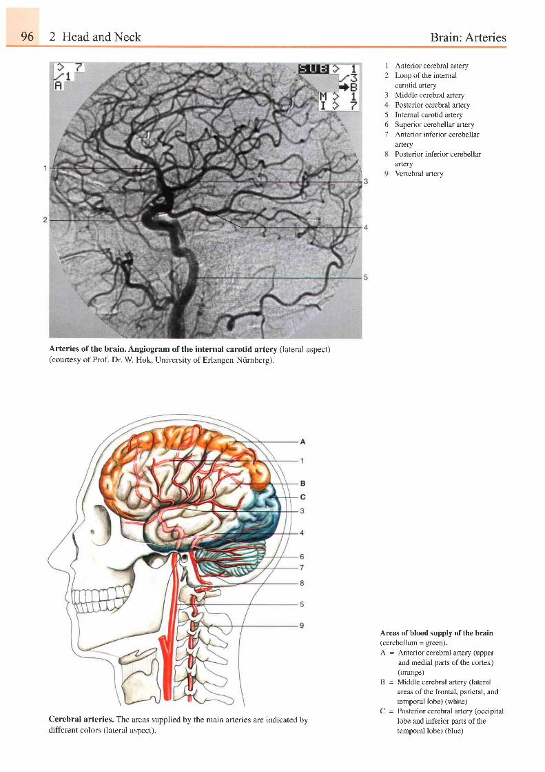
96 2 Head and Neck Brain: Arteries
Arteries of the brain. Angiogram of the(courtesy of Prof. Dr. W. Huk, University
internal carotid artery (lateral aspect)of Erlangen-Niirnberg).
12
Anterior cerebral arteryLoop of the internalcarotid arteryMiddle cerebral arteryPosterior cerebral arteryInternal carotid arterySuperior cerebellar arteryAnterior inferior cerebellararteryPosterior inferior cerebellararteryVertebral artery
3/5o7
Areas of blood supply of the brain(cerebellum = green).A = Anterior cerebral artery (upper
and medial parts of the cortex)(orange)
B = Middle cerebral artery (lateralareas ofthe frontal, parietal, andtemporal lobe) (white)
C = Posterior cerebral artery (occipitallobe and inferior parts of thetemporal lobe) (blue)
Cerebral arteries. Thedifferent colors (lateral
areas supplied by the main arteries are indicatedaspect).
by
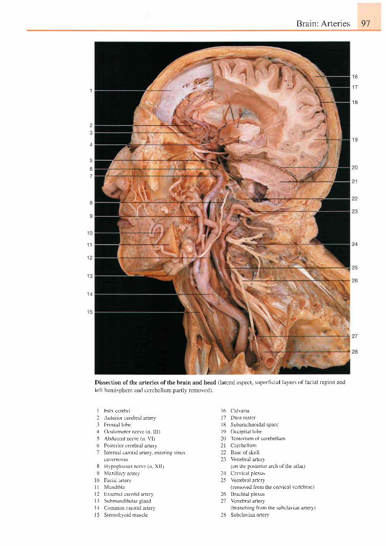
Brain: Arteries 97
1 6
1 7
1 8
Dissection of the arteries of the brain and head (lateral aspect, superficial layers of facial region and
left hemisphere and cerebellum partly removed).
1 Falx cerebri2 Anterior cerebral artery3 Frontal lobe4 Oculomotor nerve (n. III)5 Abducent nerve (n. VI)6 Posterior cerebral artery7 Internal carotid artery. entering sinus
cavernosus8 Hypoglossus nerve (n XII)9 Maxillary artery
i0 Facial arteryi I Mandiblel2 External carotid artery13 Submandibular glandl4 Common carotid arteryl5 Sternohyoid muscle
l6 Caivaria17 Dura mater18 Subarachnoidal spacel9 Occipital lobe20 Tentoriumof cerebellum21 Cerebellum22 Base of skull23 Vertebral artery
(on the posterior arch of the atlas)24 Cervical plexus25 Vertebral artery
(removed from the cervical vertebrae)26 Brachial plexus27 Vertebral artery
(branching from the subclavian artery)28 Subclavian arlery
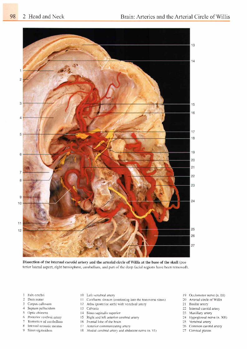
98 2 Head and Neck Brain: Arteries and the Arterial Circle of Willis
Dissection of the internal carotid artery and the arterial circle of Willis at the base of the skull (pos-terior lateral aspect, right hemisphere, cerebellum, and part of the deep facial regions have been removed)
I Falx cerebri
2 Dura mater
3 Corpus callosum
4 Septum pellucidum
5 Optic chiasma
6 Posterior cerebral artery
7 Tentorium ofcerebellum
8 lnternal acoustic meatus
9 Sinus sigmoideus
10 Left vertebral artery
1 I Confluens sinuum (continuing into the transverse sinus)
l2 Atlas (posterior arch) with vertebral artery
l3 Calvar ia
14 Sinus sagittalis superior
15 Right and left anterior cerebral artery
l6 Frontal lobe ofthe brain
l7 Anterior communicating artery
18 Medial cerebral artery and abducent nerve (n. VI)
l9 Oculomotor nerve (n. III)20 Arterial circle of Willis2l Basilar artery22 Internal carotid artery23 Maxillary artery24 Hypoglossal nerve (n. XII)25 Vertebral artery26 Common carotid artery27 Cervical plexus
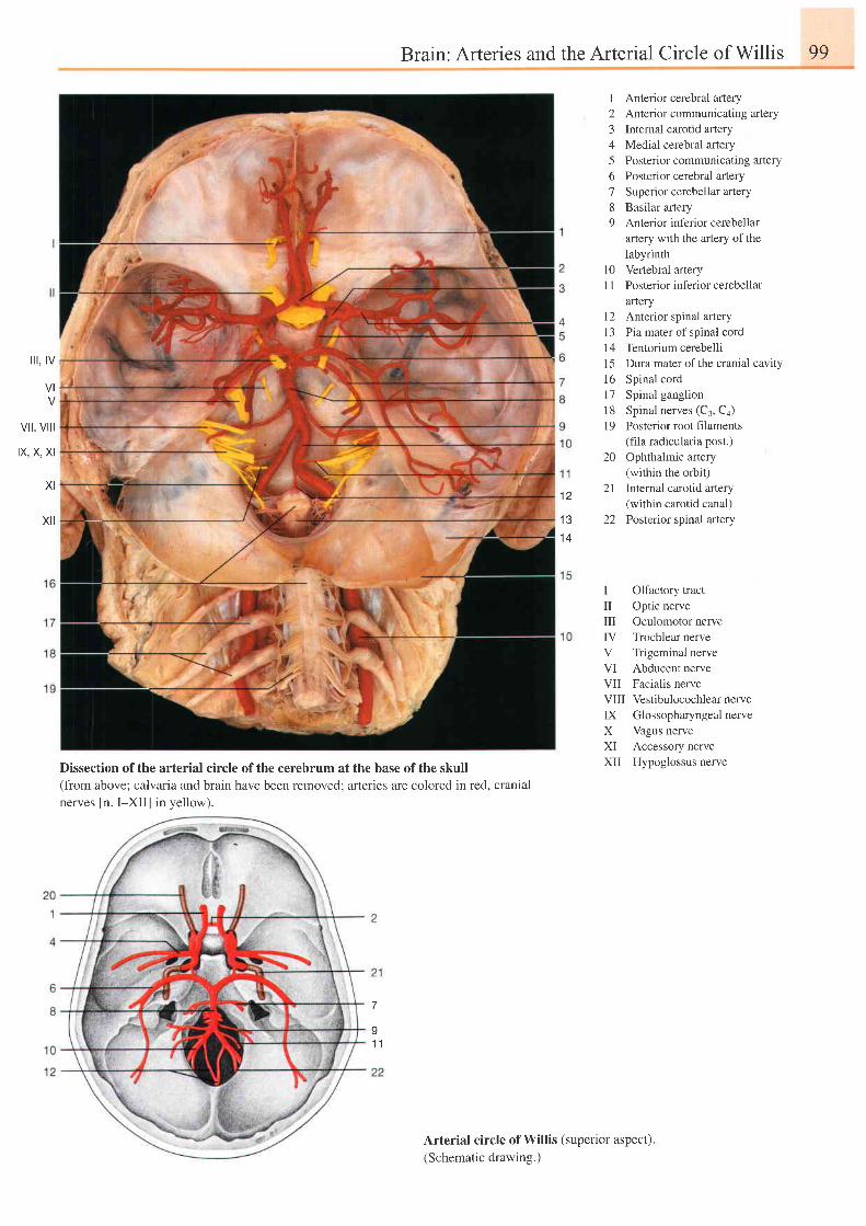
Brain: Arteries and the Arterial Circle of Willis 99
i l t , tv
VI
vil, vill
IX, X, XI
XI
xt l
12
1 31 4
I Anterior cerebral artery2 Anterior communicating artery3 Internal carotid artery4 Medial cerebral artery5 Posterior communicating artery6 Posterior cerebral arlery7 Superior cerebellar artery8 Basilar artery9 Anterior inferior cerebellar
artery with the aftery of thelabyrinth
[0 Vertebral artery1 I Posterior inferior cerebellar
artery12 Anterior spinal aftery13 Pia mater of spinal cord14 Tentorium cerebellil5 Dura mater of the cranial cavity16 Spinal cord17 Spinal ganglion18 Spinal nerves (C3, Ca)19 Posterior root filaments
(fila radicularia post.)20 Ophthalmic artery
(within the orbit)21 Internal carotid artery
(within carotid canal)22 Posterior spinal artery
I Olfactory tractil Optic nervem Oculomotor nerveIV Trochlear nerveV Trigeminal nerveVI Abducent nerveVII Facialis nerveVIII Vestibulocochlear nerveIX GlossopharyngealnerveX Vagus nerveK Accessory nerveXII Hypoglossus nerveDissection ofthe arterial circle ofthe cerebrum at the base ofthe skull
(from above; calvaria and brain have been removed; arteries are colored in red, cranialnerves [n. I-XII] in yellow).
7
I1 1
Arterial circle of Willis (superior aspect).(Schematic drawing.)
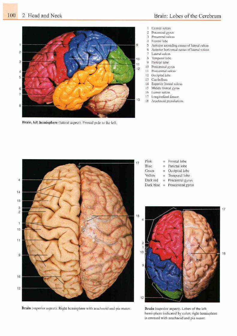
r00 2 Head and Neck Brain: Lobes of the Cerebrum
Brain, left hemisphere (lateral aspect). Frontal pole to the left
I Central sulcus2 Precentral gyrus3 Precentral sulcus4 Frontal lobe5 Anterior ascending ramus of lateral sulcus6 Anterior horizontal ramus oflateral sulcus7 Lateral sulcus8 Temporal lobe9 Parietal lobe
l0 Postcentral gyrusI I Postcentral sulcusl2 Occipital lobe13 Cerebelluml4 Superior frontal sulcus15 Middle frontal gyrusl6 Lunate sulcus17 Longitudinalflssure18 Arachnoid granulations
Pink
Blue
Green
Yellow
Dark red
Dark blue
Frontal lobeParietal lobeOccipital lobeTemporal lobePrecentral gyrusPostcentral gyrus
I
1 0
1 i
1 2
I J
1 7
.1
1 0
9
Brain (superior aspect). Lobes of the lefthemisphere indicated by color; right hemisphereis covered with arachnoid and oia mater.
Brain (superior aspect). Right hemisphere with arachnoid and pia mater.
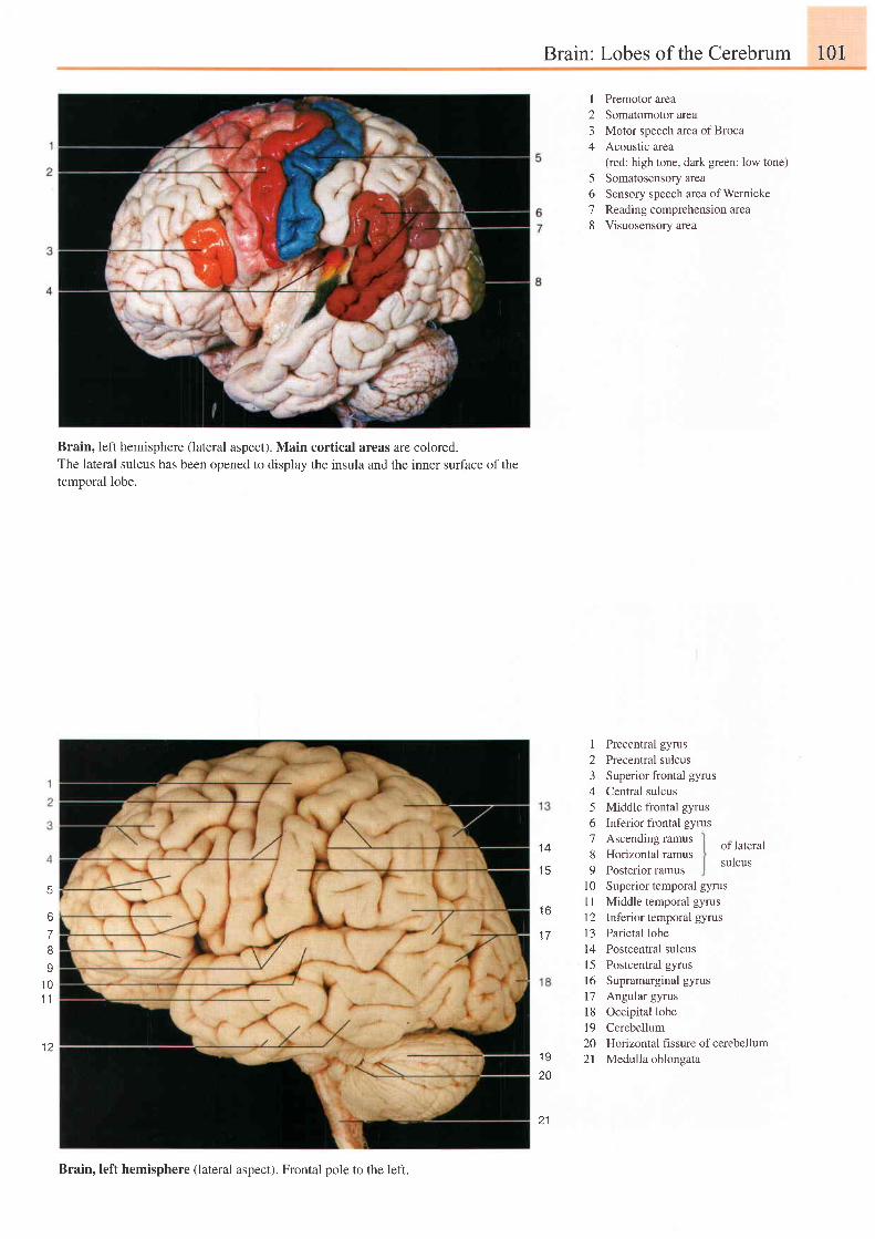
Brain: Lobes of the Cerebrum 101
Brain, left hemisphere (lateral aspect). Main cortical areas are colored.The lateral sulcus has been opened to display the insula and the inner surface of thetemooral lobe.
1 4
1 5
1 6
1 7
I
234
678
I23456789
1 0l ll 2I J
l 4l 5r6l 7l 81 9202 l
Premotor areaSomatomotor areaMotor speech area of BrocaAcoustlc area(red: high tone, dark green: low tone)Somatosensory areaSensory speech area ofWernickeReading comprehension areaVisuosensory area
Precentral gyrusPrecentral sulcusSuperior frontal gyrusCentral sulcusMiddle frontal gyrusInferior frontal gyrusAscending ramusHorizontal ramusPosterior ramusSuperior temporal gyrusMiddle temporal gyrusInferior temporal gyrusParietal lobePostcentral sulcusPostcentral gyrusSupramarginal gyrusAngular gyrusOccipital lobeCerebellumHorizontal fissure of cerebellumMedulla oblongata
of lateral
sulcus
q
7I
I1 01 1
l 1
1 920
21
Brain, left hemisphere (lateral aspect). Frontal pole to the left.
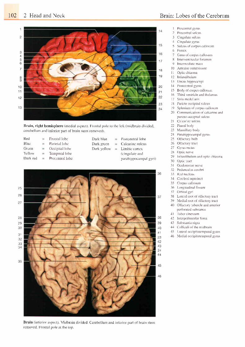
102 2 Head and Neck Brain: Lobes of the Cerebrum
A
56
7
1 01 1
1 2
Postcentral lobe
Calcarine sulcus
Limbic cortex(cingulate andparahippocampal gyri)
I23156189
l 0l 1l 2t 3t 4l 5l 61 1l 81 920
Precentral gyrus
Precentral sulcus
Cingulate sulcus
Cingulate gyrus
Sulcus of corpus cal losum
Fornix
Genu of corpus callosum
Interventricular foramen
Intermediate mass
Anterior commissure
Opt ic chiasma
Infundibulum
Uncus hippocampi
Postcentral gyrus
Body of corpus callosum
Third ventricle and thalamus
Stria medullaris
Parieto-occipital sulcus
Splenium of corpus callosum
Communication of calcarine andparieto-occipital sulcus
2l Calcarine sulcus22 Pineal body23 Mamil lary body2zl Parahippocampal gyrus25 Olfactory bulb26 Olfactory tract27 Gyrus rectus28 Optic nerve29 Infundibulum and optic chiasma30 Optic tract3l Oculomotor nerve32 Pedunculus cerebri33 Red nucleus34 Cerebral aqueduct35 Corpus callosum36 Longitudinal fissure37 Orbital gyri38 Lateral root of olfactory tract39 Medial root of olfactory tract40 Olfactory tubercle and anterior
perforated substance4l Tuber cinereum42 Interpeduncularfossa43 Substantia nigra44 Colliculi of the midbrain45 Lateral occipitotemporal gyrus46 Medial occipitotemporal gyrus
1 - l
1 8
1 9
2021
2324
Brain, right hemisphere (medial aspect). Frontal pole to the left (midbrain divided,cerebellum and inferior part of brain stem removed).
Red
BIue
Creen
Yellow
Dark red
Frontal lobe
Parietal lobe
Occipital lobe
Temporal lobePrecentral lobe
Dark blueDark greenDark yellow
25
26
28
2930
J I
323334
3B
3940A 1
234243z4
44
A E
4 b
Brain (inferior aspect). Midbrain divided. Cerebellum and inferior part of brain sremremoved Frontal pole at the top.
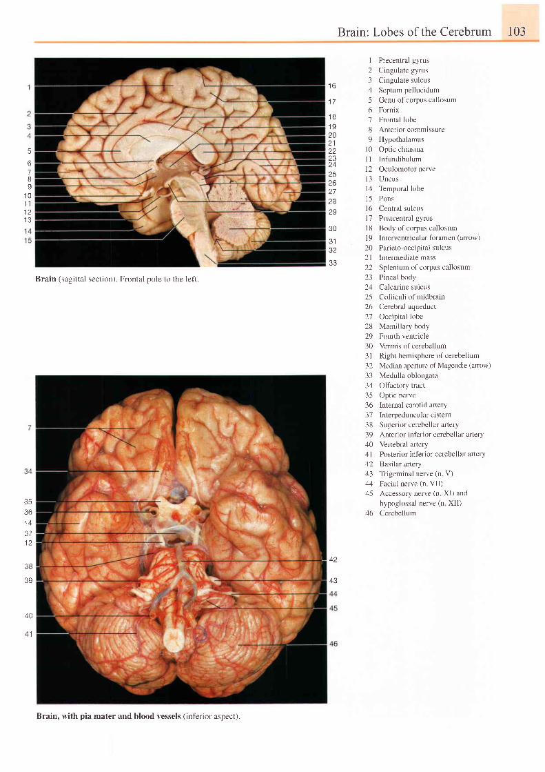
Brain: Lobes of the Cerebrum 103
1 6
1 7
1 Precentral gyrus2 Cingulate gyrus3 Cingulate sulcus4 Septum pellucidum5 Genu of corpus callosum6 Fornix7 Frontal lobe8 Anterior commissure9 Hypothalamus
l0 Optic chiasma11 Infundibuluml2 Oculomotor nervel3 Uncus14 Temporal lobe15 Pons'16
Central sulousl7 Postcentral gyrus18 Body ofcorpus callosuml9 Interventricular foramen (amow)
20 Parieto-occipital sulcus21 Intermediate mass22 Splenium of corpus callosum23 Pineal body24 Calcarine sulcus25 Colliculi of midbrain26 Cerebral aqueduct27 Occipital lobe28 Mamillary body29 Fourth ventdcle30 Vermis of cerebellum3 I Right hemisphere of cerebelium32 Median aperture of Magendie (arrow)
33 Medulla oblongata34 Olfactory tract35 Optic nerve36 Internal carotid aftery37 Interpeduncularcistern38 Superior cerebellar artery39 Anterior inferior cerebellar altery40 Vertebral artery4l Posterior inferior cerebellar artery42 Basilar artery43 Trigeminal nerve (n V)44 Facial nerve (n VII)45 Accessory nerve (n XI) and
hypoglossal nerve (n XII)46 Cerebellum
67Iq
1 01 11 21 3
1 BI Y
20z l
22I J
2526272829
30? J
32
J J
3536i 4
37t z
38
39
40
A '
Bra in tsag i t ta l sec t ion) . F ron ta l po le to the le f t .
42
43
Brain, with pia mater and blood vessels (inf'erior aspect).
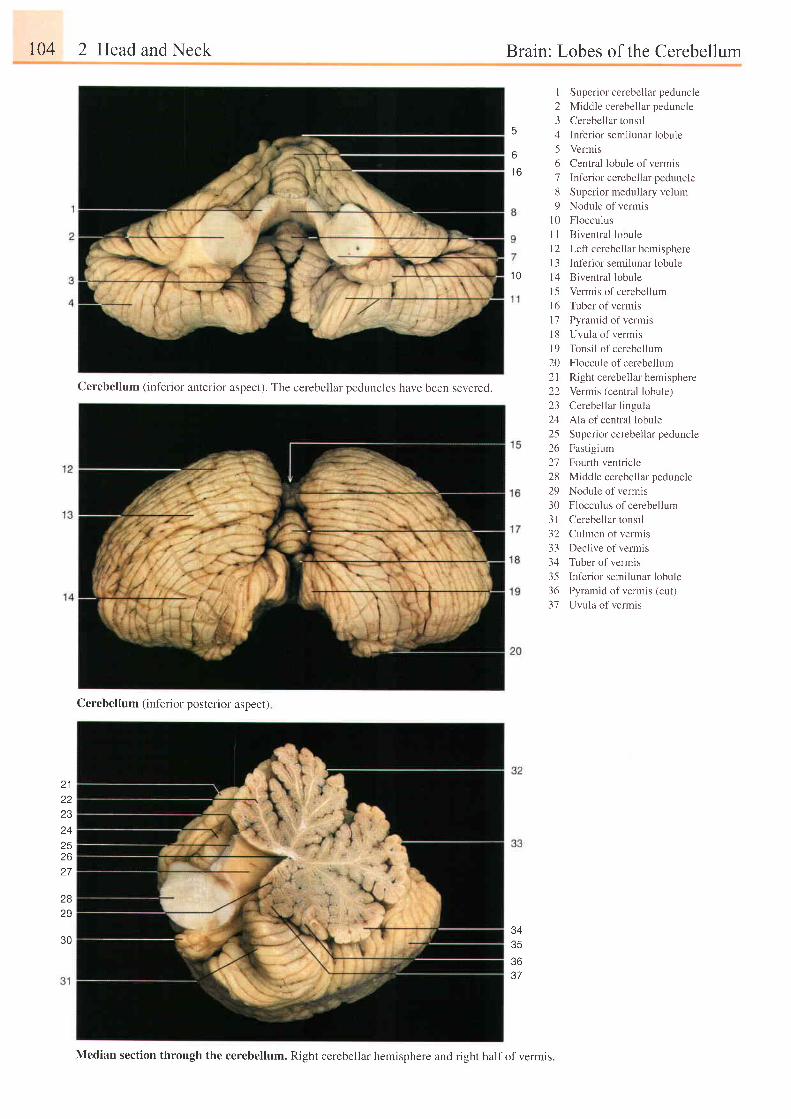
104 2 Head and Neck
Cerebellum (inferior anterior aspect). The cerebellar oeduncles have been severed.
5
6
1 6
Brain: Lobes of the Cerebellum
I Superior cerebellar peduncle
2 Middle cerebellar peduncle
3 Cerebellar tonsil
4 Inf'erior semilunar lobule
5 Vermis
6 Central lobule of vermis
7 Inferior cerebellar peduncle
8 Superior medullary velum
9 Nodule of vermis
l0 Flocculus
I I Biventral lobule
l2 Leff cerebelku hemisphere
I 3 Inferior semilunar lobule
l4 Biventral lobule
15 Vermis of cerebellum
I 6 Tuber of vermis
l7 Pyramid of vermis
18 Uvula of vermis
I 9 Tonsil of cerebellum
20 Floccule of cerebellum
2l Right cerebellar hemisphere
22 Vermis (central lobule)
23 Cerebellar lingula
24 Ala of central lobule
25 Superior cerebellar peduncle
26 Fastigium
27 Fourlh ventricle
28 Middle cerebellar peduncle
29 Nodule of vermis
30 Flocculus of cerebellurrr
3l Cerebellar tonsil
32 Culmen of vermis
33 Declive of vermis
34 Tuber of vermis
35 Inferior semilunar lobule
36 Pyramid of vermis (cut)
37 Uvula of vermis
r 0
Z J
25z o
27
2829
34353637
Cerebellum (inf'erior posterior aspect)
30
Median section through the cerebellum. Right cerebellar hemisphere and right half of vermis.
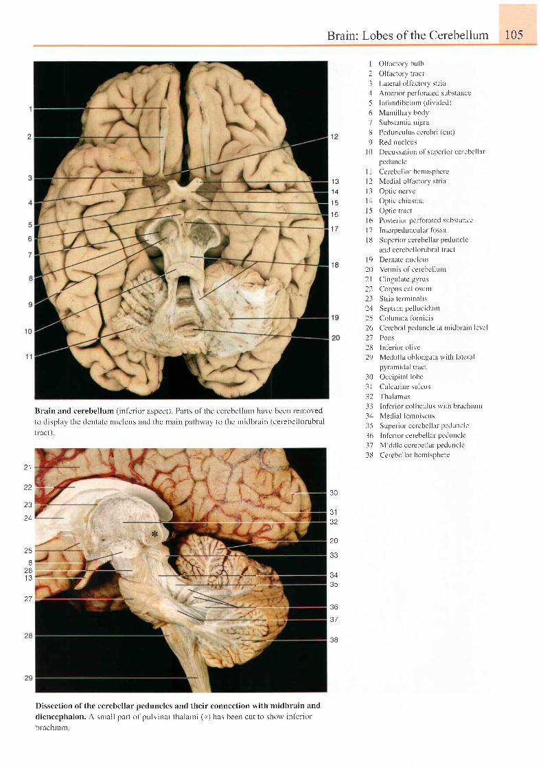
Brain: Lobes of theCerebellum 105
I Olfactory bLrlb
2 Oltactory triict
3 Lateral ollactory stria
4 Anterior perfbrated substance
5 Inf i rndibulum (div ided)
6 Mamillary body
7 Substantia nigra
8 Pedunculus cerebr i (ct t t )
9 Red nucleus
l0 Decussat ion of super ior cerebel lar
peduncle
1 31 4
1 5
1 6
1 7
1 8
1 l1 2l 3l 1t 5t 61'7I 8
l 9202 l2223242526212829
Cerebellar hemispherc
Medial olfactory stria
Optic nerve
Opt ic chiasma
Optic tract
Posterior pertbrated substance
Interpedunculiir f ossa
Superior cerebellar peduncle
and cerebellorubral tract
Dentate nucleus
Vemis of cerebellum
Cingulate gyrus
Corpus cal losutn
Stria terrninalis
Septum pellr.rcidum
Columna fbrnic is
Cerebral peduncle at midbrain level
Pons
lnf'erior olive
Medulla oblongata with lateral
Brain and cerebellum (inf-erior aspect). Parts of the cerebellum have been retnoved
to display the dentate nucleus and the main pathway to thc rnidbrain (cerebcllorubral
tract )
pyramidal tract
30 Occipital lobe
3l Calcar ine sulcus
32 Thalamus
33 Inf'erior colliculus with brachium
34 Medial lemniscus
35 Super ior cerebel lar peduncle
36 Inf'erior cerebellar peduncle
37 Middle ccrebel lar peduncle
38 Cerebel lar hemisphere
22
23
24
25
B26I J
30
J I
32
20
33
3435
36
37
3B
27
Dissection of the cerebellar peduncles and their connection with midbrain and
diencephakrn. A small part of pulvinar thalami (*) has been cut to show infertor
brachiurr
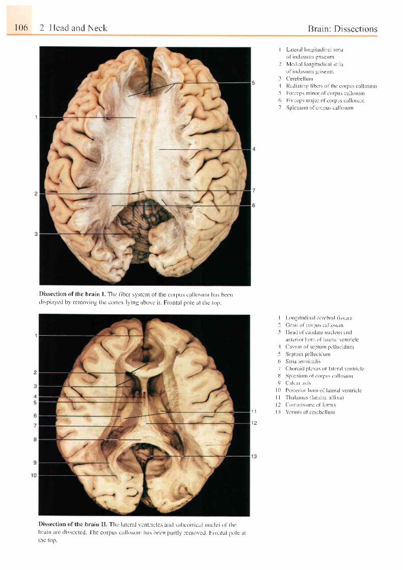
106 2 Head and Neck
Dissection of the brain I . Thc t iber system of the corpus cal losurn has becndisplayed by rcmoving thc cortex lying above i t Frontal pole at the top.
Dissection of the brain I I . The lateral ventr iclcs ancl sr.rbcolt ical nuclei of thebrain are dissected rhc corpus cal losum has becn part ly rernovcd. Fr-ontal pole atthe krp.
Brain: Dissections
I Latcral lon-s i tudinal st r ia
of indusium gr iscurn
2 Medial longi tudinal st r ia
ol inc lusiuni gr iseunr
3 Cerebel lLrrn,1 Radiat ing f lbers o1' thc corpus cal losuul
5 Forceps minor o l corpus cal losum
6 Fulceps major of colpus cal losuur
7 Splcnium ol corpus cal losutn
I Longi tudinal cercbral l lssure
2 Gcnu of corpus cal losurn
-3 Head of caudatc nucleus ancl
anler ior- horn of lateral vcntr ic le'1 Car,um o1'scptr . r rn Pel lucic iurrr-5 Septum pel lucidLrm
6 Str ia terrn inal is
7 Clhoroid p lexr . rs of lateral vcntr ic le
13 Splenrunr of corpus cal losurn
9 Calcar avis
I 0 Poster ior horn of lateral vcntr ic le
I I Thalan.rus ( lamina af l ixal
l2 Comrnissur-e of fb ln ix
l 3 Vennis of cerebel lLrnr' I I
t t
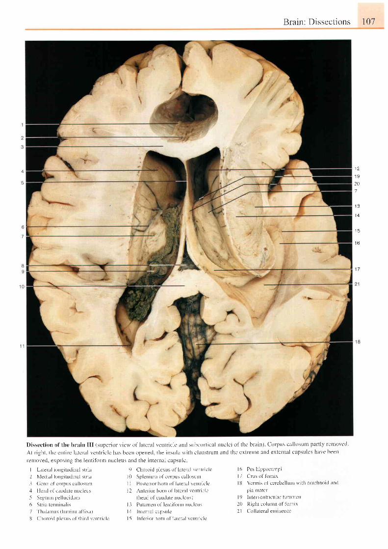
Brain: Dissections 107
Dissection of the brain I I I (superior view of lateral ventr icle i ind subcort ical nuclei of the brain). Corpus cal losutl . t part ly rer-noved.
At r ight. thc cntirc lateral ventr icle has been opened, t l ' re insula with claustrum and the extreme and external capsules havc been
Lernoved. exposing the lenti fbrm nuclens and the intelnal capsule
1 a
I Y
20I
' Q
1 4
1 5
1 [ -ateral longi t r . rd inal st r ia
2 Vlet l ia l longi tudinal st r ia
3 Cerru ol corpus cal losunr
4 l{eatl of cauclate nucleus
-5 Septurn pel luc idunr
6 S t r i a t c r I n i na l i s
7 Thalarrus ( lamina af f lxa)
8 Choroid p lcrus ol th i r -d ventr- ic le
9 Clhorc id p lexus of lateral vcntr ic le
l0 Spleniunr ofcorpus cal losunt
I I Poster- ior horn o1' lateral veutr ic lc
l2 Anter ior horn ol ' l i r tcr a l ventr ic le(head of caudate nllcl!^us )
l3 PLl tamen of lent i form nr-rc lcus
l , l Internal capsulc
I 5 ln l t r ior horn of lateral r entr- ic lc
Pes hippocampi
Crus o1' fbrnix
Vermis of cerebel lunr wi th arachnoic l and
plil Inater
lutervcntricular foramen
Right cc l lurnn of tbrnrx
Col lateral eminence
l 6l 1I t i
l 920) .1
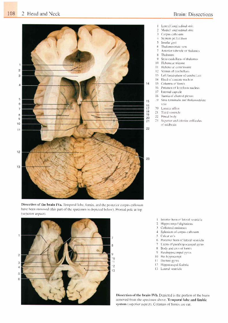
108 2 Head and Neck
II
1 0
1 l
5
o
7
Dissection of the brain IYa. Temporal lobc, fbrnix. and the posterior corpus callosumhave been removed (this part of the specimen is depicted below) Frontal pole at top(superior aspect).
7
8
I1 01 11 a
1 3
, Brain: Dissections
I Lateral longi ludinal st i la
2 Medial longi tudinal st r ia
3 Corpus c l lkrsut i
4 Septun pel luc idunr
-5 Insular gyr i
6 Thalanrostr iate r ,e in
7 Anter ior tubcrc le of thalarnus
8 Thalamus
9 Str ia rncdul lar is of thalarnus
l0 Habenular t r igone
I I Habcnul i r r commissure
l2 Vermis of cerebcllr.rnr
l3 Lef t hemisphere o1'cercbel luur
l4 Hcad of caudate nucleus
l5 Colurnns of lbrnix
l6 Putamen of lent i l i r rm nucleus
l7 I n t c rn l l capsu le
l 8 Tuen iu o l ' e ho ro i e l p l ex t r s
I t ) S t r i r t e rn r i n r l i . r nd l h r l i l t n ( ) 5 t r i l t et 3
1 61 71 81 9201 l
22
vellt
Lamina afl-rra
Third ventr ic le
Pineal body
Super ior and inf 'er io l col I iculus
of midbrain
I Inf'erior horn of lateral ventt'icle
2 H ippoc rn tpa l c l i p i t u t i ons
3 Col lateral eminencct l Splenium ofcorpus cal losunr
-5 Calcar avis
6 Poster ior horn of lateral ventr ic le
7 Uncus of parahippocalnpal gyrr,rs
8 Body and crurs o l ' lbrn ix
9 Par ahippocampal gyrr-rs
l0 Pcs hippocanrpi
I I Dentate gyrus
l2 Hippocampal fhbr ia
l3 Lateral ventr ic le
Dissection of the brain IVb. Depicted is the portion of the brainlcmoved fiom the specirnen above. Temporal lobe and limbicsystem (superior aspect) Colurnns of fbrnix are cut.
20212223
I
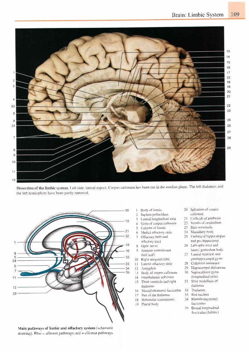
Brain: Limbic System 109
4
ZJ
t c
1 6
1 7a n
1 81 9202 1
23
25
zo
27
28
29
6
24
Dissection of the limbic system. Left side, lateral aspect. Corpus callosum has been cut in the median plane. The left thalamus and
the left hemisphere have been partly removed.
9
67
1 1
30 I Body offornix2 Septum Pellucidum
1e 3 Lateral longitudinl l str ia
4 Cenu of corPus cal losum5 Column of fornix
31 6 Medial olfactory stria32 7 Olfactory bulb and
olfactory tract19 8 optic nerve
16 9 Anterior commissurer l ^ l i h o l f \
1al0 Right temporal lobe
23 I I Lateral olfactorY stria
34 l2 Amygdala35 I 3 Body of corpus callosum
l4 I n tenha lam ic adhes ion
l5 Third ventricle and right
thalamus
l6 Mamillothalamic fasciculus
l7 Parl of the thalamus
l8 Habenu la r commissu re
l9 Pineal bodY
20 Splenium of corPus
callosum
2l Colliculi of midbrain
22 Vermis of cerebellum
23 Stria terminalis
24 Mamillary body
25 Fimbr ia of h ipPocamPus
and pes hippocamPt
26 Leftoptic tract and
lateral geniculate bodY
2'1 Laleral ventricle and
parahippocampal gyrus
28 Collateral eminence
29 Hippocampal d ig i tat ions
30 Supracallosal gYrus
(longitudinal stria)
3l Stria medullaris of
thalamus
32 Thalamus
33 Red nucleus
34 Mamillotegmental
fasciculus
35 Dorsal longitudinal
fasciculus (Schiitz)
Main pathways of limbic and olfactory system (schematic
drawing). Blue = afferent pathways; red = efferent pathways.
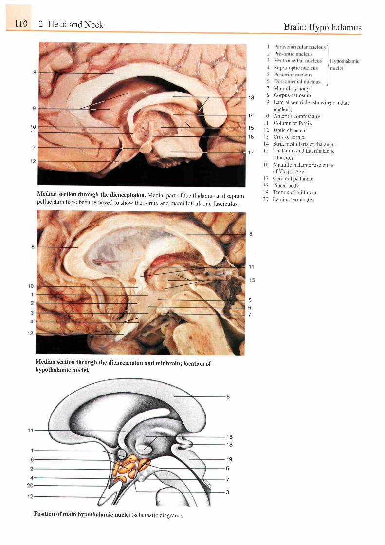
r10 2 Head and Neck
Median section through the diencephalon. Medial part of the thalamus and septumpellucidum have been removed to show the fornix and mamillothalamic fascrculus.
Median section through the diencephalon and midbrain; location ofhypothalamic nuclei.
t c
1 8
1 9
5
3
Brain: Hypothalamus
1 QI t
l e
1 4
1 6
7 Mamillary body
8 Corpus callosum
9 Lateral ventricle (showing caudatenucleus)
l0 Anterior commissureI I Column of fornix
l2 Opt ic chiasma
l3 Crus of fornix
l4 Stria medullaris of thalarlusl 5 Tha lamus and i n re r t ha lan r i c
adhesion
l6 Mamillothalamic fasciculus
of Vicq d 'Azyr
l7 Cerebral peduncle
l8 Pineal body
l9 Tectum of midbrain
20 Lamina terminalis
5
6
Position of main hypothalamic nuclei (schematic diaeram)
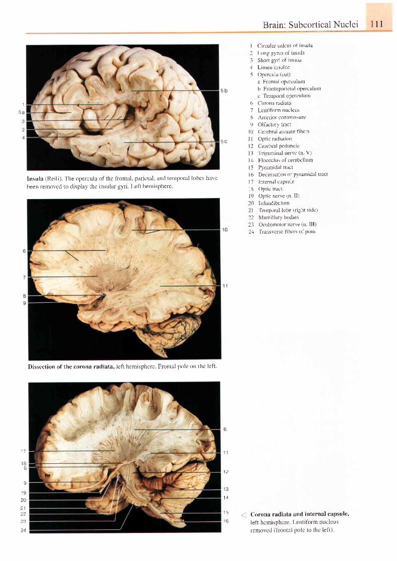
Insula (Reili). The opercula of the frontal, parietal, and temporal lobes have
been removed to display the insular gyri. Left hemisphere
Brain: Subcortical Nuclei
I Circular sulcus of insula
2 Long gyrus of insula
3 Short gyri of insula
4 Limen insulae
5 Opercula (cut)
a Frontal operculum
b Frontoparietal oPerculum
c Temporal operculum
6 Corona radiata
7 Lentifbrm nucleus
8 Anteriot commissure
9 Ollactory tlact
l0 Cerebral arcuate fibers
I I Optic radiation
12 Cerebral peduncle
l3 Trigeminal nerve (n V)
l4 Flocculus of cerebellum
15 Pyramidal tract
l6 Decussation ofPYramidal tract
I 7 Internal capsule
18 Optic tract
19 Optic nerve (n II)
20 Infundibulum
21 Ternporal lobe (right side)
22 Mamillary bodies
23 Oculomotor nerve (n. III)
24 Transverse fibers of pons
Corona radiata and internal capsule'left hemisphere. Lentifbrm nucleusremoved (frontal pole to the lef|.
l l l
1 5
1 6
1 7
1 88
9
20
2 12223
?4
1 2
1 3
1 4
Dissection of the corona radiata. lefi hemisphere. Frontal pole on the left
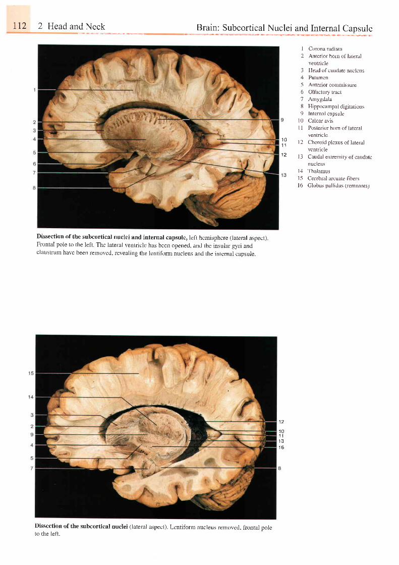
r12 2 Head and Neck
Dissection ofthe subcortical nuclei and internal capsule, left hemisphere (lateral aspect).Frontal pole to the left. The lateral ventricle has been opened, and the insular gyri andclaustrum have been removed, revealing the lentiform nucleus and the internal capsule.
Dissection ofthe subcortical nuclei (lateral aspect). Lentiform nucleus removed, frontal poleto the left.
Brain: Subcortical Nuclei and Internal Capsule
I Corona radiata2 Anterior horn oflateral
ventricle3 Head ofcaudate nucleus4 Putamen5 Anterior commissure6 Olfactory tract7 Amygdala8 Hippocampal digitat ions9 Internal capsule
10 Calcar avisI I Posterior horn of lateral
ventricle12 Choroid plexus oflateral
ventriclel3 Caudal extremity of caudate
nucleusl4 Thalamus15 Cerebral arcuate fibersl6 Globus pallidus (remnants)
1 01 1
1 2
1 2
1 01 1i 2
t o
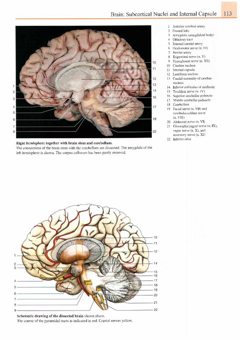
Brain: Subcortical Nuclei and Internal Capsule 113
1 0
1 1
1 2
1 3
1 4
t o
1 7
1 Anterior cerebral artery2 Frontal lobe3 Amygdala (amygdaloid body)4 Olfactory tract5 Internal carotid artery6 Oculomotor nerve (n. III)7 Basilar artery8 Trigeminal nerve (n V)9 Hypoglossal nerve (n. XII)
10 Caudate nucleus11 Internal capsule12 Lentiform nucleus13 Caudal extremity of caudate
nucleusl4 Inferior colliculus of midbrainl5 Trochlear nerve (n. IV)16 Superior cerebellar peduncle
17 Middle cerebellar pedunclel8 Cerebellum19 Facial nerve (n. VII) and
vestibulocochlear nerve(n. VIII)
20 Abducent nerve (n. VI)21 Glossopharyngeal nerve (n. IX),
vagus nerve (n. X), andaccessory nerve (n XI)
22 Inferior oliveRight hemisphere together with brain stem and cerebellum.The connections of the brain stem with the cerebellum are dissected. The amygdala of the
left hemisphere is shown. The corpus callosum has been partly removed.
Schematic drawing of the dissected brain shown above.The course of the pyramidal tracts is indicated in red. Cranial nerves yellow
1 4
1 5t o
1 1
1 8
1 9
207
8
I
21
22
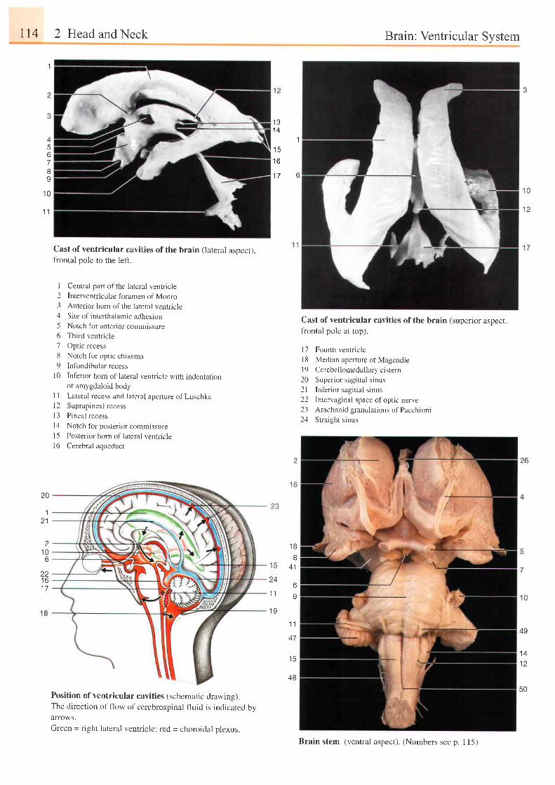
114 2 Head and Neck Brain: Ventricular System
'I ll
1 4A
67I9
1 0
1 1
20
121
1 06
zzt o1 7
1 5t o
1 7 6
Cast of ventricular cavities of the brain (lateral aspect),frontal pole to the left.
I Central part ofthe lateral ventricle2 Interventricular foramen of Monro3 Anterior horn of the lateral ventricle4 Site of interthalamic adhesron5 Notch for anter ior commissure6 Third ventricle
7 Optic recess
8 Notch for optic chiasma
9 Infundibular recess
10 Inferior horn of lateral ventricle with indentationof amygdaloid body
I 1 Lateral recess and lateral aperture of Luschkal2 Suprapineal recess1 3 Pineal recess
l4 Notch for poster ior commissure15 Posterior horn of lateral ventriclel6 Cerebral aqueduct
Position of ventricular cavities (schematic drawing).The direction of flow of cerebrospinal fluid is indicated byalTOWS.
Green = right lateral ventricle; red = choroidal plexus.
Cast of ventricular cavities of the brain (superior aspect,frontal pole at top).
17 Fourth ventriclel8 Median aperture of Magendie19 Cerebellomedullary cistern20 Superior sagittal sinus21 Inf'erior sagittal sinus22 Intervaginal space ofoptic nerve23 Arachnoid granulations of Pacchioni24 Straight sinus
t c
24
1 1
1 9
1 8
tJ
A 1
6
9
1 1
47
J J
48
49
1 A
1 2
50
Brain stem (ventral aspect). (Numbers see p. 115)
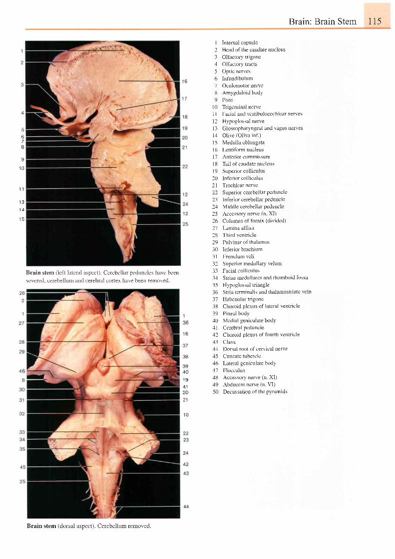
Brain: Brain Stem 115
Brain stem (left lateral aspect). Cerebellar peduncles have been
severed. cerebellum and cerebral cortex have been removed.
I Internal capsule2 Head ofthe caudate nucleus3 Olfactory trigone4 Olfactory tracts5 Optic nerves6 Infundibulum7 Oculomotor nerve8 Amygdaloid body9 Pons
l0 Trigeminal nerveI I Facial and vestibulocochlear nerves12 Hypoglossal nervel3 Glossopharyngeal and vagus nervesl4 Olive (Oliva inf.)15 Medulla oblongatal6 Lentiform nucleusl7 Anterior commissure18 Tail ofcaudate nucleusl9 Superior colliculus20 Inferior colliculus21 Trochlearnerve22 Superior cerebellarpeduncle23 Inferior cerebellar peduncle24 Middle cerebellar peduncle25 Accessory nerve (n. XI)26 Columns of fornix (divided)27 Lamina afftxa28 Third ventricle29 Pulvinar of thalamus30 Inferior brachium31 Frenulum veli32 Superior medullary velum33 Facial colliculus34 Striae medullares and rhomboid fossa35 Hypoglossaltriangle36 Str ia terminal is and thalamostr iate vein
37 Habenular trigone38 Choroid plexus of lateral ventricle39 Pineal body40 Medial geniculate body4l Cerebral peduncie42 Choroid plexus offourth ventricle43 Clava44 Dorsal root ofcervical nerve45 Cuneate tubercle16 Laleral geniculate body47 Flocculus48 Accessory nerve (n. XI)49 Abducentnerve(n VI)50 Decussation of the pyramids
1 6
1 7
5
678
I
1 0
. l I
1 31 4
1 5
38
3940.19
20
1 0
Brain stem (dorsal aspect). Cerebellum removed.
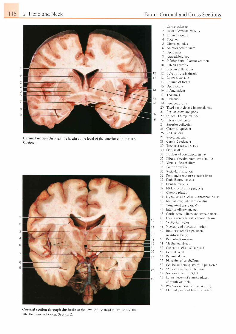
116 2 Head and Neck
Coronal section through the brain at the level of the anterior comlnissureSection I
Coronal section through the brain at the levcl ot ' the third ventr. icle and thcinterthalamic adhesion. Section 2
Brain: Coronaland Cross Sections
2
3A
56
I Corpus cal losunr
2 Heacl o l caudate nucleus
3 Internal capsulc
4 Putamen
.5 Globr. rs pal l idus
6 Antcr ior commissure
7 Optic tract
13 Anrvsdaloid body
9 Inf 'er ior horn ol ' la teral vent l ic lc
l0 Lr teral ventr ic le
I I Seprum pel lucidLrrn
10 l 2 l , obus insu la r i s ( i nsu la )
1 l l 3 Ex te rna l capsu le
1: l Colunrn ol fornix
l5 Opt ic recess12 l6 Infr - rncl ibulum
l7 Tha lan rus13 | 8 Claustruu-r
14 l 9 Len t i cu l a ransa
20 Thirc l ventr ic lc and hypothalantus
1 i I I Basilar arterl' ancl pons
22 Cortcx ol tcmporal lobe16 23 lnt 'er ior col l icr , r lus
l - l S t r pe r i o r r ' r r l l i t u l us
25 Ccrcbr-al aqueduct
26 Red nucleus
1 0
1 1
1 41 21 71 B1 9
20
1 6
2 1
22
27 Substant ia n ig la
28 Cereblal pcdurrc le
29 Trochlear nerve (n IV)
30 Gray rnattcr
3 l Nucleus ol 'oculonrotor nervc
32 Fibers of oculomotor ncrr ,e (n l l l )
33 Vermis o l 'cerebel luur
3.1 Fotu'rh r,'entliclc
3-5 Ret icular fbrrnaLion
36 Pons and t ransverse pont inc f ibers
37 Embol i fbrrn nuclcus
38 Denl-atc n l lc leus
39 Mic ld le cerebel lar peduncle:10 Choroid p lexus
11 Hypoglossal nucleus at lhontboic l l i rssa:12 Medial longi tudinal fasciculus
43 Tr igc ln inal nerve (n V )4; l ln t 'e l ior o l ivary nucleus:l-5 Colticospinal tibels :rnd arcuate fibers:16 Fourth vcntr ic le wi th choroid p lexus'17 Vcsr ibuiar nucle i
l l l \ u t l eu . t r nd l r ac l r r : r , r l i l r r i t r :
49 ln l 'cr ior ccrcbel lar pedunclc
-50-5 15 25 3
5 556515 85 9
( rest i tor-m body)
Ret ioular tbrmat iorr
Medial lemniscus
Cuncalc r rucleus of Burdach
Ccntral canal
Pyraniidal tracr
Flocculr- rs of cerebel lum
Cerebel lar hemisphcre wi th p ia ntate r'Arbor v i tae" of cerebel lum
Nucleus graci l is o l Gol l
Lateral lecess ol choroid p lexus
ol foul th ventr ic lc
60 Postcr ior infer ior cerebel lar ar tefy6 l Choroic l p lexus of latcral ventr ic le
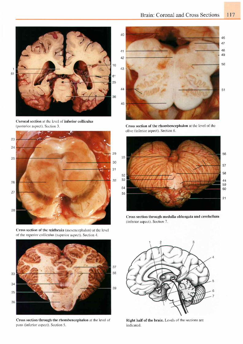
Brain: Coronal and Cross Sections ll7
46
47
4849
50
41
A a
1
6 1o i
25
57
58
445960
21
26
27
Coronal section at the level of inferior colliculus(posterior aspect). Section 3
Cross section of the midbrain (mesencephalon) at the levelof the superior colliculus (superior aspect). Section 4.
Cross section through the rhombencephalon at the level ofpons (inferior aspect). Section 5.
Cross section of the rhombencephalon at the level of theolive (inferior aspect). Section 6.
Cross section through medulla oblongata and cerebellum(inferior aspect). Section 7.
Right half of the brain. Levels of the sections are
indicated.
2933
30
J I
52q,?
O Z
trA
33
34
35
36
6
o
7
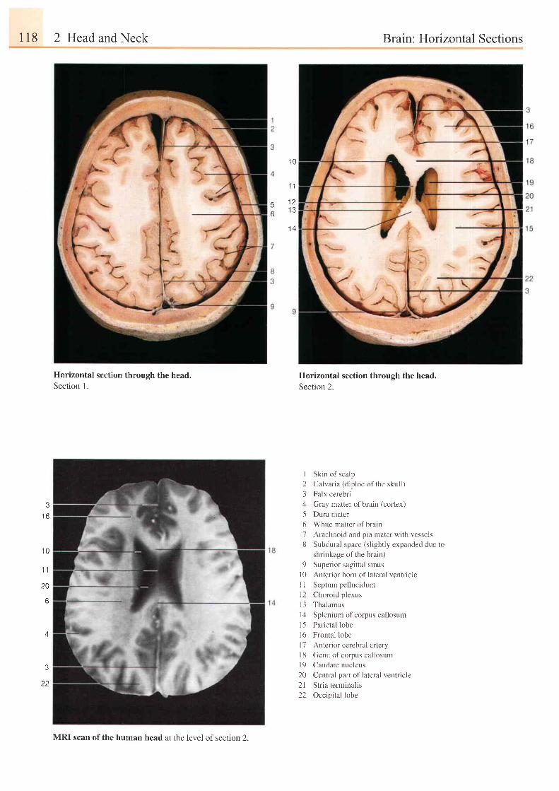
118 2 Head and Neck Brain : Hortzontal Sections
.10
'1 1
- 1 )
o
1 4
o
t o
1 0
1 1
20
A
4
3
22
Horizontal section through the head.Section L
Horizontal section through the head.
Section 2.
I Skin of scalp
2 Calvaria (diploe ofthe skull)
3 Falx cerebri
4 Gray matter of brain (cortex)
5 Dura mater
6 White matter of brain
7 Arachnoid and pia mater with vessels
8 Subdural space (slightly expanded due to
shrinkage of the brain)
9 Superior sagittal sinus
l0 Anterior horn oflateral ventricle
1 I Septum pellucidum
12 Choroid p lexus
13 Thalamus
l4 Splenium of corpus cal losum
15 Parietal lobe
l6 Frontal lobe
l7 Anterior cerebral artery
l 8 Genu of corpus callosum
l9 Caudate nucleus
20 Central part of lateral ventricle
21 Stria terminalis
22 Occipi ta l lobe
MRI scan of the human head at the level of section 2.
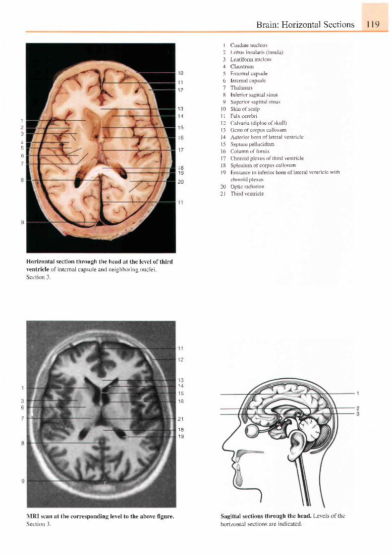
Brain: Horizontal Sections l19
Horizontal section through theventricle of internal capsule andSection 3.
head at the level of third
neighboring nuclei.
I Caudate nucleus2 Lobus insularis (insula)
3 Lentiform nucleus4 Claustrum5 Extemal capsule6 Intemal capsule7 Thalamus8 Inferior sagittal sinus9 Superior sagittal sinus
10 Skin of scalpI I Falx cerebri12 Calvaria (diploeof skull)l3 Genu ofcorpus callosuml4 Anterior horn oflateral ventricle15 Septum pellucidum16 Column of fornixl7 Choroid plexus ofthird ventricle18 Splenium ofcorpus callosum1 9 Entrance to inferior hom of lateral ventricle with
choroid plexus20 Optic radiation21 Third ventricle
Sagittal sections through the head. Levels of thehorizontal sections are indicated.
1 0
1 11 2
1
3i
5
Ct
7
1 6
1 31 4
1 5
1 81 9
20
1 7
l z
1 31 4t 3
1 6
2 1
1 8I Y
MRI scan at the corresponding level to the above figure.Section 3.
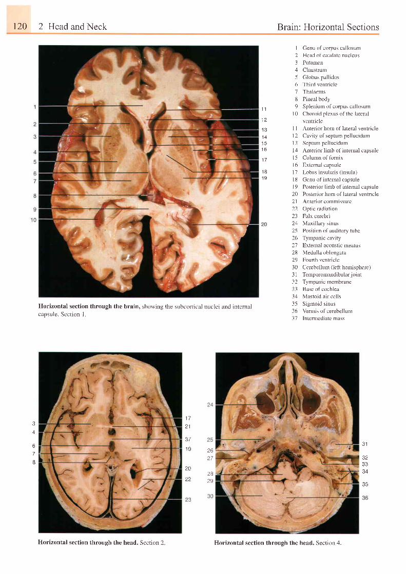
120 2 Head and Neck
Horizontal section through the brain, showing the subcortical nuclei and internalcapsule. Section l .
A
67
I
Brain: Horizontal Sections
1 1
1 a
1 41 51 6
1 7
't8
1 9
I Genu of corpus callosum2 Head of caudate nucleus3 Putamen4 Claustrum5 Globus pallidus6 Third ventricle7 Thalamus8 Pineal body9 Splenium ofcorpus callosum
10 Choroid plexus ofthe lateralventricle
l1 Anterior horn oflateral ventriclel2 Cavity ofseptum pellucidum13 Septumpellucidum14 Anterior limb of internal capsulel5 Column of fornix16 External capsule17 Lobus insularis (insula)l8 Genu of intemal capsuleI9 Posterior limb of intemal capsule20 Posterior horn oflateral ventricle21 Anterior commissure22 Opticradiation23 Falx cerebri24 Maxillary sinus25 Position ofauditory tube26 Tympanic cavity21 External acoustic meatus28 Medulla oblongata29 Fourth ventricle30 Cerebellum t lett hemisphere)3l Temporomandibular joint32 Tympanic membrane33 Base ofcochlea34 Mastoid air cells35 Sigmoid sinus36 Vermis of cerebellum37 Intermediate mass
1 7
2 1
37
I Y
20
26D 7
2B29
31
32
34
35
Horizontal section through the head. Section 2
22
Horizontal section through the head. Section 4.
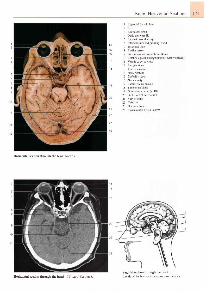
Brain: Horizontal Sections l2l
1 4
1 51 6
1 7
1 8
1 Upper lid (tarsal plate)2 Lens3 Ethmoidal sinus4 Optic nerve (n. II)5 Intemal carotid aftery6 Infundibulum and pituitary gland7 Temporal lobe8 Basilar artery9 Pons (cross section ofbrain stem)
l0 Cerebral aqueduct (beginning offourth ventricle)11 Vermis of cerebellum12 Straight sinusI 3 Transverse sinus14 Nasal septum15 Eyeball (sclera)l6 Nasal cavity17 Lateral rectus muscle18 Sphenoidal sinus19 Oculomotor nerve (n. III)20 Tentorium of cerebellum21 Skin of scalp22 Calvaria23 Occipital lobe24 Slriate cortex (visual cortex)
Sagittal section through the head.Levels of the horizontal sections are indicated.
5
6
7
I
I
21
22
z 3
24
Horizontal section through the head. Section 3.
1 4'1 5
Horizontal section through the head. (CT scan.) Section 3.
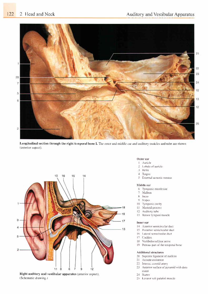
122 2 Head and Neck
1 1 8 6 7 I 1 2
Right auditory and vestibular apparatus (anterior aspect)(Schematic drawing.)
Auditory and Vestibular Apparatus
Outer ear
I Auricle
2 Lobule of auricle
3 Helix
4 Tragus
5 External acoustic meatus
Middle ear
6 Tympanic membrane
7 Malleus
8 Incus
9 Stapes
10 Tympanic cavity
I I Mastoid process
l2 Auditory tube
l3 Tensor tympani muscle
Inner ear
l4 Anterior semicircular duct
I 5 Posterior semicircular duct
16 Lateral semicircular duct
l7 Cochlea
l8 Vestibulocochlearnerve
l9 Petrous part ofthe temporal bone
Additional structures
20 Superior ligament of malleus
2l Arcuate eminence
22 Internal carotid artery
23 Anterior surface of pyramid with dura
mater
24 Stapes
25 Levator veli palatini muscle
Longitudinal section through the right temporal bone I. The outer and middle ear and auditory ossicles and tube are shown(anterior aspect).
1 8
1 0
1 7
1 3
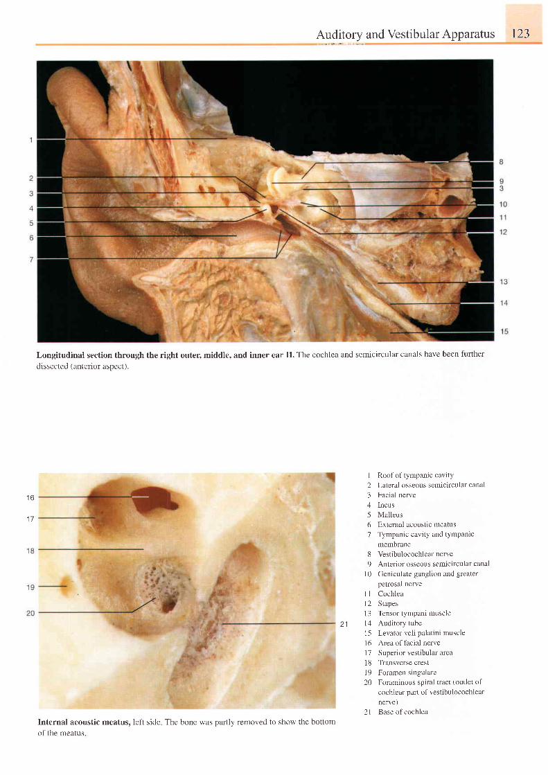
t o
1 7
Auditoryand Vestibular Apparatus 123
Longitudinal section through the right outer, middle, and inner ear II. The cochlea and semicircular canals have been further
dissected (anterior aspect).
Roof of tympanic cavity
Lateral osseous semicircular canal
Facial nerve
Incus
Malleus
Extemal acoustic meatus
Tympanic cavity and tympanlc
membrane
8 Vestibulocochlear nerve9 Anterior osseous semicircular canal
l0 Ceniculate ganglion and greaterpetrosal nerve
Cochlea
Stapes
Tensor tympani muscle
Auditory tube
Levator veli palatini muscle
Area of facial nerve
Superior vestibular area
Transverse crest
Foramen singulare
Foraminous spiral tract (outlet of
cochlear parl of vestibulocochlear
nerve)
Base of cochlea
Internal acoustic meatus, left sideof the meatus
I23/l
567
1 1l 2l 3t 41 5l 6l 71 81 920
2 1The bone was oartlv removed to show the bottom
2 1
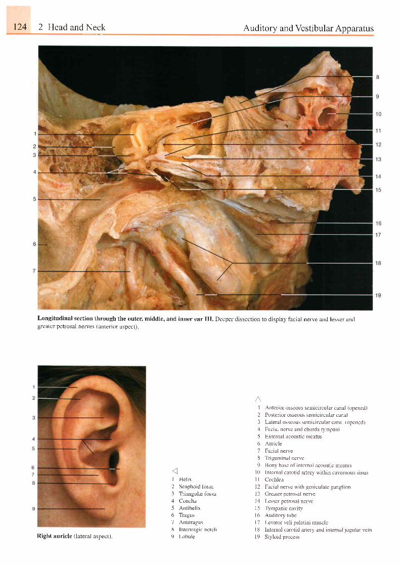
r24 2 Head and Neck Auditory and Vestibular Apparatus
Longitudinal section through the outer, middle, and inner ear III. Deeper dissection to display facial nerve and lesser andgreater petrosal nerves (anterior aspect ).
I Helix
2 Scaphoid fbssa
3 Triangular fbssa
4 Concha
5 Antihelix
6 Tragus
7 Antitragus
8 Intertragic notch
9 Lobule
A.I Anterior osseous semicircular canal (opened)
2 Posterior osseous semicircular canal
3 Lateral osseous semicircular canal (opened)
4 Facia l nerve and chorda rympani
5 Extemal acoustic meatus
6 Auricle
7 Facial nerve
8 Trigeminal nerve
9 Bony base of internal acoustic meatus
10 Internal carotid artery within cavernous sinusI I Cochlea
l2 Facial nerve with geniculate ganglion
l3 Greater petrosal nerve
14 Lesser petrosal nerve
l5 Tympanic cavity
l6 Auditory tube
l7 Levator veli palatini muscle
18 Internal carotid artery and internal jugular veinl9 Sty lo id processRight auricle (lateral aspect).
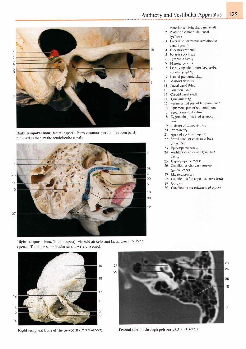
" ---*.:*l*ss***:**H*.cffis-*J::*I
2Anterior semicircular canal (red)
Posterior semicircular canal(yellow)Lateral or horizontal semicircularcanal (green)Fenestra vestibuliFenestra cochleaeTympanic cavityMastoid processPetrotympanic fissure (red Probe:chorda tympani)Lateral pterygoid PlateMastoid air cellsFacial canal (blue)Foramen ovaleCarotid canal (red)
Tympanic ringPetromastoid part of temporal bone
Squamous paft of temporal bone
Squamomastoid sutureZygomatic process of temPoralboneIncisure of tympanic ring
3
45o'7
8
q
t 0l l
121 3t4t 516t7l 8
1 920 Promontory21 Apex of cochlea (cuPula)
22 Spiral canal of cochlea at base
of cochlea23 Epitympanic recess24 Auditory ossicles and tYmPantc
cavity25 Hypotympanic recess26 Canaliculus chordae tympani
(green probe)27 Mastoid process28 Canaliculus for stapedius nerve (red)
29 Cochlea30 Canaliculus mastoideus (red probe)
1
I
A
56
Right temporal bone (lateral aspect). Petrosquamous porlion has been partly
removed to displav the semicircular canals.
2
26
1 1
l c
1 0
1 4
Right temporal bone (lateral aspect). Mastoid air cells and facial canal had been
opened. The three semicircular canals were dissected.
23
25
2 1
t 1
Right temporal bone of the newborn (lateral aspect) Frontal section through petrous part. (CT scan')
1 0
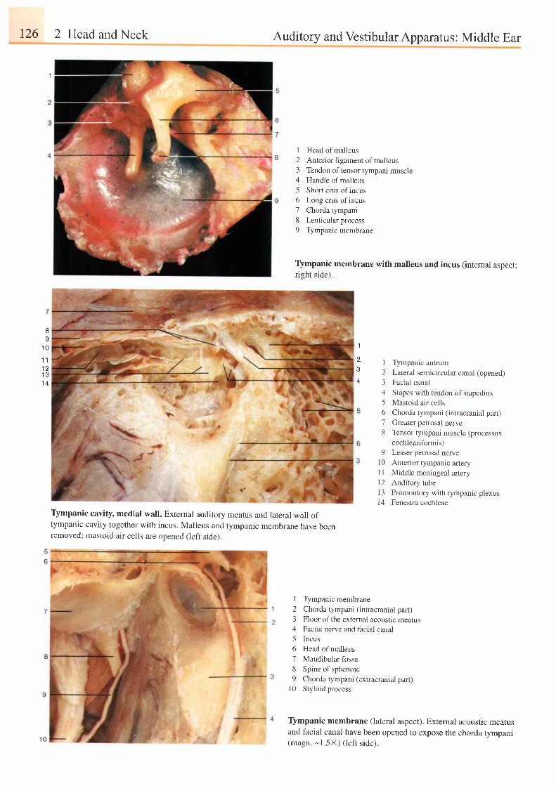
126 2 Head and Neck Auditory and Vestibular Apparatus : Middle Ear
6
Y
1 0
Tlmpanic cavity, medial wall. External auditory meatus and lateral wall oftympanic cavity together with incus. Malleus and tympanic membrane have beenremoved; mastoid air cells are opened (left side).
I Head of malleus2 Anterior ligament of malleus3 Tendon of tensor tympani muscle4 Handle of malleus5 Short crus of incus6 Long crus of incus7 Chorda tympani8 Lenticular process9 Tympanic membrane
Tlmpanic membrane with malleus and incus (intemal aspect;right side).
1 Tympanic antrum2 Lareral semicircular canal (opened)3 Facial canal4 Stapes with tendon of stapedius5 Mastoid air cells6 Chorda tympani (intracranial part)7 Greater petrosal nerve8 Tensor tympani muscle (processus
cochleariformis)9 Lesser petrosal nerve
10 Anterior tympanic arteryi I Middle meningeal arteryl2 Auditory tube13 Promontory with tympanic plexusl4 Fenestra cochleae
I Tympanic membrane2 Chorda tympani (intracranial part)3 Floor of the external acousuc mearus4 Facial nerve and facial canal5 Incus6 Head of malleus7 Mandibular fossa8 Spine of sphenoid9 Chorda tympani (extracranial part)
l0 Styloid process
Tlmpanic membrane (lateral aspect). Extemal acoustic meatusand facial canal have been opened to expose the chorda tympani(magn. - l .5x ) ( le f t s ide) .
1 11 2I J
1 4
'I
4
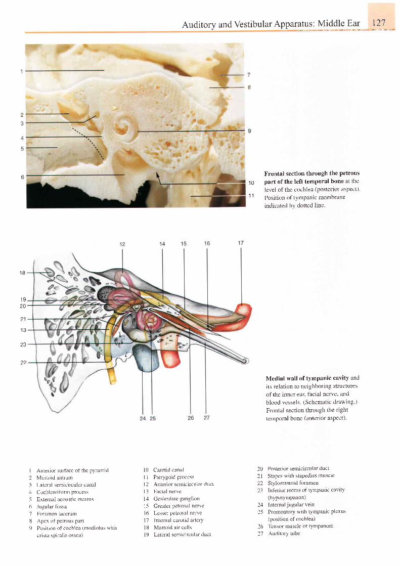
Auditory andVestibularApparatus: Middle Ear t27
s;.;
Anterior surface of the pyramid
Mastoid antrum
Lateral semicircular canal
Cochleariform process
E,xternal acoLlstic meatus
Jugular fossa
Foramen lacerum
,Apex of petrous part
Position of cochlea (modiolus with
crista spiralis ossea)
1 B
1 920
2 1
I J
23
22
23456189
e l
- ' . - 8
L0 Carotid canal
I I Pterygoid process
l2 Anterior semicircular duct
l3 Facial nerve
l4 Geniculate ganglion
15 Greater petrosal nerve
l6 Lesser petrosal nerve
17 Internal carotid artery
18 Mastoid a i r cel ls
19 Lateral semicircular duct
Frontal section through the petrous'10 part of the teft temporal bone at the
level of the cochlea (posterior aspect).11 Position of tympanic membrane
indicated by dotted line.
Medial wall of tymPanic cavitY and
its relation to neighboring structures
ofthe inner ear. facial nerve, and
blood vessels. (Schematic drawing.)
Frontal section through the right
temporal bone (anterior aspect).
20 Posterior semicircular duct2l Stapes with stapedius muscle
22 Stylomastoidforamen23 Inferior recess of tympanic cavity
(hypotympanon)24 Internal jugular vein25 Promontory with tymPanic Plexus
(position of cochlea)26 Tensor muscle of tympanum27 Auditory tube
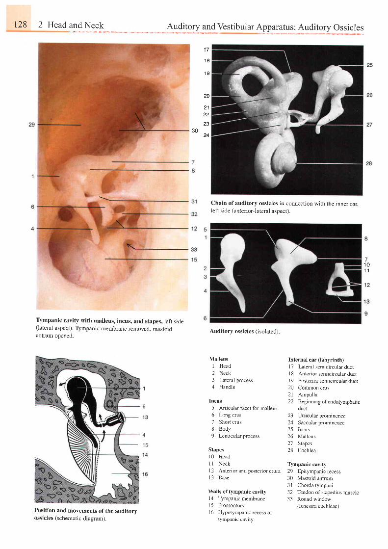
128 2 Head
1 7
' t8
1 9
30
20
2122
23
24
Chain of auditory ossicles in connection with the inner ear,left side (anterior-lateral aspect).
Auditory ossicles (isolated)
Tlmpanic cavity with malleus, incus, and stapes, left side(lateral aspect). Tympanic membrane removed, mastoidantrum ooened.
Position and movements of the auditoryossicles (schematic diagram).
1 01 1
Malleus1 Head2 Neck3 Lateral process4 Handle
Incus5 Articular facet for malleus6 Long crus7 Short crus8 Body9 Lenticular process
Stapes10 Headi I Neckl2 Anterior and posterior crura13 Base
Walls of tympanic cavity14 Tympanic membrane15 Promontoryl6 Hypotympanic recess o[
tympanic cavity
Internal ear (labyrinth)11 Lateral semicircular duct18 Anterior semicircular duct19 Posterior semicircularduct20 Common crus21 Ampulla22 Beginning of endolymphatic
duct23 Utricularprominence24 Saccular prominence25 Incus26 Malleus27 Stapes28 Cochlea
Tlmpanic cavity29 Epitympanic recess30 Mastoid antrum31 Chorda tympani32 Tendon of stapedius muscle33 Round window
(fenestra cochleae)
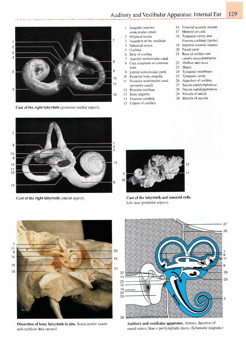
---*-3:*i*x,**J***ls*:mx*;**'-**-E-***:*
1
2
4
I Ampulla (anteriorsemicircular canal)
2 Elliptical recess3 Aqueduct of the vestibule4 Spherical recess5 Cochlea6 Base ofcochlea7 Anterior semicircular canal8 Crus commune or common
limb9 Lateral semicircular canal
l0 Posterior bony ampullaI 1 Posterior semicircular canal
(posterior canal)12 Fenestra cochleael3 Bony ampullal4 Fenestra vestibuliI 5 Cupula of cochlea
Extemal acoustic meatusMastoid air cellsTympanic cavity andfenestra cochleae (probe)
External acoustic meatusFacial canalBase of cochlea andcanalis musculotubariusMalleus and incusStapesTympanic membraneTympanic cavityAqueduct of cochleaSaccus endolymphaticusDuctus endolymphaticusMacula of utricleMacula of saccule
1 6t71 8
1 9202 l
22Z J
24252627282930
8
l e
Y
Cast of the right labyrinth (posterior-medial aspect).
Cast ofthe right labyrinth (lateral aspect).
Dissection of bony labyrinth in situ. Semicircular canalsand cochlear duct opened.
Cast of the labyrinth and mastoid cells.
Life size (posterior aspect).
Auditory and vestibular apparatus. Arrows: direction of
sound waves; blue = perilymphatic ducts. (Schematic diagram.)
1
1 1
1 4
1 11 0
z l
28
7I t
I1 4
20
1 5
211 8
1 9
781 1
I
29221 4Z J
1 2t o2425
30
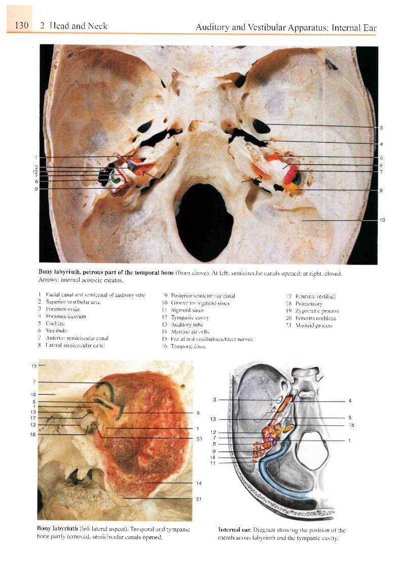
$'&. \
r30 2 Head and Neck Auditory and Vestibular Apparatus: Internal Ear
Bony labyrinth, petrous part of the temporal bone (from abovc). At left : sernicircular canals opened: at r ight. closedArrows: internal acoust ic meatus
I Facia l canal and senr icanal of audi tory tube2 Supe r i o r - ves t i bu l a l a r ca
3 Forametr ovale,l Foramen laccrurlr
5 Cochlea
6 Vest ibule
7 Arr ter ior- serr ic i rcular canall l Latcral sernic i rcular canal
1 6 -
7
'19
I 4
5I D
1
Bony labyrinth ( left lateral aspect). Tenrporal and tympanicbone palt ly removed. semicircular canals opened
Internal ear. Diagrant showing the posit ion of therrcmbranous Iabyrinth and the tvlr-rpanic cai, ' i ty.
5
67
I
2
7I
I
I J
1
7
8
I1 4. l I
l 01 lt 2t 3t '+l 5I t l
Poster ior sernic i rcuIar canel
Groove fbr s isntoic l s inus
Sigrnoic l s inr , rs
T1'nrpanic cal i t1,
Aucl torv tube
Mas to i d a i l c c l l s
Facia l and vest ibulocochlerr ncr \ csTemporal firssa
I 7 Fenest la vcst ibul i
l13 Promontory
I9 Zvgor-niLt ic p loccss
20 Fenestra cochleae
2 | Mastoid pr occss
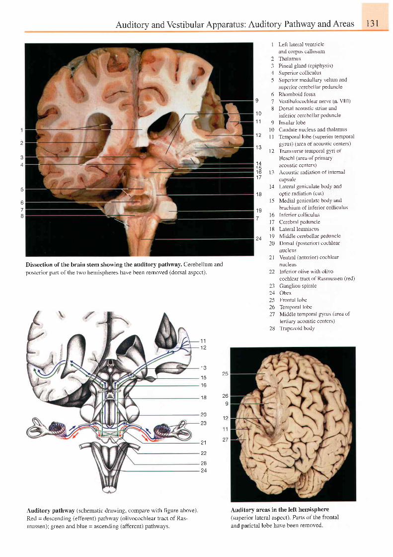
Auditory and Vestibular Apparatus: Auditory Pathway and Areas 131
6
o
78
stem showing the auditory pathway. Cerebellum andhemispheres have been removed (dorsal aspect).
Left lateral ventricleand corpus callosumThalamusPineal gland (epiphysis)
Superior colliculusSuperior medullary velum andsuperior cerebellar peduncleRhomboid fossaVestibulocochlear nerve (n. VIII)Dorsal acoustic striae andinferior cerebellar peduncleInsular lobeCaudate nucleus and thalamusTemporal lobe (superior temporalgyrus ) ( area of acoustic centers)Transverse temporal gyri ofHeschl (area of primaryacoustlc centers)Acoustic radiation of internalcapsuleLateral geniculate body andoptic radiation (cut)Medial geniculate body andbrachium of inferior colliculusInferior colliculusCerebral peduncleLateral lemniscusMiddle cerebellar peduncleDorsal (posterior) cochlearnucleusVentral (anterior) cochlearnucleusInferior olive with olivo-cochlear tract of Rasmussen (red)
Ganglion spiraleObexFrontal lobeTemporal lobeMiddle temporal gyrus (area oftertiary acoustic centers)Trapezoid body
2345
6
81 0
1 1 91 0
t 4 l l
t \ )t l
1 4
1 6 t 31 7
141 8
1 5
1 9t 6
' 1 1
l 8
24 1920
2 I
22
L J
24252627
28
Dissection of the brainposterior part of the two
1 11 2
I J
25I J
1 6
1 8 269
1 220Z J
2 1
22
2824
Auditory pathway (schematic drawing, compare with f,rgure above).Red = descending (efferent) pathway (olivocochlear tract ofRas-mussen); green and blue = ascending (afferent) pathways.
Auditory areas in the left hemisphere(superior lateral aspect). Parts of the frontaland parietal lobe have been removed.
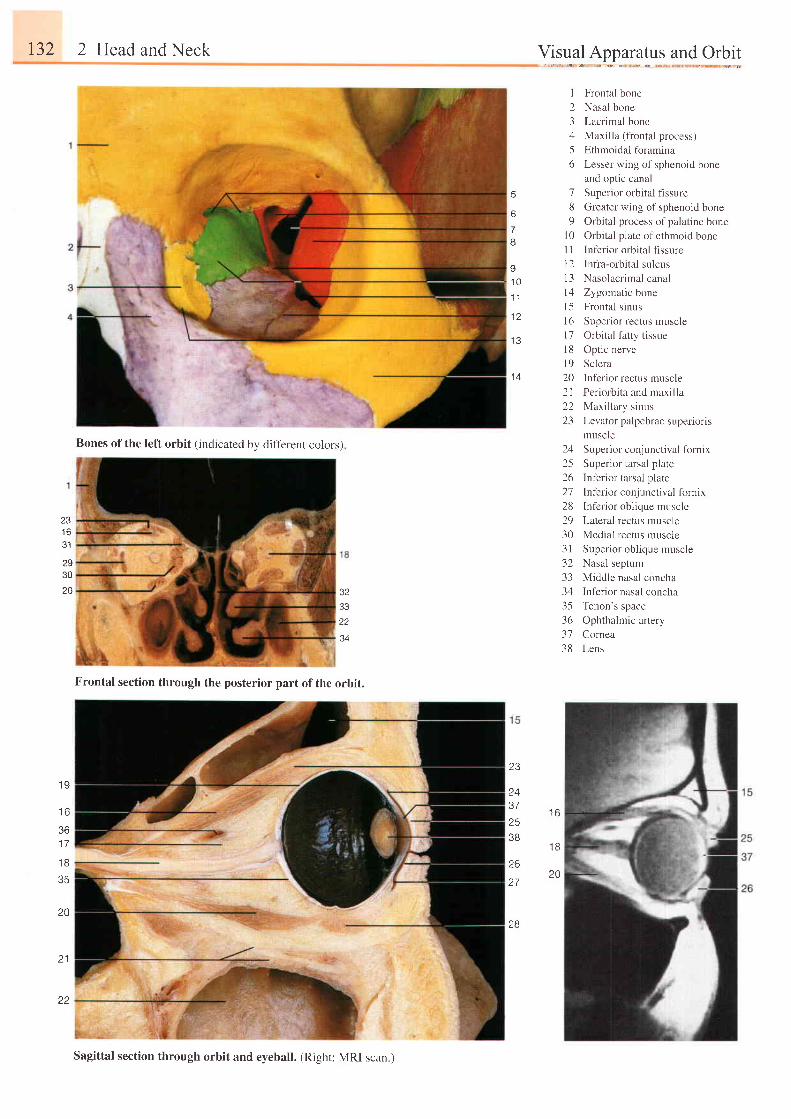
r32 2 Head and Neck
Frontal section through the posterior part of the orbit.
Visual Apparatus and Orbit
5
6
7I
I
2345o
'7
89
l 0l lt 21 3t 4l 51 6t 71 8l 9202 l22L-1
1 /
25 Superior tarsal plate
26 Inferior tarsal plate
27 Inf'erior conjunctival fornix
28 Infer ior obl ique muscle
29 Lateral rectus muscle
30 Medial rectus muscle
31 Super ior obl ique muscle
32 Nasal septum
33 Middle nasal concha
34 Inferior nasal concha
35 Tenon's space
36 Ophthalmic artery
37 Comea
38 Lens
Frontal bone
Nasal bone
Lacrimal bone
Maxilla (frontal process)
Ethmoidal fbramina
Lesser wing of sphenoid bone
and optic canal
Superior orbital flssure
Greater wing of sphenoid bone
Orbital process of palatine bone
Orbital plate of ethmoid bone
Inf'erior orbital flssure
Infra-orbital sulcus
Nasolacrimal canal
Zygomatic bone
Frontal sinus
Superior rectus muscle
Orbital fatty tissue
Optic nerve
Sclera
Inferior rectus muscle
Periorbita and maxilla
Maxillary sinus
Levator palpebrae superioris
muscle
Superior conj u nctival fornix
I1 0
1 1
1 A
1 0
1 6
2930
2B 323322
34
1 9
t o
361 7
l t '
35
20
21
23
2437253B
26
27
28
l r i
1 8
?0
Bones of the left orbit (indicated by difl'erent colors)
Sagittal section through orbit and eyeball. (Right: MRI scan.)
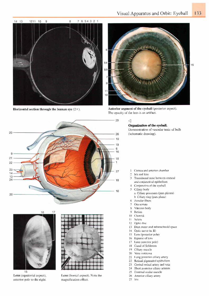
Visual Apparatus and Orbit: Eyeball 133
1 2 1 1 1 0 9
1 5Lens (equatorial aspect),anterior pole to the right.
Lens (frontal aspect). Note the
magnification effect.
1 Cornea and anterior chamber2 Iris and lens3 Transitional zone between corneal
and conjunctival epithel ium4 Conjunctiva ofthe eyeball5 Ciliary body
a Ciliary processes (pars plicata)b Ciliary ring (pars plana)
6 Zonular fibers7 Ora senata8 Vitreous body9 Retina
l0 ChoroidI I Scleral2 Optic disc13 Dura mater and subarachnoid space14 Optic nerve (n II)I 5 Lens (posterior pole)16 Equatoroflens17 Lens (anterior pole)18 Canal of Schlemm19 Ciliary muscle20 Vena vorticosa2l Long posterior ciliary artery22 Retinal pigmented epithelium23 Cenlral retinal artery and vein24 Short posterior ciliary arteries25 External ocular muscle26 Anterior ciliary artery27 Iris
Anterior segment of the eyeball (posterior aspect).The opacity of the lens is an artifact.
Organization of the eyeball.Demonstration of vascular tunic of bulb(schematic drawing).
25
1 0
1 96t o
22
zo1 41 324
1
27
1 8
1 0
Horizontal section through the human eye (2X).
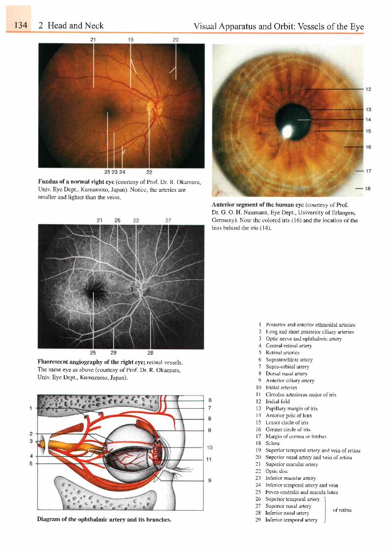
t34 2 Head and Neck Visual Apparatus and Orbit: Vessels of the Eye21
252324 22
Fundus of a normal right eye (courtesy of Prof. Dr. R. Okamura,Univ. Eye Dept., Kumamoto, Japan). Notice, the arteries aresmaller and liehter than the veins.
25 29 28
Fluorescent angiography of the right eyel retinal vessels.The same eye as above (courtesy of Prof. Dr. R. Okamura,Univ. Eye Dept., Kumamoto, Japan).
Anterior segment of the human eye (courtesy of Prof.Dr. G. O. H. Naumann, Eye Dept., University of Erlangen,Germany). Note the colored iris (16) and the location of thelens behind the iris (14).
2621
I2345678o
10l 1t 2I J
t 4151 6I7l 81 9202122z3
242526272829
Posterior and anterior ethmoidal arteriesLong and short posterior ciliary arteriesOptic nerve and ophthalmic arteryCentral retinal arteryRetinal arteriesSupratrochlear arterySupra-orbital arteryDorsal nasal arteryAnterior ciliary arteryIridial arteriesCirculus aneriosus major of irisIridial foldPupillary margin of irisAnterior pole of lensLesser circle of irisGreater circle of irisMargin of comea or limbusScleraSuperior temporal artery and vein of retinaSuperior nasal artery and vein of retinaSuperior macular arteryOptic discInferior macular arteryInferior temporal artery and vernFovea centralis and macula luteaSuperior temporal arterySuperior nasal arteryInferior nasal arteryInferior temporal arteryDiagram of the ophthalmic artery and its branches.
of retina
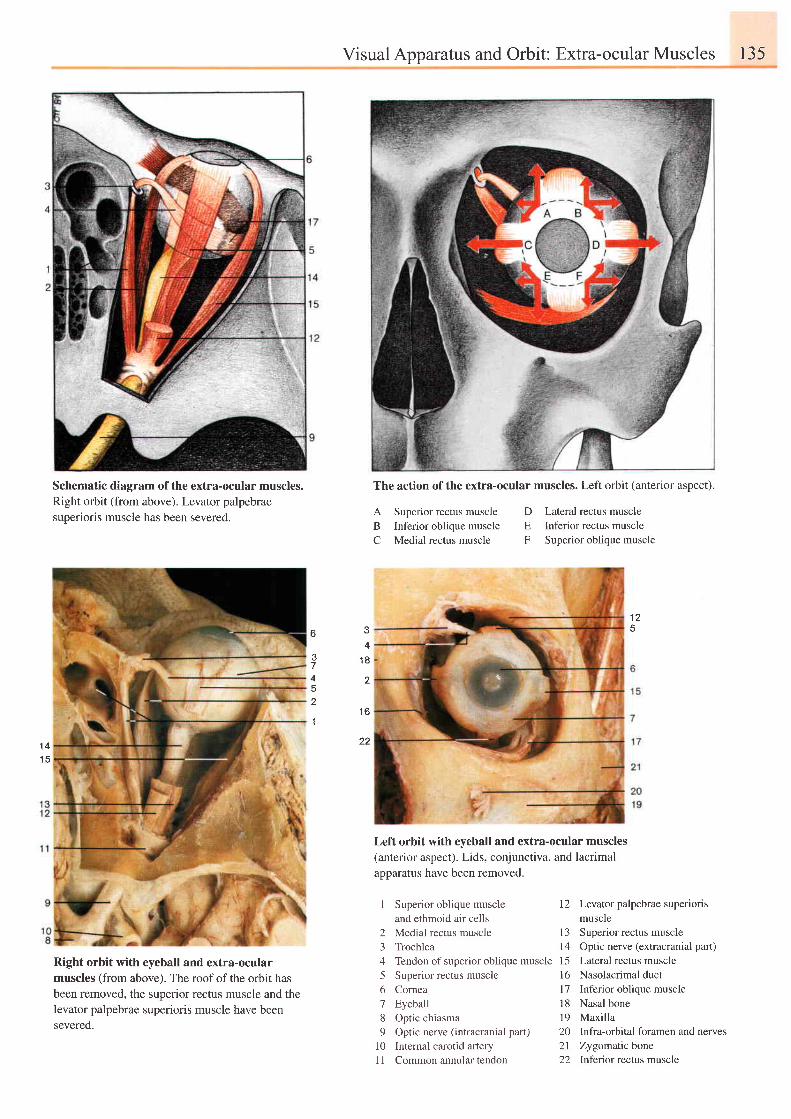
Visual Apparatus and Orbit: Extra-ocular Muscles 135
Schematic diagram of the extra-ocular muscles.Right orbit (from above). Levator palpebraesuperioris muscle has been severed.
Right orbit with eyeball and extra-ocularmuscles (from above). The roof of the orbit hasbeen removed, the superior rectus muscle and thelevator palpebrae superioris muscle have beensevered.
A Superior rectus muscleB Inferior oblique muscleC Medial rectus muscle
1 Superior oblique muscleand ethmoid air cells
2 Medial rectus muscle3 Trochlea4 Tendon of superior oblique muscle5 Superior rectus muscle6 Cornea7 Eyeball8 Optic chiasma9 Optic nerve (intracranial part)
10 Internal carotid artery11 Common annular tendon
Lateral rectus muscleInferior rectus muscleSuperior oblique muscle
I 2
1 3t4l 5l 6l 7r 81 9202 l22
Levator palpebrae superioris
muscle
Superior rectus muscle
Optic nerve (extracranial part)
Lateral rectus muscle
Nasolacrimal duct
Inferior oblique muscle
Nasal bone
Maxilla
Infra-orbital foramen and nerves
Zygomatic bone
Inferior rectus muscle
DEF
1 2
D
4
,|
4
1 8
2
1 6
22' t4t t
Left orbit with eyeball and extra-ocular muscles(anterior aspect). Lids, conjunctiva, and lacrimalapparatus have been removed.
The action of the extra-ocular muscles. Left orbit (anterior aspect).
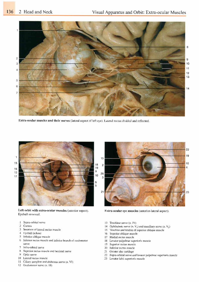
136 2 Head and Neck Visual Apparatus and Orbit: Extra-ocular Muscles
s1 0't1
1 21 3
1 4
1
1 5
Extra-ocular muscles and their nerves (lateral aspect of left eye). Lateral rectus divided and reflected.
1 5
1 8 21 9o
to 2o5
65
21
Left orbit with extra-ocular muscles (anterior aspect).Eyeball removed.
I Supra-orbital nerve2 Comea3 Insertion of lateral rectus muscle4 Eyeball (sclera)5 Inferior oblique muscle6 Inferior rectus muscle and inferior branch of oculomotor
nerve7 Infra-orbital nerve8 Superior rectus muscle and lacrimal nerve9 Optic nerve
10 Lateral rectus muscleI I Ciliary ganglion and abducens nerve (n. VI)12 Oculomotor nerve (n. III)
22
19
1 0
1 61 21-7
Extra-ocular eye muscles (anteriorJateral aspect).
13 Trochlear nerve (n. IV)14 Ophthalmic nerve (n. V1) and maxillary nerve (n. V2)15 Trochlea and tendon of superior oblique muscle16 Superior oblique muscle17 Medial rectus musclel8 Levator palpebrae superioris muscle19 Superior rectus muscle20 Inferior rectus muscle2l Greater alar cartilage22 Supra-orbital nerve and levator palpebrae superioris muscle23 Levatorlabii suoerioris muscle
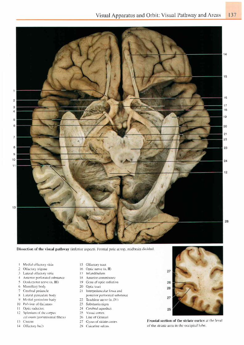
Visual Apparatus and Orbit: Visual Pathway and Areas 137
1 6
1 7
t 6
1 9
20
2 1
22
I
1 0. t 1
Dissection of the visual pathway (inf'erior aspect). Frontal pole at top, midbrain divided.
I Medial oltactory stria
2 Oltactory trigone
3 Latcral oltactory stria
4 Anterior perfbrated substance
5 Oculomotor nerve (n lll)
6 Mami l lary body
7 Cerebral peduncle
8 Latcral geniculate body
9 Medial gcniculate body
l0 Pulv inar of thalamus
I I Opt ic radiat ion
l2 Splenium of the corpus
callosum (commissural fi bers)
l 3 Cuneus
l4 Olfactory bulb
l5 Olfactory tract
16 Optic nerve (n. II)
l7 Infundibulum
l8 Anterior commissure
l9 Genu of optic radiation
20 Optic tract
2l Interpeduncular fbssa and
posterior perforated substancc
22 Trochlear nerve (n IV)
23 Substantia nigra
24 Cerebral aqueduct
25 Visual cortex
26 LineofCennari
27 Gyrus of striate cortex
28 Calcar ine sulcus
Frontal section of the striate cortex at the level
of the striate area in the occipital lobe.
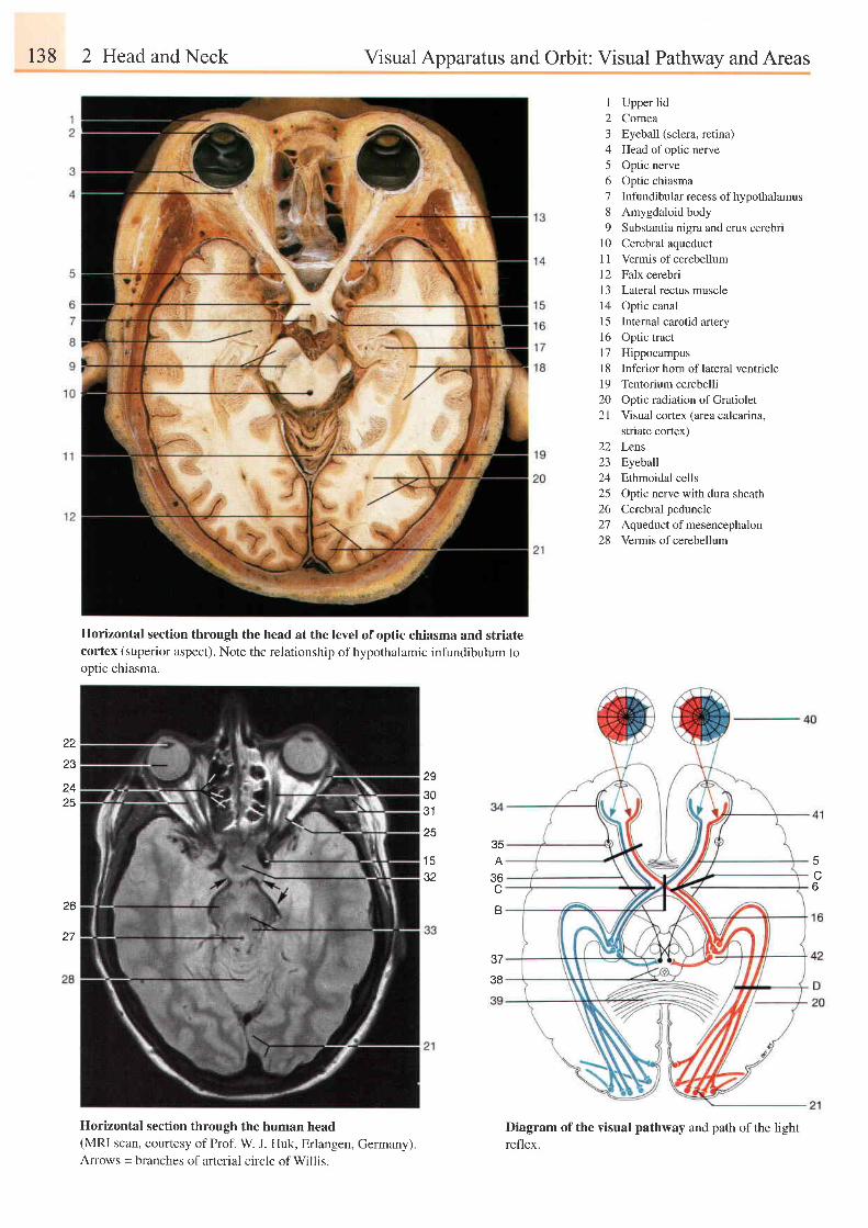
r38 2 Head and Neck Visual Apparatus and Orbit: Visual Pathway and Areas
22
23
t+
25
Horizontal section through the head at the level of optic chiasma and striatecortex (superior aspect). Note the relationship of hypothalamic infundibulum tooptic chiasma.
29
303'l
Horizontal section through the human head(MRI scan, courtesy of Prof. W J. Huk, Erlangen, Germany).Arrows = branches of arterial circle of Willis.
I Upper lid2 Cornea3 Eyeball (sclera, retina)4 Head ofoptic nerve5 Optic nerve6 Optic chiasma7 Infundibular recess of hypothalamus8 Amygdaloid body9 Substantia nigra and crus cerebri
10 Cerebral aqueduct11 Vermis of cerebellum12 Falx cerebri13 Lateral rectus muscle14 Optic canal15 Internal carotid artery16 Optic tract17 HippocampusI 8 Inferior horn of lateral ventriclel9 Tentorium cerebelli20 Optic radiation of Gratiolet2l Visual cortex larea calcarina.
striate cortex)22 Lens23 Eyeball24 Ethmoidal cells25 Optic nerve with dura sheath26 Cerebral peduncle27 Aqueductofmesencephalon28 Vermis of cerebellum
Diagram of the visual pathway and path of the lightreflex.
6
35A
JO
B
37
38
25
1 532
26

Visual Apparatus and Orbit: Visual Pathway andAreas 139
3-D reconstruction of the human visual system (MRI scan flash 40', courtesy of Prof.
W. J. Huk, University of Erlangen, Germany).
Dissection of brain stem in situ. Left hemisphere has been partly removed (compare with
MRI scan above).
29 Lateral rectus muscle30 Medial rectus muscle31 Temporalis muscle32 Hypophysis (pituitary gland)
33 Midbrain34 Ciliary nerves (long and
3536:'t I
3839404 l424344
46474849505 15253
555657
short)Ciliary ganglionOculomotor nerveAccessory oculomotornucleusColliculi of midbrainCorpus callosumVisual freldRetinaLateral geniculate bodyFrontal lobeCaudate nucleusMedial rectus muscleLateral rectus muscleSkinDiploe (skull)Dura materThalamusAnterior cerebral arteryCaudate nucleusFrontal sinusInternal capsuleLentiform nucleus (putamen)
HippocampusTemporal lobe of lefthemisphere
54
551 956
57
light reflex. If lesions of the chiasma destroy the crossing
fibers of the nasal portions of the retina (B), both tempo-
ral helds of vision are lost (bitemporal hemianopsia). If
both lateral angles of the chiasma are compressed (C),
the nondecussating fibers from the temporal retinae are
affected, resulting in loss of nasal visual fields (binasal
hemianopsia). Lesions posterior to the chiasma (D) (i. e.'
optic tract, lateral geniculate body, optic radiation, or
visual cortex) result in a loss of the entire opposite field
of vision (homonymous hemianopsia).
474S49
43
44
45
2522
23
r+o
50
21
4220
47
1 2
c l
52
In binocular vision the visual field (40) is projected uponportions of both retinae (blue and red in the drawing). Inthe chiasma the fibers from the two retinal portions arecombined to form the left optic tract. The fibers of the twoeyes remain separated from each other throughout theentire visual pathway up to their final termination in thecalcarine cortex (21). Injuries on the optic pathway pro-duce visual defects whose nature depends on the locationof the injury. Destruction of one optic nerve (A) producesblindness in the corresponding eye with loss of pupillary
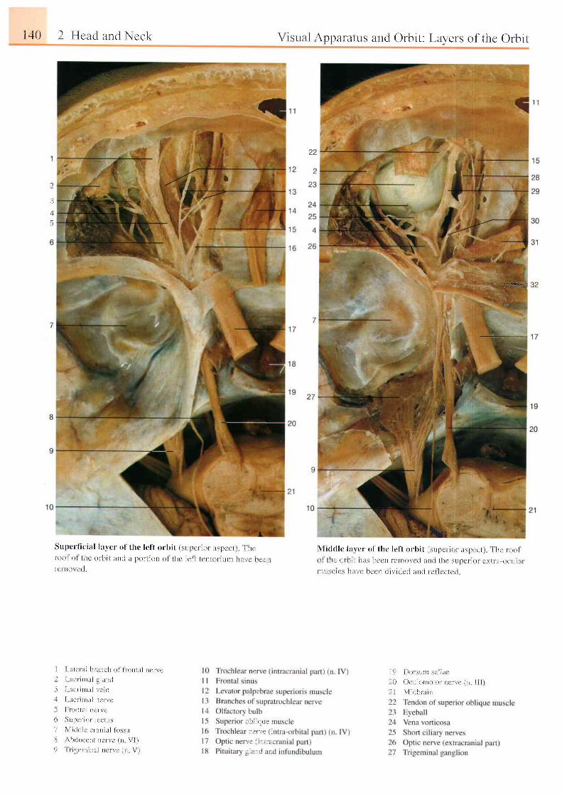
l .+0 I I l c l r r l l r r r t l \ ce k
' i a
Supcr l i c ia l lavcr u f th r : le t t r l rb i t i s r rpcr . ro r i rs l )c ! I 1 | l t (r t t ( ) l o 1 l l r c 0 r b r t a r t i l i t l ) o r l r ( ) l t r t l l h c l c n l c t ) l r ) t - l u t ) t l l . r i r . i r r . r . ll - C l l l ( ) \ C ( l
\ r : r r r l \ppara tus anc l Orb i t : Layers o f thc ( ) rb i t
1 1
-::.4b€ar.i;?
t;
tu;\ l i d t l l c i t rc r o t t l r c le f t o rb i l i sL rper l ( ' r i r sp , - ' ( ' t , i h t roo l, t l r t i j r i r ; t j I r s l t r : r ' n l o i l l o \ c r l . i l l r l l l t c \ U l ) c l i ( ) l L t i l i . t o c L r l l t r
r i r i r . i t : l t i l \ r ' i \ t a l t , i r r r i l c r - l l t n d r c l l c c t c J
' . j i ) i r r r L r n r ' t i i l t c' , i ) . r l , ' t r , , t , , r r r . : . . r , I I I
I \ l t t i i r r l r r r :
2
.i
. i
5
;r,{i,? :
{tl
#
i -
i
-1
S
9
I - : t t e t l t i h r i u t c l t o l I r - o r r l l r i n e r r r
I . r t r r r l l r l u l l r r r r l
I l r e r r l t . t i r e l t t
I l t t r r n t l t l t t c r r , '
l - l | ) l l t i l l D c f \ c
5 i l 1 ) t ' f t o l I c r l l t \
\ l l t l L l l t ' t r r r n l r l l i r s s i r
r \ h ( l L r e e l l l i l e r r c i I \ | i
I I t u c r n t n t r l i l e t \ a ( n \ l
l ) ( l l , i
\ i l r l i l I
r ' l , t t t r : , ,
-_+__--
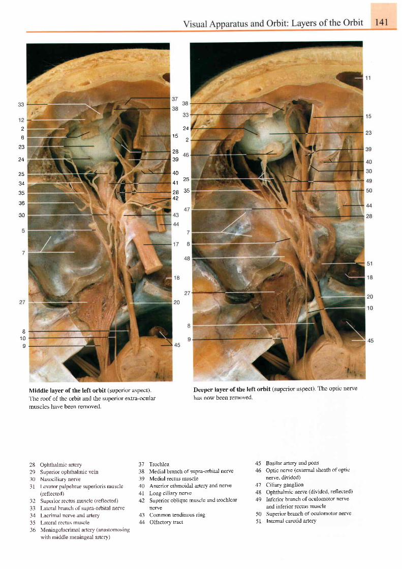
2o
241 5 2
23
24
25
34
.tc
36
30
Middle layer of the left orbit (superior aspect).The roof of the orbit and the superior extra-ocularmuscles have been removed.
28 Ophthalmic artery29 Superior ophthalmic vein30 Nasociliary nerve3l Levator palpebrae superioris muscle
(reflected)32 Superior rectus muscle (reflected)
33 Lateral branch of supra-orbital nerve34 Lacrimal nerve and artery35 Lateral rectus muscle36 Meningolacrimal artery (anastomosing
with middle meningeal artery)
40
41 25
37 Trochlea38 Medial branch of supra-orbital nerve
39 Medial rectus muscle40 Anterior ethmoidal artery and nerve41 Long ciliary nerve42 Superior oblique muscle and trochlear
nerve43 Common tendinous ring44 Olfactory tract
Deeper layer of the left orbit (superior aspect). The optic nerve
has now been removed.
45 Basilar artery and Pons46 Optic nerve (external sheath ofoptic
nerve. divided)47 Crliary ganglion48 Ophthalmic nerve (divided, reflected)
49 Inferior branch of oculomotor nerve
and inferior rectus muscle50 Superior branch of oculomotor nerve
51 Internal carotid artery
283e
28 3542
I1 0I
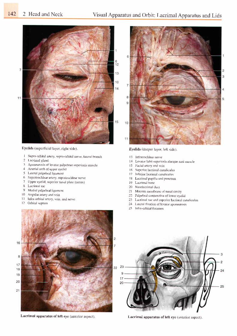
r42 2 Head and Neck Visual Apparatus and Orbit: Lacrimal Apparatus and Lids
61 2
8
1 71 8
1 9
20
2 1
Eyelids (superficial layer, right side).
1 Supra-orbital artery, supra-orbital nerve, lateral branch2 Lacrimal gland
3 Aponeurosis of levator palpebrae superioris muscle4 Arterial arch ofupper eyelid5 Lateral palpebral ligament6 Supratrochlearartery, supratrochlearnerve7 Upper eyelid, superior tarsal plate (tarsus)8 Lacrimal sac
9 Medial palpebral ligamentl0 Angularartery andveinll Infra-orbital artery, vein, and nervel2 Orbital seotum
Eyelids (deeper layer, left side).
| 3 Infratrochlear nerve
14 Levator labii superioris alaeque nasi muscle15 Facial artery and veinl6 Superior lacrimal canaliculusl7 Inf'erior lacrimal canaliculusl8 Lacrimal papilla and punctum
19 Lacrimal bone
20 Nasolacrimal duct
21 Mucous membrane of nasal cavity22 Palpebral conjunctiva oflower eyelid23 Lacrimal sac and superior lacrimal canaliculus24 Lateral fixation oflevator aponeurosis25 Infra-orbitalforamen
22 23
20
Lacrimal apparatus of left eye (anterior aspect) Lacrimal apparatus ofleft eye (anterior aspect).
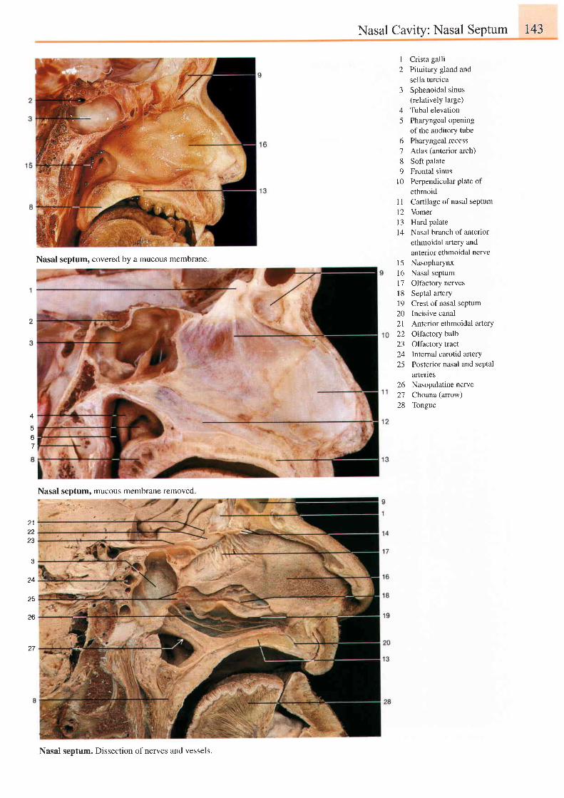
Nasal Cavity: Nasal Septum 143
3
45
678o
t 0
I
2Crista galliPituitary gland andsella turcicaSphenoidal sinus(relatively large)Tubal elevationPharyngeal openingof the auditory tubePharyngeal recessAtlas (anterior arch)Soft palateFrontal sinusPerpendicular plate ofethmoid
1 1 Cartilage of nasal septum12 Vomerl3 Hard palatel4 Nasal branch of anterior
ethmoidal artery andanterior ethmoidal nerveNasopharynxNasal septumOlfactory nervesSeptal arteryCrest of nasal septumIncisive canalAnterior ethmoidal arterYOlfactory bulbOlfactory tractInternal carotid arteryPosterior nasal and septalartenes
26 Nasopalatine nerve27 Choana (anow)28 Tongue
l 5l 61 7l 81 9202122z-)
2425
4
5
67
23
3
24
25
26
27
Nasal septum, covered by a mucous membrane.
Nasal septum, mucous membrane removed
Nasal septum. Dissection of nerves and vessels.
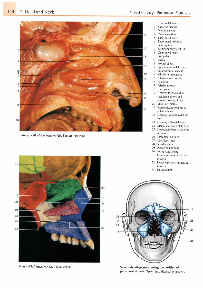
r44 2 Head and Neck Nasal Cavity: Paranasal Sinuses
I
23
56
789
1 01 l1 2l 3I 41 5l 6l 11 81 9
202 l
22
Sphenoidal sinusSuperior meatusMiddle meatusTubal elevationPharyngeal tonsilPharyngeal orifice ofauditory tubeSalpingopharyngeal foldPharyngeal recessSoft palateUvulaFrontal sinusSpheno-ethmoidal recessSuperior nasal conchaMiddle nasal conchaInferior nasal conchaVestibuleInferior meatusHard palateGrooves for the middlemeningeal artery andparietal bone (yellow)Maxillary hiatusPerpendicular process ofpalatine boneOpenings of ethmoidal aircells
23 Opening offrontal sinus24 Medial pterygoid plate (red)25 Horizontal plate ofpalatine
process26 Ethmoidal air cells27 Maxillary sinus28 Nasal septum29 fterygoid hamulus30 Nasal bone (white)31 Frontal process of maxilla
(violet)32 Palatine process ofmaxilla
(violet)33 Nasal atrium
34c
tt
I
Lateral wall ofthe nasal cavity. Septum removed.
23221 4
1 5
Schematic diagram showing the position ofparanasal sinuses. Openings indicated by arrows.
Bones of left nasal cavity, medial aspect.
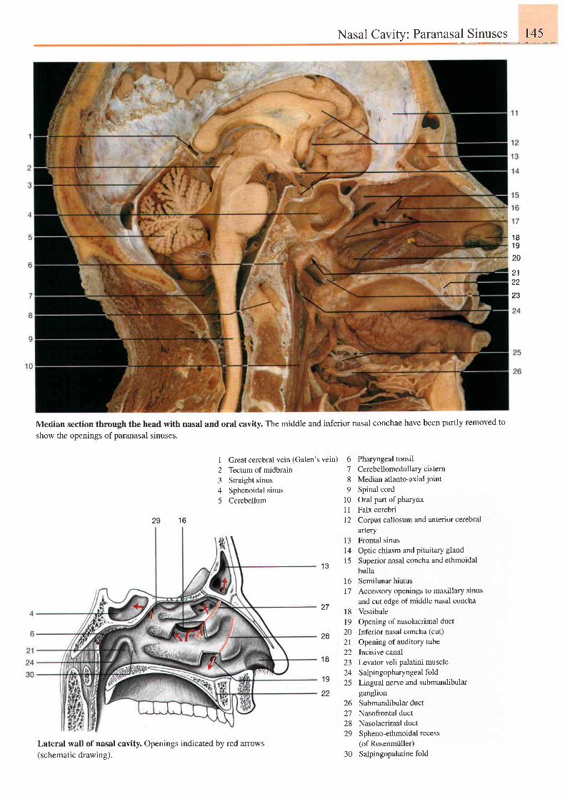
Nasal Cavity: Paranasal Sinuses 145
1 8192Q
2122
23
Median section through the head with nasal and oral cavity. The middle and inferior nasal conchae have been partly removed to
show the openings of paranasal slnuses.
1 629
I Great cerebral vein (Galen's vein)2 Tectum of midbrain3 Straight sinus4 Sphenoidal sinus5 Cerebellum
6 Pharyngeal tonsil7 Cerebellomedullarycistern8 Median atlanto-axial joint
9 Spinal cord10 Oral part of pharynx11 Falx cerebri12 Corpus callosum and anterior cerebral
artery13 Frontal sinus14 Optic chiasm and pituitary gland
15 Superior nasal concha and ethmoidalbulla
1 3
27
28
1 8
1 9
t 617
l8t 9202 l22232425
ganglion26 Submandibular duct27 Nasofrontal duct28 Nasolacrimal duct29 Spheno-ethmoidal recess
(of Rosenmiiller)30 Salpingopalatinefold
Semilunar hiatusAccessory openings to maxillary sinus
and cut edge of middle nasal conchaVestibuleOpening of nasolacrimal ductInferior nasal concha (cut)
Opening of auditory tubeIncisive canalLevator veli palatini muscleSalpingopharyngeal foldLineual nerve and submandibular
Lateral wall of nasal cavity. Openings indicated by red arrows(schematic drawing).
22
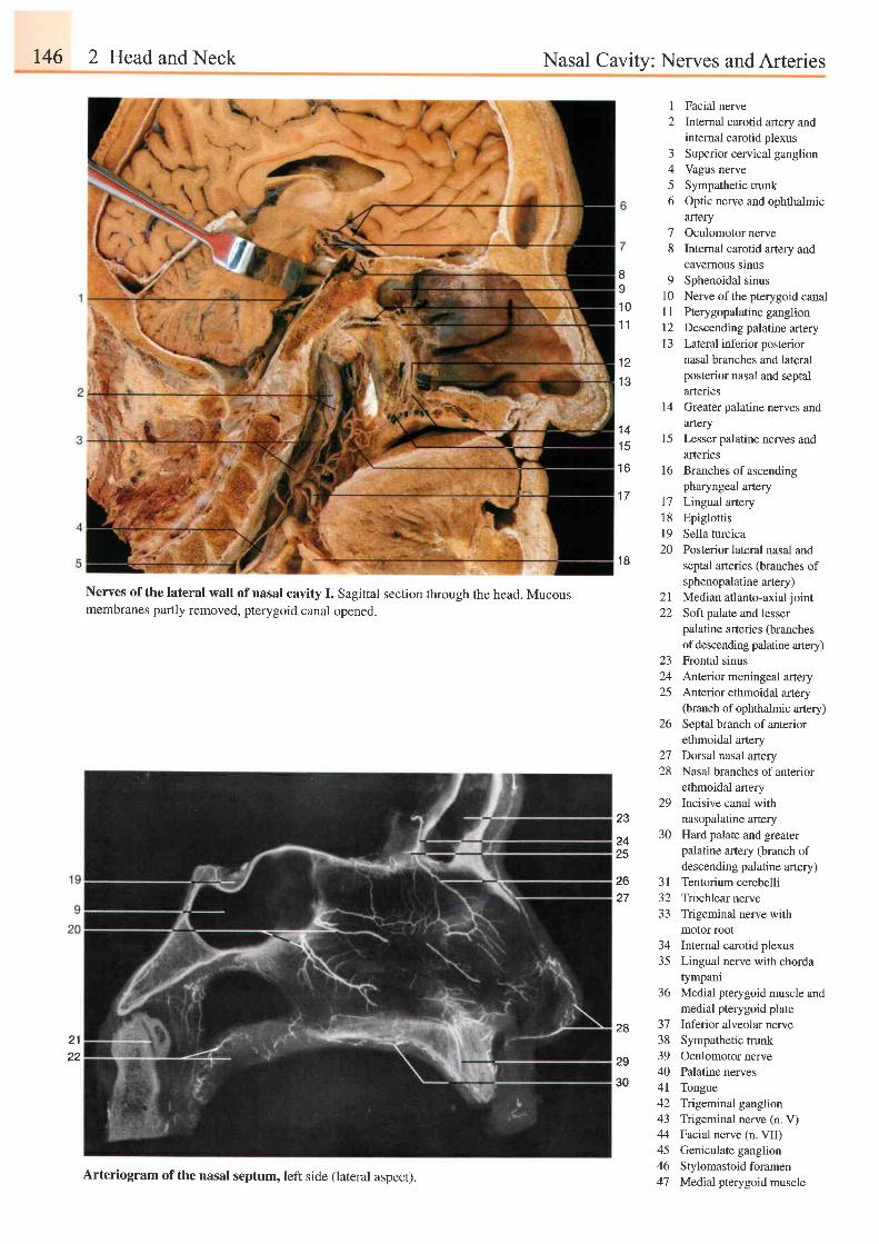
t46 2 Head and Neck Nasal Cavity: Nerves and Arteries
I
2
-1
^
56
Facial nerveInternal carotid artery andinternal carotid plexusSuperior cervical ganglionVagus nerveSympathetic trunkOptic nerve and ophthalmicarteryOculomotor nerveInternal carotid artery andcavemous srnusSphenoidal sinusNerve of the pterygoid canalPterygopalatine ganglionDescending palatine arteryLateral inferior posteriornasal branches and lateralposterior nasal and septalarteriesGreater palatine nerves andaneryLesser palatine nerves andartenesBranches of ascendingpharyngeal arteryLingual arteryEpiglottisSella turcicaPosterior lateral nasal andseptal arteries (branches ofsphenopalatine artery)Median atlanto-axial jointSoft palate and lesserpalatine arteries (branchesof descend.ing palatine artery)Frontal sinusAnterior meningeal arteryAnterior ethmoidal artery(branch of ophthalmic artery)Septal branch of anteriorethmoidal arteryDorsal nasal arteryNasal branches of anteriorethmoidal arteryIncisive canal withnasopalatine arteryHard palate and greaterpalatine artery (branch ofdescending palatine artery)Tentorium cerebelliTrochlear nerveTrigeminal nerve withmotor rootInternal carotid plexusLingual nerve with chordatympaniMedial pterygoid muscle andmedial pterygoid plateInferior alveolar nerveSympathetic trunkOculomotor nervePalatine nervesTongueTrigeminal ganglionTrigeminal nerve (n. V)Facial nerve (n. VII)Geniculate ganglionStylomastoid foramenMedial pterygoid muscle
II
1 0
1 1
78
91 0l l1 2t 3
1 21 3
l4
1 4t 5
1 6 1 6
1 7
1 8l 920
1 8
2122
Nerves of the lateral wall of nasal cavity I. sagittal section through the head. Mucousmembranes partly removed, pterygoid canal openec.
2 l22
za
2425
26
2.128
2923
?o2425
26 3 l27 32
33
3435
36
28 3738l o
2940
30 4l424344454647Arteriogram of the nasal septum, left side (lateral aspect).
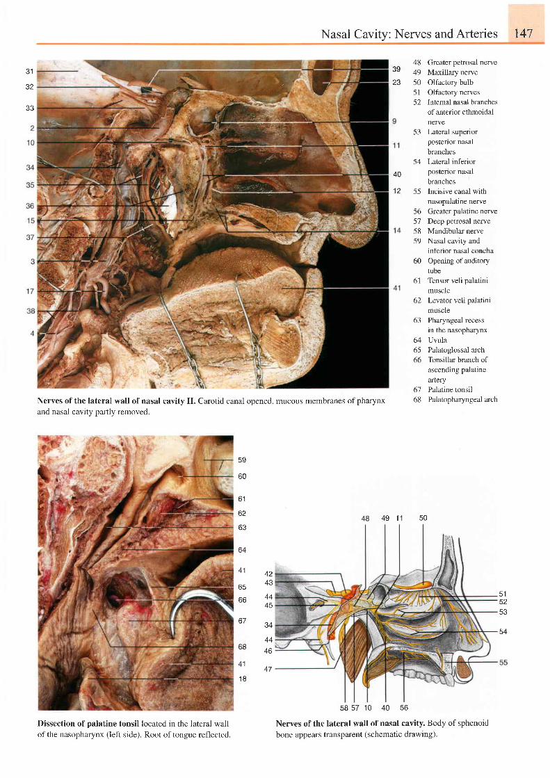
Nasal Cavity: Nerves and Arteries 147
? 1
32
J J
39
23
48 Greater petrosal nerve49 Maxillary nerve50 Olfactory bulb51 Olfactory nerves52 Intemal nasal branches
of anterior ethmoidalnerve
53 Lateral superiorposterior nasalbranches
54 Latenlinferiorposterior nasalbranches
55 Incisive canal withnasopalatine nerve
56 Greater palatine nerve57 Deep petrosal nerve58 Mandibular nerve59 Nasal cavity and
inferior nasal concha60 Openingofauditory
tube61 Tensor veli palatini
muscle62 Levator veli palatini
muscle63
646566
6768
Pharyngeal recessin the nasopharynxUvulaPalatoglossal archTonsillar branch ofascending palatinearteryPalatine tonsilPalatopharyngeal arch
40
t z
Nerves of the lateral wall of nasal cavity II. Carotid canal opened, mucous membranes of pharynx
and nasal cavity partly removed.
43
4445
50. t149
5S
60
D I
62
63
64
41
Jz+
44
46
67
bt'
41
65
bb515253
Dissection of palatine tonsil located in the lateral wall
ofthe nasopharynx (left side). Root oftongue reflected.
Nerves of the lateral wall of nasal cavity. Body of sphenoidbone appears transparent (schematic drawing).
58 57 10 40 56
1 n47
54
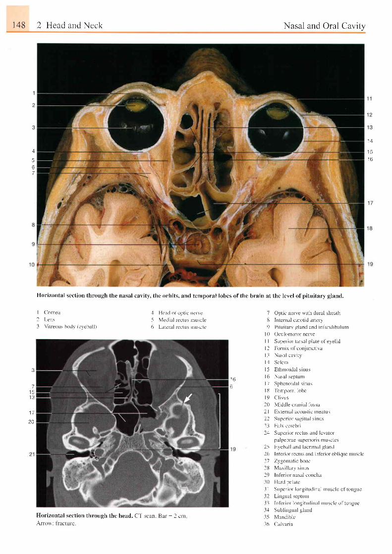
148 2 Head and Neck
I Cornca
2 Lens
3 Vi t reous body (eycbal l )
3
71 5I J
1 7
20
Nasal and Oral Cavity
A
567
1 4
t 3
l o
l
Horizontal section through the nasal cavity, the orbits, and temporal lobes ofthe brain at the level ofpituitary gland.
4 Hcad of opt ic nen'e
-5 Medial rectus nruscle
6 Latcral rectus nruscle
7 Optic nerve with dural shcath
lJ Internal carotid artery
9 Pi tu i tary g land and infundibulum
l0 Oculoniotor nerve
i t )
6
I I
t 2t 3l 1
t-5l 61 11 8t 9202 l22231 t
2526212829303 l3233343-536
Super ior tarsal p late of eyel id
Fornix of conjunctiva
Nasal cavi ty
Sclera
Ethmordal s inus
Nasal septum
Sphenoidal s inus
Temporal lobe
C l i vus
Middle crania l fossa
External acoustic lneatus
Superior sagittal sinus
Falx cerebr i
Sr.rpcrior rectus and lcvator
pulpebrae super ior is muselcs
Eyebal l and lacr imal g land
Inf'erior rectus and inferior oblique muscle
Zygonratic bone
Maxi l lary s inr . rs
Inferior nasal concha
Hard palate
Super ior longi tudinal muscle of tongue
Lingual septunr
Infer ior longi tudinal muscle of tongue
Subl ingual g land
Mandible
Calvar ia
Horizontal section through the head. CT scan. Bar = 2 crnArrow: fiacture
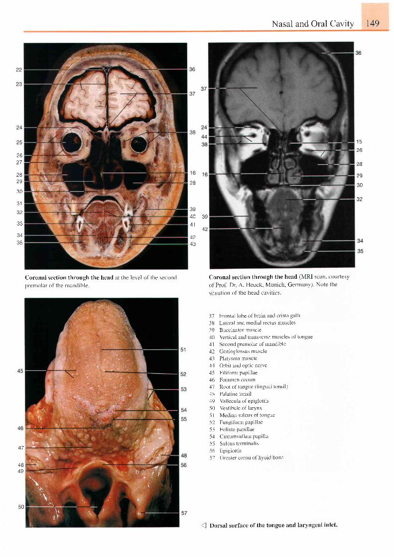
39
42
2627
2829
30
J I
32
33
Coronal section through the head at the level ofthe second
premolar of the mandible.
Nasal and Oral Cavity 149
Coronal section through the head (MRl scan, courtesy
of Prof. Dr A. Heuck, Munich, Germany). Note the
s i t ua t i on t r l t he head cav i t i e s .
37 Frontal lobe of brain and crista galli
38 Lateral and medial rectus muscles
39 Buccinator muscle
40 Vertical and transverse muscles of tongue
1l Second premolar of mandible
42 Genioglossus muscle;13 Platysma musclezl4 Orbit and optic nerve
45 Filiform papillae
46 Foramen cecum
47 Root of tongue ( l ingual tonsi l )
48 Palatine tonsil:19 Vallecula of epiglottis
.50 Vestibule of larynx
51 Median sulcus of tongue
-52 Fungifbrm papillae
53 Foliate papillae
5zl Circumval latepapi l la
55 Sulcus terminalis
56 Epiglot t is
-57 Greater cornu of hyoid bone
26
2B
29
30
32
3435
2B
39404 1
4243
4B49
{ Dorsal surface ofthe tongue and laryngeal inlet.
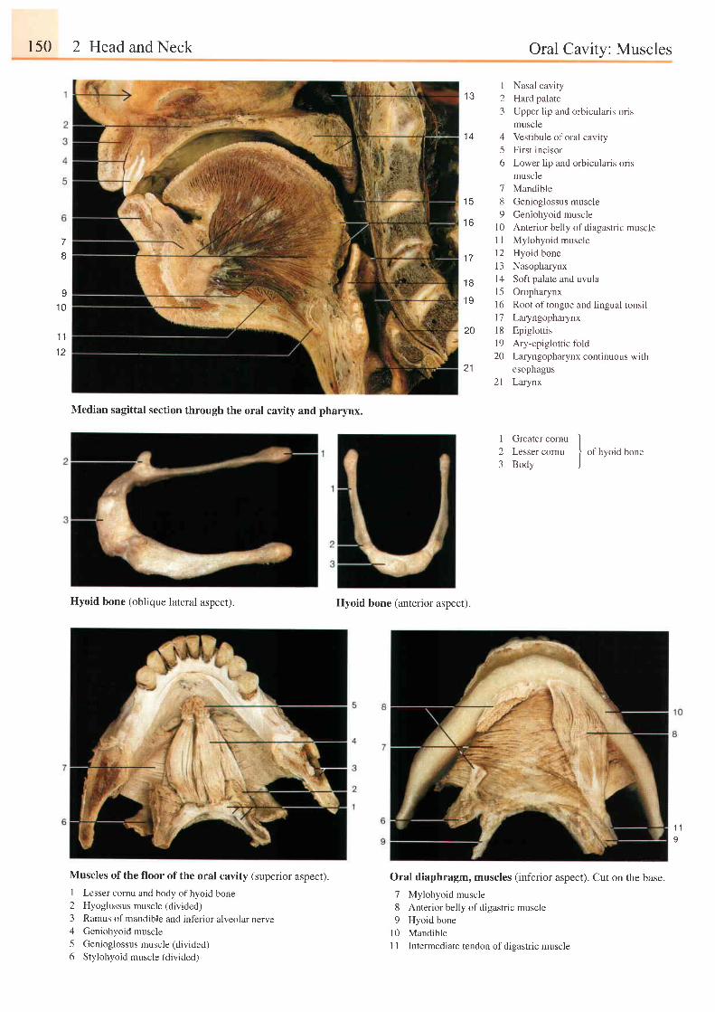
150 2 Head and Neck Oral Cavity: Muscles
7
8
I' t0
'l 1
1 2
1 3
1 4
t c
t o
1 7
20
21
1 B
1 9
I Nasal cavity2 Hard palate3 Upper lip and orbicularis oris
muscle4 Vestibule of oral cavity5 First incisor6 Lower l ip and orbicularis oris
muscle7 Mandible8 Genioglossus muscle9 Geniohyoid muscle
10 Anterior belly of diagastric muscleI I Mylohyoid muscle12 Hyoid bone13 Nasopharynx14 Soft palate and uvula15 Oropharynx16 Root of tongue and lingual tonsill7 Laryngopharynx18 Epiglottis19 Ary-epiglottic fold20 Laryngopharynx continuous with
esophagus21 Larynx
Median sagittal section through the oral cavity and pharynx.
Hyoid bone (oblique lateral aspect).
Muscles of the floor of the oral cavity (superior aspect).
1 Lesser cornu and body ofhyoid bone2 Hyoglossus muscle (divided)
3 Ramus of mandible and inferior alveolar nerve4 Geniohyoid muscle
5 Genioglossus muscle (divided)
6 Stylohyoid muscle (divided)
Hyoid bone (anterior aspect).
Oral diaphragm, muscles (inferior aspect). Cut on the base.
7 Mylohyoid muscle8 Anterior belly of digastric muscle9 Hyoid bone
l0 Mandible11 Intermediate tendon of dieastric muscle
I Greater cornu l2 Lesser cornu I of hyoid bone3 Body )
1 1o
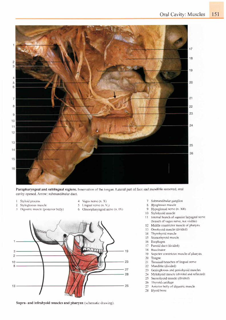
Oral Cavity: Muscles 151
1 7
1B
A
6
1
' t0
8
Parapharyngeal and sublingual regions. Innervation ofthe tongue. Lateral part offace and mandible removed, oral
cavity opened. Arrow: submandibular duct.
1 Styloid process2 Styloglossus muscle3 Digastric muscle (posterior belly)
4 Vagus nerve (n. X)5 Lingual nerve (n. V1)6 Glossopharyngeal nerve (n IX)
7 Submandibularganglion8 Hyoglossus muscle9 Hypoglossal nerve (n. XII)
10 Stylohyoid muscle1 1 Internal branch of superior laryngeal nerve
(branch of vagus nerve, not visible)l2 Middle constrictor muscle of pharynx13 Omohyoid muscle (divided)14 Thyrohyoid muscle15 Sternothyroid muscle16 Esophagus17 Parotid duct (divided)18 Buccinator19 Superior constrictor muscle of pharynx20 Tongue21 Terminal branches oflingual nerve22 Mandible(divided)23 Genioglossus and geniohyoid muscles24 Mylohyoid muscle (divided and reflected)25 Sternohyoid muscle (divided)26 Thyroid cartilage27 Anterior belly of digastric muscle28 Hyoid bone
Supra- and infrahyoid muscles and pharynx (schematic drawing).
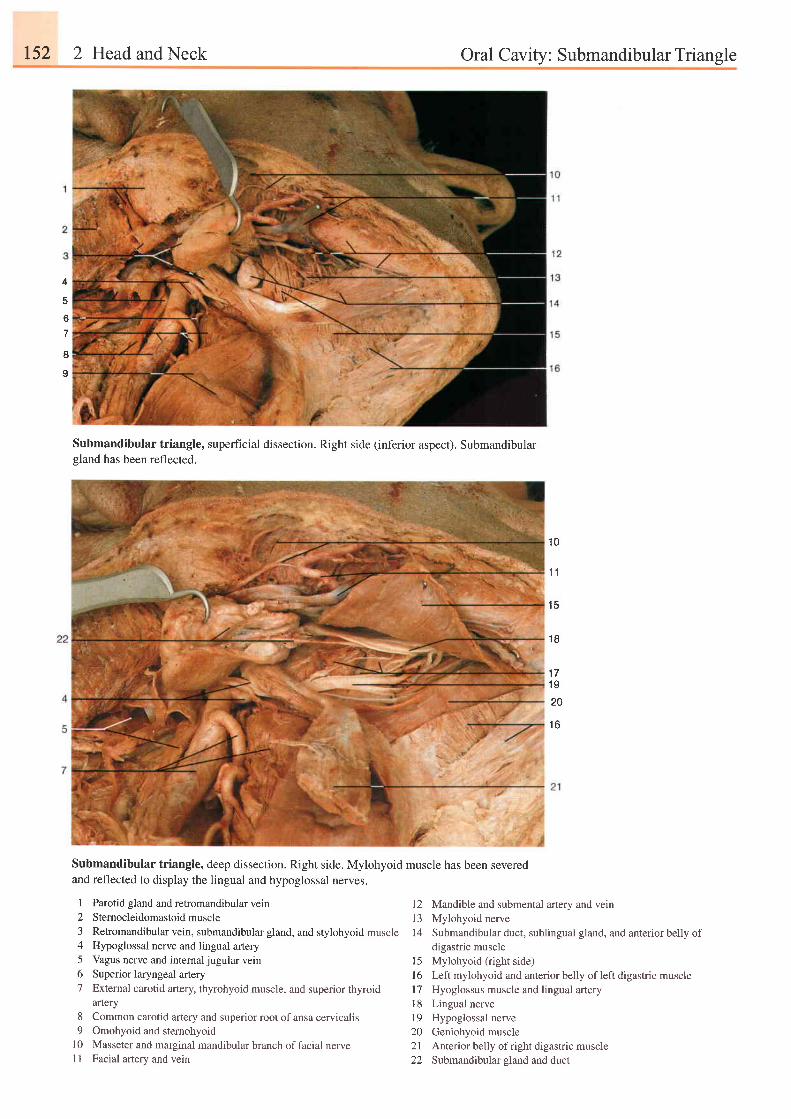
152 2 Head and Neck Oral Cavitv: Submandibular Triansle
4
6
8
I
Submandibular triangle, superfrcial dissection. Right side (inferior aspect). Submandibulargland has been reflected.
Submandibular triangle, deep dissection. Right side. Mylohyoid muscle has been severedand reflected to display the lingual and hypoglossal nerves.
1 0
1T
1 5
' Q
1 71 S
zv
1 6
I Parotid gland and retromandibular vein2 Stemocleidomastoidmuscle3 Retromandibular vein, submandibular gland, and stylohyoid4 Hypoglossal nerve and lingual artery5 Vagus nerve and internal jugular vein6 Superior laryngeal artery
Mandible and submental artery and veinMylohyoid nerveSubmandibular duct, sublingual gland, and anterior bel1y ofdigastric muscleMylohyoid (right side)Left mylohyoid and anterior belly of left digastric muscleHyoglossus muscle and lingual arteryLingual nerveHypoglossal nerveGeniohyoid muscleAnterior belly of right digastric muscleSubmandibular sland and duct
1 2I J
muscle 14
7 External carotid artery, thyrohyoid muscle, and superior thyroidartery
8 Common carotid artery and superior root of ansa cervicalis9 Omohyoid and stemohyoid
10 Masseter and marginal mandibular branch of facial nervell Facial artery and vein
1 5l 6l71 8t9202122
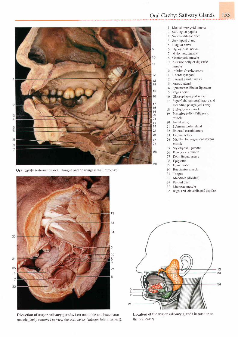
I Medial pterygoid muscle2 Sublingual papilla3 Submandibular duct4 Sublingual gland
5 Lingual nerve6 Hypoglossal nerve7 Mylohyoid muscle8 Geniohyoid muscle9 Anterior bel ly of digastr ic
muscle10 Inf'erior alveolar nerveI I Chorda tympani12 Internal carotid artery13 Parotid gland14 Sphenomandibular ligamentl5 Vagus nervel6 Glossopharyngeal nervel7 Superficial temporal artery and
ascending pharyngeal arteryl8 Styloglossus muscle| 9 Posterior bel ly ol 'digastr ic
muscle20 Facial artery21 Submandibular gland22 External carotid artery23 Lingual artery24 Middle pharyngeal constrictor
muscle25 Stylohyoid ligament26 Hyoglossus muscle27 Deep lingual arterY28 Epiglottis29 Hyoid bone30 Buccinator muscle3l Tongue32 Mandible (divided)
33 Parotid duct34 Masseter muscle35 Right and left sublingual papillae
1 2
1 31 4
1 71 81 9202 122ZJ
o t
zo
Oral cavity (internal aspect). Tongue and pharyngeal wall removed.
Dissection of major salivary glands. Left mandible and buccinator
muscle pafily removed to view the oral cavity (inferior lateral aspect).
1 3
33
34
20
J
z l I J
33
47
Location of the major salivary glands in relation to
the oral cavity.
2 1
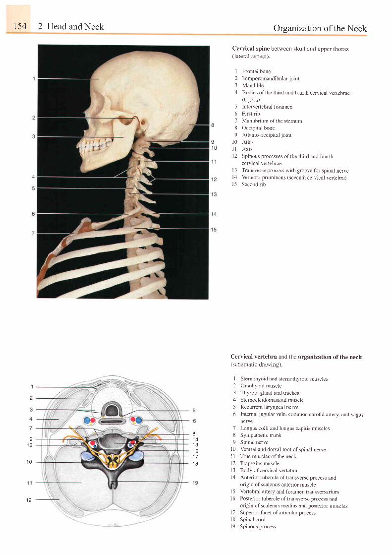
t54 2 Head and Neck
r\ ,
Organization of the Neck
91 0
Cervical spine between skull and upper thorax(lateral aspect).
I Frontal bone2 Temporomandibularjoint3 Mandible4 Bodies ofthe third and fourth cervical vertebrae
(C:, C+)5 Intervertebral foramen6 First rib7 Manubrium of the stemum8 Occipital bone9 Atlanto-occipital joint
10 Atlasl l Ax isl2 Spinous processes ofthe third and lburth
cervical vertebraeI 3 Transverse process with groove for spinal nervel4 Vertebra prominens (seventh cervical vertebra)15 Second rib
Cervical vertebra and the organization of the neck(schematic drawing).
I Sternohyoid and sternothyroid muscles2 Omohyoid muscle
3 Thyroid gland and trachea
4 Sternocleidomastoidmuscle
5 Recurrent laryngeal nerve
6 Internaljugular vein, common carotid artery, and vagusnerve
7 Longus colli and longus capitis muscles8 Sympathetic trunk
9 Spinal nerve
I 0 Ventral and dorsal root of spinal nervel1 True muscles of the neck
l2 Trapezius muscle
13 Body ofcervical vertebra
l4 Anterior tubercle of transverse process andorigin of scalenus anterior muscle
l5 Vertebral artery and foramen ffansversarium16 Posterior tubercle of transverse process and
origin of scalenus medius and posterior musclesl7 Superior facet of articular process
l8 Spinal cord
I 9 Spinous process
I1 6
81 4l ?
I J'17
1 81 0
,//,.,
12,, .,t,,
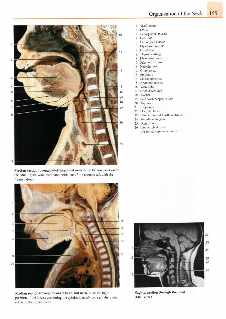
Organtzation of the Neck 155
{ Q
1 4
1 6
1 7
1 8
1 Nasal septum2 Uvula3 Genioglossus muscle4 Mandible5 Geniohyoid muscle6 Mylohyoid muscle7 Hyoid bone8 Thyroid cartilage9 Manubrium sternt
10 Sphenoidal sinusI I Nasopharynx12 Oropharynx13 Epiglottisl4 Laryngopharynxl5 Arytenoid muscle16 Vocal fold17 Cricoid cartilage18 Tracheal9 Left brachiocephalic vein
20 Thymus21 Esophagus22 Occipital lobe23 Cerebellum and fourlh ventricle24 Medulla oblongata25 Dens of axis26 Intervertebraldiscs
of cervical vertebral column
Median section through adult head and neck. Note the low position of
the adult larynx when compared with that of the neonate (cf. with the
figure below).
Median section through neonate head and neck. Note the highposition of the larynx permitting the epiglottis nearly to reach the uvula(cf. with the figure above).
1 3
1 5
1 7 22
Z J
24
25
Sagittal section through the head.(MRI scan.)
f6
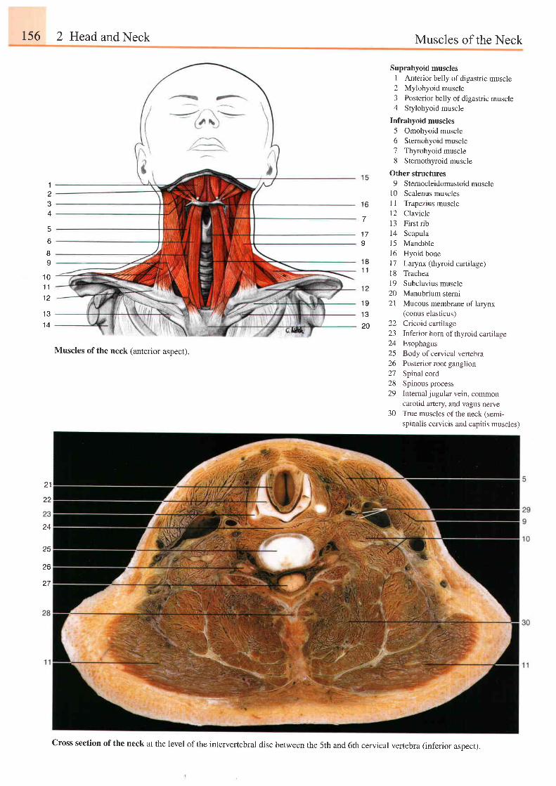
156 2 Head and Neck Muscles of the Neck
1 6
1 7o
1 81 1
1234
o
Suprahyoid musclesI Anterior belly of digastric muscle2 Mylohyoid muscle3 Posterior belly of digastric muscle4 Stylohyoid muscle
Infrahyoid muscles5 Omohyoid muscle6 Stemohyoid muscle7 Thyrohyoid muscle8 Sternothyroid muscle
Other structures9 Stemocleidomastoidmuscle
10 Scalenus musclesI I Trapezius muscle12 Clavicle13 First ribl4 Scapula15 Mandible16 Hyoid bone17 Larynx (thyroid cartilage)18 Trachea19 Subclavius muscle20 Manubrium sterni21 Mucous membrane of larynx
(conus elasticus)Cricoid cartilageInferior horn of thyroid cartilageEsophagusBody of cervical vertebraPosterior root ganglionSpinal cordSpinous processInternal jugular vein, commoncarotid artery, and vagus nerveTrue muscles of the neck (semi-spinalis cervicis and capitis muscles)
I9
1 01 11 2
1 Q
1 4
1 2
1 9
1 3
20
Muscles of the neck (anterior aspect).
2223. A
2526272829
30
z l
22
24
25
26
Z T
Cross section of the neck at the level of the intervertebral disc between the 5th and 6th cervical vertebra (inferior aspect).
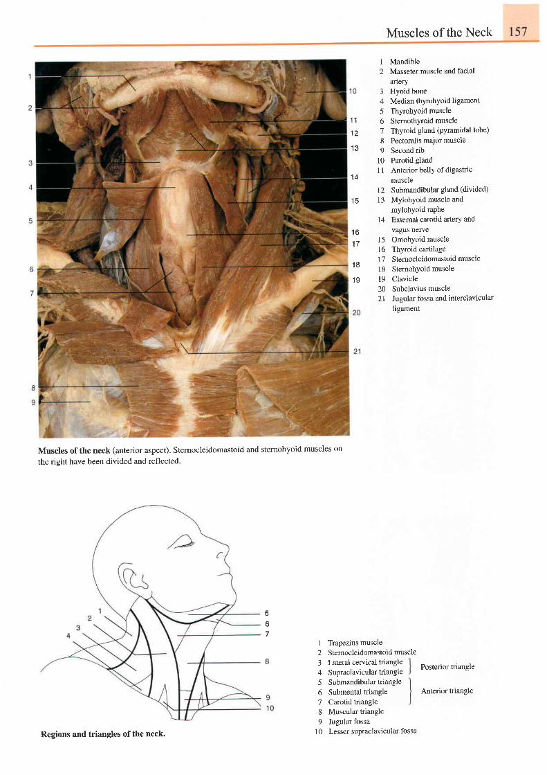
1 1
1 2
1 3
1 4
1 5
1 61 7
1 8
1 9
I
2
34
6789
t0n
Muscles of the Neck
MandibleMasseter muscle and facial
arteryHyoid boneMedian thyrohyoid ligamentThyrohyoid muscleSternothyroid muscleThyroid gland (pyramidal lobe)
Pectoralis major muscleSecond ribParotid glandAnterior belly of digastricmuscle
12 Submandibular gland (divided)
13 Mylohyoid muscle and
mylohyoid raphe14 Extemal carotid artery and
vagus nerveOmohyoid muscleThyroid cartilageSternocleidomastoid muscle
Sternohyoid muscleClavicleSubclavius muscleJugular fossa and interclavicularligament
15t61718t9202 l
Muscles of the neck (anterior aspect). Stemocleidomastoid and sternohyoid muscles on
the right have been divided and reflected.
5
67
I
234
6789
10
Trapezius muscleSternocleidomastoid muscleLateral cervical triangleSupraclavicular triangleSubmandibular triangleSubmental triangleCarotid triangleMuscular triangleJugular fossa
Posterior triangle
Anterior triangle
Regions and triangles of the neck. Lesser supraclavicular fossa
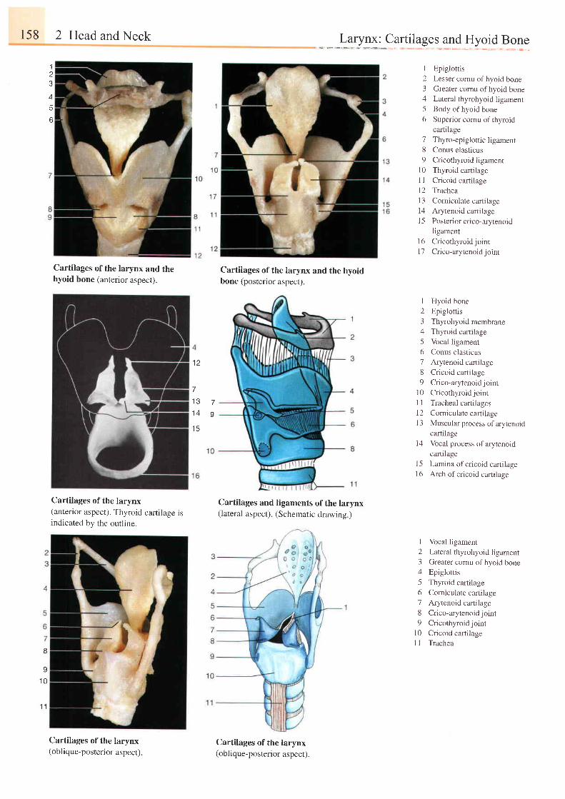
158 2 Head and Neck
Cartilages of the larynx and thehyoid bone (anterior aspect).
Cartilages of the larynx(anterior aspect). Thyroid cartilage isindicated by the outline.
I
q
1 0
1 1
Cartilages of the larynx(obl ique-posterior aspect ).
..".."L"?:y*:_c""*:1"'.k_e:_l?:+5y:i"{..P_q":1
a
4
o
Cartilages of the larynx and the hyoidbone (posterior aspect).
Cartilages and ligaments of the larynx(lateral aspect). (Schematic drawing.)
Cartilages of the larynx(obl ique-posterior aspect ).
I Epiglottis2 Lesser cornu ofhyoid bone3 Greater comu of hyoid bone4 Lateral thyrohyoid ligamenr5 Body ofhyoid bone6 Superior cornu ofthyroid
cartilage7 Thyro-epiglottic ligament8 Conus elasticus9 Cricothyroidligament
l0 Thyroid cartilageI I Cricoid cartilagel2 Tracheal3 Comiculatecartilagel4 Arytenoid cartilage15 Posteriorcrico-arytenoid
ligament16 Cricothyroidjoint17 Crico-arytenoidjoint
1 Hyoid bone2 Epiglottis3 Thyrohyoid membrane4 Thyroid cartilage5 Vocal ligament6 Conus elasticus7 Arytenoid cartilage8 Cricoid cartilage9 Crico-arytenoidjoint
10 CricothyroidjointI I Tracheal cartilages12 Corniculate cartilageI 3 Muscular process of arytenoid
cartilagei4 Vocal process ofarytenoid
cartilage15 Lamina of cricoid cartilageI 6 Arch of cricoid cartilage
I Vocal ligament
2 Lateral thyrohyoid ligament3 Greater cornu ofhyoid bone4 Epiglottis
5 Thyroid cartilage
6 Corniculatecartilage
7 Arytenoid cartilage
8 Crico-arytenoidjoint
9 Cricothyroidjoint
l0 Cricoid cartilage
I 1 Trachea
1 2
7
1 3
1 4
t 3
7o
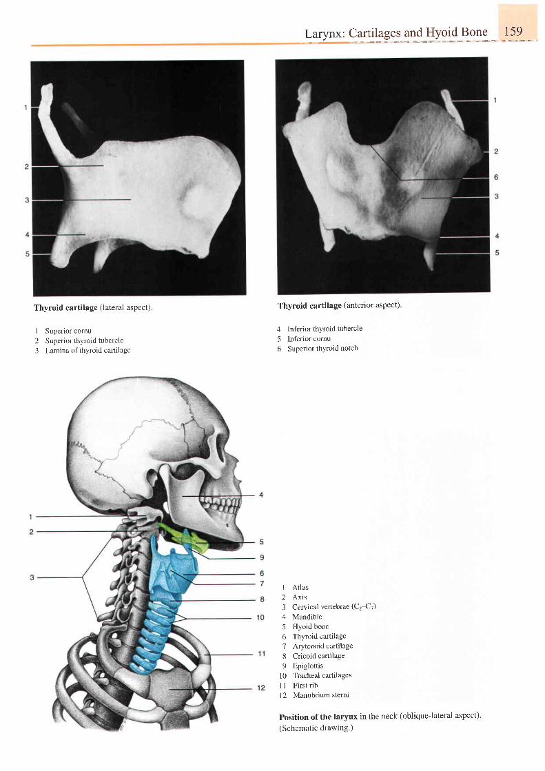
Larynx: Cartilages and Hyoid Bone 159
Thyroid cartilage (lateral aspect).
1 Superior comu2 Superior thyroid tubercle3 Lamina of thyroid canilage
Thyroid cartilage (anterior aspect).
4 Inferior thyroid tubercle
5 Inferior cornu6 Superior thyroid notch
I Atlas2 Axis3 Cervical vertebrae (Ct-tr)
4 Mandible5 Hyoid bone6 Thyroid cartilage7 Arytenoid cartilage8 Cricoid cartilage9 Epiglottis
10 Tracheal cartilages1 1 First ribl2 Manubrium sternt
Position of the larynx in the neck (oblique-lateral aspect)'
(Schematic drawing.)
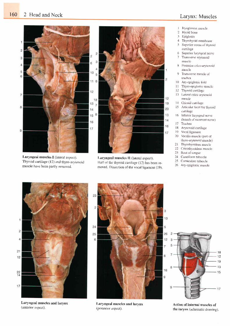
160 2 Head and Neck Larynx: Muscles
I Hyoglossus muscle2 Hyoid bone3 Epiglottis4 Thyrohyoid membrane5 Superior cornu ofthyroid
cartilage6 Superior laryngeal nerve7 Transverse arytenoid
muscle8 Posteriorcrico-arytenoid
muscle9 Transverse muscle of
trachea10 Ary-epiglottic foldl1 Thyro-epiglottic musclel2 Thyroid cartilagel3 Lateral crico-arytenoid
musclet4 Cricoid cartilage[5 Articularfacet forthyroid
cartilageInf'erior laryngeal nerve(branch of recurrent nerve)TracheaArytenoid caftilageVocal ligamentVocalis muscle (part ofthyro-arytenoid muscle)Thyrohyoideus muscleCricothyroideus muscleRoot of tongueCuneiform tubercleCorniculate tubercleAry-epiglottic muscle
Laryngeal muscles I (lateral aspect).Thyroid cartilage (12) and thyro-arytenoidmuscle have been parlly removed.
Laryngeal muscles and larynx(anterior aspect).
Laryngeal muscles II (lateral aspect).Half of the thyroid cartilage (12) has been re-moved. Dissection of the vocal ligament (19)
Laryngeal muscles and larynx(posterior aspect).
1 6
1 7l 81 920
2 l2223242526
1 81 2
1 9
1 3
1 5zz1 4
Action of internal muscles ofthe larynx (schematic drawing).
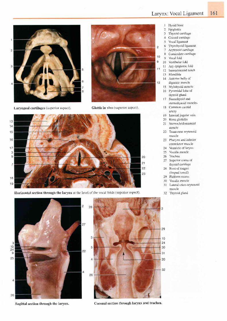
Larynx: Vocal Ligament 161
Laryngeal cartilages (superior aspect) Glottis in vivo (superior aspect).
12345o78o
1 0l 1l 21 314
1 51 6
t'7
l 8
Hyoid boneEpiglottisThyroid cartilageCricoid cartilageVocal ligamentThyrohyoid ligamentArytenoid cartilageCorniculate cartilageVocal foldVestibular foldAry-epiglottic foldInterarytenoid notchMandibleAnterior belly ofdigastric muscleMylohyoid musclePyramidal lobe ofthyroid glandSternohyoid andsternothyroid musclesCommon carotidartery
19 Internaljugularvein20 Rima glottidis21 Sternocleidomastoid
muscle22 Transversearytenoid
muscle23 Pharynx and inferior
constrictor muscle24 Ventricle of larynx25 Vocalis muscle26 Trachea27 Superior comu of
thyroid cartilage28 Root of tongue
(lingual tonsil)29 Piriform recess30 Vocalis muscle31 Lateralcrico-arytenoid
muscle32 Thyroid gland
1 0
I
1 1
1 3. A
1 5
t o
t t
I
7
1 8
1 9
20
21
22
23
31
20
?
1 024
25
29
1 02430
Horizontal section through the larynx at the level ofthe vocal folds (supenor aspect).
Sagittal section through the larynx. Coronal section through larynx and trachea.
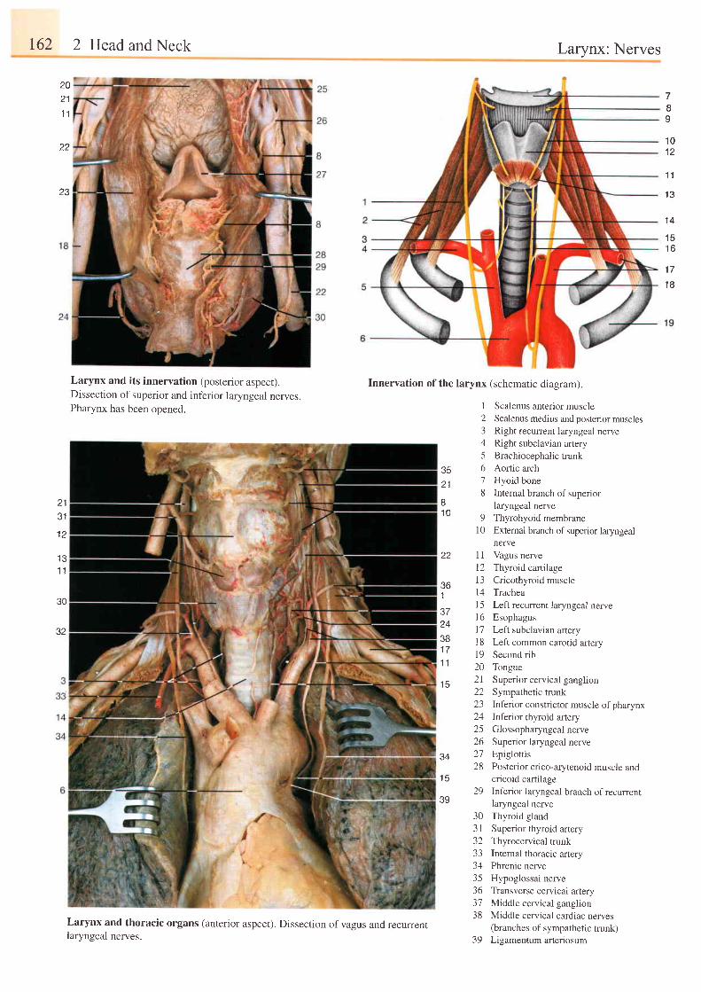
r62 2 Head and Neck Larynx: Nerves
201 l
1 1
1 1
23
1 01 2
1 1
1 3
7I9
1 4
1 51 6
1 7
1 8
Larynx and its innervation (posterior aspect).Dissection of superior and inferior laryngeal nerves.Pharynx has been opened.
Innervation of the larynx (schematic diagram).
I
2345
3 5 621
'7
8
Scalenus anterior muscleScalenus medius and posterior musclesRight recurrent laryngeal nerveRight subclavian arteryBrachiocephalic trunkAortic archHyoid boneInternal branch of superiorlaryngeal nerve
9 Thyrohyoid membrane10 External branch ofsuperior laryngeal
nerve
Vagus nerve
Thyroid cartilage
Cricothyroid muscleTrachea
Left recurrent laryngeal nerveEsophagus
Left subclavian arteryLeft common carotid arterySecond rib
Tongue
Superior cervical ganglion
Sympathetic trunkInferior constrictor muscle of pharynx
Inferior thyroid artery
Glossopharyngeal nerve
Superior laryngeal nerveEpiglottis
Posterior crico-arytenoid muscle andcricoid cartilage
Inferior laryngeal branch of recurrentlaryngeal nerve
Thyroid gland
Superior thyroid artery
Thyrocervical trunkInternal thoracic arteryPhrenic nerve
Hypoglossal nerveTtansverse cervical arteryMiddle cervical ganglion
Middle cervical cardiac nerves(branches of sympathetic trunk)Ligamentum arteriosum
J I
I J
1 1
30
32
I1 n
1 4
Jft
1
J I
24381 71 1
1 5
l l
t 21 314I 5l 61 7t 81 9202122232425262728
Jz+
1 529
39
303 1-)z
333435363738
Larynx and thoracic organs (anterior aspect). Dissection ofvagus and recurrentlaryngeal nerves. 39
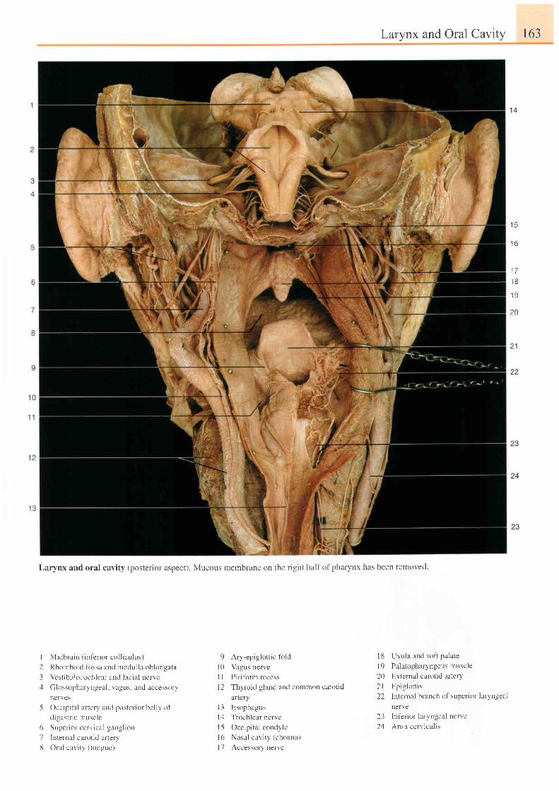
Midbrain ( inf 'er ior col l iculus)
Rhomboid firssa and medulla oblongata
Vestibr.rlocochleal and facial nerve
Glossopharyngetr l , vagus. and accessory
nerves
Occipital artery and posterior belly of
d igastr ic muscle
Super ior cerv ical gangl ion
Internal carotid artery
Oral cavrty ( tongue)
9 Ary-epiglottic fold
l0 Vagus nerve
I I Piriform recess
l2 Thyroid gland and common carotid
artery
I 3 Esophagus
I zl Trochlear nerve
l5 Occip i ta l condyle
l6 Nasal cavi ty (choanl)
I 7 Accessory nervc
Larynx and Oral Cavity 163
l8 Uvula and sof i palate
l9 Palatopharyngeus muscle
20 External carotid artery
2l Epiglot t is
22 Internal branch of superior laryngeal
nerve
23 Inlerior laryngeal nerve
24 Ansa cerv ical is
t 3
t o
1 71 8
1 9
2A
Larynx and oral cavity (posterior aspect) Mucous membrane on the right half of pharynx has been removed
I234
616
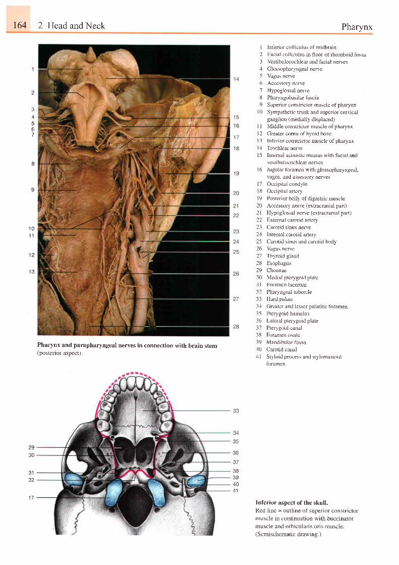
164 2 Head and Neck Pharynx
J
67
Pharynx and parapharyngeal nerves in connection with brain stem(posterior aspect).
I Inferior colliculus ofmidbrain2 Facial colliculus in floor ofrhomboid fossa3 Vestibulocochlear and facial nerves4 Glossopharyngealnerve5 Vagus nerve6 Accessory nerve7 Hypoglossal nerve8 Pharyngobasilarfascia9 Superior constrictor muscle of pharynx
I 0 Sympathetic trunk and superior cervicalganglion (medially displaced)
I I Middle constrictor muscle of pharynx12 Greater cornu ofhyoid bone1 3 Inferior consffictor muscle of pharynxl4 Trochlearnerve15 Internal acoustic meatus with facial and
vestibulocochlear nervesI 6 Jugular foramen with glossopharyngeal,
vagus, and assessory nerves17 Occipital condyle18 Occipital artery19 Posterior belly ofdigastric muscle20 Accessory nerve (extracranial part)2l Hypoglossal nerve (extracranial part)22 External carotid artery23 Carotid sinus nerve24 lnternal carotid artery25 Carotid sinus and carotid body26 Vagus nerve27 Thyroid gland28 Esophagus29 Choanae30 Medial pterygoid plate31 Foramen lacerum32 Pharyngeal tubercle33 Hard palate34 Greater and lesser palatine foramen35 Pterygoid hamulus36 Lateral pterygoid plate37 Pterygoid canal38 Foramen ovale39 Mandibular fossa40 Carotid canal41 Styloid process and stylomastoid
foramen
Inferior aspect of the skull.Red l ine = outl ine of superior constr ictormuscle in continuation with buccinatormuscle and orbicularis oris muscle.(Semischematic drawing.)
1 01 1
t z
1 3
2930
3132
1 7
37
383940
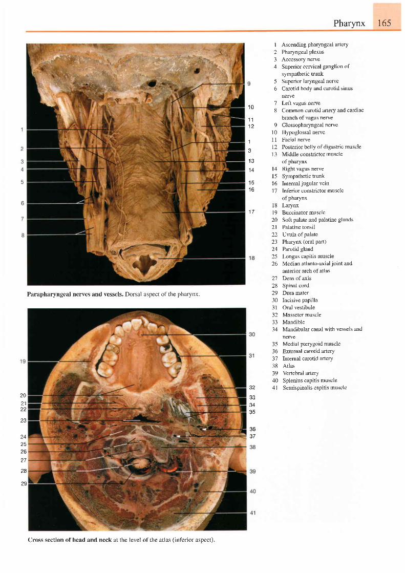
165Pharynx
1 0
1 11 2
'l
J
1 e
1 4
1 6
1 a
I
23A
Ascending pharyngeal arteryPharyngeal plexusAccessory nerveSuperior cervical ganglion ofsympathetic trunkSuperior laryngeal nerveCarotid body and carotid sinusnerve
5t)
7 Left vagus nerve8 Common carotid artery and cardiac
branch of vagus nerve9 Glossopharyngeal nerve
10 Hypoglossal nerve1l Facial nerve1 2 Posterior belly of digastric muscle13 Middle constrictor muscle
of pharynx14 Right vagus nervel5 Sympathetic trunk16 Internaljugularvein17 Inferiorconstrictormuscle
of pharynxl8 Larynx19 Buccinator muscle20 Soft palate and palatine glands2l Palatine tonsil22 Uvtlaof palate23 Pharynx (oral part)24 Parotid gland25 Longus capitis muscle26 Median atlanto-axial joint and
anterior arch of atlas27 Dens ofaxis28 Spinal cord29 Dura mater30 Incisive papilla31 Oral vestibule32 Masseter muscle33 Mandible34 Mandibular canal with vessels and
nerve35 Medial pterygoid muscle36 External carotid artery37 Internal carotid artery38 Atlas39 Vertebral artery40 Splenius capitis muscle41 Semispinalis capitis muscle
?02122
24zc
lo
27
28
?s
Parapharyngeal nerves and vessels. Dorsal aspect of the pharynx.
3eerl
s435
$637
Cross section ofhead and neck at the level of the atlas (inferior aspect).
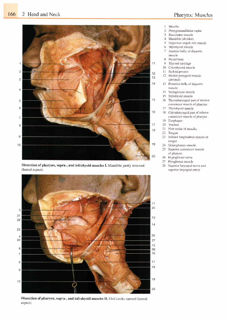
r66 2 Head and Neck Pharynx: Muscles
t a
1 3
1 4
I
234
561
6
o
10l l
t 2
1 3
t4l 5t 6
I7l 8
MaxillaPterygomandibular rapheBuccinator muscleMandible (divided)Depressor anguli oris muscleMylohyoid muscleAnterior belly of digastricmuscleHyoid boneThyroid cartilageCricothyroid muscleStyloid processMedial pterygoid muscle(divided)Posterior belly of digastricmuscleStyloglossus muscleStylohyoid muscleThyropharyngeal part of inferiorconstrictor muscle of pharynxThyrohyoid muscleCricopharyngeal part of infenorconstrictor muscle of pharynx
l9 Esophagus20 Trachea21 First molar of maxilla22 Tongte23 Inferior longitudinal muscle of
tongue24 Genioglossus muscle25 Superior constrictor muscle
of pharynx26 Hypoglossal nerve27 Hyoglossus muscle28 Superior laryngeal nerve and
superior laryngeal artery
1 9
2122
I J
Dissection of pharynx, supra-, and infrahyoid muscles I. Mandible partly removed(lateral aspect).
Dissection of pharynx, supra-, and infrahyoid muscles II. oral cavity opened (lateralasDect).
1 1
25
I J
t +
2627t c
28I D
1 7
1 8
1 9
A
24
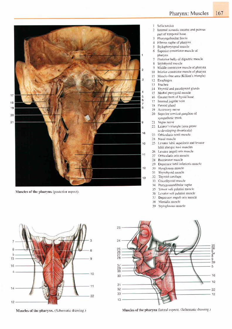
Pharynx: Muscles 167
4
678
I
1 6
1 0
1 1
' t2
1 3
2 l22
I Sella turcica2 Internal acoustic meatus and petrous
part of temporal bone3 Pharyngobasilarfascia4 Fibrous raphe of pharynx
5 Stylopharyngealmuscle6 Superior constrictor muscle of
pharynx7 Posterior belly of digastric muscle
8 Stylohyoid muscle9 Middle constrictor muscle of pharynx
10 Inferior constrictor muscle ofpharynx
1l Muscle-free area (Killian's triangle)
12 Esophagusl3 Trachea14 Thyroid and parathyroid glands
l5 Medial pterygoid muscle16 Greater horn of hyoid bonel7 Intemal jugular vein18 Parotid gland19 Accessory nerve20 Superior cervical ganglion of
sympathetic trunkVagus nerveLaimer's triangle (area Proneto developing diverticula)Orbicularis oculi muscleNasal muscleLevator labii superioris and levator
labii alaeque nasi musclesLevator anguli oris muscleOrbicularis oris muscleBuccinator muscleDepressor labii inferioris muscle
Hyoglossus muscleThyrohyoid muscleThyroid caltilageCricothyroid musclePtery gomandibular rapheTensor veli palatini muscleLevator veli palatini muscleDepressor anguli oris muscleMentalis muscleStyloglossus muscle
z-)
2425
26272829303 l7 Z
3334
Muscles of the pharynx (posterior aspect).3536313839
7
Bq
t c
t o
A
35366J48I395
25zo2728
37293830
J I
3233
t o
1 0
221 2
1 7
1 8
1 9
Muscles of the pharynx. (Schematic drawing.) Muscles of the pharynx (lateral aspect). (Schematic drawing.)
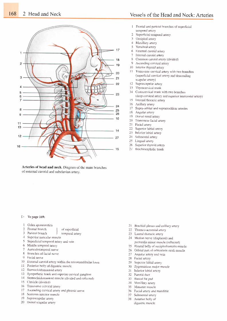
168 2 Head and Neck Vessels of the Head and Neck: Arteries
1 7
1 8
1 9
21
22
I Frontal and parietal branches ofsuperficialtemporal artery
2 Superhcial temporal artery3 Occipital artery4 Maxillary artery5 Vertebral artery6 External carotid artery7 Intemal carotid artery8 Common carotid artery (divided)9 Ascending cervical artery
l0 Inferior thyroid arteryI 1 Transverse cervical artery with two branches
(superhcial cervical artery and descendingscapular artery)
12 Suprascapularartery13 Thyrocervical trunkl4 Costocervical trunk with two branches
(deep cerr,rical artery and superior intercostal artery)15 Internal thoracic artery16 Axillary artery17 Supra-orbital and supratrochlear arteries18 Angular artery19 Dorsal nasal artery20 Transverse facial artery21 Facial artery22 Superior labial artery23 Inferior labial artery24 Submental artery25 Lingual artery26 Superior thyroid artery27 Brachiocephalictrunk
4
567
8o
t 11 3
1 2
2425261 0
Arteries of head and neck. Diagram of the main branchesofexternal carotid and subclavian artery.
I Galea aponeurotica
2 Frontal branch I of supeficial3 Parietal branch J temporal artery4 Superior auricular muscle5 Superhcial temporal artery and vein6 Middle temporal artery7 Auriculotemporalnerve
8 Branches of facial nerve9 Facial nerve
10 Extemal carotid aftery within the retromandibular fossaI 1 Posterior belly of digastric muscle12 Sternocleidomastoid arIery13 Sympathetic trunk and superior cervical ganglion14 Stemocleidomastoid muscle (divided and reflected)15 Clavicle (divided)
16 Transverse cervical arteryl7 Ascending cervical artery and phrenic nerve18 Scalenus anterior muscle19 Suprascapular artery20 Dorsal scapular artery
Brachial plexus and axillary arteryThoraco-acromial artery
Lateral thoracic artery
Median nerve (displaced) andpectoralis minor muscle (reflected)
Frontal belly of occipitofrontalis muscleOrbital part of orbicularis oculi muscleAngular artery and vein
Facial artery
Superior labial artery
Zygomaticus major muscle
Inferior labial artery
Parotid duct
Buccal fat pad
Maxillary artery
Masseter muscle
Facial artery and mandible
Submental artery
Anterior belly ofdigastric muscle
L I
22a )
24
2526272829303 132333435363738
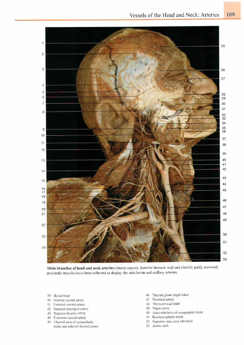
Vessels of the Head and Neck: Arteries 169
e t
JZ
33343536
38
1 61 71 8
1 9
39
404142
43
44
45
46
47
48
49
2021
50
52
53
Main branches of head and neck arteries (lateral aspect). Anterior thoracic wall and clavicle partly removed;
pectoralis muscles have been reflected to display the subclavian and axillary artefles.
39 Hyoid bone40 Internal carotid artery41 External carotid artery42 Superior laryngeal artery43 Superior thyroid artery44 Common carotid artery45 Thyroid ansa of sympathetic
trunk and int'erior thyroid artery
46 Thyroid gland (right lobe)47 Vertebral artery48 Thyrocervical trunk49 Vagus nerve50 Ansa subclavia of sympathetic trunk
51 Brachiocephalictrunk52 Superior vena cava (divided)
53 Aortic arch
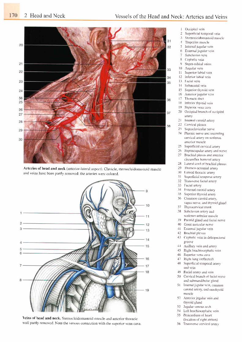
170 2 Head and Neck Vessels of the Head and Neck: Arteries and Veins
1 Occipital vein
2 Superficial temporal vein3 Sternocle idomastoidmuscle
4 Trapezius muscle
5 Internal jugular vein
6 Externaljugular vein
7 Subclavian vein
8 Cephalic vein
9 Supra-orbital veins
l0 Angular vein
1 I Superior labial vein
l2 Inf'erior labial veinl3 Facial vein
14 Submental vein
l5 Superior thyroid veinl6 Anter ior jugularvein
17 Thoracic duct
I 8 Inferior thyroid vein
l9 Superior vena cava20 Occipital branch of occipital
artery
2l Internal carotid artery
22 Cervical plexus
23 Supraclavicularnerve
24 Phrenic nerve and ascending
cervical artery on scalenus
anterior muscle
25 Superficial cervical artery
26 Suprascapular artery and nerve27 Brachial plexus and antenor
circumfl ex humeral artery28 Lateral cord of brachial plexus
29 Thoraco-acromialartery
30 Lateral thoracic artery31 Superficial temporal artery32 Transverse facial artery
33 Facial artery
34 External carotid artery35 Superior thyroid artery
36 Common carotid aftery,vagus nerve, and thyroid gland
3 1
32
5625
2627 J /
38
Arteries of head and neck (anterior-lateral aspect). Clavicle, sternocleidomastoid muscleand veins have been partly removed; the arteries were colored.
veins of head and neck. sternocleidomastoid muscle and anterior thoracicwall partly removed. Note the venous connection with the superior vena cava.
3138
39404 l4243
Thyrocervical trunk
Subclavian artery and
scalenus anterior muscle
Parotid gland and facial nerve
Great auricular nerve
External jugular veinBrachial plexus
Cephalic vein in deltopectoralgroove
44 Axillary vein and artery .
45 Right brachiocephalic vern46 Superior vena cava47 Right lung (reflected)
48 Supedicial temporal artery
and vein
49 Facial artery and vein
50 Cervical branch of facial nerve
and submandibular gland
5l Intemal jugular vein, commoncarotid arlery, and omohyoidmuscle
52 Anteriorjugular vein andthyroid gland
53 Jugular venous arch54 Lei t brachiocephal ic vein
55 Pericardium ofheart(location of right atrium)
56 Transverse cervical artery
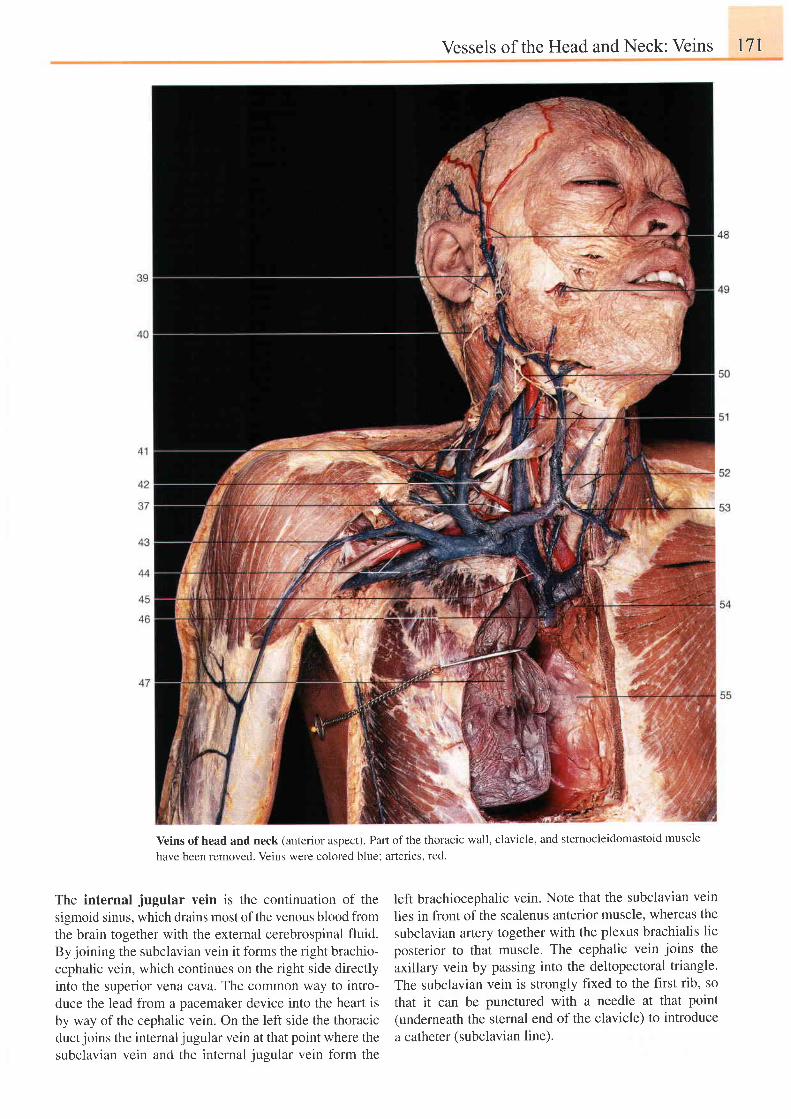
Vessels of the Head and Neck: Veins I71
Veins of head and neck (anterior aspect). Part of the thoracic wall, clavicle, and sternocleidomastoid muscle
have been removed. Veins were colored blue: arteries, red.
The internal jugular vein is the continuation of thesigmoid sinus, which drains most of the venous blood fromthe brain together with the external cerebrospinal fluid.By joining the subclavian vein it forms the right brachio-cephalic vein, which continues on the right side directlyinto the superior vena cava. The common way to intro-duce the lead from a pacemaker device into the heart isby way of the cephalic vein. On the left side the thoracicduct joins the internal jugular vein at that point where thesubclavian vein and the internal jugular vein form the
left brachiocephalic vein. Note that the subclavian vein
lies in front of the scalenus anterior muscle, whereas the
subclavian artery together with the plexus brachialis lie
posterior to that muscle. The cephalic vein joins the
axillary vein by passing into the deltopectoral triangle.
The subclavian vein is strongly frxed to the first rib, so
that it can be punctured with a needle at that point(underneath the sternal end of the clavicle) to introduce
a catheter (subclavian line).
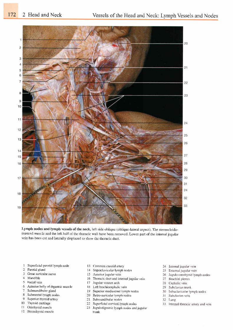
172 2 Head and Neck Vessels of the Head and Neck: Lymph Vessels and Nodes
Lymph nodes and lymph vessels of the neck, left side oblique (obliqueJateral aspecr). The sternocleido-mastoid muscle and the left half of the thoracic wall have been removed. Lower part of the intemal jugularvein has been cut and laterally displaced to show the thoracic duct.
1 Superficial parotid lymph node2 Parotid gland3 Great auricular nerve4 Mandible5 Facial vein6 Anterior belly of digastric muscle7 Submandibular gland8 Submental lymph nodes9 Superior thyroid artery
l0 Thyroid cartilagel1 Omohyoidmuscle12 Sternohyoid muscle
13 Common carotid artery14 Supraclavicular lymph nodes15 Anteriorjugular vein16 Thoracic duct and internaljugular veinl7 Jugular venous arch18 Left brachiocephalic vein19 Superior mediastinal lymph nodes20 Retro-auricular lymph nodes2l Submandibularnodes22 Superficial cervical lymph nodes23 Jugulodigastric lymph nodes andjugular
trunk
24 Internaljugularvein25 External jugular vein26 Jugulo-omohyoid lymph nodes27 Brachial plexus28 Cephalic vein29 Subclavian trunk30 Infraclavicular lymph nodes3l Subclavian vein32 Lung33 Intemal thoracic artery and vein
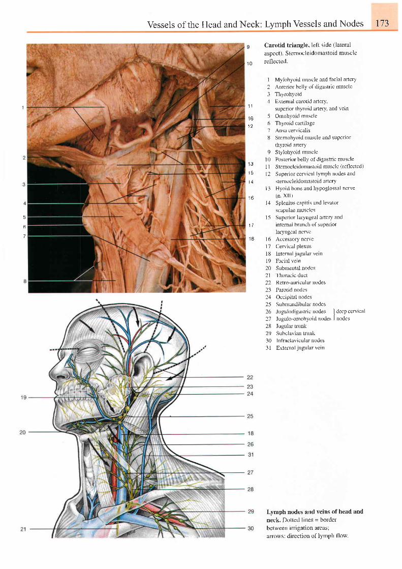
Vessels of the Head and Neck: Lymph Vessels and Nodes 173
6
Carotid triangle, left side (lateral
aspect). Sternocleidomastoid musclereflected.
1234
1 6 5A
1 278
o
l 01 3
I t
1 5 1 21 4
1 3
1 6I 4
1 51 7
Mylohyoid muscle and facial afterYAnterior belly of digastric muscleThyrohyoidExternal carotid artery,superior thyroid artery, and vetnOmohyoid muscleThyroid cartilageAnsa cervicalisSternohyoid muscle and superlorthyroid arteryStylohyoid musclePosterior belly of digastric muscleSternocleidomastoid muscle (reflected)
Superior cervical lymph nodes andstemocleidomastoid arterYHyoid bone and hypoglossal nerve(n. XII)Splenius capitis and levatorscapulae musclesSuperior laryngeal altery andintemal branch of superiorlaryngeal nerve
18 16 Accessory nerve17 Cervical plexusl8 Intemal jugular vein19 Facial vein20 Submental nodes21 Thoracic duct22 Retro-auricularnodes23 Parotid nodes24 Occipital nodes25 Submandibular nodes26 Jugulodigastric nodes I deep cervical27 Jugulo-omohyoid nodes . l nodes28 Jugular trunk29 Subclavian trunk30 Infraclavicular nodes3 | External jugular vein
Lymph nodes and veins ofhead and
neck. Dotted lines = border
between irrigation areas;
arrows: direction of lYmPh flow.
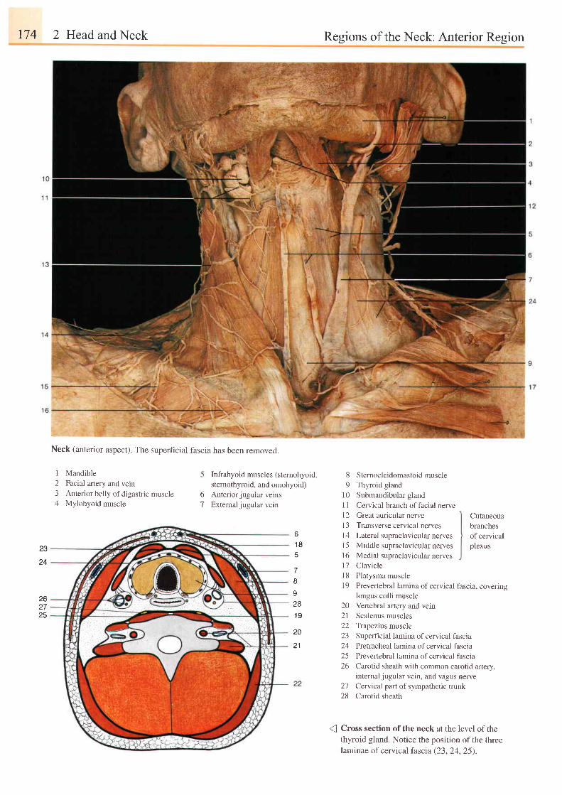
174 2 Head and Neck Regions of the Neck: Anterior Region
Neck (anterior aspect). The superhcial fascia has been removed.
1 Mandible
2 Facial artery and vein3 Anterior belly of digastric muscle4 Mylohyoid muscle
Infrahyoid muscles (stemohyoid,
sternothyroid, and omohyoid)
Anterior jugular veinsExternal jugular vein
8 Sternocleidomastoid muscle9 Thyroid gland
10 Submandibular gland1 I Cervical branch offacial nervel2 Great auricular nerve13 Transverse cervical nerves14 Lalerul supraclavicular nerves15 Middle supraclavicular nerves16 Medial supraclavicular nerves17 Claviclel8 Platysma muscle19 Prevertebral lamina ofcervical
o
7
o
1 8
Cutaneous
branches
of cervicalplexus
fascia, coveringI
928
1 9
20
2 1
23
24
262725
longus colli muscle
Vertebral artery and vein
Scalenus muscles
Trapezius muscle
Superficial lamina of cervical fascia
Pretracheal lamina of cervical fascia
Prevertebral lamina of cervical fascia
Carotid sheath with common carotid artery,internal jugular vein, and vagus nerve
Cervical part of sympathetic trunkCarotid sheath
202122z-)
242526
2128
Cross section of the neck at the level of thethyroid gland. Notice the position of the threelaminae of cervical fascta (23, 24, 25).
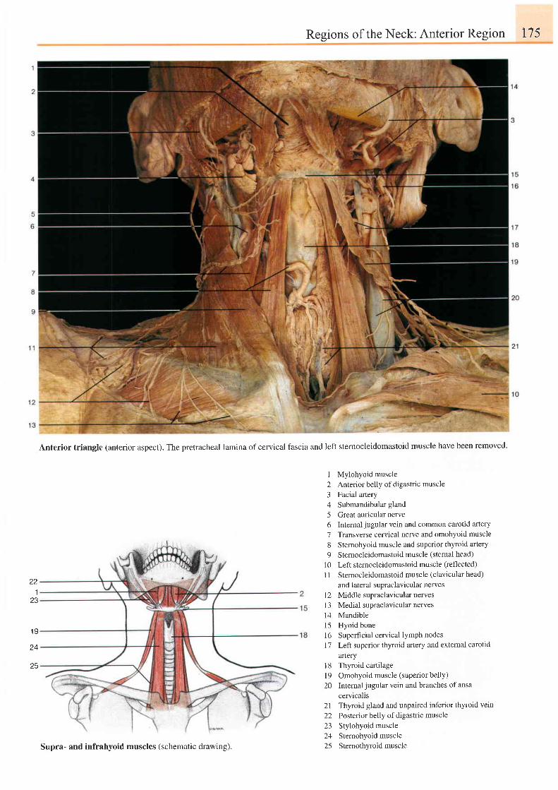
22.l
23
1 9
Regions of the Neck: Anterior Region 175
Anterior triangle (anterior aspect). The pretracheal lamina of cervical fascia and left sternocleidomastoid muscle have been removed.
1 Mylohyoid muscle2 Anterior belly of digastric muscle3 Facial artery4 Submandibular gland
5 Great auricular nerve6 Intemal jugular vein and common carotid artery
7 Transverse cervical nerve and omohyoid muscle
8 Sternohyoid muscle and superior thyroid artery
9 Sternocleidomastoid muscle (sternal head)
10 Left stemocleidomastoid muscie (reflected)
l1 Stemocleidomastoidmuscle (clavicularhead)
and lateral supraclavicular nerves12 Middle supraclavicular nerves13 Medial supraclavicular nerves14 Mandible15 Hyoid bone16 Superhcial cervical lymph nodes17 Left superior thyroid artery and extemal carotid
artery18 Thyroid cartilage19 Omohyoid muscle (superior belly)20 Intemal jugular vein and branches of ansa
cervicalis21 Thyroid gland and unpaired inferior thyroid vein
22 Posterior belly of digastric muscle23 Stylohyoid muscle24 Sternohyoid muscle25 Sternothyroidmuscle
25
Supra- and infrahyoid muscles (schematic drawing).
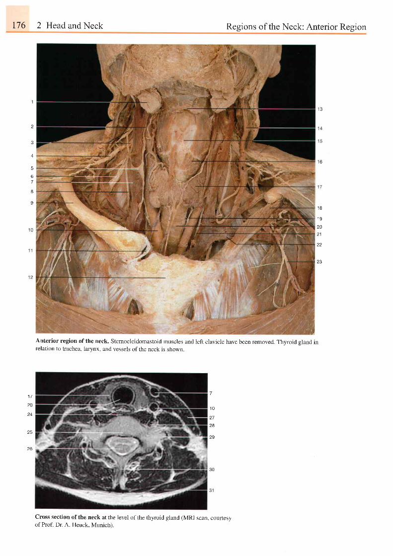
176 2 Head and Neck Regions of the Neck: Anterior Region
2
/
5
67
8
91 8
1 9
2021
23
7
1 0
27
28
29
1 7
20
24
25
z o
Anterior region of the neck. Sternocleidomastoid muscles and left clavicle have been removed. Thyroid gland inrelation to trachea, larynx, and vessels ofthe neck is shown.
Cross section of the neck at the level of the thyroid gland (MRI scan, courtesyof Prof. Dr. A. Heuck. Munich).
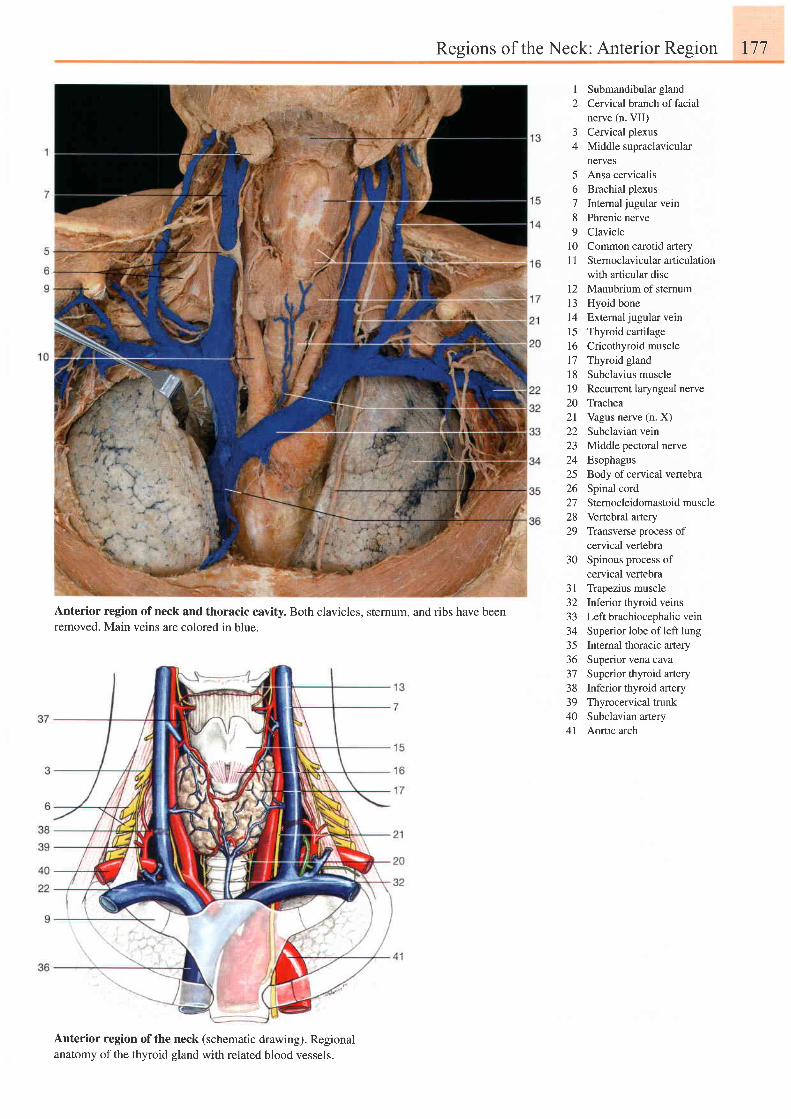
Regions of the Neck: Anterior Region 177
I2
J
4
56789
l 01 1
l 2I J
I41516171 819202 l22z3",^2526272829
30
3 13 Z
333435365 t
3839404 l
Submandibular glandCervical branch of facialnerve (n. VII)Cervical plexusMiddle supraclavicularnervesAnsa cervicalisBrachial plexusIntemal jugular veinPhrenic nerveClavicleCommon carotid arterySternoclavicular articulationwith articular discManubrium of sternumHyoid boneExternal jugular veinThyroid cartilageCricothyroid muscleThyroid glandSubclavius muscleRecurrent laryngeal nerveTracheaVagus nerve (n. X)Subclavian veinMiddle pectoral nerveEsophagusBody of cervical vertebraSpinal cordSternocleidomastoid muscleVertebral arteryTransverse process ofcervical vertebraSpinous process ofcervical vertebraTrapezius muscleInferior thyroid veinsLeft brachiocephalic veinSuperior lobe of left lungInternal thoracic arterySuperior vena cavaSuperior thyroid arteryInferior thyroid arteryThyrocervical trunkSubclavian arteryAortic arch
Anterior region of neck and thoracic cavity. Both clavicles, sternum, and ribs have beenremoved. Main veins are colored in blue.
Anterior region of the neck (schematic drawing). Regionalanatomy of the thyroid gland with related blood vessels.
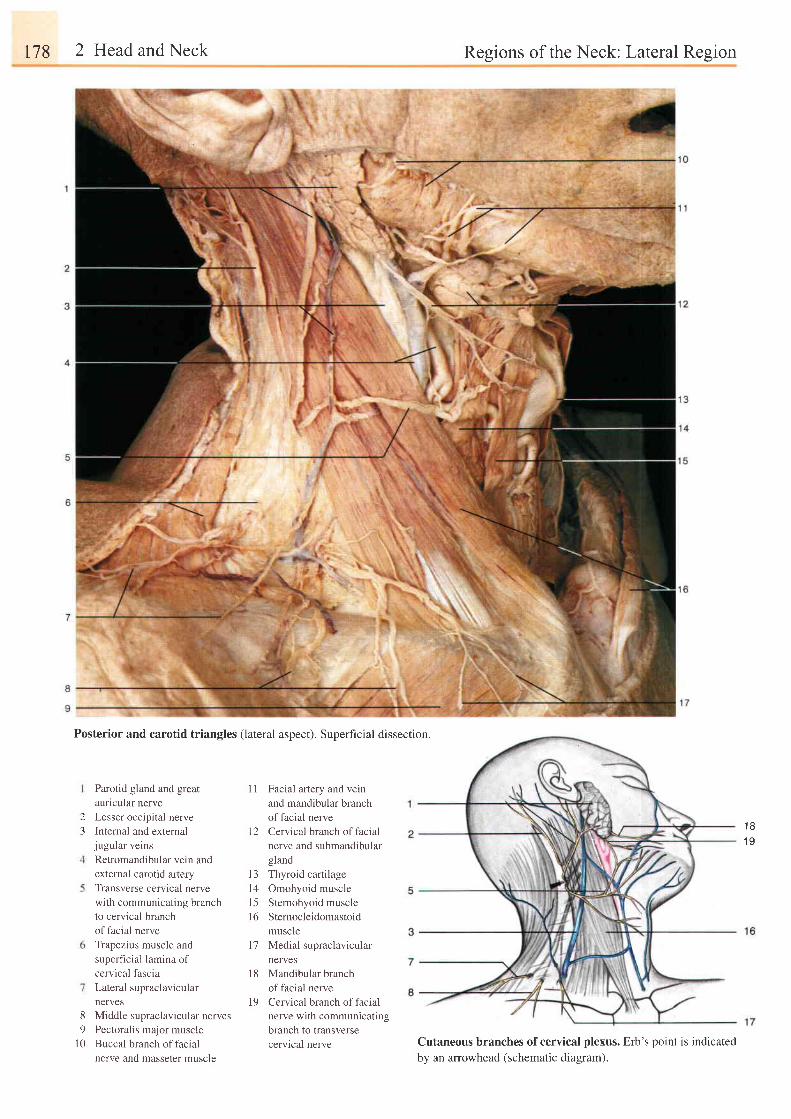
178 2 Head and Neck Resions of the Neck: Lateral Resion
Posterior and carotid triangles (lateral aspect). Superficial dissection.
23
Parotid gland and great
auricular nerve
Lesser occipital nerve
Internal and externaljugular veins
Retromandibular vein and
external carotid artery
Transverse cervical nerve
with communicating branch
to cervical branch
of facial nerve
Trapezius muscle and
superficial lamina of
cervical fascia
Lateral supraclavicular
nerves
8 Middle supraclavicular nerves
9 Pectoralis major muscle
l0 Buccal branch offacial
nerve and masseter muscle
I I Facial artery and vein
and mandibular branch
of facial nerve
I 2 Cen ical branch of facia
nerve and submandibular
gland
l3 Thyroid cartilage
14 Omohyoid muscle
l5 Sternohyoid muscle
l6 Sternocleidomastoid
muscle
17 Medial supraclavicular
nerves
18 Mandibular branch
of facial nerve
19 Cervical branch of facial
nerve with communicating
branch to transverse
cervical nerve
1 B
1 9
Cutaneous branches of cervical plexus. Erb's point is indicatedbv an arrowhead (schematic diaeram).
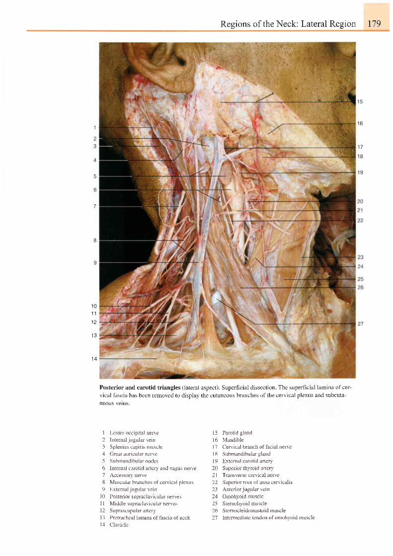
Resions of the Neck: Lateral Reeion I79
1 01 1
1 2
1 3
1 4
Posterior and carotid triangles (lateral
vical fascia has been removed to disolavneous velns.
aspect). Superficial dissection. The superfrcial lamina ofcer-
the cutaneous branches of the cervical plexus and subcuta-
1 Lesser occipital nerve2 Internal jugular vein3 Splenius capitis muscle4 Great auricular nerve5 Submandibular nodes6 Internal carotid artery and vagus nerve7 Accessory nerve8 Muscuiar branches ofcervical plexus9 External jugular vein
l0 Posterior supraclavicular nerves11 Middle supraclavicular nervesl2 Suprascapular artery13 Pretracheal lamina offascia ofneckl4 Clavicle
15 Parotid glandl6 Mandiblel7 Cervical branch offacial nervel8 Submandibular gland19 Extemal carotid artery20 Superior thyroid artery2l Transverse cervical nerve22 Superior root of ansa cervicalis23 Anteriorjugularvein24 Omohyoid muscle25 Sternohyoid muscle26 Sternocleidomastoidmuscle27 lntermediate tendon of omohyoid muscle
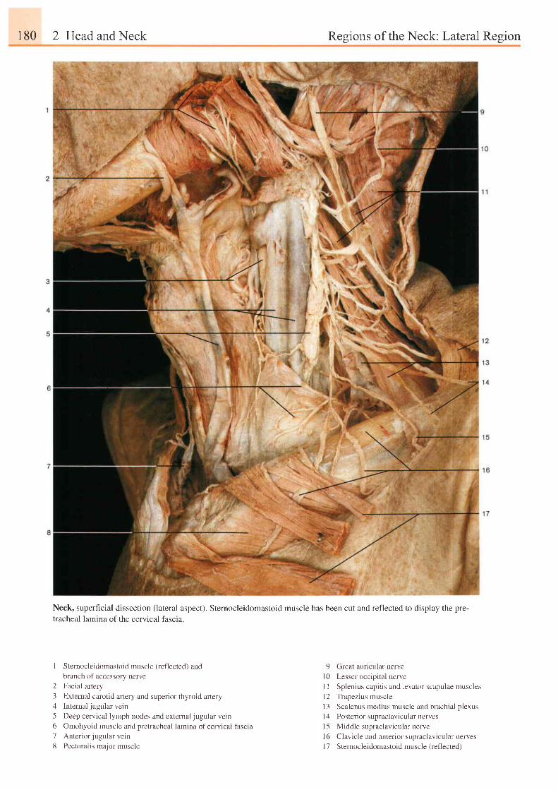
r80 2 Head and Neck Resions of the Neck: LateruI Resion
Neck, superficial dissection (lateral aspect). Sternocleidomastoid muscle has been cut and reflected to display the pre-
tracheal lamina of the cervical fascia.
I Sternocleidomastoid muscle (reflected) andbranch of accessory nerve
2 Facial artery3 External carotid artery and superior thyroid artery4 Internal jugular vein5 Deep cervical lymph nodes and extemal jugular vein6 Omohyoid muscle and pretracheal lamina of cervicai fascia7 Anteriorjugular vein8 Pectoralis major muscle
9 Great auricular nervel0 Lesser occipital nerveI I Splenius capitis and levator scapulae musclesl2 Trapezius muscle13 Scalenus medius muscle and brachial plexusl4 Posterior supraclavicular nervesl5 Middle supraclaviculu nervel6 Clavicle and anterior supraclavicular nervesl7 Sternocleidomastoid muscle (reflected)
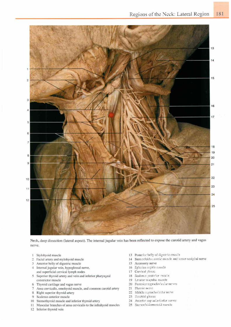
[ { i r r ( ) l ls of the Neck: Lateral Regt ln I 8 I
. #ltl:
dis',
i 3 i
&'.1::,
i
q.
- - . * { ' " *\'
1 L
1n&1,1, ! ,
t\
\ r r ' l o
w
) \ - - * *
' . )L
i t ' ) t l r r i l . . j l i l l { : r s i i l i l l l l t \ i i .
I ' r r - , ( ' t i r t J l l l , i : i r 1 l ( l t t t t i s c l c l r t l r l ] r ' \ \ f l r r , r r l l '
\ l l , l l l l \ ! i l l J l t r \ l l l t l \ l t '
- ! ! \ i l i l \ i C \ t l .
\ t i j n l t ! l t i ) \ t C t 1 0 1 l I L i \ e l r l
r . r r l , t l . r l l l ) r l l " l e t l l L t s . l C
, ' t \ l ! ' l - l ! \ l . , l l l i l ( l i l \ l ! t l l l l l I l e l \ t ' \
, i t , f l i l i l l c \ . (
I t . : . ' r ; ' : . r . , r \ l . . l l , r l l l . l \ ' .
I i r i ! r l l t . l 1 l ) i t ' \ i r \
! t : l \ ' t l i i \ L l l - r l i i r i l l r t c r t l l i l l l c l t r l t
I L l t l L t ( i c l t l r ) l l l . L : l ( l l i l i l l u : c i c
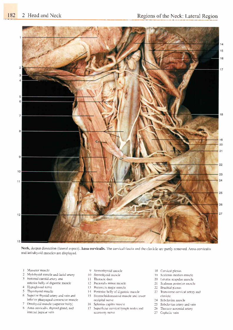
,...1.92 2 Head and Neck Regions of the Neck: Lateral Region
F
Neck, deeper dissection (lateral aspect). Ansa cervicalis. The cervical fascia and the clavicle are partly removed. Ansa cervicalisand infrahyoid muscles are displayed.
1 9
20
2 1
I23
A
56
78
Masseter muscle
Mylohyoid muscle and facial arteryExternal carotid artery andanterior belly of digastric muscleHypoglossal nerve
Thyrohyoid muscle
Superior thyroid artery and vein andinf'erior pharyngeal constrictor muscleOmohyoid muscle (superior belly)Ansa cervicalis, thyroid gland, andinternal jugular vein
9 Sternothyroid muscle
10 Sternohyoid muscle
I 1 Thoracic duct
l2 Pectoralis minor muscle
13 Pectoralis major muscle
14 Posterior belly ofdigastric musclel5 Stemocleidomastoid muscle and lesser
occipital nerve
l6 Splenius capitis musclel7 Supeficial cervical lymph nodes and
accessory nerve
l8 Cervical p lexus
l9 Scalenus medius muscle
20 Levator scapulae muscle
2l Scalenus posterior muscle
22 Brachial plexus
23 Transverse cervical artery and
clavicle
24 Subclavius muscle
25 Subclavian artery and vern
26 Thoraco-acromial artery
27 Cephalic vein
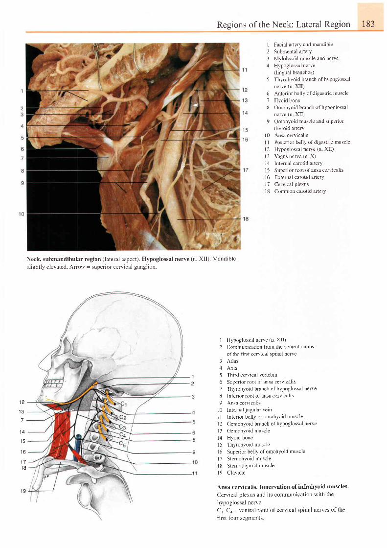
Regions of the Neck: Lateral Region 183
t z
1 3
Neck, submandibular region (lateral aspect). Hypoglossal nerve (n. XII). Mandibleslightly elevated. Arrow = superior cervical ganglion.
I Facial artery and mandible2 Submental artery3 Mylohyoid muscle and nerve4 Hypoglossal nerve
(lingual branches)5 Thyrohyoid branch of hypoglossal
nerve (n. XII)6 Anterior belly of digastric muscle7 Hyoid bone8 Omohyoidbranchofhypoglossal
nerve (n XII)9 Omohyoid muscle and superior
thyroid aftery10 Ansa cervicalis11 Posterior belly ofdigastric musclel2 Hypoglossal nerve (n XII)13 Vagus nerve (n. X)14 Intemal carotid afiery15 Superior root of ansa cervicalisl6 External carotid artery17 Cervical plexus18 Common carotid artery
1 Hypoglossal nerve (n. XII)2 Communication from the ventral ramus
of the first cervical spinal nerve3 Atlas4 Axis5 Third cervical vertebra6 Superior root of ansa cervicalis7 Thyrohyoid branch ofhypoglossal nerve
8 Int'erior root of ansa cervicalis9 Ansa cervicalis
l0 Internal jugularveinI I Inferior belly of omohyoid muscle1 2 Geniohyoid branch of hypoglossal nervel3 Geniohyoid muscle14 Hyoid bonel5 Thyrohyoid muscle16 Superior belly of omohyoid muscle17 Sternohyoid muscle'| 8 Sternothyroid muscle
l9 Clavicle
Ansa cervicalis. Innervation of infrahyoid muscles.
Cervical plexus and its communication with the
hypoglossal nerve.
C,-C+ - ventral rami of cervical spinal nerves of the
first four segments
1 4
t 3
1 6
1 1
1 8
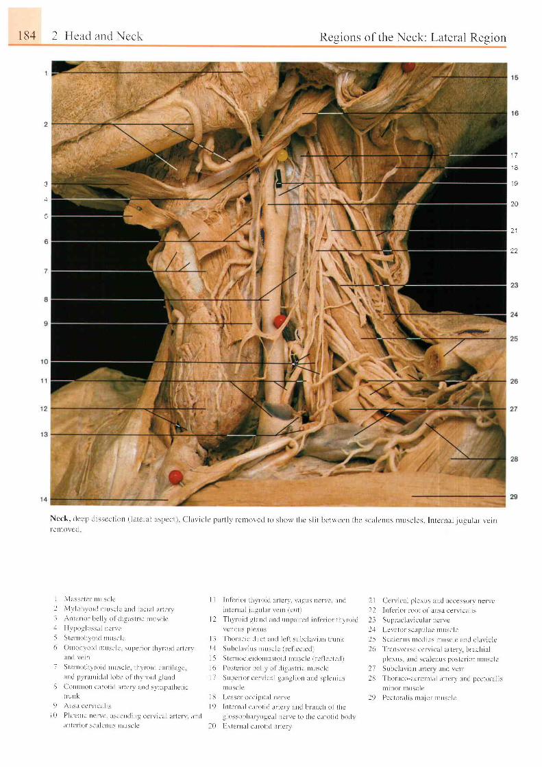
184 2 Heltt l uttr l Nr'c1.,
e c q
,1
/'i:
I
Neck, c lccP c l i sscc t ron ( la lc r -u l uspcc t ) C l l r i c lc l )a l l l \ reu t ( ) \ ' c r l to sho i l thc s l i tr-ctt tot ct l
Rcgions of the Neck: Lateral Region
:,llr,tr,lr ,irl:
'i
qJ
1 7
1 8
1 9
20
3
4
5 tt ? 1
22
)e+
betvn'een the scalcrrus ntuscles lntct 'nal iLrcular I 'c in
I l \ l t L ssc l c r n t r r se l c
I \ l r l o l r r o i t l n t r r sc l c u l t l l l c i a l a r r r : r t- l An t r ' r i o r bc l l l o l r l i g t r s t t - i c r l l r s c l e .- 1 H r po r l os :a l ne r r c
- i S t c r r r ohvo i r l n rL r sc l c6 On roh lo i d r l r r : e l c . supe r i o l t l r r r o i r l l t r t t r r
l t r t d r c rn
7 S t c r r r o l l r l r o i t l n rL r sc l c l h l r o i r l c l L t - t i l l t g c 'l un r l 1 t r l Ln t i r l l i l l obc o l t hv ro i t l s l l n t l
E ( ' o r l n ron cx to l i ( l i u t L l \ l r n t l s t r l pa l l r c t i ct r - L rnk
9 , \ nsa ec r r i c t L i i sl 0 Ph rcn i c r r c r r c . l r s ccD t l i ng cc r - r i ca l a r t en ' . t r n r l
i l l l l ( l i r ' l \ r ' , l l r ' t l l l \ I i l t l \ e l (
I I l n l e r ro t t l t r r - o i t l l u t c r \ ' . \ i l ! l u s nc r r c . un t l
i n t e r n a l . j L r u u l l r \ ( ' i n ( c u t )
l l J ' h r r o i d q l l Lnc l an t l r r n l l r i r cd i u l i ' r i o r t hv l o i i lr c r r 0 r r : ; l l c r r r s
l - l J ' ho r : r c i c t l r r c r l n r l l c f t s t r bc l t r r i t r n t r r - r nkl J S L r h c l a r i u : n r r r s c l c ( r c l l c c t e L l r
l 5 S t c rnoc l c t don ta : t o i r l n t L r sc l c ( r - c l l c c t cc l )
l 6 l ) o : t c r - i o r bc l l i o l t l i s l r : t r - i c n tL r sc l e
I7 SL rpc r i o r c c r r r ca l g l r r r g l i on l L l r l sp l c r r i u :r r L r sc l e
I I J l . es : c r occ i p i r r r l l r . r r c
l 9 I r r t en l r l c l r o t i r i l r r t c r r t i n t l bnnch o f t he
! lo:sophar r n lc l l r rcr- r c to thc c l in l t i t l bodrl0 F-r ternt l c l r r -o l i r l iLncrr
I I Ccn i c t l p l e rL r s l Lnd acccs \o r \ ne r \ cl l I n l c r i o r - r oo t o f ansu ce r - r i ca l i s
l 3 SL rp rac l a r i cu l l r nc - r - r c
l . l I -e\ ator scapulrc rnuscle
l 5 Sca l cnus n red ius r nL rsc l c an t l c l a r i c l c26
' l l 'ansrcr-sc ccrr ical ur- terr br-achia l
p l c rus - l Lnd sc l l cnus pos te r i o l r nusc l cl7 Srrbclur ien ar- tcrr l r r r l retr r
l8 Thoraco lcront i t r l l r tcr- r ' lLnt l l tecto le l isrn i r t o r - t t t t r s c l c
l 9 Pec to r u l r s r n l t j o r - n t usc l e
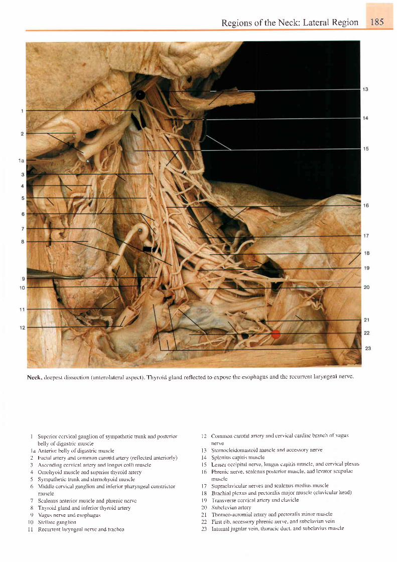
Regions of the Neck: Lateral Region 185
Neck, deepest dissection (anterolateral aspect). Thyroid gland reflected to expose the esophagus and the recuffent laryngeal nerve.
I Superior cervical ganglion of sympathetic trunk and posteriorbelly of digastric muscle
1a Anterior belly of digastric muscle2 Facial artery and common carotid artery (reflected anteriorly)3 Ascending cervical artery and longus colli muscle4 Omohyoid muscle and superior thyroid artery5 Sympathetic trunk and sternohyoid muscle6 Middle cervical ganglion and inferior pharyngeal constrictor
muscle7 Scalenus anterior muscle and phrenic nerve8 Thyroid gland and inferior thyroid artery9 Vagus nerve and esophagus
10 Stellate ganglionI 1 Recurent laryngeal nerve and trachea
12 Common carotid artery and cervical cardiac branch of vagus
nerveI 3 S ternocleidomastoid muscle and accessory nerve
l4 Splenius capitis muscle15 Lesser occipital nerve, longus capitis muscle, and cervical plexus
l6 Phrenic nerve, scalenus posterior muscle, and levator scapulae
musclel7 Supraclavicular nerves and scalenus medius muscle
18 Brachial plexus and pectoralis major muscle (clavicular head)
19 Transverse cervical artery and clavicie20 Subclavian atlery21 Thoraco-acromial artery and pectoralis minor muscle
22 First rib, accessory phrenic nerve, and subclavian vern
23 Internaljugular vein, thoracic duct, and subclavius muscle
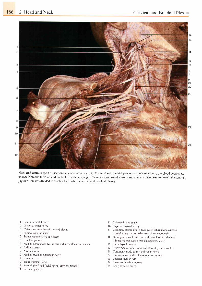
186 2 Head and Neck Cervical and Brachial Plexus
Neck and arm, deepest dissection (anterior-lateral aspect). Cervical and brachial plexus and their relation to the blood vessels areshown. Note the location and content of scalene triangle. Sternocleidomastoid muscle and clavicle have been removed; the internaljugular vein was divided to display the roots of cervical and brachial plexus.
1 3
1 4
1 5
t o
1 7
1 81 9
2021
23
I Lesser occipital nerve
2 Great auricular nerve
3 Cutaneous branches of cervical plexus
4 Supraclavicularnerve
5 Suprascapular nerve and artery6 Brachial plexus
7 Median nerve (with two roots) and musculocutaneous nerve8 Axillary artery
9 Axillary vein
10 Medial brachial cutaneous nerveI I Ulnar nerve
l2 Thoracodorsal nerve13 Parotid gland and facial nerve (cervical branch)14 Cervical plexus
15 Submandibular gland
l6 Superior thyroid artery
l7 Common carotid artery dividing in internal and external
carotid artery and superior root of ansa cervicalis
1 8 Omohyoid muscle and cervical branch of facial nervejoining the transverse cervical nerve (C2, C1)
l9 Sternohyoid muscle
20 Transverse cervical nerve and stemothyroid muscle21 Common carotid artery and vagus nerve
22 Phrenic nerve and scalenus anterior muscle23 Internaljugularvein
24 Intercostobrachial nerves
25 Long thoracic nerve
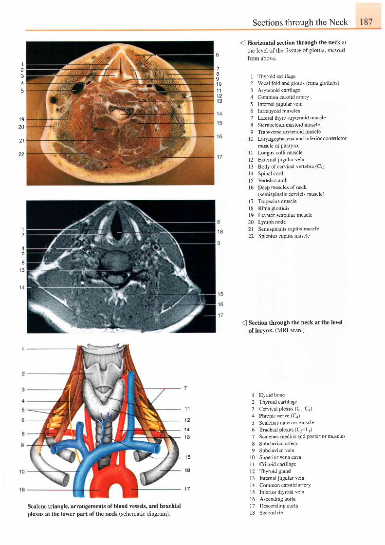
Sections through the Neck t87
Io1 01 11 21 3
I
o
4
45
8I J
1 920
22
1
( Horizontal section through the neck at
the level of the hssure of glottis, viewed
from above.
1 Thyroid cartilage2 Vocal fold and glottis (rima glottidis)
3 Arytenoid cartilage4 Common carotid arterY5 Intemal jugular vein6 Infrahyoid muscles7 Lateral thyro-arytenoid muscle8 Sternocleidomastoid muscle9 Transverse arytenoid muscle
10 Laryngopharynx and inferior constrictormuscle of pharynx
1l Longus colli muscle12 Externaljugularvein13 Body ofcervical vertebra (C5)
14 Spinal cord15 Vertebra arch16 Deep muscles ofneck
(semispinalis cervicis muscle)17 Trapezius muscle18 Rima glottidis19 Levator scapulae muscle20 Lymph node2l Semispinalis capitis muscle22 Splenius capitis muscle
( Section through the neck at the levelof larynx. (MRI scan.)
I Hyoid bone2 Thyroid cartilage3 Cervical plexus (C1-Ca)4 Phrenic nerve (Ca)
5 Scalenus anterior muscle6 Brachial plexus (C5-T1)7 Scalenus medius and posterior muscles
8 Subclavian artery9 Subclavian vein
10 Superiorvenacava11 Cricoid cartilage12 Thyroid gland13 Intemal jugular vein
14 Common carotid artery15 Inferiorthyroidvein16 Ascending aorta17 Descending aorta18 Secondrib
1 1
1 2
1 41 3
1 5
1 8
1 7
Scalene triangle, arrangements of blood vessels, and brachialplexus at the lower part of the neck (schematic diagram).
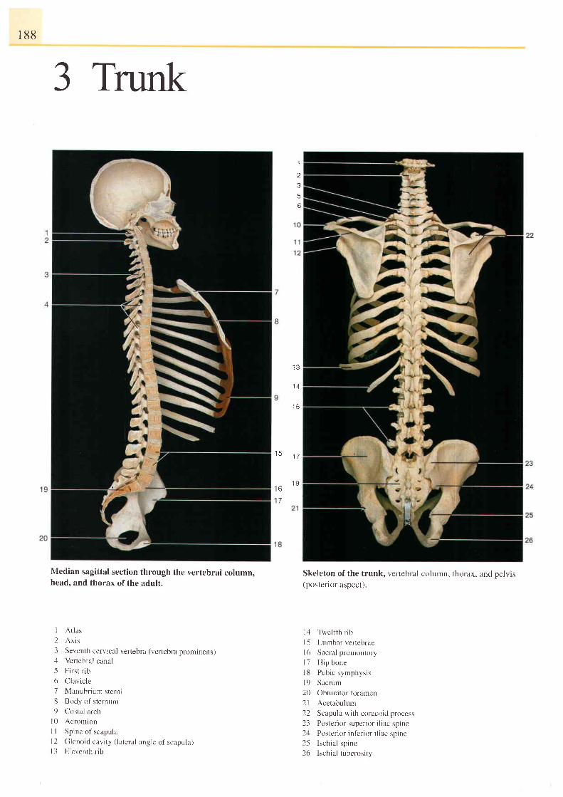
188
3 Trunk
Median sagittal section through the vertebral column,head, and thorax of the adult.
-I
)356
I J
1 4
I )
1 5
t r )
1 7
1 7
1 9
1 At las
2 Axis
3 Seventh ccrvical vertebra (vertebra prominens)zl Vertebral canal
5 First rib
6 Clavic le
7 Manubrium sterni8 Body o1'sternum
9 Costal archI0 Acromion
1 I Spine of scapulal2 Glenoid cavi ty ( lateral angle of scapula)l3 Eleventh r ib
Skeleton ofthe trunk, vertebral column, thorax, and pelvis(postenor aspect).
l4 Twelfih rib
l5 Lumbar vertebrae
l6 Sacral promontory
l 7 H ip bone
18 Pubic symphysis
l9 Sacrum
20 Obturator fbramen
21 Acetabulurn
22 Scapula with coracoid process
2.1 Po' ter ior super ior i l iac spine2ul Portcr ior infer ior i l iac:p ine25 Ischia l spine26 lschia l tuberosi ty
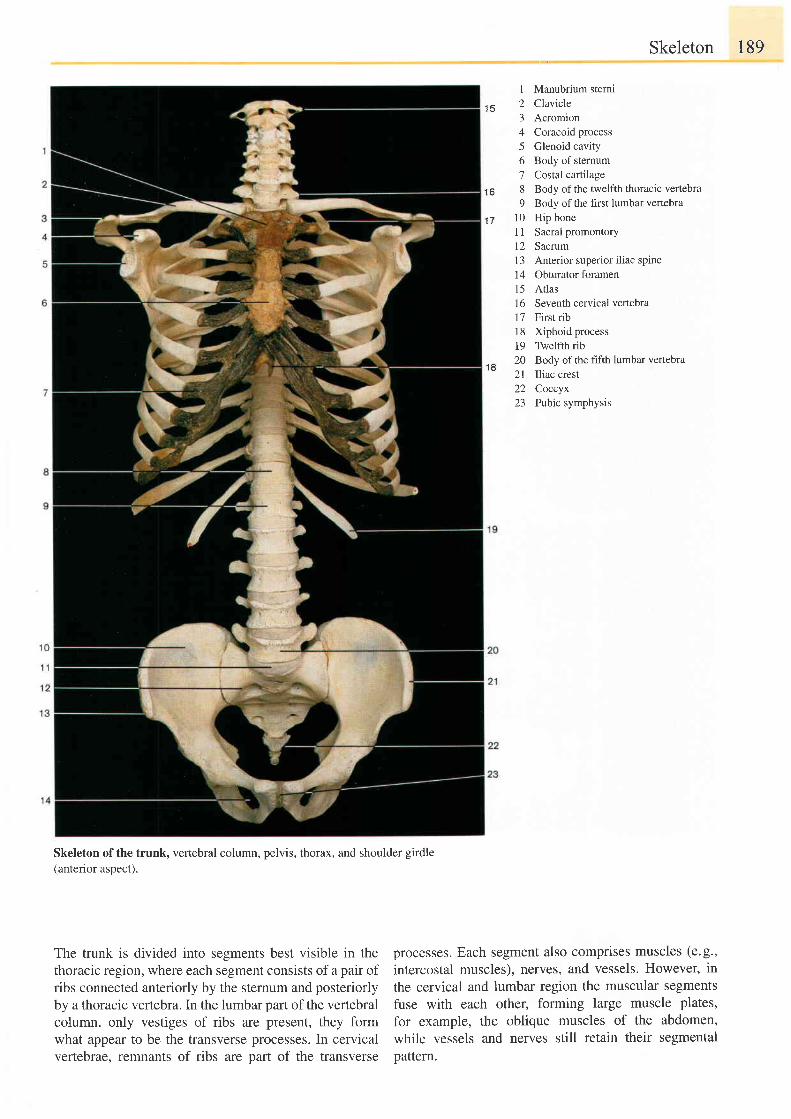
t R
Skeleton 189
1 Manubrium sterni2 Clavrcle3 Acromion4 Coracoid process5 Glenoid cavity6 Body ofsternum7 Costal cartilage8 Body ofthe twelfth thoracic vertebra9 Body of the first lumbar vertebra
10 Hip bone11 Sacral promontory12 Sacrum13 Anterior superior iliac spine14 Obturator foramenl5 Atlas16 Seventh cervical vertebra17 First rib18 Xiphoid process19 Twelfth rib20 Body of the hfth lumbar vertebra2l Iliac crest22 Coccyx23 Pubic symphysis
1 6
1 7
1 8
Skeleton ofthe trunk, vertebral column, pelvis, thorax, and shoulder girdle(anterior aspect).
The trunk is divided into segments best visible in thethoracic region, where each segment consists of a pair ofribs connected anteriorly by the sternum and posteriorlyby a thoracic veftebra. In the lumbar part of the vertebralcolumn, only vestiges of ribs are present, they formwhat appear to be the transverse processes. In cervicalvertebrae, remnants of ribs are part of the transverse
processes. Each segment also comprises muscles (e.g.,
intercostal muscles), nerves, and vessels. However, in
the cervical and lumbar region the muscular segmentsfuse with each other, forming large muscle plates,
for example, the oblique muscles of the abdomen,while vessels and nerves still retain their segmentalpattern.
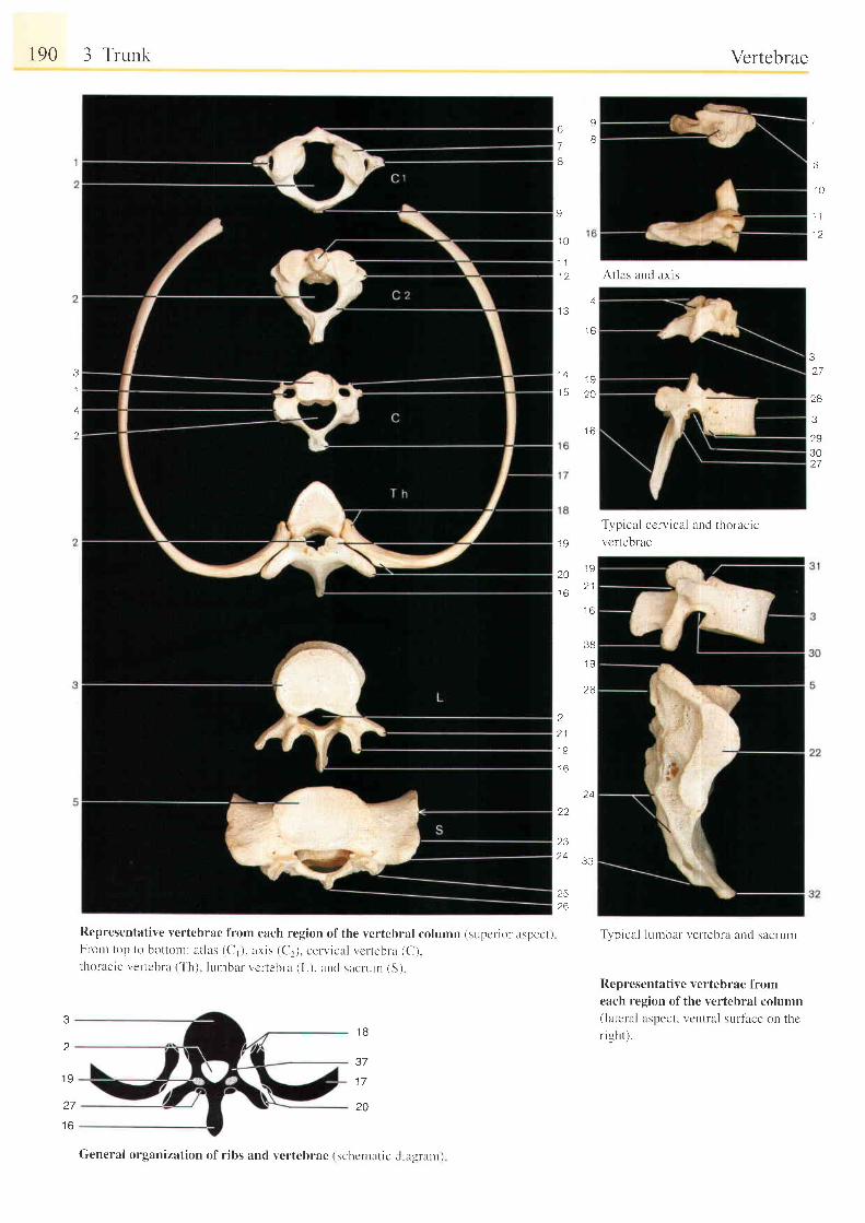
190 3 Trunk
Rcpresentative vertebrae f iom each region of the vcrtebral column (supcl ior aspect)From top to bo t ton t : a t las (C, ) . ax is (C. ) . cen , ica l vc r tebr -a (C) .Lhoracic vertcbrx ( ' l 'h). lulnbar vcrtebru (L), and sacrurn (S)
1 B
37
1 7
20
Vertebrae
t)
7
B
9
'10
1 1i ' ) .
1 9
20
J O
2
2 1
1 9
i o
g
8
7
6
1 0
1 l
1 2
At las and ax is
3a 7
28
3
?9302 7
3
1
'2.
41 3
l o
1 A
1 6
1 9
2 1
1 6
3B
26
Typical ccrvical ancl thoracrcvcrtebrae
Typical lurnbar- vertebra and sacrum
Representative vcrtebrae fromeach region ofthe vertebral column(lateral aspect. vcntral sulface on thcr igh t ) .
24
22
23
J.J
2526
1 9
27
1 6
General organization of r ibs and vertebrae (schcntatic diagr.arny
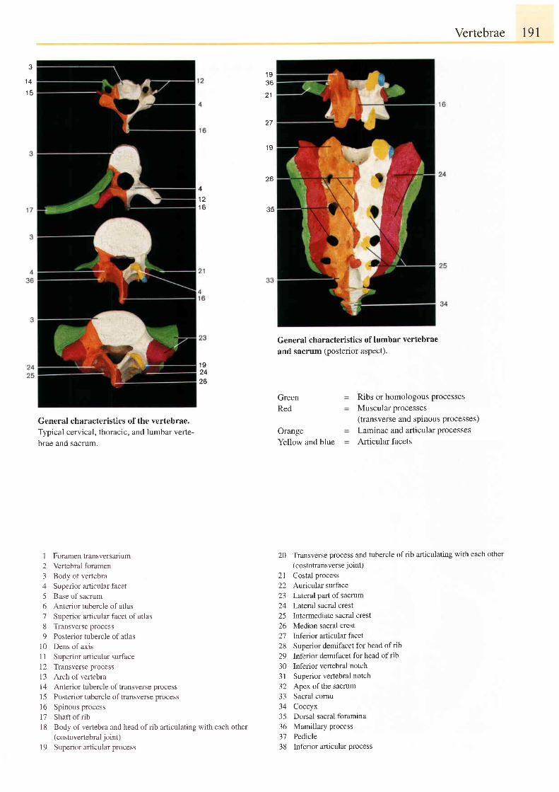
Vertebrae 191
J
1 4
1 q
1 936
27
1 9
C A
35
4
1 6
General characteristics of the vertebrae.Typical cervical, thoracic, and lumbar verte-brae and sacrum.
I Foramentransversalium2 Vertebral foramen3 Body of vertebra4 Superior afticular facet5 Base of sacrum6 Anterior tubercle of atlas7 Superior articular facet of atlas8 Transverse process9 Posterior tubercle of atlas
10 Dens of axis1 I Superior articular surface12 Transverse process13 Archofvertebra14 Anterior tubercle of transverse processI 5 Posterior tubercle of ffansverse processl6 Spinous process17 Shaft ofrib1 8 Body of vertebra and head of rib articulating with each other
(costovertebral joint)l9 Superior articular process
General characteristics of lumbar vertebrae
and sacrum (posterior aspect).
1 92426
GreenRed
OrangeYellow and blue
Ribs or homologous processesMuscular processes(transverse and spinous processes)Laminae and articular processesArticular facets
20 Transverse process and tubercle ofrib articulating with each other(costotransverse joint)
21 Costal process22 Auricular surface23 Lateral part of sacrum24 Lateral sacral crest25 Intermediate sacral crest26 Median sacral crest21 lnferior articular facet28 Superior demifacet for head of rib29 Inferior demifacet for head ofrib30 Inferior vertebral notch3l Superior vertebral notch32 Apex ofthe sacrum33 Sacral cornu34 Coccyx35 Dorsal sacral foramina36 Mamillary process37 Pedicle38 Inferior articular process
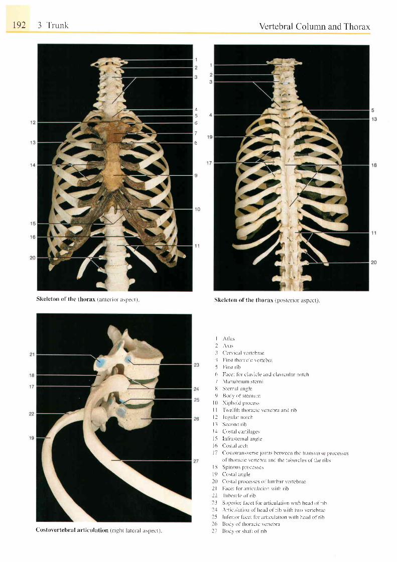
t92 3 Trunk
Skeleton of the thorax (anter-iol aspect )
Vertebral Column and Thorax
Skeleton of the thorax (posterior aspect).
I A t l as
3 Ccrvical ver tebrac
4 First thoracic vertebra
5 First r ib
6 Facct l i r r -c lavic le and c lavicular notch
7 Manubr ium sterni
13 Stcrnal angle
9 Bocli, of stertrttnt
l0 Xiphoid process
I I Tu'e l f rh thoracic vertebra and r ib
| 2 Jugular notch
l3 Sccond r i l r
l : l Costal car t i lagcs
l5 Inf ' rastcrnal angle
l6 Costal arch
I 7 Costorransversc jo ints between the t ransverse processcs
ol thor l rc ic t ,er tebra and thc tubercles of the r ibs| [ i Spinous processcs
l9 C'ostal angle
20 Cosr l l proccsses of l r , rmbar vcr tebrae
2l Facct t i r r ar t iculat ion rv i th r ib
22 Tuberclc o l ' r ib
23 Super ior f lLcet fbr ar t iculat ion wi th heacl o1' r ib2.1 Ar t iculat ion of head ol ' r ib wi th two vertcbrte2-5 Infer io l f i rcct lor ar t iculat ion wi th head of l ib2(r Bodl 'o l thoracic vertebra
27 Bodv or shaf i of r ib
A
5
6
7
B
Costovertebral art iculat ion (r ight latcral aspect)
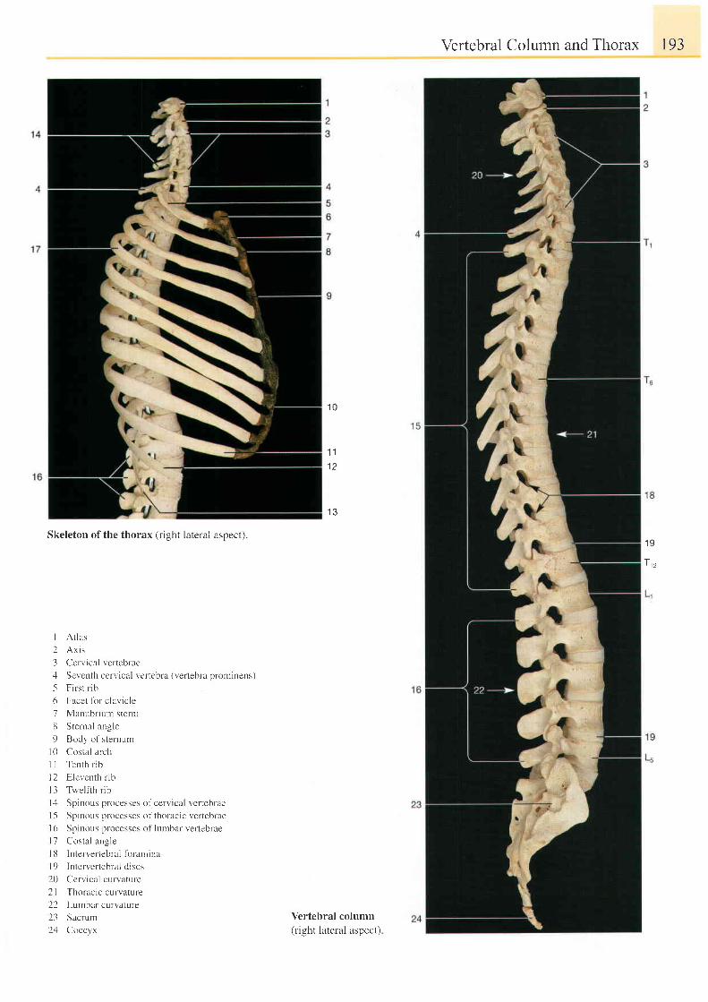
Vertebral Column and Thorax I93
Skeleton of the thorax (right lateral aspect).
I Atlas
2 Ax i s
3 Cervical vertebraezl Seventh cervical vertebra (vertebra prorninens)
5 F i r s t r i b
6 Facet fbr clavicle
7 Manubr ium sterni
8 Stelnal angle
9 Body of sternum
10 Costal arch
I I Tenth rib
l2 Eleventh r ib
l3 Twel f ih r ib
l rl Spinous processes of cervical vertebrae
| 5 Spinor.rs processes of thoracic vertebrae
I 6 Spinous processes of lumbar vertebrae
l7 Costal rngle
l8 Intervertebral tbramina
19 Interverteblal cliscs
20 Ccrvical curvature
21 Thoracic curvature
22 Lumbar curvature
23 Sacrum
2zl Coccyx
1 0
1 1
1 2
1 3
Vertebral column(right lateral aspect)
1 9
T,,
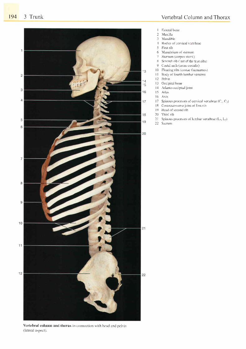
194 3 Trunk
Vertebral column and thorax in connection with head and oelvis(lateral aspect).
Vertebral Column and Thorax
I Frontal bone
2 Maxi l la
3 Mandible
4 Bodies of cervical verteDrae
5 First rib
6 Manubrium of sternum
7 Sternum (corpus sterni)
8 Seventh rib (last of the true ribs)
9 Costal arch (arcus costalis)
l0 Floating ribs (costae fluctuantes)
I I Body of fburth lumbar vertebra
l2 Pelv is
l3 Occip i ta l bone
l4 At lanto-occip i ta l jo int
15 At las
l 6 A x i s
17 Spinous processes of cervical vertebrae (Co, Cs)
I 8 Costotransverse joint of first rib
I 9 Head of second rib
20 Third r ib
21 Spinous processes of lumbar vertebrae (L2, L1)
22 Sacrum
I J
1 4t 3
t o
1 7
1 8
1 9
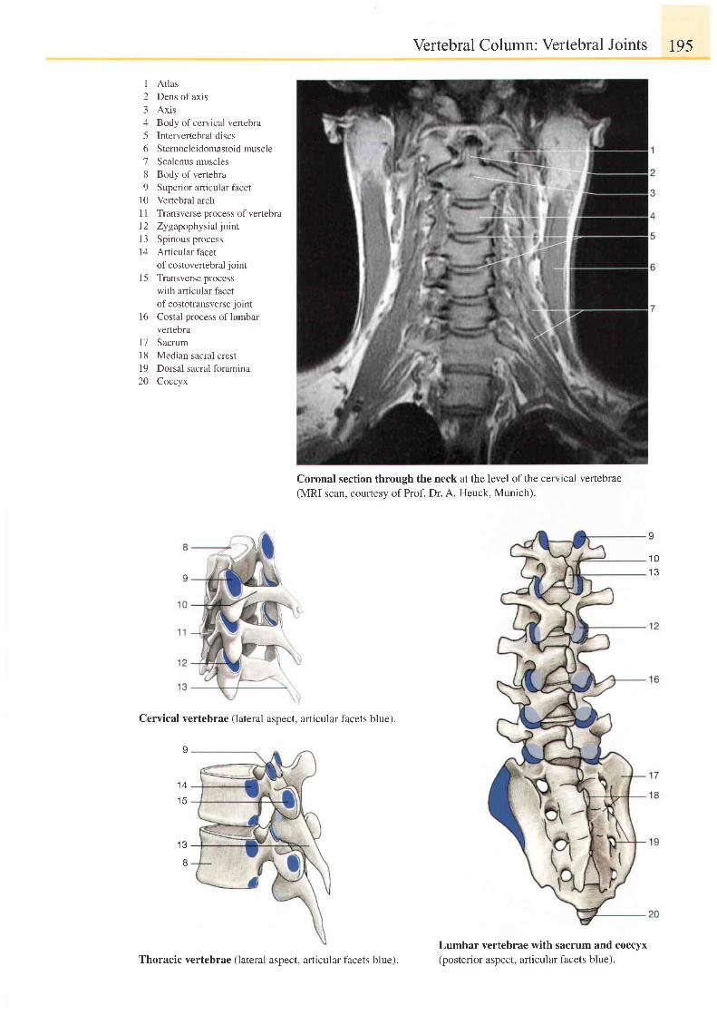
I Atlas2 Dens of axis3 Axis4 Body ofcervical vefiebra5 Intervertebral discs6 Sternocleidomastoidmuscle7 Scalenus muscles8 Body of vertebra9 Superior articular facet
10 Vertebral arch1 1 Transverse process of vertebra12 Zygapophysialjoint13 Spinous process14 Articular facet
of costovertebral jointI 5 Transverse process
with articular facetof costotransverse joint
16 Costal process oflumbarvertebra
17 Sacrum18 Median sacral crest19 Dorsal sacral foramina20 Coccyx
Cervical vertebrae (lateral aspect, afticular facets blue).
I
1 4
t e
8
Vertebral Column: Vertebral Joints I95
Coronal section through the neck at the level of the cervical vertebrae(MRI scan, courtesy of Prof. Dr. A. Heuck, Munich).
I
1 01 3
Lumbar vertebrae with sacrum and coccyx(posterior aspect, articular facets blue).Thoracic vertebrae (lateral aspect, articular facets blue).
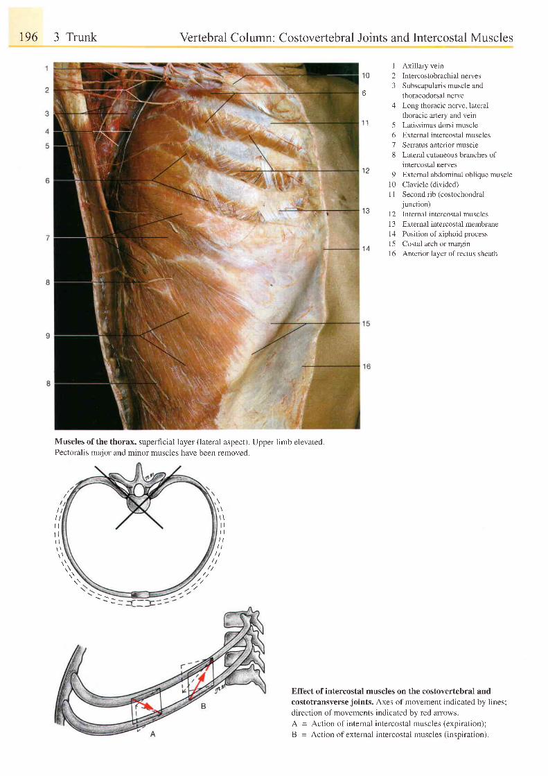
196 3 Trunk
Muscles of the thorax, superficial layer (lateral aspect). Upper limb elevated.Pectoralis major and minor muscles have been removed.
l \l lt lt t
Vertebral Column: Costovertebral Joints and Intercostal Muscles
I Axillary vein10 2 Intercostobrachial nerves
3 Subscapularis muscle and6 thoracodorsal nerve
4 Long thoracic nerve, lateralthoracic artery and vein
l1 5 Latissimus dorsi muscle6 External intercostal muscles7 Seratus anterior muscle8 Lateral cutaneous branches of
intercostal nerves1 2 9 External abdominal obl ique muscle
10 Clavicle (divided)1 I Second rib (costochondral
iunction)13 12 internal intercostal muscles
13 External intercostal membrane14 Position of xiphoidprocess
1 a 15 Costal arch or margin16 Anterior layer ofrectus sheath
ut lr lt t
//,\\ //
Effect of intercostal muscles on the costovertebral andcostotransverse joints. Axes of movement indicated by lines;
direction of movements indicated by red arrows.
A = Action of internal intercostal muscles (expiration);
B = Action of external intercostal muscles (inspiration).
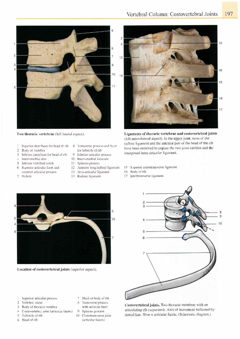
Two thoracic vertebrae (left lateral aspect)
Superior demifacet for head of rib
Body of vertebra
Inf'erior demifacet fbr head of rib
Intervertebral disc
Inferior vertebral notch
Superior articular facet and
superior articular process
Pedicle
I
23456
8 Transverse process and f'acet
fbr tubercle of rib
9 lnfer ior rn icular proce\s
l0 Interveftebral foramen
I 1 Spinous process
12 Anterior longitudinal ligament
13 Intra-ar t icular l igament
14 Radiate ligament
Vertebral Column: Costovertebral Joints 197
Ligaments of thoracic vertebrae and costovertebral joints
(left anterolateral aspect). ln the upper joint, most of the
radiate ligament and the anterior part of the head of the rib
have been removed to expose the two joint cavities and the
interposed intra-articular ligament.
I 5 Superior costotransverse ligamentl6 Body of r ibI 7 Intertransverse ligament
Ba
1 0
Location of costovertebral joints (superior aspect)
I Superior articular process
2 Vertebral canal
3 Body of thoracic vertebra
4 Costovertebral joint (articular facets)
5 Tubercle of rib
6 Head of rib
7 Shaft or body of rib8 Transverse process
with articular facet9 Spinous process
l0 Costotransverse joint(articular facets)
Costovertebral joints. Two thoracic vertebrae with an
articulating rib (separated). Axis of movement indicated by
dotted line. BIue = articular facets. (Schematic diagram.)
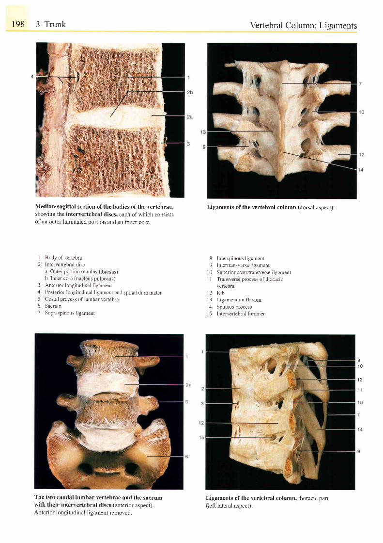
198 3 Trunk
Median-sagittal section of the bodies of the vertebrae,showing the intervertebral discs, each of which consistsof an outer laminated portion and an inner core.
12
Body of vertebraIntervertebral disca Outer portion (anulus fibrosus)b Inner core (nucleus pulposus)Anterior longitudinal ligamentPosterior longitudinal ligament and spinal dura materCostal process of lumbar veftebraSacrumSupraspinous ligament
34561
The two caudal lumbar vertebrae and the sacrumwith their intervertebral discs (anterior aspect).Anterior longitudinal ligament removed.
Vertebral Column: Lisaments
Ligaments of the vertebral column (dorsal aspect).
8 Interspinousligament9 lntertransverseligament
10 Superior costotransverse ligamentI I Transverse process of thoracrc
vertebral 2 R i b13 Ligamentum flavum| 4 Spinous processl5 Intervertebralforamen
I1 0
1 2
Ligaments of the vertebral column, thoracic part(left lateral aspect).
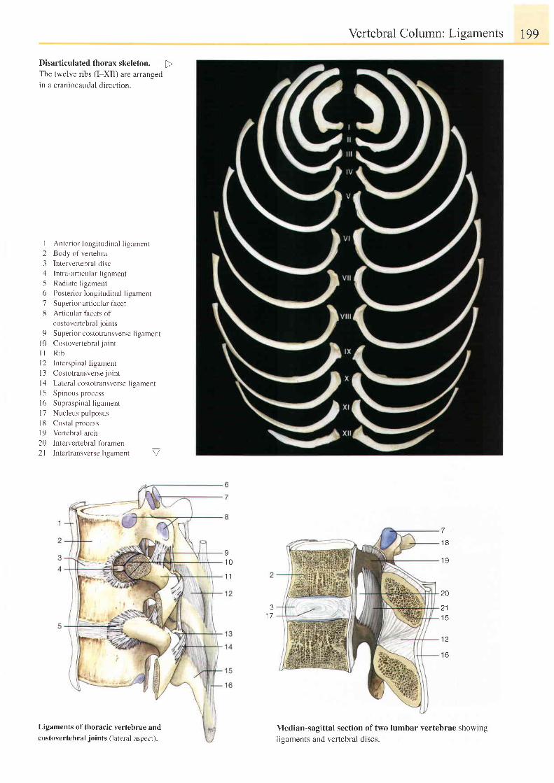
Vertebral Column: Ligaments 199
Disarticulated thorax skeleton.The twelve ribs (I-XII) are arrangedin a craniocaudal direction.
I
2345o'7
8
9l 0l ll 2l 3t 1t 5l 6t 7l 81 9202 l
Anterior longitudinal Iigament
Body of vertebra
Intervertebral disc
Intra-articular ligament
Radiate ligament
Posterior longitudinal ligament
Superior articular facet
Articular facets of
costovertebral joints
Superior costotransverse ligament
Costovertebral joint
R ib
Interspinal l igament
Costotransverse joint
Lateral costotransverse ligament
Spinous process
Supraspinal ligament
Nucleus pulposus
Costal process
Vertebral arch
Intervertebral foramen
Intertransverse ligament V
I1 0
1 1
7
1 8
1 9
20
21t c
12
1 6
31 7
Ligaments of thoracic vertebrae andcostovertebral joints (lateral aspect).
Median-sagittal section of two lumbar vertebrae showingligaments and verlebral discs.
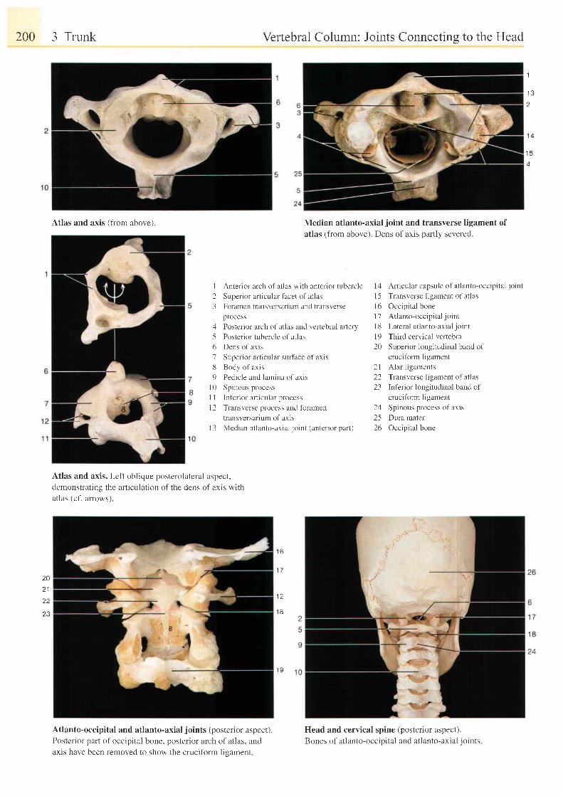
200 3 Trunk
Atlas and axis (from above).
Vertebral Column: Joints Connectins to the Head
Median atlanto-axial joint and transverse ligament ofatlas (from above). Dens of axis partly severed.
1
1 3t
1 4
1 5
4
I Anterior arch ol atlas with anterior tubercle
2 Superior articular facet of atlas
3 Foramen transversarium and transverse
process
4 Posterior arch of atlas and vertebral artery
-5 Posterior tubercle of atla:
6 Dens of axis
7 Superior articular surf'ace of axis
8 Body o f ax i s
9 Pedic le und lurnina of axis
| 0 Spinous process
| | ln fer ior ar l icul r r process
l2 T ran : re r se pn reg l . , nU l i r r amcn
transversarium of axis
l3 Median at lanto-ar ia l jornt (anter ior part )
1 7
14 Articular capsule of atlanto-occipital joint
I 5 Transverse ligament of atlas
l6 Occipital bone
17 At lanto-occip i ta l jo int
l8 Lateral atlanto-axialjoint
l9 Third cervical vertebra
20 Superior longitudinal band of
cruciform ligament
Alar ligaments
Transverse ligament of atlas
In l 'er ior longi tudinal band ol
cruciform ligament
Spinous process of axis
Dura mater
Occipital bone
2 12223
1 1
2526
Atlas and axis. Left oblique posterolateral aspect,demonstrating the articulation of the dens of axis withatlas (cf. arrows).
Atlanto-occipital and atlanto-axial joints (posterior aspect).Posterior part of occipital bone, posterior arch of atlas, andaxis have been removed to show the crucifbrm ligament
Head and cervical spine (posterior aspect).
Bones of atlanto-occipital and atlanto-axial joints
1 6
20
221 8
l 9
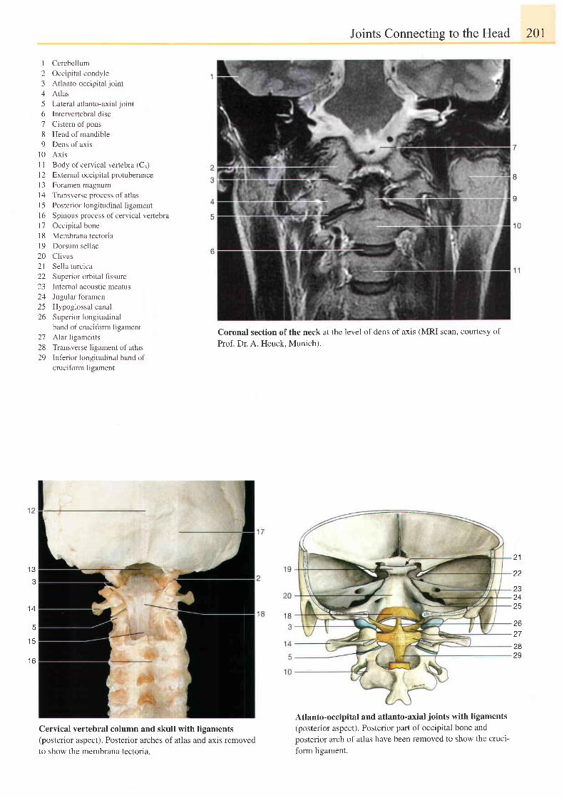
Cerebellum
Occipital condyle
Atlanto-occipital joint
Atlas
Lateral atlanto-axial joint
Intervertebral disc
Cistem of pons
Head of mandible
Dens of axis
Axrs
Body of cervical vertebra (C.)
External occipital protuberance
Foramen magnum
Transverse process of atlas
Posterior longitudinal ligament
Spinous process of cervical vertebra
Occipital bone
Membrana tectoria
Dorsum sellae
Clivus
Sel la turc ica
Superior orbital fi ssure
Internal acoustic meatus
Jugular foramen
Hypoglossal canal
Superior longitudinal
band of crucifbrm ligament
Alar ligaments
Transverse ligament of atlas
Inf 'er ior longi tudinal band oI
cruciform ligament
Coronal section of the neck
Prof. Dr. A. Heuck, Munich).
Joints Connecting to the Head
at the level of dens of axis (MRI scan, courtesy of
201
123456789
l 0l ll 2t 3t 4l 5t 6T'7t 8l 9202 l22L-)
242526
272829
. A
t c
t o
21
zz
zc
z+
25
zo
27
2829
Cervical vertebral column and skull with ligaments(posterior aspect). Posterior arches of atlas and axis removedto show the membrana tectoria.
Atlanto-occipital and atlanto-axial joints with ligaments
(posterior aspect). Posterior part of occipital bone and
posterior arch of atlas have been removed to show the cruci
form ligament.
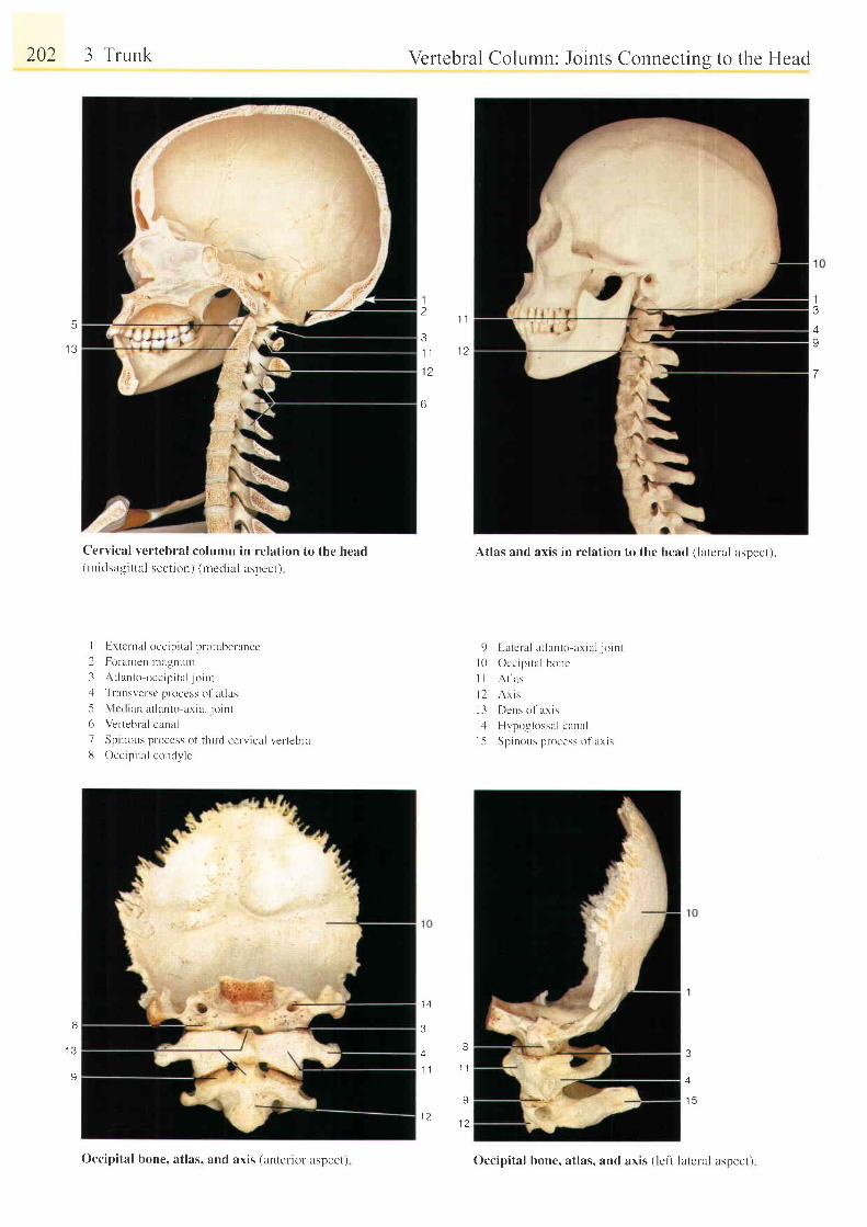
202 3 Trunk
Cervical vertebral column in relation to the head(midsa-eit tal section) (medial aspect)
I Exrernal occip i ta l protuberance
2 Foranten lnalinLlnt
3 At lanto occip i ta l jo int, l Transversc process ol 'at las
5 Mcci ian at lanto axia l jo int
6 Vertebral canal
7 Spinous process of th i rd cerv ical ver tebra13 Occipi ta l condl ' le
Vertebral Column: Joints Connectins to the Head
Atlas and axis in relation to the head (lateral asDect).
I L r t e l t l l r t l l t t t l o r r r i : r l - j o i t t tl0 Occip i ta l bone
I I A t l as
l 2 A r i s
l 3 Dens o f ax i s
l - [ [Jvpoglossal canal
l -5 Spinous process of axis
3
a4 -
1 1 1 1
9
1 ?1 2
5
I J
13/9
7
1t
t l
31 1 1 2
6
I
I J
9
3
4
1 5
Occipital bone, atlas, and axis (anterior aspect) Occipital bone, atlas, and axis (lel't latelal aspect)
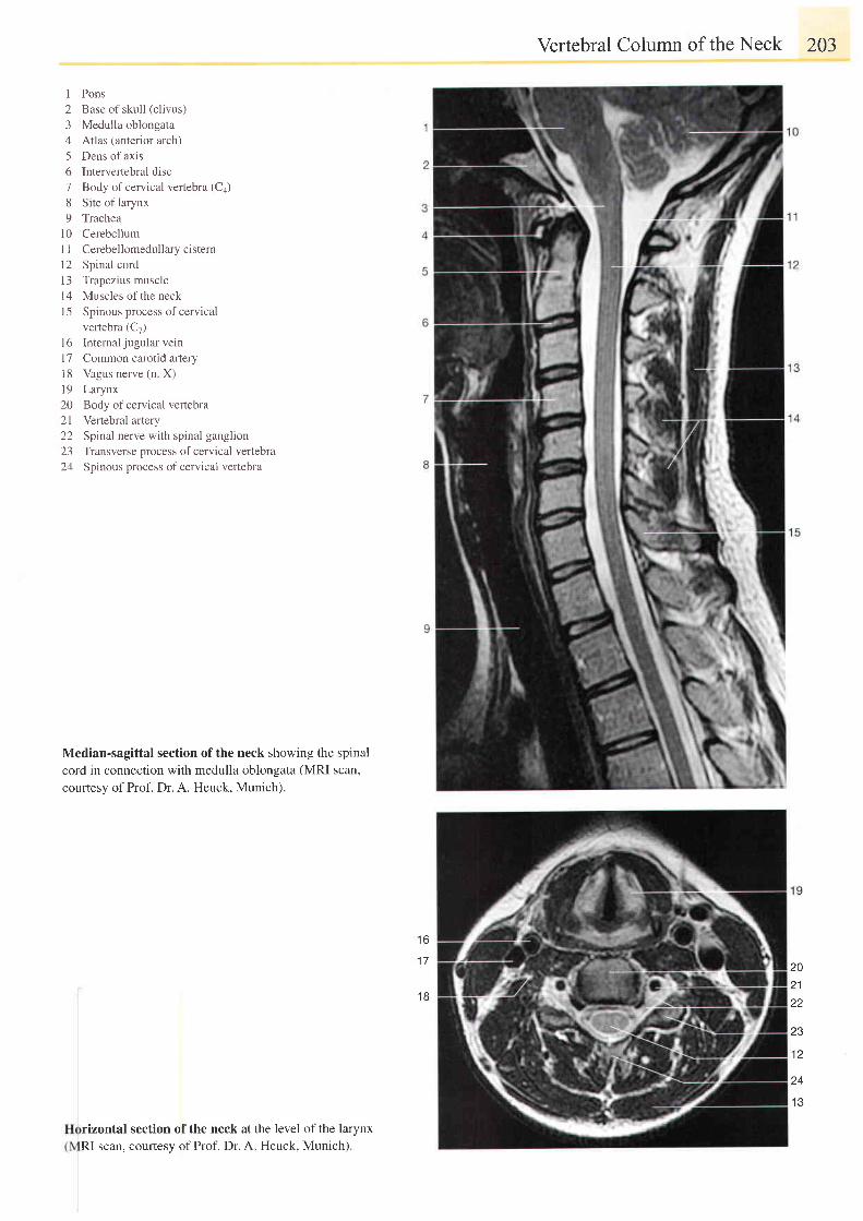
Vertebral Column of the Neck 203
I Pons2 Base of skull (clivus)3 Medulla oblongata4 Atlas (anterior arch)5 Dens of axis6 Intervertebral disc7 Body ofcervical vertebra (Ca)8 Site of larynx9 Trachea
i0 CerebellumI I Cerebellomedullary cistern12 Spinal cordl3 Trapezius musclel4 Muscles of the neckI 5 Spinous process of cervical
vertebra (C7)16 Intemal jugular vein17 Common carotid artery18 Vagusnerve(n X)l9 Larynx20 Body ofcervical vertebra21 Vertebral artery22 Spinal nerve with spinal ganglion23 Transverse process of cervical vertebra24 Spinous process of cervical vertebra
Median-sagittal section of the neck showing the spinal
cord in connection with medulla oblongata (MRI scan,
courtesy of Prof. Dr. A. Heuck, Munich).
1 7
1 6
202122
23
1 2
24
I J
rizontal section of the neck at
RI scan, courtesy of Prof. Dr. Athe level of the larynx
. Heuck, Munich).
1 8
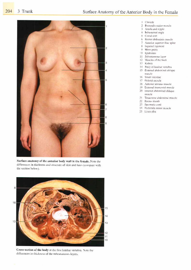
204 3 Trunk
Surface anatomy of the anterior body wall in the female. Note thedifferences in thickness and structure of skin and hairs (comoare withthe section below).
Cross section of the body at the llrst lumbar vertebra. Note thedifferences in thickness of the subcutaneous lavers.
Surface Anatomy of the Anterior Body in the Female
I Clavicle
2 Pectoralis major muscle
3 Areola and nipple
4 Infrasternal angle
5 Costal arch
6 Rectus abdominis muscle
7 Anter ior ruper ior i l iac spine
8 Inguinal ligament
9 Mons pubis
l0 Epidermis
1 I Subcutaneous layer
12 Muscles of the back
l3 Kidney
l4 Body of lumbar vertebra
15 External abdominal oblique
muscle
l6 Smal l intest ine
l7 Deltoid muscle
18 Anterior serratus muscle
l9 External intercostal muscle
20 Internal abdominal oblique
muscle
21 Transverse abdominal muscle
22 Rectus sheath
23 Spermatic cord
24 Pectoralis minor muscle
25 Linea alba
1 4
1 3
1 21 1
1 0
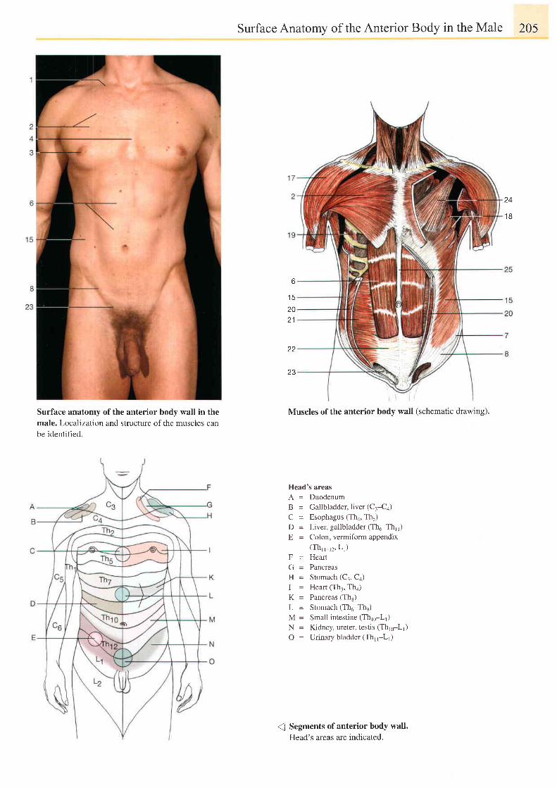
Surface anatomy of the anterior body wall in themale. Localization and structure of the muscles canbe identified.
Surface Anatomy of the Anterior Body in the Male 205
6
2021
22
23
Muscles of the anterior body wall (schematic drawing).
Head's areasA = DuodenumB = Gallbladder, liver (C:-C+)
C = Esophagus (Tha, Th5)D = Liver, gallbladder(Th6-Thlr)E = Colon, vermiform appendix
(Thr_r2, Lr)F = HeartG = PancreasH = Stomach (C:, C+)I = Heart (Th.r, Th4)K = Pancreas (Ths)L = Stomach (Th6-The)M = Small intestine (Thro-Lr)N = Kidney, ureter, testis (Thro-Lr)
O = Urinary bladder (Th"-L1)
( Segments of anterior body wall.Head's areas are indicated.
24
1 8
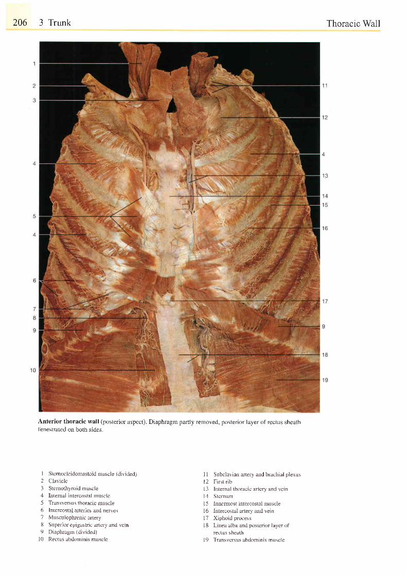
206 3 Trunk
Anterior thoracic wall (posterior aspect). Diaphragm partly removed, posterior layer ofrectus sheathfenestrated on both sides.
Thoracic Wall
I Stemocleidomastoid muscle (divided)2 Clavicle3 Sternothyroid muscle4 Internal intercostal muscle5 Transversus thoracic muscle6 Intercostal arteries and nerves7 Musculophrenicartery8 Superior epigastric artery and vein9 Diaphragm (divided)
l0 Rectus abdominis muscle
I 1 Subclavian anery and brachial plexusl2 First rib13 Internal thoracic artery and vein14 Sternuml5 Innermost intercostal musclel6 Intercostal artery and veinl7 Xiphoid processl8 Linea alba and posterior layer of
rectus sheathl9 Transversus abdominis muscle
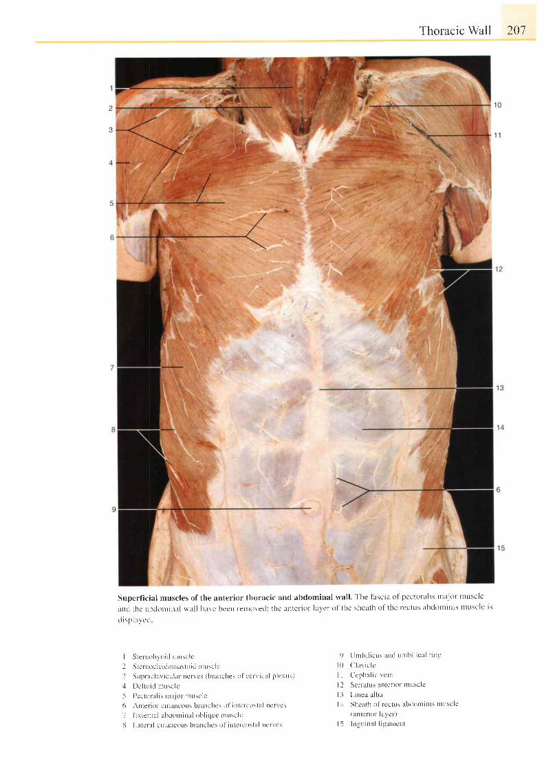
Thoracic Wall 201
Superf icial muscles of the anterior thoracic and abdominal wall . The tascia of pectoral is tnajor musclc
and thc abclontrnal wall have bccn retnoved: thc untcl ior laycr ol thc sheath of the rcctus abdorninis tnuscle is
disphyecl
qI
I Sternohvoid nrusclc
I S t c rn0c l c i dL r t nas to i d n rusc l c
3 Sup rac la r i cu l u r nc r l es ( b t ' auches o l c c r r i c l l p l c rL r s )
J De l t o i d r nL rsc l e
5 Pcctor a l is nt t jot ' r r tuscle
6 Anter ior cLr lancoLls bt 'anches ol intcrcost l t l ne t ' rcs
7 Extenral abdominal obl ic lue tnusclc
8 Latcf tL l cutru lcous branchcs ol i t l te ' rcostal t lc t l t 's
9 Urnbi l icus and urrb i l ical t ' in-g
l 0 C l l av i c l c
I I Ccpha l i c vc tn
l l S r ' r r a l t l r Jn l c r i ( ) r l nu \e l c
l 3 L i nea a l b rL
l , l Shcuth of rcct t ts abclot t t in is n luscle
(antcr ior laycr)
| 5 I ngu ina l l t gamcn l
; '
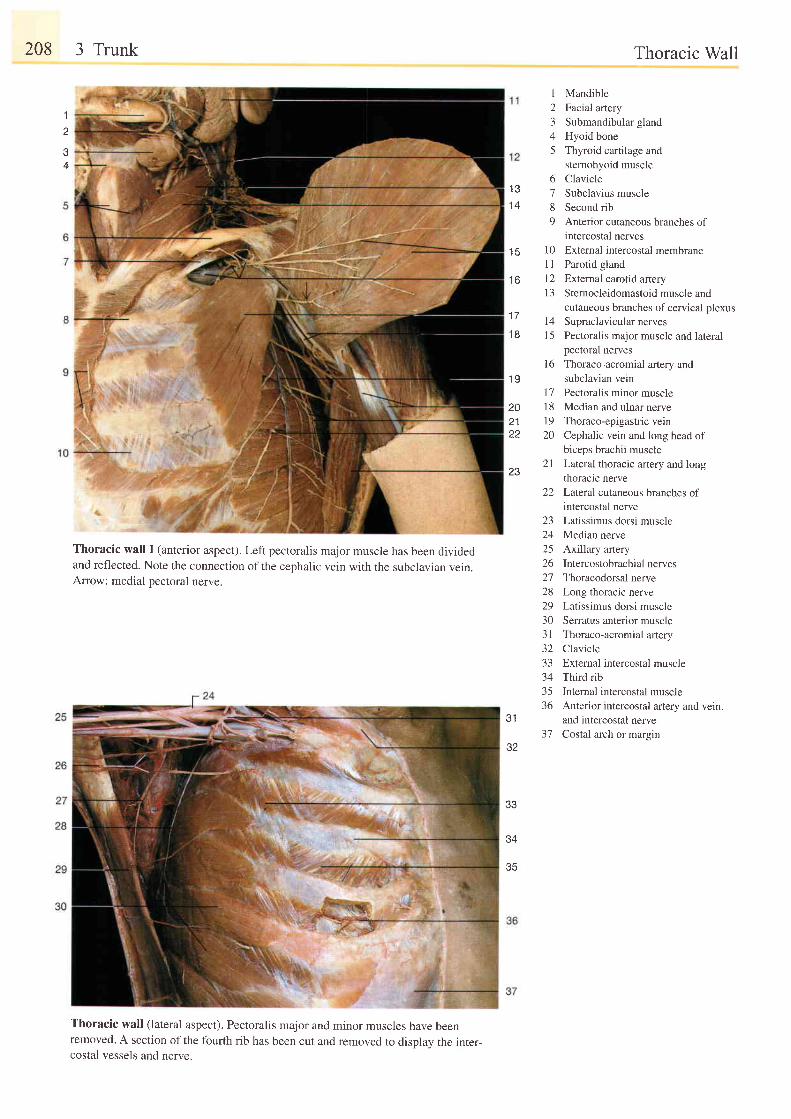
208 3 Trunk
Thoracic wall I (anterior aspect). Left pectoralis major muscle has been dividedand reflected. Note the connection of the cephalic vein with the subclavian vern.Arrow: medial pectoral nerve.
Thoracic wall (lateral aspect). Pectoralis major and minor muscles have beenremoved. A section of the fourth rib has been cut and removed to displav the inter-costal vessels and nerve.
1
I23/
5
or r 71 4 8
9
1 5 1 01 l
f o t L
1 3
1 7l +
1 8 1 5
1 61 9
I 720 1821 1922 20
2 lZ J
22
za
242526272829303 l3 Z
33343536
J I
375Z
Thoracic Wall
MandibleFacial arterySubmandibular glandHyoid boneThyroid cartilage andsternohyoid muscleClavicleSubclavius muscleSecond ribAnterior cutaneous branches ofintercostal nervesExtemal intercostal membraneParotid glandExternal carotid arteryS ternocleidomastoid muscle andcutaneous branches of cervical plexusSupraclavicular nervesPectoralis major muscle and lateralpectoral nervesThoraco-acromial artery andsubclavian veinPectoralis minor muscleMedian and ulnar nerveThoraco-epigastric veinCephalic vein and long head ofbiceps brachii muscleLateral thoracic artery and longthoracic nerveLateral cutaneous branches ofintercostal nerveLatissimus dorsi muscleMedian nerveAxillary arteryIntercostobrachial nervesThoracodorsal nerveLong thoracic nerveLatissimus dorsi muscleSerratus anterior muscleThoraco-acromial arteryClavicleExternal intercostal muscleThird ribInternal intercostal muscleAnterior intercostal artery and vein,and intercostal nerveCostal arch or margin
e
4
33
34
35
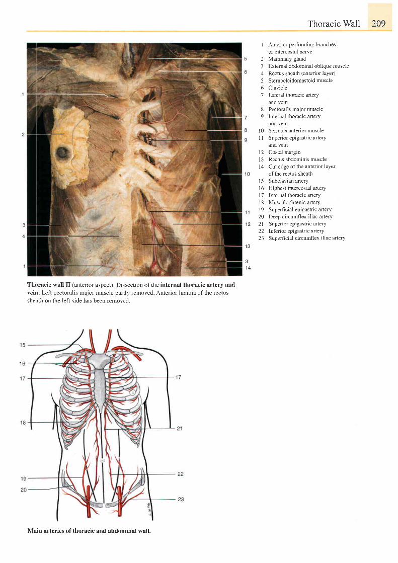
Thoracic Wall 209
1 Anterior perforating branchesof intercostal nerve
2 Mammary gland3 Extemal abdominal oblique muscle4 Rectus sheath (anterior layer)5 Sternocieidomastoidmuscle6 Clavicle7 Lateral thoracic artery
and vein8 Pectoral is major muscle9 Internal thoracic artery
and vein10 Serratus anterior muscle1 1 Superior epigastric artery
and vein12 Costal margin13 Rectus abdominis muscle14 Cut edge of the anteriorlayer
of the rectus sheath15 Subclavian artery16 Highest intercostal artery17 Internal thoracic artery18 Muscuiophrenic artery19 Superficial epigastric artery20 Deep circumflex iliac artery2l Superior epigastric afiery22 lnferror epigastric artery23 Superficial circumflex iliac anery
1 3
d
1 4
Thoracic wall II (anterior aspect). Dissection ofthe internal thoracic artery andvein. Left pectoralis major muscle partly removed. Anterior lamina of the rectussheath on the left side has been removed.
Main arteries of thoracic and abdominal wall.
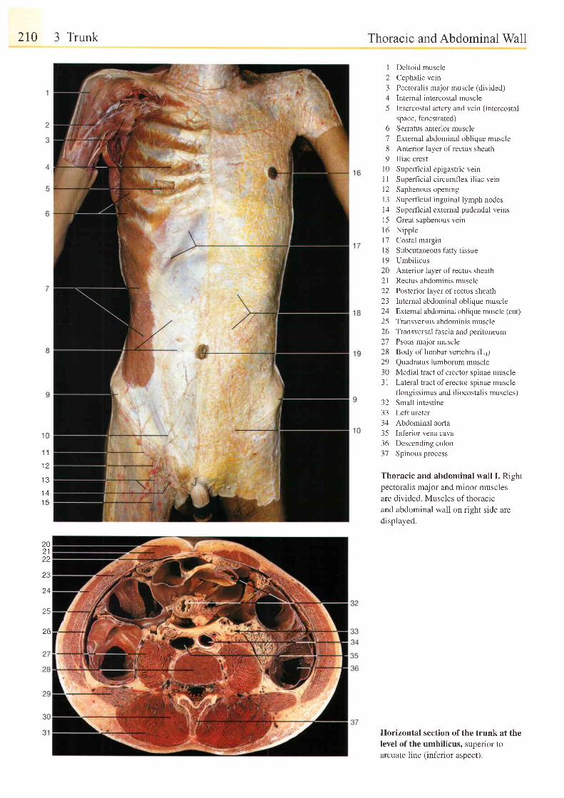
210 3 Trunk
1 3
1 41 5
Thoracic and Abdominal Wall
12345
6789
1 01 1t 21 31 4l 51 6t '71 81 9202 122L J
2425Z O
212829303 l
Deltoid muscle
Cephalic vein
Pectoralis major muscle (divided)
Internal intercostal muscle
Intercostal artery and vein (intercostal
space, fenestrated)
Serratus anterior muscle
External abdominal oblique muscle
Anterior layer of rectus sheath
Iliac crest
Superficial epigastric vein
Superf ic ia l c i rcumflex i l iac vein
Saphenous opening
Superficial inguinal lymph nodes
Superficial external pudendal veins
Great saphenous vein
Nipple
Costal margin
Subcutaneous fatty tissue
Umbiiicus
Anterior layer of rectus sheath
Rectus abdominis muscle
Posterior layer of rectus sheath
Internal abdominal oblique muscle
Extemal abdominal oblique muscle (cut)
Transversus abdominis muscle
Transversal fascia and peritoneum
Psoas major muscle
Body of lumbar vertebra (La)
Quadratus lumborum muscle
Medial tract of erector spinae muscle
Lateral tract of erector spinae muscle(longissimus and iliocostalis muscles)
32 Small intestine33 Left ureter34 Abdominal aorta35 Inferior vena cava36 Descending colon37 Spinous process
Thoracic and abdominal wall I. Rightpectoralis major and minor muscles
are divided. Muscles of thoracrcand abdominal wall on right side are
displayed.
Horizontal section of the trunk at thelevel of the umbilicus, superior toarcuate line (inferior aspect).
1 '1
202122
Z J
24
25
26
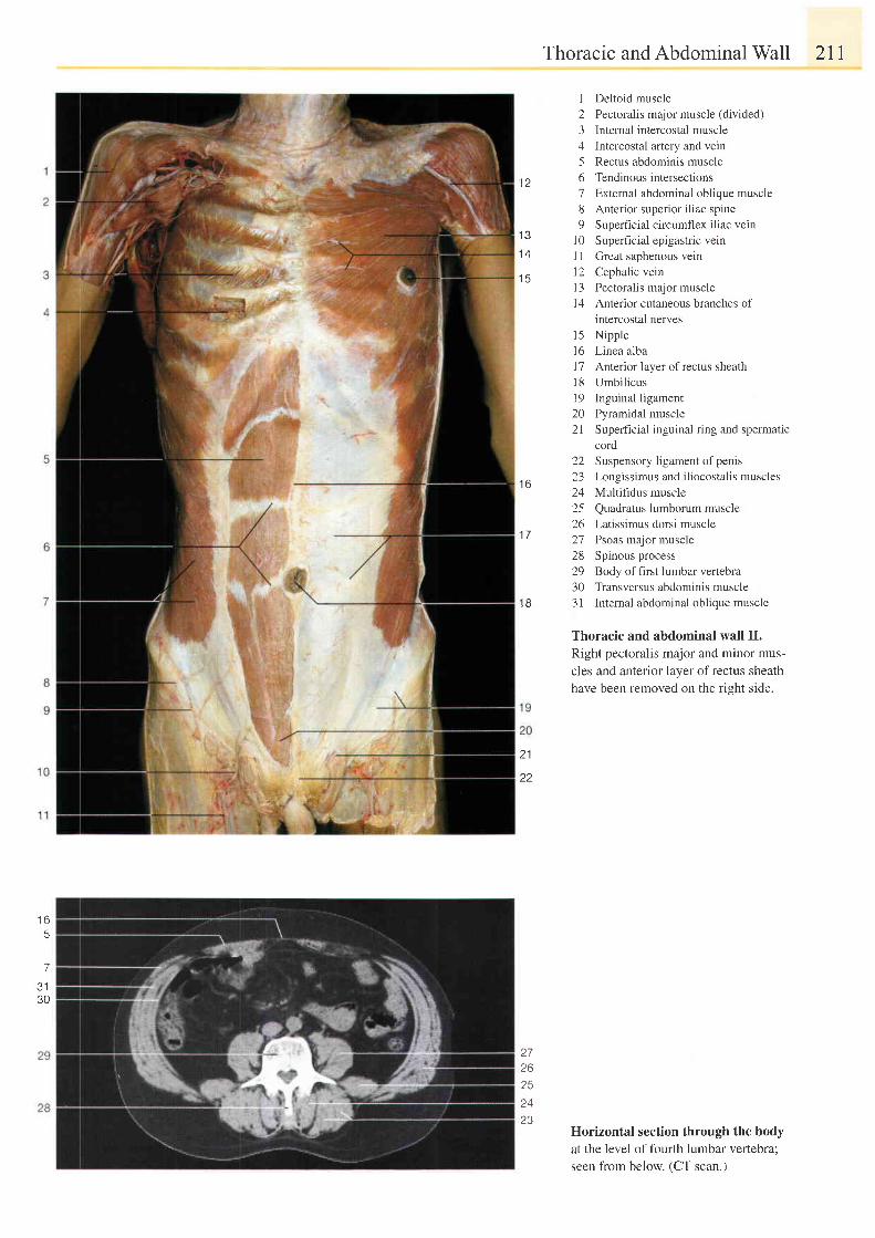
I L
Thoracic and Abdominal Wall 211
I Deltoid muscle2 Pectoralis major muscle (divided)3 Internal intercostal muscle4 Intercostal artery and vein5 Rectus abdominis muscle6 Tendinous intersections7 External abdominal oblique muscle8 Anterior superior iliac spine9 Superfrcial circumflex iliac vein
l0 Superf icial epigastr ic vein1 1 Great saphenous vern12 Cephalic veinl3 Pectoralis major muscle14 Anterior cutaneous branches of
intercostal nerves15 Nipple16 Linea alba17 Anterior layer ofrectus sheathl8 Umbil icus19 Inguinal ligament20 Pyramidal muscle2l Superficial inguinal ring and spermatic
cord22 Suspensory ligament ofpenis23 Longissimus and iliocostalis muscles24 Multifidus muscle25 Quadratus lumborum muscle26 Latissimus dorsi muscle27 Psoas major muscle28 Spinous process29 Body of first lumbar vertebra30 Transversus abdominis muscle31 Internal abdominal oblique muscle
Thoracic and abdominal wall II.
Right pectoralis major and minor mus-
cles and anterior layer ofrectus sheath
have been removed on the right side.
Horizontal section through the bodyat the level of fourth lumbar vertebra;seen from below. (CT scan.)
I J
1 4
t 3
1 7
t o
1 B
21
22
t o
5
7
J I
30
Z I
262524D2
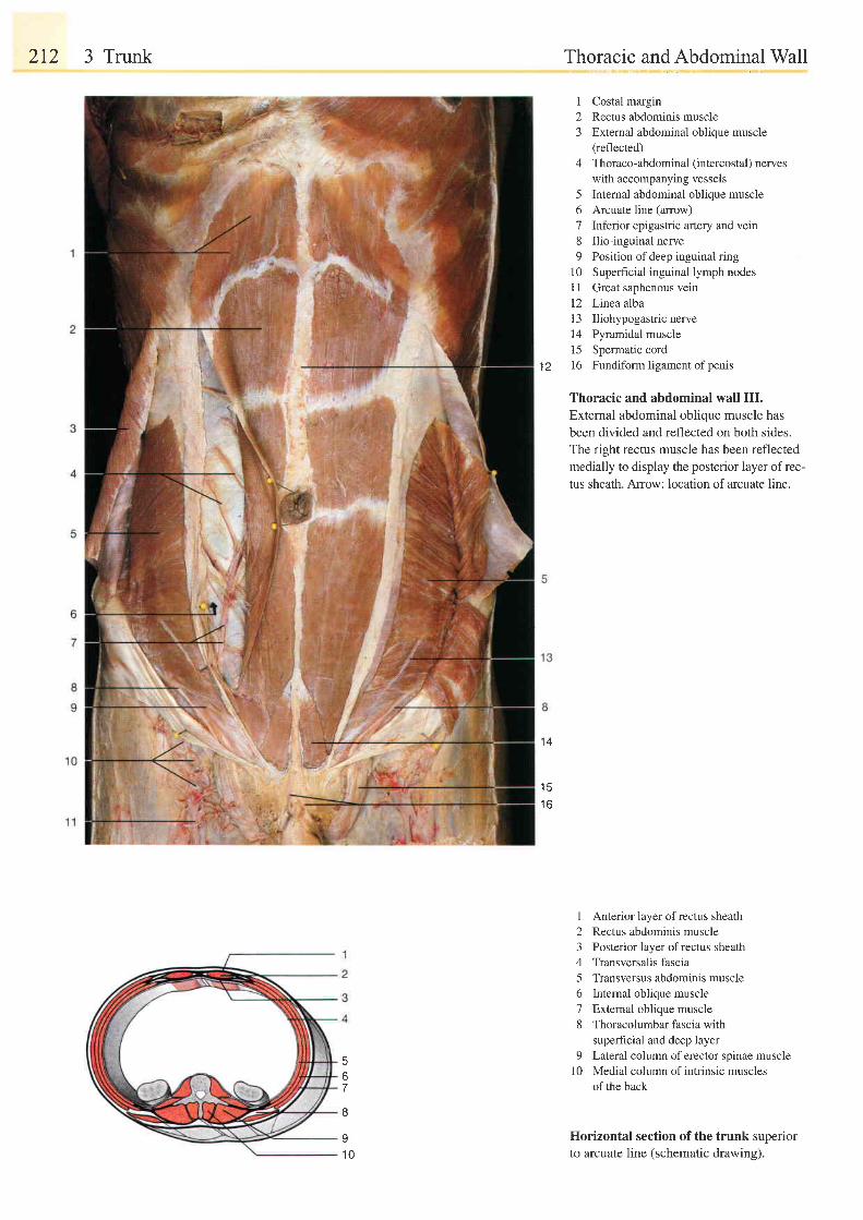
2t2 3 Trunk
t z
Thoracic and Abdominal Wall
1 Costal margin2 Rectus abdominis muscle3 External abdominal oblique muscle
(reflected)
4 Thoraco-abdominal (intercostal) nerveswith accompanying vessels
5 Internal abdominal oblique muscle6 Arcuate line (anow)7 Inferior epigastric artery and vein8 Ilio-inguinal nerve9 Position ofdeep inguinal ring
10 Superficial inguinal lymph nodesI I Great saphenous vein12 Linea alba13 Iliohypogastricnerve14 Pyramidal muscle15 Spermatic cordl6 Fundiform l igament of penis
Thoracic and abdominal wall III.
External abdominal oblique muscle has
been divided and reflected on both sides.
The right rectus muscle has been reflected
medially to display the posterior layer of rec-
tus sheath. Arrow: location of arcuate line.
1 Anterior layer of rectus sheath2 Rectus abdominis muscle3 Posterior layer ofrectus sheath4 Transversalis fascia5 Transversus abdominis muscle6 Intemal oblique muscle7 External oblique muscle8 Thoracolumbar fascia with
superFrcial and deep layer9 Lateral column of erector spinae muscle
l0 Medial column of intrinsic musclesof the back
Horizontal section of the trunk superiorto arcuate line (schematic drawing).
1 4
1 5t o
o7
I
91 0
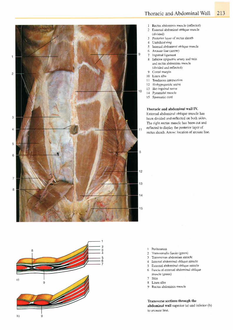
1
A
Thoracic and Abdominal Wall 213
1 Rectus abdominis muscle (reflected)
2 External abdominal oblique muscle(divided)
3 Posterior layer ofrectus sheath4 Umbilical ring5 Internal abdominal oblique muscle6 Arcuate line (arrow)
7 Inguinal ligament8 Inferior epigastric artery and vein
and rectus abdominis muscle(divided and reflected)
9 Costal margin10 Linea albaI 1 Tendinous intersection12 Iliohypogastric nerve13 Ilio-inguinal nerve14 Pyramidal muscle15 Spermatic cord
Thoracic and abdominal wall IV.
External abdominal oblique muscle has
been divided and reflected on both sides.
The right rectus muscle has been cut and
reflected to display the posterior layer of
rectus sheath. Arrow: location of arcuate line.
I
23A
5f)
PeritoneumTransversalis fascia (green)
Transversus abdominis muscleInternal abdominal oblique muscleExternal abdominal oblique muscleFascia of external abdominal obliquemuscle (green)SkinLinea albaRectus abdominis muscle
'7
89
Tfansverse sections through theabdominal wall superior (a) and inferior (b)
to arcuate line.
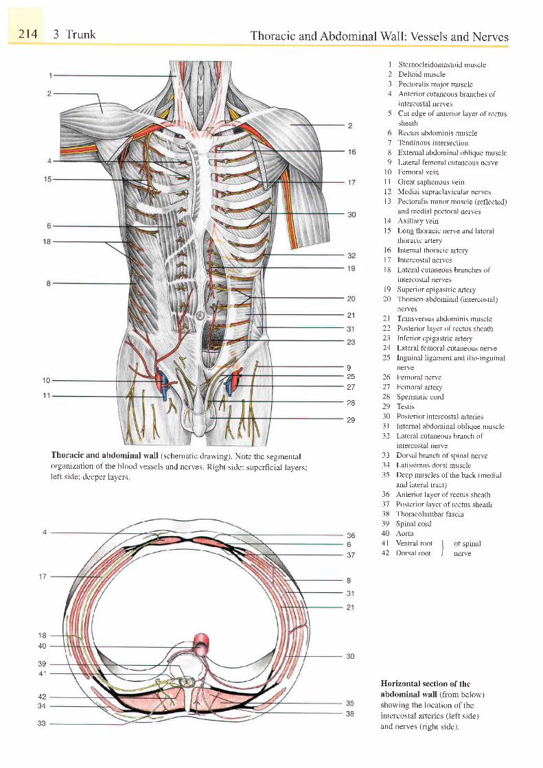
214 3 Trunk
1 840
39A 1
34
Thoracic and AbdominalWall: Vessels and Nerves
1 Sternocleidomastoidmuscle2 Deltoid muscle3 Pectoralis major muscle4 Anterior cutaneous branches of
intercostal nerves5 Cut edge of anterior layer of rectus
sheath6 Rectus abdominis muscle7 Tendinousintersection8 External abdominal oblique muscle9 Lateral femoral cutaneous nerve
10 Femoral vein1 I Great saphenous vein12 Medial supraclavicular nerves'13
Pectoralis minor muscle (reflected)and medial pectoral nerves
14 Axillary vein15 Long thoracic nerve and lateral
thoracic artery16 Internal thoracic artery17 Intercostal nerves18 Lateral cutaneous branches of
intercostal nerves19 Superior epigastric artery20 Thoraco-abdominal (intercostal)
nerves21 Transversus abdominis muscle22 Posterior layer ofrectus sheath23 Inferior epigastric artery24 Lateral femoral cutaneous nerve25 Inguinal ligament and ilio-inguinal
nerve26 Femoral nerve27 Femoral artery28 Spermatic cord29 Testis30 Posterior intercostal artenes31 Internal abdominal oblique muscle32 Lateral cutaneous branch of
intercostal nerve33 Dorsal branch of spinal nerve34 Latissimus dorsi muscle35 Deep muscles of the back (medial
and lateral tract)36 Anterior layer ofrectus sheath37 Posterior layer of rectus sheath38 Thoracolumbarfascia39 Spinal cord40 Aorta4l Ventral root
' l of soinal
I '
42 Dorsal root J nerve
Horizontal section of theabdominal wall (from below)showing the location of theintercostal arteries (left side)and nerves (right side).
92527
28
29
Thoracic and abdominal wall (schematic drawing). Note the segmentalorganization of the blood vessels and nerves. Rieht side: superficial lavers:left side: deeper lavers.
36637
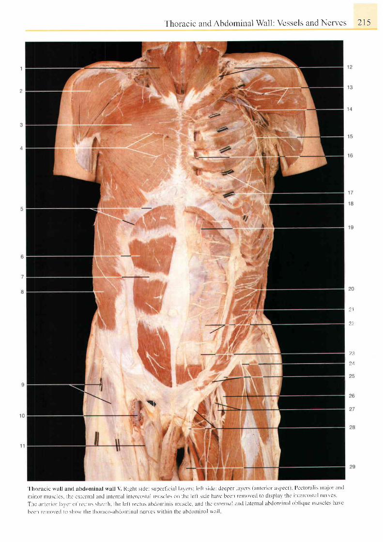
Thoracic and Abdominal Wall: Vessels and Nerves 215
r,g
-7-? 1
',}?,
23
$
tQr
::,lF,€
' Ihoracic nal l ancl abdominal lval l V. Right s ide: superf ic ia l lay 'crs: le l t s ide : deepcr lavels (auter ior aspect) Pectoral is tnr t jor and
t l l i t l t l t t l l t t s c l e s . t h c c r t e t - n a l l n c l i n t e r n a l i n t e r c c l s t a l t n t t s c l e s o n t h c l c l . t s i d c h a r ' e b c e n r e t l c l v e d t o d i s p l a y t h c i n t e r c o s t a ] n e I - , e s
T h : a r l l c r . i t l t . l a v c t . t l f r c c t r t s s h c a t h . t h e l e f t r e c t u S a b ( ( ) l ] ] i
b c . : n r cn ro re r l t o shou , t he t ho raco -abdon r i na l nc r " ' c s w i t h i n t he ahdou r i n l l wa l l
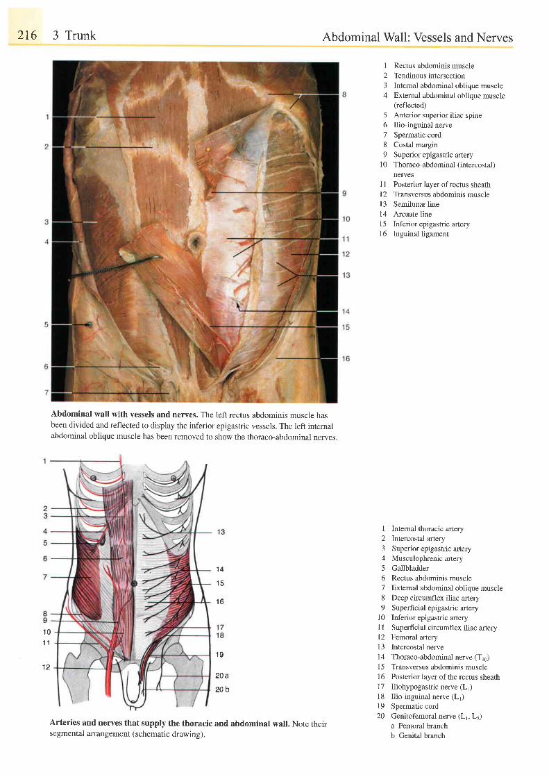
216 3 Trunk
Abdominal wall with vessels and nerves. The left rectus abdominis muscle nasbeen divided and reflected to display the inferior epigastric vessels. The left internalabdominal oblique muscle has been removed to show the thoraco-abdominal nerves.
Abdominal Wall: Vessels and Nerves
1 Rectus abdominis muscle2 Tendinousintersection3 Internal abdominal oblique muscle4 Extemal abdominal oblique muscle
(reflected)5 Anterior superior iliac spine6 Ilio-inguinal nerve7 Spermatic cord8 Costal margin9 Superior epigastric artery
10 Thoraco-abdominal (intercostal)nerves
I I Posterior layer of rectus sheath12 Transversus abdominis musclel3 Semilunar line14 Arcuate line15 Inferior epigastric artery16 Inguinal ligament
I Internal thoracic artery2 Intercostal artery3 Superior epigastric artery4 Musculophrenic artery5 Gallbladder6 Rectus abdominis muscle7 Extemal abdominal oblique muscle8 Deep circumflex iliac artery9 Superficial epigastric artery
10 Inferior epigastric artery1l Superficial circumflex iliac artery12 Femoral artery13 Intercostal nerve14 Thoraco-abdominal nerve (T16)15 Transversus abdominis muscle16 Posterior layer of the rectus sheath17 Iliohypogastric nerve (Lr)18 Ilio-inguinal nerve (L1)19 Spermatic cord20 Genitofemoral nerve (LrLz)
a Femoral branchb Genital branch
1 4
1 5
1 6
1 01 1
1 2
1 9
20a
20b
Arteries and nerves that supply the thoracic and abdominal wall. Note theirsegmental arrangement (schematic drawing).
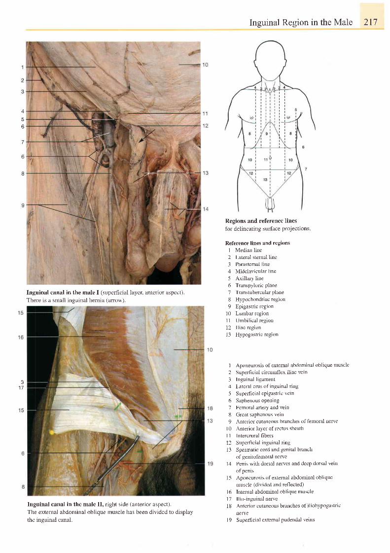
lnguinal Region in the Male 217
o
J
1 7
Inguinal canal in the male I (superficial layer, anterior aspect).
There is a small insuinal hernia (arrow).
Inguinal canal in the male II, right side (anterior aspect).The external abdominal oblique muscle has been divided to displaythe ineuinal canal.
Regions and reference lines
for delineating surface projections.
Reference lines and regions1 Median line2 Lateral sternal line3 Parastemal line4 Midclavicular line5 Axillary line6 Transpyloric plane7 Transtubercularplane8 Hypochondriac region9 Epigastric region
10 Lumbar region11 Umbilical region12 Iliac region13 Hypogastric region
1 Aponeurosis ofexternal abdominal oblique muscle
2 Superf ic ia l c i rcumflex i l iac vein
3 Inguinal ligament
4 Lateral crus of inguinal ring
5 Superficial epigastric vetn
6 Saphenous opening
7 Femoral artery and vein
8 Great saphenous vein
9 Anterior cutaneous branches offemoral nerve
10 Anterior layer ofrectus sheath
11 Intercrural fibers
12 Superficial inguinal ring
l3 Spermatic cord and genital branch
of genitofemoral nerve
14 Penis with dorsal nerves and deep dorsal vein
of penis
15 Aponeurosis ofexternal abdominal oblique
muscle (divided and reflected)
16 Internal abdominal oblique muscle
17 Ilio-inguinal nerve
1 8 Anterior cutaneous branches of iliohypogastric
nelrye
l9 Superficial external pudendal veins
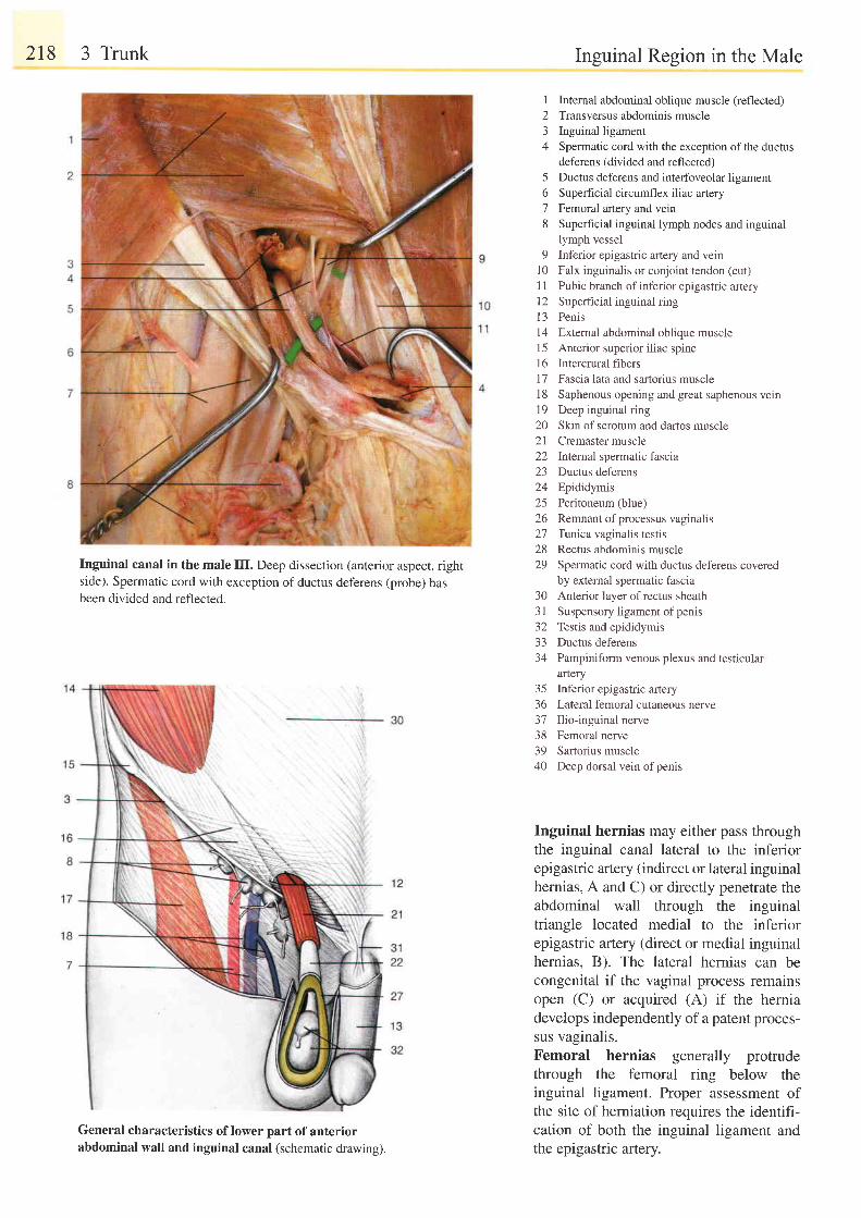
218 3 Trunk
Inguinal canal in the male III. Deep dissection (anterior aspect, rightside). Spermatic cord with exception ofductus deferens (probe) hasbeen divided and reflected.
General characteristics of lower part of anteriorabdominal wall and inguinal canal (schematic drawing).
I
23A
Inguinal Region in the Male
Internal abdominal oblique muscle (reflected)Transversus abdominis muscleInguinal ligamentSpermatic cord with the exception of the ductusdeferens (divided and reflected)Ductus deferens and interfoveolar ligamentSuperficial circumflex iliac arteryFemoral artery and veinSuperhcial inguinal lymph nodes and inguinal
5o78
lymph vessel9 Inferior epigastric artery and vein
10 Falx inguinalis or conjoint tendon (cut)1 1 Pubic branch of inferior epigastric artery12 Superficial inguinal ring13 Penis14 External abdominal oblique muscleI 5 Anterior superior i l iac spine16 Intercrural fibers17 Fascia lata and sartorius muscle18 Saphenous opening and great saphenous vein19 Deep inguinal ring20 Skin of scrotum and dartos muscle21 Cremaster muscle22 lntemal spermatic fascia23 Ductus deferens24 Epididymis25 Peritoneum (blue)26 Remnant ofprocessus vaginalis27 Ttnica vaginalis testis28 Rectus abdominis muscle29 Spermatic cord with ductus deferens covered
by external spermatic fasciaAnterior layer of rectus sheathSuspensory ligament of penisTestis and epididymisDuctus deferensPampiniform venous plexus and testiculararteryInferior epigastric arteryLateral femoral cutaneous nerveIlio-inguinal nerveFemoral nerveSartorius muscleDeep dorsal vein of penis
303 1) L
3334
35363'7383940
Inguinal hernias may either pass throughthe inguinal canal lateral to the inferiorepigastric artery (indirect or lateral inguinalhernias, A and C) or directly penetrate theabdominal wall through the inguinaltriangle located medial to the inferiorepigastric artery (direct or medial inguinalhernias, B). The lateral hernias can becongenital if the vaginal process remainsopen (C) or acquired (A) if the herniadevelops independently of a patent proces-sus vaginalis.Femoral hernias generally protrudethrough the femoral ring below theinguinal ligament. Proper assessment ofthe site of herniation requires the identifi-cation of both the inguinal ligament andthe epigastric artery.
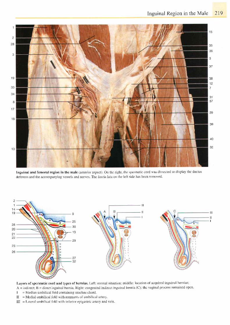
Inguinal Region in the Male 2I9
Inguinal and femoral region in the male (anterior aspect). On the right, the spermatic cord was dissected to display the ductus
deferens and the accompanying vessels and nerves. The fascia lata on the left side has been removed.
o /
38t 4
7
J I
37
29
39
40
a9
11 41 9
282021zz
I
25
30
1 3
29
ilt
t lI
ill
t l
I
\ l il , rl l \ i , ' ,
23
26
Layers of spermatic cord and types of hernias. Left: normal situation; middle: location of acquired inguinal hernias;
A = indirect; B = direct inguinal hernia. Right: congenital indirect inguinal hernia (C); the vaginal process remained open
I = Median umbilical fold containing urachus chord.
II = Medial umbilical fold with remnants of umbilical artery.
III = Lateral umbilical fold with inferior episastric anery and vein.
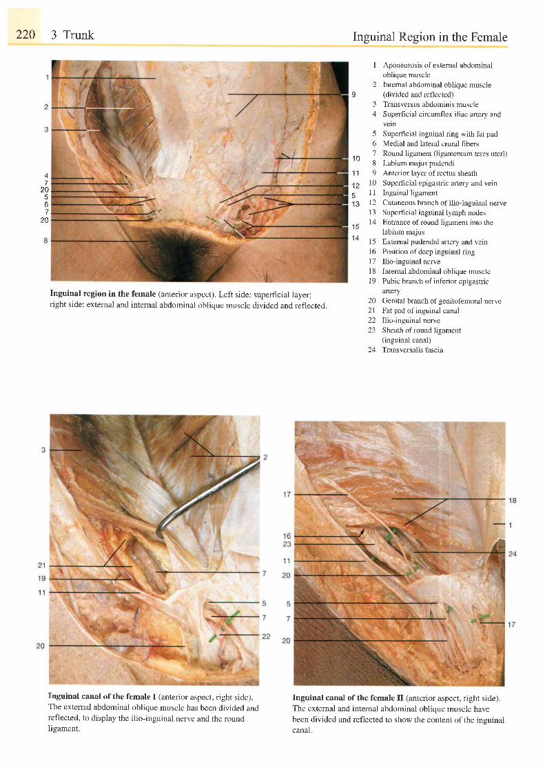
220 3 Trunk
Inguinal region in the female (anterior aspect). Left side: superficial layer;right side: external and internal abdominal oblique muscle divided and reflected.
Inguinal canal of the female I (anterior aspect, right side).The extemal abdominal oblique muscle has been divided andreflected, to display the ilio-inguinal nerve and the roundIieament.
Inguinal Region in the Female
I Aponeurosis of external abdominaloblique muscle
2 Internal abdominal oblique muscleI (divided and reflected)
3 Transversus abdominis muscle4 Superficial circumflex iliac artery and
veln5 Superficial inguinal ring with fat pad6 Medial and lateral crural fibers7 Round ligament (ligamentum teres uteri)8 Labium majus pudendi9 Anterior layer ofrectus sheath
10 Superficial epigastric artery and vein11 Inguinal ligament12 Cutaneous branch ofilio-inguinal nerve13 Superficial inguinal lymph nodes14 Entrance of round ligament into the
labium majus15 Extemal pudendal artery and vein16 Position ofdeep inguinal ring17 Ilio-inguinal nervel8 Internal abdominal oblique muscleI 9 Pubic branch of inferior epigastric
artery20 Genital branch ofgenitofemoral nerve2l Fatpad of inguinal canal22 Ilio-inguinal nerve23 Sheath ofround ligament
(inguinal canal)24 Transversalisfascia
1 0
1 1
1 251 3
4
4V
56
1 V
1 5t4
Inguinal canal of the female II (anterior aspect, right side).The external and internal abdominal oblique muscle havebeen divided and reflected to show the content of the insuinalcanal.
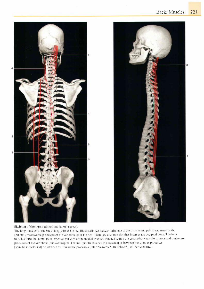
Back: Muscles 221
y
*ta4;
f,r#b
#t
Skeleton of the trunk (ckrr-sal and lateral aspect)
The long n tusc les o1 ' thc back l long iss iu rus ( l ) and i l iocos ta l i s (2 ) n rusc lc ] o r ig ina tc lL t thc sacru t r and pc lv is a t ld insc l t a t the
sptnous or t l 'ul tsvct 'sc pt 'ocesses ol ' the vertebrae or at thc r ibs. Ther-e are l lso nrusclcs that insclt at the occipital bone. Thc long
I r l u s c | c s | o r . n r t h c | a t e r a l t r a c t . r ' r h e t - e a s t l u s c l e s t l l t h e r r r c d a l
p rocesses o l the l 'e r tebrae I tnLnsver -sosp ina l (3 )and sp ino t ransvc lsa l ( .1 ) rnusc lcs lo r bc twcen the sp inous proccsscs
lspinal is rnuscle s (-5)l of bctwccn thc t lansvclsc proccsses I interlr 'ansvcrsal i i musclcs (6)l of the vet ' tebrac.
*-.fr
f
1 . -rfir
r-t, t-r
l i
\a
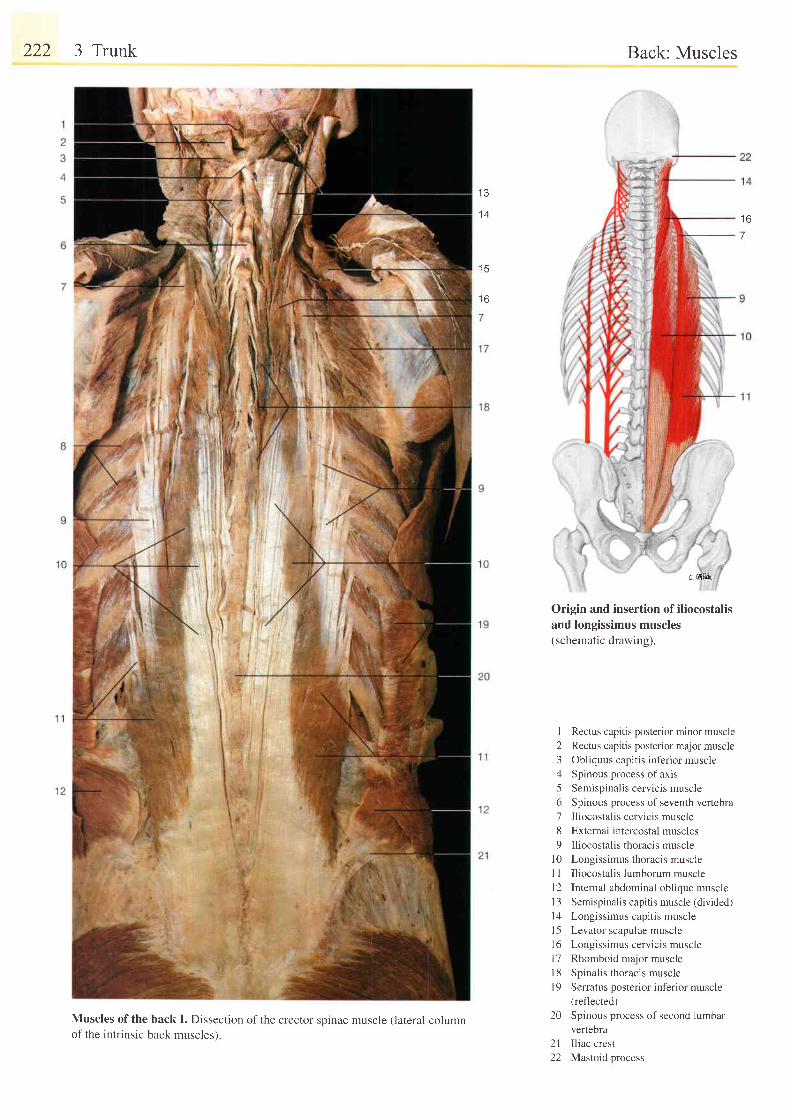
222 3 Trunk
Muscles of the back I. Dissection of the erector spinae muscle (lateral columnof the intrinsic back muscles)
I J
1 4
I J
t o
Back: Muscles
Origin and insertion of iliocostalisand longissimus muscles(schematic drawing).
1 Rectus capitis posterior minor muscle
2 Rectus capitis posterior major muscle
3 Obliquus capitis inferior muscle
4 Spinous process of axis
5 Semispinalis cervicis muscle
6 Spinous process of seventh vertebra
7 Iliocostalis cervicis muscle
8 External intercostal muscles
9 Iliocostalis thoracis muscle
l0 Longissimus thoracis muscle
I I Iliocostalis lumborum muscle
12 Internal abdominal oblique muscle
13 Semispinalis capitis muscle (divided)
l4 Longissimus capitis muscle
l5 Levator scapulae muscle
16 Longissimus cervicis muscle
l7 Rhomboid major muscle
18 Spinalis thoracis muscle
| 9 Serratus posterior inferior muscle(reflected)
20 Spinous process of second lumbar
vertebra
2l I l iac crest
22 Mastoid process
1 6
7
c ,',liLd
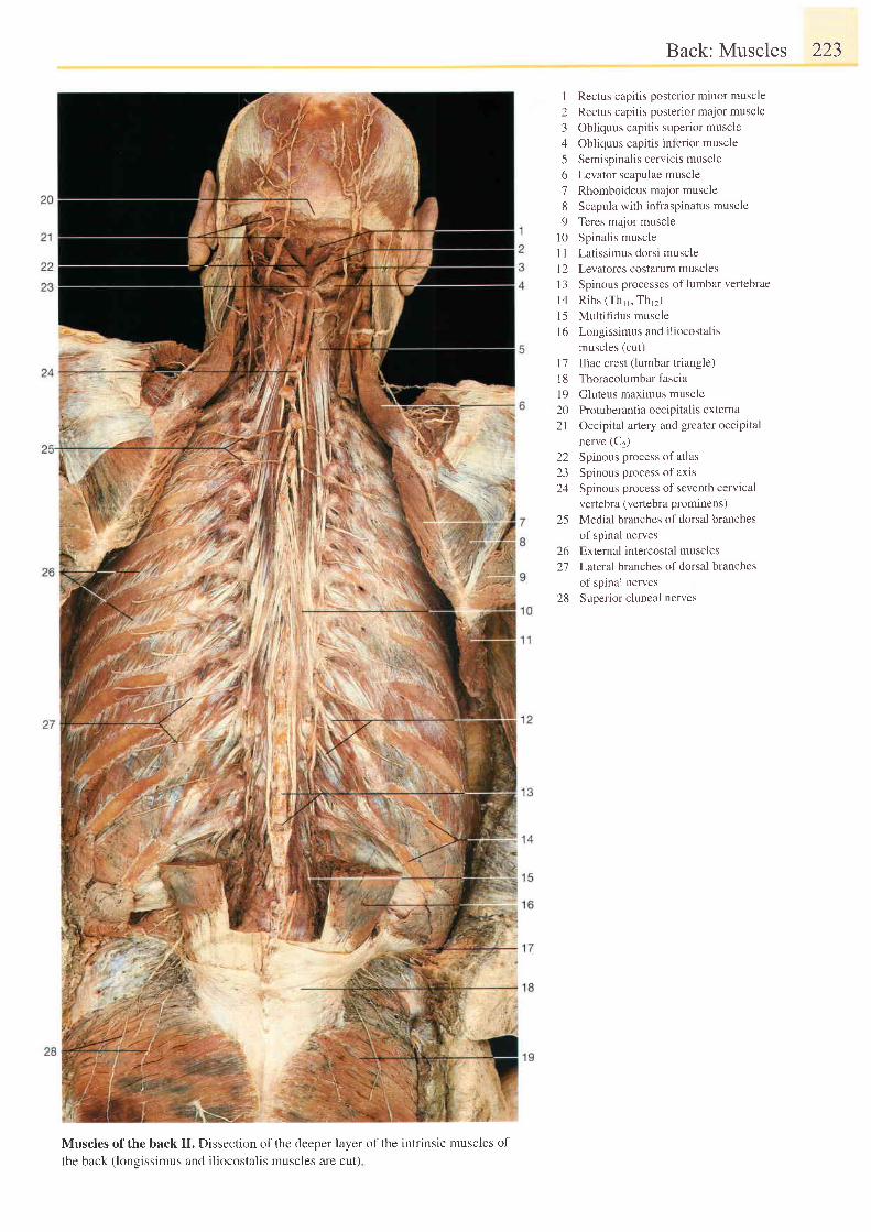
Back: Muscles 223
I Rectus capitis posterior minor muscle2 Rectus capitis posterior major muscle3 Obliquus capitis superior muscle4 Obliquus capitis inferior muscle5 Semispinalis cervicis muscle6 Levator scapulae muscle7 Rhomboideus major muscle8 Scapula with infraspinatus muscle9 Teres major muscle
10 Spinalis muscleI I Latissimus dorsi musclel2 Levatores costalum muscles13 Spinous processes of lumbar veftebraeI4 Ribs (Th1, Thr2)l5 Mult i f idus muscle16 Longissimus and iliocostalis
muscles (cut)17 Iliac crest (lumbar triangle)18 Thoracolumbarfascial9 Gluteus maximus muscle20 Protuberantiaoccipitalis extema21 Occipital artery and greater occipital
nerve (Ct)22 Spinous process of at las23 Spinous process of axis24 Spinous process of seventh cervical
vertebra (vertebra prominens)25 Medial branches of dorsal branches
of spinal nerves26 External intercostal muscles2'7 Laleral.branches of dorsal branches
of spinal nerves28 Superior cluneal nerves
Muscles of the back II. Dissection of the deeper layer of the intrinsic muscles of
the back (lonsissimus and iliocostalis muscles are cut)
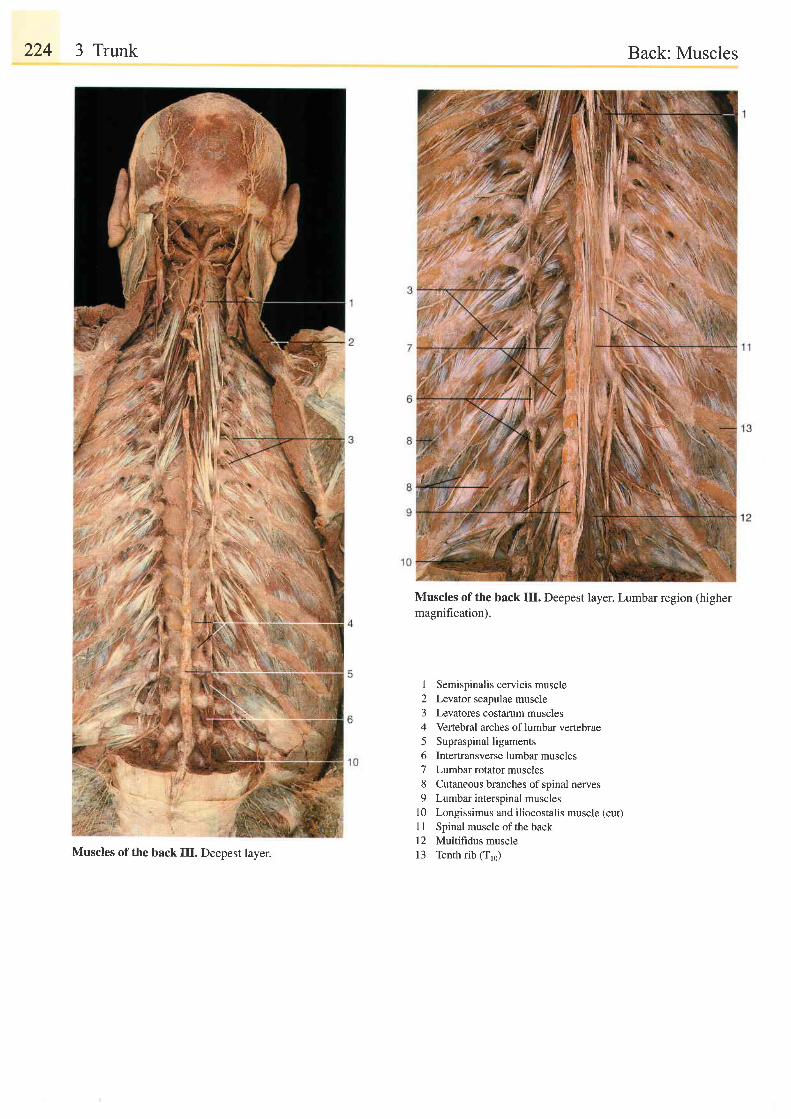
224 3 Trunk Back: Muscles
Muscles of the back III. Deepest layer.
Muscles of the back III. Deepest layer. Lumbar region (highermagnihcation).
1 Semispinalis cervicis muscle2 Levator scapulae muscle3 Levatores costarum muscles4 Vertebral arches oflumbar vertebrae5 Supraspinalligaments6 Intertransverse lumbar muscles7 Lumbar rotator muscles8 Cutaneous branches of spinal nerves9 Lumbar interspinal muscles
10 Longissimus and iliocostalis muscle (cut)I I Spinal muscle of the back12 Multifidus muscle13 Tenth rib (T,6)
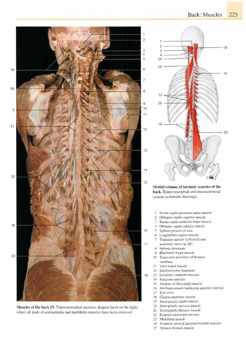
Muscles of the back IV, Transversospinal muscles, deepest layer on the right,where all parts of semispinalis and multifidus muscles have been removed.
Back: Muscles 225
Medial column of intrinsic muscles of the
back. Transversospinal and intertransversal
system (schematic drawing).
I Rectus capitis posterior minor muscle2 Obliquus capitis superior muscle3 Rectus capitis posterior major muscle4 Obliquus capitis inferior muscle5 Spinous process of axis6 Longissimus capitis muscle7 Trapezius muscle (reflected) and
accessory nerve (n. XI)8 Spinous processes9 Rhomboid major muscle
I 0 Transverse processes of thoracicvertebrae
1 I Teres major muscle12 Intertransverse ligamentsi3 Levatores costarum muscles14 Rotatores muscles15 Tendons of iliocostalis musclel6 Intertransversarii lumborum muscles (lateral)
17 I l iac crestI8 Gluteus maximus muscle19 Semispinalis capitis muscle20 Semispinalis cervicis muscle2l Semispinalis thoracis muscle22 Exlernal intercostal muscles23 Multifidus muscle24 Posteriorcervical intertransversarii muscles25 Spinalis thoracis muscle
1
34
20
1 3
J
A
9 2 51 0
1 1
t b
1 6
1 7
1 B
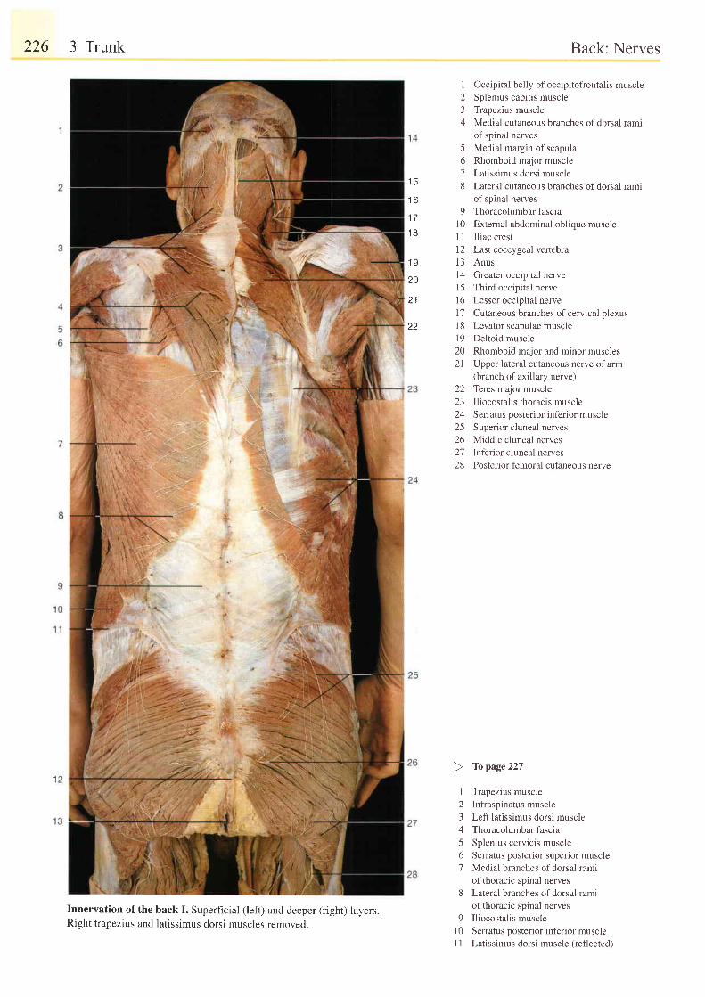
226 3 Trunk
Innervation of the back I. Superficial (left) and deeper (right) layers.Right trapezius and latissimus dorsi muscles removed.
Back: Nerves
1 Occipital belly of occipitofrontalis muscle2 Splenius capitis muscle3 Trapezius muscle4 Medial cutaneous branches ofdorsal rami
of spinal nerves5 Medial margin of scapula6 Rhomboid major muscle7 Latissimus dorsi muscle8 Lateral cutaneous branches ofdorsal rami
of spinal nerves9 Thoracolumbar fascia
10 External abdominal oblique muscleI 1 Iliac crest12 Last coccygeal veftebral3 Anus14 Greater occipital nervel5 Third occipital nerve16 Lesser occipital nerve17 Cutaneous branches ofcervical plexus18 Levator scapulae muscle19 Deltoid muscle20 Rhomboid major and minor muscles21 Upper lateral cutaneous nerve ofarm
(branch of axillary nerve)22 Teres major muscle23 Iliocostalis thoracis muscle24 Serratus posterior inferior muscle25 Superior cluneal nerves26 Middle cluneal nerves27 Inferior cluneal nerves28 Posterior femoral cutaneous nerve
I Trapezius muscle2 Infraspinatus muscle3 Left latissimus dorsi muscle4 Thoracolumbar fascia5 Splenius cervicis muscle6 Serratus posterior superior muscle7 Medial branches of dorsal rami
of thoracic spinal nerves8 Lateral branches ofdorsal rami
of thoracic spinal nerves9 Iliocostalis muscle
l0 Serratus posterior inferior musclei 1 Latissimus dorsi muscle (reflected)
t c
t o
1 7
1 8
.19
20
2 1
22
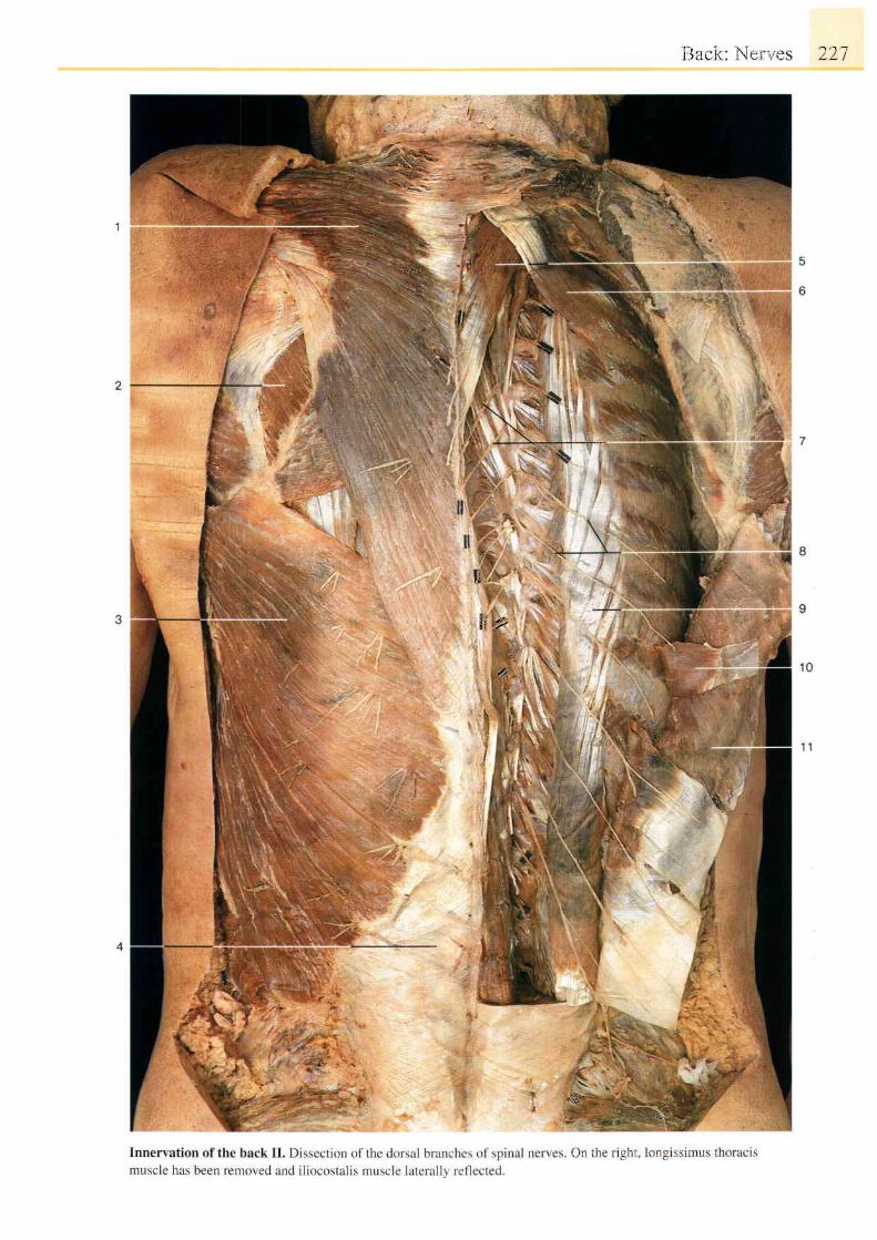
LJ, rr i . \ , ' r 1 . '
q,
t :=
ft
{ t , r
/t
-\ 'flt€:"q.
-]
j
1i
l l t , l i . : l : l r l r ' i l : ' . i i l i r i l
,{
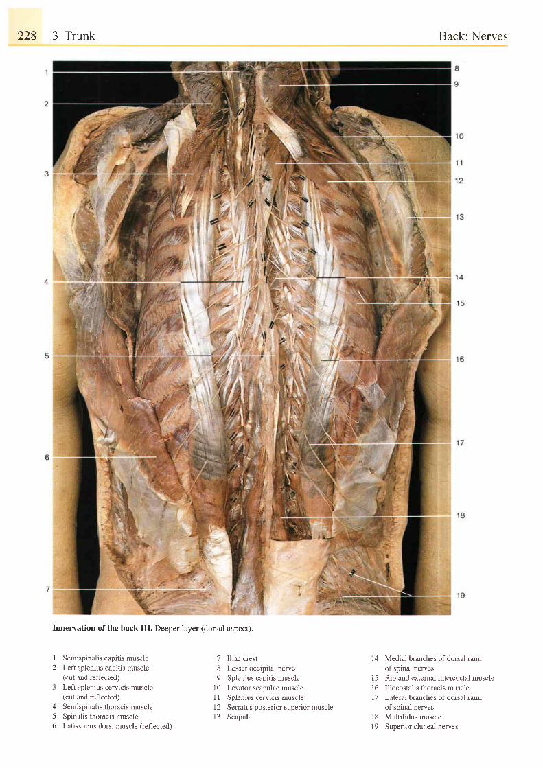
228 3 Trunk
Innervation of the back III. Deeper layer (dorsal aspect).
I
2Semispinalis capitis muscleLeft splenius capitis muscle(cut and reflected)Left splenius cervicis muscle(cut and reflected)Semispinalis thoracis muscleSpinalis thoracis muscleLatissimus dorsi muscle (refl ected)
7 Iliac crest8 Lesser occipital nerve9 Splenius capitis muscle
10 Levator scapulae muscle1 1 Splenius cervicis musclel2 Serratus posterior superior muscle13 Scapula
14 Medial branches of dorsal ramiof spinal nerves
15 Rib and external intercostal muscle16 Iliocostalis thoracis muscle7'7 Lateral branches ofdorsal rami
of spinal nerves18 Multifidus muscle19 Superior cluneal nerves
Back: Nerves
/5o
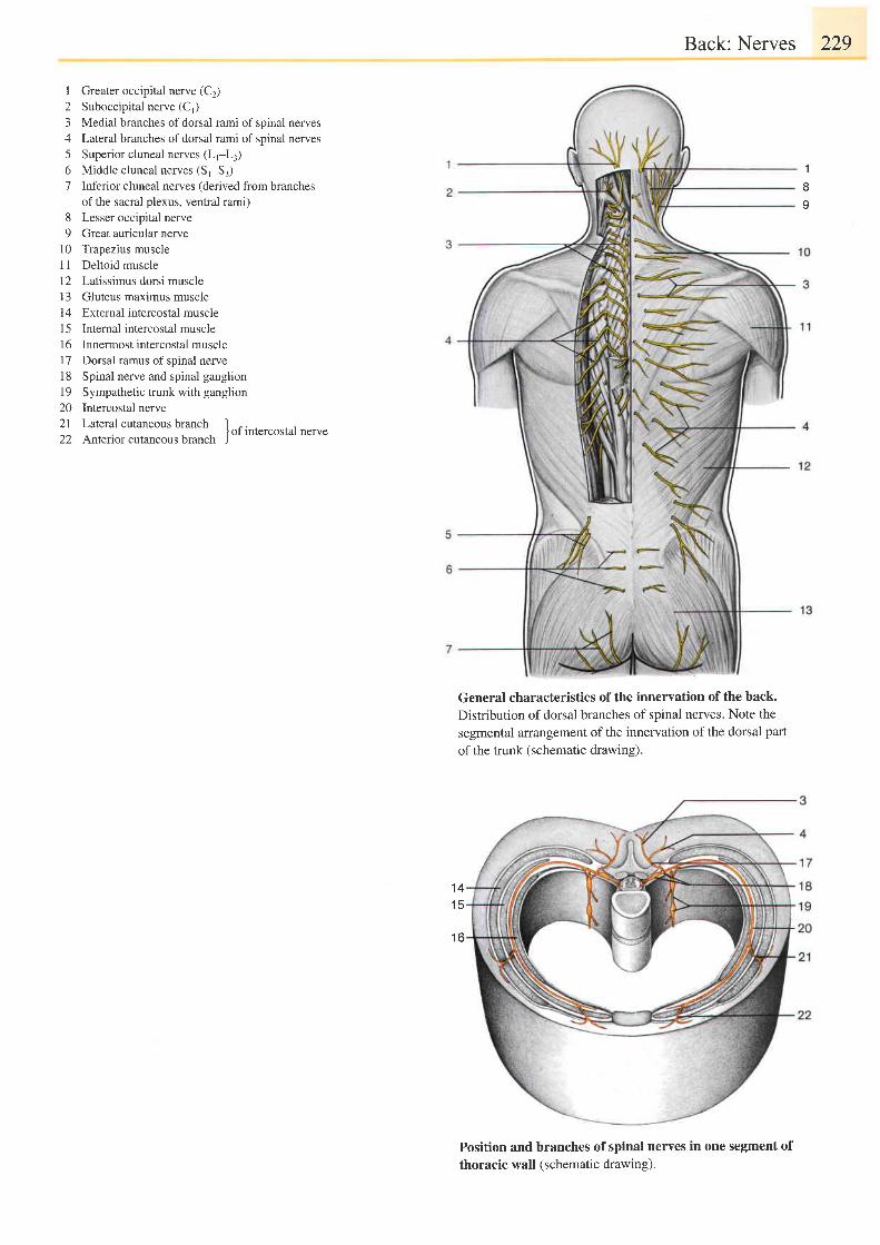
I Greater occipital nerve (C2)2 Suboccipital nerve (C,)3 Medial branches of dorsal rami of spinal nerves4 Lateral branches of dorsal rami of spinal nerves5 Superior cluneal nerves (Lr-L:)6 Middle cluneal nerves (St-S,)7 Inferior cluneal nerves (derived from branches
of the sacral plexus, ventral rami)8 Lesser occipital nerve9 Great auricular nerve
10 Trapezius muscle11 Deltoid muscle12 Latissimus dorsi musclel3 Gluteus maximus muscle14 Extemal intercostal muscle15 Internal intercostal muscle16 Innermost intercostal muscle17 Dorsal ramus of spinal nerve18 Spinal nerve and spinal ganglion19 Sympathetic trunk with ganglion20 Intercostal nerve21 Lateral cutaneous branch I ^.22 Anterior cutaneous brunch Jof
lntercostal nerve
Back: Nerves 229
General characteristics of the innervation of the back.Distribution of dorsal branches of spinal nerves. Note thesegmental arrangement of the innervation of the dorsal part
of the trunk (schematic drawing).
1
I
v
1 41 5
' t6
Position and branches of spinal nerves in one segment ofthoracic wall (schematic drawing).
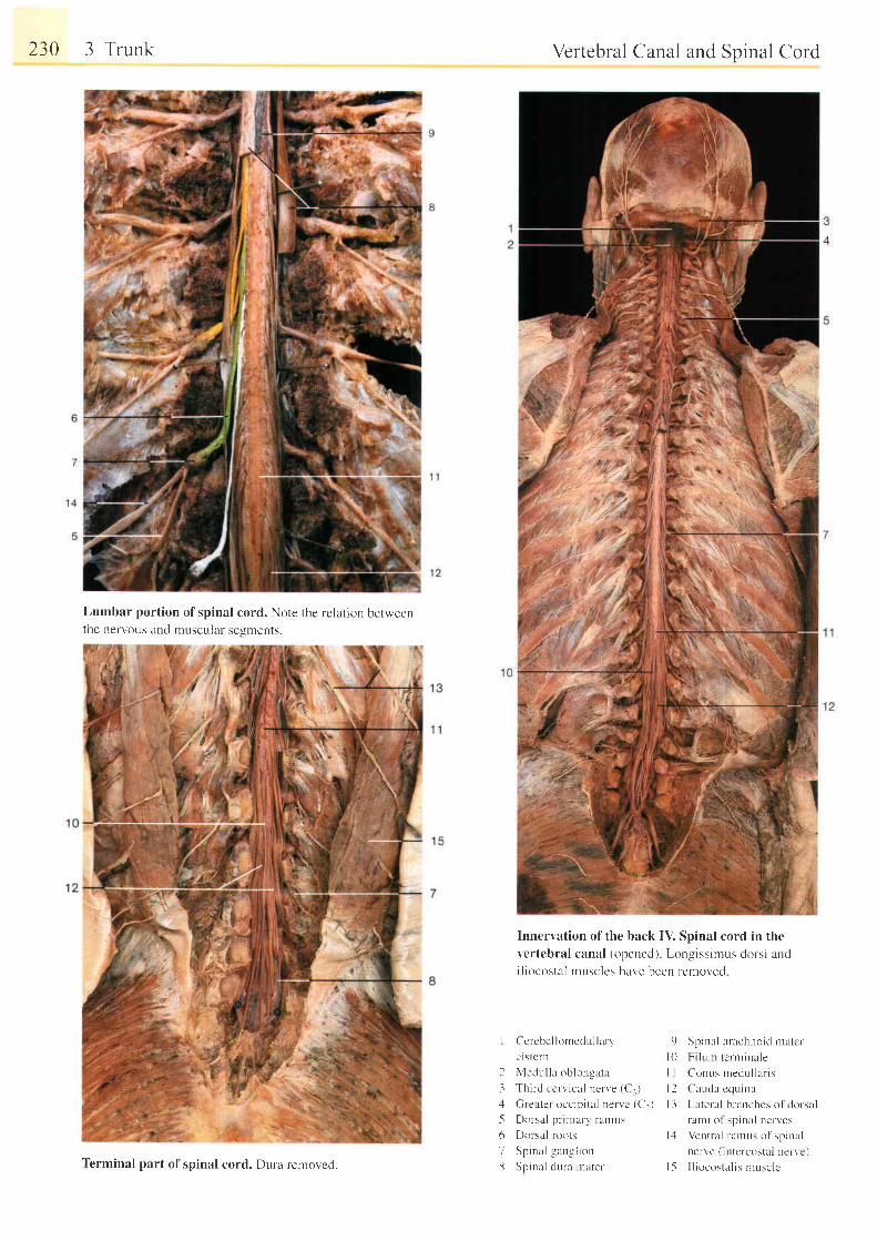
230 3 Trunk
Lumbar portion of spinal cord. Note the re lation betweenthe nervous and muscular segments.
Vertebral Canal and Spinal Cord
Innervation of the back IV. Spinal cord in thevertebral canal {opened) Longissirnus dorsi andi l iocostal musclcs ha.n'e been removed
I Cerebel lomedul lary
ct stcrn
2 Medul la oblonsata
3 Th i r d cc r v i ca l ne rve (C , ),1 Greater occip iu l nervc (C1)
-5 Dor-sal prinrary rarrrus
6 l)or sal roots
7 Spinal - langl ion13 Spinal dura rnater '
9 Spinal arachnoid matcr
l 0 F i l u rn [ c r r - n i na le
I I Conus rnedu l l a r i s
l2 Car,rd l cquinu
l 3 Latelal blanches ol dorsal
rarni of spinni nelves
l . : l Ventra l ra l rLrs of spinal
nerve ( intcrcostal nerve)
| 5 I l iocostal is nrLrsclcTerminal part of spinal cord. Dura removed.
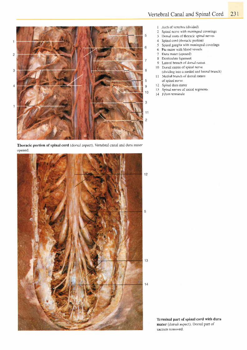
Thoracic portion of spinal cord (dorsal aspect). Vertebral canal and dura materopened.
Vertebral Canal and Spinal Cord 231
I Arch of vertebra (divided)
2 Spinal nerve with meningeal coverings
3 Dorsal roots of thoracic spinal nerves4 Spinal cord (thoracic portion)5 Spinal ganglia with meningeal coverings6 Pia mater with blood vessels7 Dura mater (opened)8 Denticulate ligament9 Lateral branch ofdorsal ramus
10 Dorsal ramus of spinal nerve(dividing into a medial and lateral branch)
11 Medial branch of dorsal ramusof spinal nerve
12 Spinal dura mater13 Spinal nerves of sacral segments14 Filum terminale
Terminal part of spinal cord with duramater (dorsal aspect). Dorsal part ofsacrum removed.
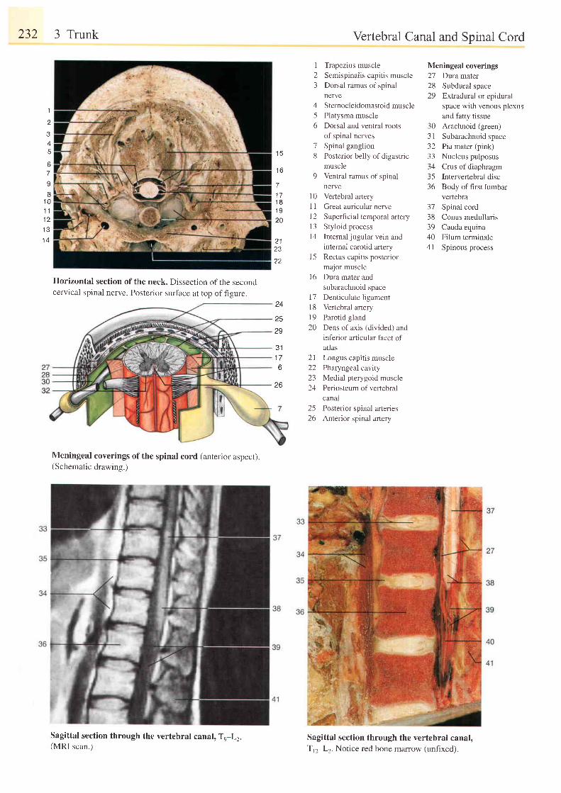
232 3 Trunk
Horizontal section of the neck. Dissection of the secondcervical spinal nerve. Posterior surface at top offigure.
Meningeal coverings of the spinal cord (anterior aspect).(Schematic drawing.)
Sagittal section through the vertebral canal, To-Lr.(MRI scan.)
Vertebral Canal and Spinal Cord
1 5
1 6
71 71 81 920
2 1I J
22
1
4
67q
I1 01 l1 21 31 4
I Trapezius muscle2 Semispinalis capitis muscle3 Dorsal ramus of spinal
nerve4 Sternocleidomastoid muscle5 Platysma muscle6 Dorsal and ventral roots
of spinal nerves7 Spinal ganglion8 Posterior bel ly ofdigastr ic
muscle9 Ventral ramus of spinal
nervel0 Vertebral artery1 1 Great auricular nerve12 Superficial temporal artery13 Styloid processl4 Intemal jugular vein and
internal carotid artery15 Rectus capitis posterior
Meningeal coverings27 Duramater28 Subdural space29 Extradural or epidural
space with venous plexusand fatty tissue
30 Arachnoid (green)31 Subarachnoid space32 Pia mater (pink)33 Nucleus pulposus34 Crus of diaphragm35 Intervertebral disc36 Body of first lumbar
vertebra37 Spinai cord38 Conus medullaris39 Cauda equina40 Filum terminale4l Spinous process
1 6
t 7l 81 920
major muscle
Dura mater and
subarachnoid space
Denticulate ligament
Vertebral artery
Parotid gland
Dens of axis (divided) and
inferior articular facet of
atlas
21 Longus capitis muscle22 Pharyngeal cavity23 Medial pterygoid muscle24 Periosteum of vertebral
canal25 Posterior spinal arteries26 Anterior spinal artery
Sagittal section through the vertebral canal,T n-Lz.Notice red bone marrow (unfixed).
24
25
29
3 11 76
26
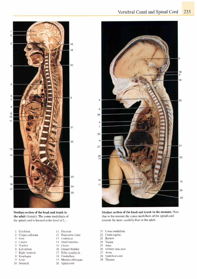
I
1 21 0
1 1
Vertebral Canal and Spinal Cord 233
Median section of the head and trunk in the neonate. Note
that in the neonate the conus medullaris of the spinal cord
extends far more caudally than in the adult.
2l Conus medullaris22 Cauda equina23 Rectum24 Yagina25 Anus26 Inferior vena cava2'7 Aorta28 Umbilical cord29 Thymus
1 8
1 9
Median section of the head and trunk inthe adult (female). The conus medullaris ofthe spinal cord is located at the level ofL1.
I Cerebrum2 Corpus callosum3 Pons4 Larynx5 Trachea6 Left atrium7 Right ventricie8 Esophagus9 Liver
l0 Stomach
I 1 Pancreas12 Transverse colon13 Umbilicus14 Small intestine15 Uterus16 Urinary bladder17 Pubic symphysisl8 Cerebellum19 Medulla oblongata20 Spinal cord
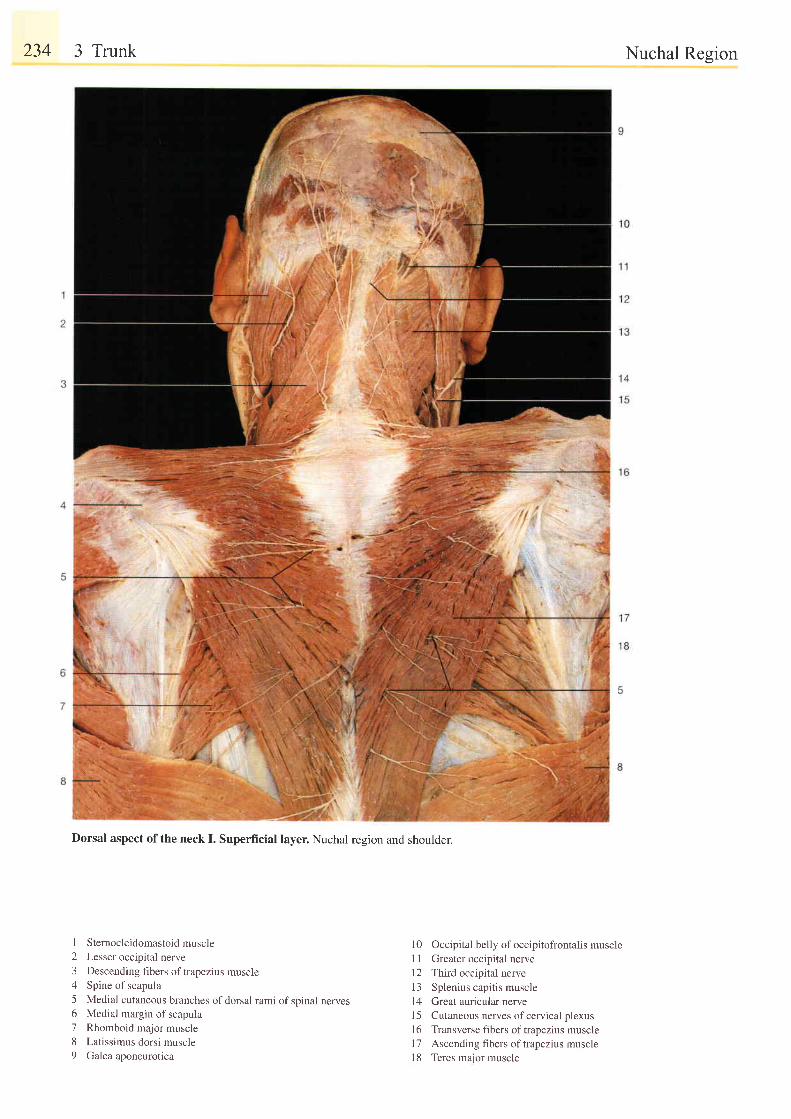
234 3 Trunk
Dorsal aspect of the neck I. Superficial layer. Nuchal region and shoulder.
1 Sternocleidomastoid muscle2 Lesser occipital nerve3 Descending fibers oftrapezius muscle4 Spine of scapula5 Medial cutaneous branches of dorsal rami of spinal nerves6 Medial margin of scapula7 Rhomboid major muscle8 Latissimus dorsi muscle9 Galea aponeurotica
Nuchal Region
l0 Occipital belly of occipitofrontalis muscle
I I Greater occipital nerve
l2 Third occipital nerve
13 Splenius capitis muscle
14 Great auricular nerve
l5 Cutaneous nerves ofcervical plexus
l6 Transverse fibers oftrapezius muscle
l7 Ascending fibers of trapezius muscle
l8 Teres major muscle
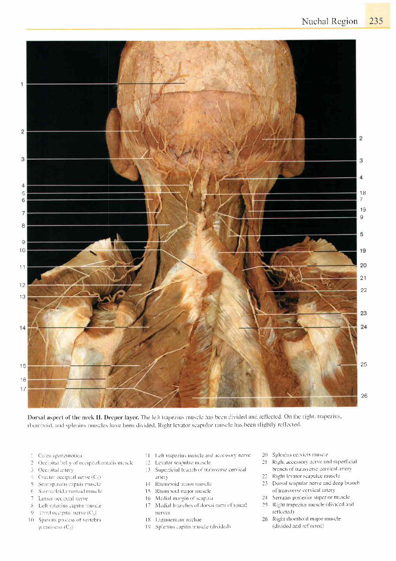
Nuchal Resion 235
A
56
1 2
I J
; l '
i..:. .:r,rl
t ( . : , : t '
l t r i l .
Dorsal aspect of the neck II . Deeper lay'er. Thc lcft t l lpezir-Ls rnusclc hus bccn di l ' idecl and ref lected. On the r ight. i t 'apczit ts.
rhouthoir i . uurl \pleuius ruusclcs i tar,e been diviclecl Right lcvatur scapr.r lac ntusclc has bccn sl ightly rel ' lectccl.
t o
7
\ l h8
I
1 0
'1 1
22
l 3
l o
1 7
t:1. ' , ' '1i. , . t . ; . ,1 :1, . ;
!
,t,, ';l:$
' i,'' I
25
26
I G l r i e l r l poncu ro t i c i . l
I Occ ip i t a l i r c l l r o l occ i l . r i t o l r - on tu l i s r uL t sc l e
3 Oce ip r t a l u r t en
i ( i r ca t c r occ i p i t r L l ne ' r - r c ( ( 1 . )
5 Sen r r sp in l l r s cap i t i s n rusc l e( r S t c r noe l c i r l omas to i d n r r - r sc l c
7 l -csscr occi l . r i l lL l ncrr e
3 | c l L : p l r ' n i L r s c l p i t r s n ru : c l e
9 ' f h i r - t i
o cc r l . r i t u l l l e r \ c (C r )
I 0 Sp inoL rs l ) r oce s \ o l \ c l t e l l n r
p r -o r i r i r i c r i : {C - )
I I Lc l ' t t n t pez ius r r usc l c l t t r t l r t c ccsso r ) ' t t c r ve
I I L.er ator- seaprr l lLc r t t t t :c lc
l - l SuPcr- l ic i l l bntnch ol I r i l l l \ \ er-sc cel v lc i t l
il fte rv
l - i Rhon rbo i t l u t i no t - n rusc l c
I 5 Rh i l t t t h i l i J r t l : l l ( , r i l t t t \ . l ( '
l ( r Nlcdia l ruaruiu o l scapt t l r t
I 7 N' lcc l ia l branchcs ol t [ r r s . t l ratr t i t t f spiu l t l
ncf \ cs
I l l L i ga r r r en tun r nuchac
l9 Sp i cn ius c r ; . r i t i s u ru :e l c ( d i v i c l c t l )
Sp len ius ce rv i c i s u tusc l c
Right acccssot 'v nervc at td supcr l ic ia l
branch ol ' t ransvcl se cerr" ical ar terv
I{ ight levator scapulac ntuscle
Dorsal scapular- nerve ancl deep branch
ol ' t r lnsversc cerv ical at ' tery
Sen atus poster ior supet- iot - tnt tsc le
Right t r lpezius Lnuscle (d iv ided ancl
leflectcd )Ri . lht rhornboid tnajor t t tuscle(c l ivrded and lef lccted)
202 l
2223
).12 5
|'.,i;P-4ill n,
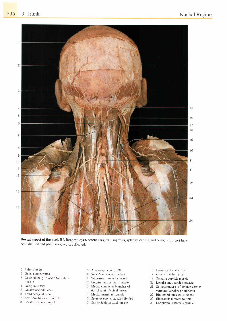
236 3 Trunk
I23
15o78
Skin of scalp
Galea aponeurotica
Occipital belly of occipitofiontalismuscle
Occipital artery
Greater occipital nerveThird occipital nerve
Semispinal is capi t is muscleLevator scapulae muscle
9 Accessory nerve (n XI)
l0 Superficial cervical artery
l1 Trapezius muscle (ref'lected)
l2 Longissimus cervicis muscle
13 Medial cutaneous branches ofdorsal rami of spinal nerves
l4 Medial margin of scapula
l5 Splenius capitis muscle (divided)
l6 Sternocle idomastoidmuscle
Nuchal Resion
I O
' t7
1 B
1 9
20
21
1 1
I J
17 Lesser occipital nervel8 Great auricular nerve19 Splenius cervicis muscle20 Longissimus cervicis muscle21 Spinous process of seventh cervical
vertebra (vertebra prominens)22 Rhomboid muscles (divided)23 Iliocostalis thoracis muscle24 Longissimus thoracis muscle
Dorsal aspect ofthe neck III. Deepest layer. Nuchal region. Trapezius, splenius capitis, and cervicis muscles havebeen divided and partly removed or ref'lected.
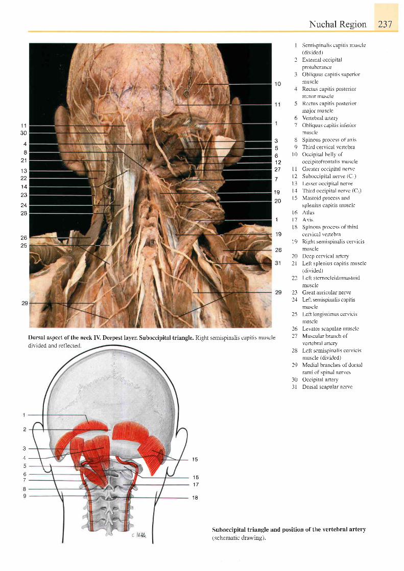
1 130
4
2 1
1 3221 423
zq
28
2625
2
1 Q
20
o7
89
1 0
l 1t 21 31 4l 5
l 61 t '7
1 81 9
1 9
2620 Deep cervical artery2l Left splenius capitis muscle
(divided)22 Leftsternocleidomastoid
muscle23 Greatauricularnerve24 Left semispinalis capitis
muscle25 Left longissimus cervicis
muscle26 Levator scapulae muscle27 Muscular branch of
vertebral artery28 Left semispinalis cervicrs
muscle (divided)29 Medial branches ofdorsal
rami of spinal nerves30 Occipital artery31 Dorsal scapular nerve
Suboccipital triangle and position of the vertebral artery(schematic drawing).
3
b
1 227
Nuchal Region 237
Semispinalis capitis muscle(divided)Extemal occipitalprotuberanceObliquus capitis superiormuscleRectus capitis posteriorminor muscleRectus capitis posteriormajor muscleVertebral arteryObiiquus capitis inferiormuscleSpinous process of axisThird cervical vertebraOccipital belly ofoccipitofrontalis muscleGreater occipital nerveSuboccipital nerve (Cr)Lesser occipital nerveThild occipital nerve (Cj)Mastoid process andsplenius capitis muscleAtlasAxisSpinous process of thirdcervical vertebraRight semispinalis cervicismuscle
Dorsal aspect of the neck IV. Deepest layer. Suboccipital triangle. Right semispinalis capitis muscledivided and reflected.
1 5
1 61 7
1 8
A
b
7
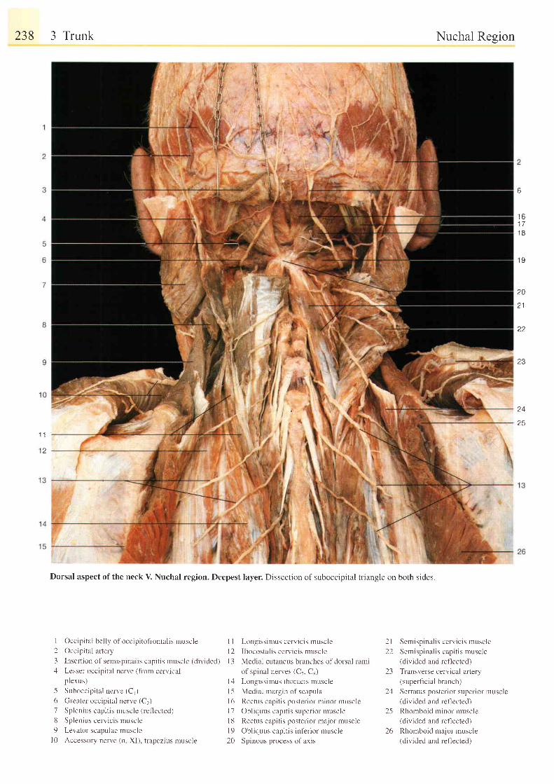
238 3 Trunk
I
Dorsal aspect of the neck V. Nuchal region. Deepest layer. Dissection of suboccipitat triangle on both sides
1 Occipital belly of occipitoftontalis muscle2 Occipital artery
3 Insertion of sen'rispinalis capitis muscle (divided),l Lesser occipital nerve (from cervical
p lexus)
-5 Suboccipital nerve (C1)
6 Greater occipital nerve (C2)
7 Splenius capitis muscle (reflected)
8 Splenius cerv ic is muscle9 Ler ator \c : lpulae muscle
l0 Accessclry nerve (n XI), trapezius muscle
I I Longissimus cervicis muscle
l2 I l iocostal is cerv ic is muscle
I 3 Medial cutaneus branches of dorsal rami
of spinal nerves (C7. Cs)
l4 Longissimus thoracis muscle
l5 Medial margin of scapula
16 Rectus capitis posterior minor muscle
l7 Obliquus capitis superior muscle
l8 Rectus capi t is poster ior major muscle
l9 Obl iquus capi t is inf 'er ior muscle
20 Spinous process of axis
Nuchal Region
2I Semispinal is cerv ic is muscle
22 Semispinal is capi t is muscJe
(divided and reflected)
23 Transverse cervical artery(superficial branch)
24 Serratus posterior superior muscle(divided and reflected)
25 Rhomboid minor muscle(divided and reflected)
26 Rhomboid major muscle(divided and rellected)
6
t o1 71 8
1 9
20
z l
23
25
1 1
12
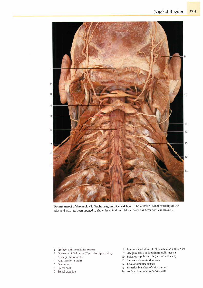
Nuchal Region 239
Dorsal aspect ofthe neck VI. Nuchal region. Deepest layer. The vertebral canal caudally ofthe
atlas and axis has been opened to show the spinal cord (dura mater has been par1ly removed).
1 Protuberantiaoccipitalisexterna2 Greater occipital nerve (C2) and occipital artery3 Atlas (posterior arch)4 Axis (posterior arch)5 Dura mater6 Spinal cord7 Spinal ganglion
8 Posterior root filaments (fila radicularia posterior)
9 Occipital belly of occipitofrontalis muscle
l0 Splenius capitis muscle (cut and reflected)11 Sternocleidomastoidmuscle12 Levaror scapulae muscle13 Posterior branches of spinal nerves
14 Arches ofcervical vertebrae (cut)
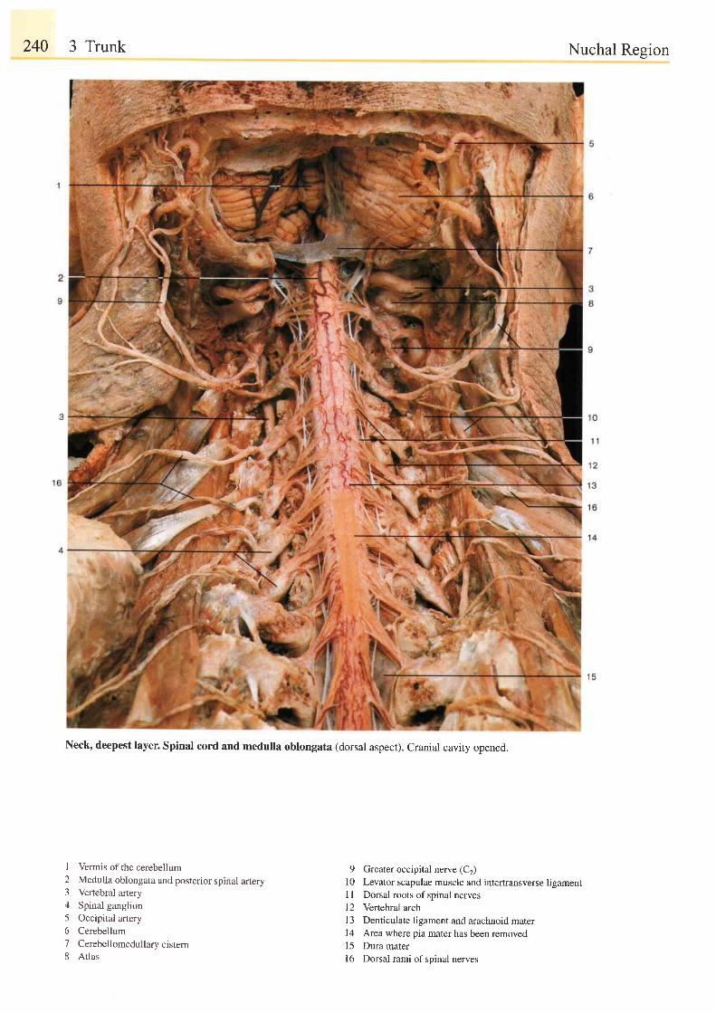
240 3 Trunk
Neck, deepest layer. Spinal cord and medulla oblongata (dorsal aspect). Cranial cavity opened.
Nuchal Region
I Vermis of the cerebellum2 Medulla oblongata and posterior spinal artery3 Vertebral artery4 Spinal ganglion5 Occipital artery6 Cerebellum7 Cerebellomedullarycistern8 Atlas
9 Greater occipital nerve (C2)10 Levator scapulae muscle and intertransverse ligament1 I Dorsal roots of spinal nerves12 Vertebral arch13 Denticulate ligament and arachnoid mater14 Area where pia mater has been removed15 Dura mater16 Dorsalrami of spinal nerves
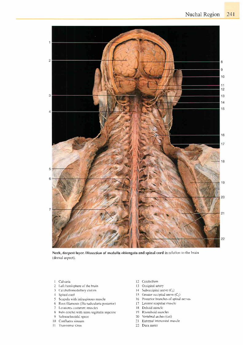
Nuchal Region 241
Neck, deepest layer. Dissection of medulla oblongata and spinal cord in relation to the brain(dorsal aspect)
I Calvaria
2 Left hemisphere of the brarn
3 Cerebellomedullary cistern
4 Spinal cord
5 Scapula with infraspinous muscle
6 Root filaments (fila radicularia posterior)
7 Levatores costarum muscles
8 Falx cerebri with sinus sagittalis superior
9 Subarachnoidal space
l0 Confluens sinuum
I I Transverse sinus
l2 Cerebellum
13 Occipital artery
l4 Suboccipital nerve (C1)
15 Greater occipital nerve (C2)
l6 Posterior branches of spinal nerves
l7 Levator scapulae muscle
18 Deltoid muscle
19 Rhomboid muscles
20 Vertebral arches (cut)
21 External intercostal muscle
22 Duramarer

242 3 Trunk
Dissection of neck and deeper layer (oblique-lateral aspect after removal of trapezius muscle).
Nuchal Resion
20
2122
23
I Protuberantia occipitalis externa2 Semispinal is capi t is muscle
3 Splenius capi t is muscle
4 Scapula
5 Splenius cerv ic is muscle
6 Posterior branches of spinal nerves7 Longissimus muscle
8 Spinous processes of thoracic vertebrae
9 iliocostalis rruscle
10 Lat iss imus dors i muscle
I I Epidermis ofthe head (scalp)
l2 Galea aponeurot ica
l3 Occipital belly of occipitofrontalis musclelzl Occipital artery
l5 Greater occipital nerve (C,)
16 Lesser occip i ta l nerve
l7 Sternocle idomastoidmuscle
I 8 Great auricular nerve
l9 Punctum nervosum
20 Transverse cervical nerve
21 Supraclavicularnerves
22 Accessory nerve (n XI)
23 Trapezius muscle (cr-rt edge)
24 Medial margin of scapula
25 Rhomboid muscle
26 Infraspinous muscle
27 Teres major muscle
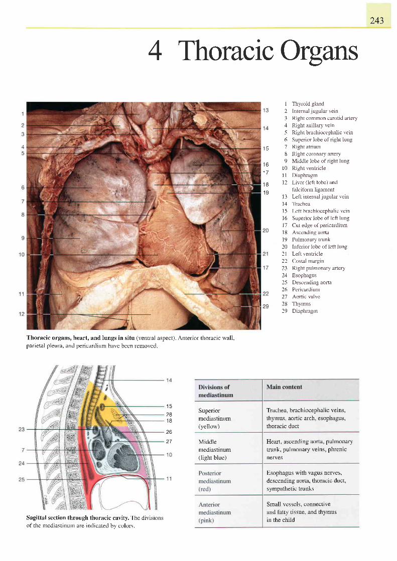
243
4 ThoracrcOrgans
t c
t o
1 7
1 8
1 9
123456'7
8q
t 0l 1t2
Thyroid glandInternal jugular veinRight common carotid arteryRight axillary veinRight brachiocephalic veinSuperior lobe of right lungRight atriumRight coronary arteryMiddle lobe of right lungRight ventricleDiaphragmLiver (left lobe) andfalciform ligamentLeft internal jugular veinTracheaLeft brachiocephalic veinSuperior lobe of left lungCut edge of pericardiumAscending aortaPulmonary trunkInferior lobe of left lungLeft ventricleCostal marginRight pulmonary arteryEsophagusDescending aortaPericardiumAortic valveThymusDiaphragm
l 31 4l 51 61 7l 81 9202 122L 3
. A
25zo
212829
Thoracic organs, heart, and lungs in situ (ventral aspect). Anterior thoracic wall,parietal pleura, and pericardium have been removed.
Sagittal section through thoracic cavity. The divisionsof the mediastinum are indicated bv colors.
I J
281 8
26
27
Superiormediastinum(yellow)
Middlemediastinum(light blue)
Trachea, brachiocephalic veins,thymus, aortic arch, esophagus,thoracic duct
Heart, ascending aorta, pulmonarytrunk, pulmonary veins, phrenicnefves
Esophagus with vagus nerves,descending aorta, thoracic duct,sympathetic trunks
Small vessels, connectiveand fatty tissue, and thymusin the child
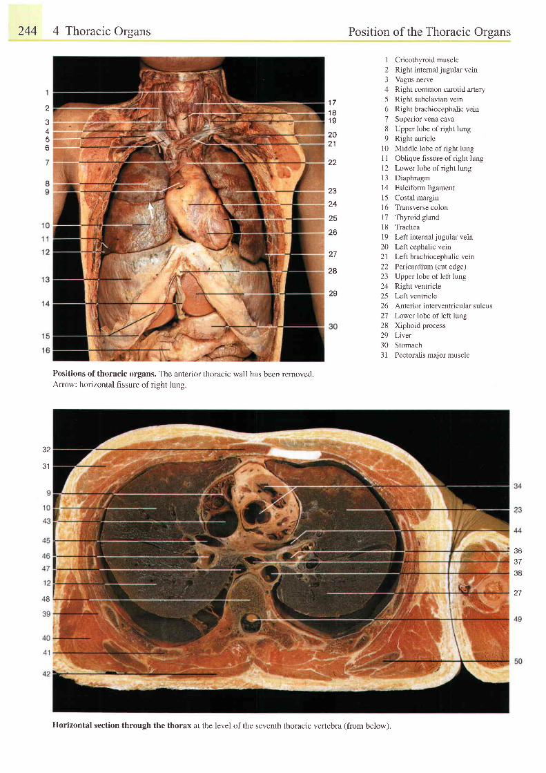
244 4 Thoracic Orsans Position of the Thoracic Orsans
1 Cricothyroid muscle2 Right intemal jugular vern3 Vagus nerve4 Right common carotid artery5 Right subclavian vein6 Right brachiocephalic vein7 Superior vena cava8 Upper lobe of right lung9 Right auricle
l0 Middle lobe of right lungI I Oblique fissure of right lung12 Lower lobe of right lung13 Diaphragm14 Falciform ligament15 Costal margin16 Transverse colon17 Thyroid glandl8 Trachea19 Left internal jugular vein20 Left cephalic vein21 Left brachiocephalic vern22 Pericarditm (cut edge)23 Upper lobe of left lung24 Right ventricle25 Left ventricle26 Anterior interventricular sulcus27 Lower lobe of left lung28 Xiphoid process29 Liver30 Stomach3l Pectoral is major muscle
t t
1 n1 9
2021
22
23
24
25
26
27
28
29
2
3
5o
32
J I
Positions ofthoracic organs. The anterior thoracic wall has been removed.Arrow: horizontal fissure of right lung.
36J I
38
27
llorizontal section through the thorax at the level of the seventh thoracic vertebra (from below).
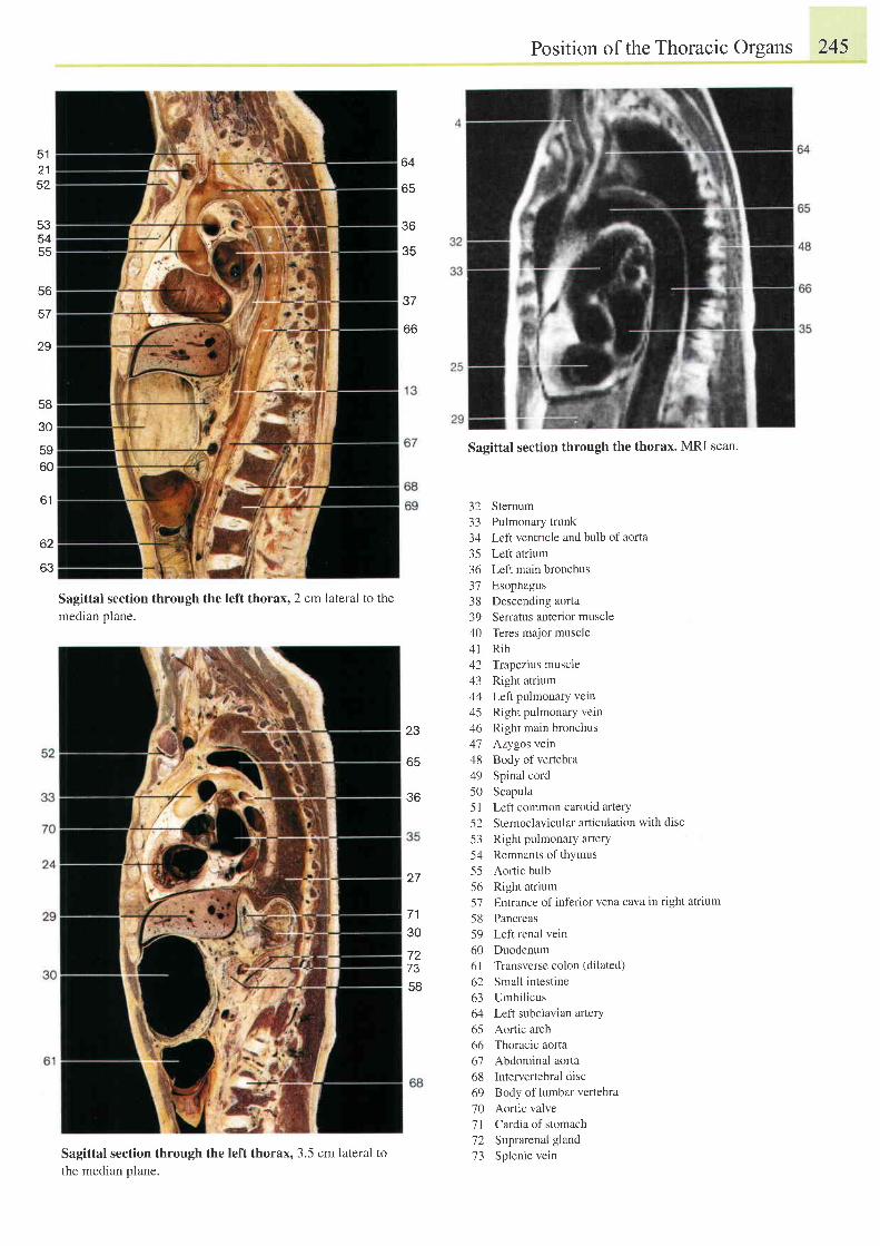
Position of the Thoracic Organs 245
512152
64
J b
35
* t /
oo
535455
co
57
29
58
30
5960
Sagittal section through the left thorax, 2 cm lateral to themedian plane.
Sagittal section through the left thorax, 3.5 cm lateral to
the median plane.
Sagittal section through the thorax. MRI scan.
32 Sternum33 Pulmonary trunk34 LefI ventricle and bulb of aorta
35 Left atrium36 Left main bronchus37 Esophagus38 Descending aorta39 Serratus anterior muscle40 Teres major muscle4t Rib42 Trapezius muscle43 Right atrium44 Left pulmonary vein45 Right pulmonary vein46 Right main bronchus47 Azygos vein48 Body of vertebra49 Spinal cord50 Scapula51 Left common carotid artery52 Sternoclavicular articulation with disc
53 Right pulmonary artery54 Remnants of thymus55 Aonic bulb56 Right atrium57 Entrance of inferior vena cava in right atrium
58 Pancreas59 Left renal vein60 Duodenum61 Transverse colon (dilated)
62 Small intestine63 Umbilicus64 Left subclavian arterY65 Aortic arch66 Thoracic aorta67 Abdominal aorta68 Intervertebral disc69 Body of lumbar vertebra70 Aortic valve'71 Cardia of stomach72 Suprarenal gland73 Splenic vein
6 1
62
OJ
23
65
36
727358
27
1 a
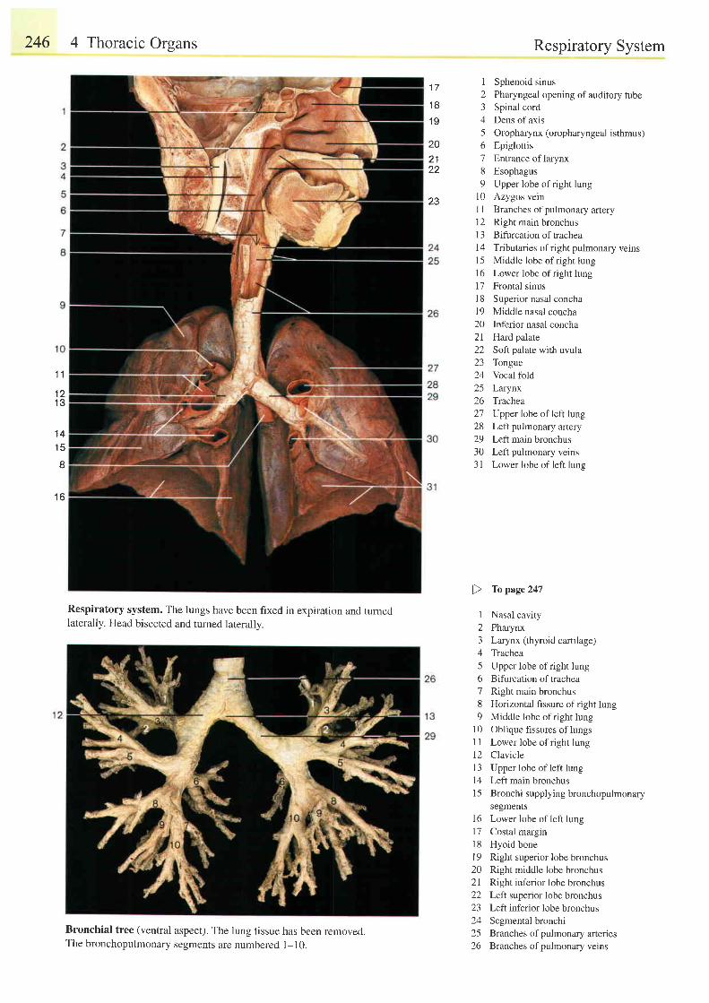
246 4 Thoracic Organs
Respiratory system. The lungs have been fixed in expiration and tumedlaterally. Head bisected and turned laterally.
Bronchial tree (ventral aspect). The lung tissue has been removed.The bronchopulmonary segments are numbered 1-10.
Respiratory System
1 Sphenoid sinus2 Pharyngeal opening of auditory tube3 Spinal cord4 Dens of axis5 Oropharynx (oropharyngeal isthmus)6 Epiglottis7 Entrance oflarynx8 Esophagus9 Upper lobe ofright lung
10 Azygos vein11 Branches ofpulmonary artery12 Right main bronchus13 Bifurcation of tracheal4 Tributaries of r ight pulmonary veins15 Middle lobe of right lung16 Lower lobe ofright lung17 Frontal sinus18 Superior nasal concha19 Middle nasal concha20 Inferior nasal concha21 Hard palate22 SofI palate with uvula23 Tongue24 Vocal fold25 Larynx26 Trachea27 Upper lobe of left lung28 Left pulmonary artery29 Left main bronchus30 Left pulmonary veins31 Lower lobe of left lung
) Topage247
I Nasal cavity2 Pharynx3 Larynx (thyroid cartilage)4 Trachea5 Upper lobe ofright lung6 Bifurcation of trachea7 Right main bronchus8 Horizontal fissure ofright lung9 Middle lobe of right lung
10 Oblique fissures oflungsI i Lower lobe ofright lung12 Clavicle13 Upper lobe ofleft lung14 Left main bronchus15 Bronchi supplying bronchopulmonary
segments16 Lower lobe of left lung17 Costal margin18 Hyoid bonel9 Right superior lobe bronchus20 Right middle lobe bronchus21 Right inferior lobe bronchus22 Left superior lobe bronchus23 Left inferior lobe bronchus24 Segmental bronchi25 Branches ofpulmonary arteries26 Branches ofpulmonary verns
1 8
1 9
202 122
1 7
23
1 1
1 21 3
'14
1 5
I
1 6
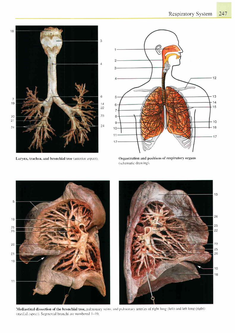
1 0
t o
1 7
6
7
8
9
1 0
1 1
1 7
202 1
24
Respiratory System 247
6
1 A
c t
23
24
Larynx, trachea, and bronchial tree (anterior aspect) C)rganization and positions of respiratory organs( schematic drawing).
Nlediastinal dissection of the bronchial tree, pulr.nonary veins. and pulmonary alteries of r ight lung ( left) and lef i lung (r ight)
(meclial aspect). Segmental bronchi arc nurnbered I l0
t \ t
1 4t c
1 9
2 526
26
25
22
23
2526
20
1 0
*1, ,;\:+,
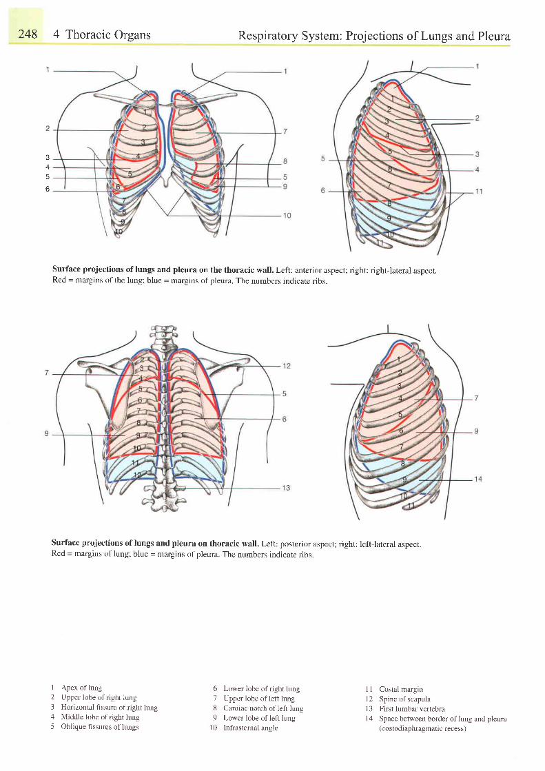
248 4 Thoracic Organs Respiratory System: Projections of Lungs and Pleura
Surface projections oflungs and pleura on the thoracic wall. Left: anterior aspect; right: right-lateral aspect.Red = margins of the lung; blue = margins of pleura. The numbers indicate ribs.
Surface projections oflungs and pleura on thoracic wall. Left: posterior aspect; right: left-lateral aspect.Red = margins of lung; blue = margins of pleura. The numbers indicate ribs.
4
o
I Apex of lung2 Upper lobe ofright lung3 Horizontal fissure ofright lung4 Middle lobe of r ight lung5 Oblique fissures of lungs
6 Lower lobe ot r ight lung
7 Upper lobe ofleft lung
8 Cardiac notch of leftlunq
9 Lower lobe of left lungl0 Infrasternal angle
11 Costal margin12 Spine of scapulal3 First lumbar vertebrat4 Space between border oflung and pleura
(costodiaphragmatic recess)
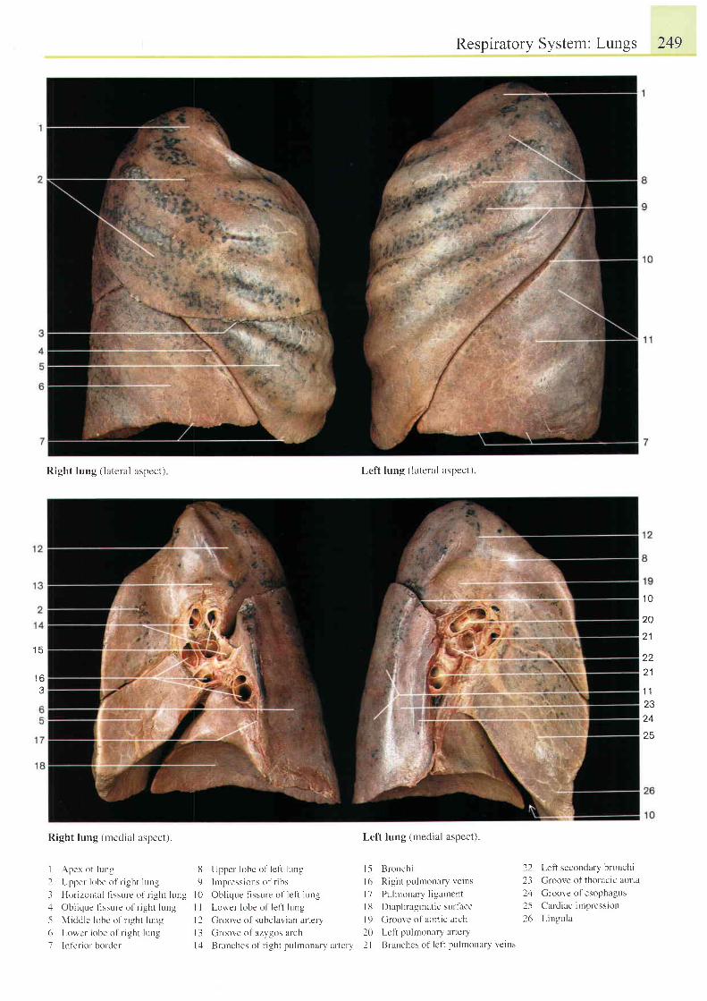
1 q
1 A
Right lung ( lateral aspect)
Right lung (rnedial aspect) .
I Apex of lun-u 8
2 Uppel lobe of l ight lung 9
3 Hor izontal t lssure of r ight lung l0,l Oblique fissure of ri-uht lung I I
5 Midcl le lobe of r ight lung 12
6 Lorvcr lobe of r ight lung 13
7 lnf 'er ior border 14
Upper lobe of le l ' t l r , rng
Impressions of r ibs
Obl ique f issure of lef i lung
Lower lobe of lefi lung
Groove of subclavian artery
Groove of azygos arch
Branches ol r ight pulmonary ar tery
l,eft lung (lateral aspect)
Left lung {nledi i l l :rspect).
15 tsronchi 22
l(r Right pulrnonary veins 23l 7 Puhronary l igamcnt 24Il l Diaphragmatic surtace 25| 9 Groove of aortic arch 2620 t-cft pulmonary arteryI I Brunches o l l c l i pu lmun l r l vc in t
Respiratory System: Lungs 249
1 0
2Q
21
222 1
1 1z5
24
25
Lefi secondary bronchi
Groove of thoracic aorta
Groove of esophagus
Cardiac irrpression
LingLrla
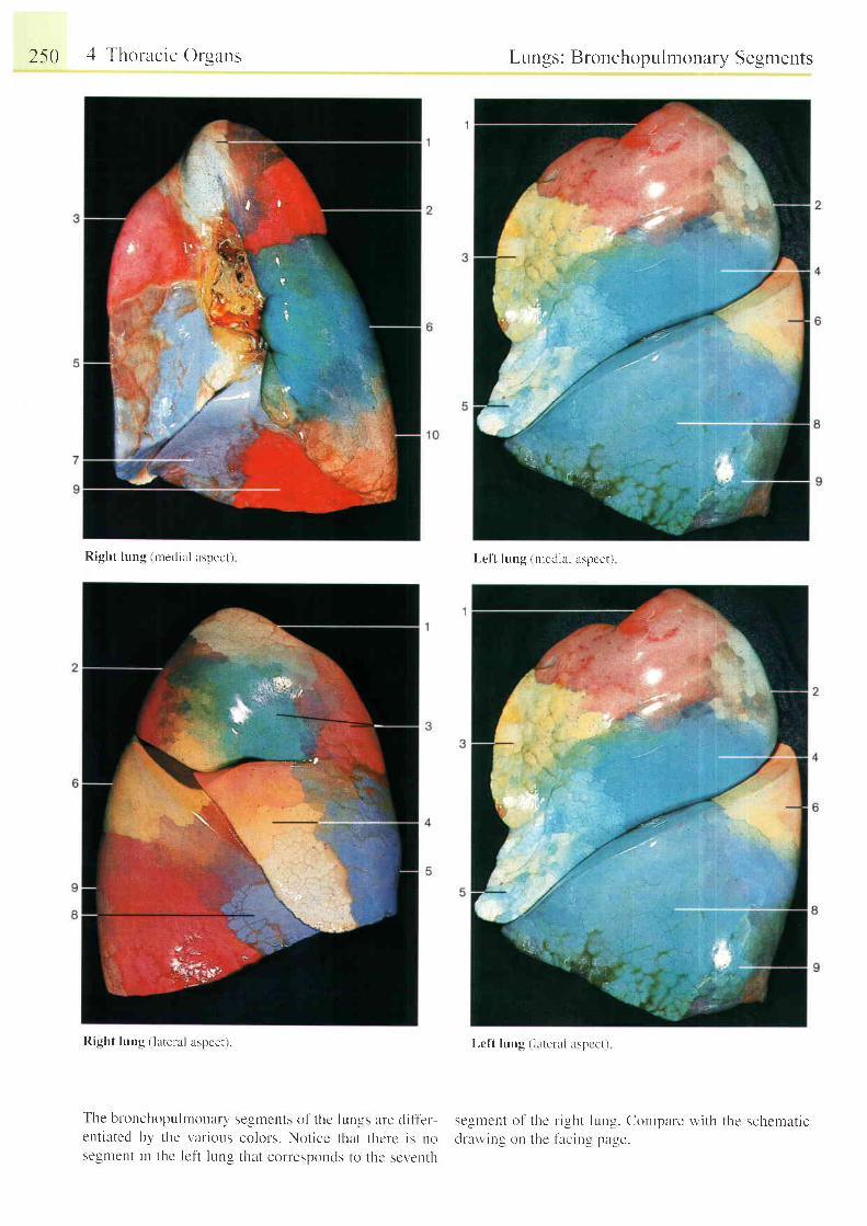
2s0 4 Thoracic Organs
Right lung (medial aspect)
The bronchopul rnonarl,ent ia ted by the var ioussegment in the lefi lr-rng
Lungs: Bronchopulmonary Segments
Lctt lung (uredial xspcct)
Lc f t lung ( l l te ra l aspcc t )
seglncnts of thc lun_us are cli l ' l-er'eo lo l s . No t i ec t h l r t t l r c l c i r nothat corresponcls to the scventh
se-s l l lcnt o l ' the r i -cht lung. Contpareclrawing on the tircin-l pa-ue .
Right lung ( lateral aspect).
with the schemat ic
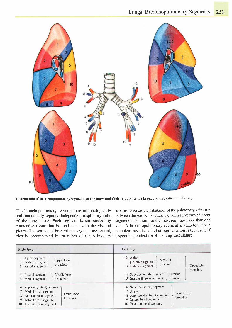
Lungs: BronchopulmonarySegments 251
Distribution of bronchopulmonary segments of the lungs and their relation to the bronchial tree (after J. F. Huber).
The bronchopulmonary segments are morphologicallyand functionally separate independent respiratory unitsof the lung tissue. Each segment is surrounded byconnective tissue that is continuous with the visceralpleura. The segmental bronchi in a segment are central,closely accompanied by branches of the pulmonary
arteries, whereas the tributaries of the pulmonary veins run
between the segments. Thus, the veins serve two adjacentsegments that drain for the most pafi into more than one
vein. A bronchopulmonary segment is therefore not a
complete vascular unit, but segmentation is the result of
a specific architecture of the lung vasculature.
Right lung
1 Apical segment
2 Posterior segment
3 Anterior segment
4 Lateral segment
5 Medial segment
6789
t0
Upper lobebronchus
Middle lobebronchus
Left lung
l+2 Apico-postenor segment
3 Anterior segment
Superiordivision Upper lobe
bronchus
4 Superior lingular segment I Inferior
5 Inferior lingular segment J division
Superior (apical) segmentMedial basal segmentAnterior basal segmentLateral basal segmentPosterior basal segment
Lower lobebronchus
6 Superior (apical) segment7 Absent8 Anteromedial basal segment9 Lateral basal segment
10 Posteriot basal segment
Lower lobebronchus
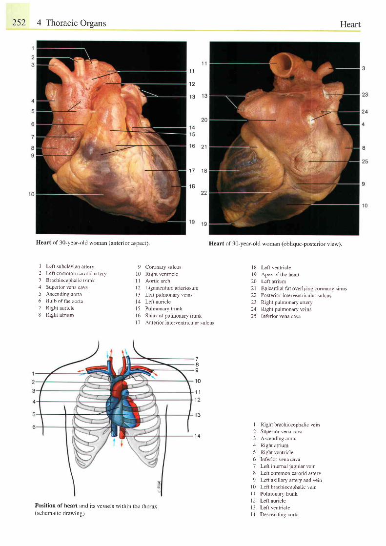
252 4 Thoracic Orsans
1 1
1 2
1 3
1 7
1 8
Heart of 30-year-old woman (anterior aspect).
I Left subclavian artery2 Left common carotid artery3 Brachiocephalic trunk4 Superior vena cava5 Ascending aorta6 Bulb of the aorta7 Right auricle8 Right atrium
9 Coronary sulcus10 Right ventricle1l Aortic arch12 Ligamentum arteriosum13 Left pulmonary veins14 Left auriclel5 Pulmonary trunkl6 Sinus ofpulmonary trunk17 Anteriorinterventricular sulcus
1 11 2
1 3
1 4
Heart of 3O-year-old woman (oblique-posterior view).
18 Left ventricle19 Apex ofthe heart20 Left atrium21 Epicardial fat overlying coronary sinus22 Posterior interventricular sulcus23 Right pulmonary artery24 Right pulmonary veins25 Inferior vena cava
I Right brachiocephalic vern2 Superior vena cava3 Ascending aorta4 Right atrium5 Right ventricle6 Inferior vena cava7 Left internal jugularvein8 Left common carotid artery9 Left axillary artery and vern
l0 Left brachiocephalic veinI I Pulmonary trunkl2 Left auriclel3 Left ventricle14 Descending aorta
78I
1 0
Position of heart and its vessels within the thorax(schematic drawing).
Heart
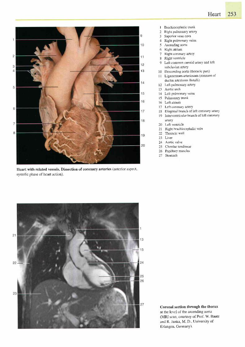
Heart with related vessels. Dissection of coronary arteries (anterior aspect,
systolic phase of heart action).
Heart 253
I Brachiocephalictrunk2 Right pulmonary arterY3 Superior vena cava4 Right pulmonary veins5 Ascending aorta6 Right atrium7 Right coronary artery8 Right ventricle9 Left common carotid artery and left
subclavian artery10 Descending aorta (thoracic pan)
1l Ligamentum arteriosum (remnant of
ductus arteriosus Botalli)12 Left pulmonary artery13 Aortic arch14 Left pulmonary veins15 Puimonary trunk16 Left atriuml7 Lefl coronary artery18 Diagonal branch ofleft coronary artery
1 9 Interventricular branch of left coronary
artery20 Left ventricle2l Right brachiocephalic vein
22 Thoracic wall23 Liver24 Aortic valve25 Chordae tendineae26 Paptllry muscles27 Stomach
Coronal section through the thoraxat the level of the ascending aorta(MRI scan, courtesy of Prof. W. Bautz
and R. Janka, M. D., UniversitY ofErlangen, Germany).
1 1
1 2
1 3
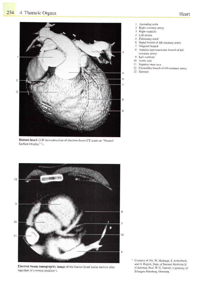
254 4 Thoracic Orsans
Human heart (3-D reconstruction of electron beam CT scans as ,,Shaded
Surface Display"') .
Electron beam tomographic image of the human heart (axial section afterinjection of contrast mediuml).
Heart
1 Ascending aorta2 Right coronary artery3 Right ventricle4 Left atrium5 Pulmonary trunk6 Septal branch ofleft coronary artery7 Diagonal branch8 Anterior interventricular branch of left
coronary artery9 Left ventricle
10 Aortic rootI I Superior vena caval2 Circumflex branch of left coronary aneryl3 Sternum
Courtesy of Drs W. Moshage, S. Achenbach,and D. Ropers, Dept of Internal Medicine II(Chairman: Prof. W. G Daniel), University ofErl angen-Ntirnberg, Germany
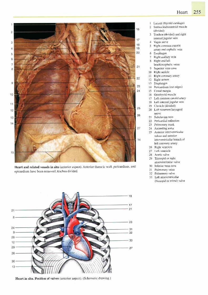
1 6
1 7
1 B1 920
zz
2324
Heart 255
I Larynx (thyroid cartilage)2 Stemocleidomastoid muscle
(divided)3 Trachea (divided) and right
intemal jugular vein4 Vagus nerve5 Right common carotid
artery and cephalic vein
6 Esophagus7 Right axillary vein8 Right and left
brachiocephalic veins
9 Superior vena cava10 Right auricle11 Right coronary artery12 Right atrium13 Diaphragm14 Pericardium (cut edges)15 Costal margin16 Omohyoid musclel7 Left common carotid arterY18 Left internaljugular vein
19 Clavicle (divided)
20 Left recurrent laryngealnerve
21 Subclavian vein22 P ericardial reflection23 Pulmonary trunk24 Ascending aorta25 Anterior interventricular
sulcus and anteriorinterventricular branch ofleft coronary artery
26 Right ventricle27 Leftventricle28 Aortic valve29 Tricuspid or right
atrioventricular valve30 Inferior vena cava31 Pulmonary veins32 Pulmonary valve33 Leftatrioventricular
(bicuspid or mitral) valve
25
26
Heart and related vessels in situ (anterior aspect). Anterior thoracic wall, pericardium, and
epicardium have been removed; trachea divided.
1 8
1 721
23
J I
J Z
33
1 l
Heart in situ. Position of valves (anterior aspect). (Schematic drawing')
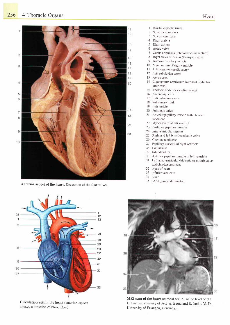
256 4 Thoracic Orsans
Anterior aspect of the heart. Dissection of the four valves.
Circulation within the heart (anterior aspect;arrows = direction of blood flow).
Heart
1 Brachiocephalictrunk2 Superior vena cava3 Sulcus terminalis4 Right auricle5 Right atrium6 Aortic valve7 Conus afteriosus (interventricular septum)8 Right atrioventricular (tricuspid) valve9 Anterior papillary muscle
l0 Myocardium of right ventricleI 1 Left common carotid artery12 Left subclavian artery13 Aortic archl4 Ligamentum arteriosum (remnant of ductus
anenosus)15 Thoracic aorta (descending aorta)l6 Ascending aorta17 Left pulmonary veinl8 Pulmonary trunkl9 Left auricle20 Pulmonic valve2l Anterior papillary muscle with chordae
tendineae22 Myocardium of left ventricle23 Posterior papillary muscle24 Interventricularseptum25 Right and left brachiocephalic veins26 Chordae tendineae21 Paplllary muscles of right ventricle28 Left atrium29 Infundibulum30 Anterior papillary muscle of left ventricle3l Left atrioventricular (bicuspid or mitral) valve
and chordae tendineae32 Apex of heart33 Inf'erior vena cava34 Liver35 Aorta (pars abdominalis)
1 11 2
.13
1 A
t c
t o
I T
1 81 9
20
21
24
22
1 11 2t o
t o
28202S22
30
31
23
MRI scan of the heart (coronal section at the level of theleft atrium; couftesy of Prof W. Bautz and R. Janka, M. D..University of Erlangen, Germany).
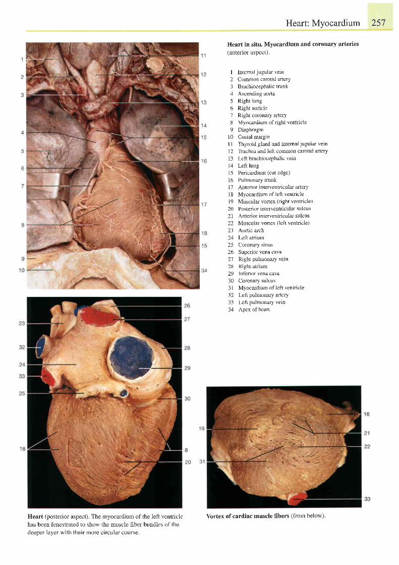
Heart: Myocardium 257
Heart in situ. Myocardium and coronary arteries
(anterior aspect).
I Internaljugular vein2 Common carotid artery3 Brachiocephalictrunk4 Ascending aorta5 Right lung6 Right auricle7 Rightcoronary artery8 Myocardium of right ventricle9 Diaphragm
10 Costal marginl1 Thyroid gland and intemal jugular vein72 Trachea and left common carotid arteryl3 Left brachiocephalic vein14 Left lung15 Pericardium (cut edge)16 Pulmonary trunkI 7 Anterior interventricular artery18 Myocardium of left ventricle19 Muscular vortex (right ventricle)20 Posterior interventricular sulcus21 Anteriorinteilentricularsulcus22 Muscular vortex (left ventricle)23 Aortic arch24 Left atrium25 Coronary sinus26 Superior vena cava27 Right pulmonary vein28 Right atrium29 Inferior vena cava30 Coronary sulcus31 Myocardium of left ventricle32 Left pulmonary artery33 Left pulmonary vein34 Apex ofheart
Heart (posterior aspect). The myocardium of the left ventriclehas been fenestrated to show the muscle fiber bundles of thedeeper layer with their more circular course.
Vortex of cardiac muscle fibers (from below).
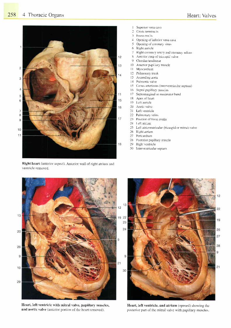
258 4 Thoracic Orsans
Right heart (anterior aspect). Anterior wall of right atrium andventricle removed.
Heart: Valves
I Superior vena cava
2 Crista terminalis
3 Fossa ovalis
4 Opening of inferior vena cava
5 Opening ofcoronary sinus
6 Right auricle
7 Right coronary artery and coronary sulcus
8 Anterior cusp of tricuspid valve
9 Chordae tendineae
l0 Anterior papillary muscle
I I Myocardium
12 Pulmonary trunk
l3 Ascending aorta
14 Pulmonic valve
l5 Conus arteriosus (interventricular septum)
| 6 Septal papi l lary muscles
l7 Septomarginal or moderator band
l8 Apex ofheart
19 Left auricle
20 Aortic valve
21 Left ventricle
22 Pulmonary veins
23 Position of fossa ovalis
24 Left atrium
25 Left atrioventricular (bicuspid or mitral) valve26 Right atrium
27 Pericardium
28 Posterior papillary muscle
29 Right ventricle
30 Interventricular septum
Heart. left ventricle. andposterior part of the mitral
atrium (opened) showing thevalve with papillary muscles.
t l
I J
1 4
7BI
1 9 22
23
Heart, left ventricle with mitral valve,and aortic valve (anterior portion of the
papillary muscles,heart removed).
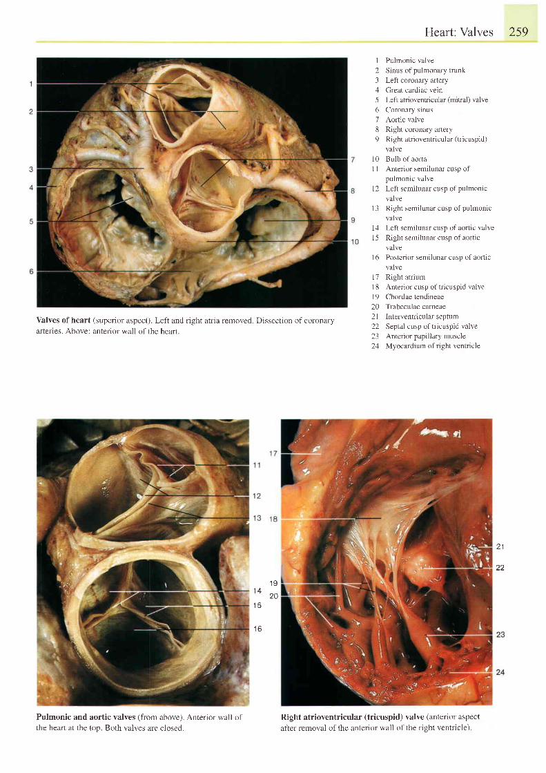
Heart: Valves
1 Pulmonic valve2 Sinus of pulmonary trunk3 Left coronary artery4 Great cardiac vein5 Left atrioventricular (mitral) valve6 Coronary sinus7 Aortic valve8 Right coronary artery9 Right atrioventricular (tricuspid)
valve10 Bulb of aorta11 Anterior semilunar cusp of
pulmonic valve12 Left semilunar cusp of pulmonic
valvel3 Right semilunar cusp of pulmonic
valve14 Left semilunar cusp ofaortic valvel5 Right semilunar cusp of aort ic
valve16 Posterior semilunar cusp of aortic
valve17 Right atrium18 Anteriorcusp of tricuspid valve19 Chordae tendineae20 Trabeculae carneae2l Interventdcularseptum22 Septal cusp of tricuspid valve23 Anterior papillary muscle24 Myocardium of right ventricle
259
Valves ofheart (superior aspect). Left and right atria removed. Dissection ofcoronaryarteries. Above: anterior wall of the hearl.
1 91 4
201 5
1 6
Pulmonic and aortic valves (fromthe heart at the top. Both valves are
above). Anterior wall of
closed.
21
22
Right atrioventricular (tricuspid)after removal of the anterior wall of
valve (anterior aspect
the right ventricle).
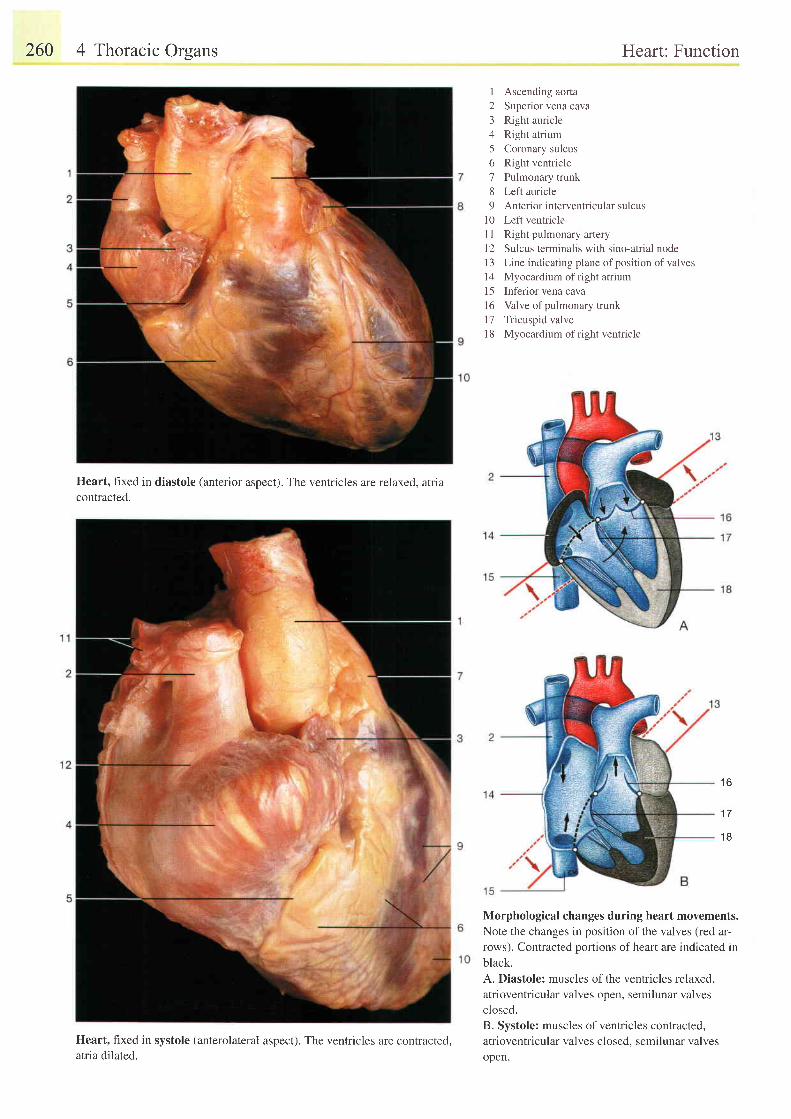
260 4 Thoracic Orsans
Heart, fixed in diastole (anterior aspect). The ventricles are relaxed, atriacontracted.
Heart, fixed in systole (anterolateral aspect). The ventricles are contracted,atria dilated.
Heart: Function
1 Ascending aorta2 Superior vena cava3 Right auricle4 Right atrium5 Coronary sulcus6 Right ventricle7 Pulmonary trunk8 Left auricle9 Anteriorinterventricularsulcus
l0 Left ventricleI I Right pulmonary arteryl2 Sulcus terminalis with sino-atrial nodeI 3 Line indicating plane of position of valvesl4 Myocardium oI r ight atr ium15 Inf'erior vena cava16 Valve of pulmonary trunkl7 Tricuspid valvel8 Myocardium of right ventricle
Morphological changes during heart movements.
Note the changes in position of the valves (red ar-rows). Contracted poftions of heart are indicated in
black.
A. Diastole: muscles of the ventricles relaxed.
atrioventricular valves open, semilunar valves
closed.B. Systole: muscles of ventricles contracted,
atrioventricular valves closed. semilunar valves
open.
t o
1 7
1 8
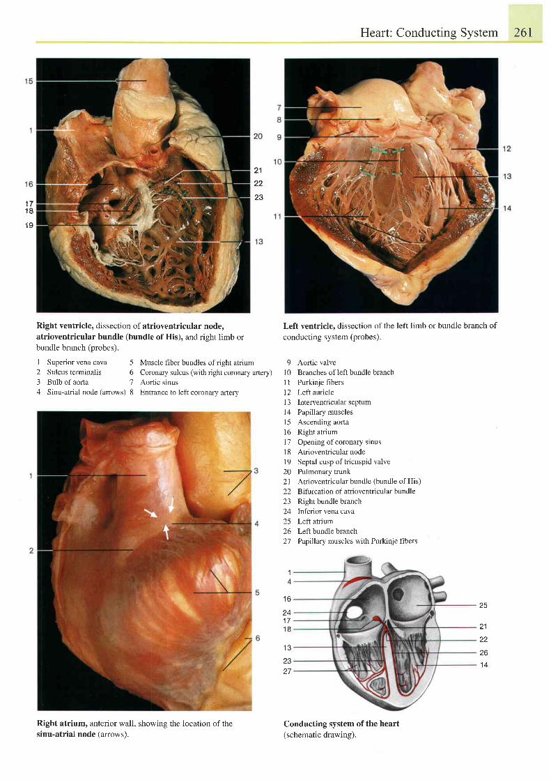
Heart: Conducting System 26I
21
22
231 71 8
1 9
Right ventricle, dissection of atrioventricular node,atrioventricular bundle (bundle of His), and right limb orbundle branch (probes).
1 Superiorvenacava 5 Musclefrberbundlesofrightatrium2 Sulcusterminalis 6 Coronarysulcus(withrightcoronaryartery)3 Bulb of aorta 7 Aortic sinus4 Sinu-atrial node (arrows) 8 Entrance to left coronary artery
Right atrium, anterior wall, showing the location of thesinu-atrial node (arrowsl.
Left ventricle, dissection of the left limb or bundle branch of
conducting system (probes).
9 Aortic valve10 Branches of leftbundle branchI I Purkinje fibers12 Left auricle13 Interventricularseptum14 Papillary musclesl5 Ascending aorta16 Right atrium17 Opening ofcoronary sinus18 Atrioventricularnode19 Septal cusp oftricuspid valve20 Pulmonary trunk21 Atrioventricular bundle (bundle of His)22 Bifurcation of atrioventricular bundle23 Right bundle branch24 Infeior vena cava25 Leftatrium26 Left bundle branch27 Pap1llary muscles with Purkinje fibers
Conducting system of the heart(schematic drawing).
1 6
241 71 8
14
2327
25
21
22
26t \ t
1 4
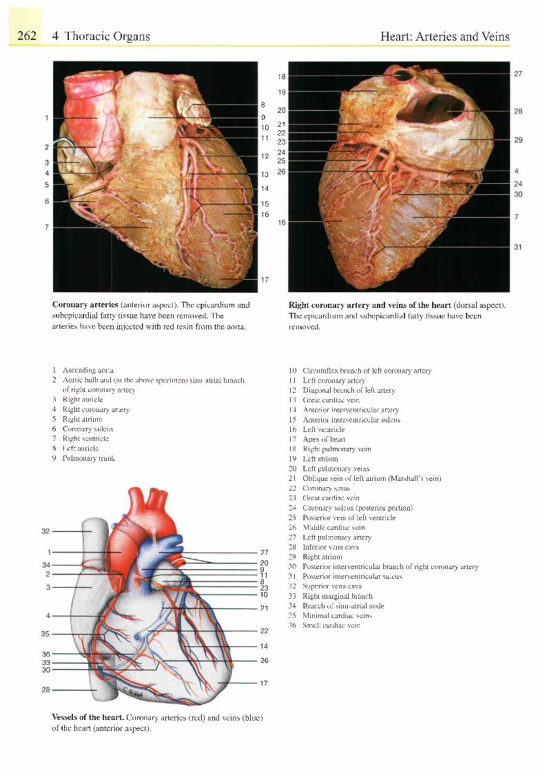
262 4 Thoracic Orsans
Coronary arteries (anterior aspect). The epicardium andsubepicardial fatty tissue have been removed. Thearleries have been injected with red resin from the aorta
I
2Ascending aorta
Aortic bulb and (in the above specimen) sinu-atrial branchof right coronary artery
Right auricle
Right coronary artery
Right atrium
Coronary sulcus
Right ventricle
Left auricle
Pulmonary trunk
3/1
56789
Vessels of the heart. Coronary arteries (red) and veins (blue)
of the heart (anterior aspect).
Heart: Arteries and Veins
Right coronary artery and veins of the heart (dorsal aspect).The epicardium and subepicardial fatty tissue have beenremoved.
1 0 Circumllex branch of left coronary arteryI I Left coronary arteryl2 Diagonal branch of left arteryl3 Great cardiac veinl4 Anterior interventricular artery15 Anteriorinterventricularsulcusl6 Left ventricle1 7 Apex of heartl8 Right pulmonary vein19 Left atrium20 Left pulmonary veins2l Oblique vein of left atrium (Marshall's vein)22 Coronary sinus23 Great cardiac vein24 Coronary sulcus (posterior portion)25 Posterior vein of left ventricle26 Middle cardiac vein21 Left pulmonary artery28 Inferior vena cava29 Right atrium30 Posterior interventricular branch of right coronary artery3 I Posterior interventricular sulcus32 Superior vena cava33 Right marginal branch34 Branch of sinu-atrial node35 Minimal cardiac veins36 Small cardiac vein
1 B
1 9
I
I1 01 t
1 2
1 3
20
2 1
Z J
2526
1 4
1 5
t o
363330
27
2091 ' tB231 0
2 1
26
1 7
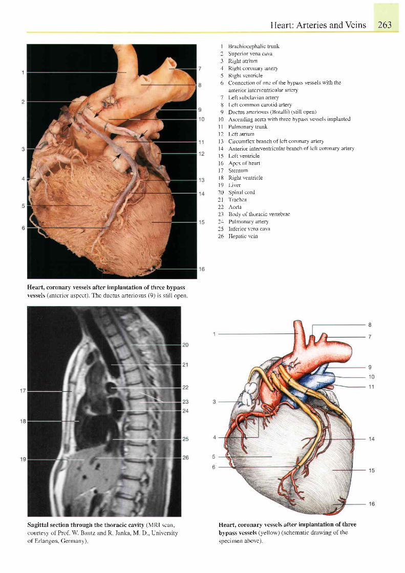
Heart, coronary vessels after implantation of three bypassvessels (anterior aspect). The ductus arteriosus (9) is still open.
Sagittal section through the thoracic cavity (MRI scan,courtesy of Prof. W. Bautz and R. Janka, M. D., Universityof Erlangen, Germany)
Heart: Arteries and Veins 263
I Brachiocephalictrunk2 Superior vena cava3 Right atrium4 Right coronary artery5 Right ventricle6 Connection of one of the bypass vessels with the
anterior interventricular artery7 Left subclavian artery8 Left common carotid artery9 Ductus arteriosus (Botalli) (still open)
l0 Ascending aorta with three bypass vessels implanted1l Pulmonary trunkl2 Left atriumI 3 Circumflex branch of left coronary arteryI 4 Anterior interventricular branch of left coronary arteryl5 Left ventricle16 Apex ofheartl7 Sternuml8 Right venriclel9 Liver20 Spinal cord21 Trachea22 Aorla23 Body of thoracic vertebrae24 Pulmonary anery25 Inferior vena cava26 Hepatic vein
Heart, coronary vessels after implantation of threebypass vessels (yellow) (schematic drawing of thesoecimen above).
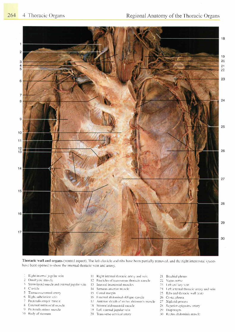
264 4 Thoracic Orsans Regional Anatomy of the Thoracic Organs
1 9202 122
ti,tt:.
tt
t
.&wr
Thoracic wall and organs (ventral aspect) The left clavicle and r ibs have becn part ial ly removed. and the r ight intelcostal spaceshave been opened to show the intcrnal thoracic vein and arlery
I Righl intental jugular vern
2 Onrohvoid nrusclc
3 Stcrnohvoid ntuscle and external lugular vein4 C lav i c l c
5 Thoraco-aclornial arterv6 Right subclavtan vcin7 Pectoral is nta jor nnsclclJ E,xternal intercostal ntuscle9 Pcctoral is nt inor l rusclc
l 0 Bod l ' o f s t c rnum
l l Ri-sht internal thoracic ar tcry and vein
l2 Fascic les of t ransr,ersus thoracis nrusclc
l3 Internal intcrcostal nruscles
l . l Serratus anter ior t luscle
l -5 Costal rnargin
l6 l rx ternal abckrmrnal obl ic lue muscle| 7 Anter ior sheath of rcctus abdonr in is rnuscle
l 8 S te rnoc le i domas to i d rnusc le
l9 Let t internal . jugr : lar vein
20 Transversc cervical arter v
2l Brachial p lexLrs
22 Vagus nerve
23 Lcf i ax i l lary vein
24 Lef l in ternal thoracic ar terv and r ,e in
25 Ribs and thoracic u,a l l (cut)
26 Costal p leura
27 Xiphoir i pnrccss
28 Super ior epigastr ic ar tcr v
29 Diaphlagr l
30 Rectr- rs abdorr in is rnuscle
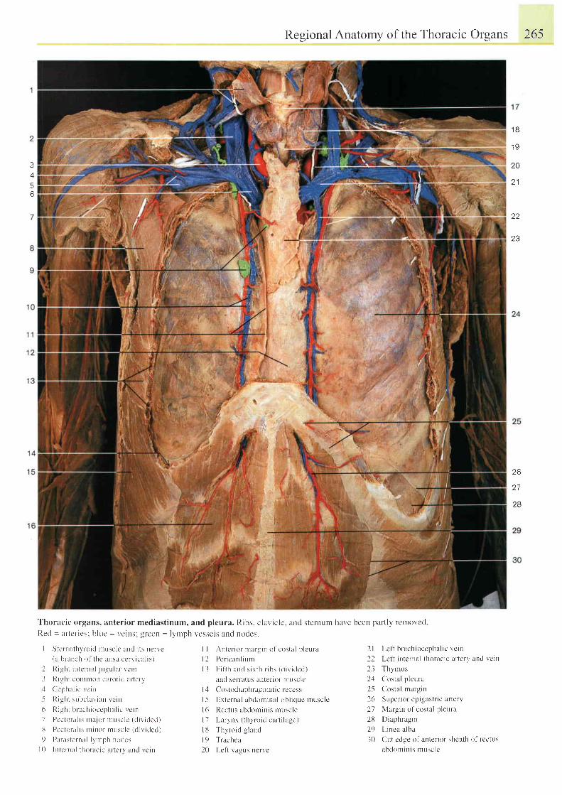
3i
56
I I
1 9
2A
o 1
22
O Q
'r:
ii
r:i{,
Regional Anatomy of the Thoracic Organs 265
tl.t;..{'"'}
2l Lcl i brachiocephal ic vc in
22 Lett internal thoracic artery arrd vcin
23 Thymus
2'1 Costal p lcura
25 Costal margin
26 Superior epigastric artery
27 Margin of costal p leura
2fl Diaphragnr
29 Linca alba
30 Cut cdge of anterror shcath of rectus
abdonr in is rnusclc
t
ti*. _
' l 'horacic organs, anterior mediastinum, and pleura. Ribs. claviclc. and sternuln have been part ly rclnoved
Recl = artcr-rcs: blr.rc = veins: green = lymph vessels and nodes.
26
27
28
I Ster nothvroid urLrsclc and i ts nen'e(a br-ur tch ol thc iursa cerv ical is)
I l { i . : l r t i n l c r r . l l i u . - u l : r r ' \ c i n
- l Ri3ht courron carot ic l ar terv
{ ( ' e ' pha l i c r c i n
5 R igh t su l r c l uv i uu r c i nI ' R r - l l r t l r r l r r ' l r i , r . c p l r ; r l i e r c i r r
7 Pcc tu ra l r s n ra . j o r n tusc l c ( d i t , i ded )
I Pectoral rs r l r inor r .uusclc (d iv ided)
9 l ) l raste ln l l Jvr lph noclcs
l 0 I n t c rna l t ho rac i c i l r t e r v l l nd vc tn
Antcr ior mirrg in o l costal p ler l ra
Pericarcli unr
F i l t h and s i x l h r i bs ( d i i i ded )
and serratus anter ior nrusclc
Costodiaphragrnat ic r cccss
External abdominal obl ic lLe musclc
Rectus abclorn in is nruscle
Larynx ( thytoid c iu ' t i lage)
Thyroid g land
Trachca
Left vagus nerve
il
t l
l 3
l 4
t 5l 6l 1I ut 920
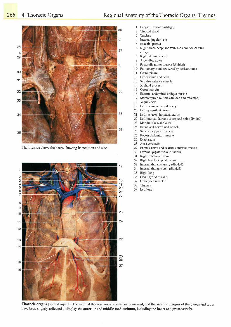
266 4 Thoracic Organs Regional Anatomy of the Thoracic Organs: Thymus
1 7
1 81 9202122
23
24
I Larynx (thyroid cartilage)2 Thyroid gland3 Trachea4 Intemal jugular vein5 Brachial plexus6 Right brachiocephalic vein and common carotid
artery7 Right phrenic nerve8 Ascending aorta9 Pectoralis minor muscle (divided)
l0 Pulmonary trunk (covered by pericardium)I 1 Costal pleura12 Pericardium and heartI 3 Serratus anterior musclel4 Xiphoid processl5 Costal margin16 External abdominal oblique muscle17 Stemothyroid muscle (divided and reflected)18 Vagus nerve19 Left common carotid artery20 Left sympathetic trunk2l Left recurrent laryngeal nerve22 Left internal thoracic artery and vein (divided)23 MNgin of costal pleura24 Intercostal nerves and vessels25 Superior epigastric artery26 Rectus abdominis muscle27 Diaphragm28 Ansa cervicalis29 Phrenic nerve and scalenus anterior muscle30 Externaljugular vein (divided)31 Right subclavian vein32 Right brachiocephalic vein33 Internal thoracic artery (divided)34 Internal thoracic vein (divided)35 Rightlung36 Cricothyroid muscle37 Omohyoid muscle38 Thymus39 Left lung
Thoracic organs (ventral aspect). The internal thoracic vessels have been removed, and the anterior margins ofthe pleura and lungshave been slightly reflected to display the anterior and middle mediastinum, including the heart and great vessels.
28
+
29
30
31
32
33
1
234567
2526
27
The thymus above the heart, showing its position and size.
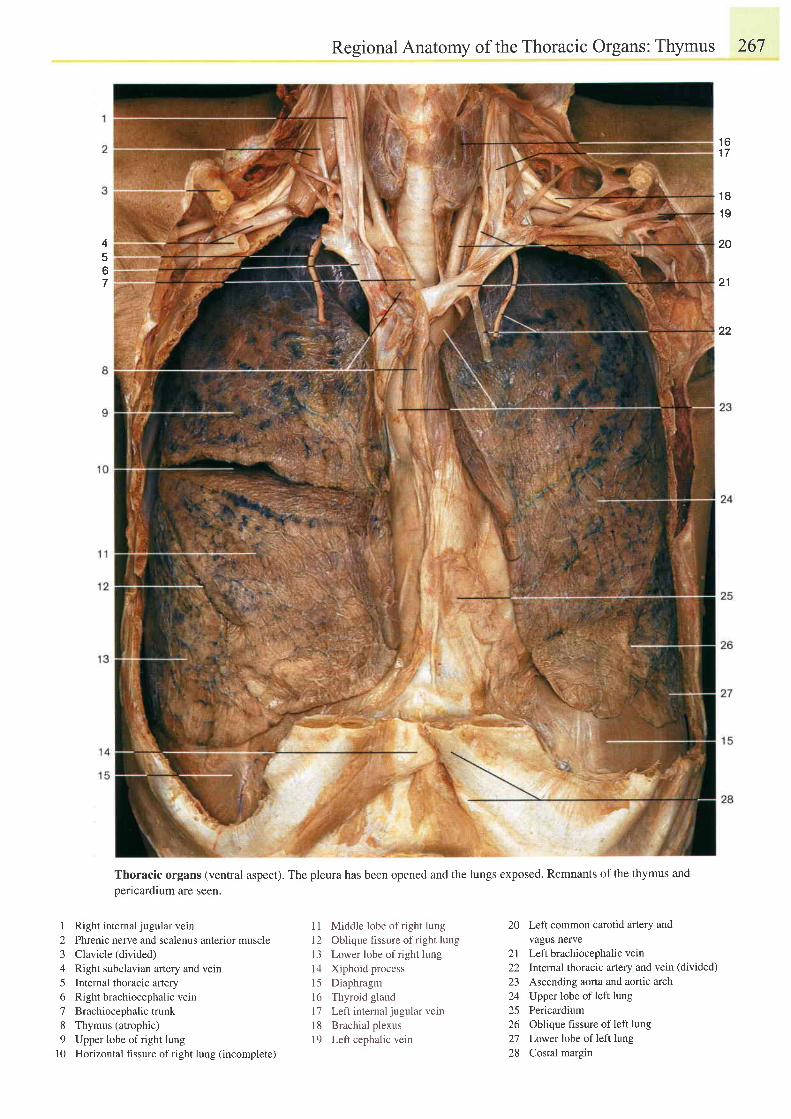
Regional Anatomy of the Thoracic Organs: Thymus 267
Thoracic organs (ventral aspect). The pleura has been opened and the lungs exposed. Remnants ofthe thymus andoericardium are seen.
1 6I I
4567
1 8
1 9
20
2 1
22
1 Right internal jugular vern2 Phrenic nerve and scalenus anterior muscle3 Clavicle (divided)
4 Right subclavian arlery and vein5 Internal thoracic artery6 Right brachiocephalic vern7 Brachiocephalictrunk8 Thymus (atrophic)9 Upper lobe ofright lung
l0 Horizontal hssure of right lung (incomplete)
11 Middle lobe of right lung12 Oblique fissure of right lung13 Lower lobe ofright lung14 Xiphoid process15 Diaphragm16 Thyroid glandl7 Lefrinternal jugular vein18 Brachial plexus19 Left cephalic vein
20 Left common carotid aftery andvagus nerve
2l Left brachiocephalic vern22 lnternal thoracic artery and vein (divided)
23 Ascending aorta and aortic arch24 Upper lobe of left lung25 Pericardium26 Oblique fissure of left lung27 Lower lobe of left lung28 Costal margin
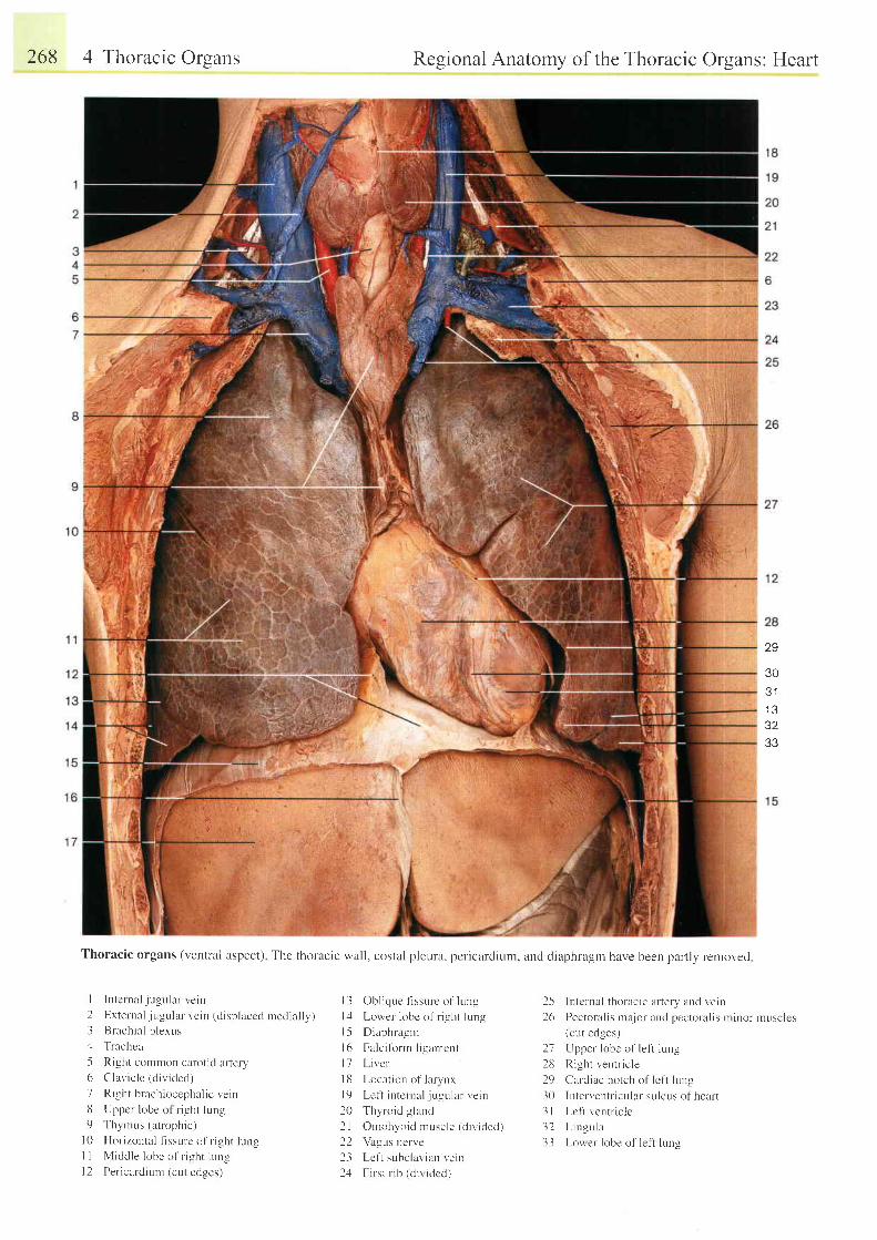
268 4 Thoracic Organs Regional Anatomy of the Thoracic Organs: Heart
Thoracic organs (r,entral aspect) Thc thoracic wall. costal pleura. pcricardium, and diaphragm have been partly removerl
29
30Q 1
1 332
i''
I ln tcr nal jugular vein2 External jugular vc in (d isplaccd rnedial ly)3 Brachial p lexuszl Trachca
5 Right common carot id ar terv6 Clavic lc (d iv idcd)
7 R i . r : h t h r re l t i oecnh r l i c r e i n
8 t ppcr ' lubc ol r i . r :hr l r rn59 Thvnrus (atrophic)
l0 Hor izontal f issure o1' r ight lungI I Middle lobe of r ight l r , rngI 2 Per icardiurn (cut edges)
l 3 Ob l i que f i s su rc o l ' l uns l
l : l Lower lobe of r i -qht iung
l5 Diaphragm
l6 Falc i tbrrn l igamcnt
l 7 L i ve r
I 8 Locat ion of larvnx
l9 Lef t intcrnal . jugular vein
20 Thvroid g land
21 Omohyoid muscle (d ivrc led)
22 Vagr-rs nerve
23 Lef t subclaviarr vein
24 First r ib (d i r , idcd)
25 lntcrnal thoracic artely aud vein
26 Pectoral is major and pectoral is minor r rusclcs(cut ed-qes)
27 Upper lobc of lef t lung
2t i Right ventr ic le
29 Cardiac notch of lef i lung
30 Lrten'entricular sulcus of heart
3 l Lc l t ven t l i c l e
32 L i ngu la
33 Lower lobe of lef t lung
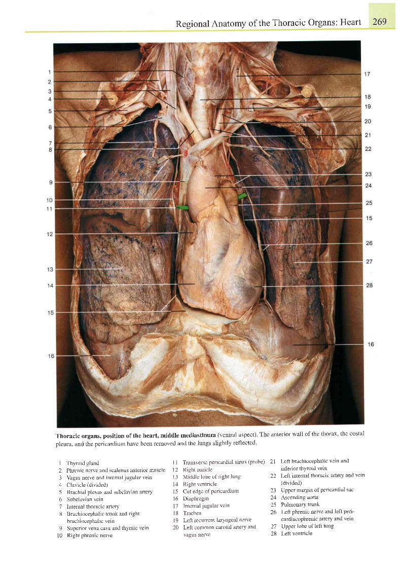
Regional Anatomy of the Thoracic Organs: Heart 269
Thoracic organs, position of the heart, middle mediastinum (ventral aspect). The anterior wall of the thorax, the costal
pleura, and the pericardium have been removed and the lungs slightly reflected.
I Thyroid gland
2 Phrenic nerve and scalenus anterior muscle
3 Vagus nerve and internal j ugular vein
4 Clavicle (divided)
5 Brachial plexus and subclavian artery
6 Subclavian vein
7 Internal thoracic aftery
8 Brachiocephalic trunk and right
brachiocephalic vein
9 Superior vena cava and thymic vein
l0 Right phrenic nerve
1 1 Transverse pericardial sinus (probe)
12 Right auricle
13 Middle lobe of right lung
14 Right ventricle
15 Cut edge ofpericardium
16 Diaphragm
17 Internal jugular vetn
l8 Trachea
19 Left reculTent laryngeal nerve
20 Left common carotid aftery and
vagus nerve
2l LeftbrachiocePhalic vein and
inferior thyroid vein22 Left internal thoracic artery and vein
(divided)23 Upper margin of Pericardial sac
24 Ascending aorta25 Pulmonary trunk26 Left phrenic nerve and left peri-
cardiacophrenic arlerY and vein
27 Upper lobe of left lung28 Leftventricle
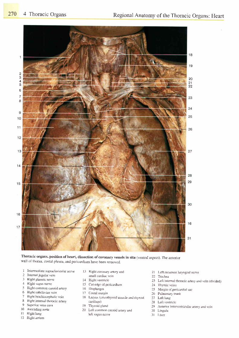
270 4 Thoracic Organs Regional Anatomy of the Thoracic Organs: Heart
Thoracic organsr position ofheart, dissection ofcoronary vessels in situ (ventral aspect). The anteriorwall of thorax, costal pleura, and pericardium have been removed.
2?
4
6
7I
202 122
I Intermediate supraclavicularnerve2 Internaljugular vein3 Right phrenic nerve4 Right vagus nerve5 Right common carotid artery6 Right subclavian vein7 Right brachiocephalic vein8 Right intemal thoracic artery9 Superior vena cava
10 Ascending aortal1 Right lungl2 Right atrium
13 Right coronary artery andsmall cardiac vein
14 Right ventriclel5 Cut edge ofpericardium16 Diaphragm17 Costal margin18 Larynx (cricothyroid muscle and thyroid
cartilage)l9 Thyroid gland20 Left common carotid arlery and
left vagus nerve
21 Leftrecurrentlaryngeal nerve22 Trachea23 Left intemal thoracic artery and vein (divided)24 Thymic veins25 Margin of pericardial sac26 Pulmonary trunk27 Leftltng28 Leftventricle29 Anterior interventricular artery and vein30 Lingula31 Liver
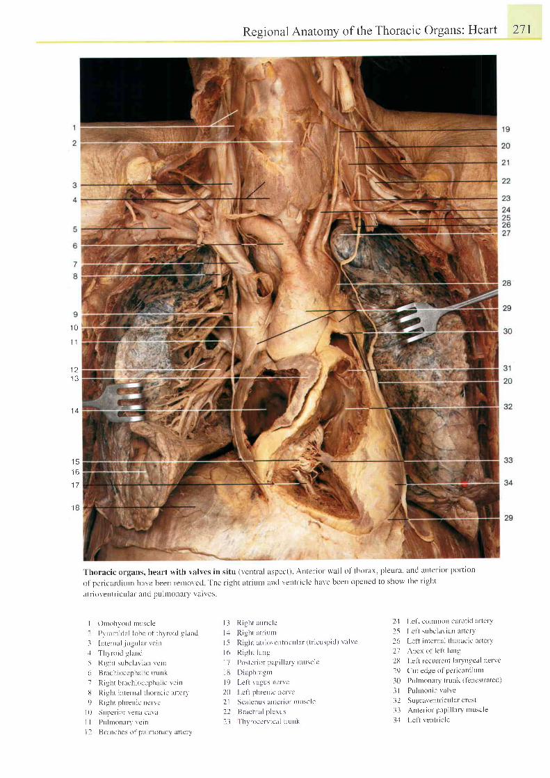
1 0
I 1
1 . )
, J
1 A
Regional Anatomy of the Thoracic Organs: Heart 271
Thoracic organs, heart with valves in situ (ventral aspect). Anterior wall of thorax, pleura. ancl anterior port ion
of pericarcl iuur havc been renrovcd The r ight atr iunt and ventt ' ic le have been opcned to show the r ight
atr ioventr iculat ' and puluouat'y valves
l r )
1 7
1 B
I Ornohvoic l rnuscle
2 Pyr-arnidal lobe of thy ' ro id g land
3 I n ten ra l j ugu la r r c i n.1 Thyr o id -q land5 Ri-sht subclavian vcin
6 Brachioccphal ic t runk
7 R i 3 h t h n r . h i , r e e p h i t l i c r e i n
13 Right interual t l roracrc ar te ly '
9 Right ph'enic ncr-vc
l 0 Super ior veua cuva
I 1 Pu ln rona ry ve in
l 2 Brruches ol pulr ronlry ar tetv
l 3 R igh t au r i c l c
l : l Right atr iunr
1-5 Right l t r iot 'et t t r ic t t l r r ( t r ic t rspid) r 'a lvc
l 6 R i - uh t i ung
I7 Postel ior papi l lary ' r t tust : lc
l13 Diaphr-agnt
l 9 I - e1 t v i t gus nc r ve
20 Lcl ' t phrenic ue rve
21 Scalcnus antcr ior l l tL lsc le
22 tsr-achia l p lcrus
23 Thyroccrv icnl t t -uuk
24 Left commorl carotid artet-y
25 Lcf t subclavian ar tery
26 Left intelnal thoracic at'tery
27 Apcx of lc l t lung
28 Left rccLlrrent laryn-qcal net vc
29 Cut edge of pericat'diltnr
30 Puhronary trlrnk (tenestrated)
3 l Pu lmon i c va l ve
32 Supraventr iculat-crest
33 Anter ior papi l lary muscle
3:1 l -e1t ventr ic le
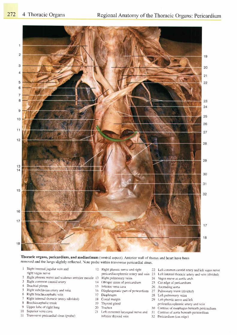
272 4 Thoracic Organs
1 31 4
Regional Anatomy of the Thoracic Organs: Pericardium
Thoracic organs' pericardium, and mediastinum (ventral aspect). Anterior wall of thorax and heart have beenremoved and the lungs slightly reflected. Note probe within transverse pericardial sinus.
I Right internaljugular vein and 12 Right phrenic nerve and rightright vagus nerve pericardiacophrenic artery and vein
2 Right phrenic nerve and scalenus anterior muscle I 3 Right pulmonary veins3 Right common carotid atery 14 oblique sinus of pericardrum4 Brachial plexus 15 Inferior vena cava5 Right subclavian artery and vein 16 Diaphragmatic part ofpericardium6 Rightbrachiocephalicvein 17 Diaphragm7 Right internal thoracic artery (divided) 18 Costal margin8 Brachiocephalic trunk 19 Thyroid gland9 Upper lobe ofright lung 20 Trachea
10 Superior vena cava 2l Leftrecurrent laryngeal nerve andI I Transverse pericardial sinus (probe) inferior thyroid vein
22z-)a /
2526212829
303 l-)z
Left common carotid ar1ery and left vagus nerveLeft intemal thoracic artery and vein (divided)
Vagus nerve at aoftic arch
Cut edge of pericardium
Ascending aorta
Pulmonary trunk (divided)
Left pulmonary veins
Left phrenic nerve and leftpericardiacophrenic artery and veinContour of esophagus beneath pericardium
Contour of aorta beneath pericardium
Pericardium (cut edge)
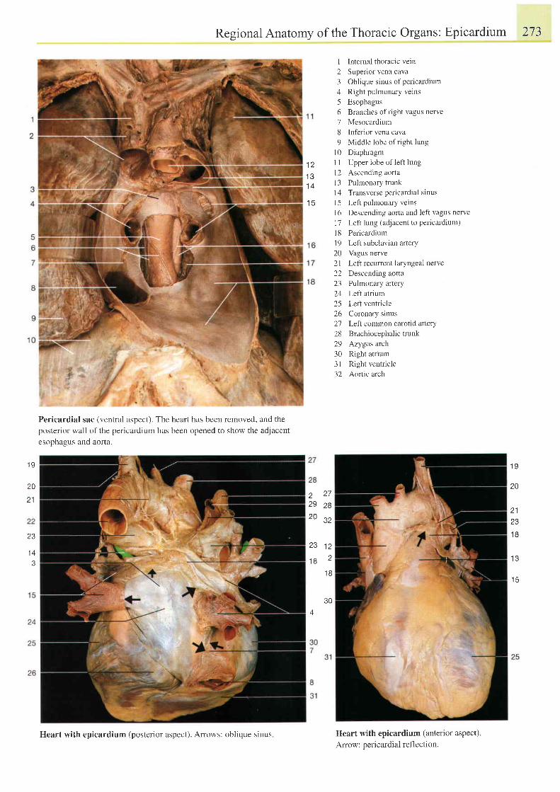
l z
1 4
1 5
Regional Anatomy of the Thoracic Organs: Epicardium 273
1 lnternal thoracic vein2 Superior vena cava3 Oblique sinus ofpericardium4 Right pulmonary verns5 Esophagus6 Branches ofright vagus nerve7 Mesocardium8 Inferior vena cava9 Middle lobe of right lung
10 DiaphragmI I Upper lobe of left lungl2 Ascending aorta13 Pulmonary trunk| 4 Transverse pericardral sinus15 Left pulmonary veins16 Descending aorta and left vagus nerve17 Left lung (adjacent to pericardium)
18 Pericardiuml9 Left subclavian artery20 Vagus nerve2l Left recurrent laryngeal nerve22 Descending aorta23 Pulmonary artery24 Left atrium25 Lefr ventricle26 Coronary sinus2'7 Left common carotid artery28 Brachiocephalic trunk29 Azygos arch30 Right atrium31 Right ventricle32 Aortic arch
Heart with epicardium (anterior aspect)Amow: pericardial reflection.
1 9
20
21
23
1 A
3
Pericardial sac (ventral aspect). The heart has been removed, and the
posterior wall of the pericardium has been opened to show the adjacent
esophagus and aorta.
1 9
a 2 7
29 ze
JZ
20
21z5
1 B
l e
1 R
23 l z
1 8 21 B
30A
Heart with epicardium (posterior aspect). Arows: oblique sinus
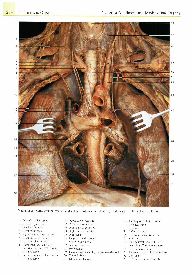
274 4 Thoracic Orsans
1 1
I J
,?,
25
6
67
I
I Supraolavicularnen'es
2 Internal jugr. r lar vc in3 Ornohyoid musclc;l Right vagLls nerve
5 Right colrtron carotid arterv6 Right subclavian ar ter l '7 B rach iocepha l i c t r unk
t t Right brachioccphal ic vcrn9 Super ior cerv ical cerrd iac branch
of r"agus nervcI 0 Inf 'er ior cerv ical cardiac orancnes
ol vagus ncrve
Azygos arch (d iv ided. l
Bifurcation of trachea
Right pu I rnonary artery
Right pulmonary veins
R igh t l ung
Esophagus and branches
of r ight vagus nervc
Inf-erior veru cavaPer icardiunr
[ -arvnx ( thvroid cart i lage. cr icothvroid rnuscle)Thyroid g land
lnternal . j r , rgular ve in
l ll 2l 3l 41 .5I 6
t 1I t il 9202 l
22
231 t
2-52621
Posterior Mediastinum: Mediastinal Orsans
r:ii':i\;
Mediastinal organs after retnoval of heart and pericardium (ventral aspect). Both lungs have been slightly reflected
Esophagus and lefi recurrenj
laryngeal nervc
Trachea
Left vagus nerve
Lcl t common carot id ar tery
Aortic arch
Lcfi recurrenr laryngeal nerve
branching ofT f rom vagus nerve
28 Le f t pulmonary veins
29 Thoracic aorta and left vagr-rs nerve30 Lcl i lung
I I Lcf i phrenic nerve (d iv ic led)
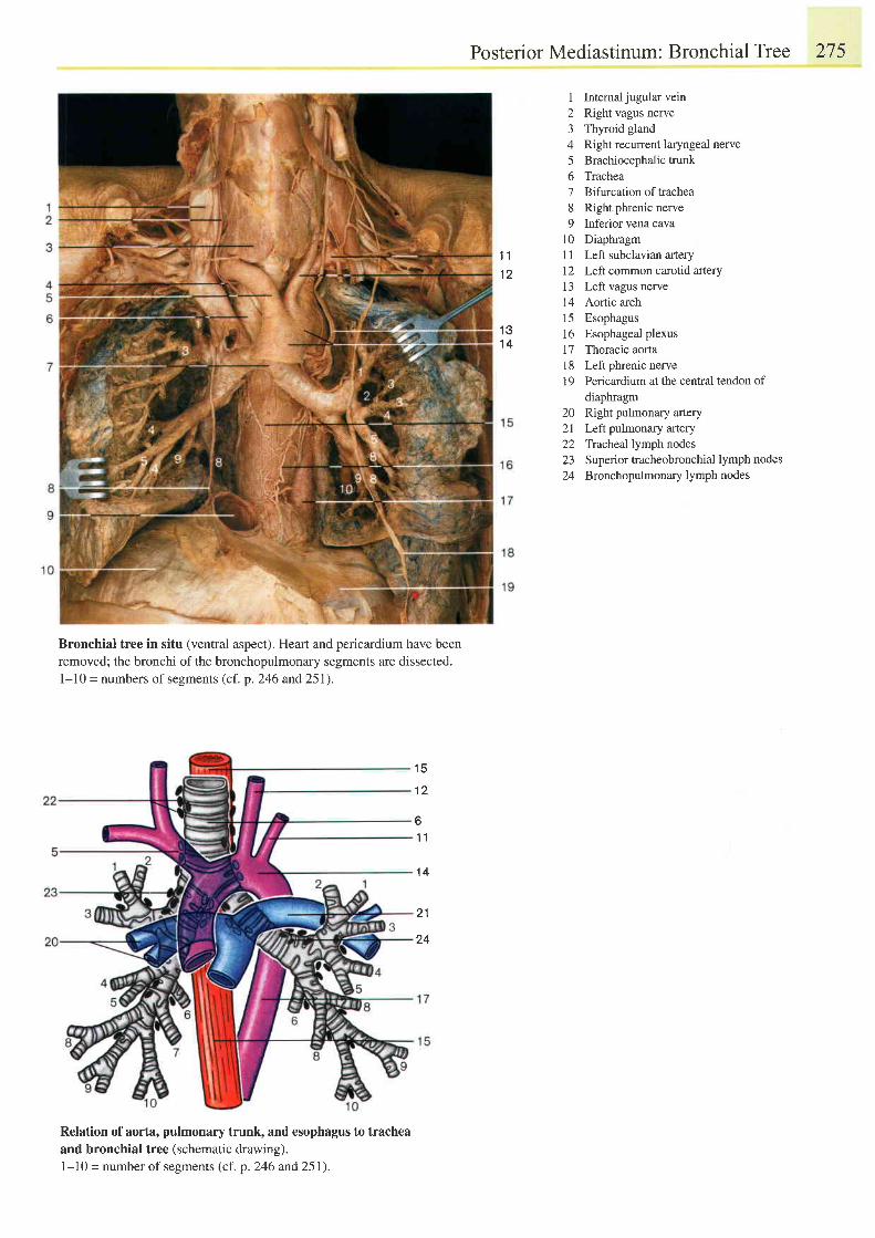
Posterior Mediastinum: Bronchial Tree 275
1 Internaljugular vein2 Right vagus neile3 Thyroid gland4 Right recurrent laryngeal nerve5 Brachiocephalictrunk6 Trachea7 Bifurcationoftrachea8 Right phrenic nerve9 Inferior vena cava
10 Diaphragm11 Left subclavian artery12 Left common carotid artery13 Left vagus nerve14 Aortic arch15 Esophagus16 Esophageal plexus17 Thoracic aorta18 Left phrenic nerve19 Pericardium at the cenffal tendon of
diaphragm20 Right pulmonary artery2l Left pulmonary artery22 Tracheal lymph nodes23 Superior tracheobronchial lymph nodes24 Bronchopulmonary lymph nodes
1 1' t2
1 31 4
Bronchial tree in situ (ventral aspect), Heart and pericardium have beenremoved; the bronchi of the bronchopulmonary segments are dissected.I - l0 = numbers of segments (cf . p. 246 and 25 1).
1 5
1 2
b
1 1
1 4
2 1
24
Relation of aorta, pulmonary trunk, and esophagus to tracheaand bronchial tree (schematic drawing).l-10 = number of segments (cf. p. 246 and 251).
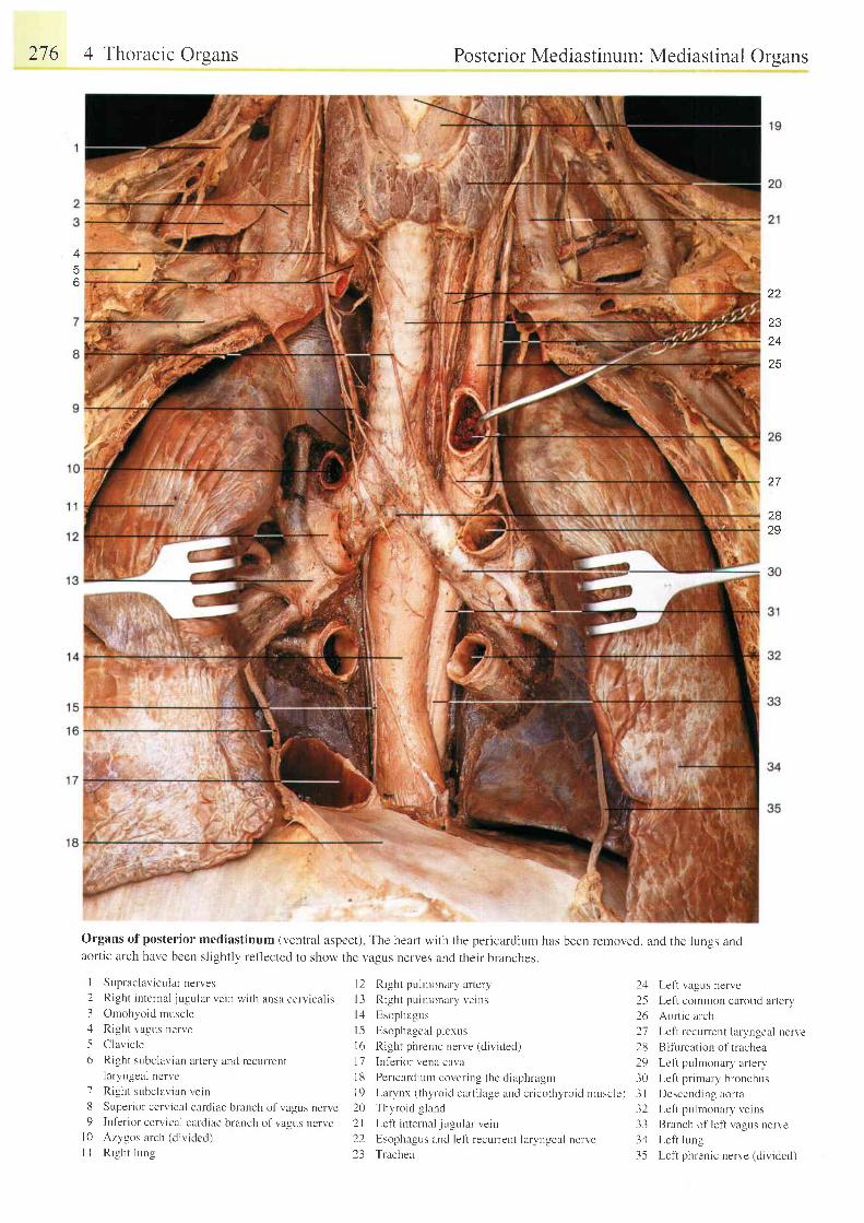
276 4 Thoracic Orsans Posterior Mediastinum: Mediastinal Orsans
2829
Organsofpos ter io rmed ias t inum(vent ra laspec t ) Thehear tw i th theper icard iun . rhasbeenremoved,andthe lungsandaortic arch have been slightly ref'lected to show the vagus nerves and their branches.
Supraclavicular nerves 12 Right pulrnonary artery 21 Left vagus nerveRight internal jugular vein wi th ansa cerv ical is l3 Right pulmonary veins 2-5 Lef t col l r ron carot id ar teryOmohyoid rnuscle lz l Esophagus 26 Aort ic archRight vagus nerve I 5 Esophageal plexus 2j Left recurrent laryngeal nerveClavicle I 6 Right phrenic nerve (divided) 28 Bifurcation of tracheaRight subclavian artery and recurrent 17 Inf'erior vena ca\.a 29 Leftpulmonary arterylaryngeal nerve 1g per icardium cover ing thc diaphragm 30 Lef t pr imary bronchusRight subclavian vein I 9 Larynx (thyroid cartilage and cricothyroid nuscle) 3 I Descending aortaSuperior cervical cardiac branch of vagus nerve 20 Thyroid gland 32 Lefi pulmonary veinsInferior cervical cardiac branch of vagus nerve 2l Left internal jugular vein 33 Branch of left vagus nerveAzygos arch (divided) 22 Esophagus and left recurrent laryngeal nerve 34 Lefi lungRight lung 23 Trachea 35 Left phrenic nerve (dividecl)
22
2324
25
27
I
23156
1d
l 0l l
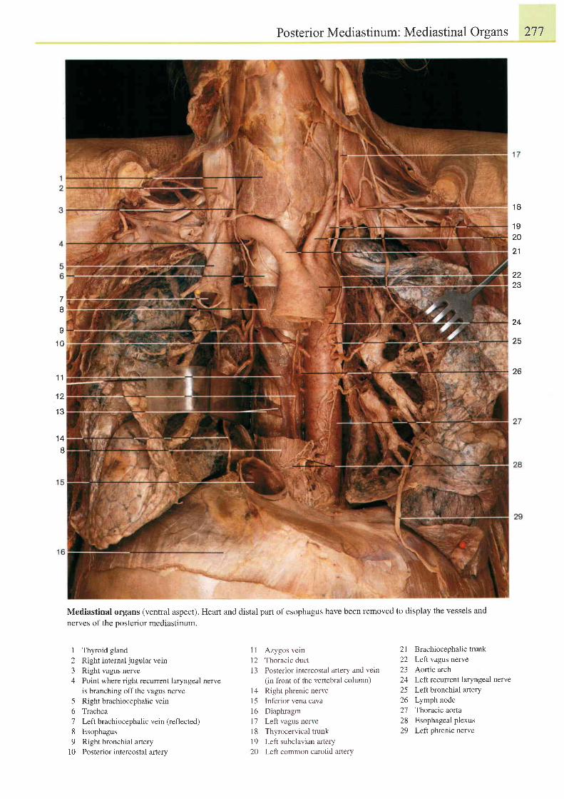
78
9
1 0
1 1
1 2
1 3
1 4
1 Thyroid gland2 Right internaljugular vern3 Right vagus nerve4 Point where right recurrent laryngeal nerve
is branching off the vagus nerve5 Right brachiocephalic vein6 Trachea7 Left brachiocephalic vein (reflected)
8 Esophagus9 Rightbronchial artery
10 Posterior intercostal artery
PosteriorMediastinum: Mediastinal Organs 277
' t8
1 920
21
2223
24
25
26
21 Brachiocephalic trunk22 LefI vagus nerve23 Aortic arch24 Leftrecwrent laryngeal nerve25 Left bronchial artery26 Lymph node2'7 Thoracic aorta28 Esophageal plexus29 Lefr phrenic nerve
Mediastinal organs (ventral aspect). Heart and distal part ofesophagus have been removed to display the vessels and
nerves of the posterior mediastinum.
11 Azygos vein12 Thoracic duct13 Posterior intercostal artery and vein
(in front of the vertebral column)14 Right phrenic nerve15 Inferior vena cava16 Diaphragml'7 Left vagus nerve18 Thyrocervical trunk19 Left subclavian artery20 LefI common carotid artery
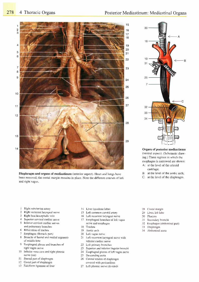
278 4 Thoracic Orsans
Diaphragm and organs ofmediastinum (anterior aspect). Heart and lungs havebeen removed; the costal margin remains in place. Note the different courses of leftand right vagus.
Posterior Mediastinum: Mediastinal Orsans
1 5
1 6a a
1 8
1 92021
22
23
24
28
26
27
Organs of posterior mediastinum(ventral aspect). (Schematic draw-ing.) Three regions in which theesophagus is narrowed are shown:A: at the level of the cricoid
cartilage;'
B: at the level of the aortic arch;C: at the level of the diaphragm.
I
23
I Right subclavian artery2 Right recurrent laryngeal nerve3 Right brachiocephalic vein4 Superior cervical cardiac nerve5 Inferior cervical cardiac nerves
and pulmonary branches6 Bifurcationof trachea7 Esophagus (thoracic part)8 Bronchi of lateral and medial sesments
of middle lobe9 Esophageal plexus and branches of
right vagus nerve10 Inferior vena cava and right phrenic
nerve (cut)I 1 Sternal part of diaphragm12 Costal part ofdiaphragm13 Falciform lieament of liver
14 Liver (quadrate lobe)15 Left common carotid artery16 Left recurrent laryngeal nervel7 Esophageal branches ofleft vagus
nerve and esophagus18 Trachea19 Aortic arch20 Left vagus nerve21 Left recurrent laryngeal nerve with
inferior cardiac nerve22 Left primary bronchus23 Superior and inferior lingular bronchi24 Esophageal plexus ofleft vagus nerve25 Descending aorta26 Central tendon ofdiaphragm
covered with pericardium27 Leftphrenic nerve (divided)
28 Costal margin29 Liver,left lobe30 Pharynx31 Secondary bronchi32 Esophagus (abdominal part)33 Diaphragm34 Abdominal aorta
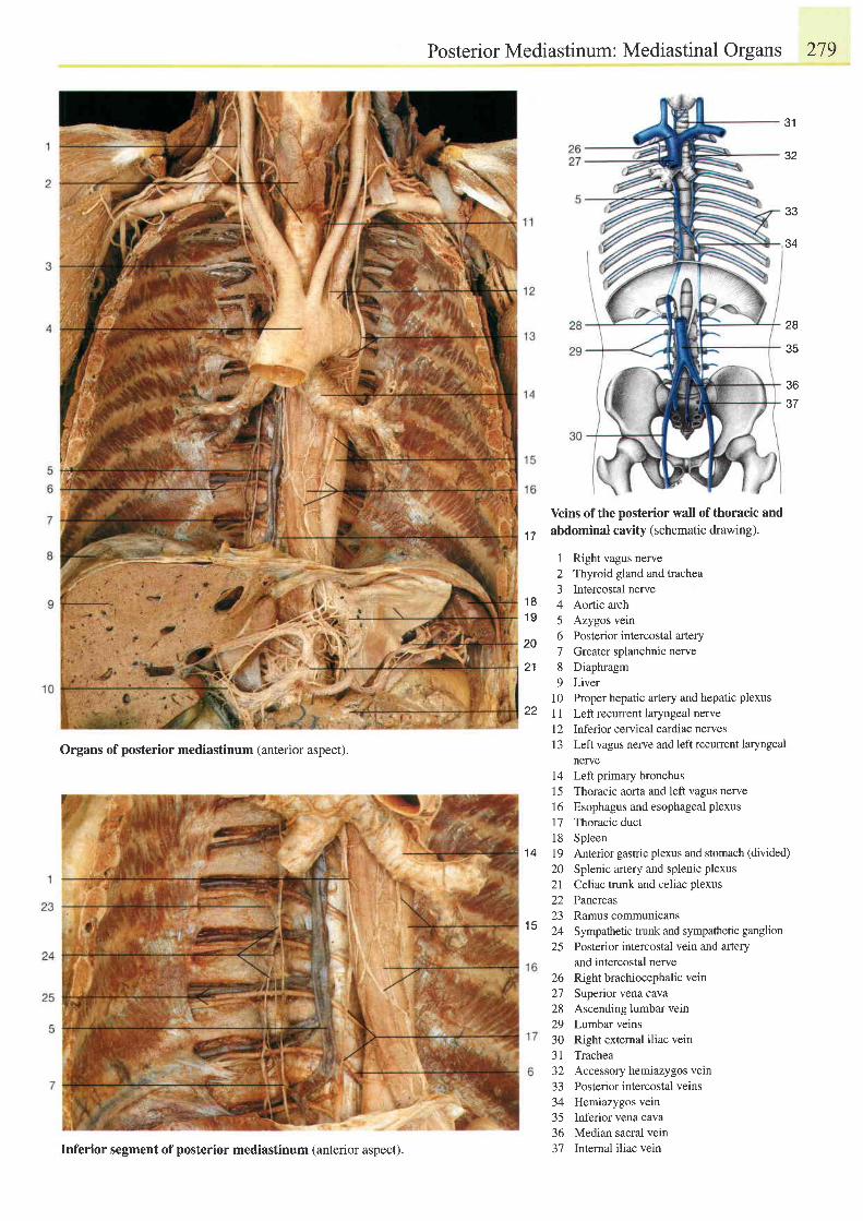
Organs of posterior mediastinum (anterior aspect).
Posterior Mediastinum: Mediastinal Organs
Yeins of the posterior wall of thoracic and
1T abdominal cavity (schematic drawing).
1 Right vagus neile2 Thyroid gland and trachea3 Intercostal nerve
18 4 Aortic arch19 5 Azygos vein
2n 6 Posterior intercostal anery7 Greater splanchnic nerve
21 8 Diaphragm9 Liver
10 Proper hepatic artery and hepatic plexus22 I 1 Left recuffent laryngeal nerve
12 Inferior cervical cardiac nervesI 3 Left vagus nerve and left recurrent laryngeal
nerve14 Left primary bronchus1 5 Thoracic aorta and left vagus nerve16 Esophagus and esophageal plexus17 Thoracic duct18 Spleen19 Anterior gastric plexus and stomach (divided)
20 Splenic artery and splenic plexus2l Celiac trunk and celiac plexus22 Pancreas23 Ramus communicans24 Sympathetic trunk and sympathetic ganglion25 Posterior intercostal vein and artery
and intercostal nerveRight brachiocephalic veinSuperior vena cavaAscending lumbar veinLumbar veinsRight external iliac veinTracheaAccessory hemiazygos veinPosterior intercostal veinsHemiazygos veinInferior vena cavaMedian sacral veinInternal iliac vein
279
31
32
33
34
28
35
3637
1 4
1 5
26272829JU
J I
az
3334J )
36J IInferior segment of posterior mediastinum (anterior aspect).
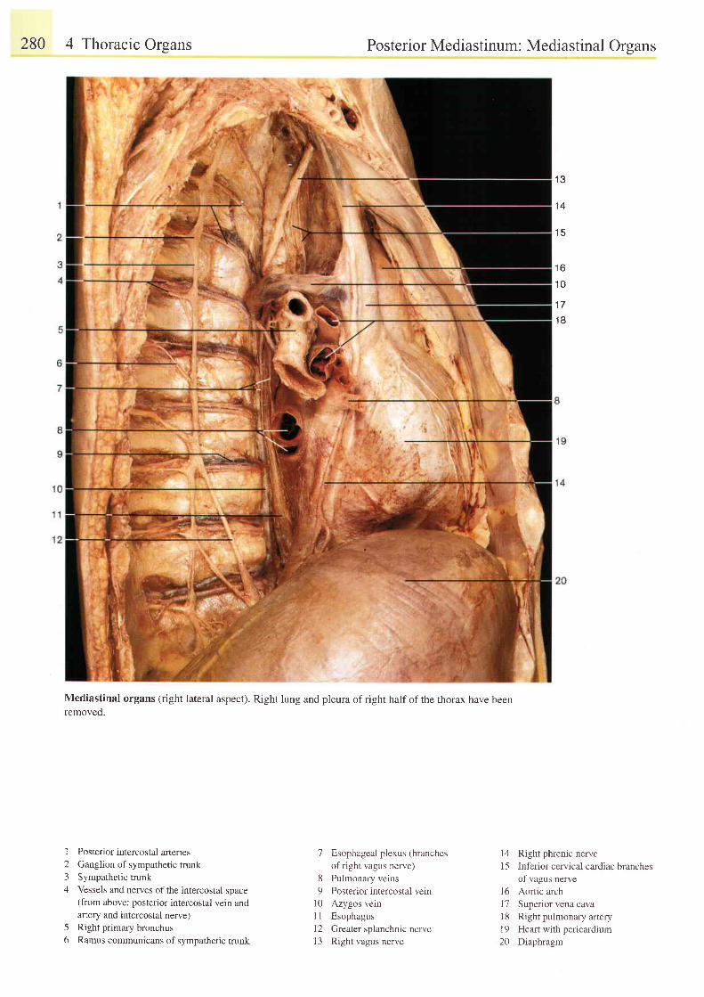
280 4 Thoracic Orsans
I
23A
5o
Posterior Mediastinum: Mediastinal Orsans
Mediastinal organs (right lateral aspect). Right lung and pleura ofright halfofthe thorax have beenremoved.
, I Q
1 4
1 5
1 6
1 0
1 7
1 B
Posterior intercostal afteriesGanglion of sympathetic trunkSympathetic trunkVessels and nerves of the intercostal space(from above: posterior intercostal vein andartery and intercostal nerve)Right primary bronchusRamus communicans of sympathetic trunk
7 Esophageal plexus (branchesof right vagus nerve)
8 Pulmonary veins9 Posterior intercostal vein
10 Azygos veinI I Esophagus12 Greater splanchnic nerve13 Right vagus nerve
14 Right phrenic nerve15 Inferior cervical cardiac branches
of vagus nerve16 Aortic arch17 Superior vena caval8 Right pulmonary artery19 Heart with pericardium20 Diaphragm
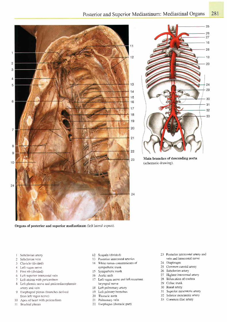
Posterior and Superior Mediastinum: Mediastinal Organs 28I
Main branches of descending aorta(schematic drawing).
25
26
28
1 3
1 4
1 51 6
z+
29
30
\ t l
Organs of posterior and superior mediastinum (left lateral aspect).
1 Subclavian artery2 Subclavian vein3 Clavicle (divided)4 Left vagus nerve5 First rib (divided)6 Left superior intercostal vein7 Left atrium with pericardium8 Left phrenic nerve and pericardiacophrenic
anery and vein9 Esophageal plexus (branches derived
from left vagus nerve)10 Apex of heart with pericardiumI I Brachial plexus
12 Scapula (divided)13 Posterior intercostal arteries14 White ramus communicans of
sympathetic trunk15 Sympathetic trunkI6 Aortic archl7 LefI vagus neile and left recurrent
laryngeal nerve18 Left pulmonary artery19 Left primary bronchus20 Thoracic aorta21 Pulmonary vein22 Esophagus (thoracic part)
23 Posterior intercostal artery andvein and intercostal nerve
24 Diaphragm25 Common carotid artery26 Subclavian atery27 Highest intercostal artery28 Bifurcation of trachea29 Celiac trunk30 Renal artery31 Superior mesentenc artery32 lnferior mesenteric artery33 Common iliac artery
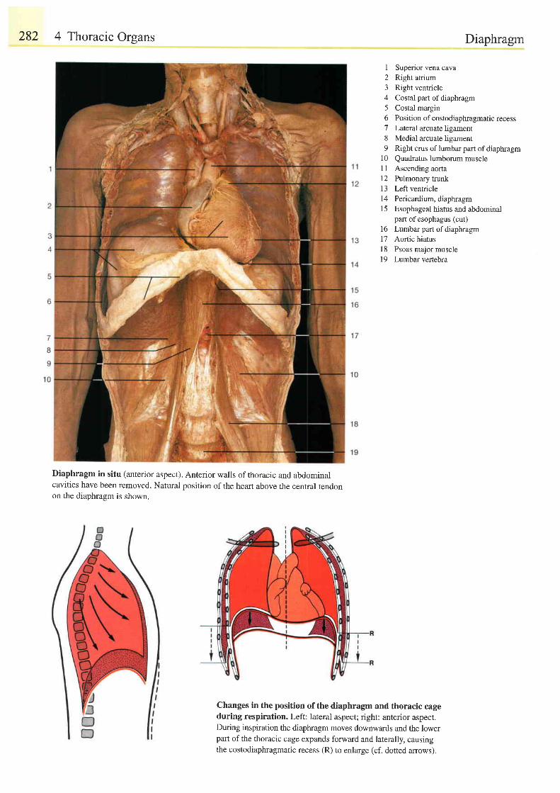
282 4 Thoracic Organs
Diaphragm in situ (anterior aspect). Anterior walls of thoracic and abdominalcavities have been removed. Natural position of the heart above the central tendonon the diaphragm is shown.
Diaphragm
I Superior vena cava2 Right atrium3 Right ventricle4 Costal part ofdiaphragm5 Costal margin6 Position of costodiaphragmatic recess7 Lateral arcuate ligament8 Medial arcuate ligament9 Right crus of lumbar part of diaphragm
10 Quadratus lumborum musclel l Ascending aorta12 Pulmonary trunk13 Left ventricle14 Pericardium, diaphragm15 Esophageal hiatus andabdominal
part of esophagus (cut)16 Lumbarpart of diaphragm17 Aortic hiatus18 Psoas major muscle19 Lumbar vertebra
Changes in the position ofthe diaphragm and thoracic cageduring respiration. Left: lateral aspect; right: anterior aspect.During inspiration the diaphragm moves downwards and the lowerpart of the thoracic cage expands forward and laterally, causingthe costodiaphragmatic recess (R) to enlarge (cf. dotted arrows).
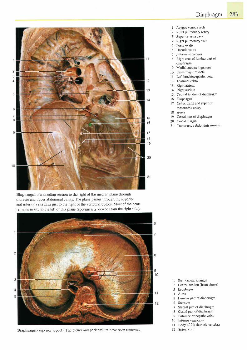
I
+
Diaphragm. Paramedian section to the right of the median plane through
thoracic and upper abdominal cavity. The plane passes through the superior
and inferior vena cava just to the right of the vertebral bodies. Most of the heart
remains in situ to the left of this plane (specimen is viewed from the right side).
Diaphragm 283
1 Azygos venous arch2 Right pulmonary artery3 Superior vena cava4 Right pulmonary vein5 Fossa ovalis6 Hepatic veins7 Inferior vena cava8 Right crus of lumbar Part of
diaphragm9 Medial arcuate ligament
l0 Psoas major muscle1 I Left brachiocephalic vein
12 Terminal cristal3 Right atrium14 Right auricle15 Central tendon ofdiaPhragm16 Esophagus17 Celiac trunk and suPerior
mesenteric artery18 Aorta19 Costal part ofdiaPhragm20 Costal margin21 Transversus abdominis muscle
I Sternocostaltriangle2 Central tendon (from above)3 Esophagus4 Aorta5 Lumbarpartof diaPhragm6 Sternum7 Sternal part of diaPhragm8 Costal part ofdiaphragm9 Entrance ofhepatic veins
10 Inferiorvenacava1 1 Body of 9th thoracic vertebra
12 Spinal cord
t c
1 6
1 7
1 81 9
20
II U
Diaphragm (superior aspect). The pleura and pericardium have been removed.
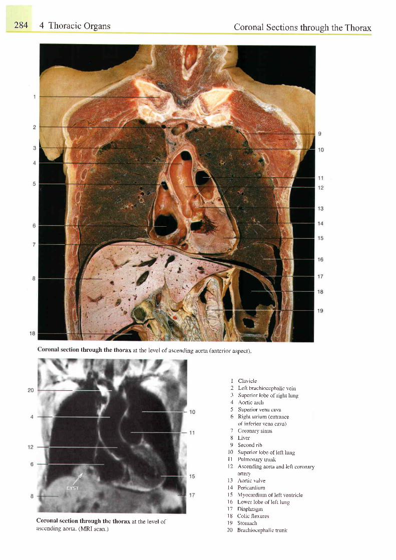
284 4 Thoracic Orsans
through the thorax at the level of ascending aorta (anterior aspect).
I
2345o
78o
1 01 1t 2
Coronal Sections throuqh the Thorax
ClavicleLeft brachiocephalic veinSuperior lobe of right lungAortic archSuperior vena cavaRight atrium (entranceof inferior vena cava)Coronary sinusLiverSecond ribSuperior lobe of left lungPulmonary trunkAscending aorta and left coronaryartery
13 Aortic valve14 Pericardium15 Myocardium of left ventricle16 Lower lobe of left lungl7 Diaphragm18 Colic flexures19 Stomach20 Brachiocephalictrunk
Coronal section through the thorax at the level ofascending aorta. (MRI scan.)
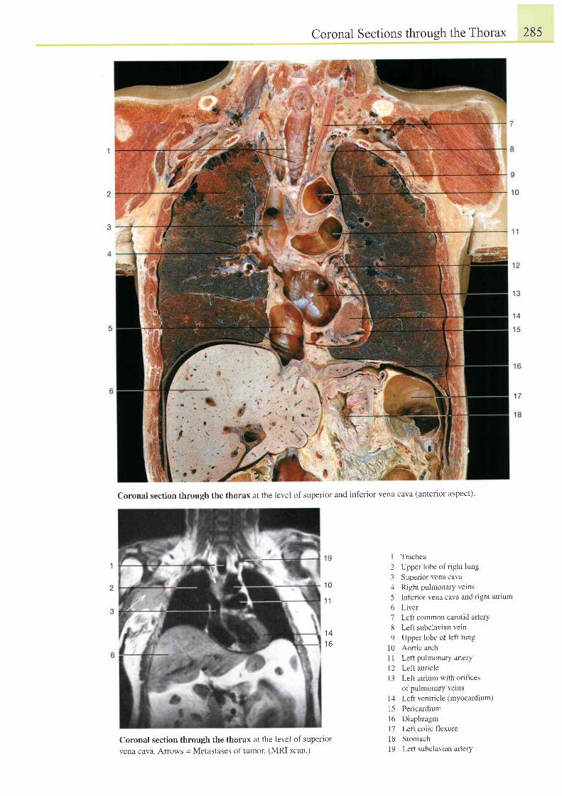
Coronal Sections through the Thorax 285
Coronal section through the thorax at the level of superior and inferior vena cava (anterior aspect)'
Coronal section through the thorax at the level of superiorvena cava. Arows = Metastases of tumor. (MRI scan.)
I Trachea2 Upper lobe ofright lung3 Superior vena cava4 Right pulmonary vetns
5 Inferior vena cava and right atrium
6 Liver7 Left common carotid arterY8 Left subclavian vein9 Upper lobe of left lung
l0 Aortic archl1 Left pulmonary arterY12 Left auricle13 Left atrium with orifices
of pulmonary vetns14 Left ventricle (myocardium)
15 Pericardiuml6 Diaphragm11 LefI colic flexure18 Stomach19 Left subclavian arlerY
t 1
1 41 6
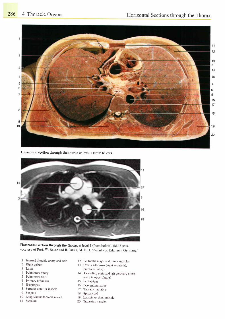
286 4 Thoracic Organs
Horizontal section through the thorax at level 1 (from below). (MRI scan,courtesy of Prof. w. Bautz and R. Janka, M. D., University of Erlangen, Germany.)
Horizontal Sections throueh the Thorax
e
o
1 0
'12
1 3e
1 4
4
6
5
1 6
1 7
1 8
1 9
20
I Intemal thoracic artery and vein2 Right atrium3 Lung4 Pulmonary artery5 Pulmonary vein6 Primary bronchus7 Esophagus8 Serratus anterior muscle9 Scapula
l0 Longissimus thoracis muscle11 Stemum
12 Pectoralis major and minor muscles| 3 Conus arteriosus {r ight ventr icle).
pulmonic valveI4 Ascending aona and left coronary artery
(only in upper figure)15 Left atriuml6 Descending aorta17 Thoracic vertebra18 Spinal cordl9 Latissimus dorsi muscle20 Trapezius muscle
Horizontal section through the thorax at level 1 (from below).
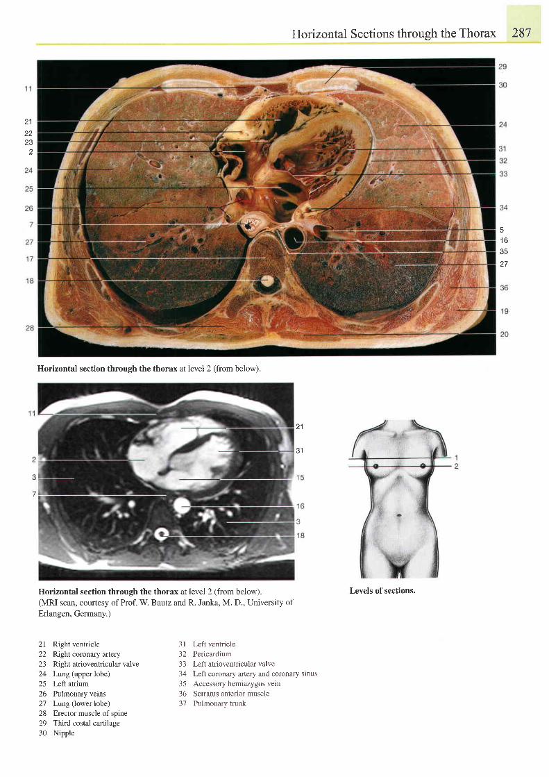
21z t
232
Horizontal section through the thorax at level 2 (from below)
Horizontal section through the thorax at level 2 (from below).(MRI scan, courtesy of Prof. W. Bautz and R. Janka, M. D., University ofErlangen, Germany.)
Horizontal Sections through the Thorax 287
Levels of sections.
1 6e(
1 '
21
21 Right ventricle22 Right coronary artery23 Right atrioventricular valve24 Lung(upperlobe)25 Left atrium26 Pulmonary veins27 Lung (lower lobe)28 Erector muscle of spine29 Third costal cartilage30 Nipple
3l Left ventricle32 Pericardium33 Left atrioventricular valve34 Left coronary artery and coronary sinus35 Accessory hemiazygos vetn36 Serratus anterior muscle37 Pulmonary trunk
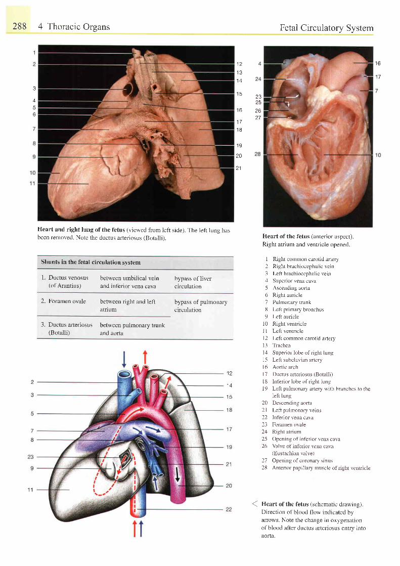
288 4 Thoracic Orsans
Heart and right lung of the fetus (viewed from left side). The left lung hasbeen removed. Note the ductus arteriosus (Botalli).
Shunts in the fetal circulation svstem
Fetal Circulatory System
Heart ofthe fetus (anterior aspect)Right atrium and ventricle opened.
12345o189
1 0l tt 2
Right common carotid artery
Right brachiocephalic vein
Left brachiocephalic vein
Superior vena cava
Ascending aorta
Right auricle
Pulmonary trunk
Left primary bronchus
Left auricle
Right ventricle
Left ventricle
Left common carotid artery
l3 Trachea
l4 Superior lobe ofright lung
15 Left subclavian artery
l6 Aortic arch
l7 Ductus arteriosus (Botalli)
l8 Inferior lobe ofright lung
19 Left pulmonary iutery with branches to theleft lung
20 Descending aorta
2l Left pulmonary verns
22 Inferior vena cava
23 Foramen ovale
24 Right atrium
25 Opening of inf'erior vena cava
26 Valve ofinf'erior vena cava(Eustachian valve)
27 Opening of coronary srnus
28 Anterior papillary muscle of right ventricle
( Heart of the fetus (schematic drawing).Direction of blood flow indicated byarrows. Note the change in oxygenationof blood after ductus arteriosus entry intoaorta.
1 31 4
t 3
t o
1 7
I
o
2,72526t o
1 7
1 B
1 9
20
21
1. Ductus venosus(of Arantius)
between umbilical veinand inferior vena cava
bypass oflivercirculation
2. Foramen ovale between right and leftatflum
3. Ductus arleriosus between pulmonary trunk(Botalli) and aorta
bypass of pulmonarycirculation
12
1 4
I J
1 8
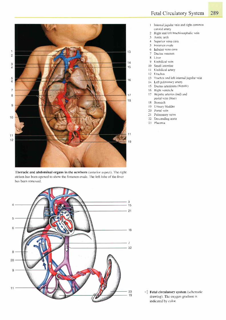
1
1 11 2
Thoracic and abdominal organs in the newborn (anterior aspect). The rightatrium has been opened to show the foramen ovale. The left lobe of the liverhas been removed.
Fetal Circulatory System 289
1 Intemal jugular vein and right commoncarotid artery
2 Right and left brachiocephalic vein3 Aortic arch4 Superior vena cava5 Foramen ovale6 Inferior vena cava7 Ductus venosus8 Liver9 Umbilical vein
10 Small intestine1l Umbilical aftery12 Urachus13 Trachea and left internaljugular vein14 Left pulmonary artery15 Ductus arteriosus (Botalli)
16 Right venfiicle17 Hepatic arteries (red) and
portal vein (blue)l8 Stomach19 Urinary bladder20 Portal vein21 Pulmonary veins22 Descending aorta23 Placenta
Fetal circulatory system (schematicdrawing). The oxygen gradient isindicated by color.
l 3
1 4' t5
t o
1 7
1 B
1 5
2 1
t o
7
22
231 9
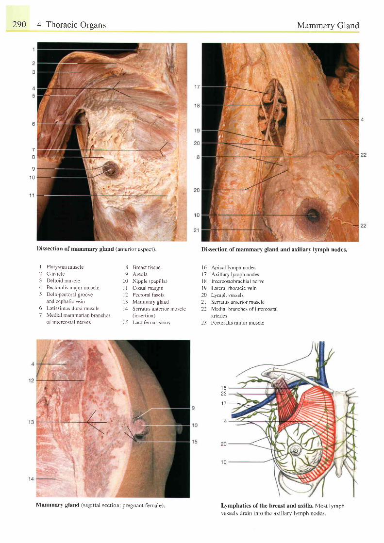
290 4 Thoracic Orsans
Dissection of mammary gland (anterior aspect).
123/
5
Platysma muscle
Clavicle
Deltoid muscle
Pectoralis major muscle
Deltopectoral groove
and cephalic vein
Latissimus dorsi muscle
Medial mammarian branchesof intercostal nerves
Mammary Gland
Dissection of mammary gland and axillary lymph nodes.
16 Apical lymph nodes
l7 Axillary lymph nodes
I 8 Intercostobrachial nerve
l9 Lateral thoracic vein
20 Lymph vessels
2l Senatus anter ior muscle
22 Medial branches of intercostal
artenes
23 Pectoralis minor muscle
Lymphatics of the breast and axilla. Most lymphvessels drain into the axillary lymph nodes.
7I
I
1 0
8 Breast tissue9 Areola
10 Nipple (papilla)I I Costal margin12 Pectoral fascia13 Mammary glandl4 Serratus anterior muscle
(insertion)l5 Lactiferous sinus
61
t o
23
1 7
Mammary gland (sagittal section; pregnant female).
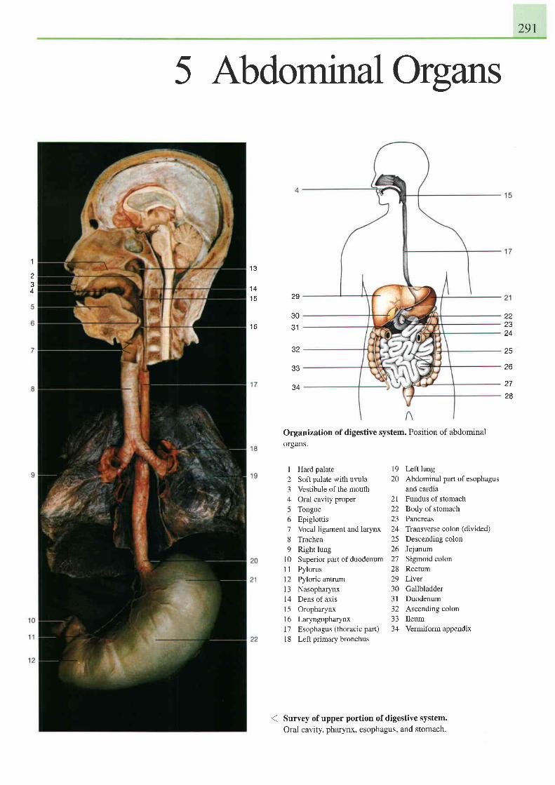
291
I
2J
4
5 AbdominalOrgans
1 3
1 41 5
I D
29
30J I
\tz
ee
34
Organization of digestive system. Position of abdominalorgans.
19 Left lung20 Abdominal part of esophagus
and cardia21 Fundus of stomach22 Body of stomach23 Pancreas24 Transverse colon (divided)25 Descending colon26 Jejunum27 Sigmoid colon28 Rectum29 Liver30 Gallbladder31 Duodenum32 Ascending colon33 Ileum34 Vermiform appendix
22Z J
z q
25
26
z t
28
I Hard palate2 Soft palate with uvula3 Vestibule of the mouth4 Oral cavity proper5 Tongue6 Epiglottis7 Vocal ligament and larynx8 Trachea9 Right lung
10 Superior part ofduodenum11 Pylorus12 Pyloric antruml3 Nasopharynx14 Dens of axis15 Oropharynx16 Laryngopharynxl7 Esophagus (thoracic pat)l8 Leftprimary bronchus
Survey of upper portion of digestive system.Oral cavity, pharynx, esophagus, and stomach.
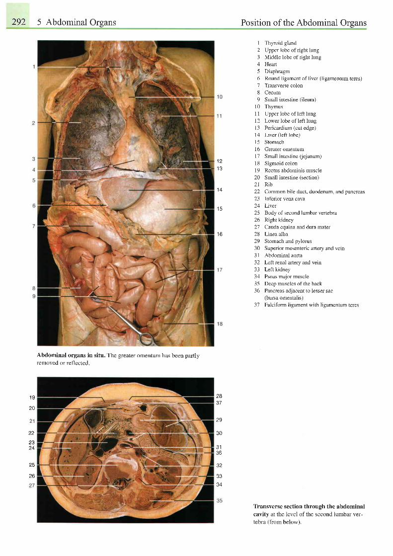
292 5 Abdominal Organs Position of the Abdominal Organs
Abdominal organs in situ. The greater omentum has been partlyremoved or reflected.
1 Thyroid gland2 Upper lobe ofright lung3 Middle lobe of right lung4 Hearl5 Diaphragm6 Round ligament of liver (ligamentum teres)7 Transverse colon8 Cecum9 Small intestine (ileum)
10 Thymus11 Upper lobe of left lung12 Lower lobe of left lung13 Pericardium (cut edge)14 Liver (left lobe)15 Stomach16 Greater omentum17 Small intestine (ejunum)18 Sigmoid colon19 Rectus abdominis muscle20 Small intestine (section)21 Rib22 Common bile duct, duodenum, and pancreas23 Inferior vena cava24 Lwer25 Body of second lumbar venebra26 Right kidney27 Catda equina and dura mater28 Linea alba29 Stomach and pylorus30 Superior mesenteric artery and vein31 Abdominal aorta32 Left renal artery and vein33 Left kidney34 Psoas major muscle35 Deep muscles of the back36 Pancreas adjacent to lesser sac
(bursa omentalis)37 Falciform ligament with ligamentum teres
T[ansverse section through the abdominalcavity at the level of the second lumbar ver-tebra (from below).
1 21 3
1 4
1 5
I D
29
\,U
J I
36
32
J 5
34
1 g
20
21
22
2324
25
28
26
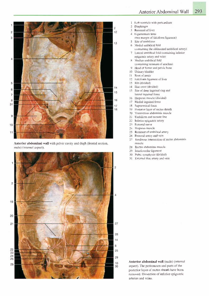
Anterior Abdominal Wall 293
1 3
1234
56
1
8
91 0t 1l 2l 31 41 5
l 6t'7t 81 9202 1222324252627
1 4
l c
t b
1 71 B
Left ventricle with pericardiumDiaphragmRemnant of liverLigamentum teres(free margin of falciform ligament)Site of umbilicusMedial umbilical fold(containing the obliterated umbilical artery)Lateral umbilical fold (containing inferiorepigastric artery and vein)Median umbilical fold(containing remnant of urachus)Head of femur and pelvic boneUrinary bladderRoot of penisFalciform ligament of liverRib (divided)Iliac crest (divided)Site of deep inguinal ring andlateral inguinal fossaIliopsoas muscle (divided)Medial inguinal fossaSupravesical fossaPosterior layer of rectus sheathTransversus abdominis muscleUmbilicus and arcuate lineInferior epigastric arteryFemoral nerveIliopsoas muscleRemnant of umbilical arteryFemoral artery and veinTendinous intersection of rectus abdominis
831
29
1 030
222324Z J
26
Anterior abdominal wall with oelvic cavitv and thish (frontal section.male) (internal aspect).
muscle28 Rectus abdominis muscle29 Interfoveolarligament30 Pubic symphysis (divided)31 External iliac artery and vein
Anterior abdominal wall (male) (internal
aspect). The peritoneum and parts of the
posterior layer ofrectus sheath have been
removed. Dissection of inferior epigastric
arteries and veins.
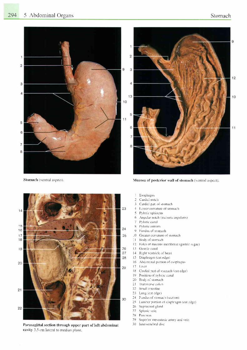
294 5 Abdominal Organs
Stomach (ventral aspect).
t 4
t o
t l
1 8
.19
20
Parasagittal sectioncavitv 3 -5 cm lateral
23
Mucosa of posterior wall of stomach (ventral aspect).
I E,sophagus
2 Cardial notch
3 Cardial part of stomach
4 Lesser curvature of stornach
5 Pvlor ic sphincter
6 Angular notch ( inc isura angular is)
7 Pylor ic canal
8 Pyloric antrurn
9 Fundus of stomach
10 Greater curvature of stomach
I I Body of stornach
l2 Folds of mucous membrane (gastr ic rugae)
l3 Gastr ic canal
l : l Right ventr ic le of heart
l5 Diaphragm (cut edge)
l6 Abdominal port ion ofesophagus
l7 L i ve r
I 8 Cardial part of stourach (cut edge)
l a P o : i t i o n o f p r l o r i e c u n r .
20 Body of stomach
2l Transverse colon
22 Srnal l in test ine
23 Lung (cut edge)
2,1 Fnndus of stomach (section)
2-5 Lunbar portion of diaphragm (cut edge)
26 Suprarenal gland
27 Sp len i c ve i n
28 Pancreas
29 Sr,rpcrior nresenteric artery and vein
30 lntervertebral d isc
262728
Z Y
through upper part of left abdominalto median olane
Stomach
30
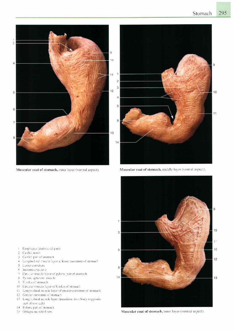
Stomach 295
1
I
1 0
\ luscu la r coa t o f s tomach. ou ter l i l \ c r ( r 'en t ra l aspec t ) \ luscul:rr coat of stomach, nt icldlc laycr ' (ventral t tsl lcct)
1 1
1 0Il
l
:
t
9
i l
I ]
l l
l 5
I : son l r i r l r L r ( r r h t l o r t t i n l l P l L r - t )( ' a r r l i . r l no t ch( ' u r t i i l l p r r r t o l s t on t re l r
I r i ng r t r t t l i n l r i n r t i : e l c l l r r c r i r I l c : : e r cu r \ l l t u f c o f : t on l r ch
l - c \ \ c l e L i l \ : t l L l l c
I r t e i r r r r l r r L l g L r l r L r - i s
( i r e L r l : L r n r u : e l c l l r r e r o f p r l o n c p t r r t o f : t o n t r c h
I ) r l o r i e s p h i r c t c l n r r r s c l e
l ' u n r l l : o l \ l ( ) n t i c l t
( i r e L r l l L r n r L r s c l c l l L r c r o l l r r r d r r s o l s t o r l a c h
I on t : r t t t t l i n l r l n rL rsc lc l r t r c l o i q r - r 'a tc r - cu f \ a tu r e o f s to t t l t ch
( i t c l r l t ' t , - t t i t l t t t t c o l s t o t n l t c l t
l - o n t i t L r r l i n l l n r i r s e l c l l r r c r ( u l r l s i t i o n f r o r r b < l d \ ' l o p t l o r - r c
p r r L ( ) l \ t o n r i r a l r I
l ) r lo t i c p l i r l o l s lo r l l i c l r
O b l i t t L r c r r L r : e l c l i b c r : \ luscular coat of stomach. inner- laYct ' (r ,enttal zlspcct )
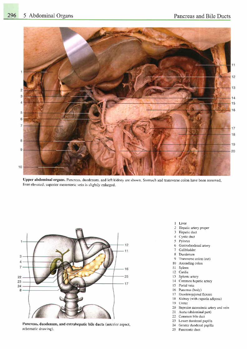
296 5 Abdominal Oreans Pancreas and Bile Ducts
1 4
1 5
1 6
Upper abdominal organs. Pancreas, duodenum, and left kidney are shown. Stomach and transverse colon have been removed,liver elevated; superior mesenteric vein is slightly enlarged.
22z5
24I
I Liver2 Hepatic artery proper3 Hepatic duct4 Cystic duct5 Pylorus6 Gastroduodenal artery7 Gallbladder8 Duodenum9 Transverse colon (cut)
l0 Ascending colon11 Spleen12 Cardia13 Splenic artery14 Common hepatic artery15 Portal vein16 Pancreas (body)17 Duodenojejunalflexure18 Kidney (withcapsulaadiposa)19 Ureter20 Superior mesenteric artery and vein2l Aorta (abdominal part)22 Common bile duct23 Lesser duodenal papilla24 Greater dtodenal papilla25 Pancreatic duct
Pancreas, duodenum, and extrahepatic bile ducts (anterior aspect,schematic drawing).
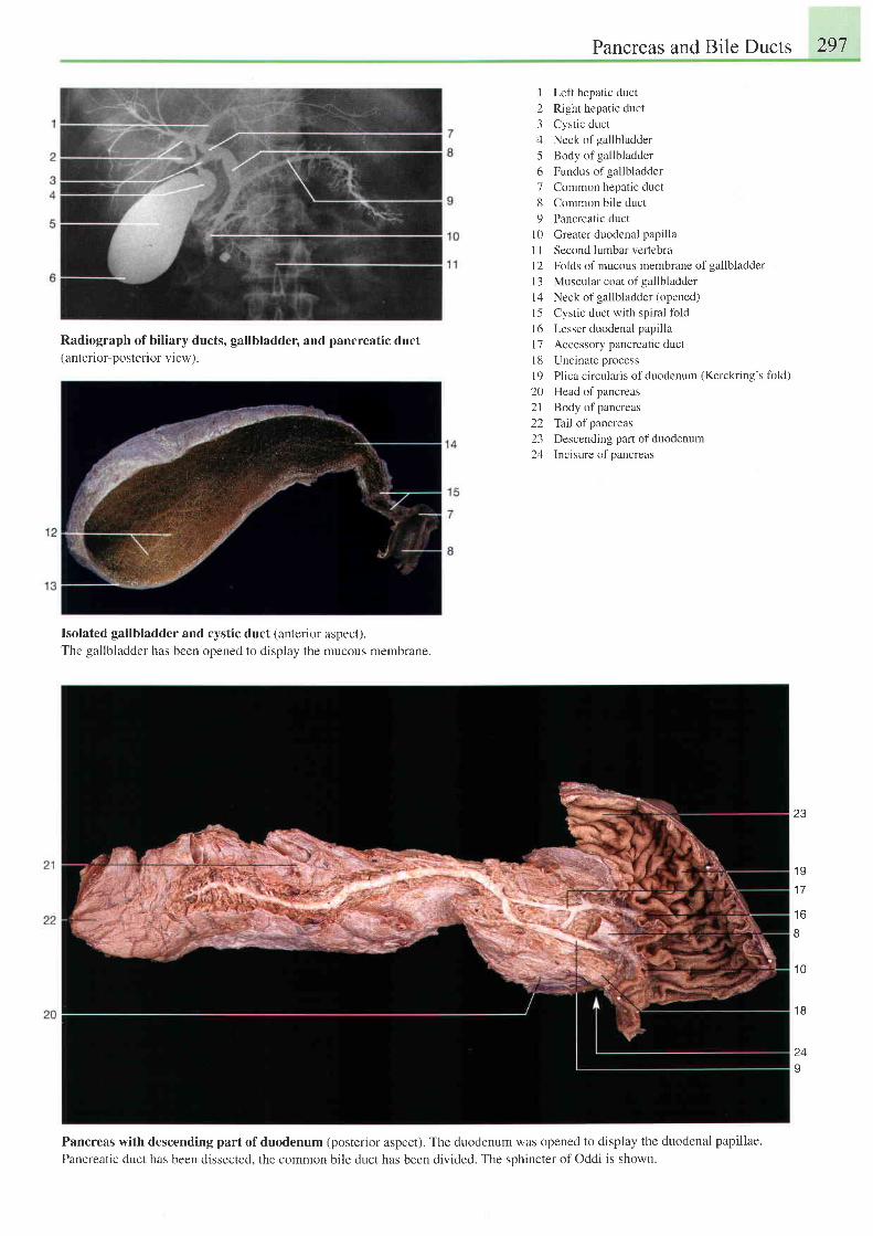
Pancreas and Bile Ducts 297
Radiograph ofbiliary ducts, gallbladder, and pancreatic duct(anterior-posterior view).
I Left hepatic duct
2 Right hepatic duct
3 Cyst ic duct
4 Neck of gallbladder
5 Body of gallbladder
6 Fundus ofgallbladder
7 Common hepatic duct
8 Common bile duct
9 Pancreatic duct
10 Greater duodenal papilla
I I Second lumbar vertebra
12 Folds of mucous membrane of gallbladder
l3 Muscular coat of gallbladder
14 Neck of gallbladder(opened)
l5 Cystic duct with spiral fold
l6 Lesser duodenal papilla
l7 Accessory pancreatic duct
I 8 Uncinate process
l9 Plica circularis ofduodenum (Kerckring's fbld)
20 Head ofpancreas
2l Body ofpancreas
22 Tailofpancreas
23 Descending panof duodenum
24 Incisure of pancreas
Isolated gallbladder and cystic duct (anterior aspect).The gallbladder has been opened to display the mucous membrane.
Pancreas with descending part of duodenum (posterior aspect). The duodenum was opened to display the duodenal papillae.
Pancreatic duct has been dissected. the common bile duct has been divided. The sphincter of Oddi is shown.
23
1 9
1 7
t o
I
1 0
I
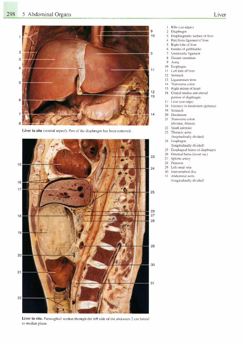
298 5 Abdominal Organs Liver
I1 0
1 21 3
23456189
1 01 ll 2l 3t 4l 5l 6
t 11 81 9202 I
Ribs (cut edges)
Diaphragm
Diaphragmatic surface of liver
Falciform ligament of liver
Right lobe of liver
Fundus of gallbladder
Gastrocolic ligament
Greater omentum
Aorta
Esophagus
Left lobe of liver
Stomach
Ligamentum teres
Transverse colon
Right atrium of heart
Central tendon and sternal
portion of diaphragm
Liver (cut edge)
Entrance to duodenum (pylorus)
Stomach
Duodenum
Transverse colon(divided, dilated)
22 Small intestine23 Thoracic aorta
(longitudinally divided)24 Esophagus
(longitudinally divided)25 Esophageal hiatus ofdiaphragm26 Omental bursa (lesser sac)27 Splenic artery28 Pancreas29 Left renal vein30 Intervertebral disc31 Abdominal aorta
(longitudinally divided)
Liver in situ (ventral aspect). Part ofthe diaphragm has been removed.
Liver in situ. Parasagittal section through the left side ofthe abdomen 2 cm lateralto median plane.
2627
28
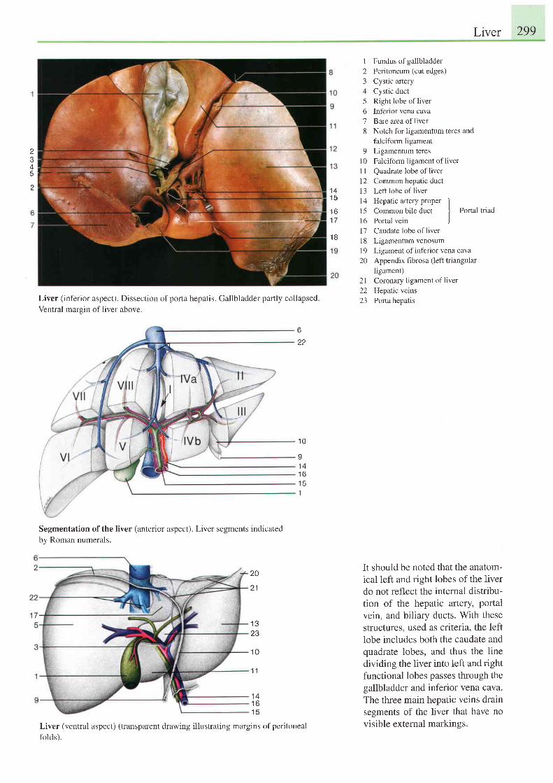
Liver 299
I
J
4
2
Liver (inferior aspect). Dissection of porta hepatis. Gallbladder partly collapsed.
Ventral margin of liver above.
Segmentation of the liver (anterior aspect). Liver segments indicatedby Roman numerals.
20
2 1
1 323
1 0
1 1
Liver (ventral aspect) (transparent drawing illustrating margins of peritoneal
folds).
Fundus of gallbladderPeritoneum (cut edges)Cystic arteryCystic ductRight lobe of liverInferior vena cavaBare area of liverNotch for ligamentum teres andfalciform ligamentLigamentum teresFalciform ligament of liver
Quadrate lobe of liverCommon hepatic ductLeft lobe of liverHepatic artery proper ICommon bile duct
I Portal triad
Portal vein )Caudate lobe of liverLigamentum venosumLigament of inferior vena cavaAppendix fibrosa (left triangularligament)Coronary ligament of liverHepatic veinsPorta hepatis
It should be noted that the anatom-ical left and right lobes of the liverdo not reflect the internal distribu-tion of the hepatic artery, portalvein, and biliary ducts. With thesestructures, used as criteria, the leftlobe includes both the caudate andquadrate lobes, and thus the linedividing the liver into left and rightfunctional lobes passes through thegallbladder and inferior vena cava.The three main hepatic veins drainsegments of the liver that have novisible external markinss.
12345o
18
q
101 l1 21 3I 4
1 51 6t7l 81920
2 12223
1 41 5
1 6
1 8
6
22
1 0
I1 4t o
1
1 4t o
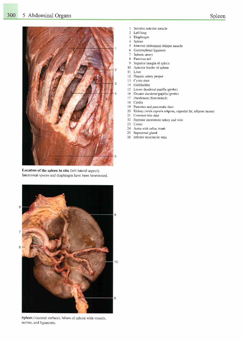
300 5 Abdominal Orsans Spleen
I Serratus anterior muscle2 Left lung3 Diaphragm4 Spleen5 External abdominal oblique muscle6 Gastrosplenicligament7 Splenic artery8 Pancreas tail9 Superior margin of spleen
10 Anterior border of spleen11 Liver12 Hepatic artery proper13 Cystic duct14 Gallbladder15 Lesser duodenal papilla (probe)1 6 Greater duodenal papilla (probe)l7 Duodenum (fenestrated)18 CardiaI 9 Pancreas and pancreatic duct20 Kidney (with capsula adiposa, capsular fat, adipose tissue)21 Common bile duct22 Superior mesenteric artery and vein23 Ureter24 Aorta with celiac trunk25 Suprarenal gland26 Inferior mesenteric vein
Location ofthe spleen in situ (left-lateral aspect).Intercostal spaces and diaphragm have been fenestrated.
Spleen (visceral surface), hilum of spleen with vessels,nerves, and ligaments.
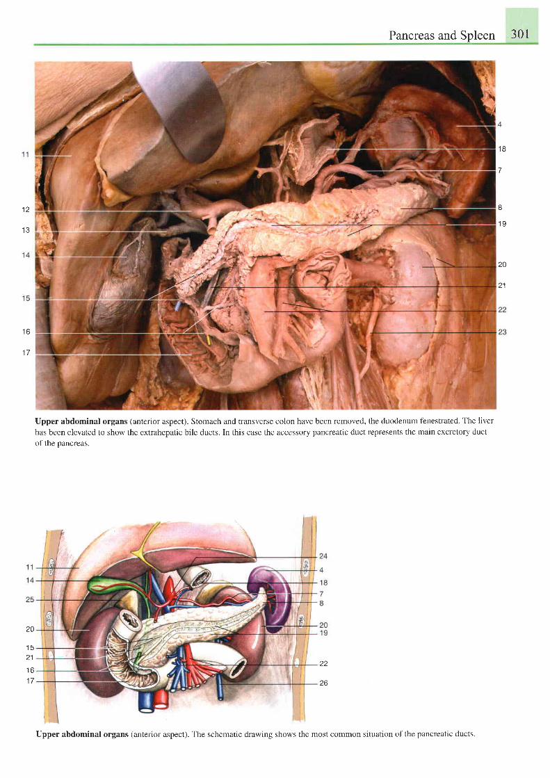
Pancreas and Spleen 301
1 2
1 3
t o
I T
1 8
7
8
1 9
20
21
Upper abdominal organs (anterior aspect). Stomach and transverse colon have been removed, the duodenum fenestrated. The liver
has been elevated to show the extrahepatic bile ducts. In this case the accessory pancreatic duct represents the main excretory duct
of the pancreas.
20
t 3
21
t o
1 7
zz
26
Upper abdominal organs (anterior aspect). The schematic drawing shows the most common situation of the pancreatic ducts.
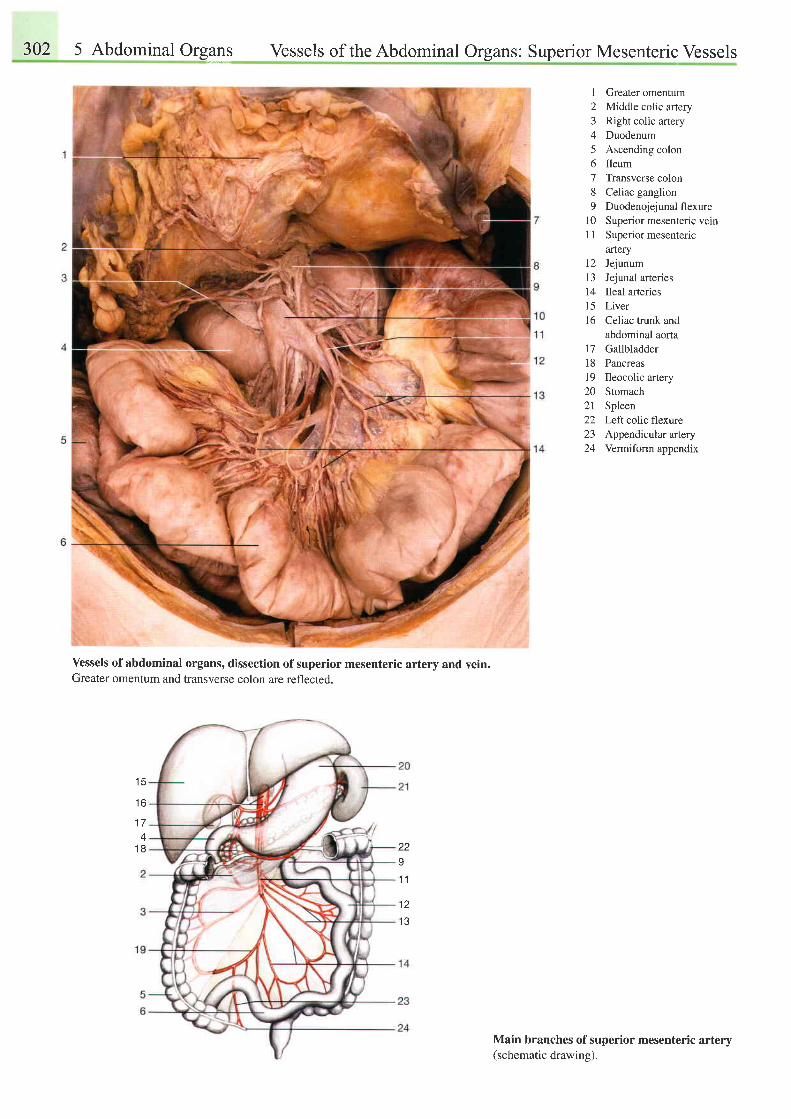
302 5 Abdominal Organs Vessels of the Abdominal Organs: Superior Mesenteric Vessels
I Greater omentum2 Middle colic artery3 Right colic artery4 Duodenum5 Ascending colon6 Ileum7 Transverse colon8 Celiac ganglion9 Duodenojejunalflexure
10 Superior mesenteric vein11 Superior mesenteric
121 3I41 5l 6
1 11 8I9202 l2223)4
arteryJejunumJejunal arteriesIleal afteriesLiverCeliac trunk andabdominal aortaGallbladderPancreasIleocolic arteryStomachSpleenLeft colic flexureAppendicular arteryVermiform appendix
Vessels of abdominal organs, dissection of sup€rior mesenteric artery and vein.Greater omentum and transverse colon are reflected.
Main branches of superior mesenteric artery(schematic drawing).
t c
t o
1 74
1 8 2291 1
12
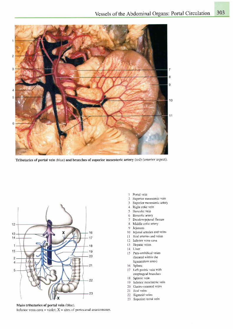
Vessels of the Abdominal Organs: Portal Circulation 303
Tributaries of portal vein (blue) and branches of superior mesenteric artery (red) ( anterior aspect).
1 2
1 31 4
1
t c
z
4
t o
1 7
1 8
1 920
21
22
23
Main tributaries of portal vein (blue).Inferior vena cava = violet; X = sites of portocaval anastomoses.
I
234
o
7
8o
l 0
l l
l 21 31415
161 7
1 8t9202 l2223
Portal veinSuperior mesenteric veinSuperior mesenteric arteryRight colic veinIleocolic veinIleocolic arteryDuodenojejunal flexureMiddle colic arteryJejunumJejunal arteries and veinsIleal arteries and veinsInferior vena cavaHepatic veinsLiverPara-umbilical veins(located within theligamentum teres)SpleenLeft gastric vein withesophageal branchesSplenic veinInferior mesenteric veinGastro-omental veinsIleal veinsSigmoid veinsSuperior rectal vein
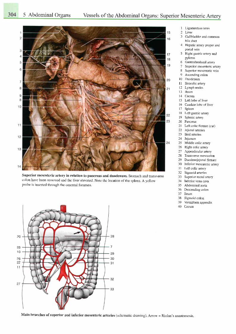
304 5 Abdominal organs vessels of the Abdominal organs: S
t c
1 6
1 7
1 8
1 9
20
21
I Ligamentum teres2 Liver3 Gallbladder and common
bile duct4 Hepatic artery proper and
portal vein5 Right gastric artery and
pylorus6 Gastroduodenal artery7 Superior mesenteric artery8 Superior mesenteric vein9 Ascending colon
10 Duodenuml1 Ileocolic artery12 Lymphnodes13 Ileum14 Cecuml5 Left lobe ofliver16 Caudate lobe ofliver17 Spleenl8 Left gastric artery19 Splenic artery20 Pancreas2l Left colic flexure (cut)22 Jejunal arteries23 Ileal arteries24 Jejunum25 Middle colic artery26 Rightcolic artery27 Appendratlarartery28 Transversemesocolon29 Duodenojejunal flexure30 Inferior mesenteric artery31 Leftcolicartery32 Sigmoid arteries33 Superior rectal artery34 Inferior vena cava35 Abdominal aorta36 Descending colon37 Ileum38 Sigmoid colon39 Vermiform appendix40 Cecum
22
Z J
20
251 0
Superior mesenteric artery in relation to pancreas and duodenum. Stomach and transversecolon have been removed and the liver elevated. Note the location of the spleen. A yellowprobe is inserted through the omental foramen.
2930J I
32
33
26221 1
Main branches of superior and inferior mesenteric arteries (schematic drawing). Arrow = Riolan's anastomosis.
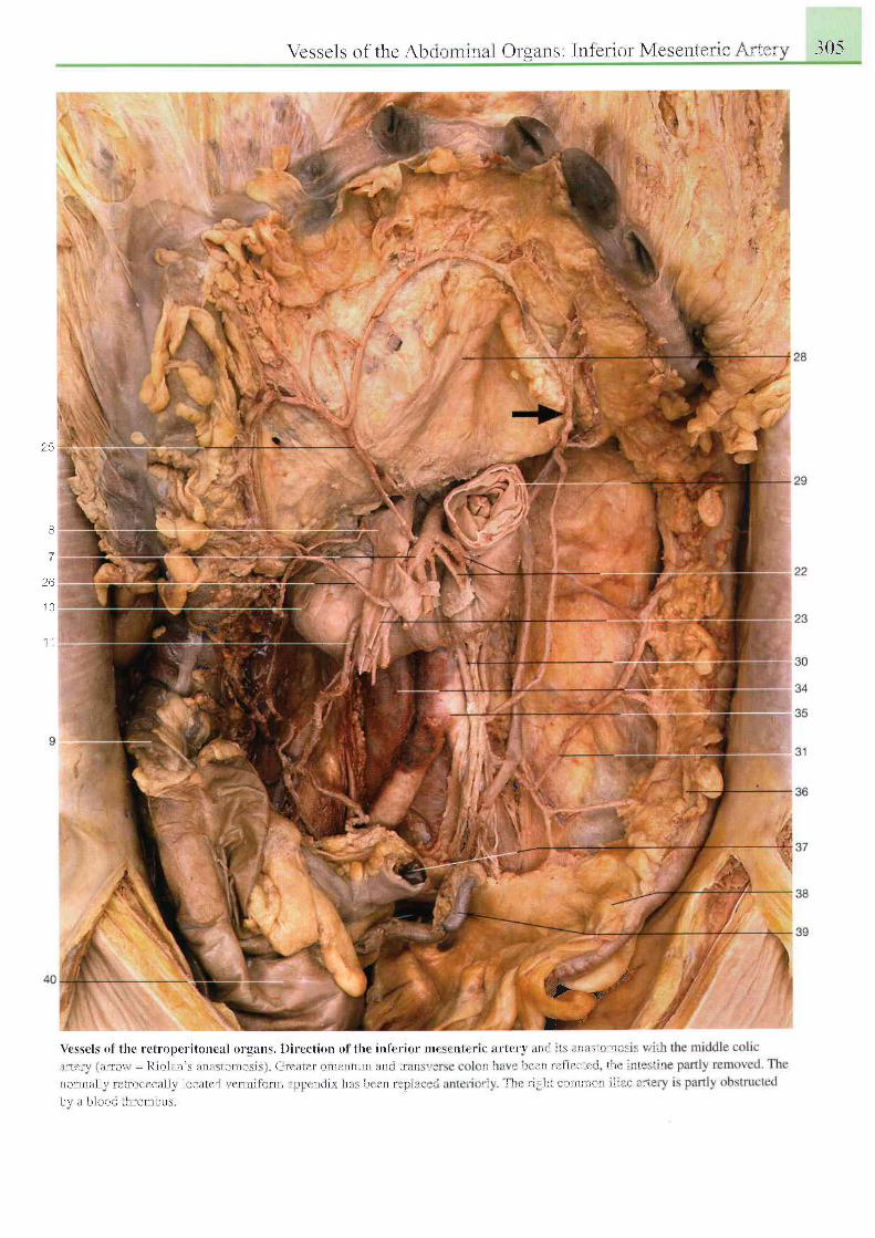
\ t c s s c l s o f t h e A l r t i , r n t l n l l i ( ) r r l l t t t : ' - l n l c r t o r \ 1 c r i - i t t e t l t r I l i r l ' ' . i 0 5
25
8
2 6
] O
1 l
" $\
i
i i '' t
\ c s s c l r o l t l t c r c l r o ; r c r i t o n c a l o r g a n s . l ) i r e c t i r l n o l t l r c i r r f c r i o r n r ( \ ( ' r t l ( r t ( i t r l c t ' \ : r l i r 1 , i . , r l l : r i L { ' l ; 1 1 ; - . , r l
i l r l c r \ L i i l r ( \ \ \ - J { l ( ) l i l n ' \ i l l l l l \ t ( r l l l ( r \ t \ , ( i t t l t l r t ( ) l t l C l ) t L r l l l . l l l ( l l r ^ r l ' . . i , l l I r i ' ! . - L i i l L - l i ! ! i . . 1 l l ) r r r r l ' ' l I
n ( ) r n l i t l i r r L ' l l ( ) ( t L l l l l \ l r r t l t l r ' r l \ c | l l l l \ \ i l l l i t l l l l ( ' n ( l l \ l L r r h c t t l t r ' l ' i , r . , ' ' l l I i . : r : l i l l ' 1 ) l i l l l i { ! i i l i t r , , 1 , . '
h r l t b l i r o . l t l t r r r n t l r r r r
1#\,
;h4,r
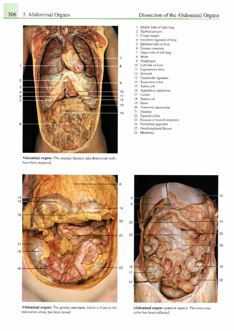
306 5 Abdominal Organs Dissection of the Abdominal Oreans"",*-...,,*"Y,.".","*",",".,
3453
Abdominal organs. The anterior thoracic and abdominal wallshave been removed.
Abdominal organs. The greater omentum, which is fixed to thetransverse colon, has been raised.
1 Middle lobe of right lung2 Xiphoid process3 Costal margin4 Falciform ligament of liver5 Quadrate lobe ofliver6 Greater omentum7 Upper lobe of left lung8 Heart9 Diaphragm
l0 Left lobe ofliver11 Ligamentum teres12 Stomach13 Gastrocolic ligament14 Transverse colonl5 Taenia coli16 Appendices epiploicae17 Cecuml8 Taenia colil9 I leum20 Transversemesocolon21 Jejunum22 Sigmoid colon23 Position of root of mesentery24 Vermiform appendix25 Duodenojejunal flexure26 Mesentery
Abdominal organs (anterior aspect). The transversecolon has been reflected.
o
1 01 11 2
I J
1 4
1 4
1 6
t t
1 8
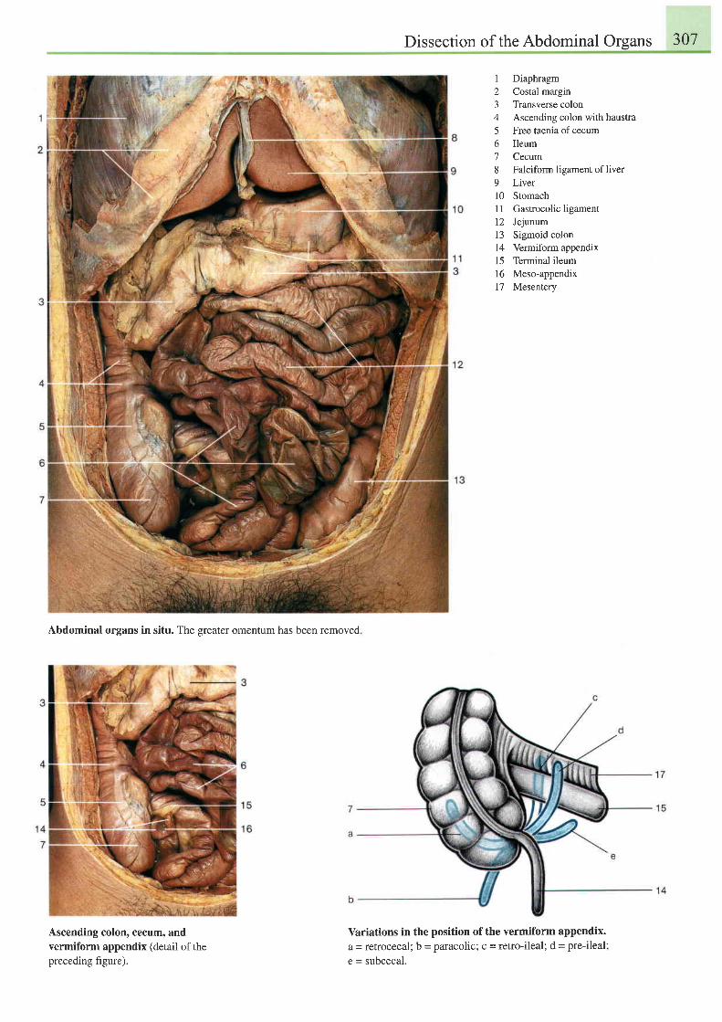
Dissection of the Abdominal Organs 307
1 Diaphragm2 Costal margin3 Transverse colon4 Ascending colon with haustra5 Free taenia ofcecum6 Ileum7 Cecum8 Falciform ligament of liver9 Liverl0 Stomachl1 Gastrocolicligament12 Jejunum13 Sigmoid colon14 Vermiform appendix15 Terminal ileum16 Meso-appendix17 Mesentery
Abdominal organs in situ. The greater omentum has been removed
Ascending colon, cecum, andvermiform appendix (detail of thepreceding figure).
Variations in the position of the vermiform appendix.a = retrocecal; b = paracolic; c = retro-ileal; d = pre-ileal;
e = subcecal.
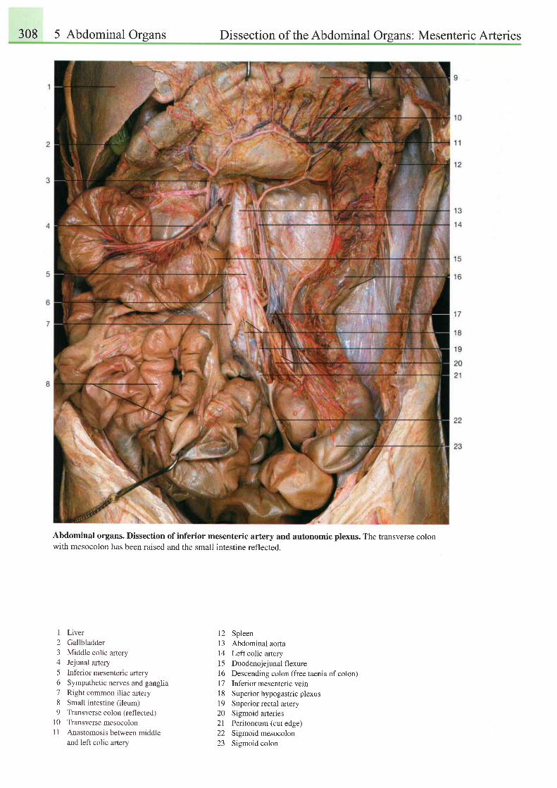
308 5 Abdominal Organs Dissection of the Abdominal Organs: Mesenteric Arteries
Abdominal organs. Dissection of inferior mesenteric artery and autonomic plexus. The transverse colonwith mesocolon has been raised and the small intestine reflected.
1 Liver2 Gallbladder3 Middle colic artery4 Jejunal artery5 Inferior mesenteric artery6 Sympathetic nerves and ganglia7 Right common iliac artery8 Small intestine (ileum)9 Transverse colon (reflected.l
10 TransversemesocolonI I Anastomosis between middle
and left colic artery
12 Spleen13 Abdominal aorta14 Lefl colic artery15 Duodenojejunal flexure16 Descending colon (free taenia ofcolon)l7 Tnferior mesenteric vein18 Superiorhypogastricplexus19 Superior rectal artery20 Sigmoid arteries21 Peritoneum (cutedge)22 Sigmoid mesocolon23 Sigmoid colon
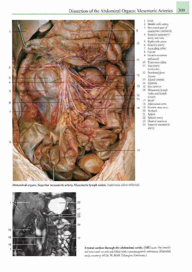
Dissection of the Abdominal Orsans: Mesenteric Arteries 309
I
23
1
51 0
o
1
8
9
1 1l 0
il
1 2
I J
l +
I 5
1 6
I 3 Jejunal arteries
lz l Jejunum
| 5 lleal arteries
l ( r Mesenter ic lymph
nodes and lymph
Liver
Middlc colic artery
Horizontal part of
duodenum (extended)
Super ior mesenter ic
artery and vein
Right colic artery
Ileocolic artery
Ascending colon
Cecum
Greater omentum
(reflected)
Transverse colon
Transverse
mesocolon
Duodeno.jejunal
f'lexure
V C S S C I S
I leum
Abdominal aor-ta
Inferior vena cava
Stomach
Spleen
Splenic artery
Head o1'pancreas
Supcr ior mesenter ic
artery
1'71 8t 9202 l222324
\
I
Abdominal organs. Superior mesenteric artery. Mesenteric lymph nodes. Transverse colon reflected
20
2 1
Z J
Frontal section through the abdominal cavity. (MRI scan: the intesti-
nal tract and vcssels are filled with a paramagnetic substance [Gadolini-
uml: courtesy of Dr W. Rddl, Erlangen, Germany.)
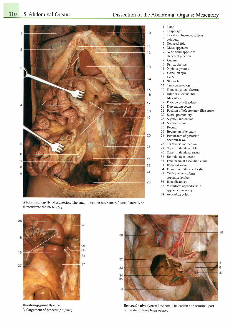
310 5 Abdominal Organs Dissection of the Abdominal Organs: Mesentery
1 4
1 51 6
1 7
1 8
1 9
I
2J
4
56789
l0l lr21 3I415r6l 7t 8t9202122232425zo27
2829303 132-l -)
3435
363'l
38
LungDiaphragmFalciform ligament of liverJejunumIleocecal foldMeso-appendixVermiform appendixIleocecal junctionCecumPericardial sacXiphoid processCostal marginLiverStomachTransverse colonDuodenojejunal flexureInferior duodenal foldMesenteryPosition of left kidneyDescending colonPosition of left common iliac arterySacral promontorySigmoid mesocolonSigmoid colonRectumBeginning ofjejunumPeritoneum of posteriorabdominal wal1Transverse mesocolonSuperior duodenal foldSuperior duodenal recessRetroduodenal recessFree taenia of ascending colonIleocecal valveFrenulum of ileocecal valveOrifice of vermiformappendix (probe)Ileocolic arteryVermiform appendix withappendicular arteryAscending colon
Abdominal cavity. Mesenteries. The small intestine has been reflected laterally todemonstrate the mesenterv.
Duodenojejunal flexure(enlargement of preceding figure).
Ileocecal valve (ventral aspect). The cecum and terminal partof the ileum have been ooened
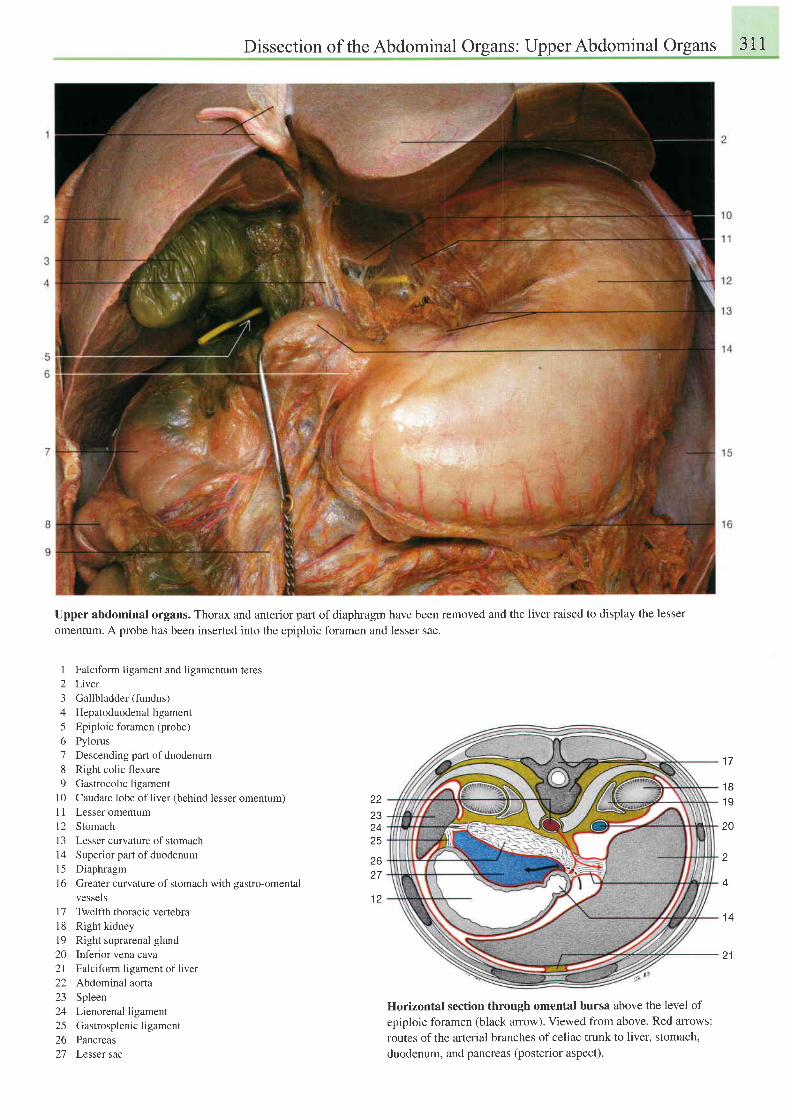
Dissection of theAbdominal Organs: UpperAbdominal Organs 311
Upper abdominal organs. Thorax and anterior part of diaphragm have been removed and the liver raised to display the lesser
omentum. A probe has been inserted into the epiploic foramen and lesser sac.
I Falciform ligament and ligamentum teres
2 Liver
3 Gallbladder (fundus)
4 Hepatoduodenalligament
5 Epiploic foramen (probe)
6 Pylorus
7 Descending part ofduodenum
8 Right colic flexure
9 Gastrocolic ligament
10 Caudate lobe of liver (behind lesser omentum)
11 Lesser omentum
l2 Stomach
l3 Lesser curvature ofstomach
14 Superior part ofduodenum
l5 Diaphragm
16 Greater curvature of stomach with gastro-omental
vessels
17 Twelfth thoracic vertebra
18 Rightk idney
l9 Right suprarenal gland
20 Inferior vena cava
2l Falciform ligament of liver
22 Abdominal aorta
23 Spleen
24 Lienorenal ligament
25 Gastrosplenicligament
26 Pancreas
27 Lesser sac
Horizontal section through omental bursa above the level of
epiploic foramen (black anow). Viewed from above. Red arrows:
routes of the arterial branches of celiac trunk to liver, stomach,
duodenum, and pancreas (posterior aspect).
22232425
2627
1 2
1 7
1 81 9
20
A
t 4
21
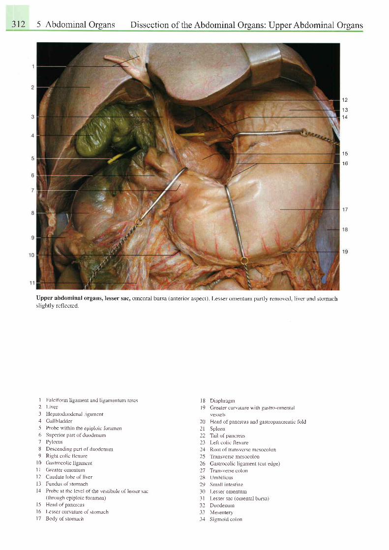
312 5 Abdominal Orsans Dissection of the Abdominal Organs: Upper Abdominal Organs
t z
1 31 4
t c
1 6
Upper abdominal organs, lesser sac, omental bursa (anterior aspect). Lesser omentum partly removed, liver and stomachslightly reflected.
1 Falciform ligament and ligamentum teres2 Liver
3 Hepatoduodenalligament
4 Gallbladder
5 Probe within the epiploic foramen6 Superior part ofduodenum
7 Pylorus
8 Descending part ofduodenum
9 Right colic flexure
10 Gastrocolic ligament
11 Greater omentum
l2 Caudate lobe ofliver
13 Fundus of stomachl4 Probe at the ievel of the vestibule of lesser sac
(through epiploic foramen)l5 Head ofpancreas
16 Lesser curvature of stomachl7 Body of stomach
l8 Diaphragml9 Greater curvature with gastro-omental
vessels20 Head ofpancreas and gastropancreatic fold21 Spleen22 Tallofpancreas23 Left colic flexure24 Root of transverse mesocolon25 Transversemesocolon26 Gastrocolic ligament (cut edge)27 Transverse colon28 Umbilicus29 Small intestine30 Lesser omentum3 I Lesser sac (omental bursa)32 Duodenum33 Mesentery34 Sigmoid colon
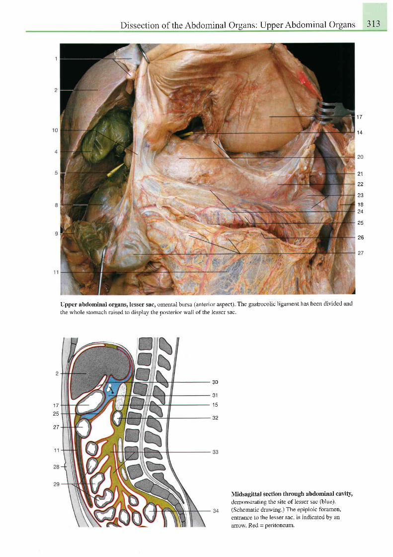
Dissection of the Abdominal Organs: Upper Abdominal Organs 313
1 a
'14
21
22
231 R
24
25
26
1 725
27
Upper abdominal organs, lesser sac, omental bursa (anterior aspect). The gastrocolic ligament has been divided and
the whole stomach raised to display the posterior wall of the lesser sac.
30
J I
l c
32
Midsagittal section through abdominal cavitydemonstrating the site of lesser sac (blue).(Schematic drawing.) The epiploic foramen,entrance to the lesser sac, is indicated by anarrow. Red = peritoneum.
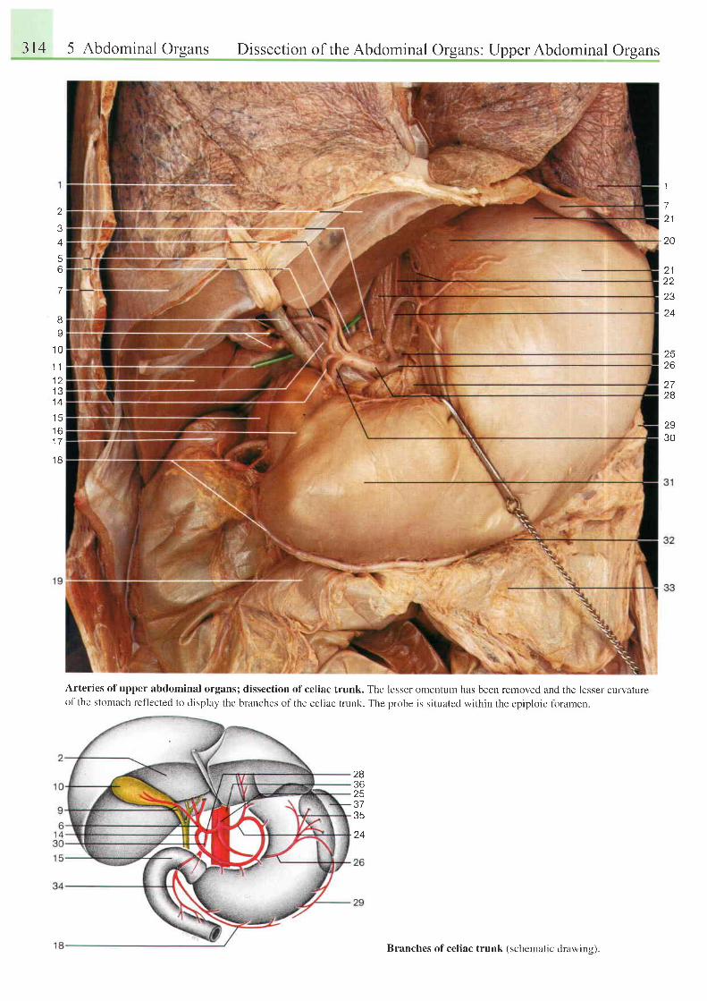
314 5 Abdominal Orsans Dissection of the Abdominal Organs: UpperAbdominal Organs
o
34
5o
7
BY
1 0
1 . 11 21 31 4
t o
1 7
t 6
1
72 1
20
2 1z 1
23
24
Arteries ofupper abdominal organs; dissection ofceliac trunk. The lesser omentum has been removed and the lesser curvatureof the stttmach reflected to display the branches of the celiac trunk. The probe is situated within the epiploic foramen.
2526
28
2930
q ,
283625
24
Branches of celiac trunk (schematic drawins).
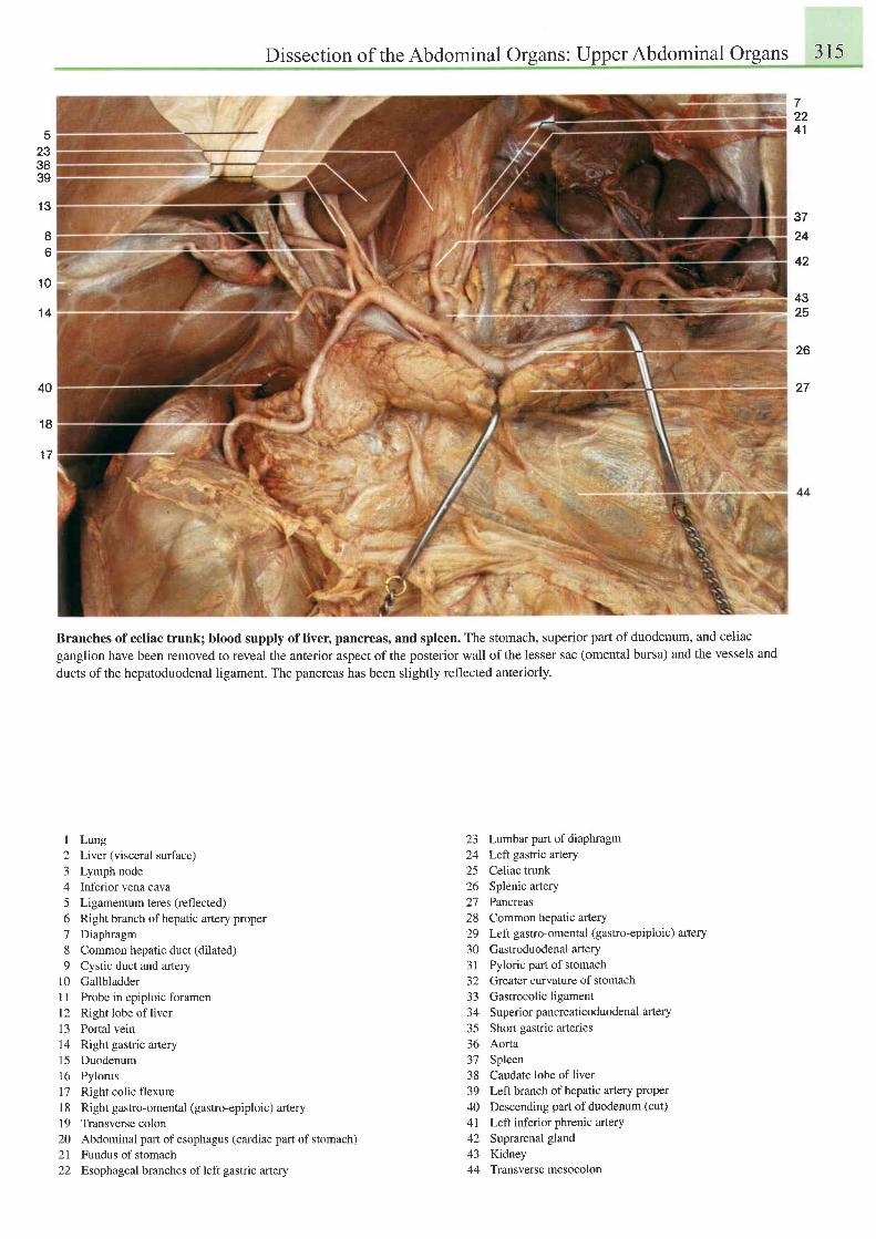
Dissection of the Abdominal Organs: Upper Abdominal Organs 315
72241
37
24
5
233839
1 3
1 0
't4
I6
42
4325
26
2740
1 8
1 7
Branches of celiac trunk; blood supply of liver, pancreas, and spleen. The stomach, superior part of duodenum, and celiacganglion have been removed to reveal the anterior aspect of the posterior wall of the lesser sac (omental bursa) and the vessels and
ducts ofthe hepatoduodenal ligament. The pancreas has been slightly reflected anteriorly.
I Lung2 Liver (visceral surface)3 Lymph node4 Inferior vena cava5 Ligamentum teres (reflected)6 Right branch of hepatic artery proper7 Diaphragm8 Common hepatic duct (dilated)9 Cystic duct and artery
10 Gallbladder11 Probe in epiploic foramenl2 Right lobe of liver13 Portal vein14 Right gastric artery15 Duodenum16 Pylorus17 Right colic flexurel8 Right gastro-omental (gastro-epiploic) artery19 Transverse colon20 Abdominal part of esophagus (cardiac part of stomach)21 Fundus of stomach22 Esophageal branches ofleft gastric artery
23 Lumbar part of diaphragm24 Left gastric artery25 Celiac trunk26 Splenic artery2'7 Pancreas28 Common hepatic artery29 Leftgastro-omental (gastro-epiploic) artery
30 Gastroduodenalartery31 Pyloric part of stomach32 Greater curvature of stomach33 Gastrocolicligament34 Superiorpancreaticoduodenal artery
35 Short gastric arteries36 Aorta37 Spleen38 Caudate lobe ofliver39 Left branch of hepatic artery proper
40 Descending part of duodenum (cut)
4l Left inferior phrenic artery42 Suprarenal gland43 Kidney44 Transversemesocolon
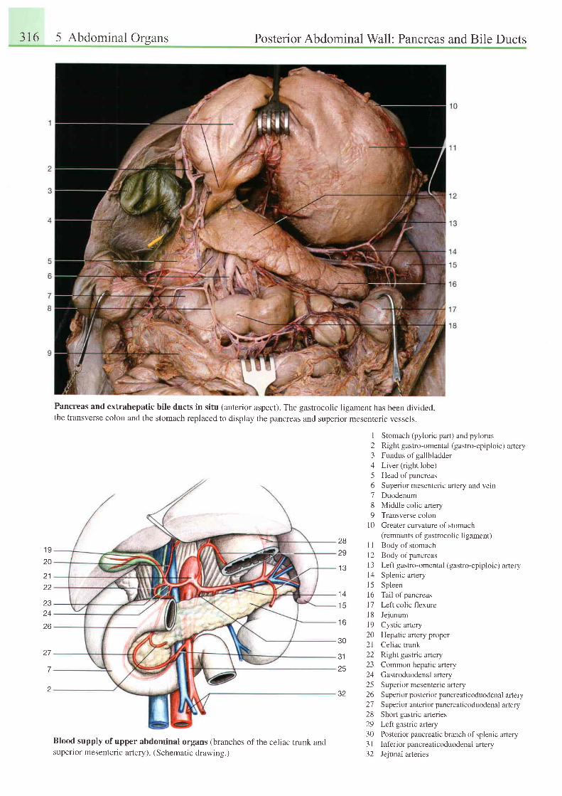
316 5 Abdominal Orsans Posterior Abdominal Wall: Pancreas and Bile Ducts
Pancreas and extrahepatic bile ducts in situ (anterior aspect). The gastrocolic ligament has been divided,the transverse colon and the stomach replaced to display the pancreas and superior mesenteric vessels.
1 920
21
23
26
27
7
z
Blood supply of upper abdominal organs (branches of the celiac trunk andsupenor mesenteric artery). (Schematic drawing.)
I Stomach (pyloric part) and pylorus2 Right gastro-omental (gastro-epiploic) artery3 Fundus ofgallbladder4 Liver (right lobe)5 Head ofpancreas6 Superior mesenteric artery and vein7 Duodenum8 Middle colic artery9 Transverse colon
l0 Greater curvature of stomach(remnants of gastrocolic ligament)
11 Body of stomachl2 Body ofpancreas13 Left gastro-omental (gastro-epiploic) artery14 Splenic arteryl5 Spleen16 Tail ofpancreasl7 Left colic flexure18 Jejunuml9 Cystic artery20 Hepatic artery proper21 Celiac trunk22 Right gastric artery23 Common hepatic artery24 Gastroduodenal artery25 Superior mesenteric artery26 Superiorposteriorpancreaticoduodenal artery27 Superior anteriorpancreaticoduodenal artery28 Short gastric afteries29 Left gastric artery30 Posterior pancreatic branch of splenic artery3 | Inf'erior pancreaticoduodenal artery32 Jejunal arteries
1 3
2829
1 4
t c
1 6
30
3125
32
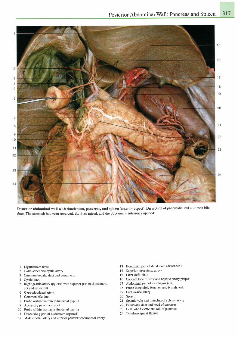
PosteriorAbdominal Wall: Pancreas and Spleen
Posterior abdominal wall with duodenum, pancreas, and spleen (anterior aspect). Dissection of pancreatic and common bile
duct. The stomach has been removed, the liver raised, and the duodenum anteriorly opened.
1 Ligamentum teres2 Gallbladder and cystic artery3 Common hepatic duct and portal vein4 Cystic duct5 Right gastric artery (pylorus with superior part of duodenum,
cut and reflected)6 Gastroduodenal artery7 Common bile duct8 Probe within the minor duodenal papilla9 Accessory pancreatic duct
10 Probe within the major duodenal papillaI 1 Descending part of duodenum (opened)
12 Middle colic artery and inferior pancreaticoduodenal artery
13 Horizontal part of duodenum (distended)
14 Superior mesenteric artery15 Liver(leftlobe)I 6 Caudate lobe of liver and hepatic artery proper
17 Abdominal part ofesophagus (cut)
18 Probe in epiploic foramen and lymph node
19 Left gastric artery20 Spleen21 Splenic vein and branches of splenic artery
22 Pancreatic duct and head ofpancreas23 Left colic flexure and tail of pancreas
24 Duodenojejunal flexure
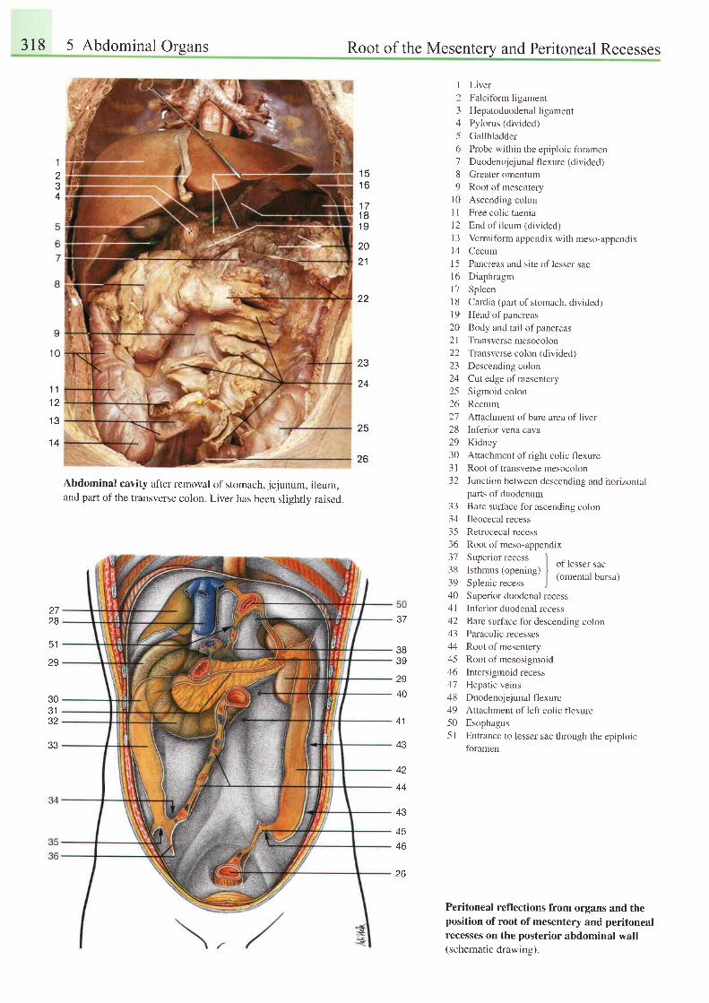
318 5 Abdominalorgans Root of the Mesentery and Peritoneal Recesses
1
2.t
4
Abdominal cavity after removal of stomach, jejunum, ileum,and part of the transverse colon. Liver has been slightly raised.
I Liver
2 Falciform ligament
3 Hepatoduodenalligament
4 Pylorus (divided)
5 Gallbladder
6 Probe within the epiploic foramen7 Duodenojejunal flexure (divided)
8 Greater omentum
9 Root of mesentery
l0 Ascending colon
I I Free colic taenia
12 End ofileum (divided)
l3 Vermiform appendix with meso-appendixl4 Cecum
15 Pancreas and site of lesser sacl6 Diaphragm
l7 Spleen
i 8 Cardia (part of stomach, divided)19 Head ofpancreas
20 Body and tail ofpancreas
21 Transversemesocolon
22 Transverse colon (divided)
23 Descending colon
24 Cut edge of mesentery
25 Sigmoid colon
26 Rectum
27 Attachment of bare area of liver28 Inferior vena cava
29 Kidney
30 Attachment of right colic flexure
31 Root oftransverse mesocolon
32 Junction between descending and horizontalparts of duodenum
Bare surface for ascending colonIleocecal recess
Retrocecal recess
Root of meso-appendix
1 51 6
1 71 8t 9
2021
22
1 0
1 11 2
1 3
1 4
23
24
25
26
33343536- ) l
3839404 l/ a
4344
46414849505 l
Superior recessIsthmus (opening)Splenic recessSuperior duodenal recess
Inferior duodenal recess
Bare surface for descending colonParacolic recesses
Root of mesentery
Root of mesosigmoid
Intersigmoid recess
Hepatic veins
Duodenojejunal flexure
Attachment of left colic flexure
Esophagus
Entrance to lesser sac through the epiploicforamen
of lesser sac(omental bursa)
30J I
JZ
3B39
2728
51
29
41
43
44
43
2940
45
46
Peritoneal reflections from organs and theposition of root of mesentery and peritonealrecesses on the posterior abdominal wall(schematic drawing).
z o
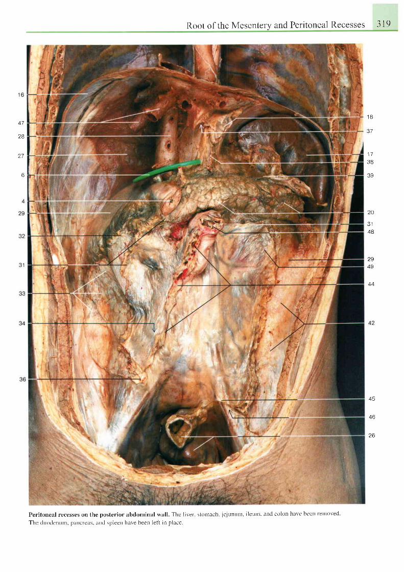
47
28
27
6
Root of the Mesentery and Peritoneal Recesses 319
Peritoneal recesses on the posterior abdominal wall. The liver, stonrach, jejunum, ileurn, and colon have been retnoved.
The duodenum. Dancreas. aud spleen have been left in place.
1 8
37
' 1 7
38
39
20
J I
4B
294S
44
45
46
26
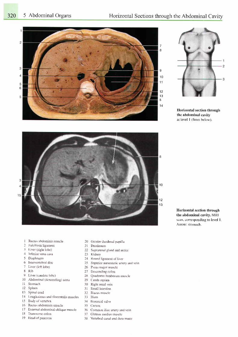
320 5 Abdominal Organs Horizontal Sections throush the Abdominal Cavitv
1 21 a
5't4
1 0
1 3
Horizontal section throughthe abdominal cavityat level I (from below).
Horizontal section throughthe abdominal cavity. MRIscan, corresponding to level 1.Arrow: stomach.
1 Rectus abdominis muscle 20 Greater duodenal papilla2 Falciform ligament 21 Duodenum3 Liver (right lobe) 22 Suprarenal gland and ureter4 Inferior vena cava 23 Kidney5 Diaphragm 24 Round ligament of liver6 Intervertebral disc 25 Superior mesenteric artery and vein7 Liver (left lobe) 26 psoas major muscle8 Rib 27 Descending colon9 Liver (caudate lobe) 28 Quadratus lumborum muscle
10 Abdominal (descending) aorta 29 Cauda equrnaI I Stomach 30 Right renal vein12 Spleen 31 Small inrestine13 Spinal cord 32 Iliacus muscle14 Longissimus and iliocostalis muscles 33 Ilium15 Bodyofvertebra 34 Ileocecalvalve16 Rectus abdominis muscle 35 Cecum17 Extemal abdominal oblique muscle 36 Common iliac artery and vein18 Transverse colon 37 Gluteus medius muscle19 Head ofpancreas 38 Vertebral canal and dura mater
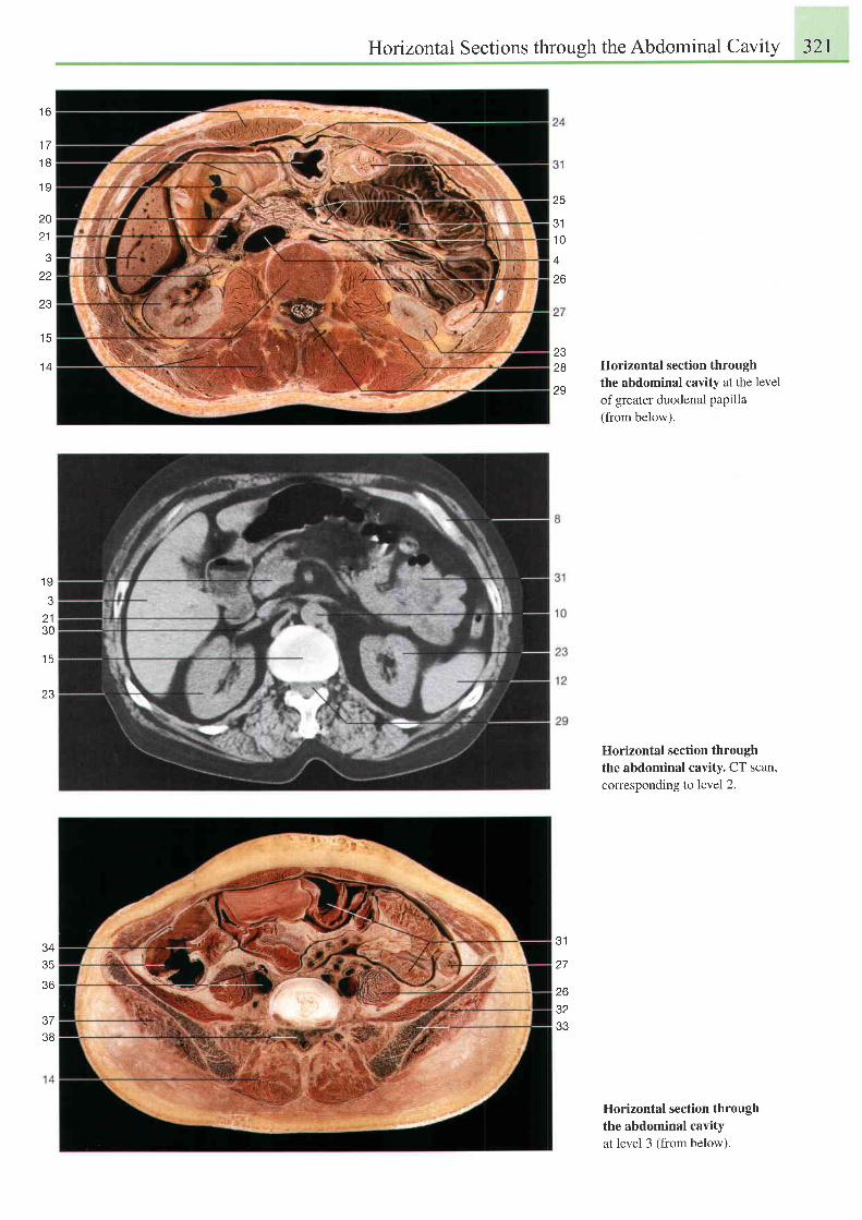
Horizontal Sections through the Abdominal Cavity 321
t o
1 7
1 8
1 9
20
2 1
z z
ZJ
25
311 0A
26
2328
Z J
1 5
1 4 Horizontal section through
the abdominal cavity at the level
of greater duodenal papilla(from below).
Horizontal section throughthe abdominal cavity. CT scan,corresponding to level 2.
Horizontal section throughthe abdominal cavityat level 3 (from below).
1 9
2 130
1 5
Z J
35
36
3738
31
263233
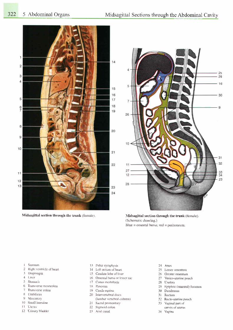
322 5 Abdomlnll 9t*l: Midsagittal Sections through the AbdominalCavity
Midsagittal section through the trunk (female) Midsagittal section through the trunk (female).(Schematic drawing.)Blue = omental bursa;19d = peritoneum.
2529
1 6
333423
1 1
t z
1 3
1 Sternum2 Right ventricle ofheart3 Diaphragm4 Liver5 Stomach6 Transverse mesocolon7 Transverse colon8 Umbil icus9 Mesentery
l0 Small intestineI I Uterus12 Urinary bladder
l3 Pubic symphysis14 Left atrium ofhearl15 Caudate lobe of liverl6 Omental bursa or lesser sacl7 Conus medullaris18 Pancreasl9 Cauda equina20 Intervertebral discs
(lumbar verlebral column)21 Sacral promontory22 Sigmoid colon23 Anal canal
24 Anus25 Lesser omentum26 Greater omentum27 Vesico-uterinepouch28 Urethra29 Epiploic (omental) foramen30 Duodenum31 Rectum32 Recto-uterine pouch33 Vaginal part of
cervix of uterus34 Vagina
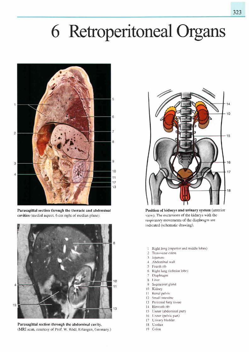
323
6 Retroperitone aI Organs
1 0
1 1
1 2
I J
1 4
1 0
Parasagittal section through the thoracic and abdominalcavities (medial aspect, 6 cm right ofmedian plane).
Parasagittal section through the abdominal cavity.(MRI scan, courtesy of Prof. W. Rddl, Erlangen, Germany.)
1 0t l
Position of kidneys and urinary system (anterior
view). The excursions of the kidneys with the
respiratory movements of the diaphragm are
indicated (schematic drawin g).
I Right lung (superior and middle lobes)2 Transverse colon3 Jejunum4 Abdominal wall5 Fourth rib6 Right lung (inferior lobe)7 Diaphragm8 Liver9 Suprarenal gland
l0 KidneyI I Renal pelvisl2 Small intestineI 3 Perirenal fatty tissuel4 Eleventh rib15 Ureter (abdominal part)l6 Ureter (pelvic part)l7 Urinary bladderl8 Urethral9 Colon
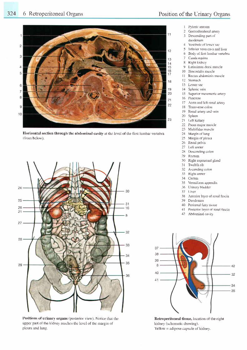
324 6 Retroperitoneal Organs Position of the Urinary Organs
24
25
zo21
27
28
311 6
8
1 2
1 31 41 5t bt t
1 B
1 920
2 1
zo
23
456189
1 0l tl 2I J
t 41 5l 61'7l 8r 9202 l22z-)a 1
25zo
272829303 1-)z
333435-JO
313839404 lA A
Pyloric antrumGastroduodenal arteryDescending part ofduodenumVestibule of lesser sacInferior vena cava and iiverBody of first lumbar vertebraCauda equinaRight kidneyLatissimus dorsi muscleIliocostalis muscleRectus abdominis muscleStomachLesser sacSplenic veinSuperior mesenteric arteryPancreasAorta and left renal arteryTransverse colonRenal artery and veinSpleenLeft kidneyPsoas major muscleMultihdus muscleMargin of lungMargin of pleuraRenal pelvisLeft ureterDescending colonRectumRight suprarenal glandTwelfth ribAscending colonRight ureterCecumVermiform appendixUrinary bladderLiverAnterior layer of renal fasciaDuodenumPerirenal fatty tissuePosterior layer of renal fasciaAbdominal cavity
Horizontal section through the abdominal cavity at the level of the first lumbar vertebra(from below).
Positions of urinary organs (posterior view). Notice that theupper part of the kidney reaches the level of the margin ofpleura and lung.
38
39
3240
4 1
Retroperitoneal tissue, location of the right
kidney (schematic drawing).
Yellow = adipose capsule ofkidney.
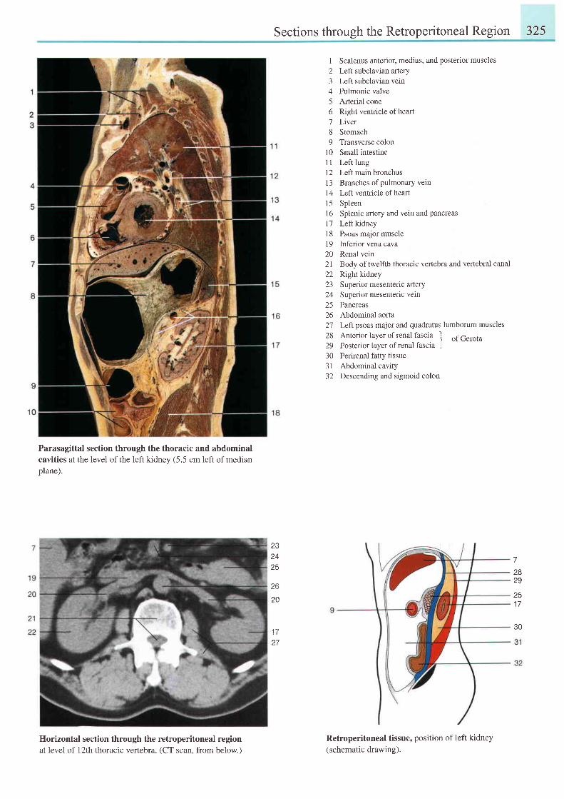
Sections through the Retroperitoneal Region 325
Parasagittal section through the thoracic and abdominalcavities at the level of the left kidnev (5.5 cm left of medianplane).
Horizontal section through the retroperitoneal regionat level of l2th thoracic vertebra. (CT scan, from below.)
1 Scalenus anterior, medius, and posterior muscles2 Left subclavian atlery3 Left subclavian vein4 Pulmonic valve5 Arterial cone6 Right ventricle ofheart7 Liver8 Stomach9 Transverse colon
10 Small intestine11 Left lung12 Left main bronchus| 3 Branches of pulmonary vein14 Left ventricle ofheart15 Spleen16 Splenic artery and vein and pancreasl7 Leftkidney18 Psoas major muscle19 Inferior vena cava20 Renal vein21 Body oftwelfth thoracic vertebra and vertebral canal22 Rightkidney23 Superior mesenteric artery24 Superior mesenteric vein25 Pancreas26 Abdominal aorta27 Left psoas major and quadratus lumborum muscles
28 Anterior layer of renal fascia -f
of Gerota29 Posterior layer of renal lascia J30 Perirenal fatty tissue3l Abdominal cavity32 Descending and sigmoid colon
Retroperitoneal tissue, position of left kidney(schematic drawing).
232425
72829
251 7
to
20
1 t
27
30
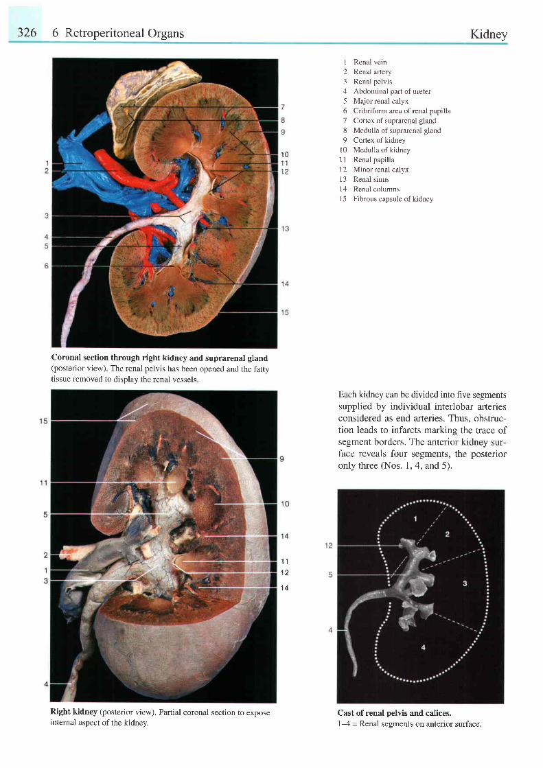
326 6 Retroperitoneal Organs Kidney
Coronal section through right kidney and suprarenal gland(posterior view). The renal pelvis has been opened and the fattytissue removed to displav the renal vessels.
Right kidney (posterior view). Partial coronal section to exposeinternal aspect of the kidney.
I Renal vein2 Renal artery3 Renal pelvis4 Abdominal part of ureter5 Major renal calyx6 Cribriform area ofrenal papilla7 Cortex of suprarenal gland8 Medulla of suprarenal gland9 Cortex ofkidney
10 Medulla of kidneyI I Renal papilla12 Minor renal calyx13 Renal sinus14 Renal columnsl5 Fibrous capsule ofkidney
Each kidney can be divided into five segmentssupplied by individual interlobar arteriesconsidered as end arteries. Thus, obstruc-tion leads to infarcts marking the trace ofsegment borders. The anterior kidney sur-face reveals four segments, the posterioronly three (Nos. 1,4, and 5).
Cast of renal pelvis and calices.1-4 = Renal segments on anterior surface.
1 01 11 2
1 1
1 2
1 4
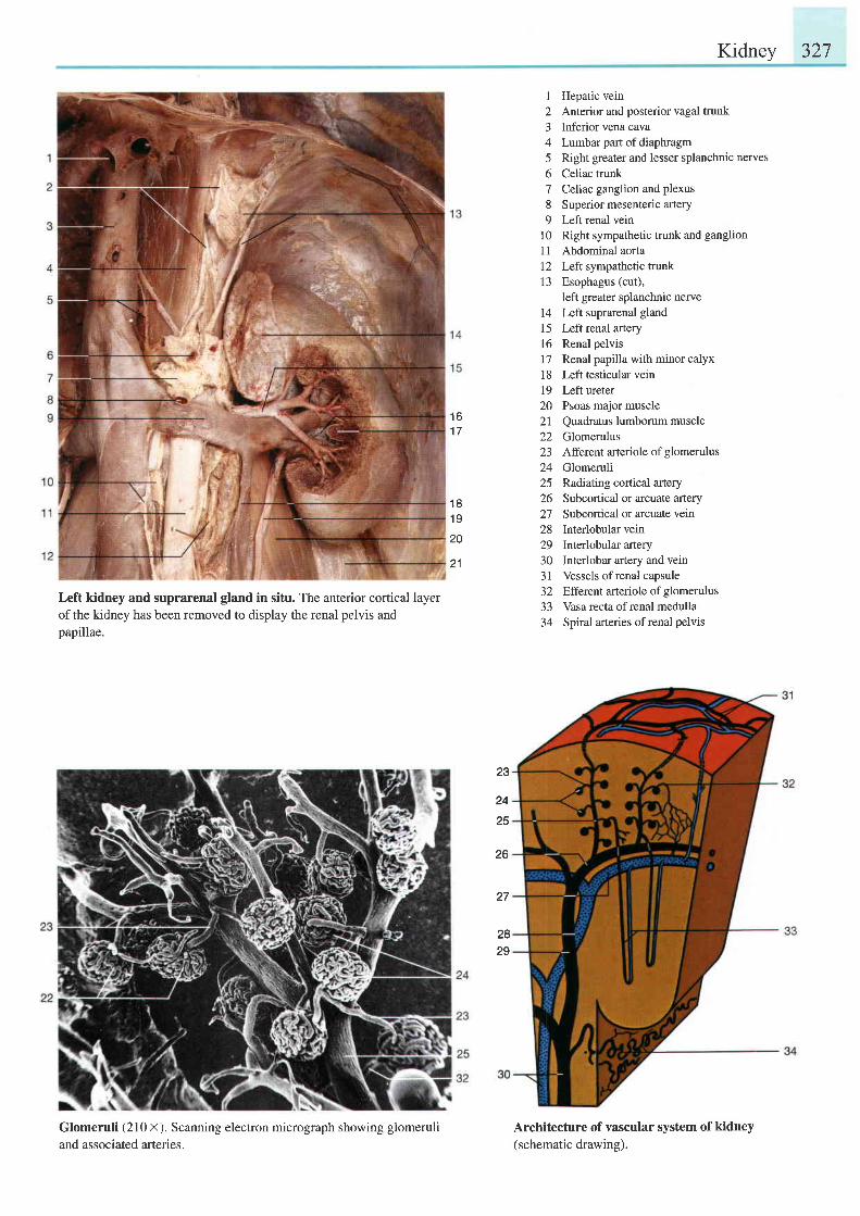
Kidney 327
Left kidney and suprarenal gland in situ. The anterior cortical layerofthe kidney has been removed to display the renal pelvis andpapillae.
Glomeruli (210 x). Scanning electron micrograph showing glomeruliand associated arteries.
I Hepatic vein2 Anterior and posterior vagal trunk3 Inferior vena cava4 Lumbar part of diaphragm5 Right greater and lesser splanchnic nerves6 Celiac trunk7 Cel\ac ganglion and plexus8 Superior mesenteric artery9 Left renal vein
10 Right sympathetic trunk and ganglion
11 Abdominal aorta12 Left sympathetic trunk13 Esophagus (cut),
left greater splanchnic nerve14 Left suprarenal gland15 Leftrenal arteryl6 Renal pelvis17 Renal papilla with minor calyx18 Left testicular vein19 Left ureter20 Psoas major muscle21 Quadratus lumborum muscle22 Glomerulus23 Afferent arteriole of glomerulus24 Glomeruli25 Radiating cortical artery26 Subcortical or arcuate artery27 Subcortical or arcuate veln28 Interlobular vein29 Interlobular artery30 Interlobar artery and vein31 Vessels ofrenal capsule32 Efferent arteriole of glomerulus33 Vasa recta of renal medulla34 Spiral arteries ofrenal pelvis
Architecture of vascular system of kidney(schematic drawing).
1 61 7
1 81 9
20
21
23
24
25
26
27
2829
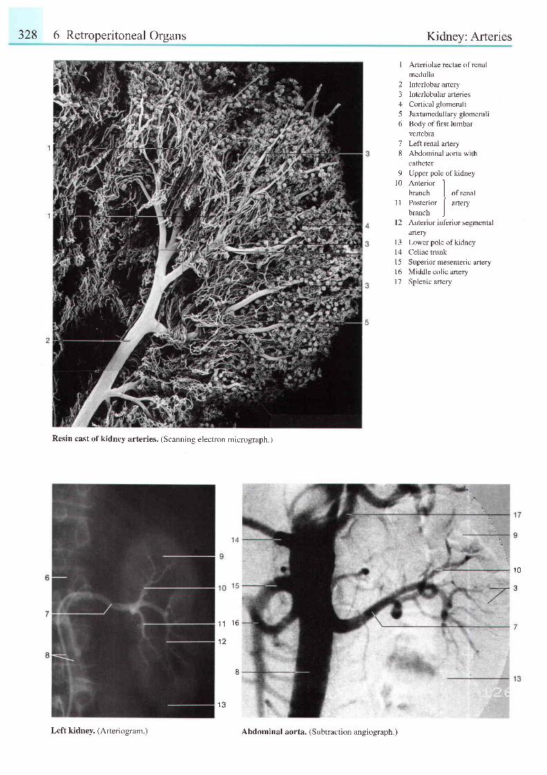
328 6 Retroperitoneal Organs Kidnev: Arteries
1 Arteriolae rectae ofrenalmedulla
2 Interlobar artery3 Interlobulararteries4 Cortical glomeruli5 Juxtamedullaryglomeruli6 Body of first lumbar
vertebra7 Left renal artery8 Abdominal aorta with
catheter9 Upper pole of kidney
10 Anterior Ibranch I ofrenal
11 Posterior I artery
branch )[2 Anterior infedor segmental
arteryl3 Lowerpoleofkidneyl4 Celiac trunk15 Superior mesenteric artery16 Middle colic artery17 Splenic artery
Resin cast ofkidney arteries. (Scanning electron micrograph.)
11 ' 16
1 2
1 3
1 0
Left kidney. (Arteriogram. ) Abdominal aorta. (Subtraction angiograph.)
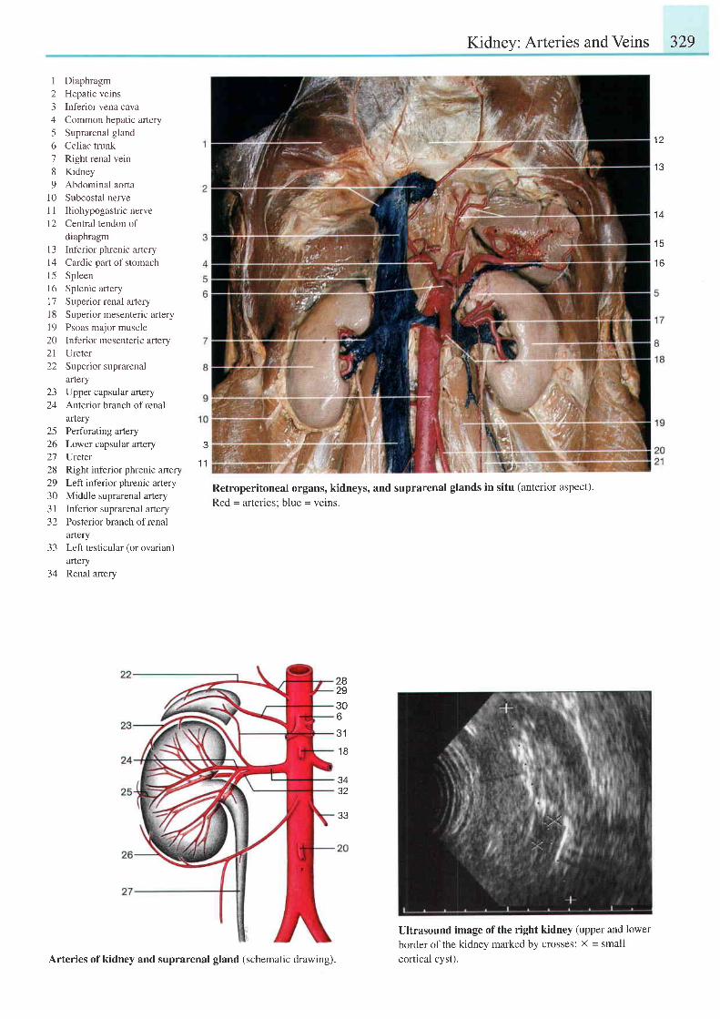
Kidney: Arteries and Veins 329
I Diaphragm2 Hepatic veins3 Inferior vena cava4 Common hepatic artery5 Suprarenal gland6 Celiac trunk7 Right renal vein8 Kidney9 Abdominal aorta
10 Subcostal nerveI I Iliohypogastric nerve12 Central tendon of
diaphragm13 Inferior phrenic artery14 Cardic part of stomachl5 Spleenl6 Splenic artery17 Superior renal arteryl8 Superior mesenteric arteryl9 Psoas major muscle20 Inferior mesenteric artery21 Ureter22 Superior suprarenal
arteryza
24
2526212829303 1-)L
-1 -t
34
Upper capsular arteryAnterior branch of renalarteryPerforating arteryLower capsular arteryUreterRight inferior phrenic arteryLeft inferior phrenic arteryMiddle suprarenal arteryInferior suprarenal arteryPosterior branch of renalafieryLeft testicular (or ovarian)arteryRenal artery
t z
1 3
1 4
t c
t o
1 1
Retroperitoneal organs, kidneys, and suprarenal glands in situ (anterior aspect).Red = arteries: blue = veins.
282930o
31
1 8
3432
Ultrasound image of the right kidney (upper and lowerborder of the kidney marked by crosses; X = smallcortical cvst).Arteries of kidney and suprarenal gland (schematic drawing).
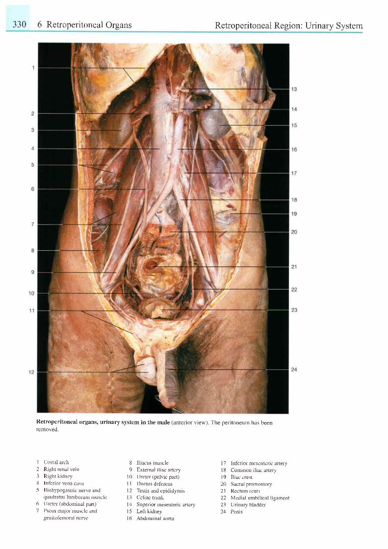
330 6 Retroperitoneal Organs
61
Retroperitoneal Region: Urinary System
Retroperitoneal organs, urinary system in the male (anterior view). The peritoneum has beenremoved.
123A
5
Costal arch
Right renal vein
Right kidney
Inf'erior vena cavaIliohypogastric nerve andquadratus lumborum muscleUreter (abdominal part)
Psoas major muscle andgenitofemoral nerve
8 Iliacus muscle9 External iliac artery
10 Ureter (pelvic pat)I I Ductus deferens12 Testis and epididymis13 Celiac trunk14 Superior mesenteric afieryl5 Left kidney16 Abdominal aorta
l7 Inf'erior mesenteric arteryl8 Common iliac arteryl9 Iliac crest20 Sacral promontory21 Rectum (cut)22 Medtal umbilical ligament23 Urinary bladder24 Penis
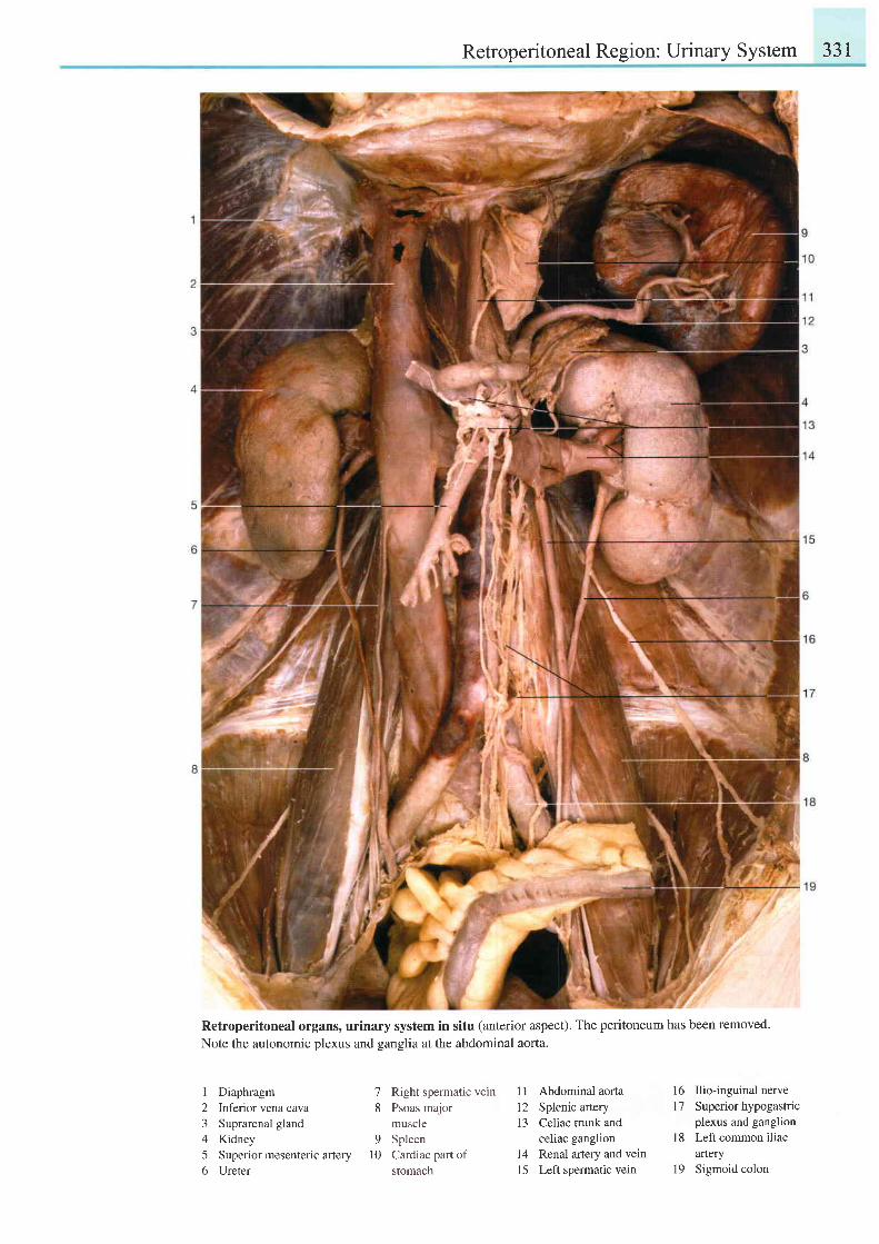
Retroperitoneal Region: Urinary System 331
Retroperitoneal organs, urinary system in situ (anterior aspect). The peritoneum has been removed.Note the autonomic plexus and ganglia at the abdominal aorta.
I Diaphragm2 Inferior vena cava3 Suprarenal gland4 Kidney5 Superior mesenteric artery6 Ureter
Right spermatic veinPsoas majormuscleSpleenCardiac pan ofstomach
11 Abdominal aorta12 Splenic atery13 Celiac trunk and
celiac ganglion14 Renal artery and vein15 Left spermatic vein
16 Ilio-inguinal nerve17 Superiorhypogastric
plexus and ganglion18 Left common iliac
afiery19 Sigmoid colon
78
9l 0
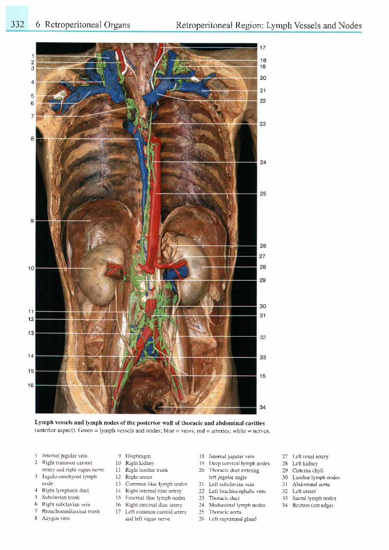
332 6 Retroperitoneal Organs Retroperitoneal Region: Lymph Vessels and Nodes
123
1 11 2
1 3
1 4
1 5
1 6
34
Lymph vessels and lymph nodes of the posterior wall of thoracic and abdominal cavities(anterior aspect). Green = lymph vessels and nodes; blue = veins; red = arteries; white = nerves.
I
2Internal jugular veinRight common carotidartery and right vagus nerveJugulo-omohyoid lymphnodeRight lymphatic ductSubclavian trunkRight subclavian veinBronchomediastinal trunkAzygos vein
9 Diaphragml0 Right kidneyl1 Right lumbar trunk12 Right ureter13 Common iliac lymph nodes14 Right internal iliac artery15 External iliac lymph nodes16 Right external iliac arteryI7 Left common carotid aftery
and left vagus nerve
18 Intemal jugular vein
19 Deep cervical lymph nodes
20 Thoracic duct entering
left jugular angle
21 Left subclavian vein
22 Left brachiocephalic vein
23 Thoracic duct
24 Mediastinal lymph nodes
25 Thoracic aorla
26 Left suprarenal gland
A
5678
27 Left renal artery28 Left kidney29 Cisterna chyli30 Lumbar lymph nodes31 Abdominal aorta32 Left ureter33 Sacral lymph nodes34 Rectum (cut edge)
1 7
1 81 9
20
21
22
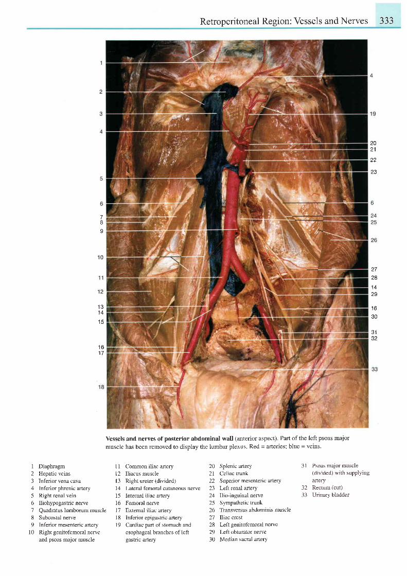
Retroperitoneal Region: Vessels and Nerves 333
2021
22
23
Io
1 31 4
1 5
t o1 7
2728
1 42S
1 630
J I
32
Vessels and nerves of posterior abdominal wall (anterior aspect). Part of the left psoas majormuscle has been removed to display the lumbar plexus. Red = arteries; blue = veins.
I Diaphragm2 Hepatic veins3 Inferior vena cava4 Inferior phrenic artery5 Right renal vein6 Iliohypogastricnerve7 Quadratus lumborum muscle8 Subcostal nerve9 Inferior mesenteric artery
l0 Right genitofemoral nerveand psoas major muscle
11 Common iliac artery12 Iliacus musclel3 Right ureter (divided)14 Lateral femoral cutaneous nerve15 Intemal iliac artery16 Femoral nerve17 Extemal iliac artery18 Inferior epigastric artery19 Cardiac partof stomach and
esophageal branches of leftgastric artery
20 Splenic artery21 Celiac trunk22 Superior mesenteric artery23 Left renal artery24 Ilio-inguinal nerve25 Sympathetic trunk26 Transversus abdominis muscle27 Iliac crest28 Left genitofemoral nerve29 Left obturator nerve30 Median sacral artery
31 Psoas major muscle(divided) with supplyingartery
32 Rectum (cut)33 Urinary bladder
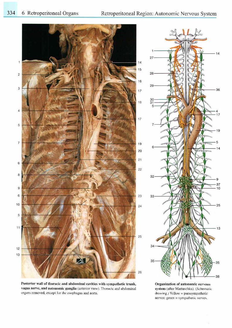
334 6 Retroperitoneal Organs Retroperitoneal Region: Autonomic Nervous System
1 4
1 5
t o
1 7
1 S
201 4
I371 0
12
1 3
Posterior wall of thoracic and abdominal cavities with sympathetic trunk,vagus nerve, and autonomic ganglia (anterior view). Thoracic and abdominalorgans removed, except for the esophagus and aorta.
Organization of autonomic nervoussystem (after Mattuschka). (Schematicdrawing.) Yellow - parasympatheticnerves; green - sympathetic nerves.
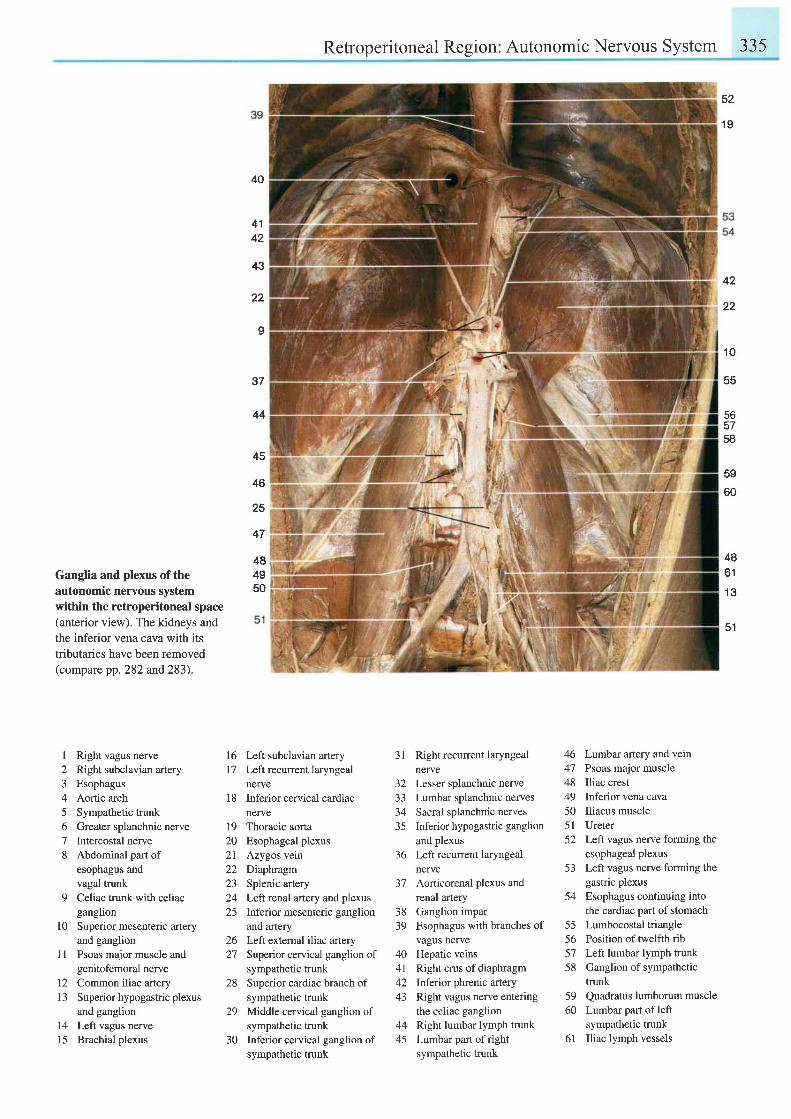
Retroperitoneal Region: Autonomic Nervous System 335
52
1 9
Ganglia and plexus of theautonomic nervous systemwithin the retroperitoneal space(anterior view). The kidneys andthe inferior vena cava with itstributaries have been removed(compare pp. 282 and 283).
I Right vagus nerve2 Right subclavian artery3 Esophagus4 Aortic arch5 Sympathetic trunk6 Greater splanchnic nerve7 Intercostal nerve8 Abdominal part of
esophagus andvagal trunk
9 Celiac trunk with celiacganglion
10 Superior mesenteric arteryand ganglion
1l Psoas major muscle andgenitofemoral nerve
12 Common iliac arteryl3 Superior hypogastric plexus
and ganglion14 Left vagus nerve15 Brachial plexus
40
4142
'lil
22
I
37
44
45
46
25
16 Left subclavian arteryl7 Leftrecurrentlaryngeal
nerve18 Inferior cervical cardiac
nerve19 Thoracic aorta20 Esophageal plexus2l Azygos vein22 Diaptuagm23 Splenic artery24 Leftrenal artery andplexus25 Inferior mesenteric ganglion
and artery26 Left extemal iliac artery27 Superior cervical ganglion of
sympathetic trunk28 Superior cardiac branch of
sympathetic trunk29 Middle cervical ganglion of
sympathetic trunk30 Inferior cervical ganglion of
sympathetic trunk
31 Rightrecurrentlaryngealnerve
32 Lesser splanchnic nerve33 Lumbar splanchnic nerves34 Sacral splanchnic nerves35 Inferior hypogastric ganglion
and plexus36 Left recurrent laryngeal
nerve37 Aorticorenal plexus and
renal artery38 Ganglion impar39 Esophagus with branches of
404 l^ 1
43
44
vagus nerveHepatic veinsRight crus of diaphragmInferior phrenic arteryRight vagus nerve enteringthe celiac ganglionRight lumbar lymph trunkLumbar part of rightsympathetic trunk
46 Lumbar artery and vein47 Psoas major muscle48 Iliac crest49 Inferior vena cava50 Iliacus muscle51 Ureter52 Left vagus nerve forming the
esophageal plexus53 Left vagus nerve forming the
gastric plexus54 Esophagus continuing into
the cardiac part of stomach55 Lumbocostaltriangle56 Position of twelfth rib57 Left lumbar lymph trunk58 Ganglion of sympathetic
trunk59 Quadratus lumborum muscle60 Lumbar part of left
sympathetic trunk61 Iliac lymph vessels
42
22
't0
bs
565758
59
60
47
484950
4A61
1 3
51
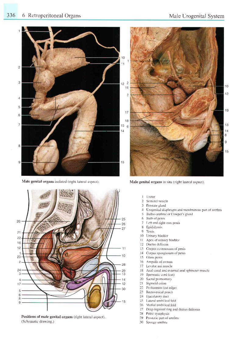
336 6 Retroperitoneal Organs Male Urogenital System
Male genital organs in situ (right lateral aspect).
Ureter
Seminal vesic le
Prostate gland
Urogenital diaphragm and membranous part of urethraBulbo-urethral or Cowper's gland
Bulb of penis
Left and right crus penis
Epididymis
Testis
Urinary bladder
Apex of urinary bladder
Ductus deferens
Corpus cavernosum of penis
Corpus spongiosum of penis
Glans penis
Ampulla of rectum
Levator ani muscle
Anal canal and extemal anal sphincter muscle
Spermatic cord (cut)
Sacral promontory
Sigmoid coion
Peritoneum (cut edge)
Rectovesical pouch
Ejaculatory duct
Lateral umbilical fold
Medial umbilical fold
Deep inguinal ring and ductus deferens
Pubic symphysis
Prostatic part of urethra
Spongy urethra
1 0
1 1
f i 2t o
J
1 0
12
1 3
1 4
81 4
2 1
z z
t o
1 2'1
232
J
41 7
568I
Male genital organs isolated (right lateral aspect),
Positions of male genital organs (right lateral aspect).(Schematic drawing )
252627
I
23456189
l 01 l1 2l 3l 4l 5l 6t 7l 8l 9202 l22L-)
a 1
252627282930
28
29t o
1 4
1 230
I J
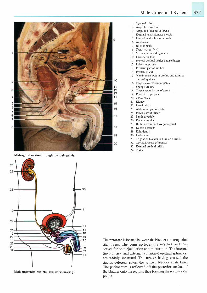
Male Urogenital System 337
I Sigmoid colon2 Ampulla of rectum3 Ampulla of ductus deferens4 External anal sphincter muscle5 Internal anal sphincter muscle6 Anal canal7 Bulb ofpenis8 Testis (cut surface)9 Median umbilical ligament
10 Urinary bladder1 I Internal urethral orifice and sphincter12 Pubic symphysis13 Prostatic part ofurethra14 Prostate glandl5 Membranous part of urethra and external
urethral sphincterl6 Corpus cavemosum ofpenis17 Spongy urethra18 Corpus spongiosum ofpenrs19 Foreskin or prepuce20 Glans penis21 Kidney22 Renal pelvis23 Abdominal part of ureter24 Pelvic part of ureter25 Seminal vesicle26 Ejaculatory duct27 Bulbo-urethral or Cowper's gland28 Ductus deferens29 Epididymis30 Umbilicus31 Trigone ofbladder and ureteric orifice32 Navicular fossa of urethra33 External urethral orifice34 Testis
The prostate is located between the bladder and urogenitaldiaphragm. The penis includes the urethra and thusserves for both ejaculation and micturition. The intemal(involuntary) and external (voluntary) urethral sphinctersare widely separated. The ureter having crossed theductus deferens enters the urinary bladder at its base.The peritoneum is reflected off the posterior surface ofthe bladder onto the rectum. thus forming the rectovesicalpouch.
1 0
1 11 21 31 44
56547
311 11 31 51 7
323334
Midsagittal section through the male pelvis.
253
261 4272829
Male urogenital system (schematic drawing).
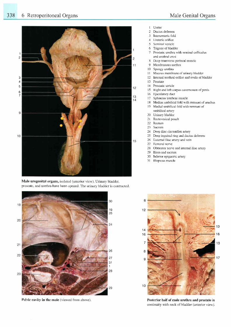
338 6 Retroperitoneal Organs Male Genital Organs
e
4
67
Male urogenital organs, isolated (anterior view). Urinary bladder,prostate, and urethra have been opened. The urinary bladder is contracted.
1 Ureter2 Ductus deferens3 Interureteric fold4 Ureteric orifice5 Seminal vesicle6 Trigone ofbladder7 Prostatic urethra with seminal colliculus
and urethral crest8 Deep transverse perineal muscle9 Membranous urethra
l0 Spongy urethra11 Mucous membrane of urinary bladder12 Intemal urethral orihce and uvula ofbladder13 Prostate14 Prostatic utricle15 Right and left corpus cavernosum ofpenis16 Ejaculatory duct17 Sphincterurethraemuscle18 Median umbilical fold with remnant of urachus19 Medial umbilical fold with remnant of
umbilical artery20 Urinary bladder21 Rectovesical pouch22 Rectum23 Sacrum24 Deep iliac circumflex artery25 Deep inguinal ring and ductus deferens26 External iliac anery and vein27 Femoral nerve28 Obturator nerve and internal iliac artery29 Ilium and sacrum30 Inferior epigastric artery31 Iliopsoas muscle
Posterior half of male urethra and prostate incontinuity with neck of bladder (anterior view).
I J
1 4
30
1 925
26
e l
1
28
1 41 6
7
I
s
1 3
1 6
' t ?
Pelvic cavity in the male (viewed from above).
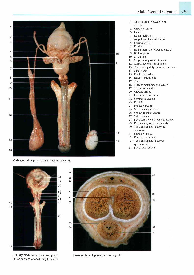
Male Genital Organs
I Apex of urinary bladder withurachus
2 Urinary bladder3 Ureter4 Ductus deferens5 Ampulla of ductus deferens6 Seminal vesicle7 Prostate8 Bulbo-urethral or Cowper's gland9 Bulb of penis
l0 Crus penisI I Corpus spongiosum ofpenis| 2 Corpus cavernosum of penisl3 Testis and epididymis with coveringsl4 Glans penis15 Fundus ofbladderl6 Head of epididymrs17 Testisl8 Mucous membrane of bladder19 Trigone ofbladder20 Ureteric orifrce21 Internal urethral oriflce22 Seminal oolliculus23 Prostate24 Prostatic urethra25 Membranous urethra26 Spongy (penile) urethra27 Skin of penis28 Deep dorsal vein ofpenis (unpaired)
29 Dorsal artery ofpenis (paired)30 Tunica albuginea of corpora
cavernosa3l Septum ofpenis32 Deep artery of penrs33 Tunica albugineaof corpus
sponglosum34 Deep fascia ofpenrs
339
Male genital organs, isolated (posterior view)
27
1 B1 920
2829
21
232425
30
3'1
32
Urinary bladder, urethra, and penis(anterior view. opened longi tudinally)
Cross section of penis (inferior aspect).
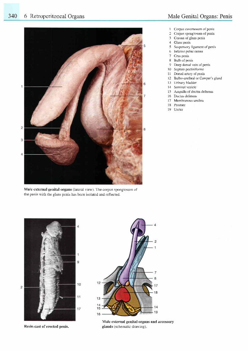
340 6 Retroperitoneal Organs Male Genital Organs: Penis
1 Corpus cavernosum ofpenrs2 Corpus spongiosum ofpenis3 Corona ofglans penis4 Glans penis5 Suspensory ligament ofpenis6 Inferior pubic ramus7 Crus penis8 Bulb of penis9 Deep dorsal vein ofpenis
l0 Septum pectiniformeI 1 Dorsal artery of penis12 Bulbo-urethral or Cowper's glandl3 Urinary bladder14 Seminal vesiclet5 Ampulla of ductus deferensl6 Ductus deferens17 Membranous urethral8 Prostatel9 Ureter
Male external genital organs (lateral view). The corpus spongiosum ofthe penis with the glans penis has been isolated and reflected.
1 3
1 41 5
t o
1 8
1 4
1 9
Male external genital organs and accessoryglands (schematic drawing).Resin cast of erected penis.
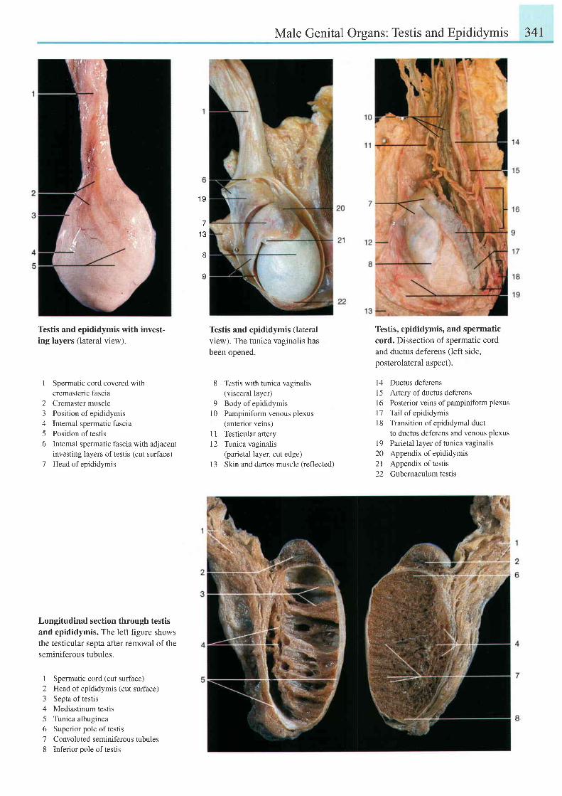
Male Genital Organs: Testis and Epididymis 341
1 9
1 3
tt
I
Testis and epididymis with invest-ing layers (lateral view).
1 Spermatic cord covered withcremasteric fascia
2 Cremaster muscle3 Position of epididymis4 Internal spermatic fascia5 Position of testis6 Intemal spermatic fascia with adjacent
investing layers of testis (cut surface)7 Head of epididymis
Longitudinal section through testis
and epididymis. The left figure shows
the testicular septa after removal of the
seminiferous tubules.
I Spermatic cord (cut surface)2 Head of epididymis (cut surface)3 Septa of testis4 Mediastinum testis5 Tunica albuginea6 Superior pole oftestis7 Convoluted seminiferous tubules8 Inferior oole of testis
Testis and epididymis (lateral
view). The tunica vaginalis has
been opened.
8 Testis with tunica vaginalis(visceral layer)
9 Body of epididymis10 Pampiniform venous plexus
(anterior veins)1 1 Testicular artery12 Tunica vaginalis
(parietal layer, cut edge)13 Skin and dartos muscle (reflected)
Testis, epididymis, and spermatic
cord. Dissection of spermatic cord
and ductus deferens (left side,
posterolateral aspect).
14 Ductus deferens15 Artery ofductus deferens16 Posterior veins ofpampiniform plexus17 Tail of epididymis18 Transition of epididymal duct
to ductus deferens and venous plexus19 Parietal layer oftunica vaginalis20 Appendix of epididymis2l Appendix of testis22 Gubemaculum testis
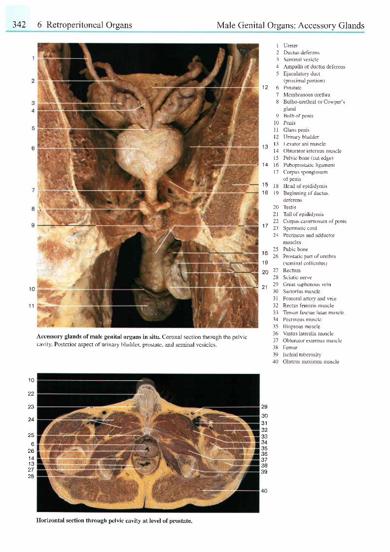
342 6 Retroperitoneal Organs Male Genital Organs: Accessory Glands
1 Ureter2 Ductus deferens3 Seminal vesicle4 Ampulla of ductus deferens5 Ejaculatory duct
(proximal portion)12 6 Prostate
7 Membranous urethra8 Bulbo-urethral or Cowper's
gland9 Bulb ofpenis
10 Penis11 Glans penis12 Urinary bladderl3 Levator ani muscle
t r J
14 Obturator internus muscle15 Pelvic bone (cut edge)
14 16 Puboprostatic ligament17 Corpus spongiosum
of penis15 18 Headofepididymis16 19 Beginning ofductus
deferens20 Testis2l Tail of epididymis
. - 22 Corpus cavernosum ofpenisa ' t' ' 23 Spermatic cord
24 Pectineus and adductormuscles
,o 25 Pub icbone'-
26 Prostatic part of urethra19 (seminal colliculus)
29 27 Rectum28 Sciatic nerve
21 29 Great saphenous vein30 Sartorius muscle31 Femoral artery and vern32 Rectus femoris muscle33 Tensor fasciae latae muscle34 Pectineus muscle35 Iliopsoas muscle36 Vastus lateralis muscle37 Obturator externus muscle38 Femur39 Ischial tuberosity40 Gluteus maximus muscle
1 0
22
23
24
25
6zo
1 4.13
2728
Accessory glands of male genital organs in situ. Coronal section through the pelviccavity. Posterior aspect of urinary bladder, prostate, and seminal vesicles.
29
303 132333435\to373839
llorizontal section through pelvic cavity at level of prostate.
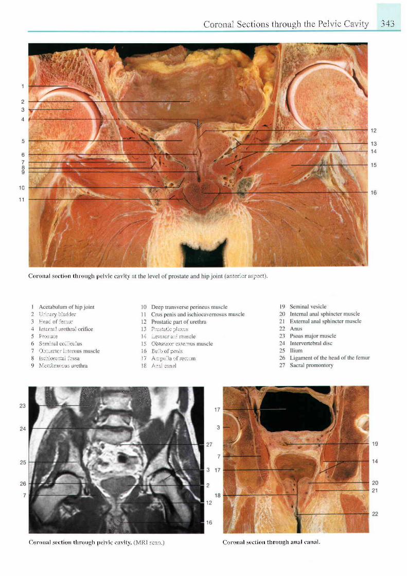
----\y
( t t r r t r r a l \ c ( ' t i on l h ro r rgh l ) ( ' i \ r ( ( ' : t \ t t \ ,
( , r r r \ l t l l \ cc l r ( )ns th rough thc Pc i r t t ' l . . r i i l ' . - 1 "+ "1
. t l , I r , , r , l \ l r \ ! l I
i l l r t l l : l , . l l l ,
l 1 ! ' r , l I f L i
l i l r ' l L . L i r r t i r i i
l ' l , , . l r l
\ ! ' l l i l i L r l i ! i
I ) l ) L r i 1 r t i l r , r i l
i - { l l r i \ ' r i i i '
\ 1 , ' 1 r , , , , y 1 , r 1 : r 1 ,
, , , , r , \ l r l r ! l r l ! \ i i -
l r i , r , r l ! r r l i l ! r r \ ,
I i i ) - L t i t i t l \ , 1 r ' \ l ( | l l
I r l t r 1 l . t 1 . 1 1 1 ''
\ t r l | L t l i , , r ' i r l l
I ' i ' ! | i r l r , | r ; !
- i . ;_ ,
#lt' "t" *
( - 0 r t l r r i l l \ r c l i o r . l l h r 0 r r g h l l ( ' i 1 r ( ( i r \ r t \ \ l l { i ' , , , r i r ( oront l sct ' t i t r r t l t l 'ough : tnul c i t t l i t l ,
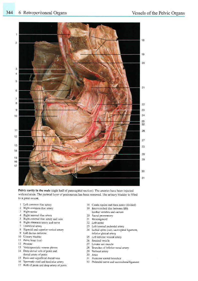
344 6 Retroperitoneal Organs Vessels of the Pelvic Organs
2532
26
1 4
1 4
1 6
1 7
Pelvic cavity in the male (right half of parasagittal section). The arteries have been injectedwith red resin. The parietal layer ofperitoneum has been removed. The urinary bladder is hlledto a great extent.
1 Left common iliac artery2 Right common iliac artery3 Right ureter4 Right intemal iliac artery5 Right external iliac artery and vein6 Right obturator artery and nerve7 Umbilical artery8 Sigmoid and superior vesical artery9 Left ductus deferens
10 Urinary bladder1 1 Pubic bone (cut)
12 Prostate13 Vesicoprostatic venous plexus14 Deep dorsal vein ofpenis and
dorsal artery of pems15 Penis and superficial dorsal vein16 Spermatic cord and testicular artery17 Bulb ofpenis and deep artery ofpenis
1 8 Cauda equina and dura mater (divided)19 Intervertebral disc between fifth
lumbar vertebra and sacrum20 Sacral promontory21 Mesosigmoid22 Leftveter23 Left internal pudendal artery24 Ischial spine (cut), sacrospinal ligament,
inferior gluteal artery25 Left inferior vesical artery26 Seminal vesicle27 Levator ani muscle28 Branches ofinferior rectal artery29 Peinealartery30 Anus31 Posterior scrotal branches32 Pudendal nerve and sacrotuberal lieament
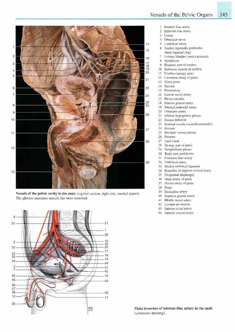
Vessels of the Pelvic Orsans 345
1 A
t c
t o
1 71 81 920
21
zz
I Intemal iliac artery2 Extemal iliac artery3 Ureter4 Obturator nerve5 Umbilical artery6 Anulus inguinalis profundus
(deep inguinal ring)7 Urinary bladder (vesica urinaria)8 Symphysis9 Prostatic part ofurethra
10 Sphinctermuscle of urethra11 Urethra (spongy part)12 Cavernous body ofpenis13 Glans penis14 Sacrum15 Promontory16 Lateral sacral arteryl7 Plexus sacralis18 Inf'erior gluteal artery19 Intemal pudendal artery20 Obturator artery2l Inferior hypogastric plexus22 Ductus deferens23 Seminal vesicle (vesicula seminalis)24 Rectum25 Prostatic venous plexus26 Prostate27 Analcanal28 Spongypartofpenis29 Pampiniform plexus30 Testis and epididymis31 Common iliac artery32 Umbilical artery33 Medial umbilical ligament34 Branches of superior vesical artery35 Urogenital diaphragm36 Deep artery ofpenis37 Dorsal artery ofpenis38 Penis39 Iliolumbar aftery40 Superior gluteal artery4l Middle rectal artery42 Levalor ani muscle43 Inf'erior rectal artery44 Inferior vesical artery
2325
26
32
zzJ J
J 4
7
26
36373B1 2
Vessels of the pelvic cavity in the male (sagittal section, right side, medial aspect).The gluteus maximus muscle has been removed.
281 1
3 1
1
39
t o40201 71 B1 9444 1
t e
Main branches of internal iliac artery in the male(schematic drawing).
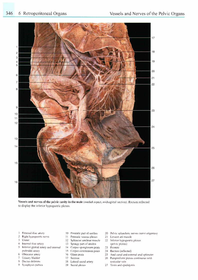
346 6 Retroperitoneal Organs Vessels and Nerves of the Pelvic Organs
A
Vessels and nerves of the pelvic cavity in the male (medial aspect, midsagittal section). Rectum reflectedto display the inferior hypogastric plexus.
123A
5
External iliac arteryRight hypogastric nerveUreterInternal iliac arteryInferior gluteal artery and internalpudendal arteryObturator aneryUrinary bladderDuctus deferensSymphysis pubica
6789
10 Prostatic part ofurethraI I Prostatic venous plexus12 Sphincter urethrae muscle13 Spongy part ofurethral4 Corpus spongiosum penis15 Corpus cavernosum penlsl6 Glans penisl7 Sacrum18 Lateral sacral artery19 Sacral plexus
20 Pelvic splanchnic nerves (nervi erigentes)2l Levalor ani muscle22 lnferior hypogastric plexus
(pelvic plexus)23 Prostate24 Rectum (reflected)25 Anal canal and external anal sphincter26 Pampiniform plexus continuous with
testicular vein27 Testis and epididymis
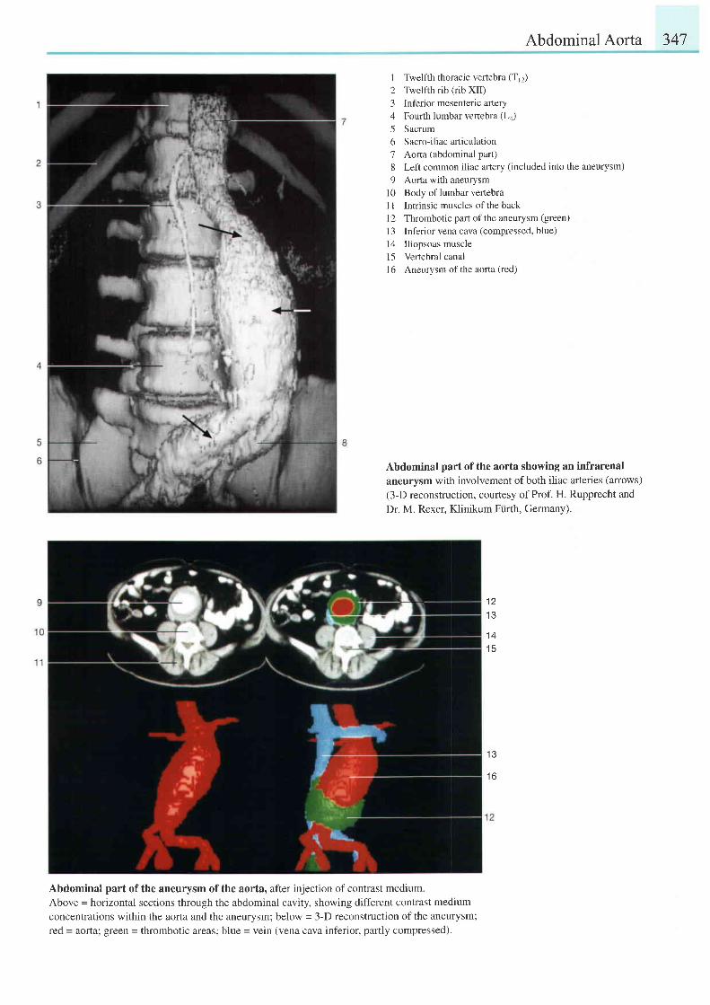
Abdominal Aorta 347
1 Twelfth thoracic vertebra (Tr2)
2 Twelfth rib (rib XII)3 Inferior mesenteric aftery4 Fourth lumbar vertebra (La)
5 Sacrum6 Sacro-iliac arliculation7 Aorta (abdominal part)8 Left common iliac artery (included into the aneurysm)
9 Aorta with aneurysml0 Body of lumbar vertebraI 1 Intrinsic muscles ofthe backI 2 Thrombotic part of the aneurysm (green)
l3 Inferior vena cava (compressed, blue)l4 lliopsoas musclel5 Vertebral canall6 Aneurysm of the aorta (red)
Abdominal part of the aorta showing an infrarenalaneurysm with involvement of both iliac arteries (anows)(3-D reconstruction, courtesy of Prof. H. Rupprecht andDr. M. Rexer, Klinikum Fiirth, Germany).
t z
I J
1 4t c
1 3
t o
Abdominal part of the aneurysm of the aorta, after injection of contrast medium.
Above = horizontal sections through the abdominal cavity, showing different contrast medium
concentrations within the aorta and the aneurysm; below = 3-D reconstruction of the aneurysm;
red = aorta; green = thrombotic areas; blue = vein (vena cava inferior, partly compressed).
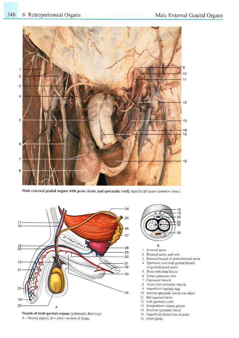
348 6 Retroperitoneal Organs Male External Genital Organs
1 41 (
BI Femoral nerve2 Femoral artery and vein3 Femoral branch ofgenitofemoral nerve4 Spermatic cord with genital branch
of genitofemoral nerve5 Penis with deep fascia6 Great saphenous vein7 Cremaster muscle8 Testis with cremaster muscle9 Superhcial inguinal ring
l0 Internal spermatic fascia (cut edge)11 Ilio-inguinal nerve12 Left spermatic cord13 Pampiniform venous plexus14 External spermatic fascia15 Superficial dorsal vein ofpenis16 Glans penis
1 51 9203430
Male external genital organs with penis, testis, and spermatic cord, superficial layers (anterior view).
35
24
25
26
27
282930
3132
33
Vessels of male genital organs (schematic drawing).A = lateral aspect; B = cross section of penis.
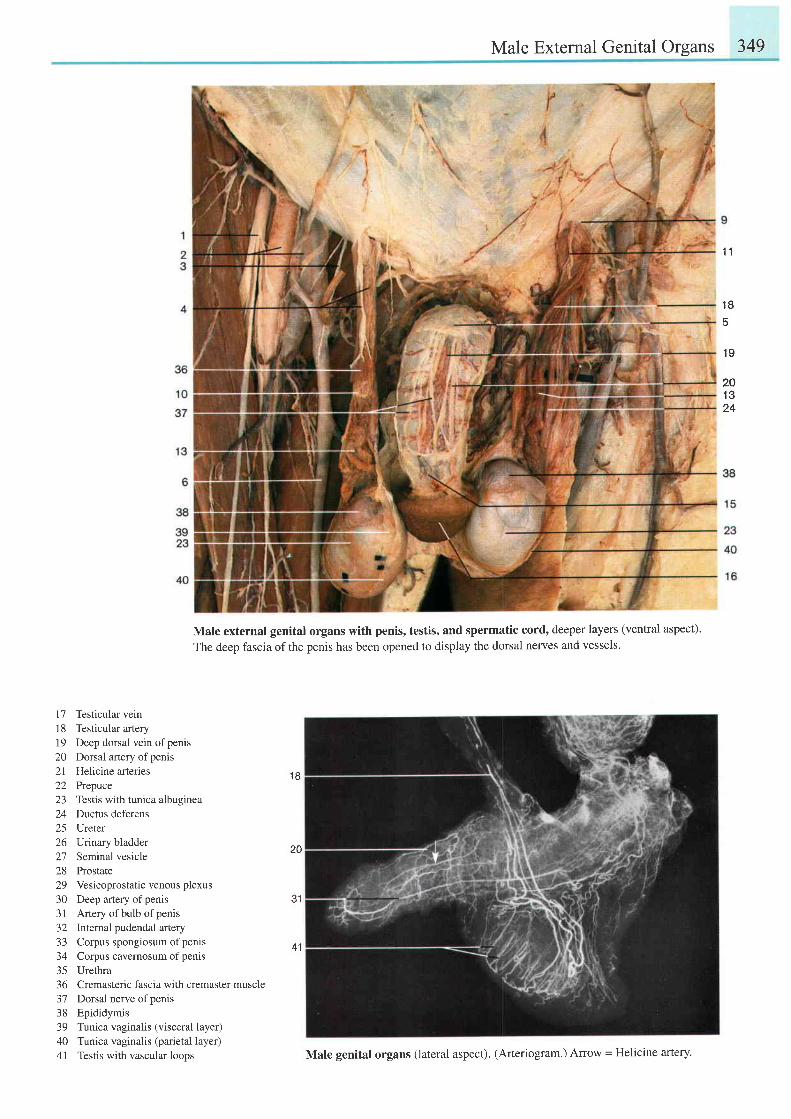
Male External Genital Organs 349
1 1
1 8
5
1 9
201 324
Male external genital organs with penis, testis, and spermatic cord, deeper layers (ventral aspect).
The deep fascia of the penis has been opened to display the dorsal nenres and vessels.
17 Testicular vein18 Testicular artery19 Deep dorsal vein ofpenis20 Dorsal artery ofpenis21 Helicine arteries22 Prepuce23 Testis with tunica albuginea24 Ductus deferens25 Ureter26 Uinary bladder27 Seminal vesicle28 Prostate29 Vesicoprostatic venous plexus30 Deep artery ofpenis3 i Artery of bulb of penis32 Intemal pudendal artery33 Corpus spongiosum ofpenis34 Corpus cavemosum of penis35 Urethra36 Cremasteric fascia with cremaster muscle37 Dorsal nerve ofpenis38 Epididymis39 Tunica vaginalis (visceral layer)40 Tunica vaginalis (parietal layer)41 Testis with vascular loops
1 B
2Q
41
Male genital organs (lateral aspect). (Arteriogram.) Arrow = Helicine arterY.
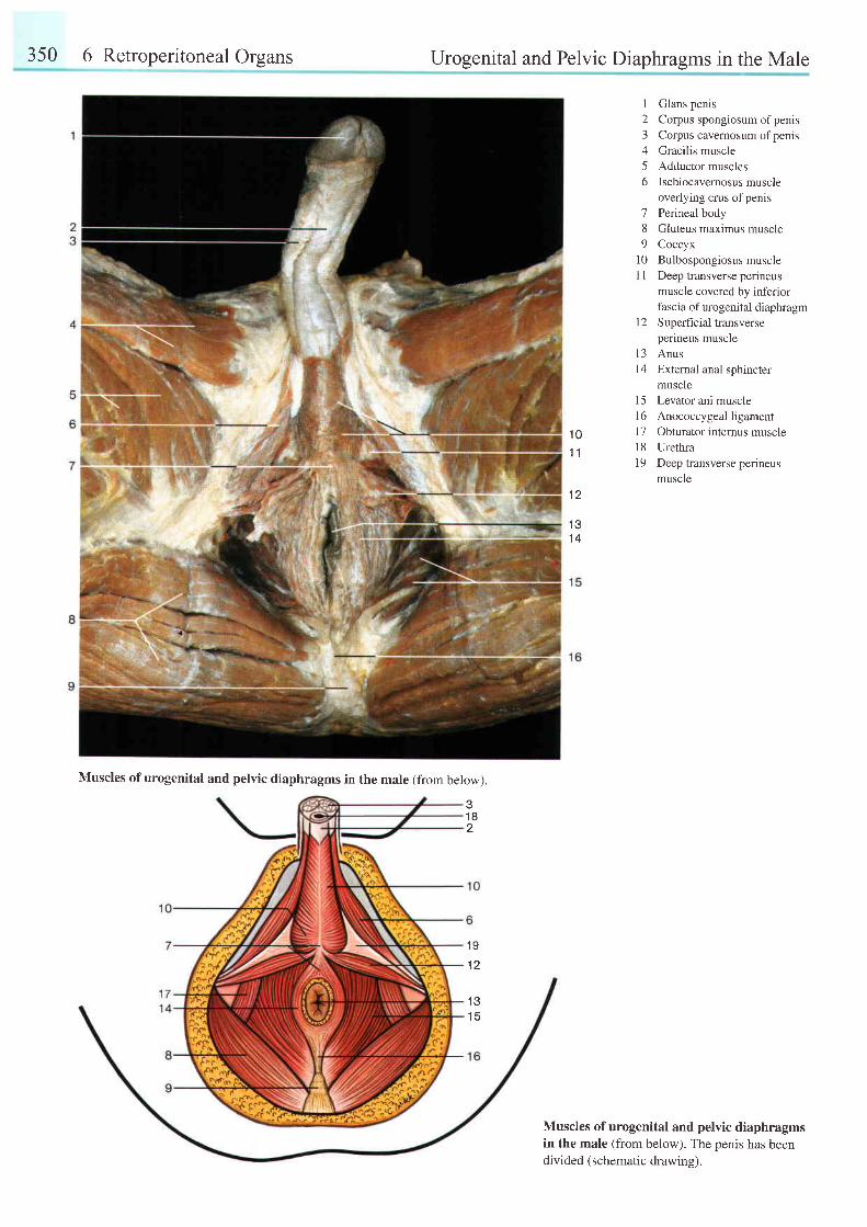
350 6 Retroperitoneal Organs
Muscles of urogenital and pelvic diaphragms in the male (from below).
Urogenital and Pelvic Diaphragms in the Male
12345o
189
10l 1
t 2
Glans penisCorpus spongiosum of penisCorpus cavernosum of penisGracilis muscleAdductor musclesIschiocavernosus muscleoverlying crus of penisPerineal bodyGluteus maximus muscleCoccyxBulbospongiosus muscleDeep transverse perineusmuscle covered by inferiorfascia of urogenital diaphragmS uperficial transverseperineus muscle
1 01 1
1 2
13 Anus14 External anal sphincter
musclel5 Levator ani muscle16 Anococcygeal ligament17 Obturator internus muscle18 Urethra19 Deep transverse perineus
muscle
1 31 4
31 82
1 9
1 2
1 31 5
Muscles of urogenital and pelvic diaphragmsin the male (from below). The penis has beendivided (schematic drawine).
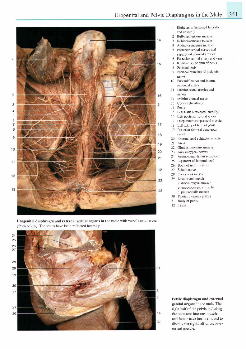
Urogenital and Pelvic Diaphragms in the Male 351
3
4t
o
tt
1 Right testis (reflected laterallyand upward)
2 Bulbospongiosusmuscle3 Ischiocavernosusmuscle4 Adductor magnus muscle5 Posterior scrotal nerves and
superficial perineal artenes6 Posterior scrotal artery and vein7 Right ar1ery of bulb of penis
8 Perineal body9 Perineal branches ofpudendal
nerve10 Pudendal nerve and intemal
pudendal arteryI I Inferior rectal arteries and
nefvesl2 Inferior cluneal nerve13 Coccyx (location)14 Penis15 Left testis (reflected laterally)16 Leftposteriorscrotal arteryl7 Deep transverse perineal muscleI 8 Le[t anery of bulb of penisl9 Posteriorfemoral cutaneous
nerve202 l22
242526272829
External anal sphincter muscleAnusGluteus maximus muscleAnococcygeal nervesAcetabulum (femur removed)Ligament of femoral headBody of ischium (cut)
Sciatic nerveCoccygeus muscleLevator ani musclea iliococcygeus muscleb pubococcygeus musclec puborectalis muscleProstatic venous plexusBody of pubisTestis
30- ) l
) L
Urogenital diaphragm and external genitat organs in the male with vessels and nerves(from below). The testes have been reflected laterally.
z3
2627
28
29
1 3
30
1 7
z l
Pelvic diaphragm and externalgenital organs in the male. Theright half of the pelvis includingthe obturator internus muscleand femur have been removed todisplay the right half of the leva-tor ani muscle.
20
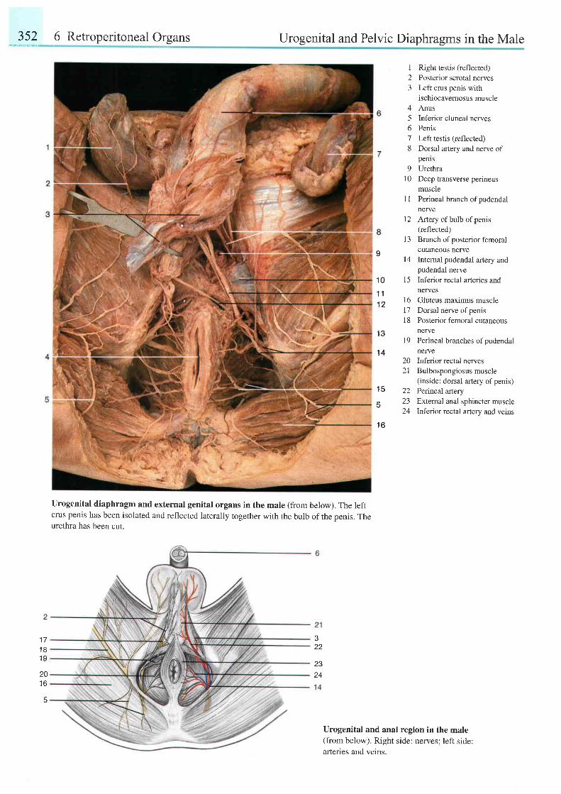
"-:it?6 Retroperitoneal Organs Urogenital and Pelvic Diaphragms in the Male
urogenital diaphragm and external genital organs in the male (from below). The leftcrus penis has been isolated and reflected laterally together with the bulb of the penis. Theurethra has been cut.
123
45678
9l 0
1 1
l 2
1 3
t 4
1 5
Right testis (reflected)Posterior scrotal nervesLeft crus penis withischiocavernosus muscleAnusInferior cluneal nervesPenisLeft testis (reflected)Dorsal artery and nerve ofpenlsUrethraDeep transverse perineusmusclePerineal branch of pudendalnerveArtery of bulb of penis(reflected)Branch of posterior femoralcutaneous nerveInternal pudendal artery andpudendal nerveInferior rectal arteries andnerves
16 Gluteus maximus muscle17 Dorsal nerve ofpenis18 Posterior femoral cutaneous
nervel9 Perineal branches ofpudendal
nerve
I
1 0
1 1l z
1 3
't4
1 5
1 6
20a 1
22z-)
24
Inferior rectal nervesBulbospongiosus muscle(inside: dorsal artery of penis)Perineal arteryExternal anal sphincter muscleInferior rectal artery and veins
J
22
23
1 71 81 9
20t o
5
Urogenital and anal region in the male(from below). Right side: nerves; left side:arteries and veins.
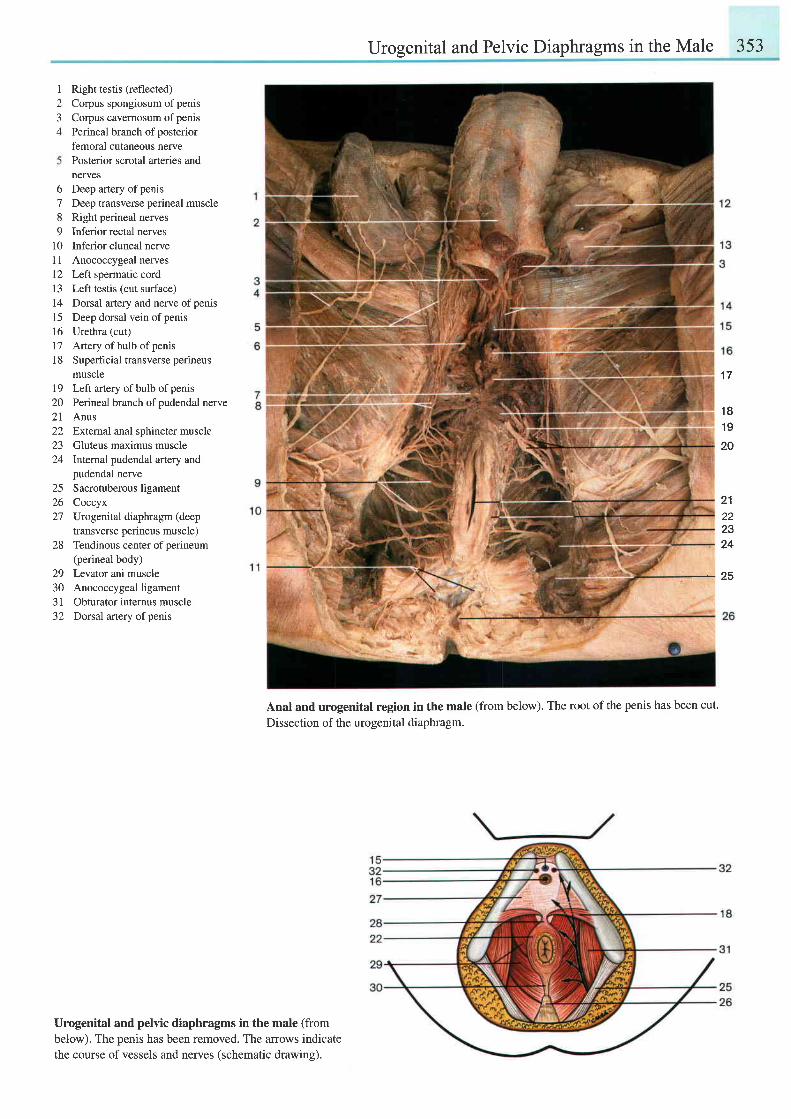
Urogenital and Pelvic Diaphragms in the Male 3s3
I
23A
Right testis (reflected)Corpus spongiosum of penisCorpus cavemosum of penisPerineal branch of posteriorfemoral cutaneous nervePosterior scrotal arteries andnerves
6 Deep artery ofpenis7 Deep transverse perineal muscle8 Right perineal nerves9 Inferior rectal nerves
10 Inferior cluneal nervel1 Anococcygeal nerves12 Left spermatic cord13 Left testis (cut surface)14 Dorsal artery and nerve ofpenisl5 Deep dorsal vein ofpenis16 Urethra (cut)
17 Attery ofbulb ofpenis| 8 Superficial transverse perineus
muscle79 Left artery of bulb of pems20 Perineal branch ofpudendal nerve21 Anus22 Extemal anal sphincter muscle23 Gluteus maximus muscle24 lntemal pudendal artery and
pudendal nerve25 Sacrotuberous ligament26 Coccyx27 Urogenital diaphragm (deep
transverse perineus muscle)28 Tendinous center ofperineum
(perineal body)29 Levator ani muscle30 Anococcygeal ligament31 Obturator internus muscle32 Dorsal anery ofpenis
Urogenital and pelvic diaphragms in the male (frombelow). The penis has been removed. The arrows indicatethe course of vessels and nerves (schematic drawing).
Anal and urogenital region in the male (from below). The root of the penis has been cut.
Dissection of the urogenital diaphragm.
1 7
1 81 9
20
21222324
25
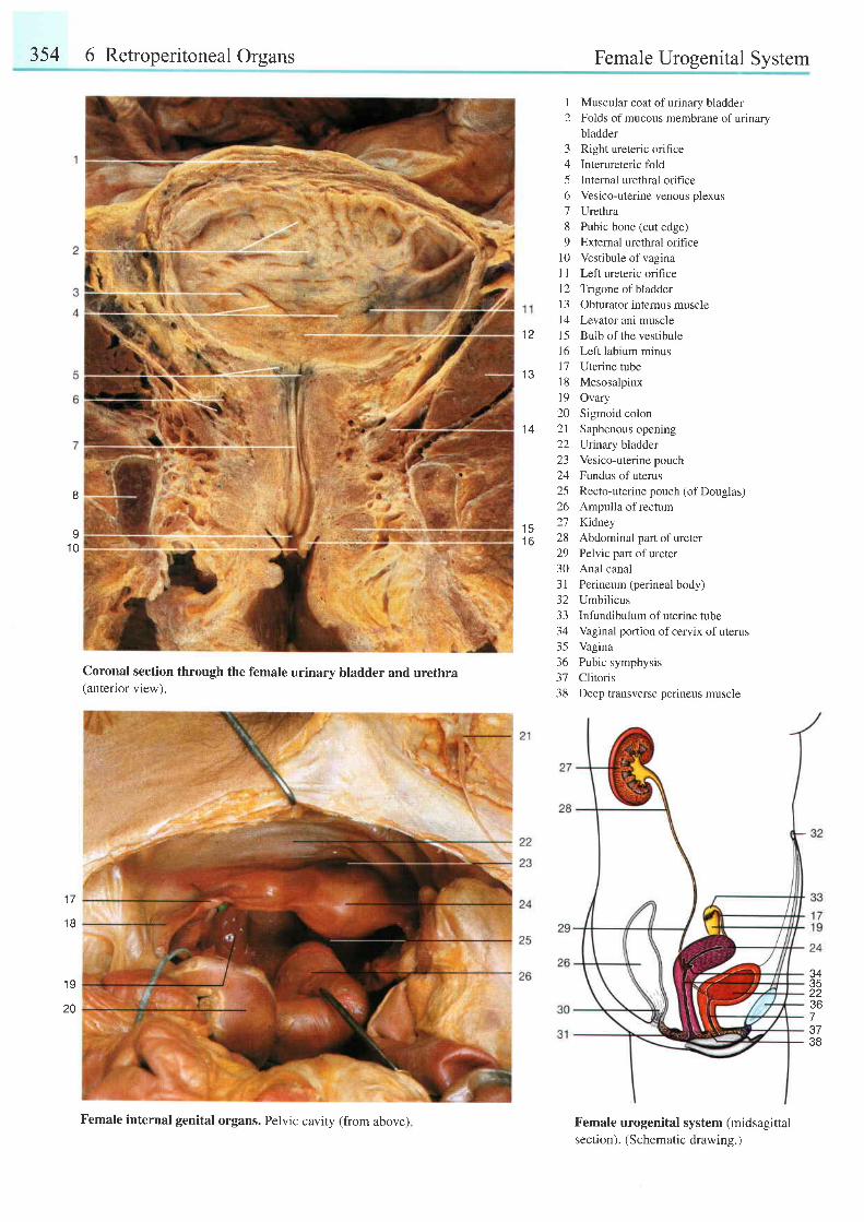
354 6 Retroperitoneal Organs Female Urogenital System
1 4
1 Muscular coat ofurinary bladder2 Folds of mucous membrane of urinary
bladder3 Right ureteric orifice4 Interureteric fold5 Internal urethral orifice6 Vesico-uterine venous plexus7 Urethra8 Pubic bone (cut edge)9 External urethral orifice
10 Vestibule ofvaginaI I Left ureteric orifice12 Trigone ofbladder13 Obturator intemus muscle14 Levator ani muscle15 Bulb of the vestibule16 Left labium minus17 Uterine tubel8 Mesosalpinxl9 Ovary20 Sigmoid colon21 Saphenous opening22 Urrnary bladder23 Vesico-uterinepouch24 Fundus of uterus25 Recto-uterinepouch (of Douglas)26 Ampulla of rectum21 Kidney28 Abdominal part of ureter29 Pelvic part ofureter30 Anal canal31 Perineum (perineal body)32 Umbilicus33 Infundibulum ofuterine tube34 Vaginal portion of cervix of uterus35 Vagina36 Pubic symphysis37 Clitoris38 Deep transverse perineus muscle
I
I1 0
'15
t o
1 7
t 6
' 19
Coronal section through the female urinary bladder and urethra(anterior view).
Female urogenital system (midsagittalsection). (Schematic drawing.)
34
22367J /
38
20
Female internal genital organs. Pelvic cavity (from above)
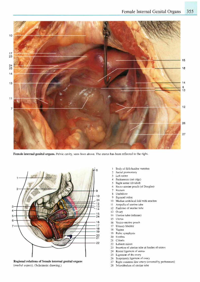
Female Internal Genital Organs 355
1 5
1 8
1 4
61 3
. t ' f
23
2425
1 4
Female internal genital organs. Pelvic cavity, seen from above. The uterus has been reflected to the right.
Regional relations of female internal genital organs(medial aspect). (Schematic drawing.)
I Body of fifth lumbar vertebra2 Sacral promontory3 Left ureter4 Peritoneum (cut edge)5 Right ureter (divided)6 Recto-uterine pouch (ofDouglas)
7 Rectum8 Umbilicus9 Sigmoid colon
10 Median umbilical fold with urachusI 1 Ampulla of uterine tube12 Fimbriae of uterine tube13 Ovary14 Uterine tube (isthmus)15 Uterus16 Vesico-uterine pouch17 Urinary bladder18 Vagina19 Pubic symphysis20 Urethra2l Clitoris22 Labium minus23 Insertion of uterine tube at fundus of uterus24 Round ligament of uterus25 Ligamentoftheovary26 Suspensoryligamentof ovary27 Right common iliac artery (covered by peritoneum)
28 Infundibulum ofuterine tube
I
1 0281 21 31 4
1 524
1 61 71 81 9202'l
22
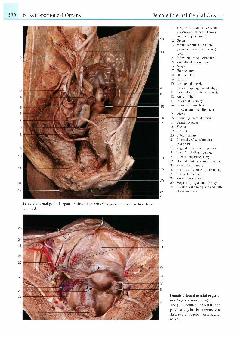
356 6 Retroperitoneal Organs
ilil'4:
Female internal genital organs in situ. Right half of the pelvis and sacrunr havc bcenrernoved
Female Internal Genital Orsans
I Body of t-rfth lurnbal vertebra,
suspensory ligament of ovary,
and sacral promontory26 2 Ilreter
3 Medial umbilical ligament
r e ( rcmnant of umbi l ical ar tery)( cu t )
4 Infundibulum ofuter ine tube
5 Ampul la of uter ine tube
6 Ovary
7 Uterine artery
8 Uterine tube
9 Rectunr
l0 Levator ani muscle
4 (pelvic diaphragm cut edge)
6 I I External anal sphincter muscle
l2 Anus (probe)
I 3 lnternal iliac artery1 4
iS I ,1 Remnant of urachus(median umbi l ical l igament)
Uterus
Round l igament of uterus
Urinary bladder
Vagina
Cl i torrs
Lab ium m inus
External orihce of urethra(red probe)
-' rii;
..1'F :
, ' l1 J
1 51 6 t 61 7 t j
1 8l 9202 l
22 Vaginal oritice (green probe)
23 Lateral urnbi l ical l igament
.1 g 2rl Inf'erior epigastric altery
25 Obturator aftery, vein. and nerve
26 Erternal iliac artery1 I 27 Recto-uterine pouch (of Douglas)
28 Recto-uterine fold
19 Vc\ ieo-uler inc pour 'ha v
30 Su rpenso r l J i gamen t o l - o r l t r y
3 I Greater vestibular gland and hulb
2j o l the vest ibule
22
Female internal genital organsin situ tseen l ' rom above.1.The peritoneurn at the left half ofpelvic cavity has been removed todisplay uterine tube, vessels, andnerves
t o
1 7
6
30
72 7
28
2
Y
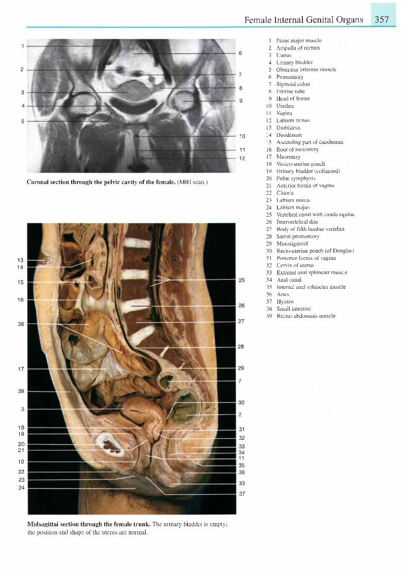
Female Internal Genital Orsans 357
1 0
'1 1
I
23456789
1 0l ll 21 3t 41 5l 61 7l 81 9
Psoas major muscleAmpulla of rectumUterusUrinary bladderObturator intemus musclePromontorySigmoid colonUterine tubeHead of femurUrethraVaginaLabium minusUmbilicusDuodenumAscending part of duodenumRoot of mesenteryMesenteryVesico-uterine pouchUrinary bladder (collapsed)Pubic symphysisAnterior fornix of vaginaClitorisLabium minusLabium majusVeftebral canal with cauda equinaIntervertebral discBody of fifth lumbar vertebraSacral promontoryMesosigmoidRecto-uterine pouch (of Douglas)Posterior fornix of vaginaCervix of uterusExternal anal sphincter muscleAnal canalIntemal anal sphincter muscleAnusHymenSmall intestineRectus abdominis muscle
1 31 4
1 6
38
Coronal section through the pelvic cavity of the female. (MRl scan )
Midsagittal section through the female trunk. The urinary bladder is empty;the position and shape of the uterus are normal.
202 l2223242526272829303 l32333435-1r)
373839
J I
32JJ
J4
1 l
36
a e
37
1 81 9
2021
1 0
22Z J
24
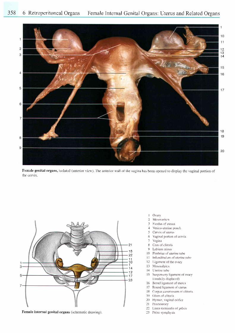
358 6 Retroperitoneal Organs Female Internal Genital Organs: Uterus and Related Organs
Female genital organs, isolated (anterior view). The anterior wall of the vagina has been opened to display the vaginal portion ofthe cervix.
1 l
I L
1 31 4
1 81 0
20
21
1 5zz
1 11 011 41 21 7z5
I
23456'7
89
l 0I I1 21 3t 11 5
Ovary
Mesovarium
Fundus of uterus
Vesico-uterine pouch
Cervix of uterus
Vaginal portion of cervix
Vagina
Crus of clitoris
Labium minus
Fimbriae of uterine tube
Infundibulum of uterine tube
Ligament of the ovary
Mesosalpinx
Uterine tube
Suspensory ligament of ovary(caudally displaced)
Broad ligament of uterus
Round ligament of uterus
Corpus cavernosum of clitons
Glans of clitoris
Hymen, vaginal orifice
Promontory
Linea terminalis of pelvis
Pubic symphysis
l 6l ll 81 9202 l2223Female internal genital organs (schematic drawing).

Female Internal Genital Organs: Uterus and Related Organs 359
1
22
21 2J
1 3
4
I
23456189
t 01 lt 2l 3t 41 5l 6
l 7
1 47
1 51 6
4
Fundus of uterus
Uterine tube
Ligament of the ovary
Ovary
lnfundibulum of uterine tube
Fimbriae of uterine tube
Ureter
Rectum
Apex of urinary bladder and
median umbilical ligament
Urinary bladder
Round ligament of uterus
Mesosalpinx
Mesovarium
Recto-uterine pouch (of Douglas)
Suspensory ligament of ovary
Scarr ing of orary { lb l lowing
ovulation)
Abdominal opening of uterine6
7
8
tube18 Body ofuterusl9 Cervical canal20 Vaginal portion of cervix of uterus
(congestion)21 Vagina22 Mucous membrane of uterus23 Anterior tbrnix of vagina
45
' t8'19
zv
2 1
Female internal genital organs, isolated (superior-posterior view ).
Right ovary and uterine tube, isolated (superior-posterior view). The fimbriae of the
uterine tube have been reflected to show the abdominal ostium.
Uterus and related organs (posterior view). The posterior wall of the uterus has been
onened.
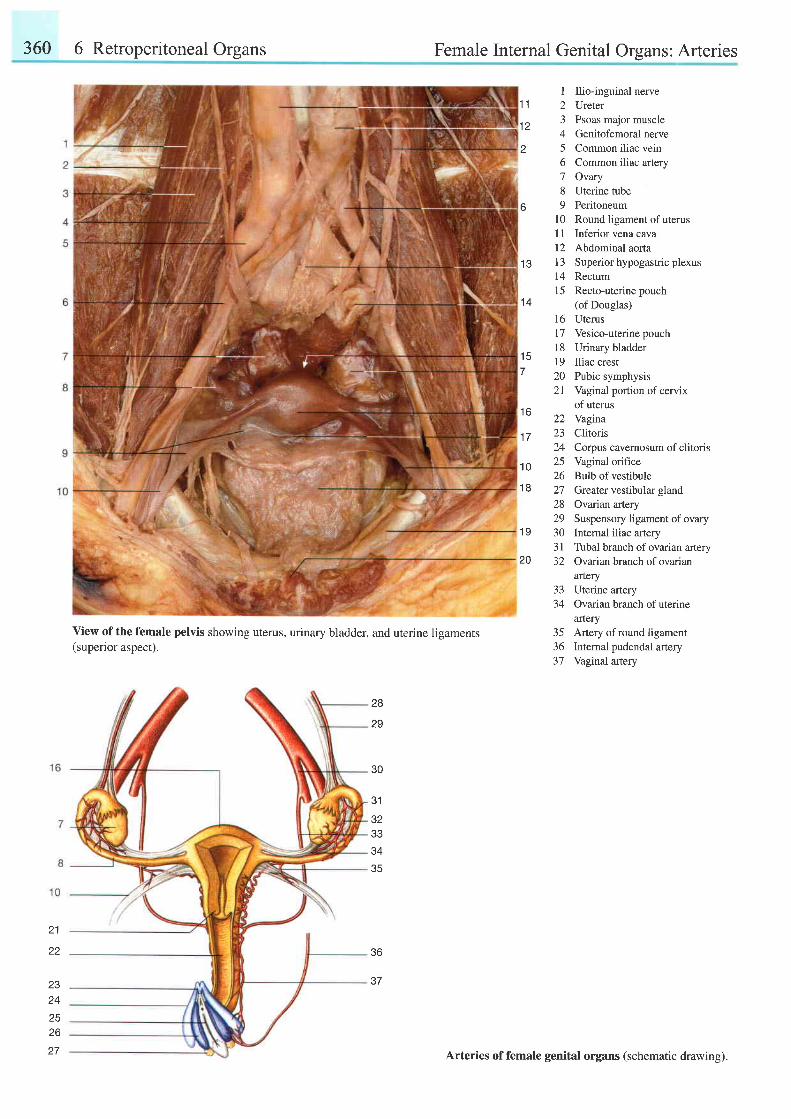
360 6 Retroperitoneal Organs Female Internal Genital Orqans: Arteries
View of the female pelvis showing uterus, urinary bladder, and uterine ligaments(superior aspect).
30
31
32e e
34
35
I Ilio-inguinal nerve1-t 2 Ureter
12 3 Psoas major muscle4 Genitofemoral nerve
2 5 Common iliac vern6 Common iliac artery7 Ovary8 Uterine tube
6 9 Peritoneuml0 Round ligament of uterusI I Inferior vena cava12 Abdominal aorta
1 3 l3 Superior hypogasffic plexus14 Rectum15 Recto-uterine pouch
14 (of Douglas)16 Uterus17 Vesico-uterinepouchl8 Urinarv bladder
15 19 Iliac crest7 z0 Pubic symphysis
21 Vaginal portion ofcervixof uterus
lo 22 Yagina
17 23 Clitoris24 Corpus cavernosum ofclitoris
1O 25 Vaginal orifice26 Bulb of vestibule
18 27 Greater vestibular gland28 Ovarian artery29 Suspensory ligamentofovary
19 30 Internal iliac artery3 | Tubal branch of ovarian anery
20 32 Ovarian branch of ovarianartery
33 Uterine artery34 Ovarian branch ofuterine
artery35 Artery of round ligament36 Internal pudendal artery37 Vaginal artery
28
29
21
22 36
2324
2526
37
Arteries of female genital organs (schematic drawing).
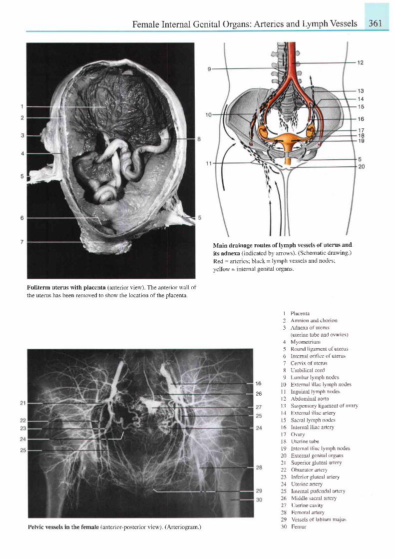
Female Internal Genital Organs: Arteries and Lymph Vessels 361
1 31 41 5
1 6
1 71 81 9
Fullterm uterus with placenta (anterior view). The anterior wall ofthe uterus has been removed to show the location of the placenta.
Main drainage routes of lymph vessels of uterus and
its adnexa (indicated by arrows). (Schematic drawing.)
Red = arteries; black = lymph vessels and nodes;
yellow = internal genital organs.
1 Placenta2 Amnion and chorion3 Adnexa ofuterus
(uterine tube and ovaries)4 Myometrium5 Round ligament of uterus6 Intemal orifice of uterus7 Cervix of uterus8 Umbilical cord9 Lumbar lymph nodes
10 External illac lymph nodes1l Inguinal lymph nodes12 Abdominal aofta13 Suspensory ligamentof ovary14 External iliac artery15 Sacral lymph nodes16 Intemal iliac arteryl7 Ovary18 Uterine tube19 Internal iliac lymph nodes20 External genital organs2l Superior gluteal artery22 Obturaror artery23 Inferior gluteal artery24 Uterine artery25 Internal pudendal artery26 Middle sacral aftery27 Ulerine cavity28 Femoral artery29 Vessels of labium majus30 Femur
1 6
26
Pelvic vessels in the female (anterior-posterior view). (Arteriogram.)
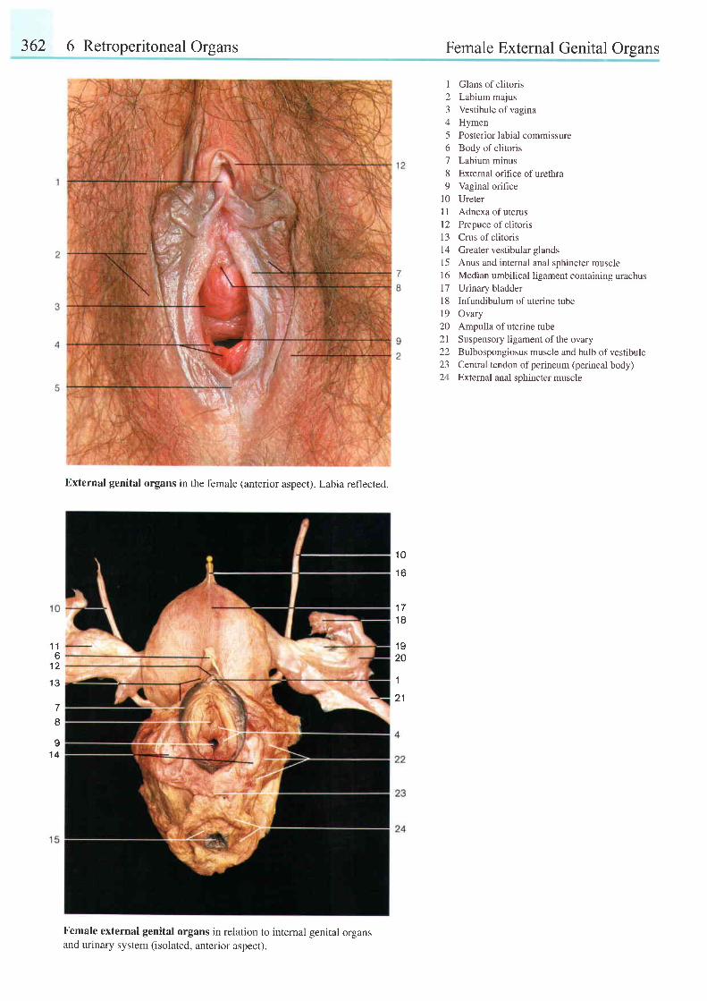
362 6 Retroperitoneal Organs Female External Genital Orsans
1 Glans of clitoris2 Labium majus3 Vestibule ofvagina4 Hymen5 Posterior labial commissure6 Body of clitoris7 Labium minus8 External orifice ofurethra9 Vaginal orifice
10 UreterI 1 Adnexa of uterusl2 Prepuce ofclitoris13 Crus of clitoris14 Greatervestibular glands15 Anus and internal anal sphincter muscle16 Median umbilical ligament containing urachus17 Urinary bladderl8 Infundibulum ofuterine tube19 Ovary20 Ampulla of uterine tube21 Suspensory ligament ofthe ovary22 Bulbospongiosus muscle and bulb ofvestibule23 Central tendon ofperineum (perineal body)24 Exlernal anal sphincter muscle
1 16
1 2
1 3
7I
I1 4
External genital organs in the female (anterior aspect). Labia reflected.
Female external genital organs in relation to internal genital organsand urinary system (isolated, anterior aspect).
1 0
1 6
1 ' l
1 8
1 920
1
2 1
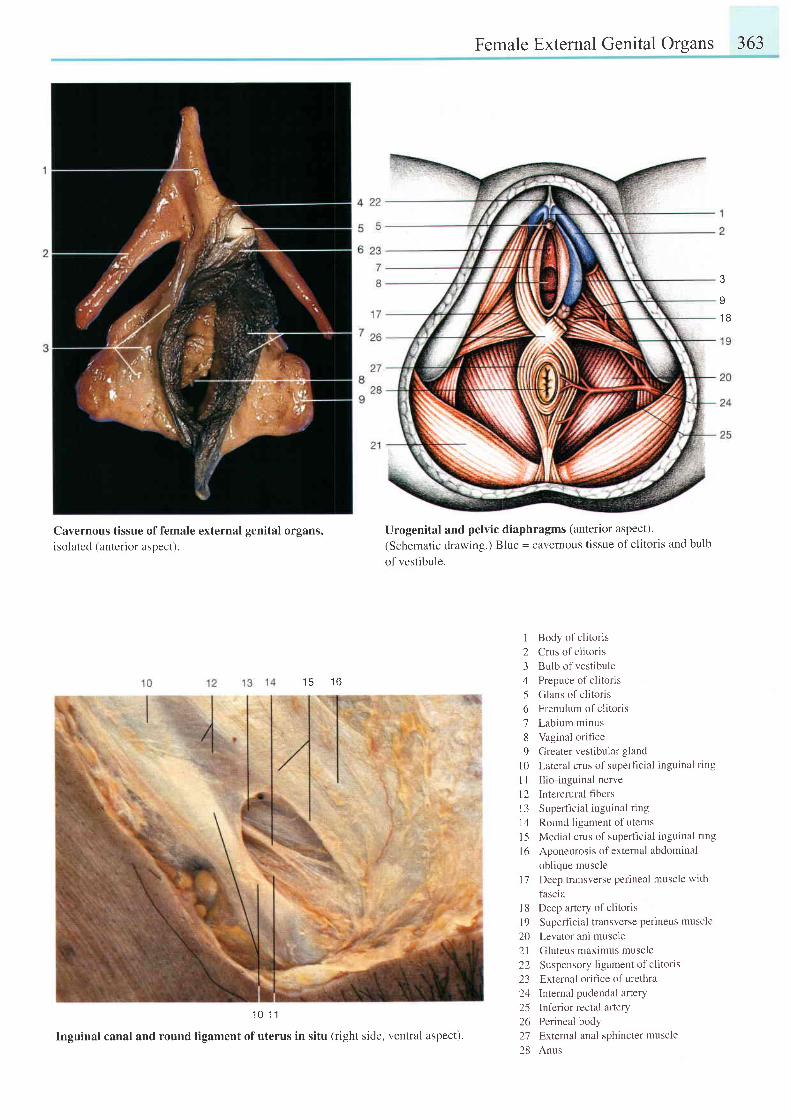
Female External Genital Organs 363
3
v
1 B
Cavernous tissue of female external genital organs,isolated (anterior aspect).
1 5 1 6
1 0 1 1
Inguinal canal and round ligament ofuterus in situ (right side, ventral aspect).
Urogenital and pelvic diaphragms (anterior aspect).
(Schematic drawing.) Blue = cavernous tissue of clitoris and bulb
of vestibule.
I
2345o'7
89
l 0l lI 2l 31 4l 5l 6
Body of clitorisCrus of clitorisBulb of vestibulePrepuce of clitorisGlans of cl i tor isFrenulum of clitorisLabium minusVaginal orificeGreater vestibular glandLateral crus of superficial inguinal ringIlio-inguinal nerveIntercrural f-rbersSuperficial inguinal ringRound ligament of uterusMedial crus of superficial inguinal ringAponeurosis of external abdominaloblique muscleDeep transverse perineal muscle withfascia
I 8 Deep artery of clitoris
l9 Superficial transverse perineus muscle
20 Levator ani muscle
21 Gluteus maximus muscle
22 Suspensory ligament of clitoris
23 External orifice of urethra
24 lnternal pudendal ar ter l
25 lnferior rectal artery
26 Perineal body
27 External anal sphincter muscle
28 Anus
t 7
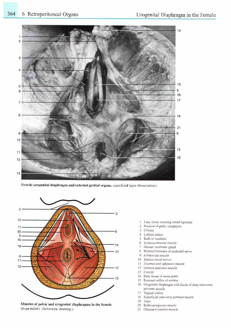
364 6 Retroperitoneal Organs Urogenital Diaphragm in the Female
1 5
61 6
I I
Female urogenital diaphragm and external genital organs, superficial layer (from below).
1 720
tt1 6
1 8
Muscles of pelvic and urogenital diaphragms in the female(from below). (Schematic drawins.)
I Fatty tissue encasing round ligament2 Position of pubic symphysis3 Clitoris4 Labium minus5 Bulb of vestibule6 Ischiocavernosusmuscle7 Greater vestibular gland8 Perineal branches ofpudendal nerve9 Levator ani muscle
l0 Inferior rectal nervesI I External anal sphincter musclel2 Gluteus maximus muscle13 Coccyxl4 Fatty t issue o[ mons pubisl5 External orifice of urethra16 Urogenital diaphragm with fascia ofdeep transverse
perineus musclel7 Vaginal orificel8 Superficial transverse perineal musclel9 Anus20 Bulbospongiosusmuscle21 Obturator internus muscle
1 1
1 2
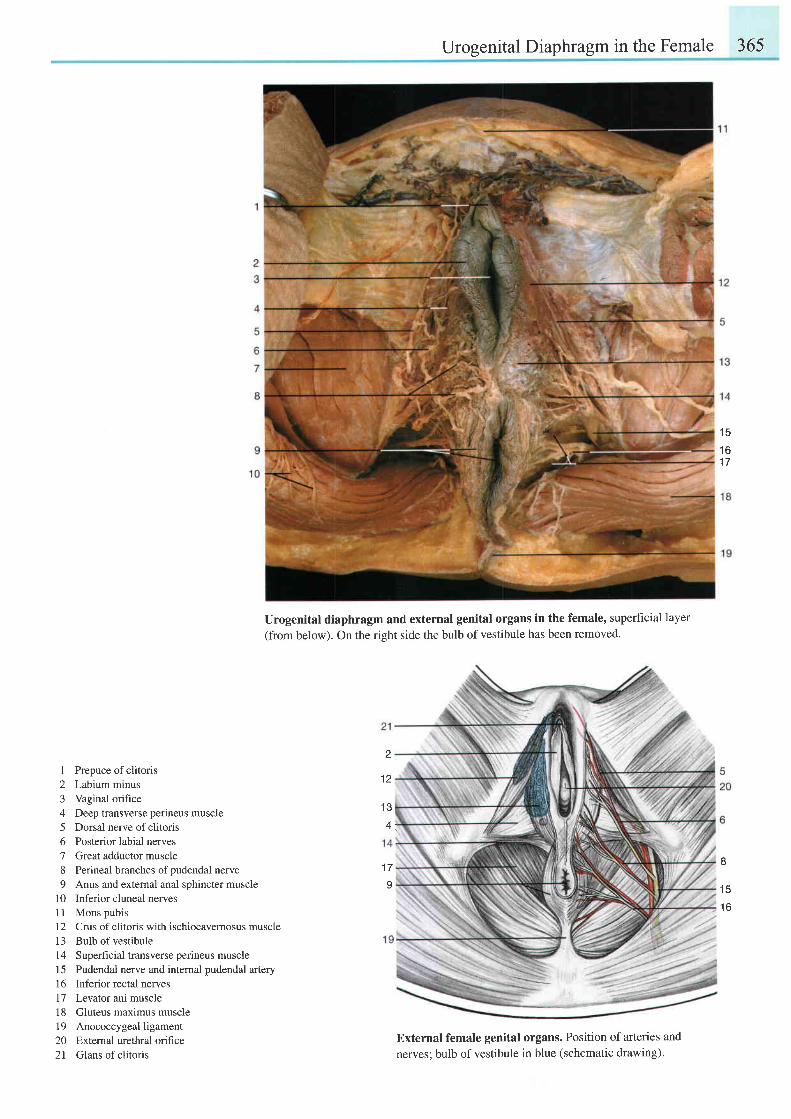
Urogenital Diaphragm in the Female 365
1 5
1 61 7
1 Prepuce ofclitoris2 Labium minus3 Vaginal orifrce4 Deep transverse perineus muscle5 Dorsal nerve of clitoris6 Posterior labial nerves7 Great adductor muscle8 Perineal branches ofpudendal nerve9 Anus and extemal anal sphincter muscle
l0 Inferior cluneal nervesl l Mons pubis12 Crus of clitoris with ischiocavernosus muscle13 Bulb of vestibule14 Superficial transverse perineus muscle15 Pudendal nerve and intemal pudendal artery16 Inferior rectal nerves17 Levator ani muscle18 Gluteus maximus muscle19 Anococcygealligament20 External urethral orifice21 Glans of clitoris
Urogenital diaphragm and external genital organs in the female, superficial layer(from below). On the right side the bulb of vestibule has been removed.
1 7
I
External female genital organs. Position of arteries and
nerves; bulb of vestibule in blue (schematic drawing).
2
1 2
I
1 5
1 6
l e
4 ; .
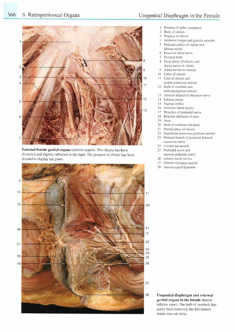
366 6 Retroperitoneal Organs Urogenital Diaphragm in the Female
External female genital organs (inferior aspect). The clitoris has beendissected and slightly reflected to the right. The prepuce of clitoris has beendivided to display the glans.
I Position of pubic symphysis2 Body of clitoris3 Prepuce of clitoris4 Adductor longus and gracilis muscles5 External orifice ofvagina and
labium minus
Posterior labial nerve
Perineal body
Deep arlery of clitoris and
dorsal nerve of clitoris
Adductor brevis muscle
Glans of slitoris
Crus of clitoris and
ischiocavernosus muscle
Bulb of vestibule and
bulbospongiosus muscle
13 Anterior branch ofobturator nervel4 Labium minusl5 Vaginal orificel6 Posterior labial nervesl7 Branches ofpudendal nerve18 External sphincterof anusl9 Anus20 Bulb of vestibule (divided)2l Dorsal artery of clitoris22 Supefficial transverse perineus muscie23 Perineal branch ofposterior femoral
cutaneous nerve
Levator ani muscle
Pudendal nerve and
internal pudendal aftery
Inferior rectal nerves
Gluteus maximus muscle
Anococcygeal ligament
Urogenital diaphragm and externalgenital organs in the female (lateral
inferior view). The bulb of vestibule haspartly been removed; the left labiummlnus was cut away.
6'7
8
91 0I l
1 2
2425
262728
22
232425
26
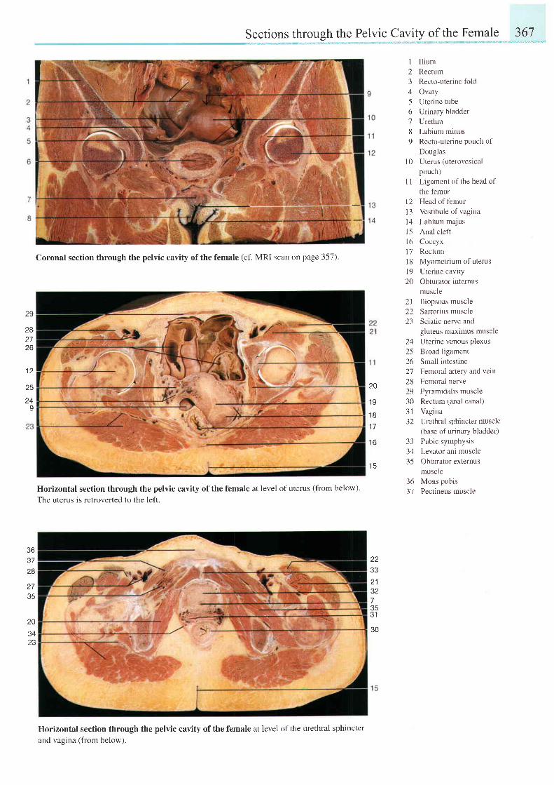
Sections through the Pelvic Cavity of the Female 367
l 0
I
234
6789
21 Iliopsoas muscle22 Sartorius muscle23 Sciatic nerve and
a l l
2526272829303 l32
333435
36 Mons pubis37 Pectineus muscle
IIiumRectumRecto-uterine foldOvaryUterine tubeUrinary bladderUrethraLabium minusRecto-uterine pouch ofDouglasUterus (uterovesicalpouch)Ligament of the head ofthe femurHead of femurVestibule of vaginaLabium majusAnal cleftCoccyxRectumMyometrium of uterusUterine cavityObturator internusmuscle
l t
t 2t 3l 41 51 6l 71 8l 920
29
2827Z D
1 2
25
24I
Coronal section through the pelvic cavity of the female (cf. MRI scan on page 357).
Horizontal section through the pelvic cavity of the female at level of uterus (from below).
The uterus is retroverted to the left.
Horizontal section through the pelvic cavity of the female at level of the urethral sphincter
and vagina (from below).
gluteus maximus muscleUterine venous plexusBroad ligamentSmall intestineFemoral artery and veinFemoral nervePyramidalis muscleRectum (anal canal)VaginaUrethral sphincter muscle(base of urinary bladder)Pubic symphysisLevator ani muscleObturator externusmuscle
20
1 9
1 8
1 7
1 6
1 5
363728
2735
zz
213273531
en20
Z J
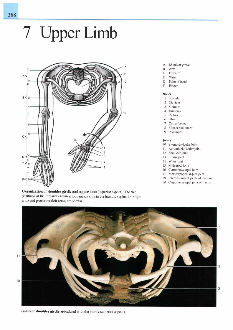
368
7 lJpper Limb
1 4
1 5t o
1 7
1 8
A Shoulder girdleB ArmC ForearmD WristE Palm ofhandF Finger
Bones1 Scapula2 Clavicle3 Sternum4 Humerus5 Radius6 Ulna7 Carpal bones8 Metacarpal bones9 Phalanges
Joints10 Stemoclavicularjointl l Acromioc lav icu la r jo in rl2 Shoulderjointl3 Elbowjoint14 Wrist joint15 Midcarpal joint16 Carpometacarpal joint17 Metacarpophalangeal jointI 8 Interphalangeal joints of the handl9 Carpometacarpal joint of thumb
r-Da,rJ"-1"
Organization of shoulder girdle and upper limb (superior aspect). The twopositions of the forearm essential to manual skills in the human, supination (rightarm) and pronation (left arm). are shown.
Bones ofshoulder girdle articulated with the thorax (superior aspect).
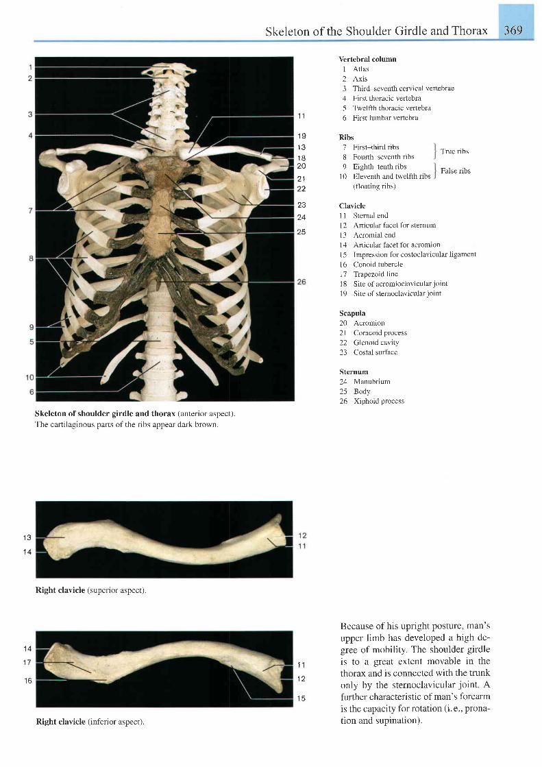
Skeleton of the Shoulder Girdle and Thorax 369
1 1
1 9t Q
1 8ZU
22
Vertebral column1 Atlas2 Axis3 Third-seventhcervicalvertebrae4 First thoracic veftebra5 Twelfth thoracic vertebra6 First lumbar vertebra
Ribs7 First-third ribs8 Fourth-seventh ribs9 Eighth-tenth ribs
10 Eleventh and twelfth ribs(floating ribs)
Clavicle
True ribs
False ribs
1 1 Sternal end.12
Articular facet for sternuml3 Acromial endl4 Art icular facet for acromionl5 Impression for costoclavicular ligament16 Conoid tubercle17 Trapezoid line18 Site of acromioclavicularjointl9 Site of stemoclavicular joint
Scapula20 Acromion2l Coracoid process22 Glenoid cavity23 Costal surface
Sternum24 Manubrium25 Body26 Xiphoid process
Because of his upright posture, man'supper limb has developed a high de-gree of mobility. The shoulder girdleis to a great extent movable in thethorax and is connected with the trunkonly by the sternoclavicular joint. Afurther characteristic of man's forearmis the capacity for rotation (i. e., prona-tion and supination).
23
24
25
I Q
1 4
Skeleton of shoulder girdle and thorax (anterior aspect).The cartilaginous parts of the ribs appear dark brown.
Right clavicle (superior aspect).
1 1
l z
1 4
1 7
t o
Right clavicle (inferior aspect).
I E
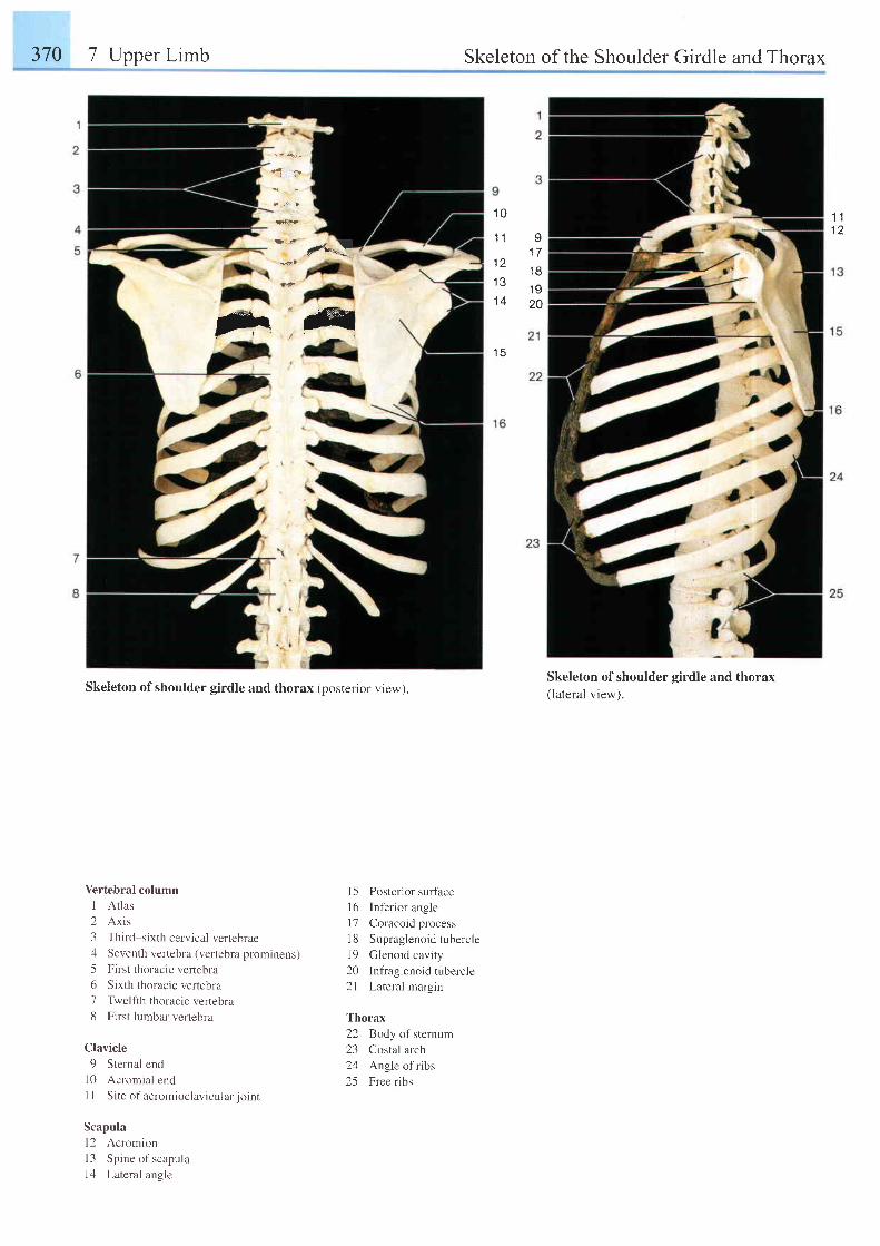
370 7 Upper Limb Skeleton of the Shoulder Girdle and Thorax
drt4^4-I
1 11 2
1 0
1 {
t 1
1 a
1 4
1 5
v1 71 8i O
20
flr,try
I 5 Posterior surfaceI 6 Inferior anglel7 Coracoid process18 Supraglenoid tuberclel9 Glenoid cavity20 Infraglenoidtubercle21 Lateral margin
Thorax22 Body ofsternum23 Costal arch24 Angle of ribs25 Free ribs
Skeleton of shoulder girdle and thorax(lateral view).Skeleton of shoulder girdle and thorax (posterior view).
Vertebral column
I At las
2 Axis
3 Third-sixth cervical verlebrae4 Seventh vertebra (vertebra prominens)
5 First thoracic vertebra
6 Sixth thoracic vertebra
7 Twelfth thoracic vertebra
8 First lumbar vertebra
Clavicle
9 Sternal end
10 Acromial endI I S i t e o f ac rom ioc la r i cu l a r j o i n t
Scapula
l2 Acromion
I 3 Spine of scapula
14 Lateral angle
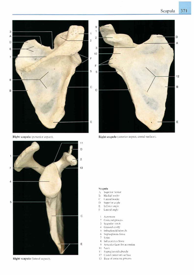
Scapula 371
Right scapula (postel iur aspect) Right scapula (anterior aspect. costal surfacc)
Scapula
A Supcr ior bordct
B Medial border
C Lateral bordet'
I ) Supcr ior anglc
E lnf'erior angle
F [ -atcra] angle
I Acr-omion
2 C-'uracoid process
i Scapu la r no t ch.1 ( i lenoic l car i ty
-5 lnf raglcnoidtuberclc(r Supraspinous tossa
7 Sp ine
l l ln f raspinous lbssa
9 Ar-ticr-rlar facet lbr acroltllon
l 0 Neck
I I Sr-rpraglcnoid tubercle
| 2 Costal (xnter ior) sur lacc
I 3 Base ol 'coracoid processRight scapula ( latelal aspect)
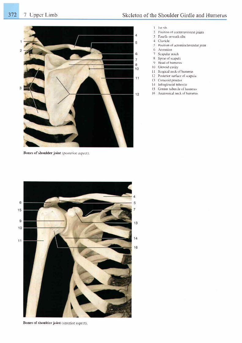
372 7 Upper Limb Skeleton of the Shoulder Girdle and Humerus
6
7I1 0
1 lst r ib2 Position of costotransverse joints3 Fourth-seventh ribs4 Clavicle5 Position of acromioclavicularjoint6 Acromion7 Scapular notch8 Spine of scapula9 Head of humerus
l0 Glenoid cavityI 1 Surgical neck ofhumerus12 Posterior surface of scapulaI 3 Coracoid process14 Infraglenoid tuberclel5 Greater tubercle of humerusl6 Anatomical neck of humerus
Bones of shoulder joint (posterior aspect).
Bones of shoulder joint (anterior aspect).
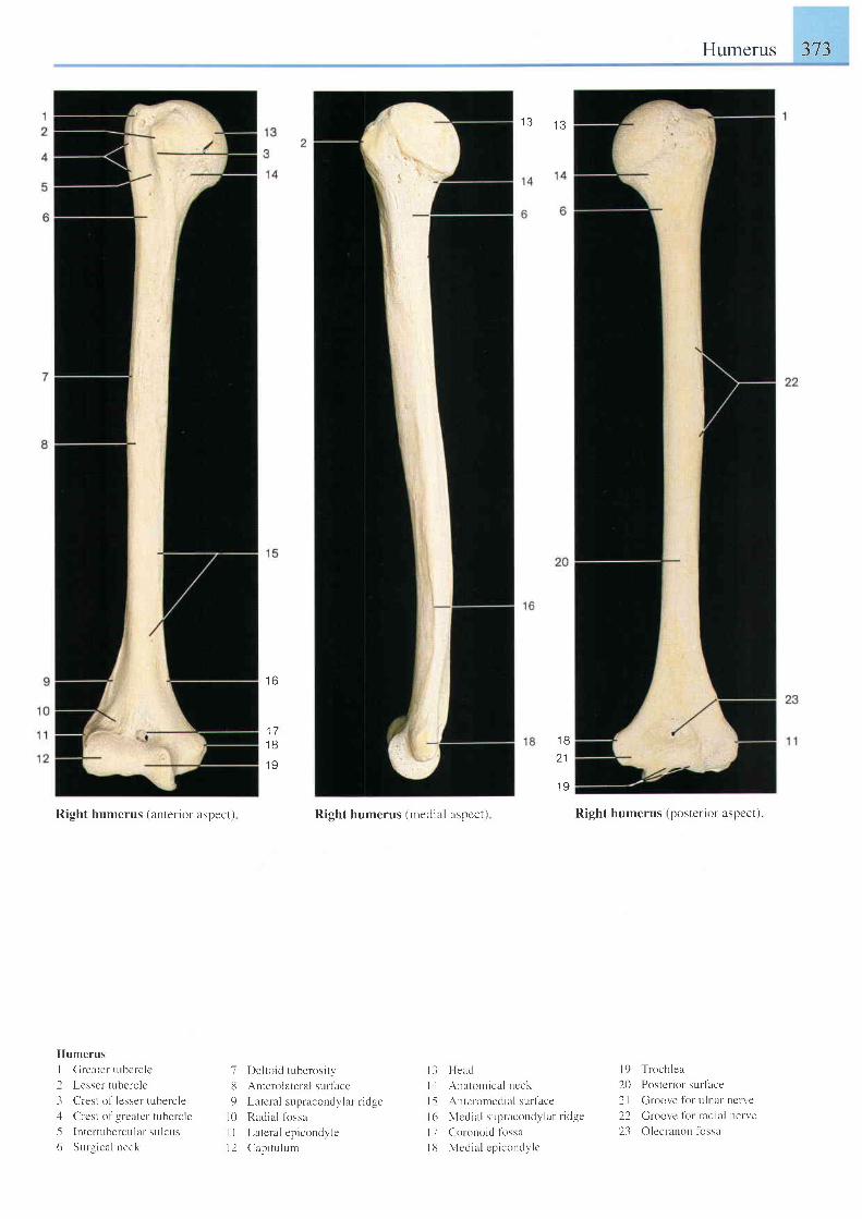
Humerus 373
1 31 3
Right humerus (anterior aspect)
Humerus
I Clreater tubcrc lc
2 Lesser tubercle
3 Crest of lessel tubelc le
4 Crcst o1'grcater t r - rberc le
5 lnter tubercular sulcus
6 Sur'-qical neck
1 6
1 71 B
1 9
7 Del to id tuberosi ty,
lJ Anterolateralsur l lceg Lr le| iL l :upraeonclr l r I r ic lge
l0 Radial lbssa
I I Lateral epicondyle
l 2 Cap i t u l um
Right humerus (medial aspcct) Right humerus (posterior aspect).
l9 Trochlea
20 Postcr iot 'sur iace
21 Groove for u lnat-nctve
22 Groove for radial nerve
23 Olecranon tbssa
1 Ba 1
1 9
l - l I lerLd
lJ . \ ratornical ncck
l-5 .Autcrolnedial sur face
I ( r N l c t l r i r l \ un r i r eonJ ) l a r l i . l ge
l7 Cor-onoid fossa
l8 l \ {ecl ia l cpicondylc
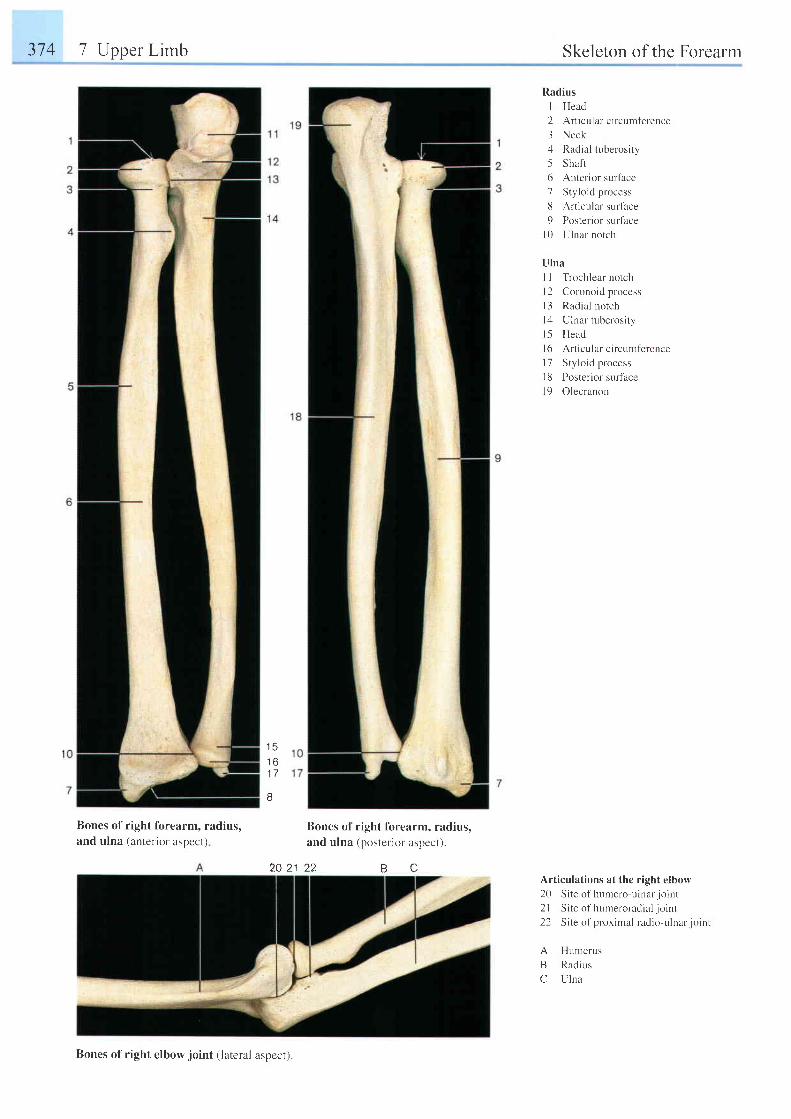
374 7 Upper Lirnb Skeleton of the Forearm
Radius
I Head
2 Art icular c i rcumference
3 Neck
4 Radial tuberosity
5 Shafi
6 Anterior surf'ace
7 Sty lo id process
8 Art icular surtace
9 Posterior surfhce
l0 U lna l no t ch
Ulna
I I Trochlear notch
I 2 Coronoid process
l3 Radial notch
I ' l Ulnar tuberosi ty
l -5 Head
l6 Art icular c i rcumf 'erence
l7 Sty lo id process
I 8 Posterior surface
l9 Olecranon
I D
t o
1 7
8
Bones of right forearm, radius,and ulna (anterior aspect)
Bones of right fbrearm, radius,and ulna (posterior aspect).
20 21 22 cBArticulations at the right elbow20 Site of humero-ulnar. joint2l Site of humeror-adial joint22 Site of proximal radio-ulnar joint
A HumerusB Radrr,rsC U lna
Bones of right elbow joint (lateral aspecr).
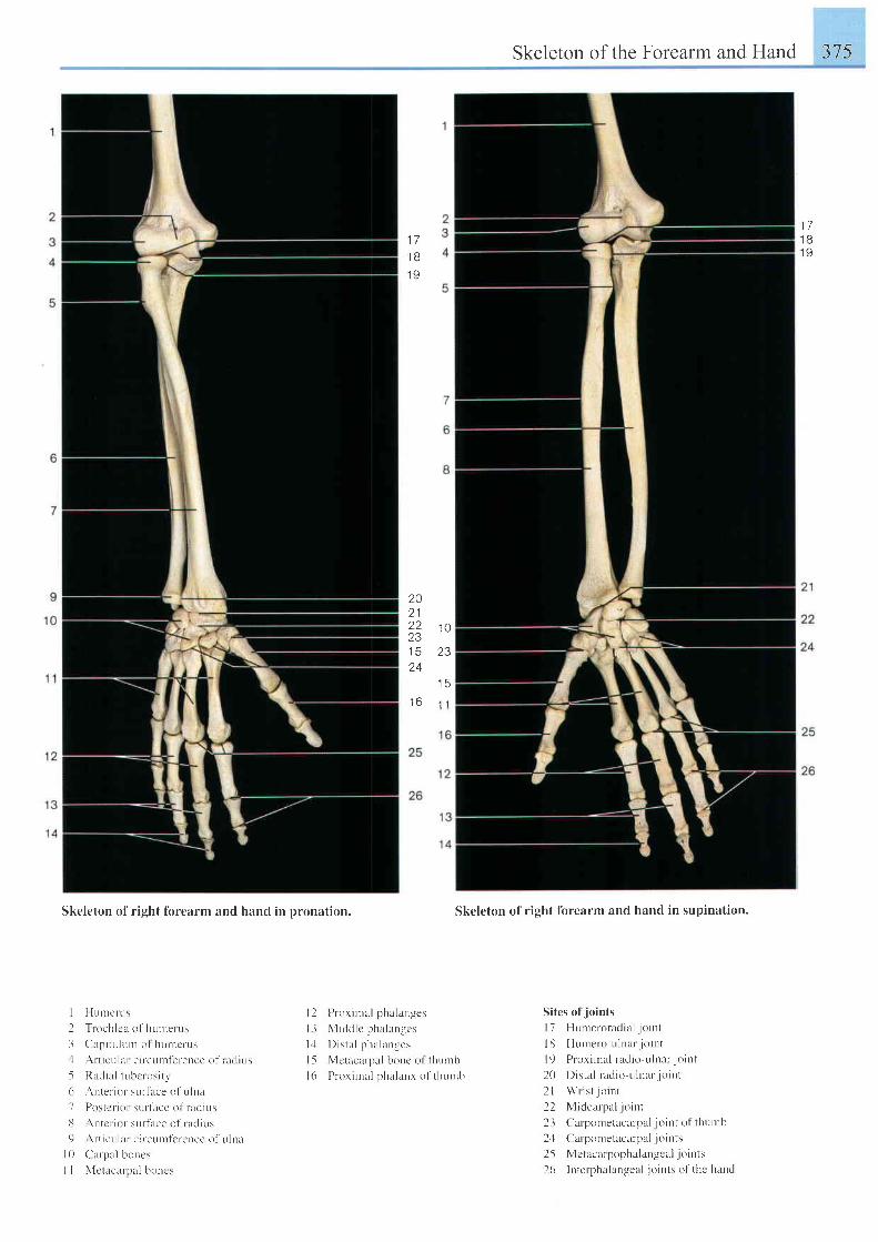
Skeleton of r ight fbrearm and hand in pronation.
I I IunrerLrs
I l r oeh l ca o l humerus
3 C lp i t uJun r o l hu r r c rus
J A r t i cu l a r c i r cum le lence o f r ac l i us
5 Radral lube'r osi t l '( r Anter ior sLrr l i rce of u lna
7 Postcr- ior sLrr tuce of radius
U ' \ntcr ior sur- t lce ol r acl i t rs
9 ' \ r - t icrr lar c i rcunrterence of t r ln l
l 0 Ca rpa l bo r res
I I \ ' [e t rcar- ; l r l boncs
Skeleton of the Forearm and Hand 375
Skcleton of right forearm and hand in supination.
Sites of jo ints
l7 Hr,rnreroradia l - jo int
l lJ Flumero r , r lnar jo i r t t
l 9 P rox i r na l r ad io -u l na r . j o i n t
20 Dista l racl io-uluar. jo i r r t
2 l W r i s t j o i n r
22 Mrdcalpal jo int
23 Carpometacarpal jo inr of thunrt ' r
2 ' l Calponretacarpal . io ints
25 Me tac l r r popha lungc ; r l . i o i t t t .
26 Interphalangeal . jo ints o l thc hand
1 7
1 B1 9
1 71 B1 9
20o 1
2 2 1 0231 5 2 324
I 5t b | 1
| 2 [ ) ror iural phalanges
l3 M idd l c pha lan rcs
l : l I ) is ta l phalanges
l-5 Mctacar-pal bonc ol thr : ruh
1(r I ' ror i rnal phalanx of thrrrnb
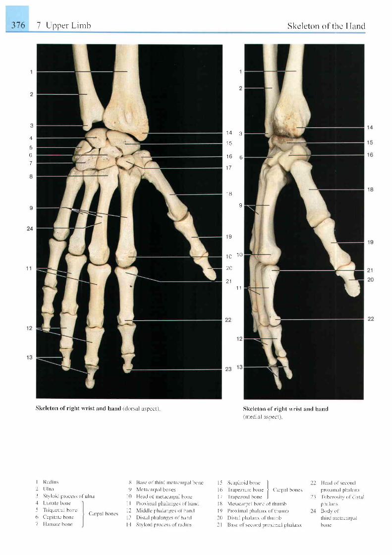
376 7 Upper L imb
Skeleton of right wrist and hand (dorsal aspect)
I Racl ius
2 Ll lna
3 Stv lo id proccss of u ln l
1 4 3
l c
1 6 6
1 7
1 B
v
1 9
1 O 1 0
. A
a 1
'I 1
l -5 Scaphoid bonc II t , I n rpez i r r r u h , , r c
| ( ; r r ' | r r l h , ' r r , .
I 7 Tn rn t - z , r i d honc Jl l l N ' lc tacar-pal bone of thumb
l9 P r ox i n ra l pha lanx o1 t hu r rL r
l 0 D i : t a l pha lanx o f t l r umb
2 I Basc of seconcl proxiural phalanr
Skeleton of r ight wrist and hand(rnecl ial asDect)
Skeleton of the Hand
t 1 Flcucl o l scconr l
p r -o r i nu r l ph l L l a r r r
Tubc ros i t r o l i l i s t u l
;.rilLlan r
Bor l r o l
th i r -d nrctacar pul
bonc
t3
56
7
. l Lunate bone
-5 'friquctr
al Lroue
6 Capi tate bonc
7 Harrate bonc
d
9t 0il
t 21 3t .1
Base of th i rd r .nctacarpal honr.
Metacarpal borres
Heacl of nretacurpal bonc
ProximlLl phalanccs ol hanr l
Middlc phalanges ol ' hant l
Dista l phalangcs ol hanr l
St1 ' lo id proccss ol radius
Carpal boncs
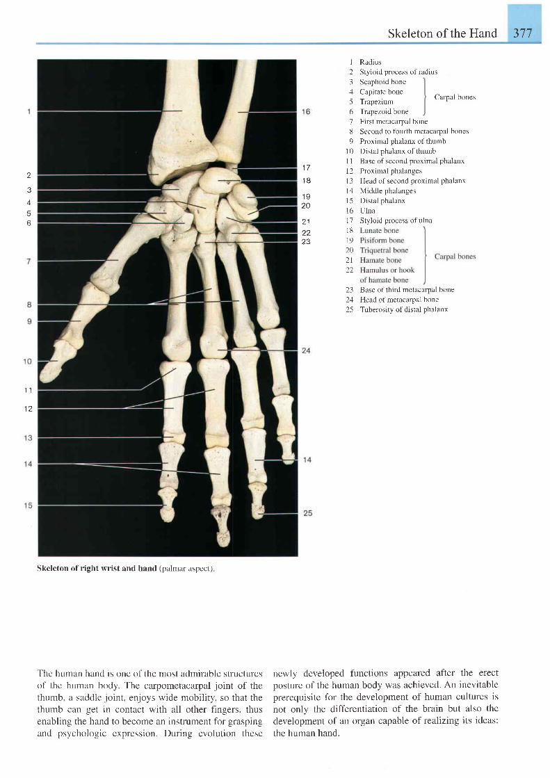
Skeleton of the Hand 1 a - l) t l
2
456
1 7
1 8
1 920
2 1
2,7
1234
6l89
1 0l ll 2l 3t 4l 5l 6r '7l 8\ 9202 l22
231 /
25
RadiusStyioid process of radiusScaphoid bone
Capitate bone
Trapezium
Trapezoid bone
Carpal bones
First metacarpal bone
Second to fourth metacalpal bones
Proximal phalanx of thumb
Distal phalanx of thumb
Base of second proximal phalanx
Proximal phalanges
Head of second proximal phalanx
Middle phalanges
Distal phalanx
Ulna
Styloid process of ulna
Base of third metacarpal bone
Head of metacarpal bone
Tuberosity of distal phalanx
1 l
12
Skeleton of right wrist and hand (palmar aspect)
The human hand rs one of the most admirable structuresof the human body. The carpometacarpal joint of thethumb, a saddle joint, enjoys wide mobility, so that thethumb can get in contact with all other fingers, thusenabling the hand to become an instrument for graspingand psychologic expression. During evolution these
newly developed functions appeared after the erectposture of the human body was achieved. An inevitableprerequisite for the development of human cultures isnot only the differentiation of the brain but also thedevelopment of an organ capable of realizing its ideas:the human hand.
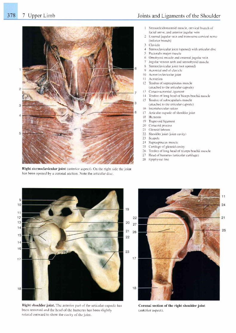
378 7 Upper Limb Joints and Ligaments of the Shoulder
I Sternocleidomastoid muscle, cervical branch of
lacial nerve, and anteriorjugular vein
2 External iugular vein and transverse cervical nerve(inferior branch)
Clavicle
Sternoclavicular joint (opened) with articular disc
Pectoralis major muscle
Omohyoid muscle and external jugular vein
Jugular venous arch and sternohyoid muscle
Sternoclavicular joint (not opened)
Acromial end of clavicle
Acromioclavicular joint
Acromron
Tendon of supraspinatus muscle
(attached to the articular capsule)
Coraco-acromial ligament
Tendon of long head of biceps brachii muscle
Tendon of subscapularis muscle
(attached to the articular capsule)
lntertubercular sulcus
An ie u l a r capsu le o f shou lde r j o i n t
Humerus
Trapezoid ligament
Coracoid process
Clenoid labrum
Shoulder joint (oint cavity)
Scapula
Supraspinatus muscle
Cartilage of glenoid cavity
Tendon of long head of triceps brachii muscle
Head of humerus (articular cartilage)
Epiphysial line
3456'7
6
9l 0l lt 2
l 3t 41 5
l 61 7l 81 9202 l22L-)
2425/.o
2128
Right sternoclavicular joint (anterior aspect).has been opened by a coronal section. Note the
On the right side the joint
articular disc.
I1 0
1 1
1 2I J
1 4
1 5
1 4
t o
1 7
22
27
28
Right shoulder joint. The anterior part of the articular capsule hasbeen removed and the head of the humerus has been slishtlvrotated outward to show the cavitv of the ioint.
Coronal section of the right shoulder joint(antenor aspect).
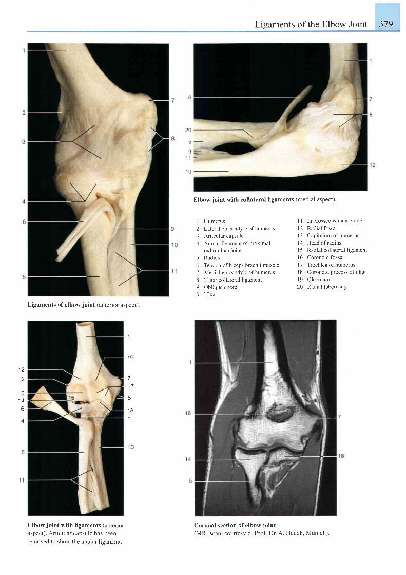
Lisaments of the Elbow Joint 379
Elbow joint with collateral ligaments (medial aspect)
1 2
1 1
1 4
6
4
Ligaments of elbow joint (anterior aspect)
1 6
71 7
I
1 B6
Elbow joint with ligaments (anterior
aspect). Articular capsule has beenremoved to show the anular ligament.
I Humerus
2 Lateral epicondyle of humerus
3 Art icular capsule
4 Anular l igament of proximal
radio-ulnar jo int
5 Radius
6 Tendon of biceps brachii muscle
7 Medial epicondyle of humerus
8 Ulnar col lateral l igament
9 Obl ique chord
l 0 U l n a
Coronal section of elbow joint
(MRI scan, courtesy of Prof Dr. A. Heuck, Munich).
I I Interosseous membrane
l2 Radial fossa
l3 Capitulum of humerus
l4 Head ofradius
l5 Radial collateral ligament
l6 Coronoid fossa
l7 Trochlea of humerus
I 8 Coronoid process of ulna
l9 Olecranon
20 Radial tuberosity
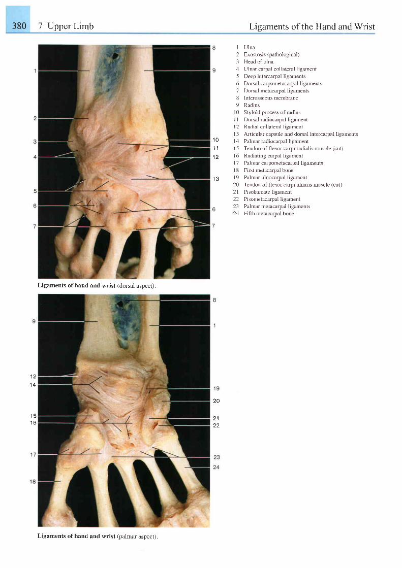
380 7 Upper Limb Liqaments of the Hand and Wrist
I Ulna2 Exostosis(pathological)3 Head of ulna4 Ulnar carpal collateral ligament5 Deep intercarpal ligaments6 Dorsal carpometacarpal ligaments7 Dorsal metacarpal ligaments8 Interosseousmembrane9 Radius
10 Styloid process ofradiusI 1 Dorsal radiocarpal ligamentl2 Radial collateral ligamentI 3 Anicular capsule and dorsal intercarpal ligamentsl4 Palmar radiocarpal ligament15 Tendon offlexor carpi radialis muscle (cut)16 Radiating carpal ligament17 Palmarcarpometacarpal ligaments18 First metacarpal bone19 Palmar ulnocarpal ligament20 Tendon offlexor carpi ulnaris muscle (cut)21 Pisohamate ligament22 Pisometacarpal ligament23 Palmar metacarpal ligaments24 Fiflh metacarpal bone
1 21 4
1 51 6
1 01 1
1 2
i 2
2A
2 122
Ligaments of hand and wrist (dorsal aspect).
Ligaments of hand and wrist (palmar aspect).
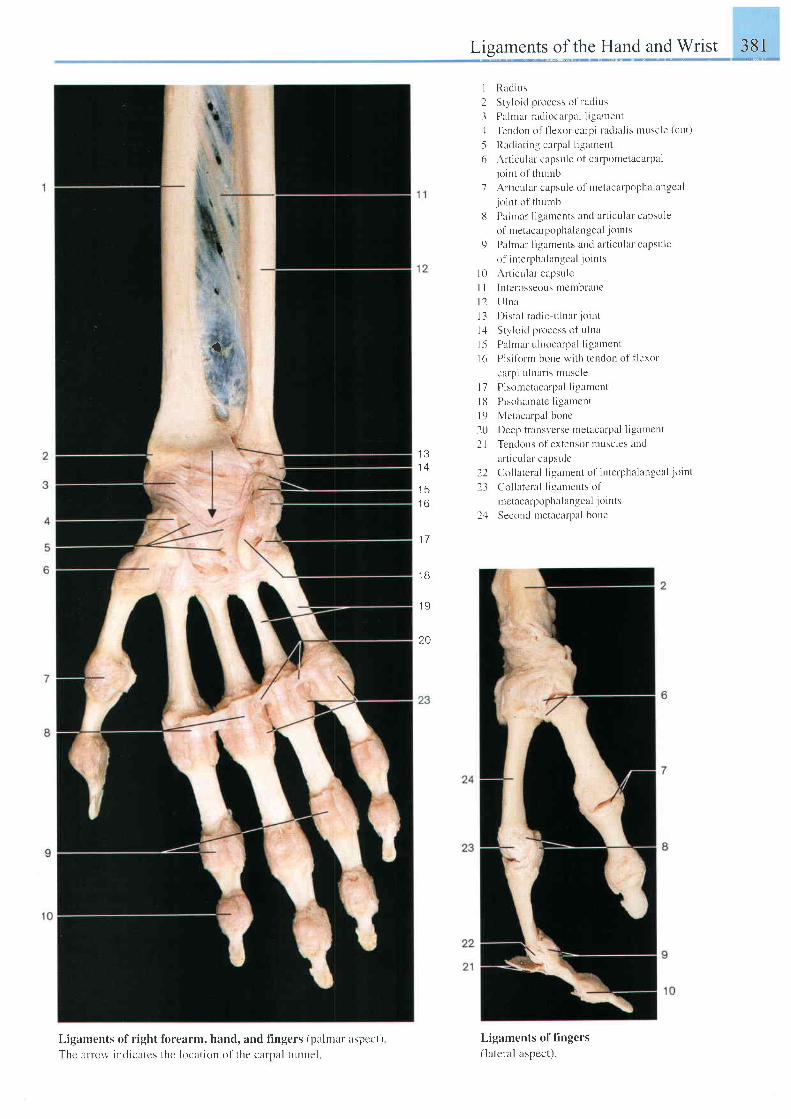
fbrearm, hand, and f ingers (palmal aspcct)
the location of the carnal tunnel
Ligaments of the Hand and Wrist 381
I Rad ius
2 Sty lo id proccss of radius
3 Palmar radiocarpal l igament: l Tendon ol ' f leror crrp i radia l is muscle (ct l t )
5 Radiar ing carpr l l igament
6 Arr icular capsule of carpontetacarpal
. jo int o l ' thurnb
7 Art icular capsule of tnetacarpophalangcal
. jo int of thr , rmb
8 Palnrar l igaments aud ar t icular capsule
( ) l r nc tucJ rp ( )ph l l i r n . r : eu l j , ' i t t l t
9 Pah'nar l igaments and ar t icular capsule
of intcrphalangeal jo ints
Art icular capsule
Intcl'osseous nrembrane
U lna
Dista l radio-ulnar. jo int
Sty lo ic l process of u lna
Palmar u lnocarpal l igarrcnt
Pisifor-m bone with tcndon of flerot'
car-p i u lnar is nruscle
Pisornetacarpal l tgament
Pisoharnate l igamcnt
Mctrcarpal bonc
Deep trausvcrse metacarpal li-gamcnt
Tendons of extensor tnttscles lncl
articr,rlal capsule
Col lateral l igarnent of intc lphalangeir l jo int
Col latcral l igamerr ls of
rnctacarpophalangeal jo ints
Second metacarpitl bone
Ligaments of hngers(lateral aspect).
t
l 0l tt 2l 3t,1l 5I t f
I J
1 A
t 3
i o
1 7
'18
1 9
20
l 1
I I J
l 9
2i)21
2223
24
Ligaments of r ight
Thc arlorv indicatcs
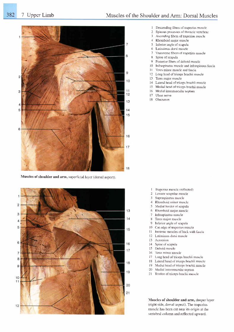
382 7 Upper Limb Muscles of the Shoulder andArm: Dorsal Muscles
Muscles of shoulder and arm, superficial layer (dorsal aspect)
1 Descending fibers of trapezius muscle2 Spinous processes ofthoracic vertebrae3 Ascending fibers of trapezius muscle4 Rhomboid major muscle5 Inferior angle of scapula6 Latissimus dorsi muscle7 Transverse fibers oftrapezius muscle8 Spine of scapula9 Posterior hbers of deltoid muscle
10 Infraspinatus muscle and infraspinous fascia11 Teres minor muscle and fascia12 Long head oftriceps brachii muscle13 Teres major musclel4 Lateral head oftriceps brachii musclel5 Medial head of triceps brachii muscle16 Medial intermuscular seotuml7 Ulnar nervel8 Olecranon
1 Trapezius muscle (reflected)2 Levator scapulae muscle3 Supraspinatus muscle4 Rhomboid minor muscle5 Medial border of scapula6 Rhomboid major muscle7 Infraspinatus muscle8 Teres major muscle9 Inferior angle of scapula
10 Cut edge oftrapezius musclel1 Intrinsic muscles of back with fascia12 Latissimus dorsi muscle13 Acromion14 Spine of scapula15 Deltoid muscle16 Teres minor musclel7 Long head oftriceps brachii muscle1 8 Lateral head of triceps brachii muscle19 Medial head of triceps brachii muscle20 Medial intermuscular septum21 Tendon oftriceps brachii muscle
Muscles of shoulder and arm, deeper layer(right side, dorsal aspect). The trapeziusmuscle has been cut near its origin at thevertebral column and reflected upward.
1 1I L
t . t
' t4
1 5
1 3
1 4
I
1 01 1
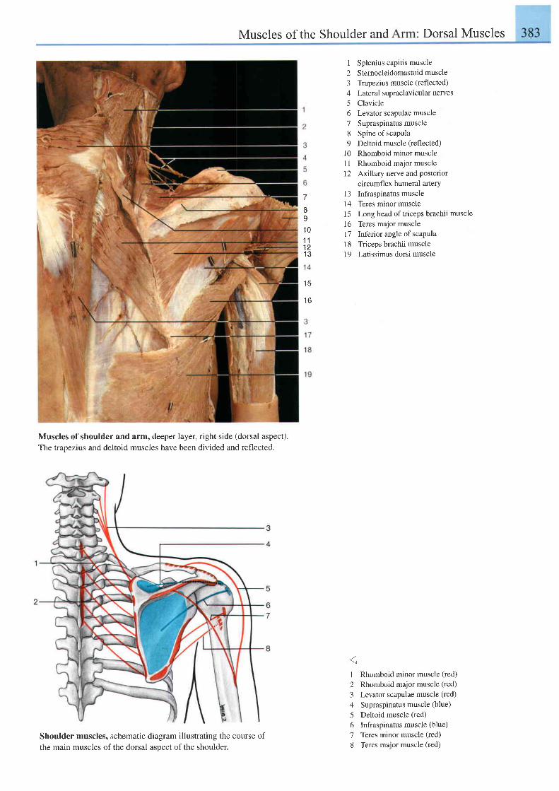
Muscles of the Shoulder and Dorsal Muscles
Shoulder muscles, schematicthe main muscles of the dorsal
diagram illustrating the course ofasoect of the shoulder.
Splenius capitis muscleSternocleidomastoid muscleTrapezius muscle (reflected)
Lateral supraclavicular nervesClavicleLevator scapulae muscleSupraspinatus muscleSpine of scapulaDeltoid muscle (reflected)Rhomboid minor muscleRhomboid major muscleAxillary nerve and posteriorcircumflex humeral arteryInfraspinatus muscleTeres minor muscleLong head of triceps brachii muscleTeres major muscleInferior angle of scapulaTriceps brachii muscleLatissimus dorsi muscle
1 Rhomboid minor muscle (red)
2 Rhomboid major muscle (red)
3 Levator scapulae muscle (red)
4 Supraspinatus muscle (blue)
5 Deltoid muscle (red)
6 Infraspinatus muscle (blue)
7 Teres minor muscle (red)
8 Teres major muscle (red)
7
x
I' t0
1 1t 1' t a
t R
l o
I
2345o78o
1 01 lt 2
1 3t 41 51 6l 71 8t 9
Muscles of shoulder and arm, deeper layer, right side (dorsal aspect).The trapezius and deltoid muscles have been divided and reflected.
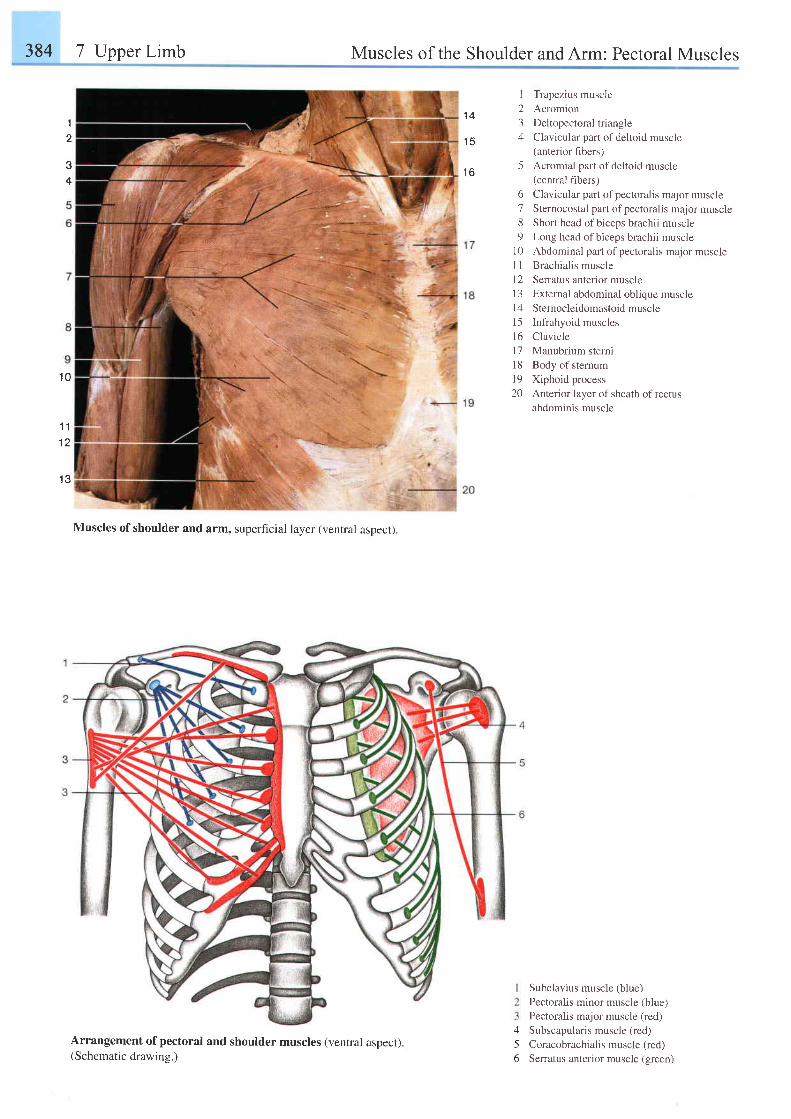
384 7 Upper Limb Muscles of the Shoulder andArm: Pectoral Muscles
34
t
2
1 0
1 11 2
1 3
Muscles of shoulder and arm, superficial layer (ventral aspect).
I Trapezius muscle2 Acromion3 Deltopectoral triangle4 Clavicular part of deltoid muscle
(anterior hbers)5 Acromial part of deltoid muscle
(central fibers)6 Clavicular parl of pectoralis major muscle7 Sternocostal part of pectoralis major muscle8 Short head ofbiceps brachii muscle9 Long head ofbiceps brachii muscle
10 Abdominal part of pectoralis major muscle'| 1 Brachialis muscle
l2 Senatus anterior muscle13 External abdominal oblique musclel4 Sternocleidomastoid muscle15 Infrahyoid musclesl6 Claviclel7 Manubrium sterni18 Body of sternuml9 Xiphoid process20 Anterior layer of sheath of rectus
abdominis muscle
456
Subclavius muscle (blue)
Pectoral is minor muscle (b lue1
Pectoralis major muscle (red)
Subscapularis muscle (red)
Coracobrachialis muscle (red)
Serratus anterior muscle (green)
1 4
1 5
t o
Arrangement of pectoral and shoulder muscles (ventral aspect).(Schematic drawing.)
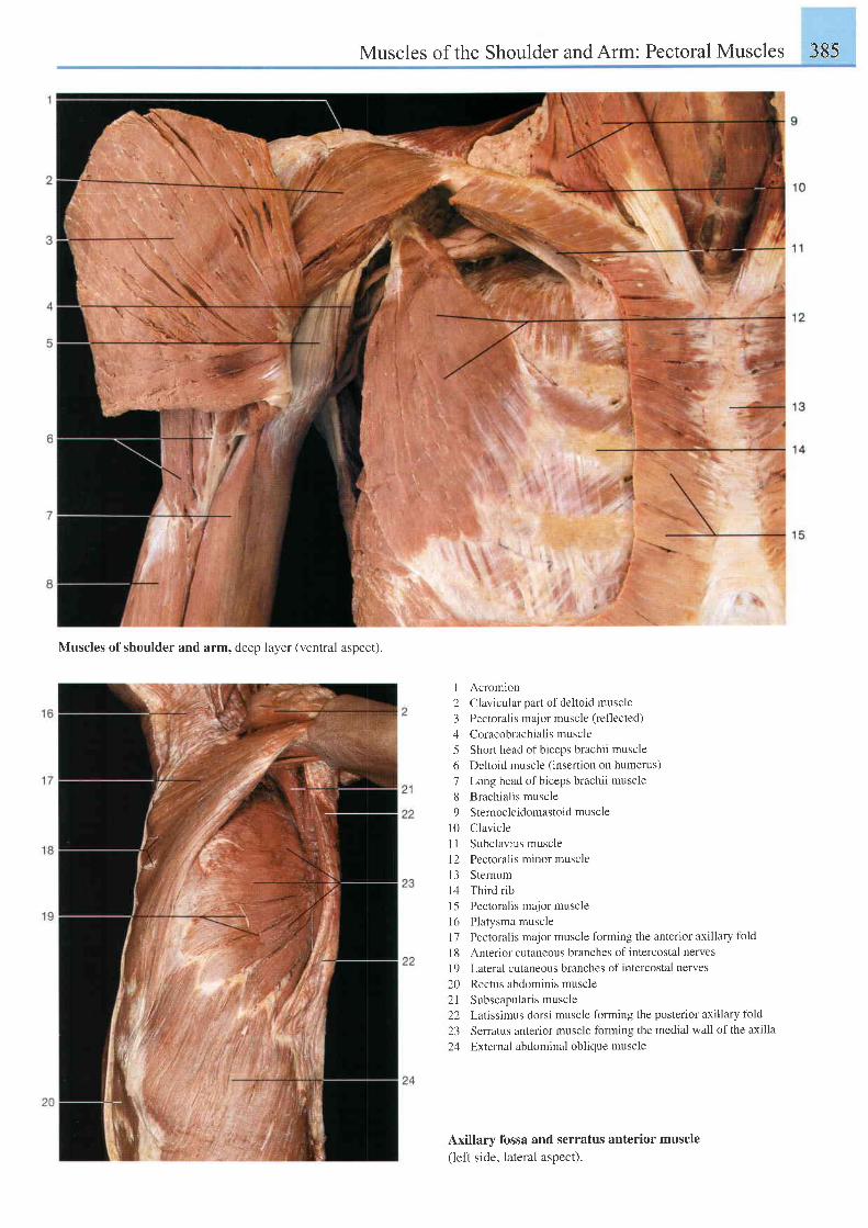
Muscles of the Shoulder and Arm: Pectoral Muscles 385
Muscles of shoulder and arm, deep layer (ventral aspect).
I Acromion2 Clavicular part of deltoid muscle3 Pectoralis major muscle (reflected)
4 Coracobrachialis muscle5 Short head ofbiceps brachii muscle6 Deltoid muscle (insertion on humerus)7 Long head of biceps brachii muscle8 Brachialis muscle9 Stemocleidomastoidmuscle
l0 ClavicleI I Subclavius muscleI2 Pectoral is minor muscle13 Sternum14 Thirdrib15 Pectoralis major musclel6 Platysma muscle17 Pectoralis major muscle forming the anterior axillary fold
I 8 Anterior cutaneous branches of intercostal nervesl9 Lateral cutaneous branches ofintercostal nerves
20 Rectus abdominis muscle2l Subscapularismuscle22 Latissimus dorsi muscle forming the posterior axillary fold
23 Serratus anterior muscle forming the medial wall of the axilla
24 External abdominal oblique muscle
Axillary fossa and serratus anterior muscle(left side, lateral aspect).
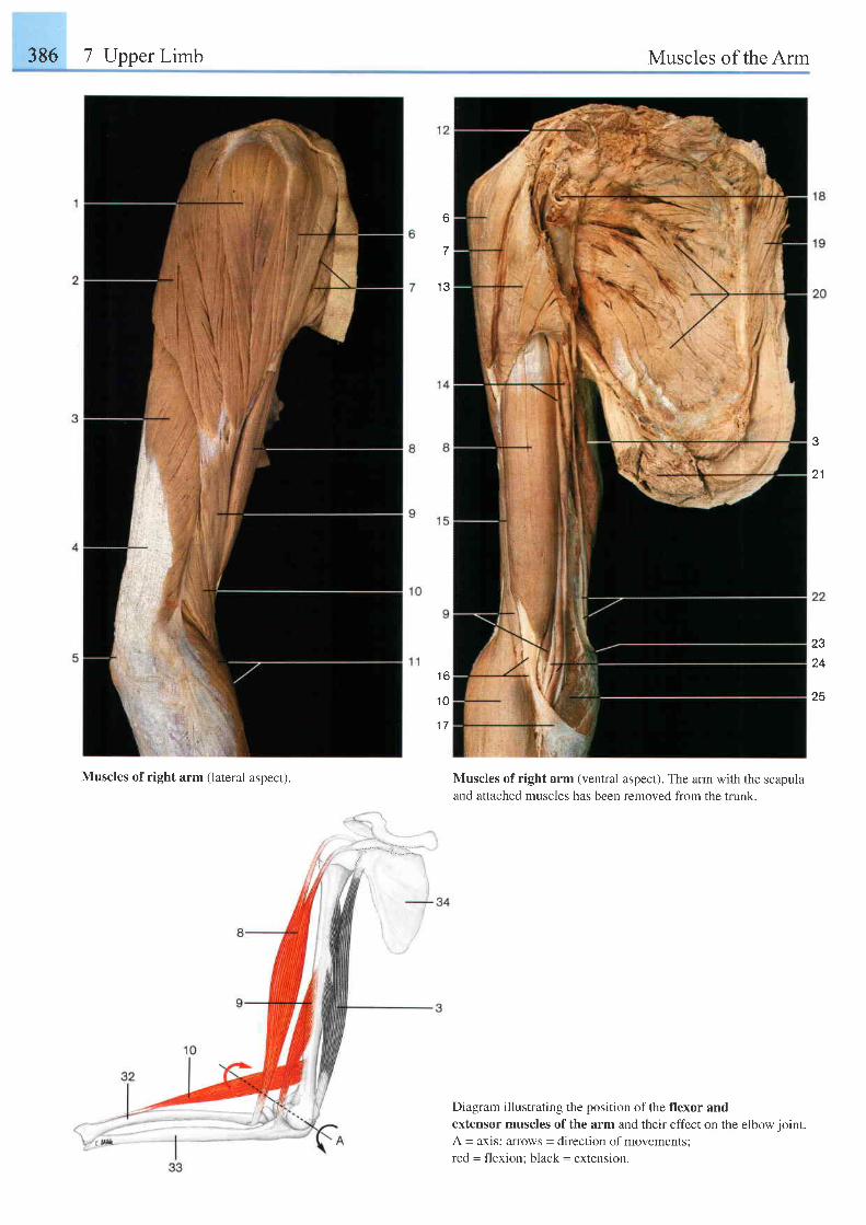
386 7 Upper Limb Muscles of the Arm
7
1 3
1 6
' t0
1 7
e
2 1
23
24
25
Muscles of right arm (lateral aspect). Muscles of right arm (ventral aspect). The arm with the scapulaand attached muscles has been removed from the trunk.
Diagram illustrating the position of the flexor andextensor muscles of the arm and their effect on the elbow ioint.A = axis; arrows = direction of movementsired = flexion: black = extenslon.
n--.';.1 ::,;/
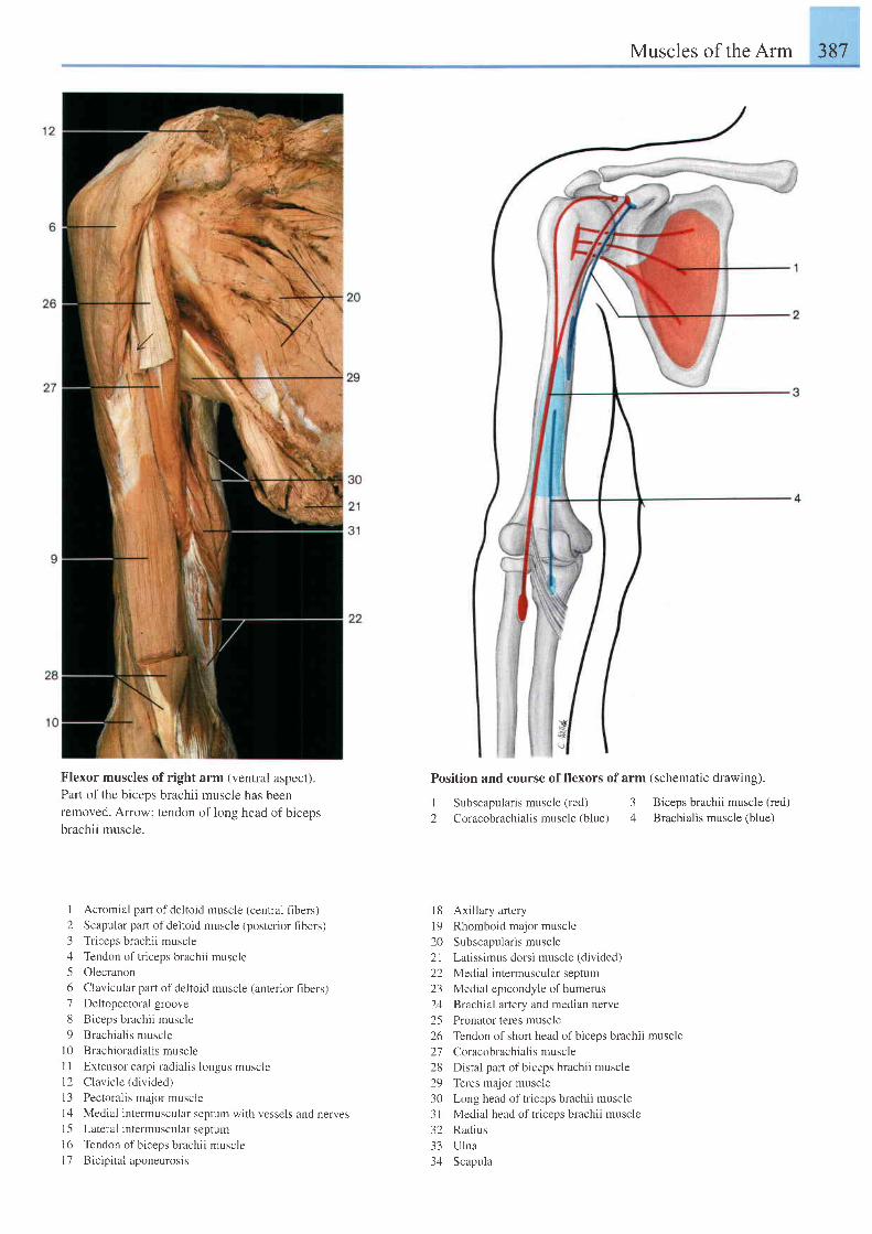
Muscles of the Arm 387
Flexor muscles of right
Part of the biceps brachiiremoved. Arrow: tendon
brachii muscle.
arm (ventral aspect).muscle has been
of long head of biceps
Position and course of flexors of arm
I Subscapularis muscle (red) 32 Coracobrachialis muscle (blue) 4
(schematic drawing).
Biceps brachii muscle (red)Brachialis muscle (blue)
I Acromial part of deltoid muscle (central fibers)
2 Scapular part of deltoid muscle (posterior fibers)3 Triceps brachii muscle
4 Tendon oftriceps brachii muscle
5 Olecranon
6 Clavicular part of deltoid muscle (anterior fibers)7 Deltopectoral groove
8 Biceps brachii muscle
9 Brachialis muscle
l0 Brachioradia l is muscle
1 1 Extensor carpi radialis longus muscle12 Clavicle (divided)
l3 Pectoralis major muscle
1zl Medial intermuscular septum with vessels and nerves15 Lateral intermuscular septum
16 Tendon of biceps brachii muscle
| 7 Bicipital aponeurosis
l8 Axillary arteryl9 Rhomboid major muscle20 Subscapularismuscle21 Latissimus dorsi muscle (divided)22 Medial intermuscular septum23 Medial epicondyle of humerus24 Brachial artery and median nerve25 Pronator teres muscle26 Tendon of short head of biceps brachii muscle27 Coracobrachialismuscle28 Distal part of biceps brachii muscle29 Teres major muscle30 Long head of triceps brachii muscle3l Medial head of triceps brachii muscle32 Radius33 Ulna34 Scapula
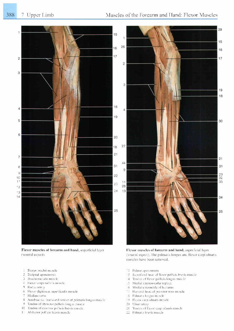
388 7 Upper L inrb
Flexor musclcs of l i r rearm and hand, supcr ' l ' ic ia l la ler
( r ' en t r a l as l ) cc t ) .
I B i ccps b rach i i n rL r sc l c
2 B i c i p i t e l r poncu l - os i s
3 Bn t c l t i o ra r l i u l i : r l u sc l e. l I r lcror carpr nrdi l r l is nruscle
-5 Raclnr l ar tcr-r( r F l c ro r r l i g i t o r - u r l supe t f i c i l l i s n rusc l c
7 \ ' l cd i a r r ne r r e
i l At t tcbr-achi l t l lascia ant l tcnt lon of l l r ln t r r is longrrs nruscJc9 l cnc l o r r o1 l t bduc to r l t o l l i c i s l ongL rs r nL rsc l c
l 0 ' l e r r don
o l c \ t cnso r po l l r c i s b r c l i s n tusc l c
I I AhdL rc to r po l l i c i s b l c ' r i s n tL r sc l c
Mr.rsclc-s ol- thc Foreartn and Hand: Flexor Muscles
2 3
'-) 4
Fleror nruscles of fbrearrn and hand, super-l iciul l rrver-( \ i r i r . r i i r s p c c l ) T h c p l l r n a n s l o n g u s a n d l l c r o r - c u r p i u l n a r r s
ntus! lr ' \ hl l \ c ircclt t 'et loved
1 I I ) . l i n l L r l r l ) onc [u i t s i s
I I \ i r | . ' r - l i e i r i i h c rL t l o l l l c r o r po l l i c i s b re r i s n r t r sc l c
i + i e r r r l o r r r r l l l c r o r po l l i c i s l on !us n rusc l c
I 5 \ l r ' L l l r l i n t c r r t t i r s cu l l f s cp tu l l r
I ( r \ 1 . ' t l r l r l cp i ! o l l ( l \ l e o f hu r t r e r -us
I I I l r I r a l i r l h ca r l 0 l p r - ona to r - [ c r cs n t r r sc l c
i i i ) ; r t n l l L r i s l o n l L r s r l ] L r s c l c
l , ) I l ! ' \ ( ) f c l r r I i L r l n I r i s n rusc l e
l 0 [ i r l r r u r t c r - r
l l i e n . l r r n o l i l c r o r ca rp i L r l n l t r - i s musc l c
l l l ) l r l r r [ L r i \ l ) l c \ i \ n rL r \ c ] e
2332
B
s1 t )1 . 1
1 2
I J
1 4
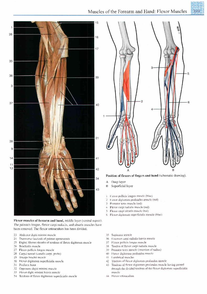
,':il.Muscles of the Forearm and Hand: Flexor Muscles _iffi
1 4
1 1t e
Flexor muscles offorearm and hand, middle layer (ventral aspect).
The palmaris longus, flexor carpi radialis, and ulnaris muscles havebeen removed. The flexor retinaculum has been divided.
23 Abductor digiti minimi muscle24 Transverse fasciculi of palmar aponeurosis25 Digital fibrous sheaths oftendons offlexor digitorum muscle26 Brachial is muscle27 Flexor pollicis longus muscle28 Carpal tunnel (canalis carpi, probe)29 Triceps brachii muscle30 Flexor digitorum superficialis muscle3 I Pisiform bone32 Opponens digiti minimi muscle33 Flexor digiti minimi brevis muscle34 Tendons offlexor digitorum superficialis muscle
Position of flexors of fingers and hand (schematic drawing).
A Deep layerB Superficial layer
I Flexor pollicis longus muscle (blue)
2 Flexor digitorum profundus muscle (red)
3 Pronator teres muscle (red)
4 Flexor carpi radialis muscle (red)
5 Flexor carpi ulnaris muscle (red)
6 Flexor digitorum superficialis muscle (blue)
35 Supinator muscle36 Extensor carpi radialis brevis muscle37 Flexor pollicis longus muscle38 Tendon offlexor carpi radialis muscle39 Pronator teres muscle (insertion of radius)40 Flexor digitorum profundus muscle4l Lumbrical muscles42 Tendons offlexor digitorum profundus muscle43 Tendons of f'lexor digitorum profundus muscle having passed
through the divided tendons of the flexor digitorum superhcialismuscle
44 Flexor retinaculum
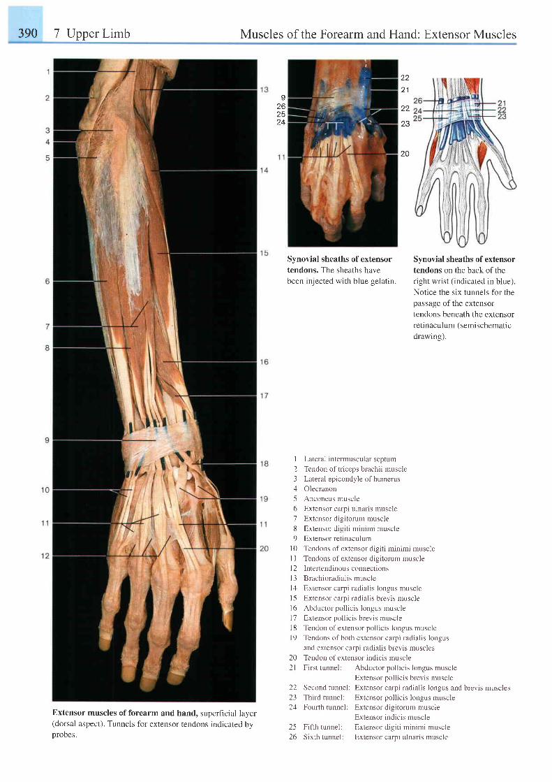
390 7 Upper Limb Muscles of the Forearm and Hand: Extensor Muscles
Izo2524
z z
z l
22
23
zv
Extensor muscles offorearm and hand, superficial layer(dorsal aspect). Tunnels for extensor tendons indicated byprobes.
Synovial sheaths of extensor
tendons on the back of theright wrist (indicated in blue).
Notice the six tunnels for thepassage of the extensor
tendons beneath the extensorre t inacu lum (semischemat ic
drawing).
Lateral intermuscular septum
Tendon of triceps brachii muscle
Lateral epicondyle of humerus
Olecranon
Anconeus muscle
Extensor carpi ulnaris muscle
Extensor digitorum muscle
Extensor digiti minimi muscle
Extensor retinaculum
Tendons of extensor digiti minimi muscle
Tendons of extensor digitorum muscle
Intertendinou s connections
Brachioradialis muscle
Extensor carpi radialis longus muscle
Extensor carpi radialis brevis muscle
Abductor pollicis longus muscle
Extensor pollicis brevis muscle
Tendon of extensor pollicis longus muscle
Tendons of both extensor carpi radialis longus
and extensor carpi radialis brevis muscles
Tendon of extensor indicis muscle
First tunnel: Abductor pollicis longus muscle
Extensor pollicis brevis muscle
Second tunnel: Extensor carpi radialis longus and brevis muscles
Third tunnel: Extensor pollicis longus muscle
Fourth tunnel: Extensor digitorum muscle
Extensor indicis muscle
Fifih tunnel: Extensor digiti minimi muscle
Sixth tunnel: Extensor carpi ulnaris muscle
Synovial sheaths of extensortendons. The sheaths havebeen injected with blue gelatin.
123156'7
89
l 0l lt 2l 31 4l 5t 61'7l 8l 9
202 1
22/-)24
25zo
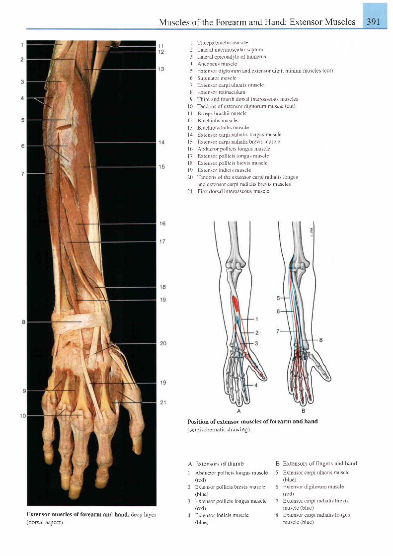
Muscles of the Forearm and Hand: Extensor Muscles 391
I Triceps brachii muscle2 Laleral intermuscular septum3 Lateral epicondyle ofhumerus4 Anconeus muscle5 Extensor digitorum and extensor digiti minimi muscles (cut)
6 Supinator muscle7 Extensor carpi ulnaris muscle8 Extensorretinaculum9 Third and fourth dorsal interosseous muscles
l0 Tendons ofextensor digitorum muscle (cut)
I I Biceps brachii musclel2 Brachial is musclel3 Brachioradialis musclel4 Extensor carpi radialis longus musclel5 Extensor carpi radialis brevis musclel6 Abductor pollicis longus musclel7 Extensor pollicis longus musclel8 Extensor pollicis brevis muscle19 Extensor indicis muscle20 Tendons of the extensor carpi radialis longus
and extensor carpi radialis brevis muscles21 First dorsal interosseous muscle
Position of extensor muscles of forearm and hand(semischematic drawing).
I
Extensors of thumb
Abductor pollicis longus muscle(red)Extensor pollicis brevis muscle(blue)Extensor pollicis longus muscle(red)Extensor indicis muscle(blue)
Extensors of fingers and hand
Extensor carpi ulnaris muscle(blue)Extensor digitorum muscle(red)Extensor carpi radialis brevismuscle (blue)Extensor carpi radialis longusmuscle (blue)
B
5
Extensor muscles of forearm and(dorsal aspect).
hand, deep layer
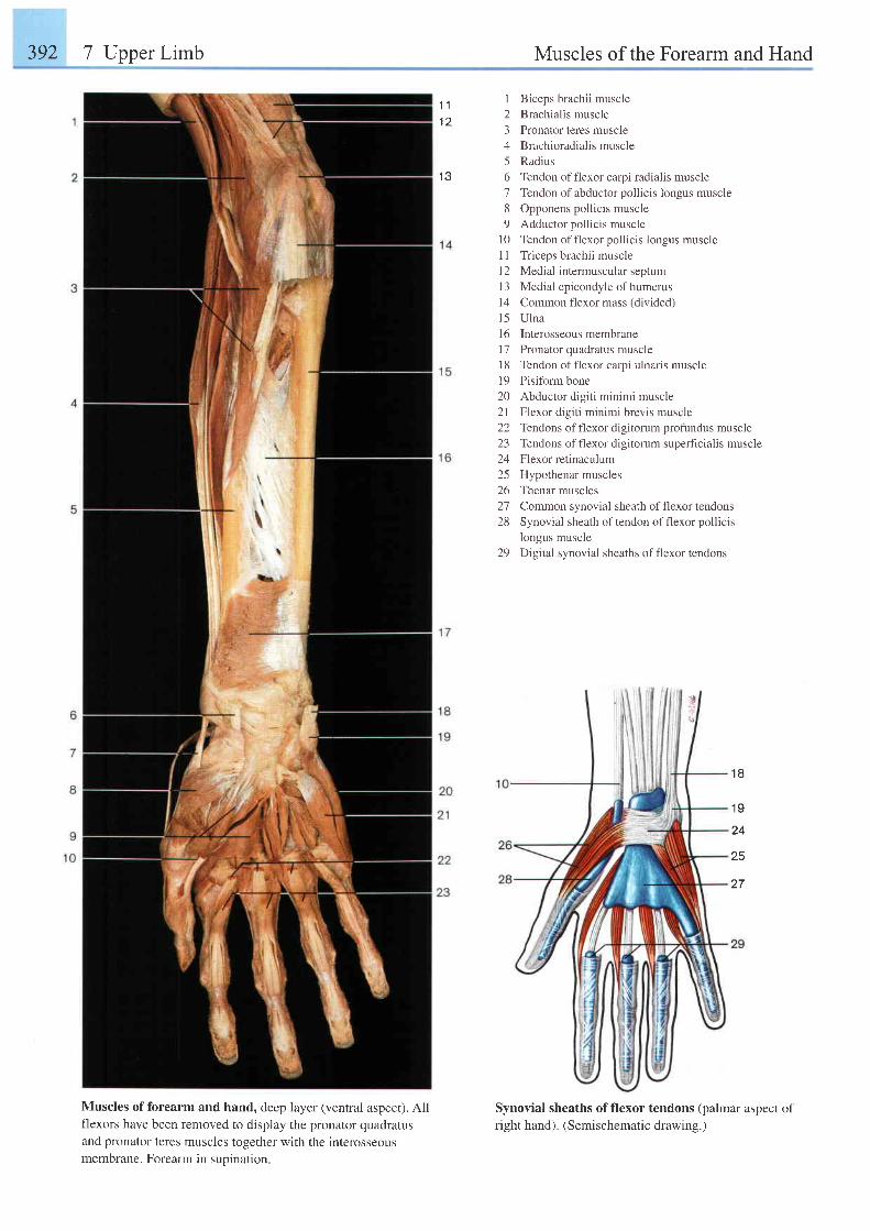
392 7 Upper Limb Muscles of the Forearm and Hand
Muscles of forearm and hand, deep layer (ventral aspect). Allflexors have been removed to display the pronator quadratusand pronator teres muscles together with the interosseousmembrane. Forearm in suoination.
1 Biceps brachii muscle2 Brachialis muscle3 Pronator teres muscle4 Brachioradialis muscle5 Radius6 Tendon of flexor carpi radialis muscle7 Tendon of abductor pollicis longus muscle8 Opponens pollicis muscle9 Adductor pollicis muscle
10 Tendon of flexor pollicis longus muscle11 Triceps brachii musclel2 Medial intermuscular septum13 Medial epicondyle of humerusl4 Common flexor mass (divided)l5 Ulna16 Interosseous membrane17 Pronator quadratus muscle18 Tendon of flexor carpi ulnaris muscle19 Pisiformbone20 Abductor digiti minimi muscle21 Flexor digiti minimi brevis muscle22 Tendons offlexor digitorum profundus muscle23 Tendons offlexor digitorum superficialis muscle24 Flexorretinaculum25 Hypothenar muscles26 Thenar muscles27 Common synovial sheath of flexor tendons28 Synovial sheath of tendon of flexor pollicis
Iongus muscle29 Digital synovial sheaths offlexor tendons
Synovial sheaths of flexor tendons (palmar aspect ofright hand). (Semischematic drawing.)
1 11 2
I J
1 8
1 9
24
25
27
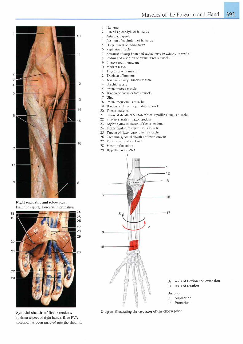
Muscles of the Forearm and Hand 393
1 91 0
242526
2728
29
\ . i '
! 1tl1-. 'r
l , '
22
1 Humerus
2 Lateral epicondyle ofhumerus
3 Articular capsule
4 Position of capitulum of humerus
5 Deep branch of radial nerve
6 Supinator muscle
7 Entrance of deep branch ofradial nen/e to extensor muscles
8 Radius and insertion ofpronator teres muscle
9 Interosseous membrane
l0 Median nerve
I I Triceps brachii muscle
12 Trochlea of humerus
l3 Tendon ofbiceps brachi i muscle
14 Brachial artery
15 Pronator teres muscle
l6 Tendon of pronator teres muscle
17 Ulna
l8 Pronator quadratus muscle
l9 Tendon offlexor carpi radialis muscle
20 Thenar muscles
2l Synovial sheath of tendon of flexor pollicis longus muscle
22 Fibrous sheath of flexor tendons
23 Digital synovial sheath offlexor tendons
24 Flexor digitorum superficialis muscle
25 Tendon offlexor carpi ulnaris muscle
26 Common synovial sheath of f'lexor tendons
27 Position of pisifbrm bone
28 Flexor retinaculum
29 Hypothenar muscles
8
Right supinator and elbow joint(anterior aspect) Forearm in pronation.
Synovial sheaths of flexor tendons(palmar aspect of right hand). Blue PVA
solution has been injected into the sheaths.
20
2 1
A Axis of flexion and extension
B Axis of rotation
Arrows:
S Supination
P Pronation
:.4
23
Diagram illustrating the two axes of the elbow joint.
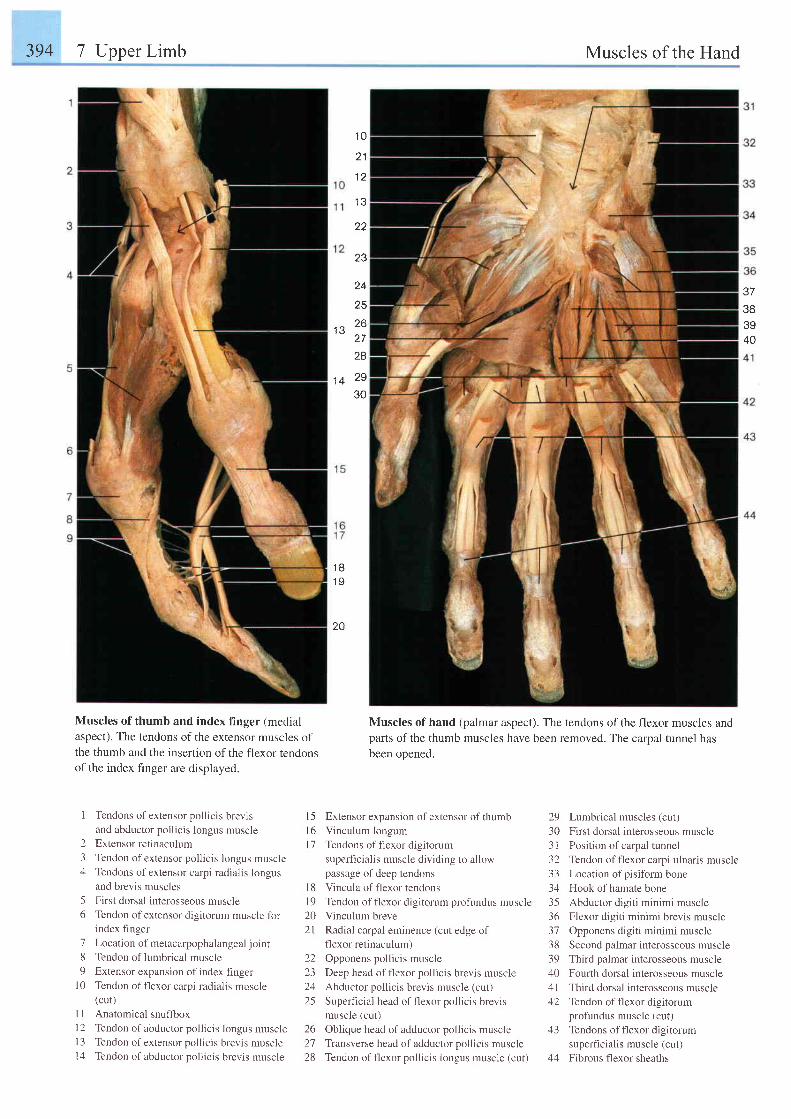
394 7 Upper Limb Muscles of the Hand
1 0
2 1
t 1
22
23
24
25
.," 26I I
28
tq 29
30
37
383940
1 B1 9
20
Muscles of thumb and index finger (medialaspect). The tendons of the extensor muscles ofthe thumb and the insertion of the flexor tendonsofthe index finger are displayed.
Muscles of hand (palmar aspect). The tendons of the flexor muscles andparts of the thumb muscles have been removed. The carpal tunnel hasbeen opened.
I Tendons of extensor pollicis brevisand abductor pollicis longus muscle
2 Extensor retinaculum
3 Tendon of extensor pollicis longus muscle4 Tendons ofextensor carpi radialis longus
and brevis muscles
5 First dorsal interosseous muscle6 Tendon ofextensor digitorum muscle for
index finger
7 Location of metacarpophalangeal joint
8 Tendon of lumbrical muscle9 Extensor expansion of index finger
l0 Tendon of flexor carpi radialis muscle(cut)
I I Anatomical snuffboxI 2 Tendon of abductor pollicis longus musclel3 Tendon ofextensor pollicis brevis muscle14 Tendon ofabductor pollicis brevis muscle
Extensor expansion of extensor of thumb
Vinculum longum
Tendons of t lexor d ig i torum
superficialis muscle dividing to allowpassage of deep tendons
Vincula of flexor tendons
Tendon of flexor digitorum profundus muscleVinculum breve
Radial carpal eminence (cut edge offlexor retinaculum)
Opponens pollicis muscle
Deep head of flexor pollicis brevis muscle
Abductor pollicis brevis muscle (cut)
Superficial head of f'lexor pollicis brevismuscle (cut)
26 Oblique head of adductor pollicis muscle27 Transverse head of adductor pollicis muscle
28 Tendon of f'lexor pollicis longus muscle (cut)
29 Lumbrical muscles (cut)30 First dorsal interosseous muscle31 Position ofcarpal tunnel32 Tendon of flexor carpi ulnaris muscle33 Location ofpisiform bone34 Hook of hamate bone35 Abductor digiti minimi muscle36 Flexor digiti minimi brevis muscle37 Opponens digiti minimi muscle38 Second palmar interosseous muscle39 Third palmar interosseous muscle40 Fourth dorsal interosseous muscle41 Third dorsal interosseous muscle42 Tendon offlexor digitorum
profundus muscle (cut)43 Tendons offlexor digitorum
superfi cialis muscle (cut)44 Fibrous flexor sheaths
1 5l 6l 1
l 8l 9202 l
22z-)
2425
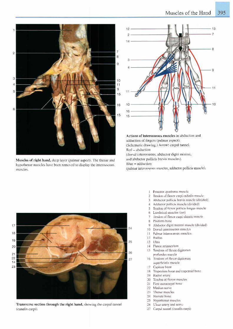
Muscles of the Hand 395
1 01 tI
t o
t c
Muscles of right hand, deep layer (palmar aspect'). The thenar and
hypothenar muscles have been removed to display the interosseousmuscles.
Transverse section through the right hand, showing the carpal tunnel(canalis carpi).
Actions of interosseous muscles in abduction and
adduction of fingers (palmar aspect).
(Schematic drawing.) Arow: catpal tunnel.
Red = abduction(dorsal interosseous, abductor digiti minimi,
and abductor pollicis brevis muscles).
Blue = adduction(palmar interosseous muscles, adductor pollicis muscle)
I Pronator quadratus muscle2 Tendon of f'lexor carpi radiaiis muscle3 Abductor pollicis brevis muscle (divided)
4 Adductor pollicis muscle (divided)
5 Tendon offlexor pollicis longus muscle6 Lumbrical muscles (cut)
7 Tendon offlexor carpi ulnaris muscle8 Pisiform bone9 Abductor digiti minimi muscle (divided)
10 Dorsal interosseous musclesI I Palmar interosseous muscles12 Radiusl3 U lna14 Flexor retinaculum15 Tendons offlexor digitorum
profundus muscleI 6 Tendons of flexor digitorum
superf-rcialis muscle17 Capitate bonel8 Trapezium bone and trapezoid bonel9 Radial artery20 Tendon offlexor muscles21 First metacarpal bone22 Median nerve23 Thenar muscles24 Hamate bone25 Hypothenar muscles26 Ulnat artery and nerve27 Carpal tunnel (canalis catpi)
1 0
1 6
t c
1 7
1 B
1 9
20
2 1
l +
23
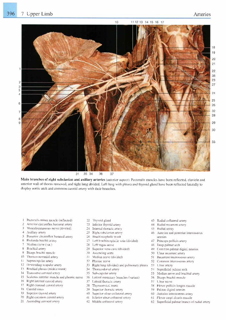
396 7 Upper Limb Arteries
I
5A
1 8
1 9
2Aa 1
1 1
3B2327
24
26
3228
29
30
31 35 34 36
Main branches ofright subclavian and axillary arteries (anterior aspect). Pectoralis muscles have been reflected, clavicle andanterior wall ofthorax removed, and right lung divided. Left lung with pleura and thyroid gland have been reflected laterally todisplay aortic arch and common carotid artery with their branches.
37
I Pectoralis minor muscle (reflected)
2 Anterior circumflex humeral artery3 Musculocutaneous nerve(divided)zl Axillary artery
5 Posterior circumf'lex humeral artery6 Profunda brachii artery7 Median nerve (var.)
8 Brachial artery
9 Biceps brachii musclel0 Thoraco-acromial arteryI I Suprascapular artery
1 2 Descending scapular artery13 Brachial plexus (middle trunk)l4 Transverse cervical afteryl5 Scalenus anterior muscle and phrenic
16 Right internal carotid arteryl7 Right external carotid arteryl8 Carot id s inusl9 Superior thyroid artery20 Right common carotid artery21 Ascending cervical artery
22 Thyroid gland 43 Radial collateral artery23 Inferior thyroid artery 44 Radial recuffent artery24 Internal thoracic artery 45 Radial artery25 Right subclavian artery 46 Anterior and posterior interosseous26 Brachiocephalic trunk arteries21 Left brachiocephalic vein (divided) 47 Princeps pollicis artery28 Left vagus nerve 48 Deep palmar arch29 Superior vena cava (divided) 49 Common palmar digital arteries30 Ascending aorta 50 Ulnar recurrent artery3l Median nerve (d iv ided) 5 l Recurrent interosseous ar tery32 Phrenic nerve 52 Common interosseous artery33 Right lung (divided) and pulmonary pleura 53 Ulnar artery34 Thoracodorsal artery 5zl Superficial palmar arch35 Subscapular artery 55 Median nerve and brachial artery36 Lateral mammary branches (variant) 56 Biceps brachii muscle
37 Lateral thoracic artery 57 Ulnar nerve38 Thyrocervical trunk 58 Flexor pollicis longus muscle39 Superior thoracic artery 59 Palmar digital arteries40 Superior ulnar collateral artery 60 Anterior interosseous artery41 lnferior ulnar collateral artery 61 Flexor carpi ulnaris muscle42 Middle collateral artery 62 Superficial palmar branch of radial artery
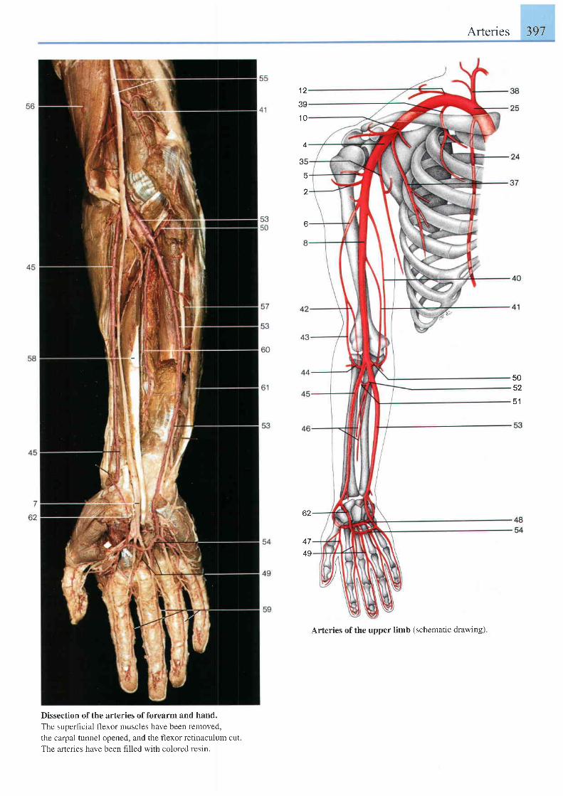
Arteries 397
1 2
39
1 0
4
35
5
z
5052
51
62
4749
Arteries of the upper limb (schematic drawing)
Dissection of the arteries of forearm and hand.
The superficial flexor muscles have been removed,
the carpal tunnel opened, and the flexor retinaculum cut.
The arteries have been filled with colored resin.
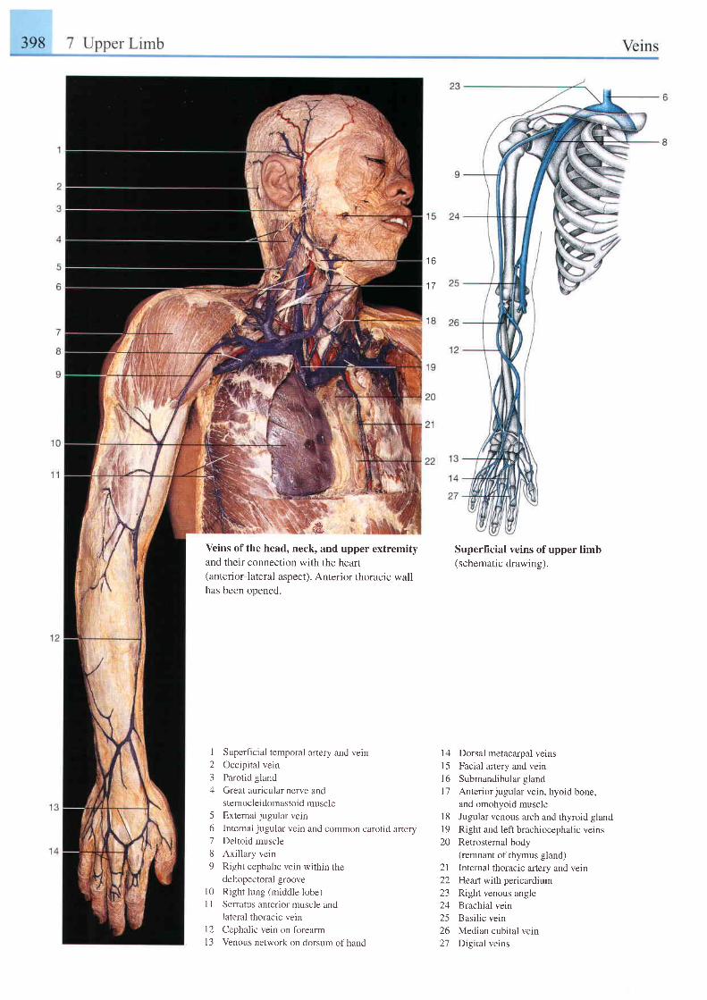
t o
1eVeins of the head, neck, and upper extremityand their connection with the heart(anterior-lateral aspect). Anterior thoracic wallhas been ooened.
I
234
Superficial temporal artery and vein
Occipital vein
Parotid gland
Great auricular nerve andsternocleidomastoid muscleExternai jugular vein
Intemal jugular vein and common carotid arteryDeltoid muscle
Axillary vein
Right cephalic vein within thedeltopectoral groove
Right lung (middle lobe)
Serratus anterior muscle andlateral thoracic vein
Cephalic vein on forearm
Venous network on dorsum of hand
Superficial veins of upper limb(schematic drawing).
l4 Dorsal metacarpal veinsl5 Facial artery and veinl6 Submandibular glandl7 Anteriorjugular vein, hyoid bone,
and omohyoid muscle18 Jugular venous arch and thyroid glandl9 Right and left brachiocephalic veins20 Retrosternal body
(remnant of thymus gland)21 Internal thoracic artery and vein22 Heart with pericardium23 Right venous angle24 Brachial vein25 Basiiic vein26 Median cubital vein27 Digital veins
56189
1 0l l
t 2l 3
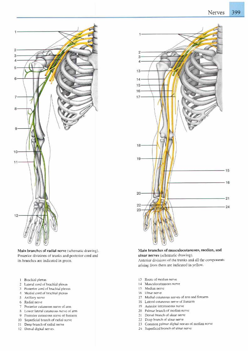
Main branches of radial nerve (schematic drawing).Posterior divisions of trunks and posterior cord andits branches are indicated in s.reen.
1 Brachial plexus
2 Lateral cord of brachial plexus
3 Posterior cord of brachial plexus
4 Medial cord ofbrachial plexus
5 Axillary nerve
6 Radial nerve
7 Posterior cutaneous nerve of arm
8 Lower lateral cutaneous nerve of arm
9 Posterior cutaneous nerve offorearm
10 Supeficial branch ofradial nerve
1 1 Deep branch of radial nerve
12 Dorsal digital nerves
Nerves
Main branches of musculocutaneous' median, and
ulnar nerves (schematic drawing).Anterior divisions of the trunks and all the componentsarising from them are indicated in yellow.
13 Roots of median nerve14 Musculocutaneous nerve15 Median nerve16 Ulnar nerve1 7 Medial cutaneous nerves of arm and forearmI 8 Lateral cutaneous nerve of forearm19 Anterior interosseous nerve20 Palmar branch ofmedian nerve21 Dorsal branch ofulnar nerve22 Deep branch ofulnar nerve23 Common palmar digital nerves of median nerve
24 Suoerficial branch ofulnar nerve
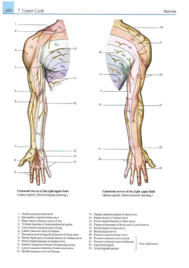
400 7 Upper Limb Nerves
Thg
- 3
Cutaneous nerves of the right upper limb(ventral aspect). (Semischematic drawing.)
1 Medial supraclavicular nerve2 Intermediate supraclavicular nerve3 Upper lateral cutaneous nerve of arm4 Terminal branches of intercostobrachial nerves5 Lower lateral cutaneous nerve ofarm6 Lateral cutaneous nerve offorearm7 Terminal branch of superficial branch of radial nerve8 Palmar digital nerve of thumb (branch of median nerve)9 Palmar digital branches of median nerve
10 Anterior cutaneous branches ofintercostal nervesI 1 Lateral cutaneous branches of intercostal nervesI 2 Medial cutaneous nerye of forearm
Thr
Cutaneous nerves of the right upper limb(dorsal aspect). (Semischematic drawing.)
1 3 Palmar cutaneous branch of ulnar nerve14 Palmar branch of median nerveI 5 Palmar digital branches of ulnar nerve1 6 Cutaneous branches of dorsal rami of spinal nerves17 Dorsal branch ofulnar nerve18 Dorsal digital nervesl9 Posteriorsupraclavicularnerve20 Posterior cutaneous nerve of arm I2l Posterior cutaneous nerve of forearm I22 Superficial branch I
from radial nerve
23 Dorsal digital branches
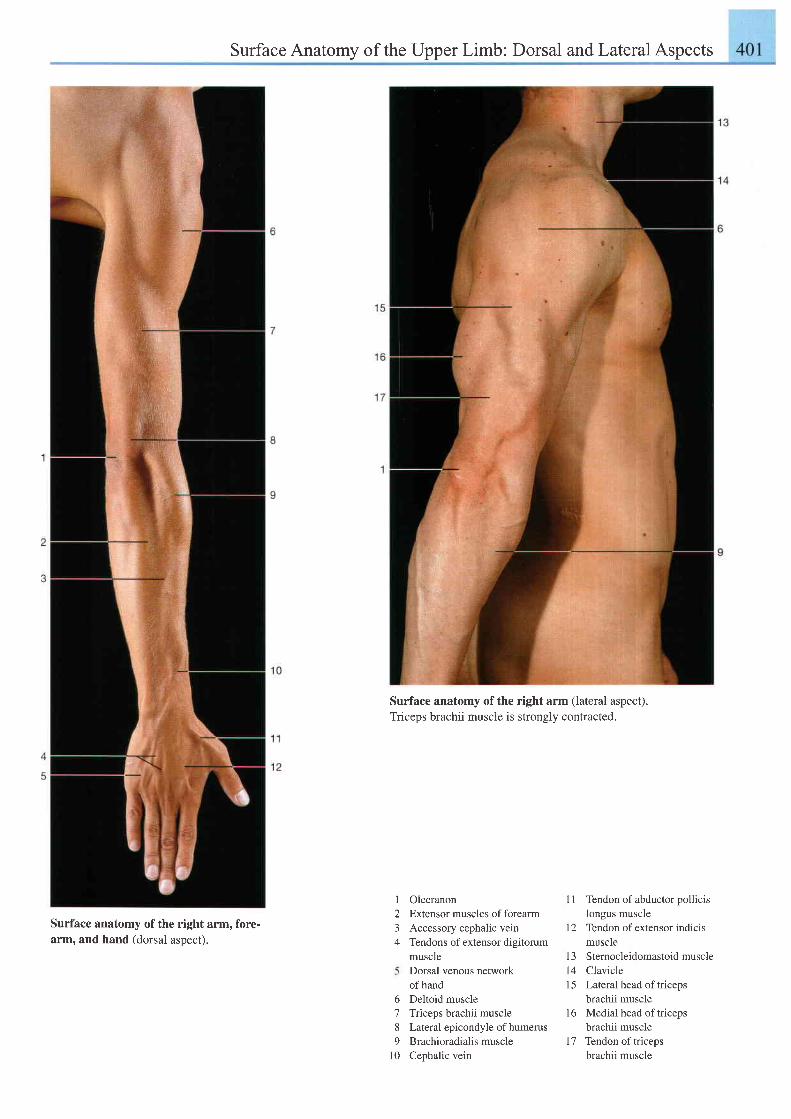
Surface Anatomy of the Upper Limb: Dorsal andLateral Aspects
Surface anatomy of the right arm (lateral aspect).Triceps brachii muscle is strongly contracted.
Surface anatomy of the right arm, fore-arm. and hand (dorsal asoect).
I
23+
b
789
t 0
OlecranonExtensor muscles of forearmAccessory cephalic veinTendons of extensor digitorummuscleDorsal venous networkof handDeltoid muscleTriceps brachii muscleLateral epicondyle of humerusBrachioradialis muscleCephalic vein
I 1 Tendon of abductor polhcrslongus muscle
l2 Tendon ofextensor indicismuscle
13 Sternocleidomastoid muscle14 Clavicle15 Lateral head oftriceps
brachii muscle16 Medial head of triceps
brachii muscle17 Tendon oftriceps
brachii muscle
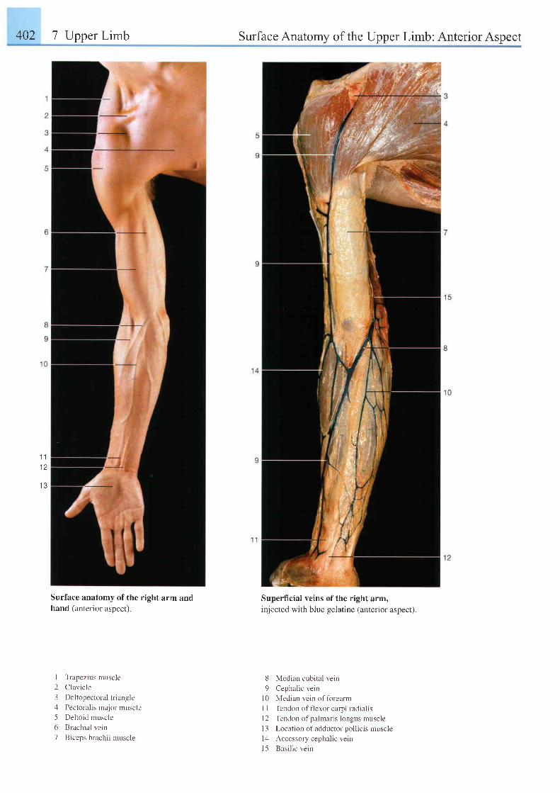
402 7 Upper Limb Surface Anatomy of the Upper Limb: Anterior Aspect
1 l1 2
1 3
Surface anatomy of the right arm andhand (anterior aspect).
I Trapezius muscle
2 Clavicle
3 Deltopectoral triangle
4 Pectoralis major muscle
5 Deltoid muscle
6 Brachial vein
7 Biceps brachii muscle
Superficial veins of the right arm,injected with blue gelatine (anterior aspect).
8 Median cubital vein
9 Cephalic vein
10 Median vein of forearm
I I Tendon of flexor carpi radialis
l2 Tendon of palmaris longus muscle
I 3 Location of adductor pollicis muscle
l4 Accessory cephal ic vein
l5 Basi l ic vein
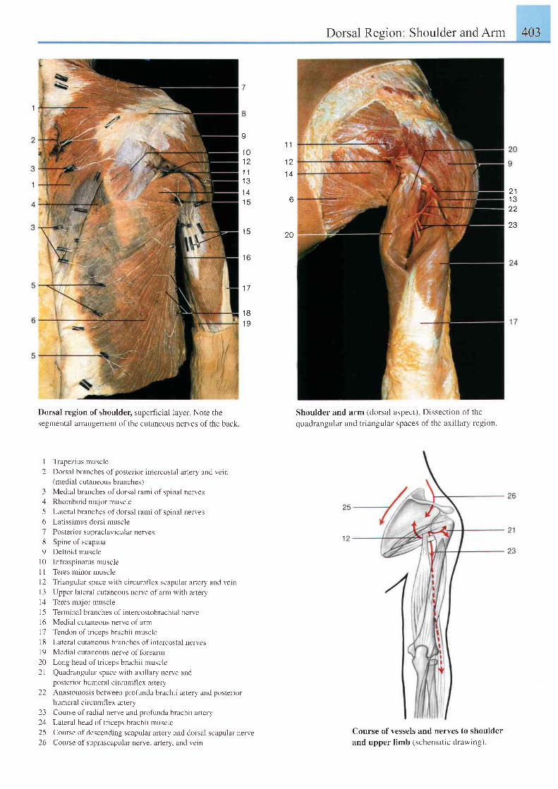
Dorsal Region: Shoulder and Arm 403
a
1 01 21 J
I J
1 4t 3
1 5
t o
1 7
1 B1 9
Dorsal region ofshoulder, superficial layer. Note the
segmental arrangement of the cutaneous nerves of the back.
I Trapezius muscle
2 Dorsal branches of posterior intercostal artery and vein(medial cutaneous branches)
3 Medial branches of dorsal rami of spinal nerves4 Rhomboid major muscle
5 Lateral branches of dorsal rami of spinal nerves
6 Latissimus dorsi muscle
7 Posterior supraclavicular nerves
8 Spine of scapula
9 Deltoid muscle
l0 Infraspinatus muscle
11 Teres minor muscle
l2 Triangular space with circumflex scapular artery and vein
I 3 Upper lateral cutaneous nerve of arm with artery
14 Teres major muscle
I 5 Terminal branches of intercostobrachial nerve
16 Medial cutaneous nerve of arm
l7 Tendon of triceps brachii muscle
1 8 Lateral cutaneous branches of intercostal nerves
l9 Medial cutaneous nerve offorearm
20 Long head of triceps brachii muscle
2). Quadrangular space with axillary nerve andposterior humeral circumflex aflery
22 Anastomosis between profunda brachii artery and posterior
humeral circumfl ex artery
23 Course of radial nerve and profunda brachii artery
24 Lateral head oftriceps brachii muscle
25 Course of descending scapular artery and dorsal scapular nerve
26 Course of suprascapular nerve, artery, and vein
Shoulder and arm (dorsal aspect)
quadrangular and triangular spaces
1 .1
1 21 4
6
20
2 11 Q
22
23
Dissection of the
of the axillary region.
Course of vessels and nerves to shoulderand upper limb (schematic drawing).
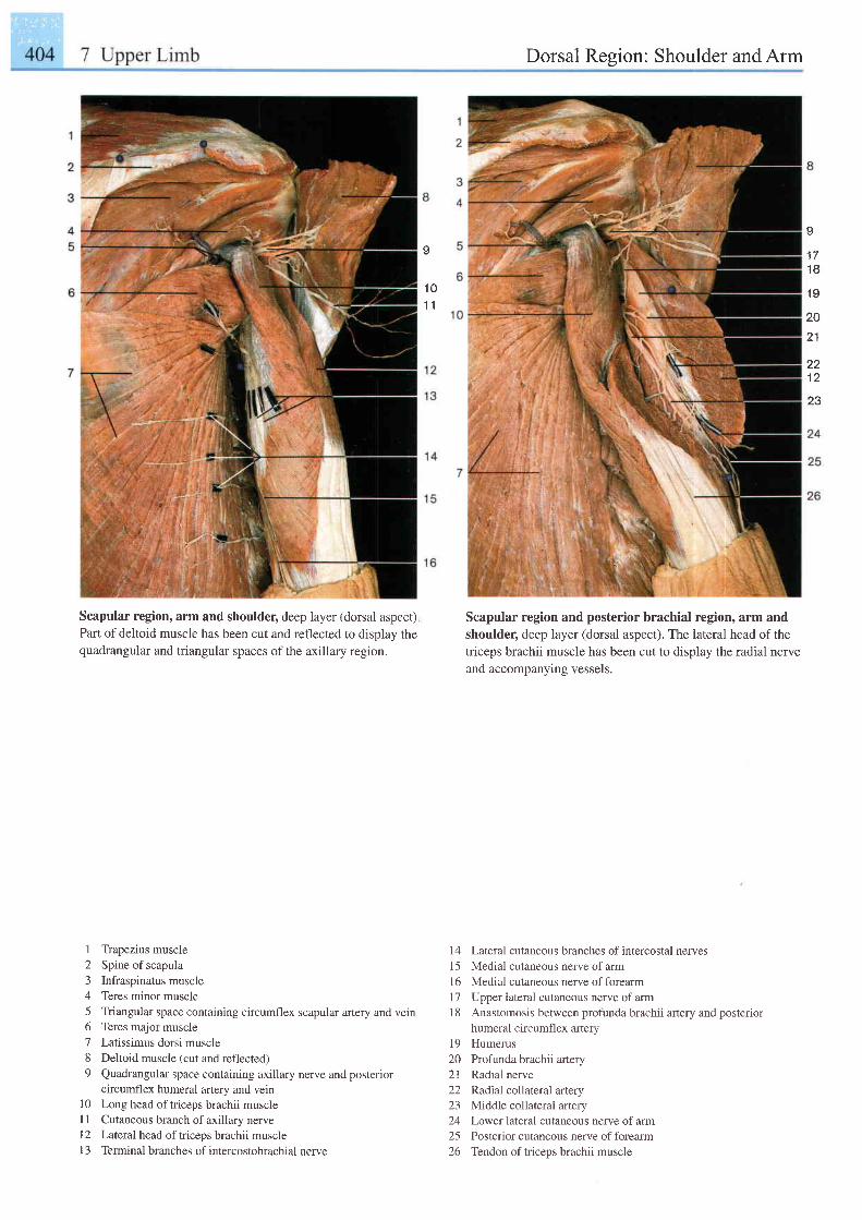
' t t t . "
. ,
Dorsal Region: Shoulder and Arm
I
' t0' t 1
Scapular region, arm and shoulder, deep layer (dorsal aspect),Part of deltoid muscle has been cut and reflected to display thequadrangular and triangular spaces of the axillary region.
Scapular region and posterior brachial region, arm andshoulder, deep layer (dorsal aspect). The lateral head of thetriceps brachii muscle has been cut to display the radial nerveand accompanying vessels.
I
1 71 8
1 9
20
21
221 2
23
1 Trapezius muscle2 Spine of scapula3 Infraspinatus muscle4 Teres minor muscle5 Triangular space containing circumflex scapular artery and vein6 Teres major muscle7 Latissimus dorsi muscle8 Deltoid muscle (cut and reflected)9 Quadrangular space containing axillary nerve and posterior
circumflex humeral artery and veinl0 Long head oftriceps brachii muscle[ 1 Cutaneous branch of axillary nerveL2 Lateral head oftriceps brachii musclel3 Terminalbranches of intercostobrachial nerve
14 Lateral cutaneous branches ofintercostal nerves15 Medial cutaneous nerveof arm16 Medial cutaneous nerve offorearm17 Upper iateral cutaneous nerve ofarmI 8 Anastomosis between profunda brachii artery and posterior
humeral circumflex arterv19 Humerus20 Profunda brachii arlery21 Radial nerve22 Radial collateral artery23 Middle collateral artery24 Lowerlateral cutaneous nerve of arm25 Posterior cutaneous nerve offorearm26 Tendon oftriceps brachii muscle
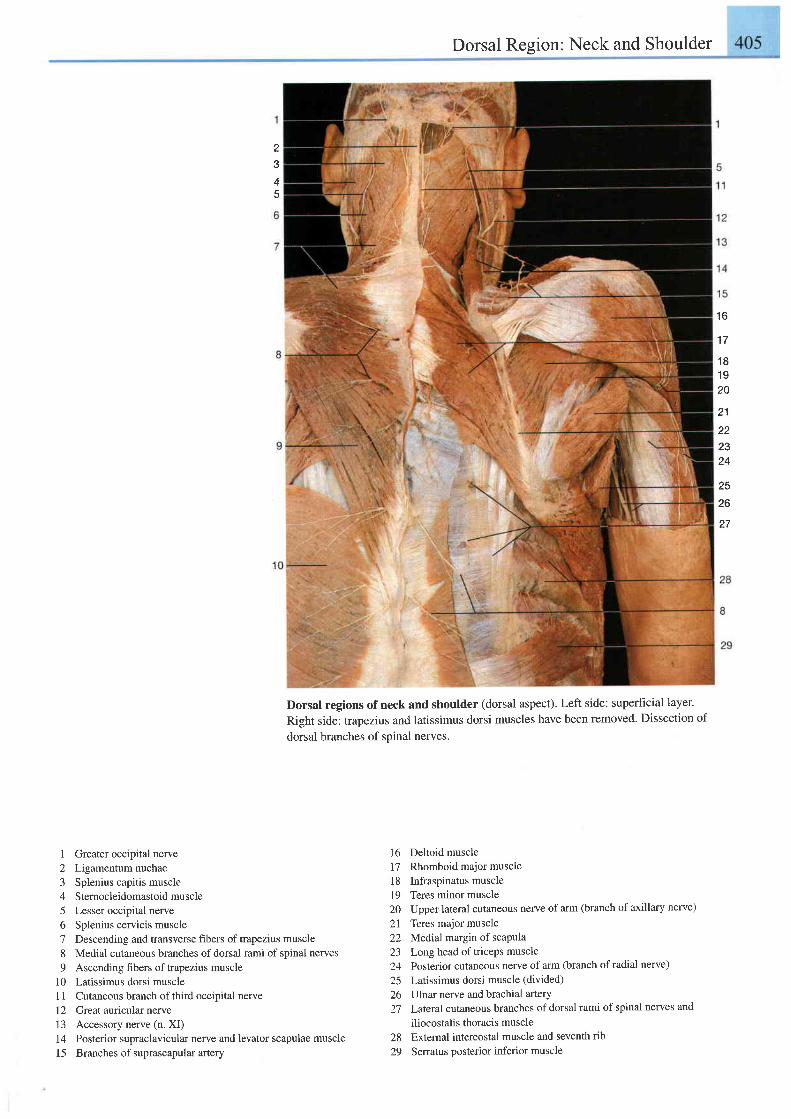
1 Greater occipital nerve2 Ligamentum nuchae3 Splenius capitis muscle4 Sternocleidomastoid muscle5 Lesser occipital nerve6 Splenius cervicis muscle7 Descending and transverse fibers of trapezius muscle8 Medial cutaneous branches of dorsal rami of spinal nerves9 Ascending fibers oftrapezius muscle
l0 Latissimus dorsi muscle1 1 Cutaneous branch of third occipital nerve12 Great auricular nerve13 Accessory nerve (n. XI)14 Posterior supraclavicular nerye and levator scapulae muscle15 Branches of suprascapularartery
Dorsal Region: Neck and Shoulder
Dorsal regions ofneck and shoulder (dorsal aspect). Left side: superltcial layer.
Right side: trapezius and latissimus dorsi muscles have been removed. Dissection of
dorsal branches of spinal nerves.
16 Deltoid muscle17 Rhomboid major muscle18 Infraspinatus muscle19 Teres minor muscle20 Upper lateral cutaneous nerve of arm (branch of axillary nerve)
21 Teres major muscle22 Medial, margin of scaPula23 Long head of triceps muscle24 Posterior cutaneous nerve ofarm (branch ofradial nerve)
25 Latissimus dorsi muscle (divided)
26 Ulnar nerve and brachial artery27 Lateralcutaneous branches of dorsal rami of spinal nerves and
iliocostalis thoracis muscle
28 Extemal intercostal muscle and seventh rib
29 Senatus posterior inferior muscle
23
4
. t A
1 7
1 8't9
20
21
222324
25
26
1 t
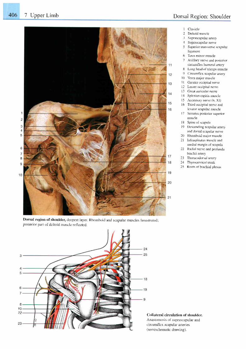
406 7 Upper Limb Dorsal Region: Shoulder
I
2345
67
89
l 0l lt 2l 3t 4l 5t 6
1 7
l 81 9
202 1
22
ClavicleDeltoid muscleSuprascapular arterySuprascapular nerveSuperior transverse scapularligamentTeres minor muscleAxi l lary nerve and posteriorcircumflex humeral arteryLong head of tr iceps muscleCircumfl ex scapular arteryTeres major muscleGreater occipital nerveLesser occipital nerveGreat auricular nerveSplenius capitis muscleAccessory nerve (n. XI)Third occipital nerve andlevator scapulae muscleSerratus posterior superiormuscleSpine of scapulaDescending scapular arteryand dorsal scapular nerveRhomboid major muscleInfraspinatus muscle andmedial margin of scapulaRadial nerve and profundabrachii artery
2
6
7Io
23 Thoracodorsal artery24 Thyrocervical trunk25 Roots of brachial plexus
Dorsal region ofshoulder, deepest layer. Rhomboid and scapular muscles fenestrated;posterior part of deltoid muscle reflected.
B1 022
Collateral circulation of shoulder.Anastomosis of suprascapular andcircumfl ex scapular arteries(semischematic drawing).
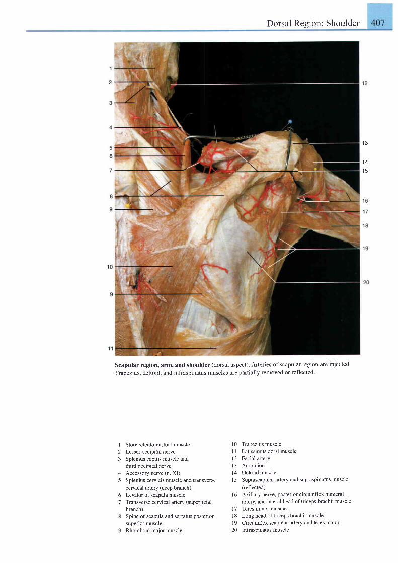
Dorsal Region: Shoulder
Scapular region, arm, and shoulder (dorsal aspect). Arteries of scapular region are injected.
Trapezius, deltoid, and infraspinatus muscles are partially removed or reflected.
1 3
1 4't5
I
23
4
5
67
Stemocleidomastoid muscleLesser occipital nerveSplenius capitis muscle andthird occipital nerveAccessory nerve (n. XI)Splenius cervicis muscle and transversecervical artery (deep branch)Levator of scapula muscleTransverse cervical artery (superficialbranch)Spine of scapula and serratus posteriorsuperior muscleRhomboid major muscle
10 Trapezius musclel1 Latissimus dorsi musclel2 Facial artery13 Acromion14 Deltoid muscle15 Suprascapular artery and supraspinatus muscle
(reflected)
16 Axillary nerve, posterior circumflex humeralartery and lateral head of triceps brachii muscle
17 Teres minor muscle1 8 Long head of triceps brachii muscle19 Circumflex scapular artery and teres major20 Infraspinatusmuscle
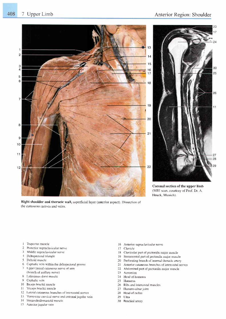
408 7 Upper Limb Anterior Resion: Shoulder
23
1 7
1 3
1 4
1 5
1 6' t7
1 8
Right shoulder and thoracic wall, superficial layer (anterior aspect). Dissection ofthe cutaneous nerves and vetns.
I Trapezius muscle2 Posterior supraclavicular nerve3 Middle supraclavicular nerve4 Deltopectoraltriangle5 Deltoid muscle6 Cephalic vein within the deltopectoral groove7 Upper lateral cutaneous nerve of am
(branch of axillary nerve)8 Latissimus dorsi muscle9 Cephalic vein
10 Biceps brachii muscleI I Triceps brachii muscle12 Laleral cutaneous branches ofintercostal nervesl3 Transverse cervical nerve and extemaljugular veinl4 Sternocleidomastoid musclel5 Anteriorjugularvein
16 Anterior supraclavicular nerve17 Clavicle18 Clavicular part of pectoralis major muscle19 Sternocostal part ofpectoralis major muscle20 Perforating branch of internal thoracic artery21 Anterior cutaneous branches of intercostal nerves22 Abdominal part of pectoralis major muscle23 Acromion24 Headofhumerus25 Humerus26 Ribs and intercostal muscles27 Humero-ulnarjoint28 Head ofradius29 Ulna30 Brachial artery
I
Coronal section of the upper limb(MRI scan, couftesy of Prof. Dr. A.Heuck. Munich).
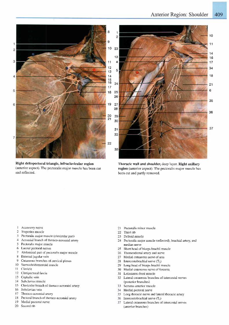
Anterior Region: Shoulder
1 0
1 0
1 1
1 21 3't4
1 51 61 7
23
't3
4
5
24
3132
33
18 2526
19 2728
1' t
1 41 61 7
34
1 8
21
o
35
3620 2921 go
Right deltopectoral triangle, infraclavicular region(anterior aspect). The pectoralis major muscle has been cutand reflected.
I Accessory nerve2 Trapezius muscle3 Pectoralis major muscle (clavicular part)4 Acromial branch of thoraco-acromial artery5 Pectoralis major muscle6 Lateral pectoral nerves7 Abdominal part of pectoralis major muscle8 Extemal jugular vein9 Cutaneous branches ofcervical plexus
l0 Sternocleidomastoidmuscle11 Clavicle12 Clavipectoralfascia13 Cephalic vein14 Subclavius muscle15 Clavicular branch of thoraco-acromial artery16 Subclavian veinl7 Thoraco-acromial artery1 8 Pectoral branch of thoraco-acromial artery19 Medial pectoral nerve20 Second rib
Thoracic wall and shoulder, deep layer. Right axillaryregion (anterior aspect). The pectoralis major muscle hasbeen cut and partly removed.
2l Pectoralis minor muscle22 Thirdib23 Deltoid muscle24 Pectoralis major muscle (reflected), brachial artery, and
median nerve25 Short head of biceps brachii muscle26 Thoracodorsal artery and nerve27 Medial cutaneous nerve of arm28 Intercostobrachial nerve (T2)29 Long head of biceps brachii muscle30 Medial cutaneous nerve offorearm3l Latissimus dorsi muscle32 Lateral cutaneous branches ofintercostal nerves
(posterior branches)Seffatus anterior muscleMedial pectoral nerveLong thoracic nerve and lateral thoracic arteryIntercostobrachial nerve (T3)Lateral cutaneous branches of intercostal nerves(anterior branches)
J J
3+
35365 t
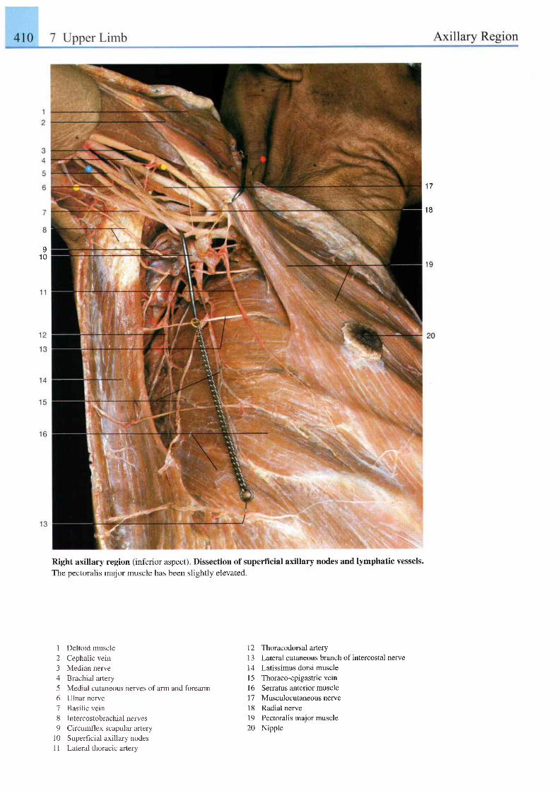
1 7
t 8
I1 0
Right axillary region (inferior aspect). Dissection of superficial axillary nodes and lymphatic vessels.The pectoralis major muscle has been slightly elevated.
1 Deltoid muscle2 Cephalic vein3 Median nerve4 Brachial arlery5 Medial cutaneous nerves of arm and forearm6 Ulnar nerve7 Basilic vein8 Intercostobrachial nerves9 Circumflex scapular artery
10 Superficial axillary nodesI I Lateral thoracic artery
12 Thoracodorsal artery1 3 Lateral cutaneous branch of intercostal nerve14 Latissimus dorsi musclel5 Thoraco-epigastric veinl6 Serratus anterior muscle17 Musculocutaneousnerve18 Radial nervel9 Pectoralis major muscle20 Nipple
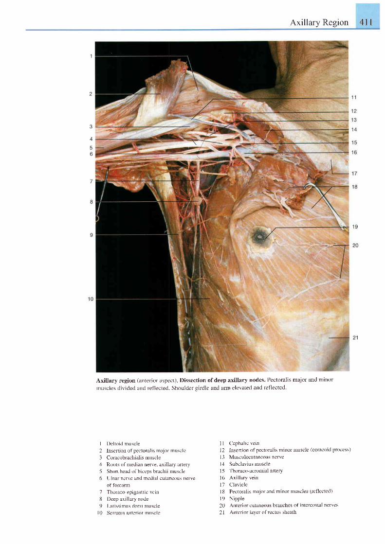
Axillary Region 4ll
Axillary region (anterior aspect). Dissection of deep axillary nodes. Pectoralis major and minor
muscles divided and reflected. Shoulder sirdle and arm elevated and reflected.
I Deltoid muscle
2 Insertion of pectoralis major muscle
3 Coracobrachial is muscle
4 Roots ofmedian nerve, axillary artery
5 Short head of biceps brachii muscle
6 Ulnar nerve and medial cutaneous nerve
of forearm
7 Thoraco-epigastricvein
8 Deep axillary node
9 Latissimus dorsi muscle
l0 Serratus anterior muscle
1l Cephalic veinl2 Insertion ofpectoralis minor muscle (coracoid process)
13 Musculocutaneous nervel4 Subclavius muscle15 Thoraco-acromial arteryl6 Axillary veinl7 Claviclel8 Pectoralis major and minor muscles (reflected)
19 Nipple20 Anterior cutaneous branches of intercostal nerves
21 Anterior layer ofrectus sheath
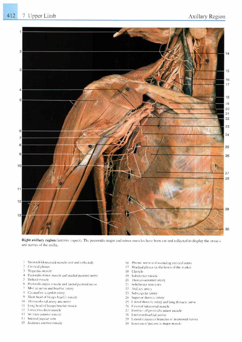
412 7 Upper Lirnb Axillary Region
i":iiiiri::r
L lrL',.; i i . r
l o
1 7
27
28
t a t
I Y
2Az l
22
Z J
24I.i'rl f '
:1
I 6 l)hrerric lrcr vc uncl ascending ccrvicill arteryI 7 Brachial p lerus (at the levels of thc t r unks)
I l l ( l l a v i c l e
| 9 Subc lav i us n rusc l c
20' I l rot ' l tco- l rcr-ornia l ar tery
2 l Suhc la r . i an r , e i n l cu r )l l Ax i l l a r v a r t c r v
23 Subscapular ar tcrv
2.1 SLrpcr ior thoracic ar tcrv
25 Latelel thomcic urtery and krng thor-acrc nert.e26 Erterrn l intercostal ntuscle) l lnsct ' t ion o1' pectoral is l l inr t r r ruscle
l8 Iutcrcostobrachial nerrc '
l9 Lateral cutar)e 'ous br-anches of intcrcostal nerves
30 luscrt ion o1 pectoral is rnajor muscle
Right axi l lary region (arrtcr ior aspcct). The pcctolal is rrajor ancl minur lnuscles have been cut and ref lected to display the vesselsancl nerr, 'cs of thc axi l la
I Stcntoclc i t lomasloid ntusclc (cut and rc l ' lectet l )2 Ccr-r icul p lcrus
3 ' l ' r
apczius nrr- rsc le. l Pcctoral is nt inor nruscle and nrccl ia l pectoral ncn'e5 Dci lo id ntuscle
6 Pectoral is nur jor-r rusclc ancl lateral pectorul ncnc7 Nlccl ian ncrr . .e and brachiul ar- tcrv13 Cir-cr , rnr f lex scapular ar tcry() Short hcat l o l b icel ts brachi i musclc
l0 Thortrcodor-sal l r r torv tnd ncr- ' " 'cI I l - ong hcud o l ' b i ceps b rach i t t l u sc l ci l La t i s s i n rus t l o t ' s i musc l c| . l S . t t i r l L t s ; l t ) l c f i ( r l i l l u \ L l L .
l : l I r r tcrnal . j r - ruular veiu
l -5 Scalerrus anter ior r rusclc
e. / t " '
f" /-.r'
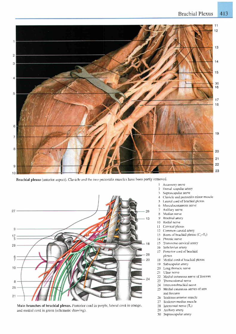
Brachial Plexus 413
30t o
1 71 8
3
1 75
29
Brachial plexus (anterior aspect). Clavicle and the two pectoralis muscles have been partly removed.
1
234561
268
1 3 91 01 l1 21 3t 4
1 8 1 5l 61 7
28
20 181 9202 122
9AL-)
2425
262'7282930
Accessory nerve
Dorsal scapular artery
Suprascapular nerve
Clavicle and pectoralis minor muscle
Lateral cord of brachial PlexusMusculocutaneous nerve
Axillary nerve
Median nerve
Brachial artery
Radial nerve
Cervical plexus
Common carotid aftery
Roots of brachial plexus (Cs-Tr)
Phrenic nerve
Transvetse cervical arterY
Subclavian artery
Posterior cord of brachial
plexus
Medial cord of brachial plexus
Subscapular artery
Long thoracic nerve
Ulnar nerve
Medial cutaneous nerve of forearm
Thoracodorsal nerve
Intercostobrachial nerve
Medial cutaneous netves of arm
and forearm
Scalenus anterior muscle
Scalenus medius muscle
Intercostal nerve (Tr)
Axillary artery
Suprascapular artery
Main branches of brachial plexus. Posterior cord in purple, lateral cord in orange,
and medial cord in green (schematic drawing).
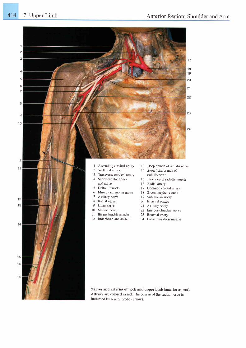
4t4 7 Upper Limb Anterior Region: Shoulder and Arm
1
z
5
6
7
1 81 A
20
1234
56789
1 01 l1 2
Ascending cervical artery
Vertebral artery
Transverse cervical anerySuprascapular artery
and nerve
Deltoid muscle
Musculocutaneous nerveAxillary nerve
Radial nerve
Ulnar nerve
Median nerve
Biceps brachii muscle
Brachioradialis muscle
l3 Deep branch ofradialis nervel4 Superfrcial branch of
radialis nerveI 5 Flexor carpi radialis muscle16 Radial artery17 Common carotid arteryl8 Brachiocephalictrunk19 Subclavian arlery20 Brachial plexus2l Axillary artery22 Intercostobrachialnerve23 Brachial artery24 Latissimus dorsi muscle
Nerves and arteries of neck and upper limb (anterior aspect).Afteries are colored in red. The course of the radial nerve isindicated by a wire probe (arrow).
1 7
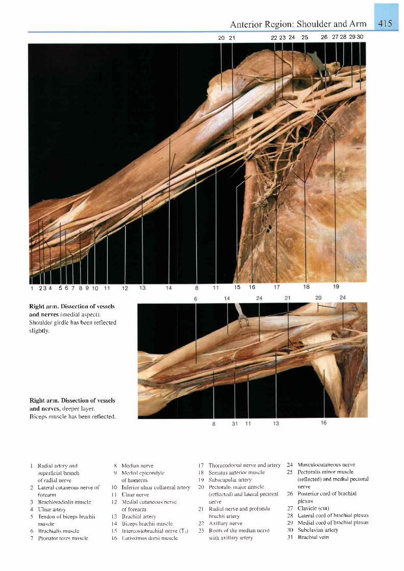
Anterior Resion: Shoulder and Arm 415
2 3 4 5 6
Right arm. Dissection of vessels
and nerves (medial aspect).
Shoulder girdle has been reflected
sl ightly
Right arm. Dissection of vesselsand nerves, deeper layer.Biceos muscle has been reflected.
3/l
5
Radial aftery and
superficial branch
of radial nerve
Lateral cutaneous nerve of
fbrearm
Brachioradialis muscle
Ulnar artery
Tendon of biceps brachii
muscle
Brachialis muscle
Pronator teres muscle
6'7
8 Median nerve9 Medial epicondyle
of humerusl0 Inferior ulnar collateral arteryI 1 Ulnar nervel2 Medial cutaneous nerve
of forearm13 Brachial arteryl4 Biceps brachii musclel5 Intercostobrachial nerve (T.)16 Latissimus dorsi muscle
17 Thoracodorsal nerve and artery
l8 Serratus anterior muscle
19 Subscapular artery
20 Pectoral is major muscle
(reflected) and lateral Pectoralnerve
2l Radial nerve and profunda
brachii artery
22 Axillary nerve
23 Roots of the median nerve
with axillary artery
2223 24 25 26 2728 2930
24 Musculocutaneous nerve25 Pectoralis minor muscle
(reflected) and medial pectoralnerve
26 Posterior cord ofbrachialplexus
27 Clavicle (cut)28 Laleral cord ofbrachial Plexus29 Medial cord ofbrachial Plexus30 Subclavian artery3l Brachial vein
21
15 ' t 6I
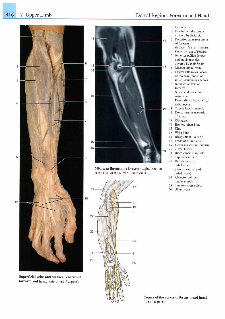
416 7 Upper Limb Dorsal Region: Forearm and Hand
MRI scan through the forearm (sagittal sectionat the level of the humero-ulnar ioint).
211 9
1 Cephalic vein2 Brachioradialismuscle
covered by its fascia3 Posterior cutaneous nerve
of forearm(branch of radialis nerve)
4 Cephalic vein offorearm5 Extensor pollicis longus
and brevis musclescovered by their fascia
6 Median cubital vein'7 Lateral cutaneous nerves
of forearm (branch ofmusculocutaneous nerve)
8 Intermedian vein offorearm
9 Superhcial branch ofradial nerve
10 Dorsal digital branches ofradial nerve
I I Triceps brachii musclel2 Dorsal venous network
of hand13 Olecranon14 Humero-ulnar joint15 Ulnal6 Wrist joint17 Biceps brachii muscle18 Trochlea ofhumerus19 Flexor muscles offorearm20 Carpal bones21 Brachioradialismuscle22 Supinator muscle23 Deep branch of
radial nerve(ramus profundus ofradial nerve)Abductor pollicislongus muscleExtensor retinaculumUlnar nerve
Course of the nerves to forearm and hand(dorsal aspect).
1 7
1 86 . , ,
1 4
7
1 9
20
2526
Superficial veins and cutaneous nerves offorearm and hand (anteromedial aspect).
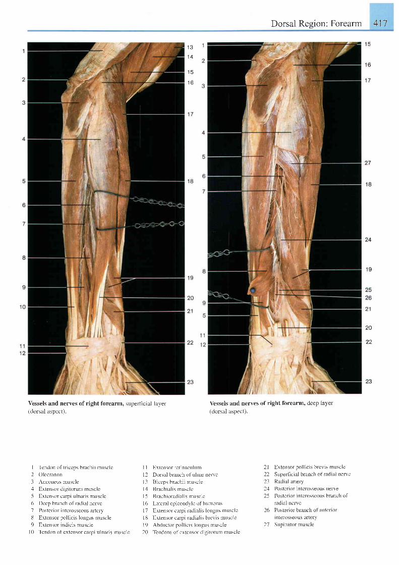
Dorsal Region: Forearm 417
1 3
1 4
t o
20
22
Vessels and nerves ofright forearm, superficial layer(dorsal aspect).
I Tendon of triceps brachii muscle
2 Olecranon
3 Anconeus muscle
4 Extensor digitorum muscle
5 Extensor carpi ulnaris muscle
6 Deep branch of radial nerve
7 Posterior interosseous artery
8 Extensor pol l ic is longus muscle
9 Extensor indicis muscle
| 0 Tendon of extensor carpi ulnaris muscle
Vessels and nerves of right forearm, deep layer
(dorsal aspect).
I I Extensor retinaculum
I 2 Dorsal branch of ulnar nerve
l3 Biceps brachi i musclc
l4 Brachial is muscle
l5 Brachioradia l is muscle
l6 Lateral epicondyle of humerus
l7 Extensor carpi radia l is longus muscle
l8 Extensor carpi radialis brevis muscle
l9 Abductor pol l ic is longus muscle
20 Tendons ofextensor digitorurn muscle
21 Extensor pollicis brevis muscle
22 Superficial branch ofradial nerve
23 Radial artery
24 Posterior interosseous nerve
25 Posterior interosseous branch of
radial nerve
26 Poster ior branch ol anter ior
interosseous artery
27 Supinator muscle
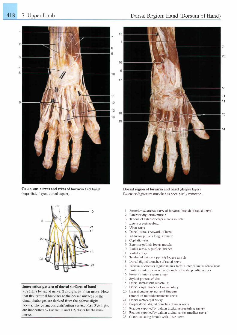
418 7 Upper Limb Dorsal Region: Hand (Dorsum of Hand)
1 0
211 1
I J
Cutaneous(superficial
nerves and veins of forearm and handlayer, dorsal aspect).
1 0
251 3
Innervation pattern of dorsal surfaces of hand2tlzdigits by radial nerve,2tlzdigits by ulnar nerve. Notethat the terminal branches to the dorsal surfaces of thedistal phalanges are derived from the palmar digitalnerves. The cutaneous distribution varies; often 3rlz digitsare innervated by the radial and Illz digits by the ulnarnerve.
Dorsal region offorearm and hand (deeper layer).Extensor digitorum muscle has been partly removed.
I Posterior cutaneous nerve of forearm (branch of radial nerve)
2 Extensor digitorum muscle
3 Tendon ofextensor carpi ulnaris muscle
4 Extensor retinaculum
5 Ulnar nerve
6 Dorsal venous network ofhand
7 Abductor pollicis longus muscle
8 Cephalic vein
9 Extensor pollicis brevis muscle
l0 Radial nerve, superficial branch
I I Radial artery
12 Tendon ofextensor pollicis longus muscle
l3 Dorsal digital branches ofradial nerve
l4 Tendons of extensor digitorum muscle with interlendinous connections
I 5 Posterior interosseus nerve (branch of the deep radial nerve)
l6 Posterior interosseous artery
l7 Styloid process of ulna
l8 Dorsal interosseus muscle IV
19 Dorsal carpal branch of radial artery
2O Lateral cutaneous nerve offorearm(branch of musculocutaneous nerve)
2l Dorsal metacarpal artery
22 Proper dorsal digital branches ofulnar nerve
23 Regions supplied by palmar digital nerves (ulnar nerve)
24 Regions supplied by palmar digital nerves (median nerve)
25 Communicatins branch with ulnar nerve
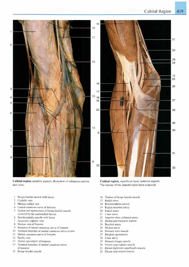
Cubital Region
2324
1 325
26
1 7
1 3 t B1 9
20
Cubital region (anterior aspect), dissection of cutaneous nerves
and veins.
I Biceps brachii muscle with fascia
2 Cephalic vein
3 Median cubital vein
4 Lateral cutaneous nerve offorearm
5 Tendon and aponeurosis ofbiceps brachii muscle(covered by the antebrachial fascia)
6 Brachioradialis muscle with fascia
7 Accessory cephalic vein
8 Median vein of forearm
9 Branches oflateral cutaneous nerve offorearml0 Terminal branches ofmedial cutaneous nerve ofarmI I Medial cutaneous nerve of forearm
12 Basi l ic vein
l3 Medial epicondyle of humerus
l4 Terminal branches ofmedial cutaneous nerveof forearm
l5 Biceps brachii muscle
Cubital region, superficial layer (anterior aspect).
The fasciae of the muscles have been removed.
16 Tendon ofbiceps brachii muscle17 Radial nervel8 Brachioradialismusclel9 Radial recurrent artery20 Radial artery2l Ulnar nerve22 Superior ulnar collateral artery23 Medial intermuscular septum24 Brachial artery25 Median nerve26 Pronator teres muscle2'7 Bicrpilal aponeurosis28 Ulnar artery29 Palmaris longus muscle30 Flexor carpi radialis muscle31 Flexor digitorum superltcialis muscle32 Flexor carpi ulnaris muscle
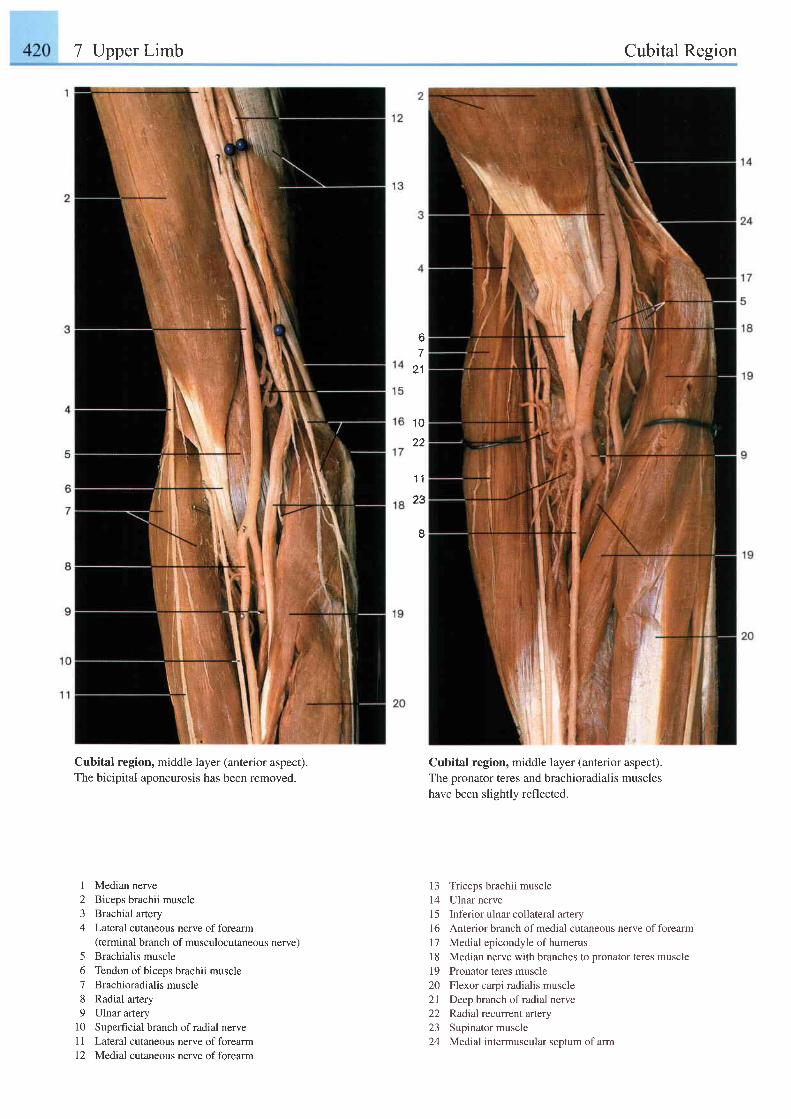
7 Upper Limb Cubital Region
6.,
21
1 0
22
1 1
23
I
Cubital region, middle layer (anterior aspect).The bicipital aponeurosis has been removed.
Cubital region, middle layer (anterior aspect).The pronator teres and brachioradialis muscleshave been slightly reflected.
13 Triceps brachii muscle14 Ulnar nerve15 Inferiorulnarcollateral artery16 Anterior branch of medial cutaneous nerve of forearm17 Medial epicondyle of humerus18 Median nerve with branches to Dronator teres muscle19 Pronator teres muscle20 Flexor carpi radialis muscle21 Deep branch ofradial nerve22 Radial recunent aftery23 Supinator muscle24 Medial intermuscular septum of arm
I2J
4
5678o
1 01 lI L
Median nerveBiceps brachii muscleBrachial arteryLateral cutaneous nerve of forearm(terminal branch of musculocutaneous nerve)Brachialis muscleTendon of biceps brachii muscleBrachioradialis muscleRadial arteryUlnar arterySuperficial branch of radial nerveLateral cutaneous nerve of forearmMedial cutaneous nerve of forearm
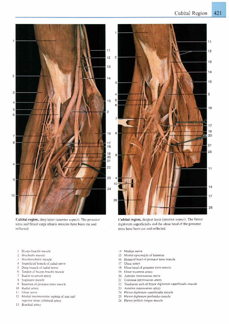
layer (anterior aspect). The pronator
ulnaris muscles have been cut and
Cubital Region
Cubital region, deepest layer (anterior aspect). The flexordigitorum superficialis and the ulnar head of the pronatorteres have been cut and reflected.
14 Median nervei5 Medial epicondyle of humerusl6 Humeral head of pronator teres muscle17 Ulnar arteryl8 Ulnar head of pronator teres musclel9 Ulnar recurrent artery20 Anterior interosseous nerve21 Common interosseous artery22 Tendinous arch of flexor digitorum superficialis muscle23 Anterior interosseous artery24 Flexor digitorum superficialis muscle25 Flexor digitorum profundus muscle26 Flexor pollicis longus muscle
1 1
1 2
1 5 A
6
1 6
1 71 8
1 9 q2021
22
Cubital region, deepteres and flexor carpireflected.
i Biceps brachii muscle2 Brachial is muscle3 Brachioradialismuscle4 Superficial branch ofradial nerve5 Deep branch ofradial nerve6 Tendon of biceps brachii muscle7 Radial recurrent artery8 Supinator muscle9 Insertion of pronator teres muscle
l0 Radial arteryI I Ulnar nerve12 Medial intermuscular septum of arm and
superior ulnar collateral arteryl3 Brachial artery
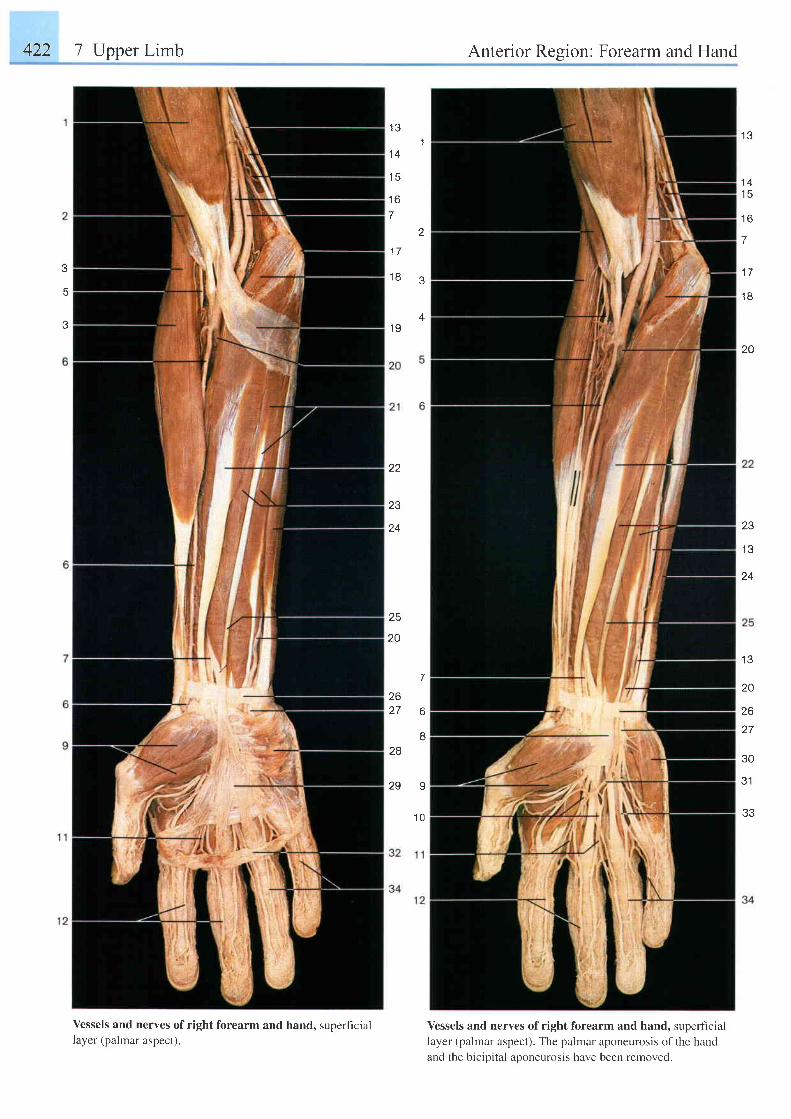
422 7 Upper Limb Anterior Resion: Forearm and Hand
1 31
1 4
1 5
1 6
7
2
1 7
1 8 3
1 9
1 3
1 4t c
t o
7
1 7
1 8
20
24
tc
20
23
1 3
24
t \ t
20
26t-7
,5U
3.1
33
7
262 7 6
B28
2 9 9
1 0
Vessels and nerves of right forearm and hand, superficiallayer (palmar aspect).
Vessels and nerves of right forearm and hand, superficiallayer (palmar aspect). The palmar aponeurosis of the handand the bicipital aponeurosis have been removed.
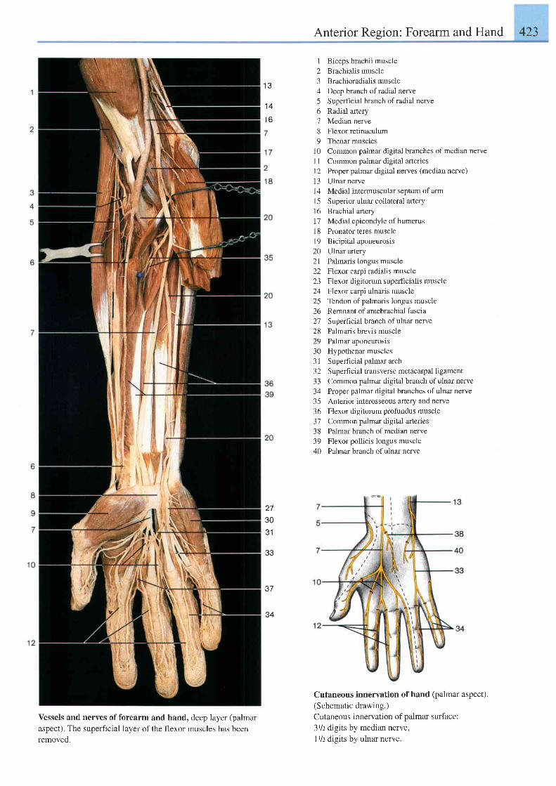
Anterior Region: Forearm and Hand 423
Vessels and nerves of forearm and hand, deep layer (palmaraspect). The superficial layer ofthe flexor muscles has beenremoved.
1 Biceps brachii muscle2 Brachialis muscle3 Brachioradialis muscle4 Deep branch ofradial nerve5 Superhcial branch ofradial nerve6 Radial artery7 Median nerve8 Flexor retinaculum9 Thenar muscles
l0 Common palmar digital branches of median nerve1l Common palmar digital arteriesI 2 Proper palmar digital nerves (median nerve)13 Ulnar nervel4 Medial intermuscular septum of arm15 Superior ulnar collateral artery16 Brachial artery17 Medial epicondyle of humerus18 Pronator teres musclel9 Bicipital aponeurosis20 Ulnar artery21 Palmaris longus muscle22 Flexor carpi radialis muscle23 Flexor digitorum superficialis muscle24 Flexor carpi ulnaris muscle25 Tendon of palmaris longus muscle26 Remnant ofantebrachial fascia27 Superficial branch ofulnar nerve28 Palmaris brevis muscle29 Palmar aponeurosis30 Hypothenar muscles31 Superficial palmar arch32 Superficial transverse metacarpal ligament33 Common palmar digital branch of ulnar nerve34 Proper palmar digital branches of ulnar nerve35 Anterior interosseous artery and nerve36 Flexor digitorum profundus muscle37 Common palmar digital arteries38 Palmar branch ofmedian nerve39 Flexor pollicis longus muscle40 Palmar branch ofulnar nerve
Cutaneous innervation of hand (palmar aspect).(Schematic drawing.)Cutaneous innervation of palmar surface:31/z digits by median nerve,I t/z digits by ulnar nerve.
{ ?
1 4
1 6
273031
33
37
34
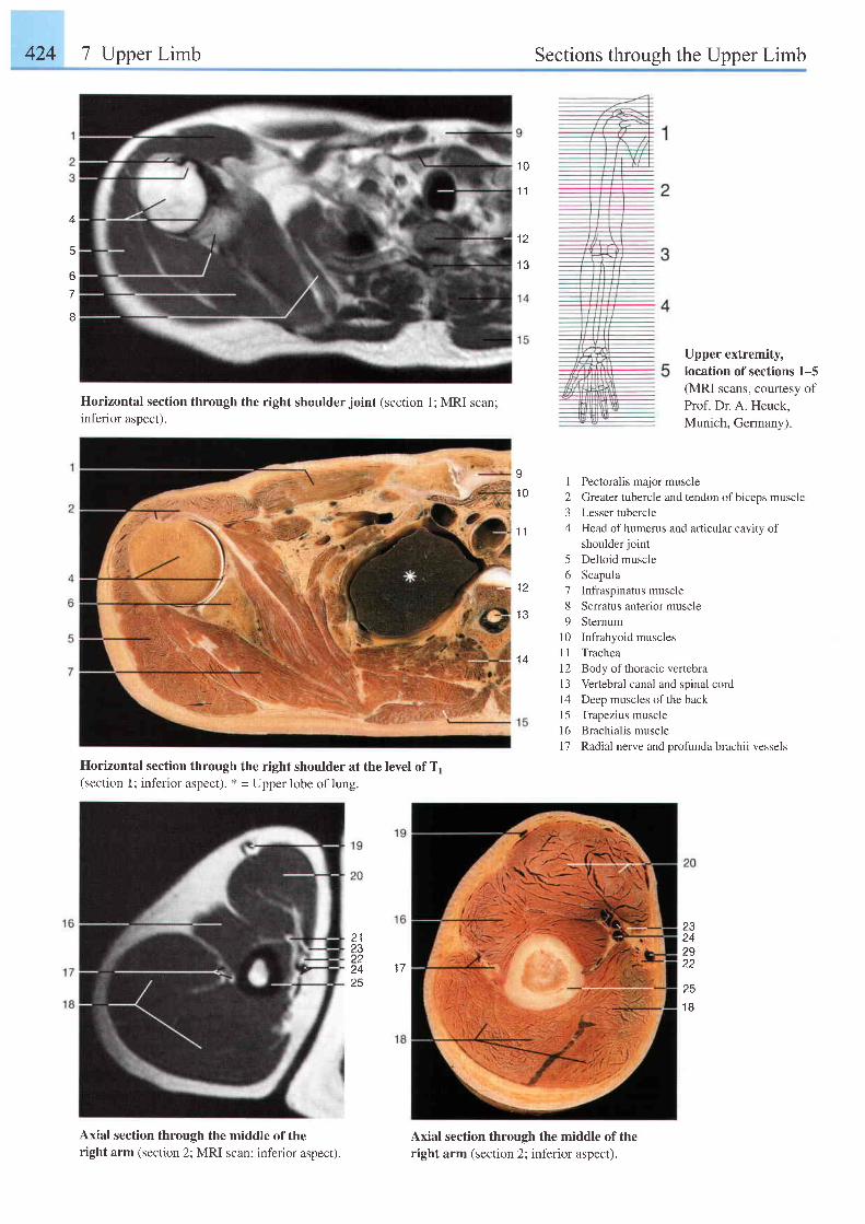
424 7 Upper Limb Sections through the Upper Limb
5
tj
7
I
1 0
1 1
t 1
1 3
Upper extremity,Iocation of sections 1-5(MRI scans, courtesy ofProf. Dr. A. Heuck,Munich, Germany).
Horizontal section through the right shoulder joint (section 1; MRI scan;inferior aspect).
Horizontal section through the right shoulder at the level ofTl(section 1; inferior aspect). * = Upper lobe of lung.
Axial section through the middle of theright arm (section 2; MRI scan; inferior aspect).
s1 0
1 1
t z
Axial section through the middle of theright arm (section 2; inferior aspect).
I Pectoralis major muscle2 Greater tubercle and tendon of biceps muscle3 Lesser tubercle4 Head ofhumerus and articular cavity of
shoulder joint5 Deltoid muscle6 Scapula7 Infraspinatus muscle8 Serratus anterior muscle9 Sternum
10 Infrahyoid musclesI 1 Trachea12 Body ofthoracic vertebra13 Vertebral canal and spinal cord14 Deep muscles of the back15 Trapezius musclel6 Brachialis muscle17 Radial nerve and profunda brachii vessels
2123zz24 1725
23242922
251 8
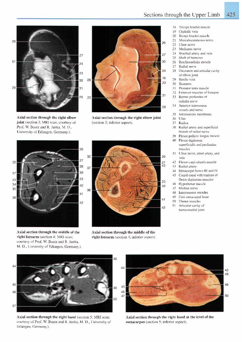
Sections through the Upper Limb 425
Axial section through the right elbowjoint (section 3; MRI scan; courtesy ofProf. W Bautz and R. Janka. M. D..University of Erlangen, Germany.)
Axial section through the middle of theright forearm (section 4; MRI scan;courtesy of Prof. W. Bautz and R. Janka,M. D., University of Erlangen, Germany.).
Axial section through the right elbowjoinl(section 3; inferior aspect).
Axial section through the middle of the
right forearm (section 4; inferior aspect).
18 Triceps brachii muscle19 Cephalic vein20 Biceps brachii muscle2l Musculocutaneousnerve22 Ulnar nerve23 Medianus nerve24 Brachial artery and vein25 Shaft ofhumerus26 Brachioradialis muscle27 Radial nerve28 Olecranon and articular cavity
of elbow jointBasilic veinHumerusPronator teres muscleExtensor muscles of forearmRamus profundus ofradialis nerveAnterior interosseusvessels and nerve
35 Interosseous membrane36 Ulna37 Radius38 Radial arlery and superficial
branch of radial nerve39 Flexor pollicis longus muscle40 Flexor digitorum
superficialis and profundusmuscles
41 Ulnar nerve, ulnar artery, andvelnFlexor carpi ulnaris muscleRadial arteryMetacarpal bones III and IVCarpal canal with tendons offlexor digitorum musclesHypothenar muscleMedian nerveInterosseous musclesFirst metacarpal boneThenar musclesArticular cavity ofhumeroradial joint
26
Z I
30
z q
23
J I22
24
23
31
29
29303 t32-l -l
34
37
333435
26
L I
43
2340
/ a
4344
45
46
4849505 1
41
4348
45
46
Axial section through the right hand (section 5; MRI scan;courtesy of Prof. W. Bautz and R. Janka, M. D., University ofErlangen, Germany.).
Axial section through the right hand at the level of themetacarpus (section 5; inferior aspect).
49
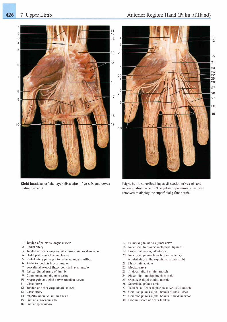
426 7 Upper Limb Anterior Region: Hand (Palm of Hand)
1 ' tt z
1 c 1
45
1 4 2 0
1 56
1 11 3
1 4
27
281 7
30
1 9
21
2324222526
20
1 6
c'
291 7
1 8
1 9
Right hand, superficial layer, dissection of vessels and nerves(palmar aspect).
I Tendon of palmaris longus muscle2 Radial artery3 Tendon offlexor carpi radialis muscle and median nerve4 Distal part of antebrachial fascia5 Radial artery passing into the anatomical snuffbox6 Abductor pollicis brevis muscle7 Superfrcial head offlexor pollicis brevis muscle8 Palmar digital artery of thumb9 Common palmar digital arteries
10 Proper palmar digital nerves (median nerve)1 1 Ulnar nerve12 Tendon offlexor carpi ulnaris musclel3 Ulnar ateryl4 Superhcial branch ofulnar nervel5 Palmaris brevis musclel6 Palmar aponeurosis
Right hand, superficial layer, dissection of vessels and
nerves (palmar aspect). The palmar aponeurosis has been
removed to display the superficial palmar arch.
l7 Palmar digital nerves (ulnar nerve)l8 Superhcial transverse metacarpal ligamentl9 Proper palmar digital arteries20 Superhcial palmar branch of radial artery
(contributing to the superficial palmar arch)2l Flexor retinaculum22 Median nerve23 Abductor digiti minimi muscle24 Flexor digiti minimi brevis muscle25 Opponens digiti minimi muscle26 Superficial palmar arch27 Tendons offlexor digitorum superhcialis muscle28 Common palmar digital branch of ulnar nerve29 Common palmar digital branch of median nerve30 Fibrous sheath offlexor tendons
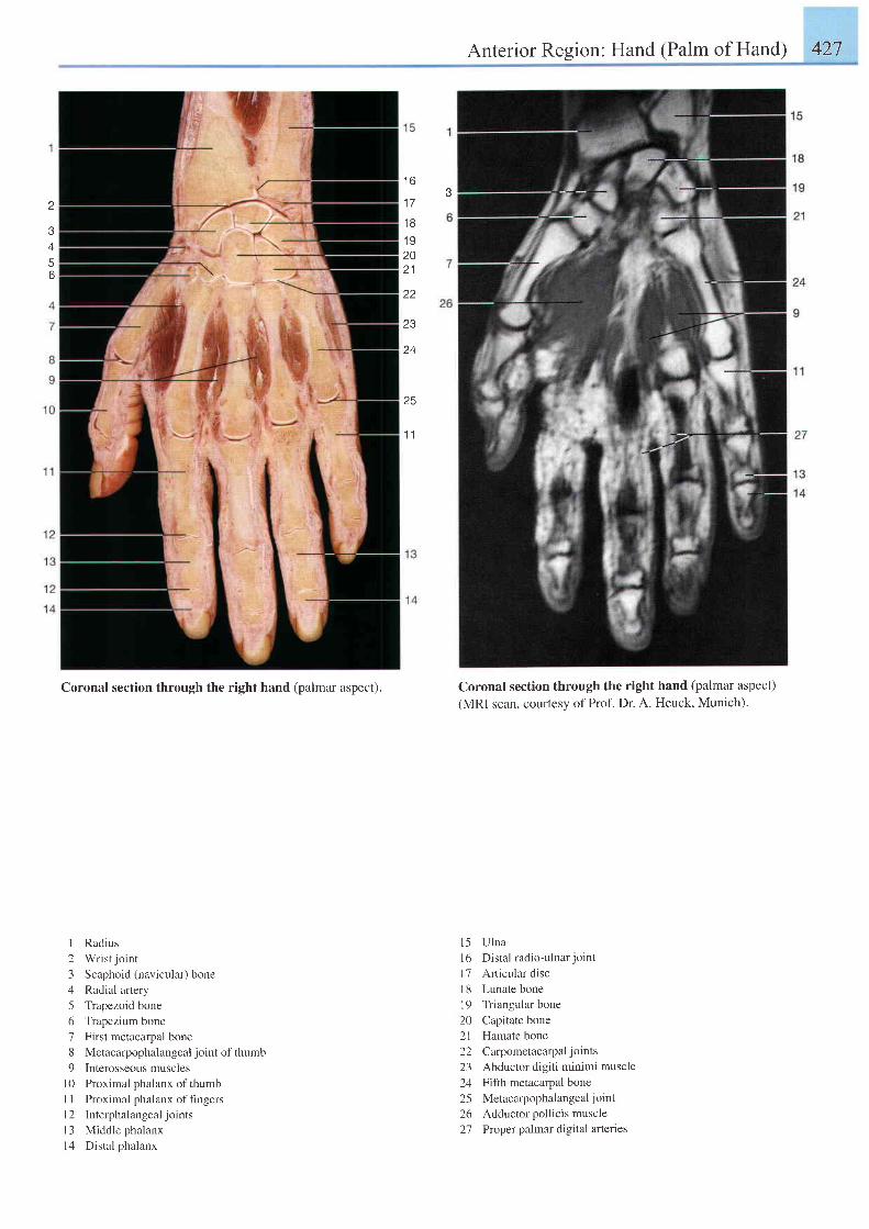
Anterior Region: Hand (Palm of Hand) 427
I O?
1 7
4
56
1 81 92021
zz
23
24
25
1 1
Coronal section through the right hand (palmar aspect).
I Radius
2 Wrist jo int
3 Scaphoid (navicular) bone
4 Radial artery
5 Trapezoid bone
6 Trapezium bone
7 First metacarpal bone
8 Metacarpophalangeal joint of thumb
9 Interosseous muscles
l0 Proximal phalanx of thumb
I I Proximal phalanx of fingers
l2 Interphalangealjoints
l3 Middle phalanx
l4 Distal phalanx
Coronal section through the right hand (palmar aspect)(MRI scan, courtesy of Prof. Dr. A. Heuck, Munich)'
l 5 U lna
l6 Distal radio-ulnarjoint
l7 Articular disc
I 8 Lunate bone
l9 Triangular bone
20 Capitate bone
21 Hamate bone
22 Carpometacarpal joints
23 Abductor digiti minimi muscle
24 Fifth metacarpal bone
25 Metacarpophalangealjoint
26 Adductor pollicis muscle
27 Proper palmar d ig i ta l ar ter ies
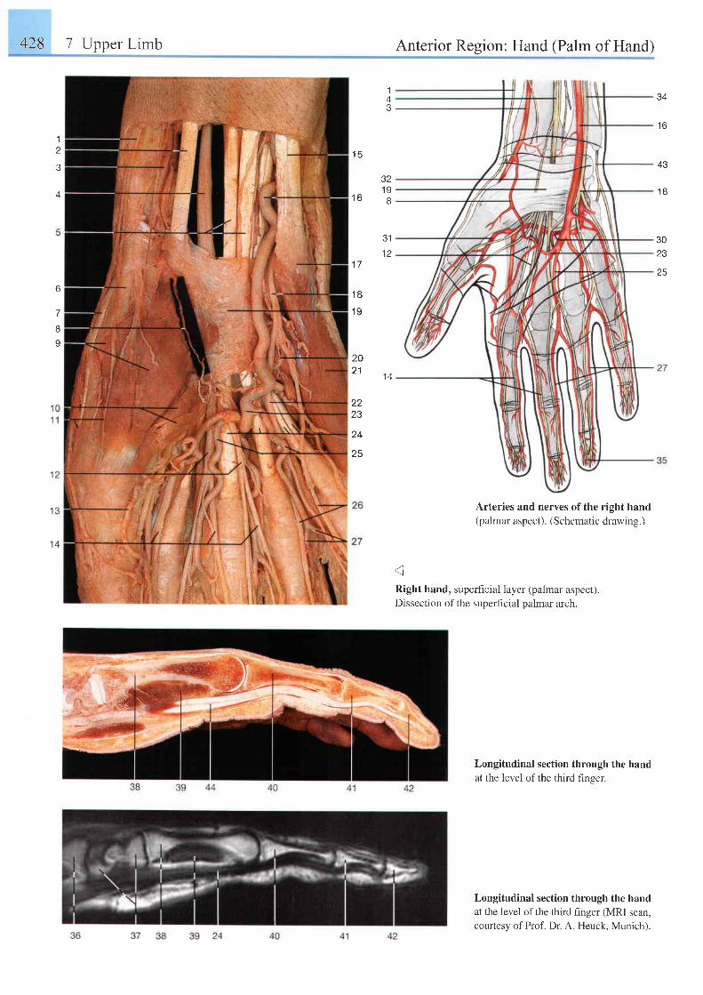
428 I Upper Limb Anterior Region: Hand (Palm of Hand)
1
4
6
7
I
I
1 5
A A
t o
43
1 B
3023
25
1A
321 9
r o g
1 7
1 8' t9
3112
20z l
22?3
24
25
1 4
@
Right hand, superficial layer (palmar aspect).Dissection of the superficial palmar arch.
Arteries and nerves of the right hand(palmar aspect). (Schematic drawing.)
Longitudinal section through the handat the level of the third finser
Longitudinal section through the handat the level of the third finger (MRl scan,courtesy of Prof. Dr A. Heuck, Munich).
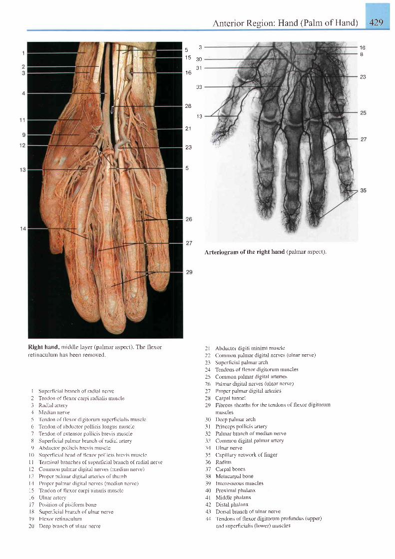
Anterior Resion: Hand (Palm of Hand) 429
5 -1 5 s o
a 1
1 6
e a
28
I J
21
23
t p
I
23
Right hand, middle layer (palmar aspect). The flexor
retinaculum has been removed.
I Superficial branch of radial nerve
2 Tendon of flexor carpi radialis muscle
3 Radial artery
4 Median nerve
5 Tendon of flexor digitorum superficialis muscle
6 Tendon of abductor pollicis longus muscle
7 Tendon of extensor pollicis brevis muscle
8 Superficial palmar branch of radial artery
9 Abductor pol l ic is brevis muscle
l0 Superficial head offlexor pollicis brevis muscle
I 1 Terminal branches of superficial branch of radial nerve
l2 Common palmar digital nerves (median nerve)
l3 Proper palmar digital arteries of thumb
l4 Proper palmar digital nerves (median nerve)
15 Tendon of flexor carpi ulnaris muscle
l6 Ulnar ar tery
l7 Position of pisifbrm bone
l8 Superficial branch ofulnar nerve
l9 Flexor retinaculum
20 Deep branch ofulnar nerve
Arteriogram of the right hand (palmar aspect).
2l Abductor digiti minimi muscle22 Common palmar digital nerves (ulnar nerve)23 Superficial palmar arch24 Tendons of flexor digitorum muscles2-5 Common palmar digital arteries26 Palmar digital nerves (ulnar nerve)27 Proper prlmar digital arteries28 Carpal tunnel29 Fibrous sheaths for the tendons of flexor digitorum
muscles30 Deep palmar arch3 I Princeps pollicis artery32 Palmar branch of median nerve33 Common digital palmar artery34 Ulnar nerve35 Capillary network ofhnger36 Radius37 Carpal bones38 Metacarpal bone39 Interosseousmuscles40 Proximal phalanx4l Middle phalanx42 Distal phalanx43 Dorsal branch ofulnar nerve44 Tendons of flexor digitorum profundus (upper)
and superficialis (lower) muscles
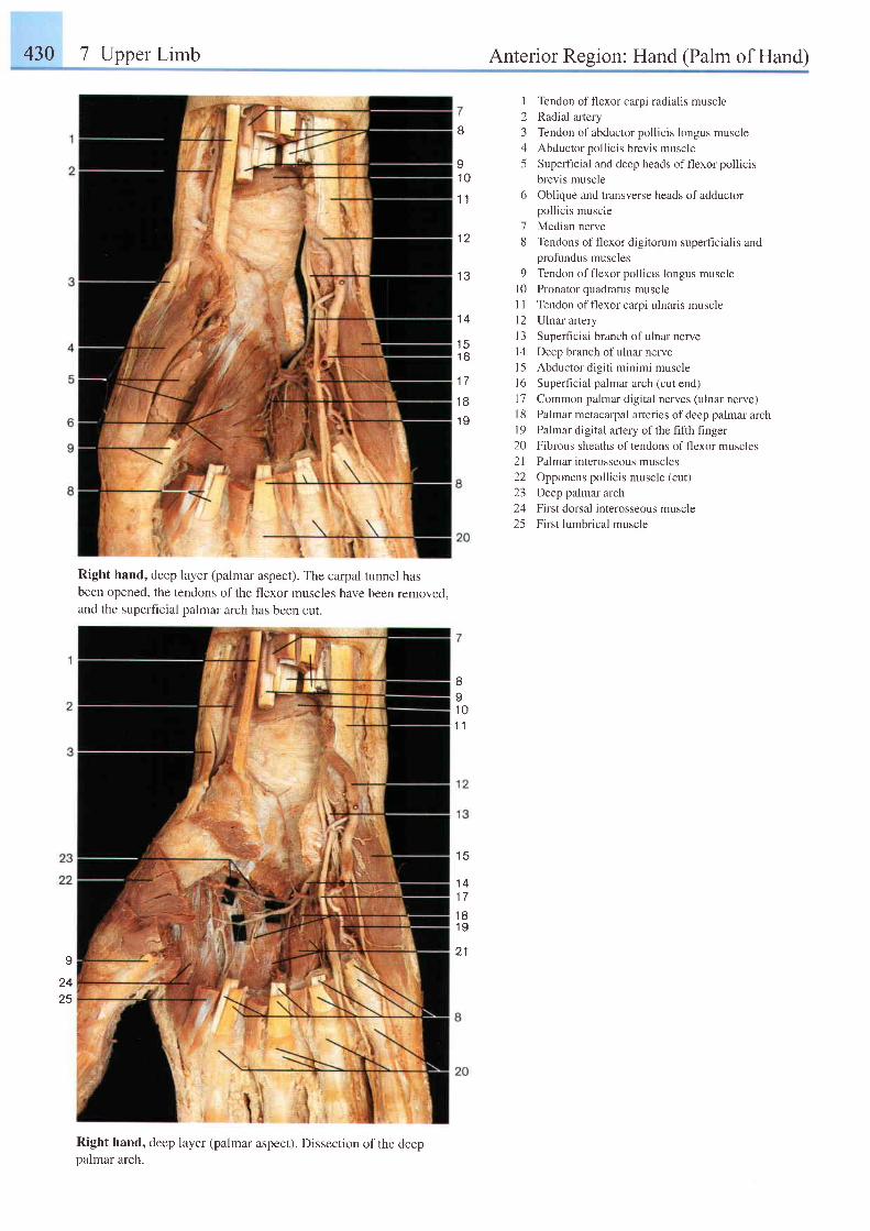
430 7 Upper Limb Anterior Region: Hand (Palm of Hand)
tJ
I1 0
1 1
1 2
r 2
I Tendon offlexor carpi radialis muscle
2 Radial artery
3 Tendon of abductor pollicis longus muscle
4 Abductor pollicis brevis muscle
5 Superhcial and deep heads offlexor pollicis
brevis muscle
6 Oblique and transverse heads of adductorpollicis muscle
7 Median nerve
8 Tendons of flexor digitorum superhcialis andprofundus muscles
9 Tendon offlexor pollicis longus muscle
l0 Pronator quadratus muscle
I I Tendon offlexor carpi ulnaris muscle
l2 Ulnar artery
I 3 Superficial branch of ulnar nerve
l4 Deep branch of ulnarnerve
l5 Abductor digiti minimi muscle
l6 Superficial palmzr arch (cut end)
17 Common palmar digital nerves (ulnar nerve)
18 Palmar metacaryal arteries of deep palmar arch
19 Palmar digital artery of the fifth finger
20 Fibrous sheaths of tendons of flexor muscles
2l Palmar interosseous muscles
22 Opponens pollicis muscle (cut)
23 Deep palmar arch
24 First dorsal interosseous muscle
25 First lumbrical muscle
1 4
1 5t o
1 7'18
1 0
Right hand, deep layer (palmar aspect). The carpal tunnel hasbeen opened, the tendons of the flexor muscles have been removed,and the superficial palmar arch has been cut.
Right hand, deep layer (palmar aspect). Dissection of the deeppalmar arch.
II1 01 1
1 5
1 41 7
1 81 9
2 1I
2425
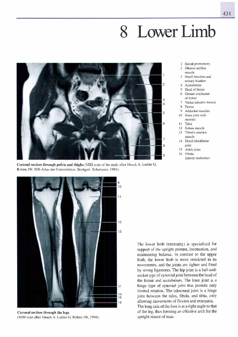
431
8 Lowerlirnb
1 Sacral promontory2 Gluteus medius
muscle3 Small intestine and
urinary bladder4 Acetabulum5 Head of femur6 Greater trochanter
of femur7 Vastus lateralis muscle8 Femur9 Adductor muscles
10 Knee joint with
Coronal section through pelvis and thighs (MRI scan of the mde after Heuck A, Luttke G,Rohen JW. MR-Atlas der Extremitdten. Stuttsart: Schattauer. 1994).
81 0
1 1
menlsclTibiaSoleus muscleTibialis anteriormuscle
14 Distal tibiofibularjoint
l5 Ankle jointl6 Fibula
(lateral malleolus)
The lower limb (extremity) is specialized forsupport of the upright posture, locomotion, andmaintaining balance. In contrast to the upperlimb, the lower limb is more restricted in itsmovements, and the joints are tighter and frxedby strong ligaments. The hip joint is a ball-and-socket type of synovialjoint between the head ofthe femur and acetabulum. The knee joint is ahinge type of synovial joint that permits onlylimited rotation. The talocrural joint is a hingejoint between the talus, fibula, and tibia, onlyallowing movements of flexion and extension.The long axis ofthe foot is at a right angle to thatof the leg, thus forming an effective arch for theupright stance of man.
l t1213
12
1 3
1 41 5
t o
Coronal section through the legs(MRI scan after Heuck A, Luttke G, Rohen JW 1994).
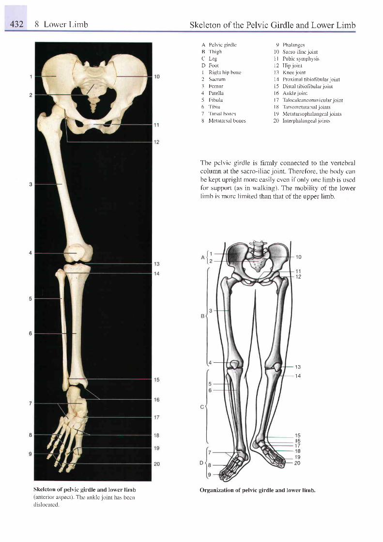
432 8 Lower Limb Skeleton of the Pelvic Girdle and Lower Limb
A Pelvic girdleB ThighC LegD FootI Right hip bone2 Sacrum3 Femur4 Patella5 Fibula6 Tibia7 Tarsal bones8 Metatarsal bones
9 Phalanges10 Sacro-i l iacjointI I Pubic symphysis12 Hipjointl3 Knee joint14 Proximal tibiofibular joint15 Distal tibiofibular jointl6 Ankle jointl7 Talocalcaneonavicular jointl8 Tarsometatarsal jointsl9 Metatarsophalangeal joints20 Interphalangeal joints
The pelvic girdle is hrmly connected to the vertebralcolumn at the sacro-iliac joint. Therefore, the body canbe kept upright more easily even if only one limb is usedfor support (as in walking). The mobility of the lowerIimb is more limited than that of the upper limb.
1 0
1 11 2
1 3
t c1 61 71 8- 1 0
20
Skeleton of pelvic girdle and lower limb(anterior aspect). The anklejoinr has beendislocated.
Organization of pelvic girdle and lower limb.
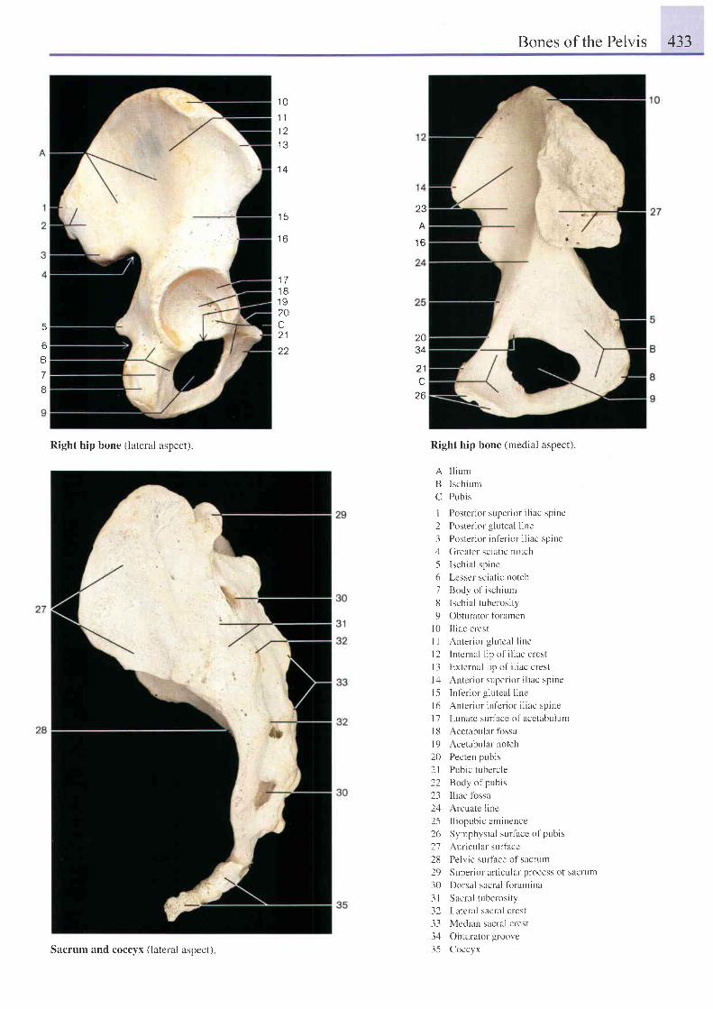
Bones of the Pelvis 433
1 0
1 1
1 3
1 6
23
A
I O
1 71 81 920C2 1
22
5
6
B
7
8
2034
2 1c
26
Right hip bone (lateral aspect). Right hip bone (medial aspect).
A I l i um
B lschium
C Pubis
I P t r s t e r i o r : upe r i o r i l i ae sp ine
2 Poster ior g luteal l ine
. l Pos t c r i o r i n l e r i o r i l i l e : p i ne
4 Greater sciatic notch
5 Ischia l spine
6 Lesser sciatic notch
7 Body ol ischium
8 Ischia l tuberosi ty
9 Obturatol fbramen
10 I l iac crest
I I Anter ior g lutcal l ine
I 2 Intcrnal l ip of i l iac crest
I 3 External l ip of i l iac crest
l 4 An te r i o r : upe r i , r r i l i l ' J sp i ne
l5 Inf 'er ior g luteal l ine
l 6 An te r i o r i n l e r i o r i l i r e sp i ne
l7 Lr.rnate surfhce of acetabulum
l8 Acetabular fbssa
l9 Acetabular notch
20 Pecten pubis
2l Pubic tr.rbercle
22 Body of pubis
23 Iliac fossa
24 Arcuate line
2-5 I l iopubic eminence
26 Syrnphysial surface of pubis
27 Auricular surface
28 Pelvic surface of sacrum
29 Superior articular process of sacrum
30 Dorsal sacral fbramina
3 1 Sacral tuberosity
32 Lateral sacral crest
33 Median sacral crest
34 Obturator groove
35 CoccyxSacrum and coccyx (lateral aspect)
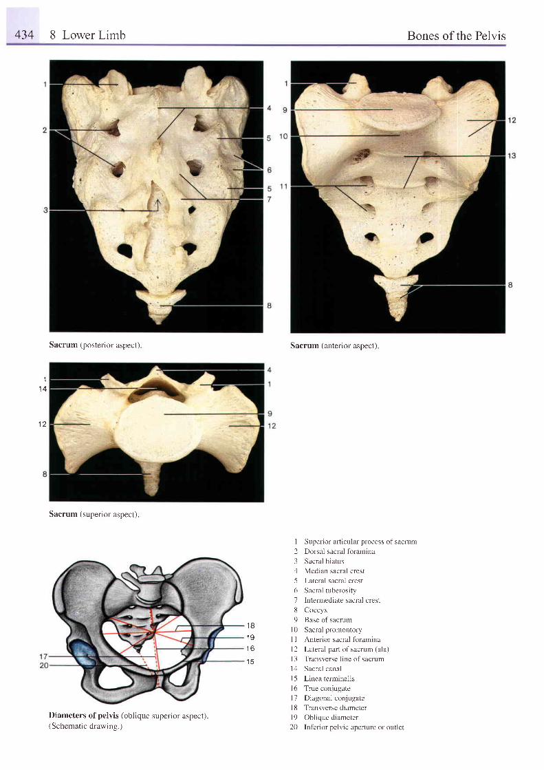
434 8 Lower Limb Bones of the Pelvis
'I
1 A
1 2
Sacrum { posterior aspect }
Sacrum (superior aspect).
Diameters of pelvis (oblique superior aspect).(Schematic drawing.)
Sacrum (anterior aspect)
Superior afticular process oi sacmm
Dorsal sacral fbramina
Sacral hiatus
Median sacral crest
Lateral sacral crest
Sacral tuberosity
Intermediate sacral crest
Coccyx
Base of sacrum
Sacral promontory
Anterior sacral foramina
Lateral part of sacrum (ala)
Transverse line of sacrum
Sacral canal
Linea terminalis
True conjugate
Diagonal conjugate
Transverse diameter
Oblique diameter
Inferior pelvic aperture or outlet
1 8
1 9
t o
1 5
1234567U9
l 0l l1 2l 3t 1l 5l 6t 1t 8t 920
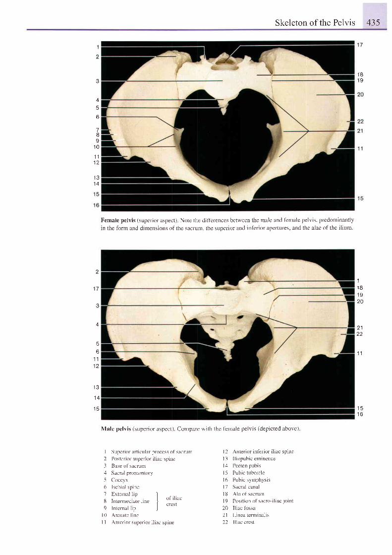
Skeleton of the Pelvis 435
1 81 9
-7
8I
1 0
i l
t z
1 31 4
4
o
4
56
1 11 2
1 3
1 4
1 5
Female pelvis (superior aspect) Note the difl'erences between the male and f'emale pelvis, predominantly
in the form and dimensions of the sacrum. the suoerior and inf'erior apertures, and the alae of the ilium
Male pelvis (superior aspect). Compare with the female pelvis (depicted above)
11 81 q
20
211 Z
1 1
1 51 6
I Superior articular process of sacrum
2 Poster ior super ior i l iac spine
3 Base of sacrum
4 Sacral promontory
5 Coccyx
6 Ischia l spine
7 External l in I. . I u f i l i ae
d I n te rmed ra te l l ne II crest
9 l n t e rna l l i p )l0 Arcuate line
I I Anterior superior iliac sprne
I 2 Anterior inferior iliac sprne
l3 I l iopubic eminence
l4 Pecten pubis
I 5 Pubic tubercle
l6 Pubic symphysis
l7 Sacral canal
l8 Ala of sacrum
| 9 Pos i l i on r r f sac ro - i l i a c j o i n t
20 Iliac fossa
21 Linea terminal is
22 Iliac crest
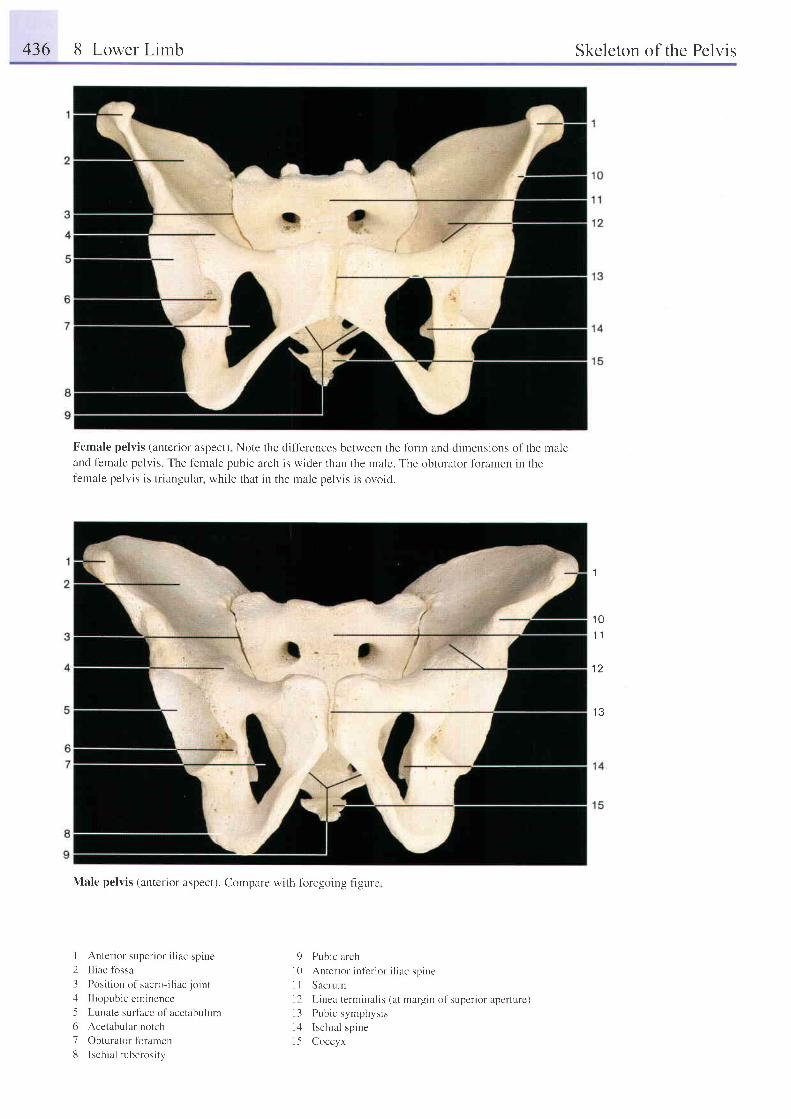
436 8 Lower Limb Skeleton of the Pelvis
Female pelvis (anterior aspect) Note the difl'erences between the tbrm and dirnensions of the maleand female pelvis. The female pubic arch is wider than the male. The obturator fbramen in thefemale pelvis is tr iangular, while that in the male pelvis is ovoid.
Male pelvis (anterior aspect). Compare with foregoing figure
1
1 01 1
1 2
1 3
I Anter ior super ior i l iac spine2 Iliac fossa
3 Posi t ion o1'sacro- i l iac jo int .
4 I l iopubic eminence
-5 Lunate surlace of acetabulr-rm
6 Acetabular notch
7 Obturator fbramen
13 Ischia l tuberosi ty
9 Pubic arch
l0 Anter ior in l 'er ior i l iac spine
I I Sacrurn
l2 L inea termirul is (at margin of super ior apertr . r re)l3 Pubic symphysis
l4 Ischia l spine
l-5 Coccyx
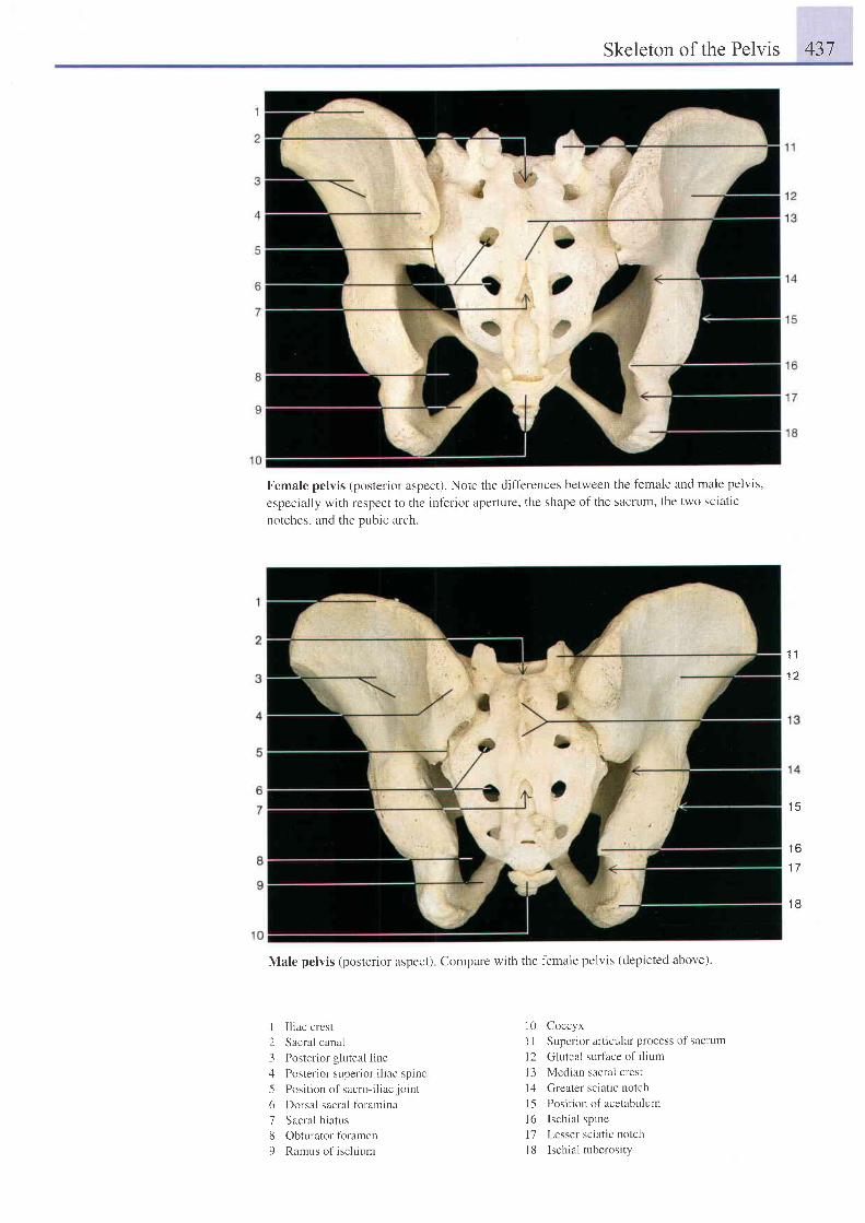
Skeleton of the Pelvis 437
Female pelvis (posterior aspect). Note the diff'erences between
especially with respect to the inf'erior aperture, the shape of the
notches. and the oubic arch.
the female and male pelvis,
sacrum, the two sciatic
1 1
1 5
1 6
1 7
1 8
Male pelvis (posterior aspect) Compare with the female pelvis (depicted above).
I Iliac crest
2 Sacral canal
3 Posterior gluteal line
4 Poster ior super ior i l iac spine
5 Pos i t i on o l s r c ro - i l i a c j t ' i n t
6 Dorsal sacral tbramina
7 Sacral h iatus
8 Obturator foramen
9 Ramus of ischium
l0 Coccyx
I I Superior articular process of sacrutr-t
I 2 Gluteal surface of ilium
l3 Median sacral crest
l4 Creater sciatic notch
I 5 Position of acetabulum
16 Ischia l spine
l7 Lesser sciatic notch
l8 Ischial tuberosity
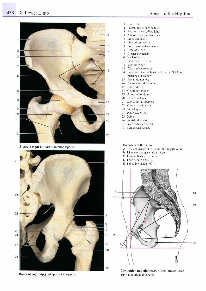
438 8 Lower Limb Bones of the Hip Joint
1 3 -
I I l iac crest
2 Lateral part of sacrum (ala)
. l Pos i t r on o f sac ro - i l i ae j o i n t
4 An te r i o r supe r i t r r i l i a c sp i ne
5 Linea terminal is
6 I l iopubic eminence
7 Bony margin of acetabulum
8 Head of femur
9 Greater trochanter
l0 Neck of femur
I I Intertrochanteric line
12 Shaft of t'emur
13 Fifth lumbar vertebra
l4 Imitation intervertebral disc
vertebra and sacrum
l5 Sacral promontory
l6 Anterior sacral foramina
17 Pubic tubercle
lt3 Obturator foramen
19 Ramus of ischium
20 Lesser trochanter
21 Dorsal sacral foramina
22 Greater sciatic notch
23 Ischia l spine
24 Pubic syrrphysis
25 Pubis
26 Ischial tuberosity
Z7 Inter t rochanter iccrest
28 Symphysial sur lace
between fifth lumbar
Diameters of the pelvis
A True conjugate ( l l - l 1.5 cm) (Conjugata vera)
B Diagonal conjugate (12 5-13 cm)
C Largest diameter of pelvis
D Inf-erior pelvic aperture
E Pelvic inclination (60")
Inclination and diameters of the female pelvis,r ight half (medial aspect)
I87
1 0 2 8
v
1 01 1
Bones of right hip ioint (anterior aspect).
Bones of right hip joint (posterior aspect).
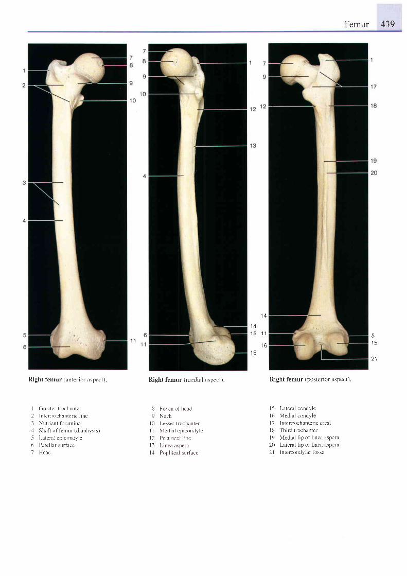
Femur 439
1 4
1 41 5 1 1
I O
1 6
51 5
2 1
Right f'emur (anterior aspect)
Gr-eater trochanter
I t r ter t fochantc l ic l ine
Nut l ient foramina
Shal t of fenrur (d iaphysis)
Lateral epicondyle
Patc l lar surface
Head
Right femur (medial aspect)
Fovea of head
Neck
Lesser trochanter
Medial epicondyle
Pect ineal l ine
Linea aspera
Poplrteal sr,rrlace
Right femur (posterior aspect)
l -5 Lateral condyle
l6 Medial condyle
I 7 lntertrochanteric crest
| 8 Third trochanter
l9 Medial l ip of l inea aspera
20 Lateral lip of linea aspera
2l Intercondylar fossa
6
9
t 0il
t 2t 3t 4
I)31561
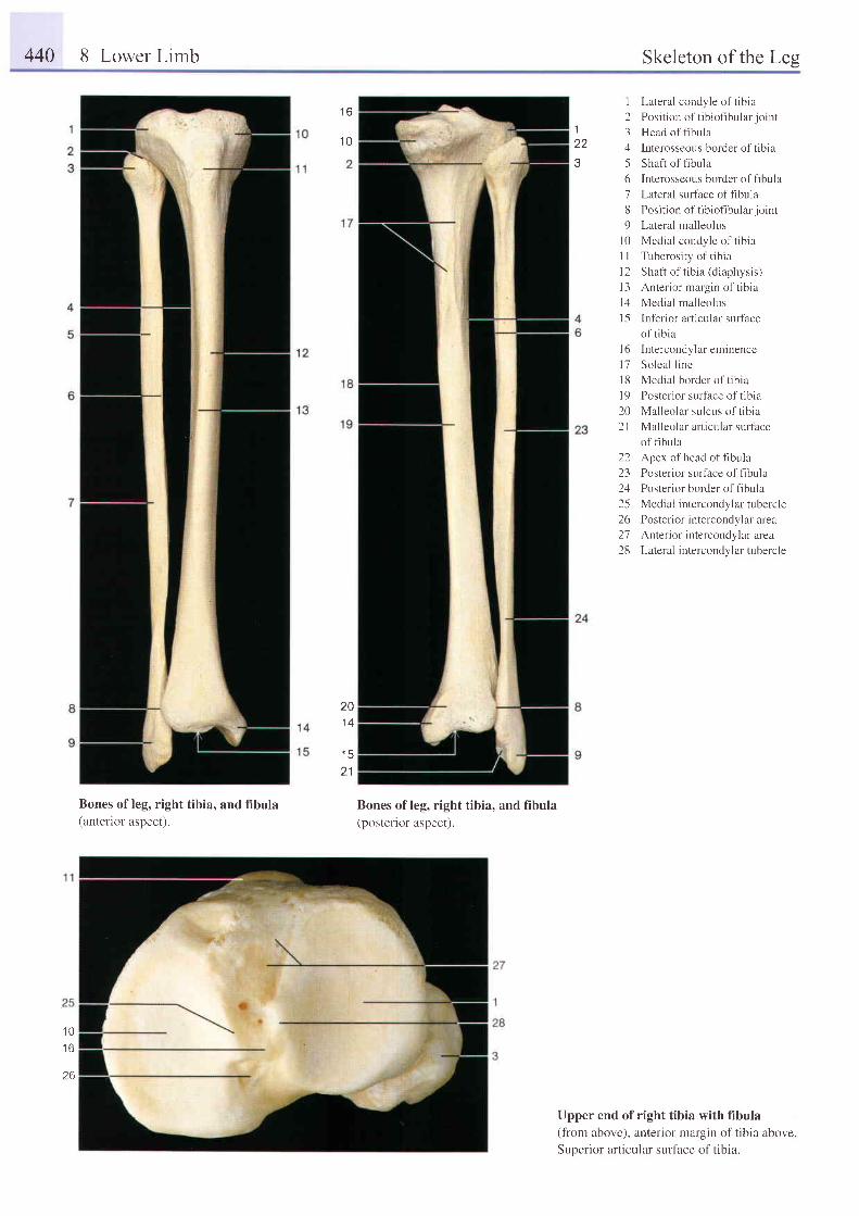
440 8 Lower Limb Skeleton of the Les
Bones of leg, right tibia, and fibula(antenor aspect).
Bones of leg, right tibia, and fibula(posterior aspect).
1z z
I Lateral condyle of tibia
2 Posi t ionof t ib iof ibular jo int
3 Head offibula
4 Interosseous border of tibia
5 Shaft of fibula
6 Interosseous border of fibula
7 Lateral surtace offibula
8 Posi t ion of t ib iof ibular jo int
9 Lateral malleolus
l0 Medial condyle of t ib ia
1 I Tuberosity of tibia
12 Shaft of tibia (diaphysis)
l - l An te r i o r ma rg in o f t i b i i
l4 Medial mal leolus
I 5 Inf'erior articular surtace
of t ib ia
l6 Intercondylareminence
l7 Soleal l ine
l8 Medial border of tibia
I 9 Posterior surface of tibia
20 Mal leolar sulcus of t ib ia
21 Malleolar articular surf'ace
of fibula
22 Apex ofhead offlbula
23 Posterior surface of fibula
24 Posterior border of fibula
25 Medial intercondylar tubercle
26 Posterior intercondylar area
27 Anterior intercondylar area
28 Lateral intercondylar tubercle
Upper end of right tibia with fibula(from above), anterior margin of tibia above.Superior articular surface of tibia.
1 6
1 0
20
'15
2 1
'10
I U
1 0
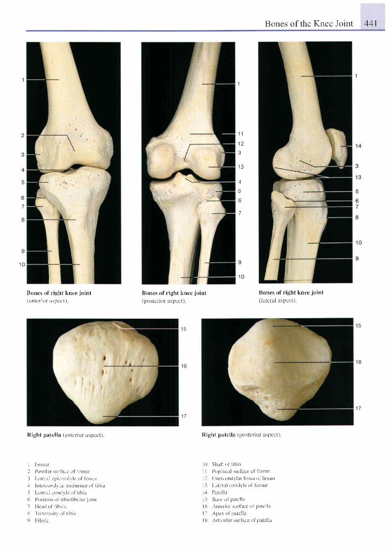
Bones of the Knee Joint 44I
1 1
1 2?
4
6
6
7
Boncs o f r igh t knee jo in t
(antcl ' ror aspcct).
Right patel la (anterior aspect)
I Fer l r - r r
2 Patc l lar-sur- lace ol fenrur
J Latcral c l . r iconclv lc o l le r rur: l Intercond.v lar enr inence of t ib i r
.5 Lareral coudl lc o l t ib ia
6 Pos i t i on o l t i b i o f i bu l a r j o i n t
7 Heacl of t lbul lL
13 Tube los i r y ' o f ' t i b i a
9 F ib tL l u
Bones of r ight knee.joint(p0stenor uspect)
Bones o f r igh t knee. io in t( lateral aspect)
Right patel la (posteriol aspect).
I 0 Shaft of t ib i^
I I Popl i tcal sur face ol lentur
l l Intcrcondylar- Iossa of f 'entur
l i Lateral condyle of fernur
l -1 Patc l la
l 5 Basc o l ' pa te l l a
| 6 Anter ior surtace of patc l l l
l7 Apcr of patc l la
I l l Al t rcular- surf i rce of patc l la
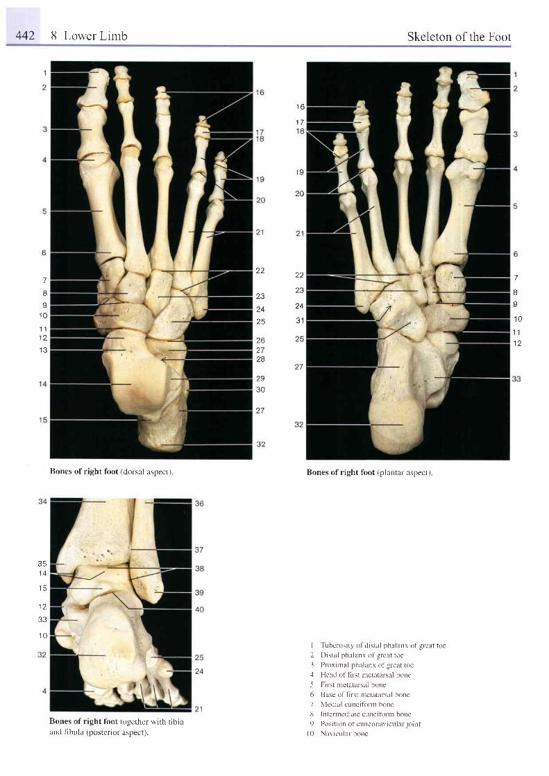
442 8 Lower Limb Skeleton of the Foot
t o
1 7
1 8
1 9
20
7
A
.10
1 t1 2
z z
ZJ
24
3 1
7
I
I1 0
1 11 2
1 3
22
23
24
25
262728
2930
2 7
32
t q
Bones of right foot (dorsal aspect)
Bones of right foot togerher with tibiairnd fibula (posterior aspect).
Bones of right foot (plantar aspect)
I Tuberosrty of distal phalanx of great toe
2 Distal phalanx of great toe
3 Proximal phalanx of great toe;l Head of flrst metatarsal bone
.5 First metatarsal bone
6 Base of flrst Inetatarsal bone
7 Medial cunei f i rm bone
lJ Interrnediate cuneiform bone
9 Posi t ion of cuneonavicr . r lar jo int
l 0 Nav i cu la r bone
a a
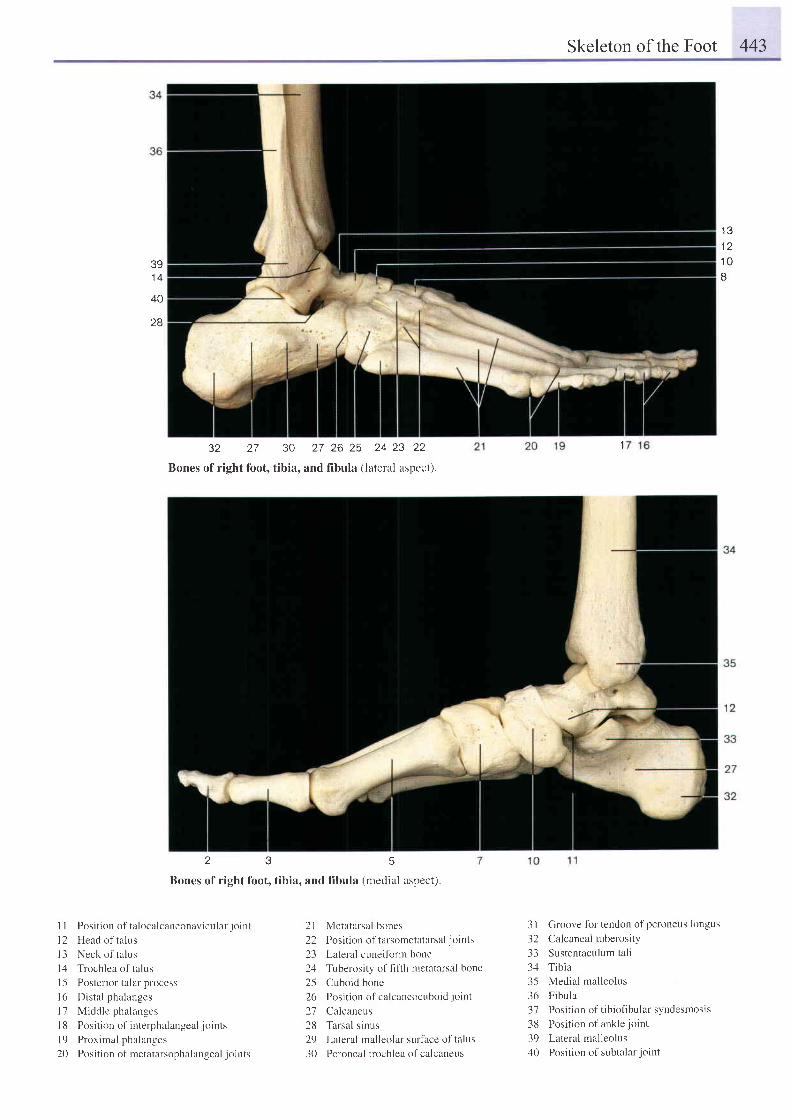
Skeleton of the Foot 443
. i Q
1 21 0tJ
391 j
40
28
32 27 30 27 26 25 24 23 22
Bones ofright foot, tibia, and fibula (lateral aspect).
2 3 5
Bones of right foot, tibia, and fibula (medial aspect).
I I Posi t ion of ta localcaneonavicular jo int
l2 Head of ta lus
l3 Neck of ta lus
14 Trochlea of ta lus
1 5 Postetior talar process
16 Dista l ph:r langes
l7 Middle phalanges
I 8 Position of interphalangeal joints
l9 Proximal phalanges
20 Position of metatarsophalangeal joints
2l Metatarsal bones
22 Position of tarsometatarsal joints
23 Lateral cuneifbrm bone
24 Tuberosity of f-rfth metatarsal bone
25 Cuboid bone
26 Posi t ion of calcaneocLrboid jo int
27 Calcaneus
28 Tarsal s inus
29 Lateral malleolar surthce of talus
30 Peroneal trochlea of calcaneus
3 I Groove fbr tendon of peroneus longus32 Calcanealtuberosity33 Sustentaculum tali34 Tibia35 Medial malleolus36 Fibula37 Position of tibiofibular syndesmosis38 Position of ankle joint
39 Lateral malleolus40 Position of subtalarjoint
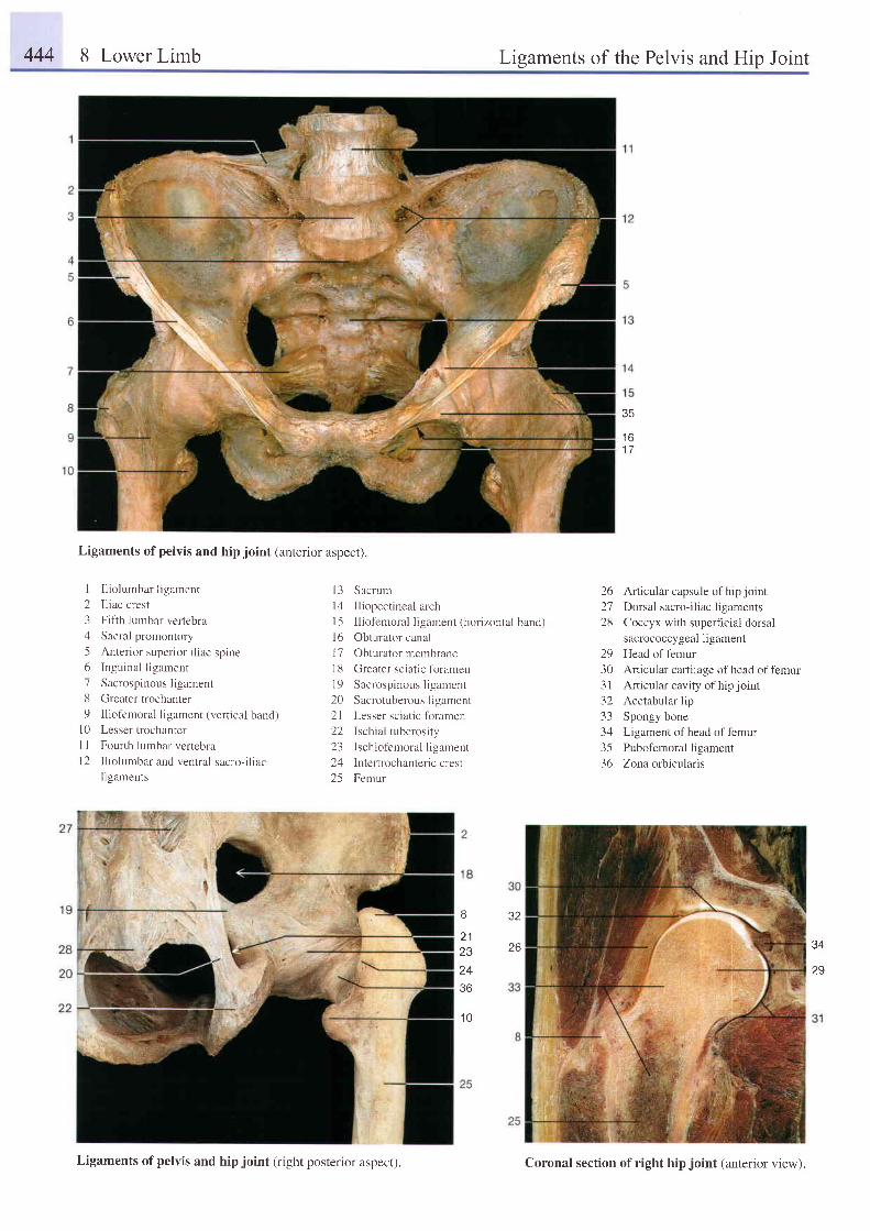
444 8 Lower Limb Ligaments of the Pelvis and Hip Joint
t o1 7
Ligaments of pelvis and hip joint (anrerior aspect).
I I l io lumbar l igament
2 Iliac crest
3 Fifth lumbar vertebra
4 Sacral promontory
5 Anterior superior iliac spine6 Ingr.rinal ligament
7 Sacrospinous ligament
8 Greater trochanter
9 Iliofemoral ligament (vertical band)l0 Lesser trochanter
I I Fourth lumbar vertebral2 Iliolumbar and ventral sacro-iliac
l igaments
l3 Sacrum
l4 Iliopectineal arch
l5 Iliofemoral ligament (horizontal band)
l6 Obturator canal
17 Obturator membrane
18 Greater sciatic fbramen
l9 Sacrospinous l igament
20 Sacrotuberous ligament
2l Lesser sciatic foramen
22 Ischial tuberosity
23 Ischiof'emoral ligament
24 Intertrochanteric crest
25 Femur
8
2 1232436
1 0
26 Articular capsule ofhipjoint27 Dorsal sacro-iliac ligaments28 Coccyx with superticiai dorsal
sacrococcygeal ligament29 Head of femur30 Articular cartilage of head of femur3 | Art icular cavity of hip joint32 Acetabular lip33 Spongy bone34 Ligament of head of f'emur35 Pubofemoralligament36 Zona orbicularis
J I
26 34
29
Ligaments of pelvis and hip joint (right posterior aspect). Coronal section of right hip joint (anterior view).
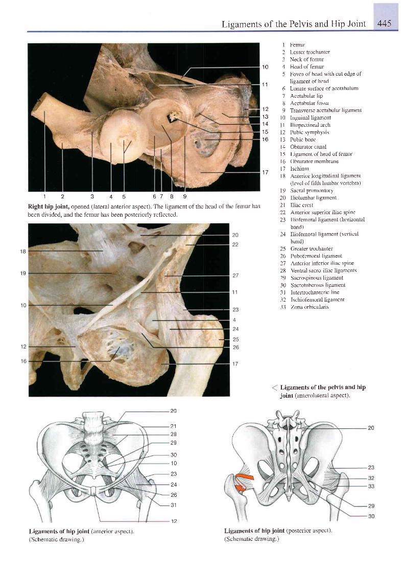
Ligaments of the Pelvis and Hip Joint 445
1 0
1 Femur2 Lesser trochanter3 Neck of femur4 Head of femur5 Fovea ofhead with cut edge of
ligament of head6 Lunate surface of acetabulum7 Acetabular lip8 Acetabular fossa9 Transverse acetabular ligament
10 Inguinal ligament1l Iliopectineal arch12 Pubic symphysis13 Pubic bonel4 Obturator canal15 Ligament of head of femurl6 Obturator membrane17 Ischium18 Anterior iongitudinal ligament
(level of hfth lumbar vertebra)19 Sacral promontory20 Iliolumbar ligament21 Iliac crest22 Anterior superior i l iac spine23 Iliofemoralligament(horizontal
band)24 Iliofemoral ligament (vertical
band)25 Greater trochanter26 Pubofemoralligament27 Anterior inferior i l iac spine28 Ventral sacro-iliac ligaments29 Sacrospinousligament30 Sacrotuberous ligament3l Intertrochantericline32 Ischiofemoral ligament33 Zonaorbicularis
( Ligaments of the pelvis and hipjoint (anterolateral aspect).
1 1
1 21 31 41 5t o
1 7
1 2 3 4 5 6 7 I I
Right hip joint, opened (lateral anterior aspect). The ligament of the head of the femur has
been divided. and the femur has been oosteriorlv reflected.
20
212829
301 0
Z J
24
26
31
Ligaments of hip joint (anterior aspect).(Schematic drawing.)
Ligaments of hip joint (posterior aspect).(Schematic drawing.)
1 2
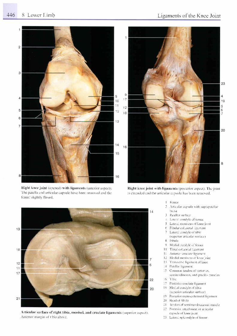
446 8 Lower Limb Ligaments of the Knee Joint
I1 01 1
I J
I
1 7
a a
1 B
I Y
5
7
Right knee.ioint (opened) with l igaments (anrel ior aspccr)The patel la and art iculal capsule ha.n'e been r-euror, 'ccl ancl thelenru l s l igh t lv f lexed
Right knee joint with l igaments (posterior aspect). The . jointis extencled arrcl the art icular capsulc has been relnovcd.
I
2Femur
Art icr , r lar capsule wirh supr apatel lar
bur-sa
Palc l lar-sr . r r face
Latela l coudvlc o l f emur
Latela l nreniscr-rs o l 'knee jo int
FibLr lar col lateral l igarrcnt
Latcr-a l condvle of t ib ia(super ior arr icular sur lucc.)
Fibula
Medial condr, le o l ' lcnrur-
T i b i a l co l l a t e ra l l i gumcn t
Antcr ior cruciate l igarncnL
Medra l men i scus o l ' knce j o i r r r
Tr nnsl'erse ligilnrcnt of kltee
Patc l lar I igalnent( i l n ) n t i l D l e D L l o n r ) l \ i t t l i l t i u .
se r r i t end iuosus . anc l g r ac i l i s n rusc l cs
l 6 T i b i a
I 7 Postel ior cnrc iute l igautent
l lJ Medial concly le of t ib ia(supcr ior ar t icular sul lacc)
l 9 Posrer ior nreniscofcnrola l l is tntcnt
20 Hcad of t lbula
2 | Tendon of scrni rnernbranosLrs r . r ruscle
f l Postcr- i ( ) r - at tuchrrent o l ' er l icLr lar
capsu le o f knee j o i n t
l i Lntcral epicondvle o l ' Icrnur
t {+l.l
-5
61
1 8
1 a
1 1
1 7
E9
t 0
l 2t 3
1.5
Art icular surlace of r ight t ibia, menisci, and cruciate l igarnents (supcrior rrspccr., .An tc r io r rnarg i l r o f t ib ia abovc
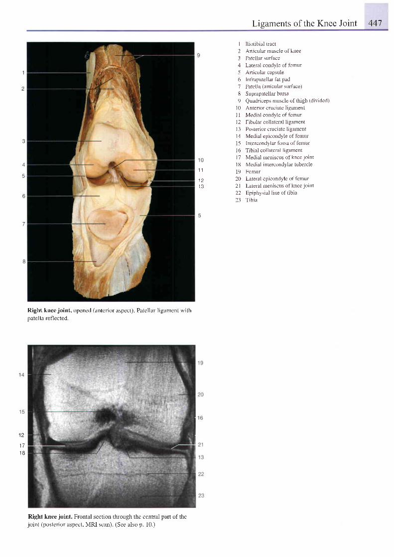
Lisaments of the Knee Joint 447
1 1
1 21 3
I Iliotibial tract2 Articular muscle of knee3 Patellar surface4 Lateral condyle of femur5 Articular capsule6 Infrapatellar fat pad7 Patella (articular surface)8 Suprapatellar bursa9 Quadriceps muscle of thigh (divided)
l0 Anterior cruciate ligament1 1 Medial condyle of femurl2 Fibular collateral ligament13 Posterior cruciate ligamentl4 Medial epicondyle of femur15 Intercondylar fossa offemurl6 Tibial collateral ligamentl7 Medial meniscus of knee joint
l8 Medial intercondylar tubercle19 Femur20 Lateral epicondyle offemur21 Lateral meniscus ofkneejoint22 Epiphysial line of tibia23 Ttbia
t z
1 7'18
Right knee joint, opened (anterior aspect). Patellar ligament with
oatella reflected.
Right kneejoint. Frontal section through the central part ofthejoint (posterior aspect, MRI scan). (See also p. 10.)
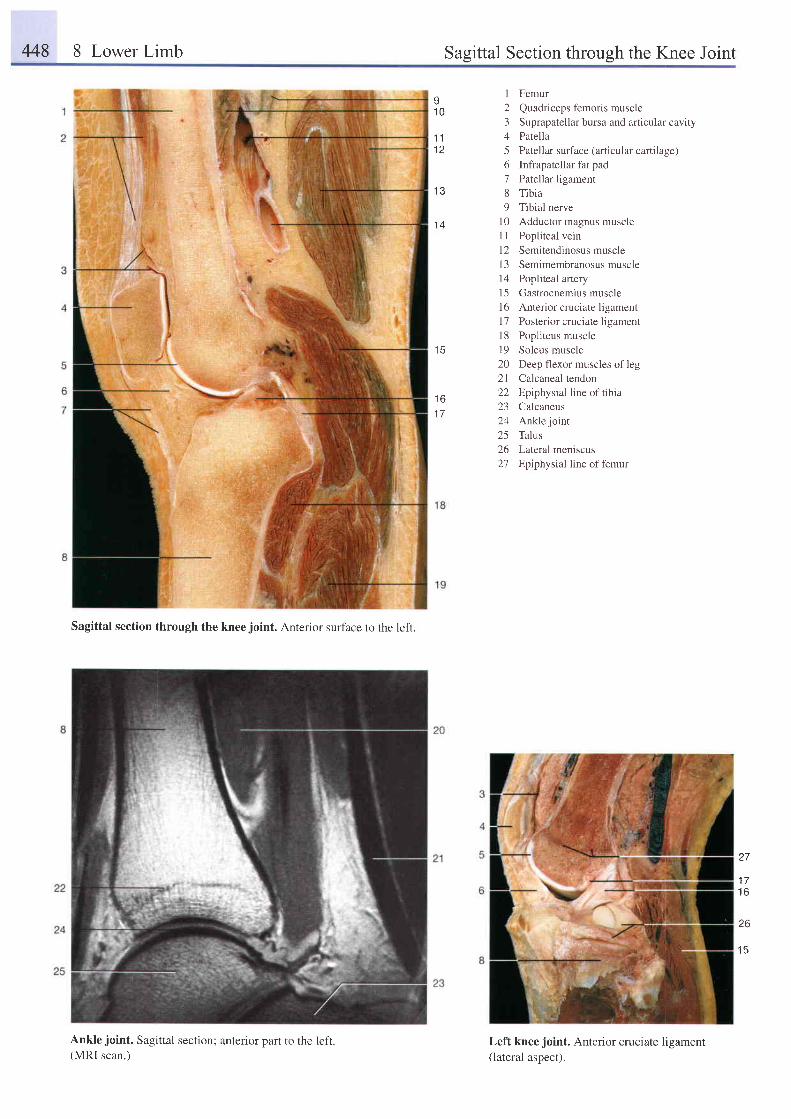
448 8 Lower Limb Sasittal Section throush the Knee Joint
Sagittal section through the kneejoint. Anterior surface to the left.
Ankle joint. Sagittal section; anterior paft to the left.(MRI scan.)
1 Femur2 Quadriceps femoris muscle3 Suprapatellar bursa and articular cavity4 Patella5 Patellar sufface (articular cartilage)6 Infiapatellar fat pad7 Patellar ligament8 Tibia9 Tibial nerve
l0 Adductor magnus muscleI I Popliteal veinl2 Semitendinosus muscle13 Semimembranosus musclel4 Popliteal artery15 Gastrocnemius muscle16 Anterior cruciate ligamentl7 Posterior cruciate ligament18 Popliteus muscle19 Soleus muscle20 Deep flexor muscles of leg21 Caicaneal tendon22 Epiphysial line of tibia23 Calcaneus24 Anklejoint25 Talus26 Lateral meniscus27 Epiphysial line of femur
Left knee joint. Anterior cruciate ligament(lateral aspect).
I1 0
1 11 2
1 3
1 4
1 61 7
) 7
t l
t o
26
. t K
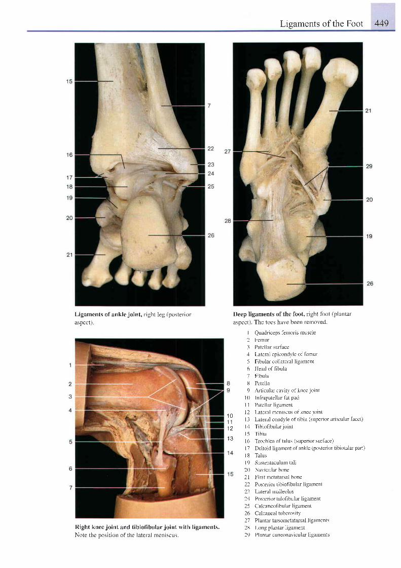
Ligaments of the Foot 449
Ligaments of ankle joint, right leg (posteriorasDect)
Right knee joint and tibiofibular joint with ligaments.Note the oosition of the lateral meniscus.
Deep ligaments of the foot, right fbot (plantar
aspect). The toes have been removed.
I Quadriceps femoris muscle
2 Femur
3 Patellar surface
4 Lateral epicondyle offemur
5 Fibular collateral ligament
6 Head offibula
7 Fibula
8 Patella
9 Articular cavity of knee joint
10 Infrapatellar fat pad
I I Patellar ligament
12 Lateral meniscus ofkneejoint
l3 Lateral condyle of tibia (superior articular facet)
l4 Tib iof ibular jo int
l5 Tib ia
l6 Trochlea oftalus (superior surface)
l7 Deltoid ligament of ankle (posterior tibiotalar pan.)
l8 Talus
l9 Sustentaculum tali
20 Navicular bone
21 First metatarsal bone
22 Posterior tibiofibultu ligament
23 Lateral malleolus
24 Posterior talofibular ligament
25 Calcaneofibularligament
26 Calcanealtuberosity
27 Plantartarsometatarsalligaments
28 Long plantar ligament
29 Plantar cuneonavicular ligaments
1 01 11 2
1 3
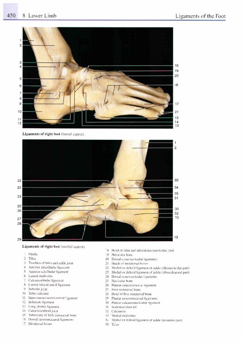
450 8 Lower Limb Lisaments of the Foot
1 8
1 9
20
' I AD
7
o
1 11 2
1 7
2 1
t c
1 41 Q
Ligaments of right foot (lateral aspect).
34
3 1
22
Z J
z 4
30321 0
Z J
26
28
29
Ligaments of right foot (medial aspect)
1 Fibula
2 Tibia
3 Trochlea of ta lus and ankle jo int4 Anterior tibiofibular ligament
5 Anterior taloflbular ligament
6 Lateral malleolus
7 Calcaneof ibular l igamentll Lateral talocalcaneal ligament
9 Subtalar jo int
l0 Tuber calcanei
I I Interosseous talocalcaneal ligament
l2 Bi furcate l igament
l3 Long plantar l igament
l4 Calcaneocuboidjo int
l -5 Tuberosity of fifih metatarsal bonel6 Dorsal tarsometatarsal ligaments
17 Metatarsal bones
I 8 Head of ta lus and ta localcaneonavicular jo int
19 Navicular bone
20 Do rsu l r ' uncunav i cu l r r I i gamen ts
2l Heads of metatarsal bones
22 Medial or de ltoid ligarnent of ankle (tibionavicular part)
23 Medial or deltoid ligarnent of ankle (tibiocalcaneal part)
2zl Dorsal cuneonavicular l igaments
25 Navicular bone
26 Plantar cuneonavicular l igament
27 First metatarsal bone
28 Head of first metatarsal bone
29 Plantar tarsometatarsal l igaments
30 Plantarcalcaneonavicular l igament
31 Sustentaculurn ta l i
32 Calcaneus
33 Mecl ia l mal leolus
3,1 Medial or deltoid ligament of ankle (posterior part)
35 Talus
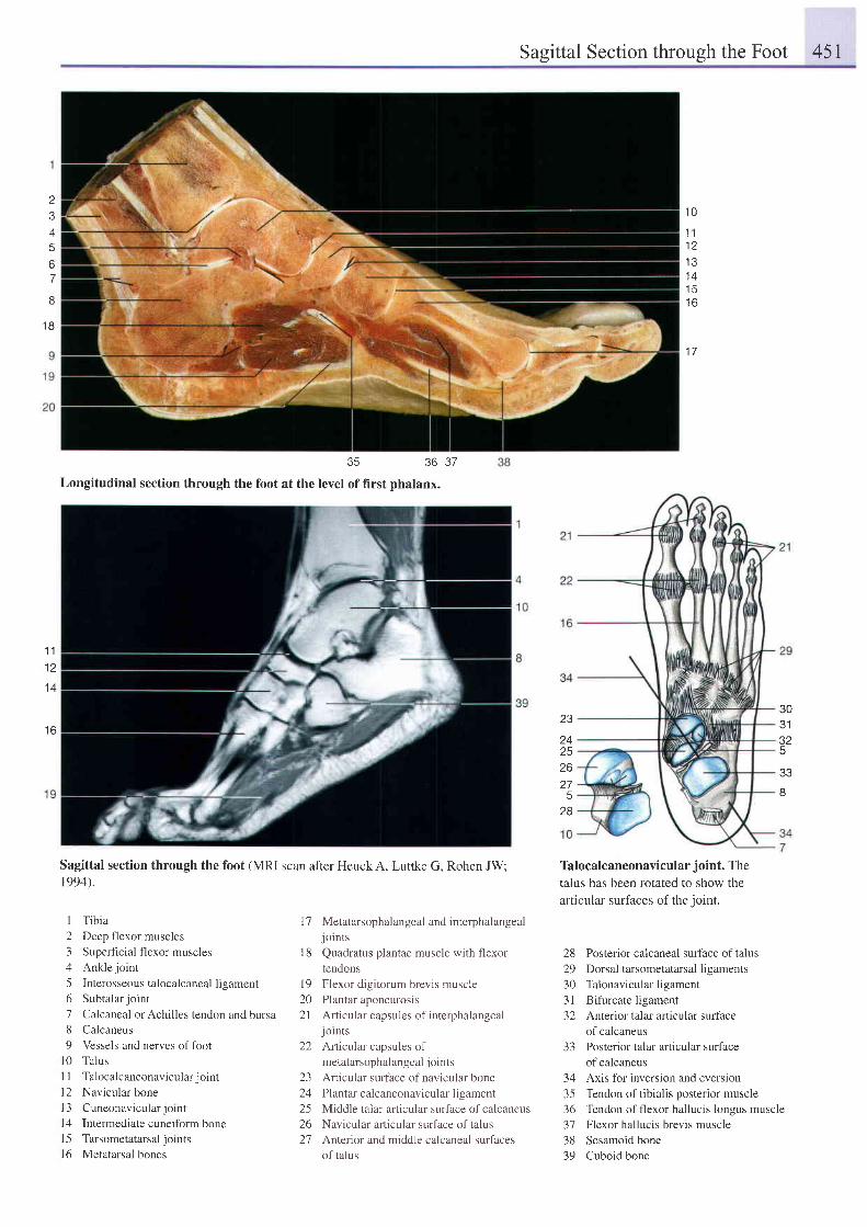
Sasittal Section throush the Foot 45r
4
67
I
1 8
35 36
Longitudinal section through the foot at the level of first phalanx.
Sagittal section through the foot (MRI scan after Heuck A, Luttke G, Rohen JW;1994).
1 0
1 11 21 31 4l c
t o
1 a
Talocalcaneonavicular joint. The
talus has been rotated to show the
art icular surfaces of the joint.
28 Posterior calcaneal surface of talus29 Dorsaltarsometatarsalligaments30 Talonavicularligament31 Bifurcate ligament32 Anterior talar articular surface
of calcaneus33 Posterior talar articular surface
of calcaneus34 Axis for inversion and eversron35 Tendon oftibialis posterior muscle36 Tendon of flexor hallucis longus muscle37 Flexor hallucis brevis muscle38 Sesamoid bone39 Cuboid bone
37
1 1
1 2
1 4
t o23
t +2526275
28
303132
e e
8
I Tibia
2 Deep flexor muscles
3 Superficial flexor muscles
4 Ankle joint
5 Interosseous talocalcaneal ligament
6 Subtalar jo int
7 Calcaneal or Achilles tendon and bursa8 Calcaneus
9 Vessels and nerves of foot
l0 Talus
| | Talocalcaneonavicular jo int
l2 Navicular bone
l3 Cuneonavicularjornt
l4 Intermediate cuneiform bone
l5 Tarsometatarsal jotnts
l6 Metatarsal bones
Metatarsophalangeal and interphalangeal
JOlnts
Quadratus plantae muscle with flexor
tendons
Flexor digitorum brevis muscle
Plantar aponeurosis
Articular capsules of interphalangeal
JolntsArticular capsules of
metatarsophalangeal j oints
Articular surface of navicular bone
Plantar calcaneonavicular ligament
Middle talar articular surface of calcaneus
Navicular articular surface of talus
Anterior and middle calcaneal surfaces
of talus
T7
1 8
1 9202 l
22
L-)
1 ,4
252621
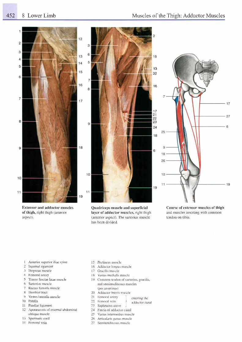
452 8 Lower Limb Muscles of the Thish: Adductor Muscles
1 5
't3
20
1 6
1 a
2122t.t
24
Extensor and adductor musclesof thigh. right thigh ( anteriorasDect).
I Anterior superior iliac spine2 Inguinal ligament3 Iliopsoas muscie4 Femoral artery5 Tensor fasciae latae muscle6 Sartorius muscle7 Rectus femoris muscle8 Iliotibial tract9 Vastus lateralis muscle
l0 Patella11 Patellar ligament12 Aponeurosis of external abdominal
oblique muscle13 Spermatic cord14 Femoral vein
Quadriceps muscle and superficiallayer of adductor muscles, right thigh(anterior aspect). The sartorius musclehas been divided.
15 Pectineus musclel6 Adductor longus musclel7 Gracilis musclel8 Vastus medialis muscle19 Common tendon of sartorius, gracilis,
and semitendinosus muscles(pes anserinus)
20 Adductor brevis muscle21 Femoral arterv I- | entering the22 Femoral vein I adductor canalI
23 Saphenous nerve )24 Fascia of adductor canal25 Vastus intermedius muscle26 Articularis genus muscle27 Semitendinosusmuscle
Course of extensor muscles of thighand muscles inserting with commontendon on tibia.
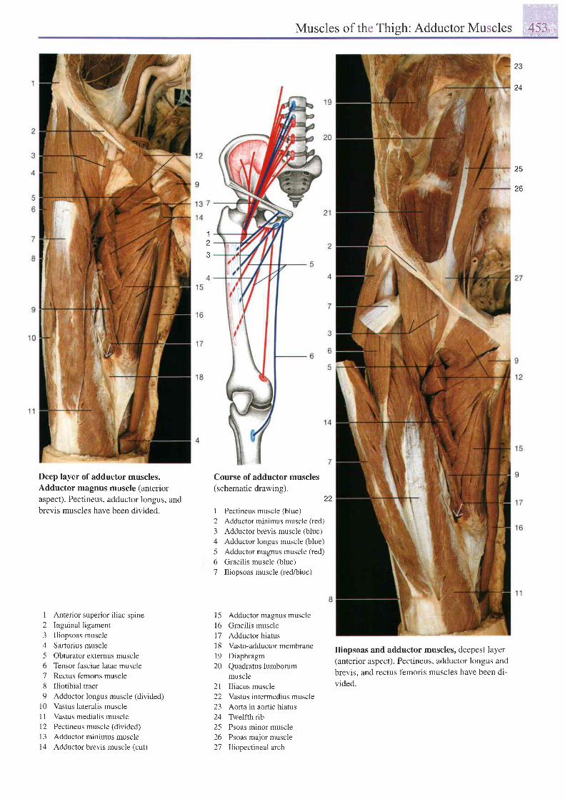
, . ' ' , "
Muscles of the Thigh: Adductor Muscles ,,':4'$,J.,.,,,.,',,
23
24
zo
1z
Deep layer of adductor muscles.Adductor magnus muscle (anterioraspect). Pectineus, adductor longus, andbrevis muscles have been divided.
1 Anterior superior iliac spine2 Inguinal ligament3 lliopsoas muscle4 Sartorius muscle5 Obturator extemus muscle6 Tensor fasciae latae muscle7 Rectus femoris muscle8 Iliotibial tract9 Adductor longus muscle (divided)
l0 Vastus lateralis musclel1 Vastus medialis musclel2 Pectineus muscle (divided)[3 Adductor minimus musclel4 Adductor brevis muscle (cut)
Course of adductor muscles(schematic drawing).
22
1 Pectineus muscle (blue)2 Adductor minimus muscle (red)3 Adductor brevis muscle (blue)4 Adductor longus muscle (blue)5 Adductor magnus muscle (red)
6 Gracilis muscle (blue)7 Iliopsoas muscle (red/blue)
15 Adductor magnus muscle16 Gracilis muscle17 Adductor hiatus18 Vasto-adductormembrane19 Diaphragm20 Quadratus lumborum
muscle2l Iliacus muscle22 Vastus intermedius muscle23 Aorta in aortic hiatus24 Twelfth rib25 Psoas minor muscle26 Psoas major muscle27 Iliopectineal arch
Iliopsoas and adductor muscles, deepest layer(anterior aspect). Pectineus, adductor longus and
brevis, and rectus femoris muscles have been di-
vided.
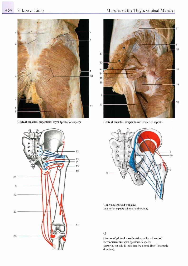
454 8 Lower Limb Muscles of the Thish: Gluteal Muscles
7
I
1 2
1 0
g t 3
1 41 0
1 5
1 6
6
Gluteal muscles, superficial layer (posterior aspect). Gluteal muscles, deeper layer (posterior aspect).
Course of gluteal muscles(posterior aspect; schematic drawing).
Course of gluteal muscles (deeper layer) and ofischiocrural muscles (posterior aspect).Sartorius muscle is indicated bv dotted line (schematic
drawins).
1 2
1 31 4
1 5
1 9
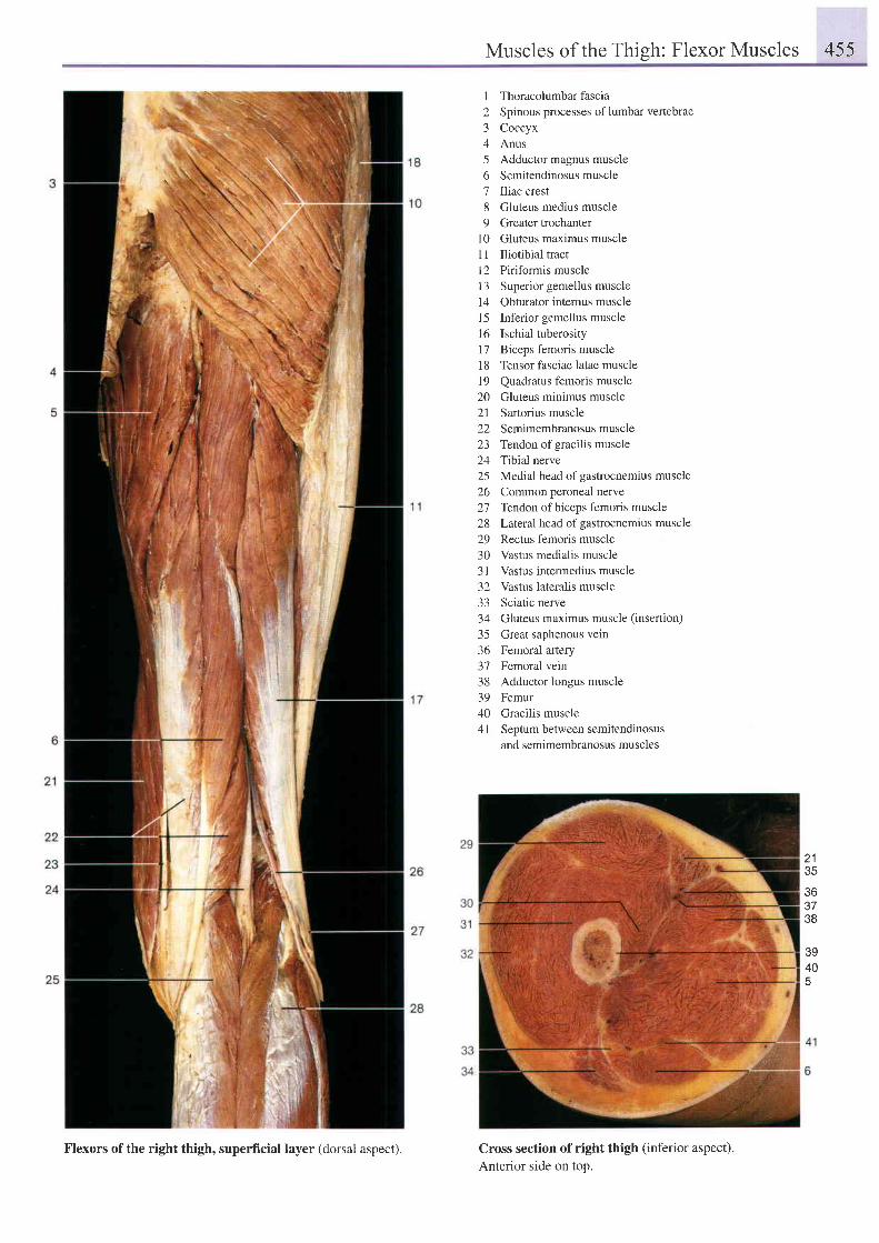
Muscles of the Thish: Flexor Muscles 455
I Thoracolumbar fascia2 Spinous processes oflumbar vertebrae3 Coccyx4 Anus5 Adductor magnus muscle6 Semitendinosusmuscle7 Iliac crest8 Gluteus medius muscle9 Greater trochanter
l0 Gluteus maximus muscleI 1 Iliotibial tractl2 Piriformis muscle13 Superior gemellus muscle14 Obturator intemus musclel5 Inferior gemellus muscle16 Ischial tuberosity17 Biceps femoris muscle18 Tensor fasciae latae musclel9 Quadratus femoris muscle20 Gluteus minimus muscle21 Sartorius muscle22 Semimembtanosusmuscle23 Tendon of gracilis muscle24 Tibial nerve25 Medial head of gastrocnemius muscle26 Common peroneal nerve27 Tendon of biceps femoris muscle28 Laleral head of gastrocnemius muscle29 Rectus femoris muscle30 Vastus medialis muscle3l Vastus intermedius muscle32 Vastus lateralis muscle33 Sciatic nerve34 Gluteus maximus muscle (insertion)
35 Great saphenous vein36 Femoral artery37 Femoral vein38 Adductor longus muscle39 Femur40 Gracilis muscle41 Septum between semitendinosus
and semimembranosus muscles
z l
J5
363738
39405
Cross section of right thigh (inferior aspect).Anterior side on top.
Flexors of the right thigh, superficial layer (dorsal aspect).
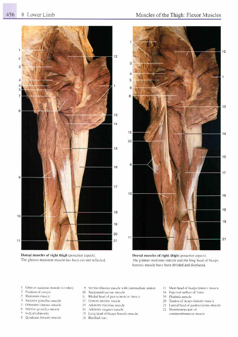
4s6 8 Lower Lirnb Muscles of the Thish: Flexor Muscles
. t
J
\[a
o
.yri:,i
Dorsal muscles of right thigh (posterior aspect)The gluteus maximus muscle has been cut and ref- lccted
I Cluteus maximus muscle (d iv ided)
2 Posi t ion of coccyx
3 Pir i fbrmis rnuscle
4 Supcr ior gentel lus muscle5 Obturator internus ntuscl '6 L r l ' e r i o r gcme l l u : m r r : e l c7 Ischia l tuberosi ty
8 Quadratus t'entoris ntr.rscle
Dorsal muscles of right thigh (posterior aspect).Thc gluteus maximus muscle and the long head of bicepsIemoris muscle have been divided and displaced
9 Scnr i tendinosus musclc wi th interrnedi i r te tendon
l0 Semimembranosus muscle
I I Medial head of gast t 'ocnemius rnuscle
l2 Gluteus lnedius nruscle
l3 Adductor nr in i rnus muscle
1,1 Adductor magnus muscle
l-5 Long head of biceps f-entoris muscle
l6 I l ior ib ia l t ract
l7 Short head of b iceps l 'emor is mnscle
l8 Popliteal surface of t'emur
l9 Plantaris muscle
20 Tendon of b iceps femoris rnuscle
2l Lateral head of gastrocnemius muscle
22 Membranous part of
semimembranosus muscle
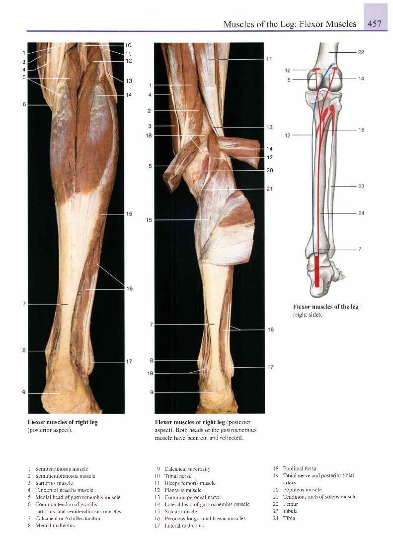
Muscles of the Leq: Flexor Muscles
1
3
5
t 01 11 2
1 3
1 4
1 4
1 2
20
Flexor muscles of right leg(posterior aspect).
I
23ll
56
78
Semitendinosus muscle
Semimembranosus muscle
Sartorius muscle
Tendon of gracilis muscle
Medial head of gastrocnemius muscle
Common tendon of gracilis,
saftorius, and semitendinosus muscles
Calcaneal or Achilles tendorr
Medial malleolus
Flexor muscles of right leg (posterioraspecl). Both heads of the gastrocnemiusmuscle have been cut and reflected.
9 Calcaneal tuberosityl0 Tibial nerve11 Biceps femoris muscle12 Plantaris muscle13 Common peroneal nerve14 Lateral head of gastrocnemius muscle15 Soleus muscle16 Peroneus longus and brevis muscles11 Lateralmalleolus
Flexor muscles of the leg(right side).
18 Popliteal fossa19 Tibial nerve and posterior tibial
afiery20 Popliteus muscle21 Tendinous arch of soleus muscle22 Femur23 Fibula24 Tibia
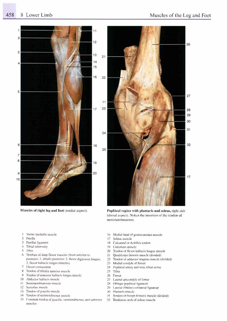
458 8 Lower Limb Muscles of the Les and Foot
Muscles of right leg and foot (medial aspect).
l
2345o
'7
89
1 0l l1 2t 31 41 5
Vastus medialis muscle
Patella
Patellar ligament
Tibial tuberosity
Tibia
Tendons of deep flexor muscles (from anterior toposterior: l. tibialis posterior; 2 flexor digitorum longus;3 flexor hallucis longus muscles)Flexor retinaculum
Tendon of tibialis anterior muscle
Tendon of extensor hallucis longus muscleAbductor hallucis muscle
Semimembranosus muscle
Sartorius muscle
Tendon of gracilis muscle
Tendon of semitendinosus muscle
Common tendon of gracilis, semitendinosus, and sartoriusmuscles
Popliteal region with plantaris and soleus, right side
(dorsal aspect). Notice the inserlion of the tendon of
semimembranosus.
16 Medial head of gastrocnemius muscle
l7 Soleus muscle
l8 Calcaneal orAchilles tendon
l9 Calcaneus muscle
20 Tendon offlexor hallucis longus muscle
21 Quadriceps f'emoris muscle (divided)
22 Tendon of adductor magnus muscle (divided)
23 Medial condyle of femur
24 Popliteal artery and vein, tibial nerve
25 Tibia
26 Femur
27 Latenl epicondyle of femur
28 Oblique popliteal ligament
29 Lateral (fibular) collateral ligament
30 Plantaris muscle
31 Tendon of biceps femoris muscle (divided)
32 Tendinous arch of soleus muscle
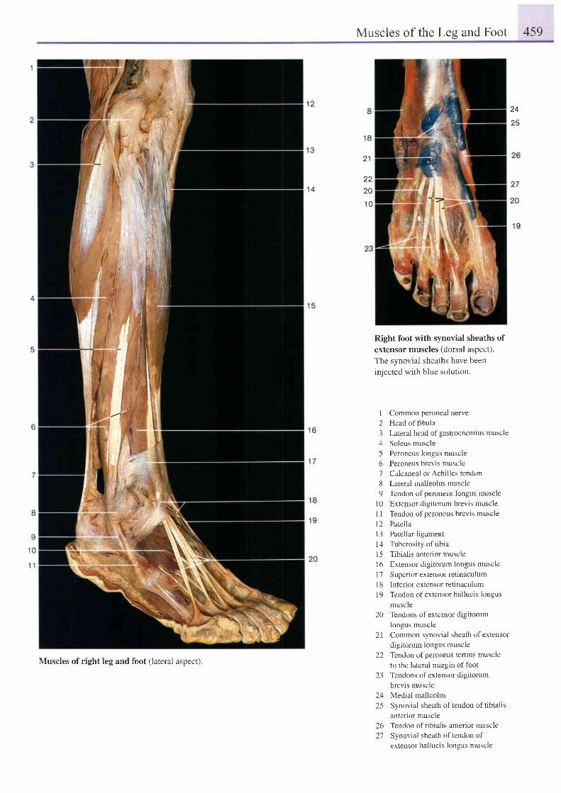
Muscles of the Les and Foot 459
24
21
2220
1 0
Right foot with synovial sheaths ofextensor muscles (dorsal aspect).The synovial sheaths have beeninjected with blue solution.
I Common peroneal netve2 Headoffibula3 Lateral head ofgastrocnemius muscle4 Soleus muscle5 Peroneus longus muscle6 Peroneus brevis muscle7 Calcaneal orAchilles tendon8 Lateral malleolus muscle9 Tendon ofperoneus longus muscle
l0 Extensor digitorum brevis muscleI 1 Tendon ofperoneus brevis muscle12 Patella13 Patellar ligament14 Tuberosity of tibial5 Tibialis anterior muscle16 Extensor digitorum longus muscle17 Superior extensor retinaculumI 8 Inferior extensor retinaculum19 Tendon of extensor hallucis longus
muscle20 Tendons of extensor digitorum
longus muscle21 Common synovial sheath of extensor
digitorum longus muscle22 Tendon ofperoneus tertius muscle
to the lateral margin of foot23 Tendons of extensor digitorum
brevis muscle24 Medial malleolus25 Synovial sheath of tendon of tibialis
anterior muscle26 Tendon oftibialis anterior muscle27 Synovial sheath oftendon of
extensor hallucis longus muscle
Muscles of right leg and foot (lateral aspect).
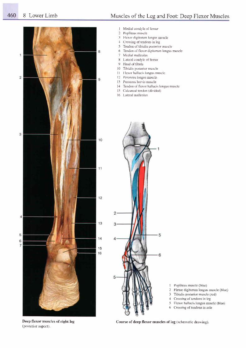
460 8 Lower Limb Muscles of the Leg and Foot: Deep Flexor Muscles
I Medial condyle of femur2 Popliteus muscle3 Flexor digitorum longus muscle4 Crossing oftendons in leg5 Tendon of tibialis posterior muscle6 Tendon of flexor digitorum longus muscle7 Medial malleolus8 Lateral condyle offemur9 Head of fibula
10 Tibialis posterior muscleI I Flexor hallucis longus muscle12 Peroneus longus musclel3 Peroneus brevis muscle14 Tendon of flexor hallucis longus muscle15 Calcaneal tendon (divided)l6 Lateral malleolus
1 4
1 51 6
I Popliteus muscle (blue)2 Flexor digitorum longus muscle (blue)3 Tibialis posterior muscle (red)4 Crossing oftendons in leg5 Flexor hallucis longus muscle (blue)6 Crossing oftendons in sole
Deep flexor muscles of right leg(posterior aspect).
Course of deep flexor muscles of leg (schematic drawing).
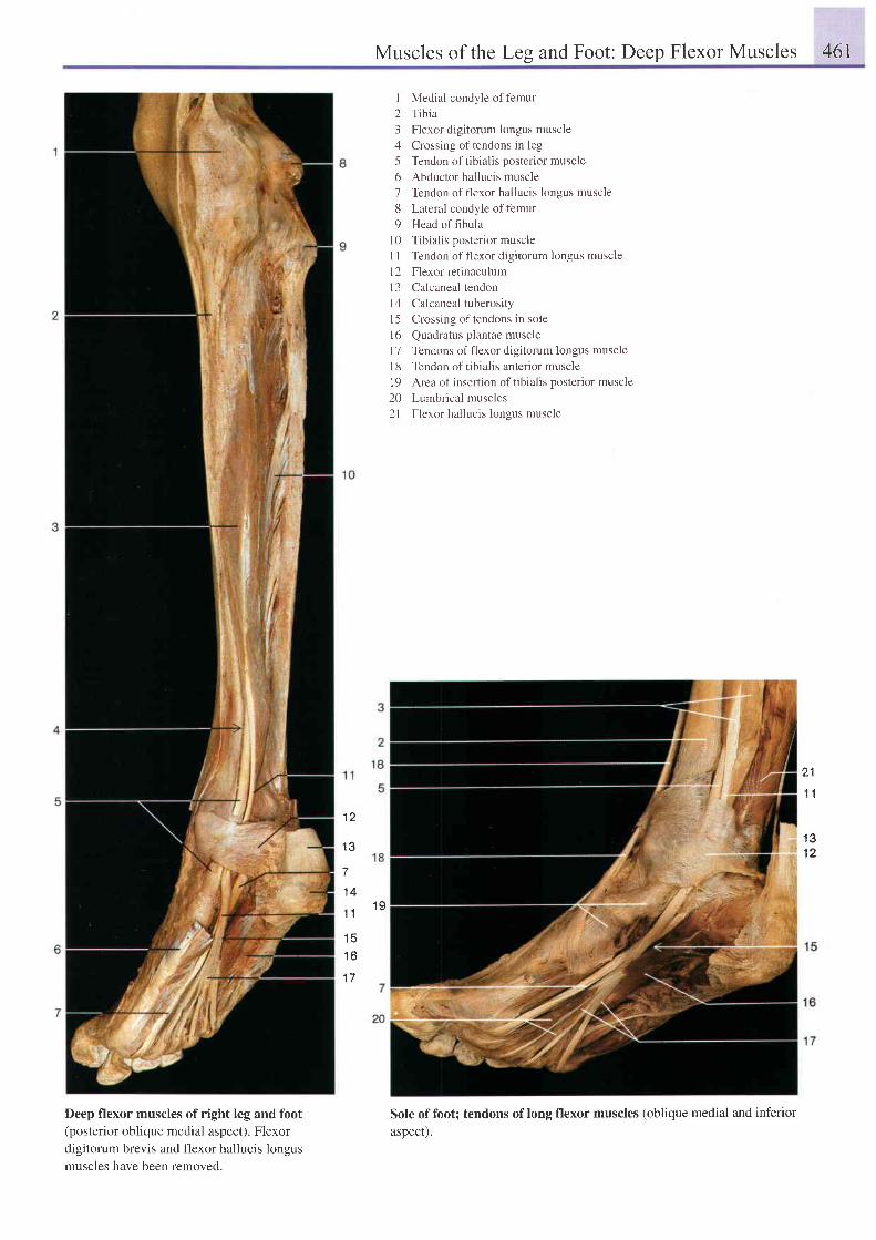
Muscles of the Leg and Foot: Deep Flexor Muscles 46I
Deep flexor muscles of right leg and foot(posterior oblique medial aspect). Flexordigitorum brevis and flexor hallucis longusmuscles have been removed.
I Medial condyle of f'emur2 Tibia3 Flexor digitorum longus muscle4 Crossing of tendons in leg5 Tendon of tibialis posterior muscle6 Abductor hallucis muscle7 Tendon of flexor hallucis longus muscle8 Lateral condyle offemur9 Head of fibula
10 Tibialis posterior muscleI I Tendon of flexor digitorum longus musclel2 Flexor retinaculuml3 Calcaneal tendonl4 Calcaneal tuberosityl5 Crossing of tendons in sole16 Quadratus plantae musclel7 Tendons of flexor digitorum longus musclel8 Tendon of tibialis anterior muscle19 Area of insertion oftibialis posterior muscle20 Lumbrical muscles2l Flexor hallucis longus muscle
Sole of foot; tendons of long flexor muscles (oblique medial and inferiorasoect).
21
1 1
1 31 2
1 2
1 3
7
1 4
1 1 1 9
1 6
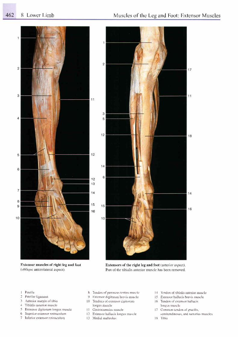
462 8 Lower Limb Muscles of the Les and Foot: Extensor Muscles
1 2
1 4
1 5
1 6
Extensor muscles of(oblique anterolateral
right leg and foot
aspect).Extensors of the right leg and foot (anterior aspect)
Part of the tibialis anterior muscle has been removed.
1 Patella2 Patellar ligament3 Anterior margin of tibia4 Tibialis anterior muscle5 Extensor digitorum longus muscle6 Superior extensor retinaculum7 Inf'erior extensor retinaculum
8 Tendon ofperoneus tertius muscle
9 Extensor digitorum brevis muscle
l0 Tendons ofextensor digitorumlongus muscle
I I Gastrocnemius muscle
l2 Extensor hal luc is longus muscle
l3 Medial malleolus
14 Tendon of tibialis anterior musclel5 Extensor hallucis brevis muscle16 Tendon ofextensor hallucrs
longus musclel7 Common tendon of gracilis,
semitendinosus, and sartorius muscles18 Tibia
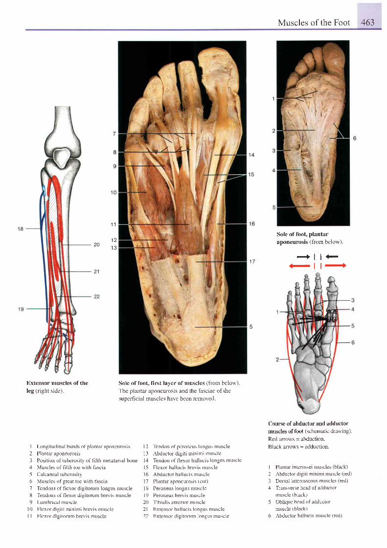
Muscles of the Foot 463
Extensor muscles of theleg (right side).
Sole of foot, first layer of muscles (fiom below)The plantar aponeurosis and the fasciae of thesuoerhcial muscles have been removed.
Sole of foot, plantaraponeurosis (from below).
+l l+-l t+
Course of abductor and adductor
muscles of foot (schematic drawing).
Red arrows = abduction.
Black arrows = adduction.
I Plantar interossei muscles (black)2 Abductor digiti minimi muscle (red)
3 Dorsal interosseous muscles (red)
4 Transverse head of adductormuscle (black)
5 Oblique head of adductormuscie (black)
6 Abductor hallucis muscle (red)
20
1 1
1 21 3
1 Longitudinal bands ofplantar aponeurosis 12 Tendon ofperoneus Iongus muscle
2 Plantar aponeurosis 13 Abductor digiti minimi muscle
3 Position of tuberosity of fifth metatarsal bone | 4 Tendon of flexor hallucis longus muscle
4 Muscles of flfih toe with fascia 15 Flexor hallucis brevis muscle
5 Calcaneal tuberosity 16 Abductor hallucis muscle
6 Muscles of great toe with fascia 17 Plantar aponeurosis (cut)
7 Tendons of flexor digitorum longus muscle l8 Peroneus longus muscle
8 Tendons offlexor digitorum brevis muscle 19 Peroneus brevis muscle
9 Lumbrical muscle 20 Tibialis anterior muscle
10 Flexor digiti minimi brevis muscle 21 Extensor hallucis longus muscle
1 I Flexor digitorum brevis muscle 22 Extensor digitorum longus muscle
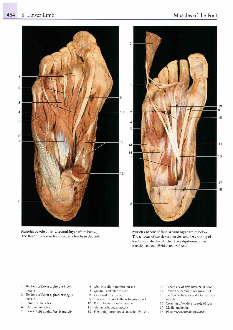
464 8 Lower Limb Muscles of the Foot
1 5I
1 0
Muscles of sole of foot, second layer (from below).The flexor digitorum brevis muscle has been divided.
3A
5
Tendons of flexor digitorum brevismuscleTendons of flexor digitorum longusmuscleLumbrical musclesInterossei musclesFlexor digiti minimi brevis muscle
1 3
1 4
6 Abductor digiti minimi muscle
7 Quadratus plantae muscle
8 Calcaneal tuberosity
9 Tendon offlexor hallucis longus musclel0 Flexor hallucis brevis muscle
11 Abductor hallucis muscle
12 Flexor digitorum brevis muscle (divided)
Muscles of sole of foot, second layer (from below).The tendons of the flexor muscles and the crossing oftendons are displayed. The flexor digitorum brevismuscle has been divided and reflected.
13 Tuberosity offifth metatarsal bone
14 Tendon ofperoneus longus muscle
I 5 Transverse head of adductor hallucis
muscle
16 Crossing oftendons in sole offoot
17 Medial malleolus
I 8 Plantar aponeurosis (divided)
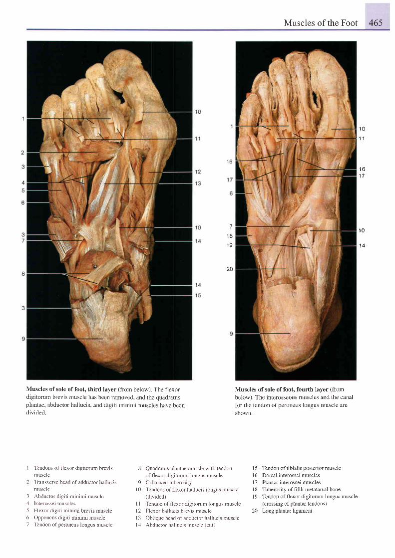
Muscles of the Foot 465
1 0
1 1
1 61 7
Muscles of sole of foot, third layer (from below). The flexordigitorum brevis muscle has been removed, and the quadratusplantae, abductor hallucis, and digiti minimi muscles have beendivided.
1 8
1 9
20
Quadratus plantae muscle with tendon
of flexor digitorum longus muscle
Calcaneal tuberosity
Tendons of flexor hallucis longus muscle(divided)
I I Tendon offlexor digitorum longus muscle
12 Flexor hallucis brevis muscle
l3 Oblique head of adductor hallucis muscle
14 Abductor hallucis muscle (cut)
Muscles of sole of foot, fourth layer (frombelow). The interosseous muscles and the canalfor the tendon of peroneus longus muscle areshown.
15 Tendon of tibialis posterior muscle16 Dorsal interossei muscles17 Plantar interossei muscles18 Tuberosity offifth metatarsal bone19 Tendon of flexor digitorum longus muscle
(crossing of plantar tendons)20 Long plantar ligament
1 0
1 4
Tendons of flexor digitorum brevismuscleTransverse head of adductor hallucismuscleAbductor digiti minimi musclelnterossei musclesFlexor digiti minimi brevis muscleOpponens digiti minimi muscleTendon of peroneus longus muscle
8
91 0
3A
56'7
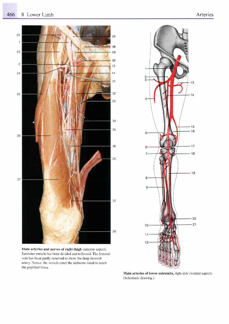
466 8 Lower Limb Arteries
48
2g
301 ?
1 4
1 51 6
20
21
Main arteries and nerves of right thigh (anterior aspect).Sartorius muscle has been divided and reflected. The femoralvein has been partly removed to show the deep femoralartery. Notice: the vessels enter the adductor canal to reachthe popliteal fossa.
Main arteries of lower extremity, right side (ventral aspect).(Schematic drawing.)
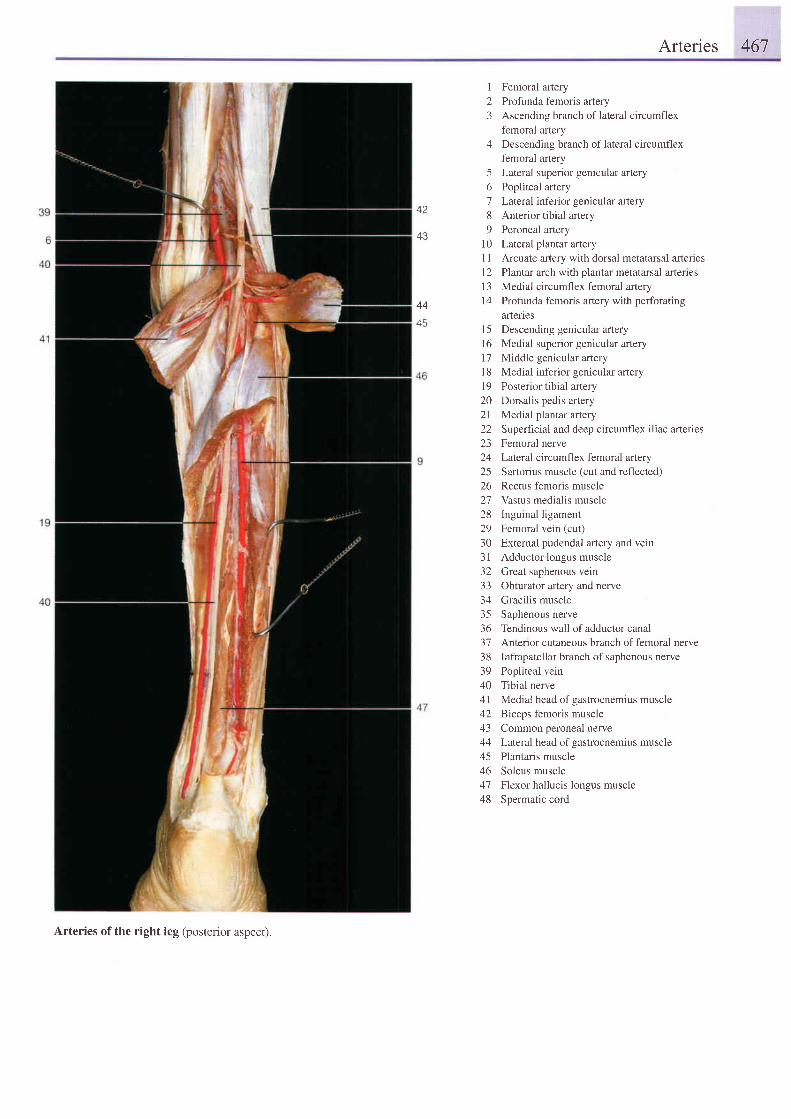
Arteries 467
44
45
I Femoral artery2 Profunda femoris artery3 Ascending branch of lateral circumflex
femoral artery4 Descending branch oflateral circumflex
f-emoral artery5 Lateral superior genicular artery6 Popliteal artery7 Lateral inferior genicular artery8 Anterior tibial artery9 Peroneal aftery
10 Lateral plantar artery11 Arcuate artery with dorsal metatarsal arteries12 Plantar arch with plantar metatarsal arteries13 Medial circumflex femoral artery14 Profunda femoris artery with perforating
artenes15 Descending genicular afiery16 Medial superior genicular artery17 Middle genicular arteryl8 Medial inferior genicular arlery19 Posterior tibial artery20 Dorsalis pedis artery21 Medial plantar artery22 Superhcial and deep circumflex iliac arteries23 Femoral nerve24 Lateral circumflex femoral artery25 Sartorius muscle (cut and reflected)26 Rectus femoris muscle27 Vastus medialis muscle28 Inguinal ligament29 Femoral vein (cut)30 External pudendal artery and vein31 Adductor longus muscle32 GreaI saphenous vein33 Obturator artery and nerve34 Gracilis muscle35 Saphenous nerve36 Tendinous wall of adductor canal37 Anterior cutaneous branch of femoral nerve38 Infrapatellar branch of saphenous nerve39 Popiiteal vein40 Tibial nerve41 Medial head of gastrocnemius muscle42 Biceps femoris muscle43 Common peroneal nerve44 Lateral head of gastrocnemius muscle45 Plantaris muscle46 Soleus muscle47 Flexor hallucis longus muscle48 Spermatic cord
Arteries of the right leg (posterior aspect).
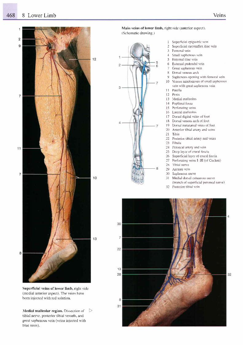
468 8 Lower Limb Veins
Main veins of lower limb, right side (anterior aspect).(Schematic drawing.)
6
I Superficial epigastric vern2 Superf icial circumflex i l iac vein3 Femoral vein4 Small saphenous vein5 Extemal iliac vein6 External pudendal vein7 Creat saphenous vein8 Dorsal venous arch9 Saphenous opening with femoral vein
10 Venous anastomoses of small saphenousvein with great saphenous vetn
I I Patella12 Penis13 Medial malleolus14 Popliteal fossa15 Perforating veins16 Lateral malleolusl7 Dorsal digital veins offoot18 Dorsal venous arch offoot19 Dorsal metatarsal veins of foot20 Anterior tibial artery and veins2l Tibia22 Posterior tibial artery and veins23 Fibula24 Peroneal artery and vein25 Deep layer of crural fascia26 Supedcial layer of crural fascia27 Perforating veins I-III (ofCockett)28 Tibial nerve29 Arcuate vein30 Saphenous nerve31 Medial dorsal cutaneous nerve
(branch of superficial peroneal nerve)32 Posterior tibial vein
1 328
Superficial veins of lower limb, right side(medial anterior aspect). The veins havebeen injected with red solution.
Medial malleolar region. Dissection oftibial nerve, posterior tibial vessels, andgreat saphenous vein (veins injected withblue resin).
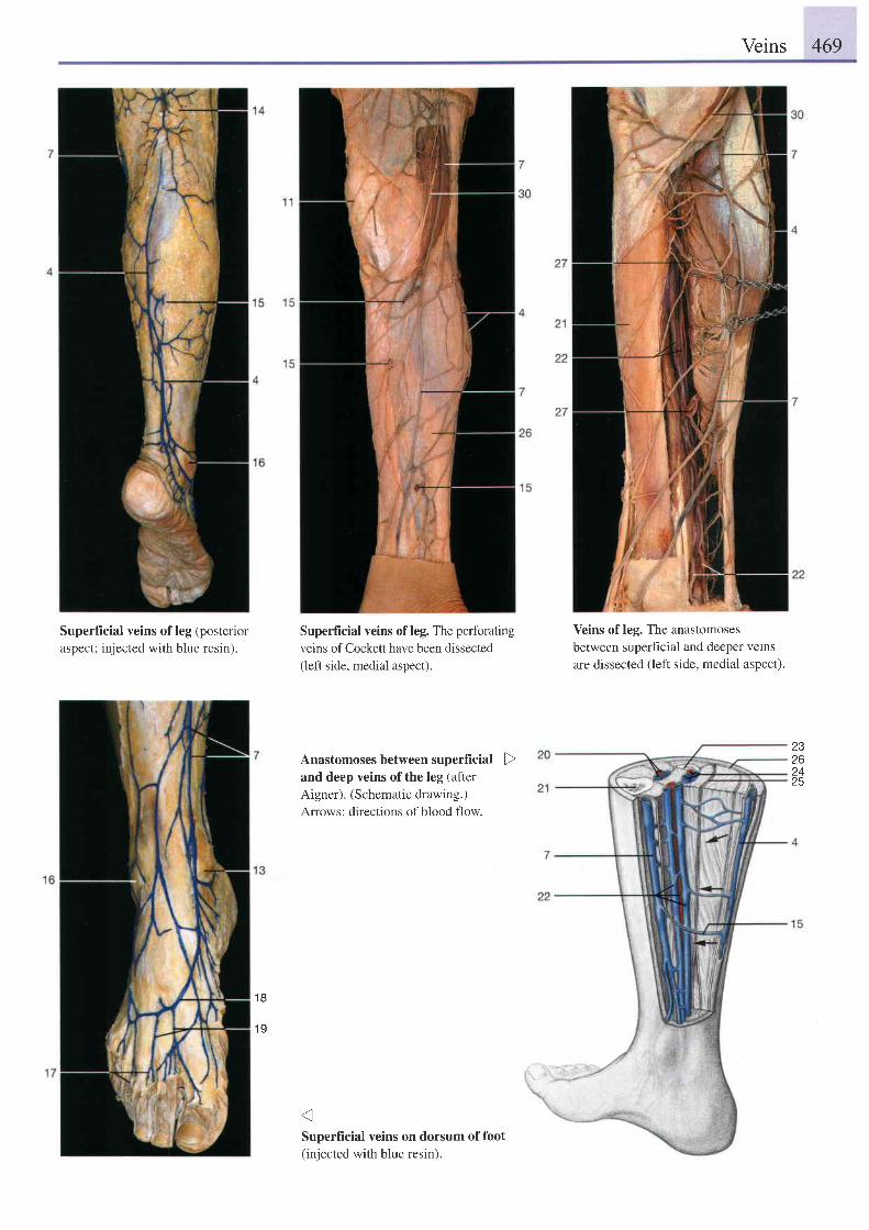
Veins 469
Superficial veins of leg (posterioraspect; injected with blue resin).
Superficial veins of leg. The perforatingveins of Cockett have been dissected(left side, medial aspect).
Anastomoses between superficial Dand deep veins of the leg (afterAigner). (Schematio drawing.)Arrows: directions of blood flow.
Superficial veins on dorsum of foot(injected with blue resin).
Veins of leg. The anastomosesbetween superf cial and deeper veinsare dissected (left side, medial aspect).
23zo2425
1 8
1 9
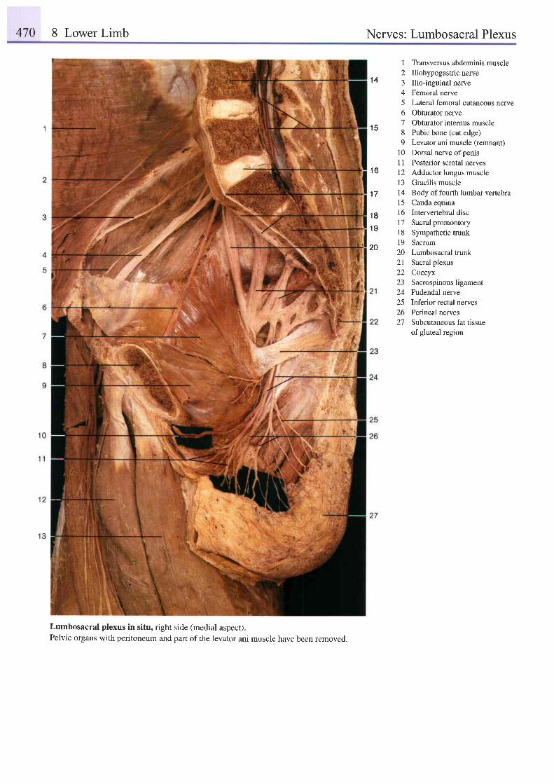
470 8 Lower Limb Nerves: Lumbosacral Plexus
1 4
1 5
I Transversus abdominis muscle2 Iliohypogastricnerve3 Ilio-inguinal nerve4 Femoral nerve5 Lateral femoral cutaneous nerve6 Obturator nerve7 Obturator intemus muscle8 Pubic bone (cut edge)9 Levator ani muscle (remnant)
10 Dorsal nerve ofpenisI I Posterior scrotal nerves12 Adductor longus musclel3 Gracilis muscle14 Body of fourth lumbar vertebra15 Cauda equina16 Intervertebral disc17 Sacral promontory18 Sympathetic trunk19 Sacrum20 Lumbosacral trunk21 Sacral plexus22 Coccyx23 Sacrospinousligament24 Pudendal nerve25 Inferior rectal nerves26 Perineal nerves27 Subcutaneous fat tissue
of gluteal region
1 6
1 7
1 8
1 9
20
Lumbosacral plexus in situ, right side (medial aspect).Pelvic organs with peritoneum and part of the levator ani muscle have been removed.
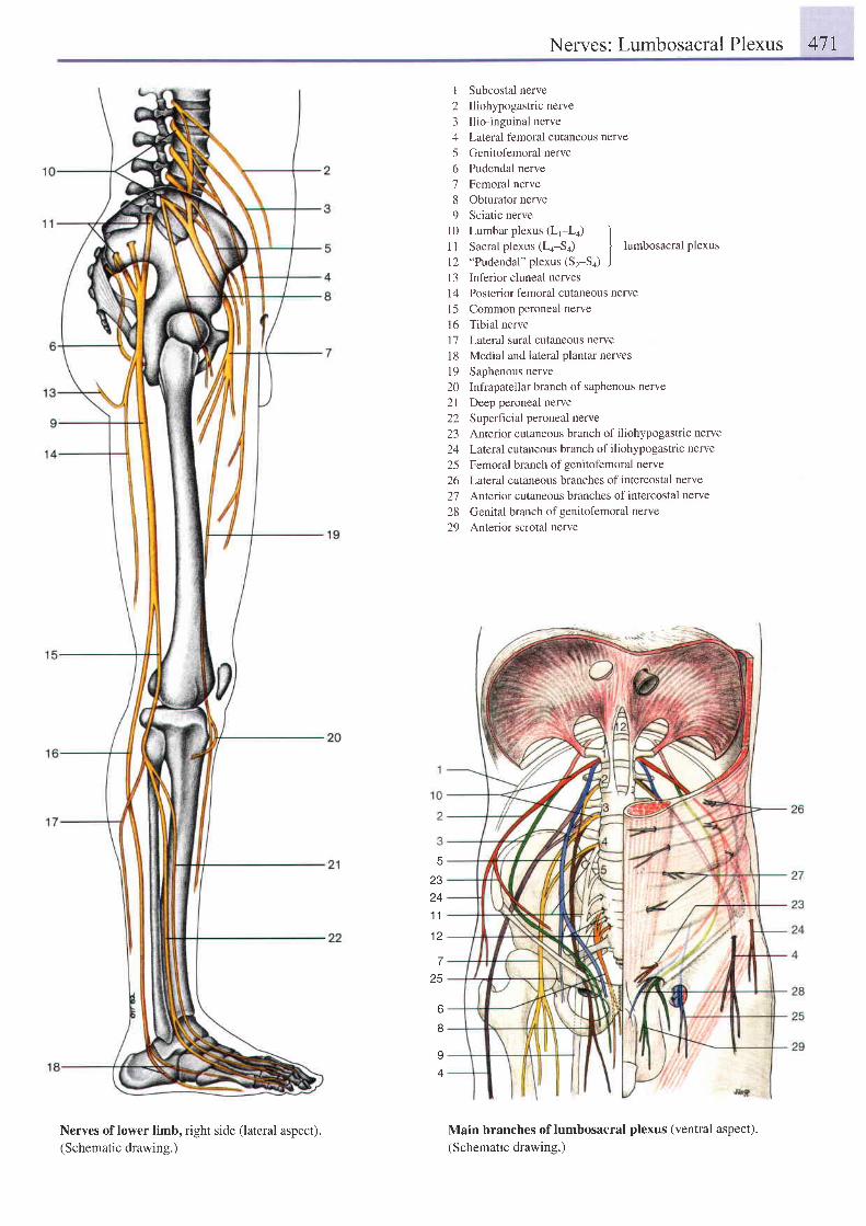
Nerves: Lumbosacral Plexus 471
23z+
1 1
1 2
7
25
o
8
I
I Subcostal nerve2 Iliohypogastricnerve3 Ilio-inguinal nerve4 Lateral femoral cutaneous nerve5 Genitofemoral nerve6 Pudendal nerve7 Femoral nerve8 Obturator nerve9 Sciatic nerve
l0 Lumbar plexus (L1-La) I1 I Sacral plexus (La-Sa) | lumbosacral plexus
12 "Pudendal"plexus (Sz-S+) J13 Inferior cluneal nerves14 Posterior femoral cutaneous nervel5 Common peroneal nervel6 Tibial nerve1'1 Lateral sural cutaneous nerveI 8 Medial and lateral plantar nervesl9 Saphenous nerve20 Infrapatellar branch of saphenous nerve21 Deep peroneal nerve22 Superhcial peroneal nerve23 Anterior cutaneous branch of iliohypogastric nerve24 Laterul cutaneous branch ofiliohypogastric nerve
25 Femoral branch ofgenitofemoral nerve26 Lateral cutaneous branches of intercostal nerve27 Anlelior cutaneous branches ofintercostal nerue
28 Genital branch ofgenitofemoral nerve29 Anterior scrotal nerve
Main branches of lumbosacral plexus (ventral aspect).(Schematic drawing.)
Nerves of lower limb, right side (lateral aspect).(Schematic drawing.)
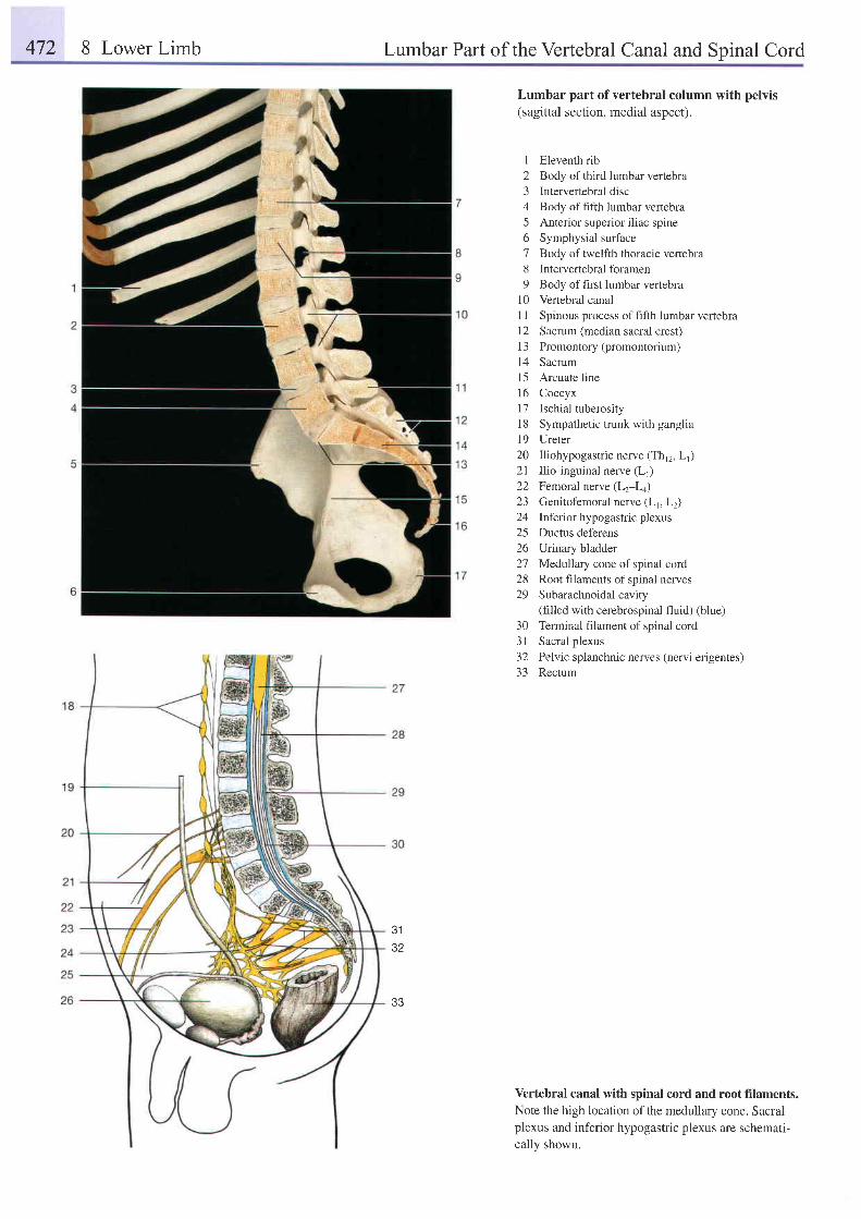
472 8 Lower Limb Lumbar Part of the Vertebral Canal and Spinal Cord
3132
Lumbar part of vertebral column with pelvis(sagittal section, medial aspect).
I Eleventh rib2 Body of third lumbar vertebra3 Intervertebral disc4 Body of fifth lumbar vertebra5 Anterior superior i l iac spine6 Symphysial surface7 Body oftwelfth thoracic vertebra8 Intervertebralforamen9 Body of first lumbar vertebra
10 Vertebral canalI I Spinous process ofhfth lumbar vertebra12 Sacrum (median sacral crest)13 Promontory (promontorium)14 Sacrum15 Arcuate line16 Coccyx17 Ischial tuberosity18 Sympathetic trunk with ganglia19 Ureter20 Iliohypogastric nerve (Thr2, Ll)21 Ilio-inguinal nerve (L1)22 Femoral nerve (L2-La)23 Genitofemoral nerve (Lr, Lz)24 lnfenor hypogastric plexus25 Ductus def'erens26 Urinry bladder27 Medullary cone of spinal cord28 Root filaments of spinal nerves29 Subarachnoidalcavity
(filled with cerebrospinal fluid) (blue)30 Terminal filament of spinal cord31 Sacral plexus32 Pelvic splanchnic nerves (nervi erigentes)33 Rectum
Vertebral canal with spinal cord and root filaments.Note the high location of the medullary cone. Sacralplexus and inferior hypogastric plexus are schemati-cally shown.
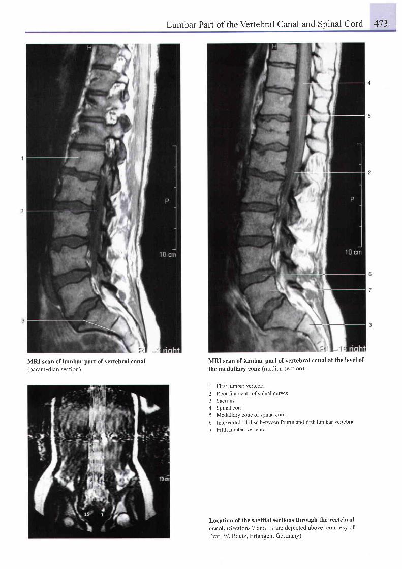
Lumbar Part of the Vertebral Canal and Spinal Cord 473
MRI scan of lumbar part of vertebral canal(paramedian section).
MRI scan of lumbar part of vertebral canal at the level of
the medullary cone (median section).
I First lumbar vertebra2 Root fllaments of spinal nerves3 Sacrum4 Spinal cord5 Medullary cone of spinal cord6 Intervertebral disc between fburth and fifth lumbar vertebra
7 Fifth lumbar vertebra
Location of the sagittal sections through the vertebralcanal. (Sections 7 and I 1 are depicted above; courtesy of
Prof. W Bautz, Erlangen, GermanY).
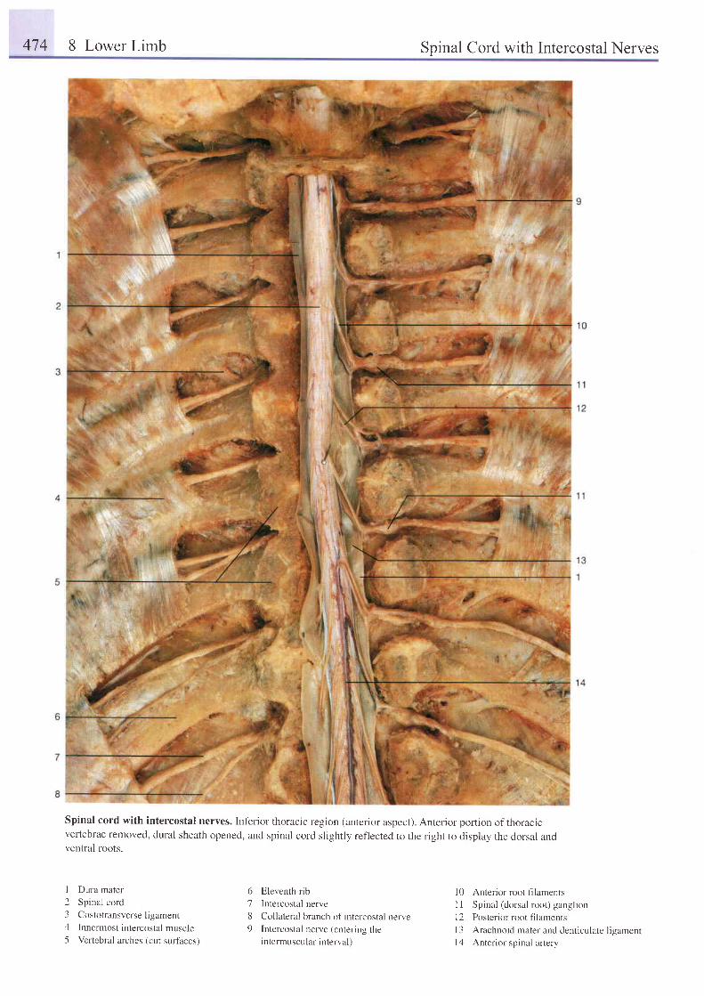
474 8 Lower Limb Spinal Cord with Intercostal Nerves
Spinal cord with intercostal nerves. Inf'erior thoracic region (anterior aspect). Anterior portion of thoracrcvertebrae removed, dural sheath opened, and spinal cord slightly ref'lected to the right to itisplay the dorsal andventral roots.
I Dura mater
2 Spinal cord
3 Costotransverseligament
4 Innermost intercostal muscle5 Vertebral arches (cut surfaces)
6 Eleventh rib
7 Intercostal nerve
8 Collateral branch of intercostal nerve
9 Intercostal nerve (entering theintermuscular interval)
l0 Anterior root filaments
I I Spinal (dorsal root) ganglion
l2 Posterior root fllaments
l3 Arachnoid mater and denticulate ligamentl4 Anterior spinal artery
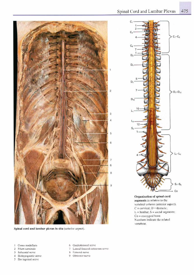
Spinal Cord and Lumbar Plexus 475
Spinal cord and lumbar plexus in situ lanterior aspect).
I Conus medullaris2 Filum terminale3 Subcostal nerve4 Iliohypogastricnerve5 Ilio-inguinal nerve
6 Genitofemoral nerve'7
Lateral femoral cutaneous nerve
8 Femoral nerve
9 Obturator nerve
- L O
Organization of spinal cord
segments in relation to the
vertebral column (anterior aspect).
C = cervical; D = thoracic:
L = lumbar; S = sacral segments;
Co = coccygeal bone.
Numbers indicate the related
vertebrae.
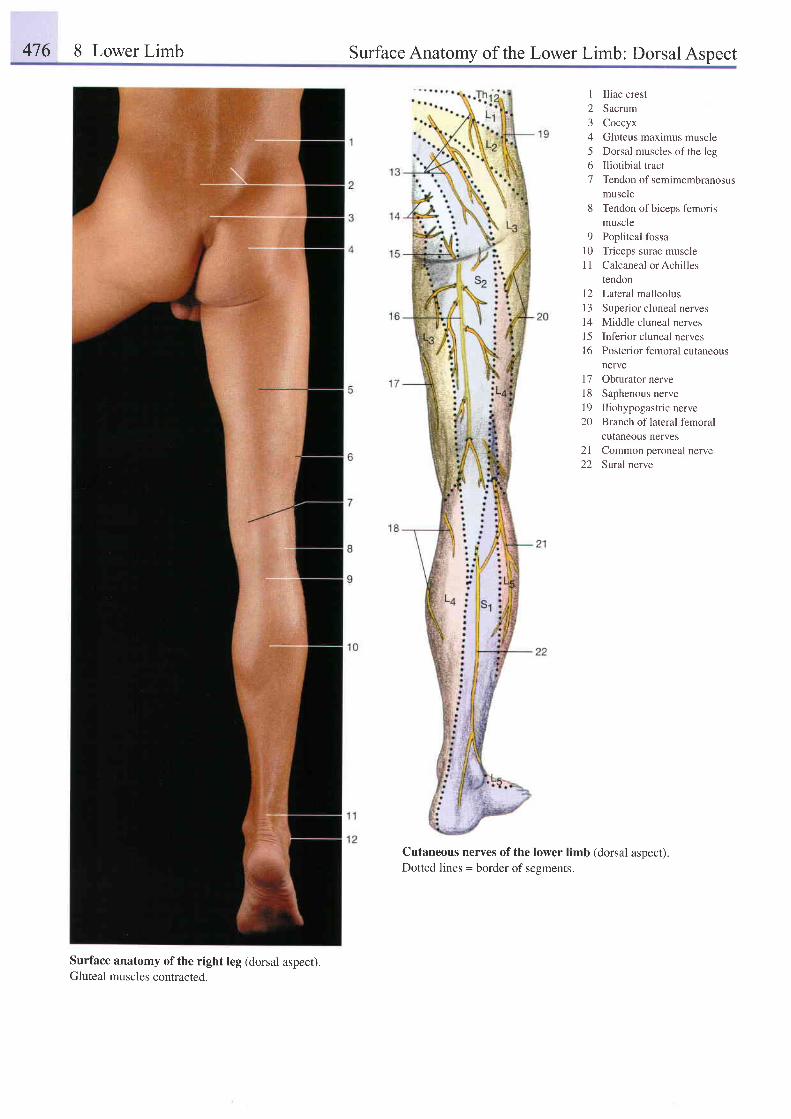
476 8 Lower Limb Surface Anatomy of the Lower Limb: Dorsal Aspect
1 Iliac crest2 Sacrum3 Coccyx4 Gluteus maximus muscre5 Dorsal muscles of the leg6 Iliotibial tract7 Tendon of semimembranosus
muscle8 Tendon ofbiceps femorrs
muscle9 Popliteal fossa
10 Triceps surae muscleI I Calcaneal or Achilles
tendon12 Lateralmalleolus13 Superior cluneal nerves14 Middle cluneal nervesl5 Inferior cluneal nerves16 Posteriorfemoral cutaneous
nerve17 Obturator nerve18 Saphenous nervel9 Iliohypogastric nerve20 Branch of lateral femoral
cutaneous nerves2l Common peroneal nerve22 Sural nerve
Cutaneous nerves of the lower limb (dorsal aspect).Dotted lines = border of sesments.
Surface anatomy ofthe right leg (dorsal aspect).Gluteal muscles contracted.
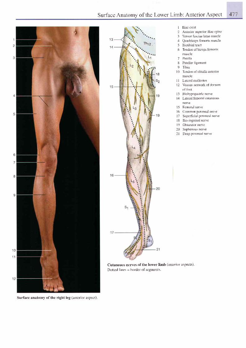
Surface Anatomy of the Lower Limb: Anterior Aspect 477
1 3
1 4Th t2 ,
L1
'- 52
Cutaneous nerves of the lower limb (anterior aspects).Dotted lines = border of segments.
1 Iliac crest2 Anterior superior iliac spine3 Tensor fasciae latae muscle4 Quadriceps femoris muscle5 Iliotibial tract6 Tendon ofbiceps femorts
muscle7 Patella8 Patellar ligament9 Tibia
10 Tendonof tibialis anteriormuscle
1l Lateral malleolus12 Venous network of dorsum
of fbot13 Iliohypogastric nerve14 Lateral femoral cutaneous
nerve15 Femoral nerve16 Common peroneal nerve17 Superficial peroneal nerve18 Ilio-inguinal nervel9 Obturator nerve20 Saphenous nerve2l Deep peroneal nerve
L2
1 8
Surface anatomy of the right leg ( anterior aspect ).
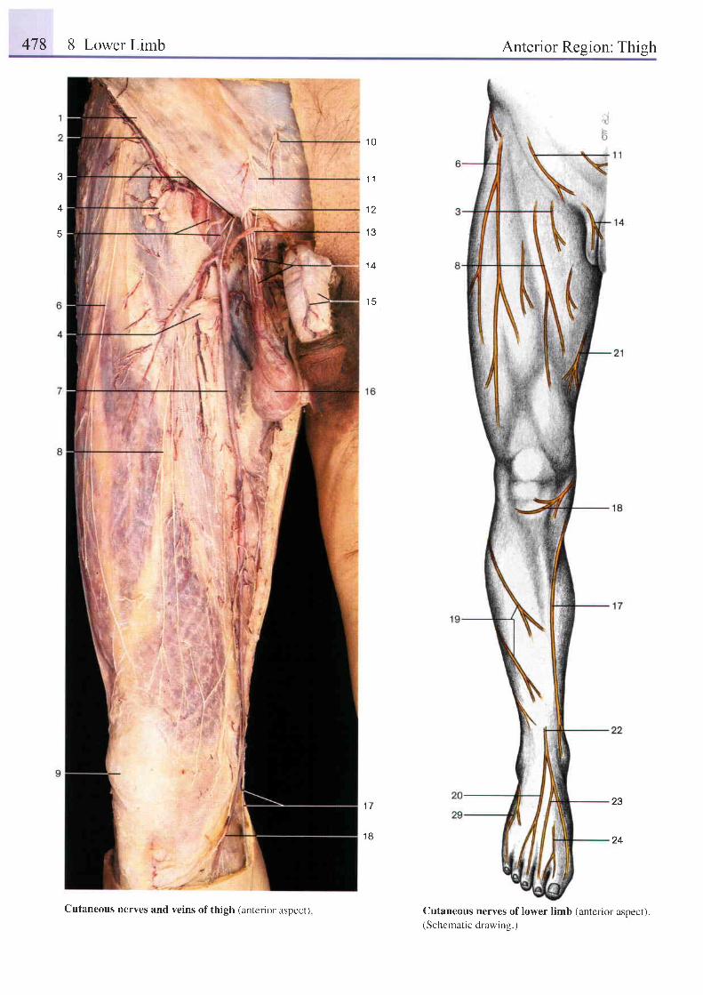
478 8 Lower Limb Anterior Region: Thigh
3
4
' t0
1 2
1 3
1 4
1 5
23
24
1 7
1 8
Cutaneous nerves of lower limb (anterior aspect).(Schematic drawine.)
Cutaneous nerves and veins of thigh (anterior aspecr)
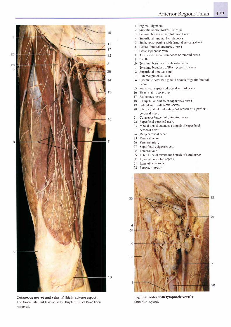
Anterior Resion: Thieh 479
Cutaneous nerves and veins ofthigh (anterior aspect).
The fascia lata and fasciae of the thieh muscles have been
removed.
I Inguinal ligament
2 Superhcial circumflex iliac vein
3 Femoral branch ofgenitofemoral nerve
4 Superticial inguinal lymph nodes
5 Saphenous opening with femoral artery and vein
6 Lateral femoral cutaneous nerve
7 Great saphenous vein
8 Anterior cutaneous branches offemoral nerve
9 Patella
l0 Terminal branches of subcostal nerve
1 I Terminal branches of iliohypogastric nerve
l2 Superficial inguinal ring
l3 External pudendal vein
14 Spermatic cord with genital branch of genitofemoral
nerve
15 Penis with supelficial dorsal vein of penis
l6 Testis and its coverings
l7 Saphenous nerve
l8 Infiapatellar branch of saphenous nerve
19 Lateral sural cutaneous nerves
20 Intermediate dorsal cutaneous branch of superficial
peroneal nerve
21 Cutaneous branch of obturator nerve
22 Superfrcial peroneal nerve
23 Medial dorsal cutaneous branch of superhcial
peroneal nerve
24 Deep peroneal nerve
25 Femoral nerve
26 Femoral artery
27 Super l ic ia l epigastr ic vein
28 Femoral vein
29 Lateral dorsal cutaneous branch of sural nerve
30 Inguinal nodes (enlarged)
3l Lympathic vessels
32 Sartorius muscle
Inguinal nodes with lymphatic vessels(anterior aspect).
-t\-
1 1
27
12
631
30
32
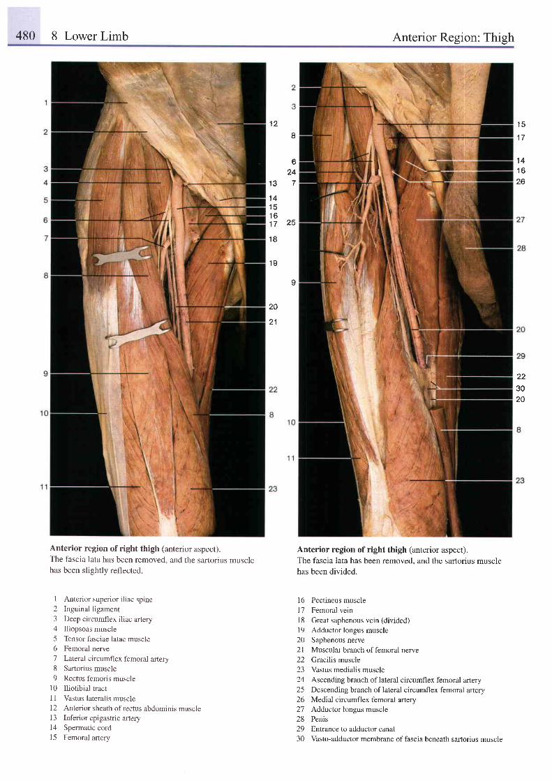
480 8 Lower Limb Anterior Region: Thigh
1 2I
624
1 3 7
1 4i E
1 61 7 2 5
1 8
1 9
I
20
21
1 5
1 7
1 41 626
223020
Anterior region of right thigh (anterior aspect).The fascia lata has been removed, and the sartorius musclehas been slightly reflected.
I Anterior superior i l iac spine2 Inguinal ligament3 Deep circumflex iliac artery4 Iliopsoas muscle5 Tensor fasciae latae muscle6 Femoral nerve7 Lateral circumflex femoral artery8 Sartorius muscle9 Rectus femoris muscle
l0 Iliotibial tracrl l Vastus lateralis muscle12 Anterior sheath of rectus abdominis muscle13 Inferior epigastric artery14 Spermatic cord15 Femoral artery
Anterior region of right thigh (anterior aspect).The fascia lata has been removed, and the sartorius musclehas been divided.
l6 Pectineus muscle17 Femoral vein18 Great saphenous vein (divided)19 Adductor longus muscle20 Saphenous nerve2l Muscular branch of femoral nerve22 Gracilis muscle23 Vastus medialis muscle24 Ascending branch oflateral circumflex femoral artery25 Descending branch oflateral circumflex femoral artery26 Medial circumflex femoral artery27 Adductor longus muscle28 Penis29 Enlrance to adductor canal30 Vasto-adductor membrane of fascia beneath sartorius muscle
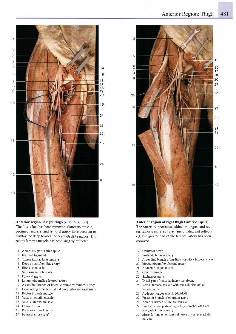
Anterior Region: Thigh
1 5
261 71 82027
28
29
30
1 623
67II
1 4
1 5
1 61 71 81 920
1 5
21
22
23
1 6
6
78I
Anterior region of right thigh (anterior aspect).The fascia lata has been removed. Sartorius muscle,pectineus muscle, and femoral artery have been cut todisplay the deep femoral artery with its branches. Therectus femoris muscle has been slightly reflected.
I Anterior superior i l iac spine2 Inguinal ligament3 Tensor fasciae latae muscle4 Deep circumflex iliac artery5 Iliopsoas muscle6 Sartorius muscle (cut)
7 Femoral nerve8 Lateral circumflex femoral artery9 Ascending branch of lateral circumflex femoral artery
10 Descending branch oflateral circumflex femoral arteryI I Rectus femoris musclel2 Vastus medialis muscle13 Vastus lateralis muscle14 Femoral vein15 Pectineus muscle (cut)
16 Femoral artery (cut)
Anterior region of right thigh (anterior aspect).
The sartorius, pectineus, adductor longus, and rec-
tus femoris muscles have been divided and reflect-
ed. The greater part of the femoral artery has been
removed.
17 Obturatornerve18 Profunda femoris artery19 Ascending branch of medial circumflex femoral artery20 Medial circumflex femoral artery21 Adductor longus muscle22 Gracilis muscle23 Saphenous nerve24 Distal part of vasto-adductor membrane25 Rectus femoris muscle with muscular branch of
femoral nerve26 Adductor longus muscle (divided)27 Posterior branch of obturator nerve28 Anterior branch of obturator nerve29 Point at which perforating artery branches off from
profunda femoris artery30 Muscular branch of femoral nerve to vastus medialis
muscle
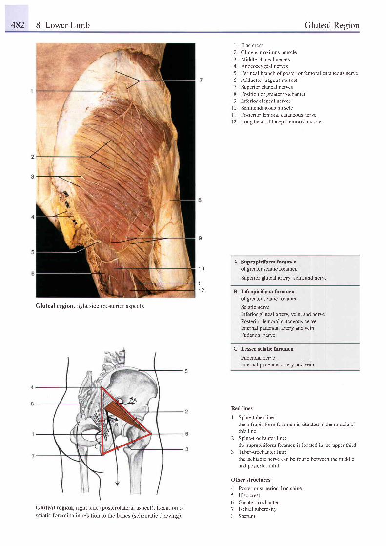
482 8 Lower Limb Gluteal Region
Gluteal region, right side (posterior aspect).
Gluteal region, right side (posterolateral aspect). Location ofsciatic foramina in relation to the bones (schematic drawing).
I Iliac crest
2 Gluteus maximus muscle
3 Middle cluneal nerves
4 Anococcygeal nerves
5 Perineal branch of posterior femoral cutaneous nerve
6 Adductor magnus muscle
7 Superior cluneal nerves
8 Position of greater trochanter
9 Inferior cluneal nerves
10 Semitendinosus muscle
I 1 Posterior femoral cutaneous nerve
12 Long head of biceps femoris muscle
A Suprapiriformforamenof greater sciatic foramen
Superior gluteal artery, vein, and nerve
B Infrapiriformforarnenof greater sciatic foramen
Sciatic nerveInferior gluteal artery, vein, and nervePosterior femoral cutaneous nerveInternal pudendal artery and veinPudendal nerve
C Lesser sciatic foramen
Pudendal nerveInternal pudendal artery and vein
Red lines
1 Spine-tuber line:the infrapiriform foramen is situated in the middle ofthis line
2 Spinetrochanterline:the suprapiriform foramen is located in the upper third
3 Tuber-trochanterline:the ischiadic nerve can be found between the middleand posterior third
Other structures
4 Posterior superior iliac sprne5 Iliac crest6 Greater trochanter7 Ischial tuberosity8 Sacrum
1 0
t 11 2
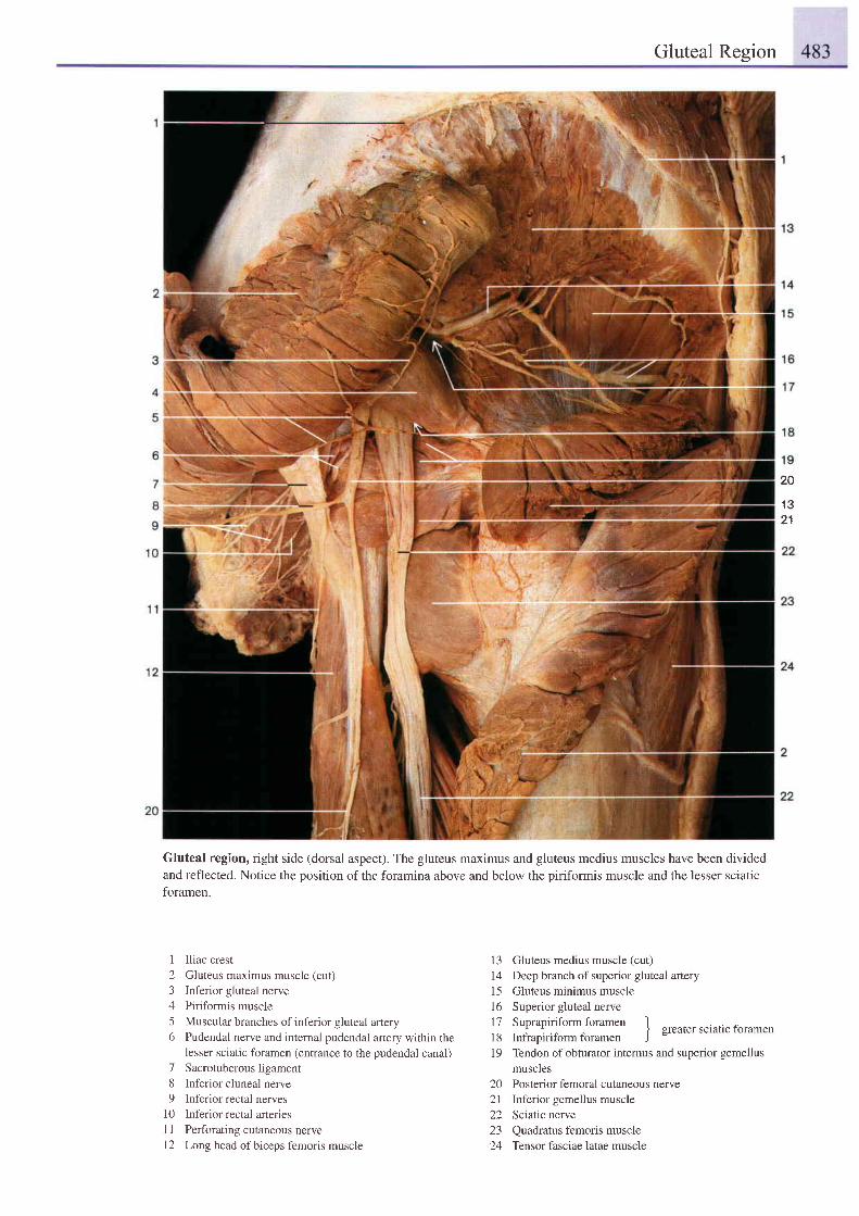
Gluteal Resion
20
1 321
Gluteal region, right side (dorsal aspect). The gluteus maximus and gluteus medius muscles have been dividedand reflected. Notice the position of the foramina above and below the piriformis muscle and the lesser sciaticforamen.
1 Iliac crest2 Gluteus maximus muscle (cut)3 Inferior gluteal nerve4 Piriformis muscle5 Muscular branches of inferior gluteal artery6 Pudendal nerve and internal pudendal artery within the
lesser sciatic foramen (entrance to the pudendal canal)7 Sacrotuberous ligament8 Inferior cluneal nerve9 Inferior rectal nerves
10 Inferior rectal arteries1 I Perforating cutaneous nerve12 Long head of biceps femoris muscle
13 Gluteus medius muscle (cut)
I 4 Deep branch of superior gluteal artery15 Gluteus minimus muscle16 Superior gluteal nerve| 7 Suprapiriform foramen1g Infrapirifbrm foramen J Sreatet sciatic foramen
19 Tendon ofobturator internus and superior gemellusmuscles
20 Posterior femoral cutaneous nerve21 Inferior gemellus muscle22 Sciatic nerve23 Quadratus femoris muscle24 Tensor fasciae latae muscle
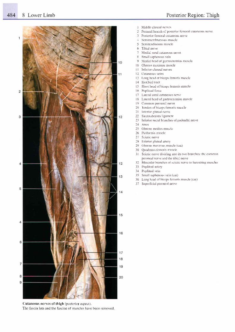
484 8 Lower Limb Posterior Resion: Thish
I Middle cluneal nerves
2 Perineal branch ofposterior femoral cutaneous nerve
3 Posterior f'emoral cutaneous nerve
4 Semimembranosus muscle
5 Semitendinosusmuscle
6 Tibial nerve
7 Medial sural cutaneous nerve
8 Small saphenous vein
9 Medial head of gastrocnemius muscle
l0 Cluteus maximus muscle
I I lnferior cluneal nerves
12 Cutaneous veins
l3 Long head of biceps f'emoris muscle
14 Iliotibial tract
l5 Short head ofbiceps femoris muscle
16 Popliteal fossa
l7 Lateral sural cutaneous nerve
l8 Lateral head of gastrocnemius muscle
l9 Common peroneal nerve
20 Tendon of biceps femoris muscle
2l lnferior gluteal nerve
22 Sacrotuberous ligament
23 Inferior rectal branches of pudendal nerve
24 Anus
25 Gluteus medius muscle
26 Piritbrmis muscle
27 Sciatic nerve
28 Inferior gluteal artery
29 Gluteus maximus muscle (cut)
30 Quadratus f'emoris muscle
3l Sciatic nerve dividing into its two branches: the common
peroneal nerve and the tibial nerve
32 Muscular branches of sciatic nerve to hamstring muscles
33 Popliteal artery
34 Popliteal vein
35 Small saphenous vein (cut)
36 Long head of biceps femoris muscle (cut)
37 Superficial peroneal nerve
Cutaneous nerves ofthigh (posterior aspect).The fascia lata and the fasciae of muscles have been removed
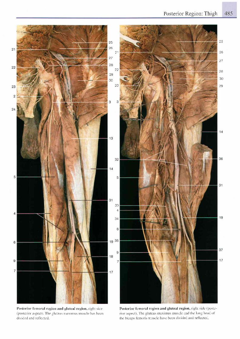
Posterior Resion: Thish 485
26
2 7
28
30
29
25
26, 1
27
2822
29
3023
334
34
t
Posterior f'cmoral region and gluteal region, right sidc(postr-r ior aspcct). The -uluteus maxilnus rrusclc has beendividcd ancl ref lected
Postcrior femoral region and gluteal region, right side (poste-
r iol aspect) Thc gluteus ntaxintus muscle and the long heacl ol '
thc biccps lernoris muscle har. 'e been divided and ref- lccted
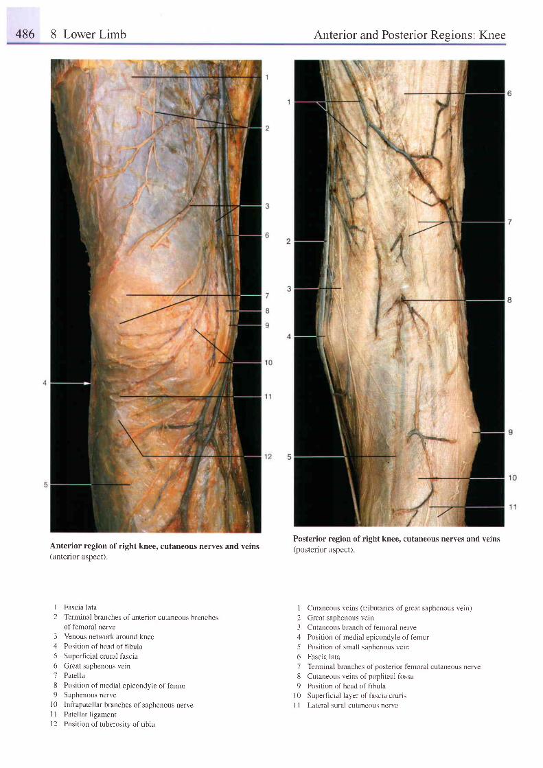
486 8 Lower Limb Anterior and Posterior Resions: Knee
Anterior region of right knee, cutaneous nerves and veins(anterior aspect).
I Fascia lata
2 Terminal branches of anterior cutaneous branchesof femoral nerve
3 Venous network around kneezl Position ofhead offibula
5 Superficial crural fascia
6 Great saphenous vein
7 Patella
8 Position of medial epicondyle of femur9 Saphenous nerve
l0 Infrapatellar branches of saphenous nerveI I Patellar ligament
l2 Position of tuberosity oftibia
Posterior region of right knee, cutaneous nerves and veins(posterior aspect).
I Cutaneous veins (tributaries ofgreat saphenous vein)
2 Great saphenous vein
3 Cutaneous branch of femoral nerve
4 Position of medial epicondyle of femur
5 Posi t ion of smal l saphenous vein
6 Fascia lata
7 Terminal branches ofposterior femoral cutaneous nerve
8 Cutaneous veins of popliteal fossa
9 Position ofhead offibula
| 0 Superficial layer of fascra crurrs
I 1 Lateral sural cutaneous nerve
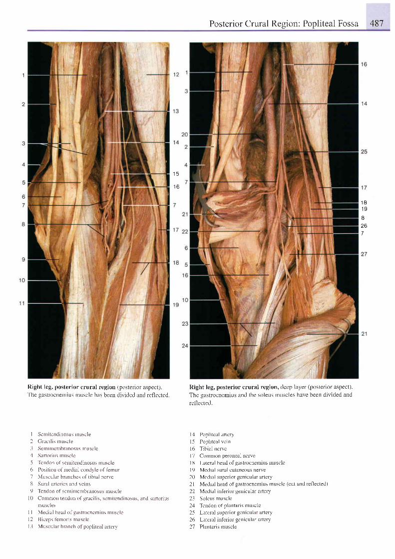
Posterior Crural Region: Popliteal Fossa 487
1 6
1 81 9
z6
7
Right leg, posterior crural region (posterior aspect).The gastrocnemius muscle has been divided and reflected.
I Semitendinosusmuscle
2 Graci l is muscle
3 Semimembranosus muscle,1 Sartorius muscle
5 Tendon of semitendinosus muscle
6 Position of medial condyle of femur
7 Muscular branches of tibial nerve
8 Sural arteries and veins
9 Tendon of semimembranosus muscle
l0 Common tendon of gracilis, semitendinosus, and sartoriusmuscles
I I Medial head of gastrocnemius muscle
l2 Biceps femoris muscle
13 Muscular branch of popliteal artery
Right leg, posterior crural region, deep layer (posterior aspect).The gastrocnemius and the soleus muscles have been divided andref'lected.
l4 Popliteal artery
l -5 Poplr teal vein
l6 Tib ia l nerve
l7 Common peroneal nerve
l8 Lateral head of gastrocnemius muscle
l9 Medial sural cutaneous nerve
20 Medial superior genicular artery
2l Medial head of gastrocnemius muscle (cut and reflected)
22 Medial inferior genicular artery
23 Soleus muscle
24 Tendon of plantaris muscle
25 Lateral superior genicular artery
26 Lateral inferior genicular artery
27 Plantaris muscle
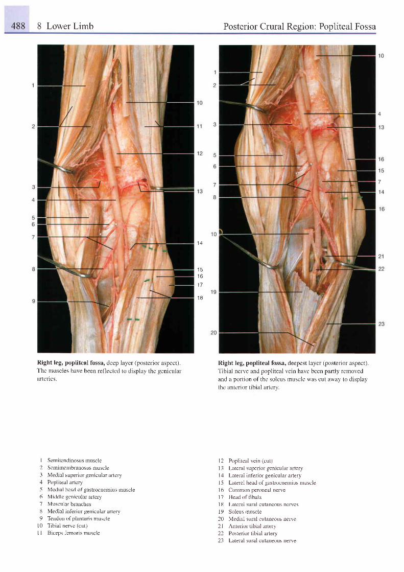
488 8 Lower Limb Posterior Crural Region: Popliteal Fossa
1 4
1 6
1 7
1 8
Right leg, popliteal fossa, deep layer (posterior aspect).The muscles have been reflected to display the genicularafierles.
I Semitendinosusmuscle2 Semimembranosusmuscle3 Medial superior genicular artery4 Popliteal artery5 Medial head of gastrocnemius muscle6 Middle genicular artery7 Muscular branches8 Medial inferior genicular artery9 Tendon ofplantaris muscle
l0 Tibial nerve (cut)I I Biceps femoris muscle
Right leg, popliteal fossa, deepest layer (posterior aspect).
Tibial nerve and popliteal vein have been partly removed
and a portion of the soleus muscle was cut away to display
the anterior tibial artery.
12 Popliteal vein (cut)I 3 Lateral superior genicular arteryl4 Lateral inferior genicular aftery15 Lateral head of gastrocnemius musclel6 Common peroneal nerve17 Head of flbula18 Lateral sural cutaneous nerves19 Soleus muscle20 Medial sural cutaneous nerve2l Anterior tibial artery22 Posterior tibial artery23 Lateral sural cutaneous nerve
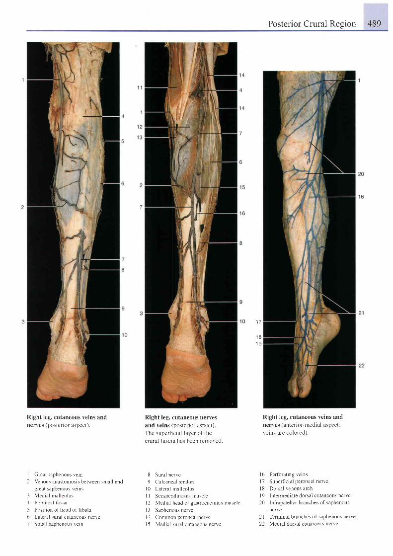
Posterior Crural Resion 489
1 7
1 81 9
Right leg, cutaneous veins andnerves (posteilor aspect).
I Great saphenous vein
2 Venous anastomosis between small andgreat serphenous veins
3 Mcd ia l r na l l eo l us;l Popliteal lirssa
5 Position of head of frbula
6 Latcral sural cutaneous nerve
7 Smal l saphenous vein
Right leg, cutaneous nerves
and veins (posterior aspect)
The superf icial layer o1'the
crural fascia has been rernoved.
8 Sural nerve
9 Calcaneal tendon
l0 Lateral mal leolus
I I Semitendinosus muscle
l2 Medial head of gastrocncmius muscle
l3 Saphenous nerve
lrl Comrnon peroneal nerve
l5 Medial sural cutaneous nerve
Right leg, cutaneous veins and
nerves (anterior-medial aspect;
veins are colored).
l6 Perforating veins
l7 Superficial peroneal nerve
l8 Dorsal venous arch
I 9 Intermediate dorsal cutaneous nsrve
20 Infiapatellar branches of saphenous
nerve
2l Terminal branches of saphenous nerve
22 Medial dorsal cutaneous nerve
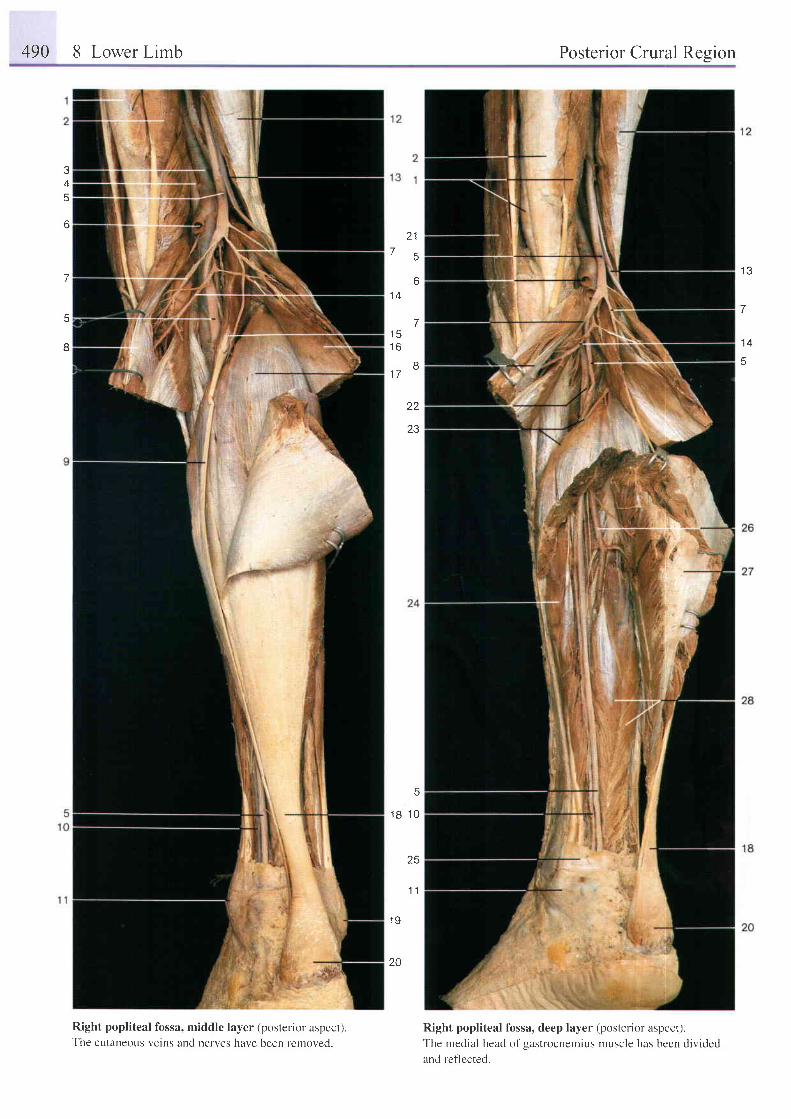
490 8 Lower Limb Posterior Crural Resion
1 l
5
o
a
A
6
J
5
8
7
1 A
t c1 A
1 7
1 3
7
1 4
B
22
23
tr
1 8 1 0
25
1 1
1 9
20
Right popliteal fossa, middle layer (posterior aspecr).The cutaneous veins and nerves have been removed.
Right popliteal fossa, deep layer (posterior aspect).The medial head of gastrocnemius muscle has been divided
and ref lected.
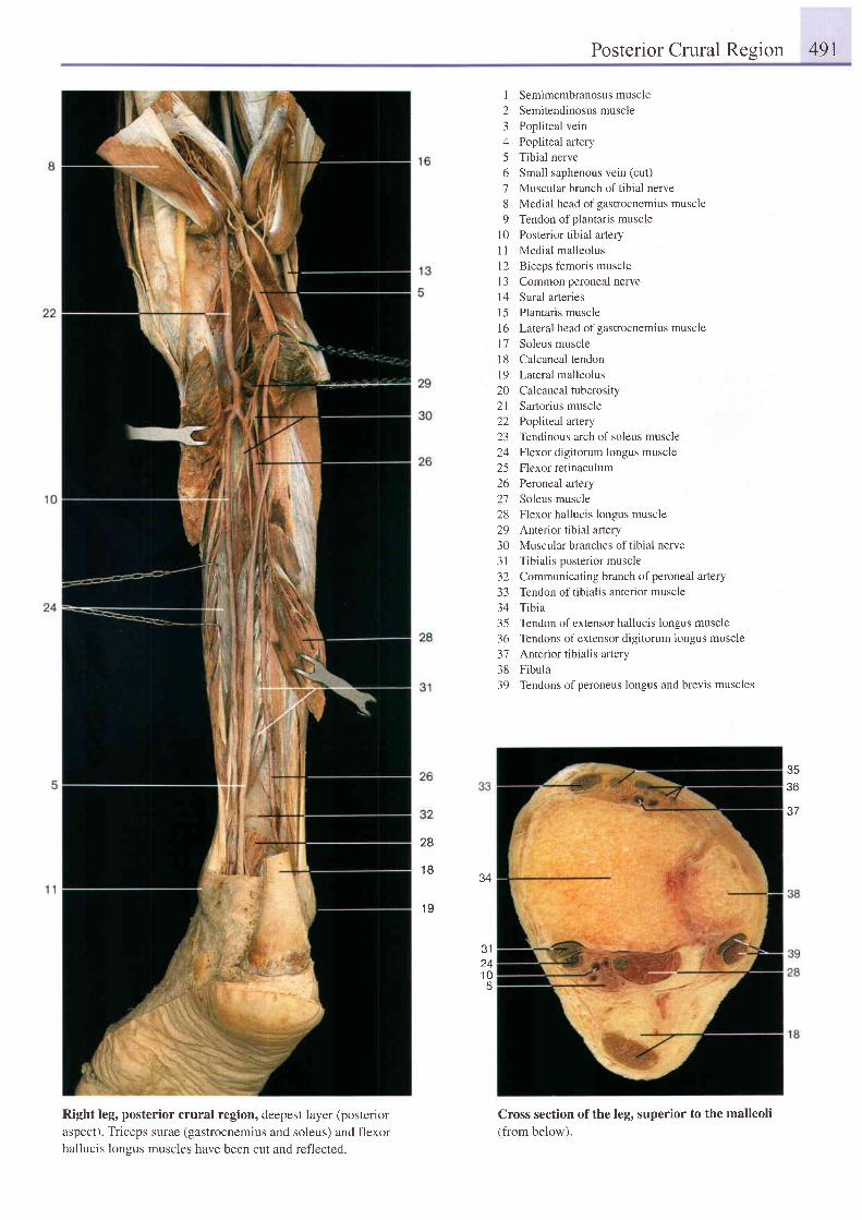
Posterior Crural Resion 491
Right leg, posterior crural region, deepest layer (posterioraspect). Triceps surae (gastrocnemius and soleus) and flexorhallucis longus muscles have been cut and reflected.
1 Semimembranosusmuscle2 Semitendinosusmuscle3 Popliteal vein4 Popliteal artery5 Tibial nerve6 Small saphenous vein (cut)
7 Muscular branch of tibial nerve8 Medial head of gastrocnemius muscle9 Tendon ofplantaris muscle
10 Posterior tibial aftery11 Medial malleolusl2 Biceps femoris muscle13 Common peroneal nerve14 Sural arteries15 Plantaris muscle16 Lateral head ofgastrocnemius muscle17 Soleus muscleI 8 Calcaneal tendon19 Lateral malleolus20 Calcanealtuberosity2l Sartorius muscle22 Popliteal artery23 Tendinous arch of soleus muscle24 Flexor digitorum longus muscle25 Flexor retinaculum26 Peroneal artery27 Soleus muscle28 Flexor hallucis longus muscle29 Anterior tibial aftery30 Muscular branches oftibial nerve31 Tibialis posterior muscle32 Communicating branch ofperoneal artery33 Tendon oftibialis anterior muscle34 Tibia35 Tendon of extensor hallucis longus muscle36 Tendons ofextensor digitorum longus muscle37 Anterior tibialis artery38 Fibula39 Tendons ofperoneus longus and brevis muscles
Cross section of the leg, superior to the malleoli(from below).
36
e-7
28
18 34
1 9
31241 0
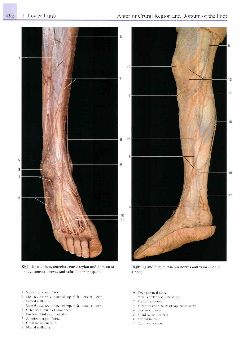
492 8 Loner L in tb
I{ ight leg and fbot. anterior crural region :urcl dorsunr off i tot. ctt tnneous ncncs and r,eins (untcrior lsl)cct i
I SL rpc r l i c i u l c r - L r ra l i u sc i l LI \ l c t i i l L l . u l i r l r ' ous b t l r r r ch o l ' su l t c r l i c i l t l l ) ! ' l o t ) e i r l nc r \ c- l I i t l e r r r l r r l r l l eo l L r s
l - t , r i , - ' r : r l e ut i lncoLls h l t r tc l r o l sLrpcrf ie i l r l l tcronelr l ncr i t .. - ( L r l i l r ) r ' { r L r \ l r r l t r t c l t ( ) l s t i l a l t t e t r c( ) 11 ) \ i l r ( ) r r r r l t L rhc ros i t v o l t i b i a7 , \ n t c r i o r r na r ! . i n o l t i b i t rt ( i r c t L t s l p l t cno r r s r c i n
9 N l cd i l r l n ra l l co l L r s
Antcrior Crural Rcgion and Dorsum of the Foot
Right lcu and lbot, cutaueous nerves and reins (nrc(l i l r l
r t s l l cc t l
i 0 I ) c r ' p p c r - o n c u l n c r - r c
I I \ ' , 'Dr rL rs i r r t l r 0 l d0r -s r rDr o l Io ( ) (
I I I ) r \ r t l o l l r r l P l L t u ' l l i r
l - l l n l r . U l r l c l l i u b r L n e h c : o 1 s a p h e n o u s r r c n c
I 1 S l rp l t . ' r to r rs r tc r r r '
i 5 S n r . L l l s u p h c n o u s r c i n
l ( r P i r l o r . L t i n L r c i n
I I ( ' : r l e l r r t c l r l t c n t L r r t

1 7
1 8
Right leg and dorsum offoot, cutaneous nerves and veins (lateral aspect)'
I
2345
6'7
89
Position of fibula
Sural nerve
Small saphenous vein
Calcaneal tendon
Lateral calcaneal branches of
sural nerve
Venous network at lateral malleolus
Cutaneous branch of sural nerve
Tendon of peroneus brevis muscle
Tendons of extensor digitorum
longus muscle
10 Fascia crurisI I Superfrcial peroneal nerve12 Position oftibia13 Lateral cutaneous branch
'l
14 Medial cutaneous branch J15 Lateral malleolus16 Dorsal digital nerves17 Dorsal venous arch18 Deep peroneal nerve
of superficialperoneal nerve
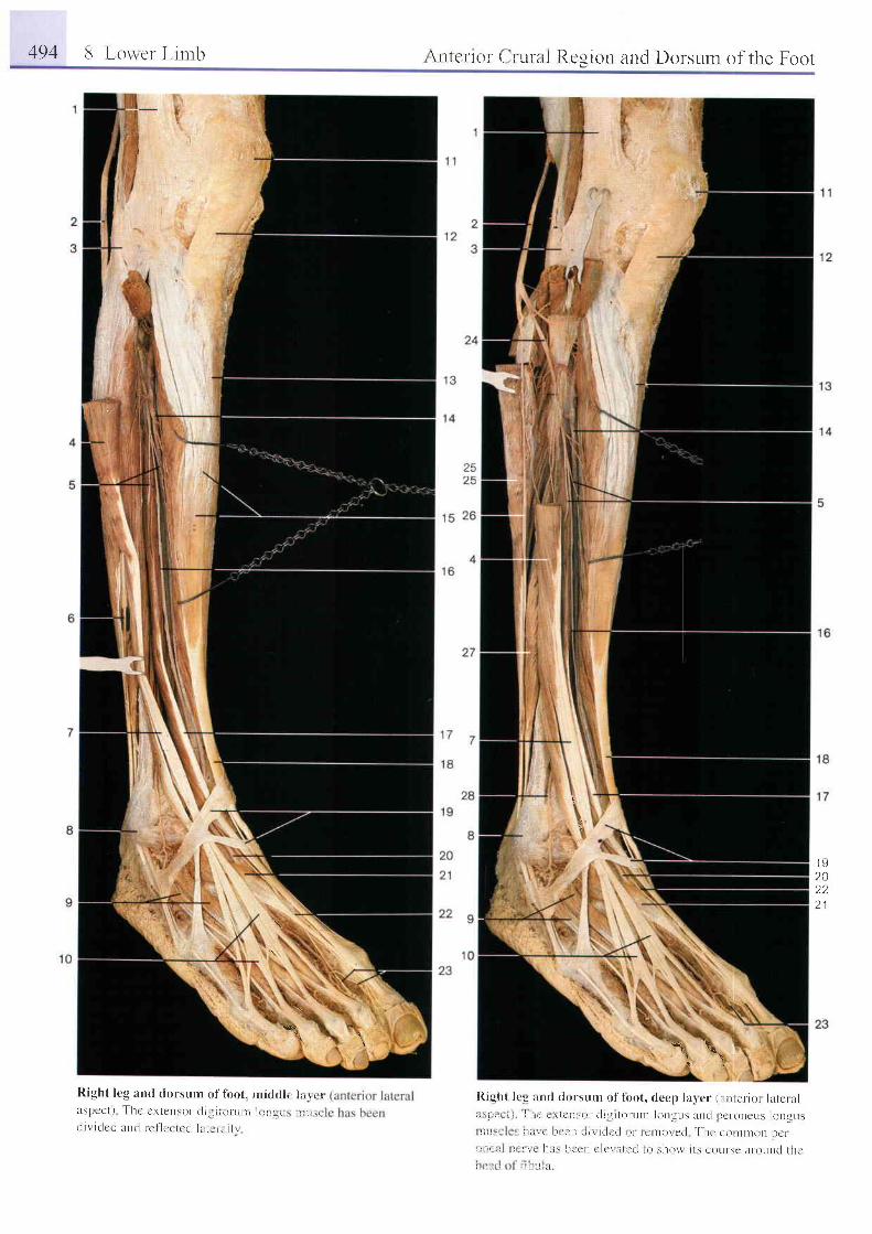
494 E Lou'cr Lir trb \n tcnor ' ( r 'L r ra l Rcg ion and Dorsum o f the Foot
\
d
t v
20D '
2 1
\ \*\ 1:
Right l cg and dorsunr o f loo t . n r idd l r l r r r l ri t s l ) c c l ) T h e c - r i c n s o r r l i ! i i i l i - u n t l ( , I t r L , i r . i r . ,
c l r r i r l c c i l n r l r c i ' l c r t e c l l l r t i r i i i l i
I t igh t l r .g u r rd dors r rn r o f lbo t , c lccp la rc r (u l rLcr - io r l l l cn r l( i \ p i i , ! i ' r . r , i c l t . r ' r ' t l i g r lo r - t r rn lo t rg t rs a r rc j l t c r -oncus longusl l t i , . . 1 t ; r " t l r c r ' I L l i r i t i r t t l L r l l 0 t ] t o \ e ( l I ' l t c c o n t t l l O n l t e t
i i l ; i . , r - r \ L l t . r \ l t c e r r c l c t t r t i t l I o s ] 1 0 $ i t s c 0 t r | s c l t t o L r D c l t l t c
i , - . , I i , l : 1 r ; . 1
-*[
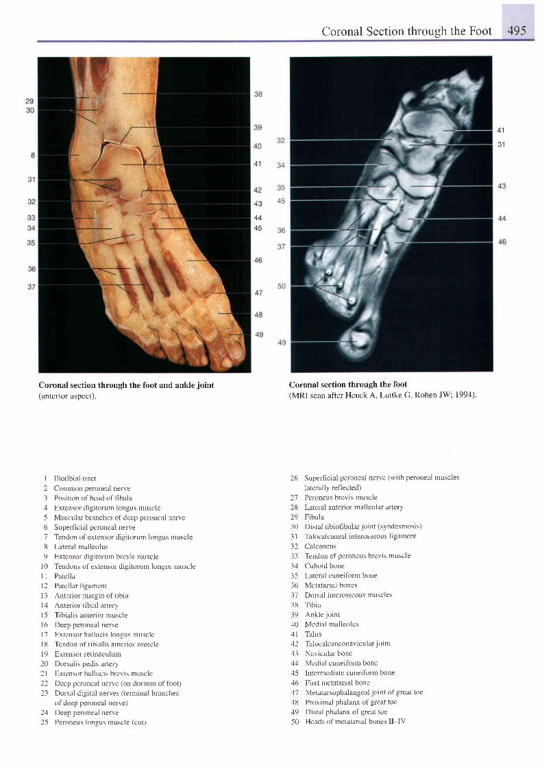
Coronal Section throueh the Foot 495
31
43
4445
Coronal section through the foot and ankle joint
(anterior aspect).
I Iliotibial tract
2 Common peroneal nerve
3 Position ofhead offibula
4 Extensor digitorum longus muscle
5 Muscular branches of deep peroneal nerve
6 Superficial peroneal nerve
7 Tendon ofextensor digitorum longus muscle
8 Lateral malleolus
9 Extensor digitorum brevis muscle
l0 Tendons of extensor digitorum longus muscle
I 1 Patella
l2 Patellar ligament
l 3 An te r i o r ma rg in o f t i b i a
l4 Anteriortibial artery
15 Tibialis anterior muscle
16 Deep peroneal nerve
l7 Extensor hallucis longus muscle
l8 Tendon of tibialis anterior muscle
19 Extensorretinaculum
20 Dorsalis pedis artery
2l Extensor hallucis brevis muscle
22 Deep peroneal nerve (on dorsum offbot)
23 Dorsal digital nerves (terminal branches
of deep peroneal nerve)
24 Deep peroneal nerve
25 Peroneus longus muscle (cut)
Coronal section through the foot(MRI scan after Heuck A, Luttke G, Rohen JW; 1994).
26 Superficial peroneal nerve (with peroneal muscles
laterally reflected)27 Peroneus brevis muscle28 Lateral anterior malleolar artery29 Fibula30 Distal tibiofibular joint (syndesmosis)
3l Talocalcaneal interosseous ligament
32 Calcaneus33 Tendon ofperoneus brevis muscle34 Cuboid bone35 Lateral cuneiform bone36 Metatarsal bones37 Dorsal interosseous muscles38 Tibia39 Ankle joint
40 Medial malleolus41 Talus42 Talocalcaneonar icular joint
43 Navicular bone44 Medial cuneiform bone45 Intermediate cuneiform bone46 First metatarsal bone47 Metatarsophalangeal joint of great toe48 Proximal phalanx of great toe49 Distal phalanx of great toe50 Heads of metatarsal bones II-IV

496 8 Lower Limb Dorsum of the Foot
t o
1 7
Dorsum of the right foot, superficial layer (anterloraspect).
Superfi cial peroneal nerve
Superior extensor retinaculum
Lateral malleolus
Venous network of lateral malleolus andtributaries of small saphenous vernLateral dorsal cutaneous nerve (branch ofsural nerve)
Intermediate dorsal cutaneous nerveTendons of extensor digitorum longus muscleDorsal digital nerves
Dorsum of the right foot, superficial layer. The fascia of thedorsum has been removed.
I
23I
678
9 Tendon of tibialis anterior muscle
10 Saphenous nerve
I I Venous network of medial malleolus andtr ibutar ies of greet raphenous vein
l2 Medial mal leolus
l3 Medial dorsal cutaneous nervesl4 Dorsal venous arch
l5 Dorsal digital nerve (ofdeep peroneal
nerve)
I 6 Tendon of extensor hallucis longus muscle
I 7 Dorsal digital arteries18 Peroneal musclesI 9 Deep plantar branch of dorsalis pedis
afiery anastomosing with plantar arch20 Extensor digitorum longus muscle21 Extensor hallucis longus muscle22 lnferior extensor retinaculum23 Extensor hallucis brevis muscle
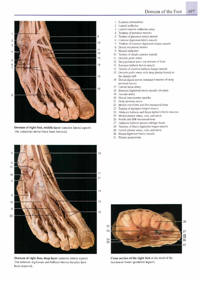
Dorsum of the Foot 497
1 Extensor retinaculum2 Lateral malleolus3 Lateral anterior malleolar artery4 Tendons ofperoneal muscles5 Tendon ofperoneus tertius muscle6 Extensor digitorum brevis muscle7 Tendons of extensor digitorum longus muscle8 Dorsal metatarsal arteries9 Medial malleolus
l0 Tendon of tibialis anterior muscle1 I Dorsalis pedis anery12 Deep peroneal nerve (on dorsum offoot)l3 Extensor hallucis brevis muscle14 Tendon ofextensor hallucis longus muscle15 Dorsalis pedis artery with deep plantar branch to
the plantar archDorsal digital nerves (terminal branches of deepperoneal nerve)Lateral tarsal arteryExtensor digitorum brevis muscle (divided)Arcuate arteryDorsal interosseous musclesDeep peroneal nerveMedial cuneiform and first metatarsal boneTendon of peroneus longus muscleAbductor hallucis and flexor hallucis brevis musclesMedial plantar aftery, vein, and nerveFourth and fifth metatarsal boneAdductor hallucis muscle (oblique head)Tendons of flexor digitorum longus muscleLateral plantar artery, vein, and nerveFlexor digitorum brevis musclePlantar aponeurosis
Dorsum of right foot, middle layer (anterior lateral aspect)The cutaneous nerves have been removed.
Dorsum of right foot, deep layer (anterior lateral aspect).The extensor digitorum and hallucis breves muscles havebeen removed.
1 0
1 11 2
1 3
1 4
1 5
1 6
l 6
1'71 81 9202 122L-)
2425262'72829303 1
282930
31
23
2425
Cross section of the right foot at the level of the
metatarsal bones (posterior aspect).
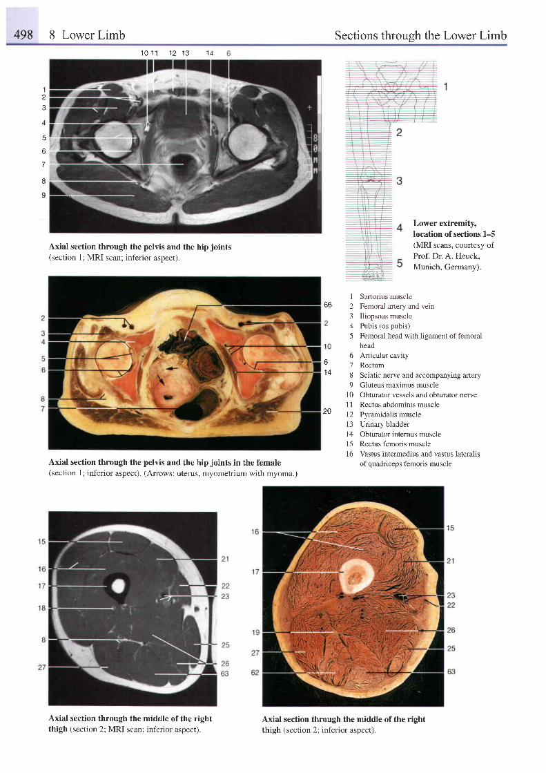
498 8 Lower Limb Sections throush the Lower Limb1 0 1 1 1 2 1 3
Axial section through the pelvis and the hip joints(section 1; MRI scan; inferior aspect).
Axial section through the pelvis and the hip joints in the female(section 1; inferior aspect). (Arrows: uterus, myometrium with myoma.)
Axial section through the middle of the rightthigh (section 2; MRI scan; inferior aspect).
12J
I
5
Sartorius muscleFemoral artery and veinIliopsoas musclePubis (os pubis)Femoral head with ligament of femoralhead
^ 6 Articular cavityo 7 Rectum14 8 Sciatic nerve and accompanying anery
9 Gluteus maximus muscle10 Obturator vessels and obturator nerve11 Rectus abdominis muscle
20 l2 \ramidalis muscle13 Urinary bladder14 Obturator internus muscle15 Rectus femoris muscle16 Vastus intermedius and vastus lateralis
of quadriceps femoris muscle
1 A
1
3
4
6
I
I
Lower extremity,location of sections 1-5(MRI scans, courtesy ofProf. Dr. A. Heuck,Munich, Germany).
66
Axial section through the middle of the rightthigh (section 2; inferior aspect).
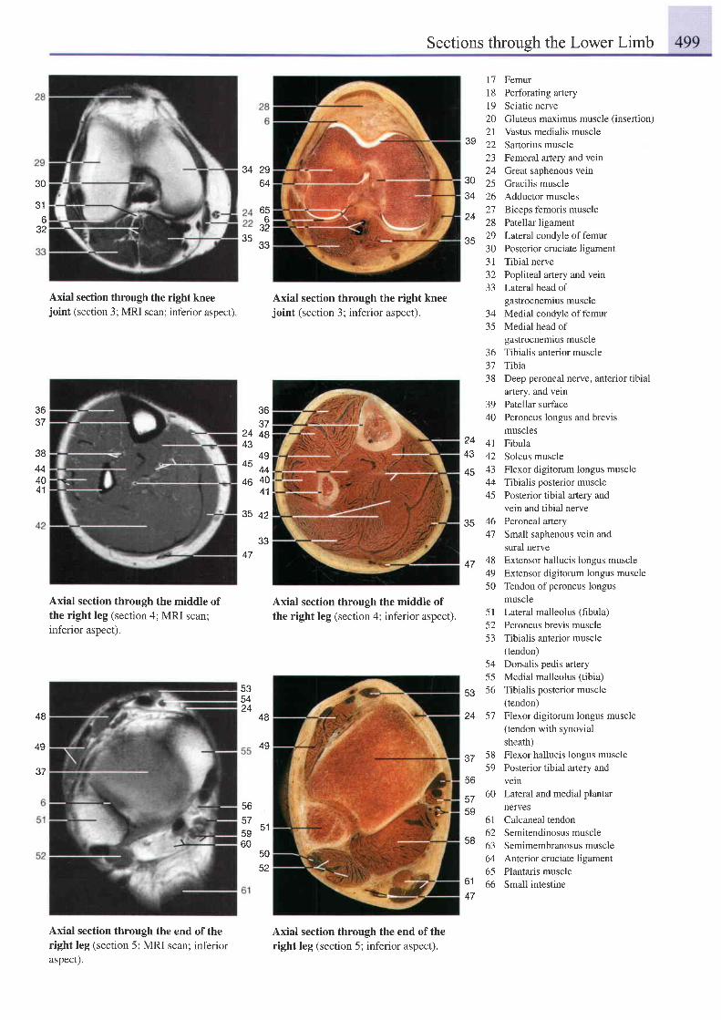
Sections through the Lower Limb
30
316
32
Axial section through the right kneejoint (section 3; MRI scan; inferior aspect).
Axial section through the middle ofthe right leg (section 4; MRI scan;inferior aspect).
Axial section through the end of theright leg (section 5; MRI scan; inferioraspect).
Axial section through the right kneejoint (section 3; inferior aspect).
Axial section through the middle ofthe right leg (section 4; inferior aspect).
Axial section through the end of theright leg (section 5; inferior aspect).
17 Femur18 Perforating artery19 Sciatic nerve20 Gluteus maximus muscle (insertion)2l Vastus medialis muscle22 Sartorius muscle23 Femoral artery and vein24 Great saphenous vein25 Gracilis muscle26 Adductor muscles27 Biceps femoris muscle28 Patellar ligament29 Lateral condyle offemur30 Posterior cruciate ligament31 Tibial nerve32 Popliteal artery and vein33 Lateral head of
gastrocnemius muscle34 Medial condyle of femur35 Medial head of
gastrocnemius muscle36 Tibialis anterior muscle37 Tibia38 Deep peroneal nerve, anterior tibial
artery, and vein39 Patellar surface40 Peroneus longus and brevis
musclesFibulaSoleus muscleFlexor digitorum longus muscleTibialis posterior musclePosterior tibial artery andvein and tibial nervePeroneal arterySmall saphenous vein andsural nerveExtensor hallucis longus muscleExtensor digitorum longus muscleTendon of peroneus longusmuscle
2964
J4
656
32e t"" g3
39
3034
24
35
36e'f
24 4843
494F' - 4446 40
41
36J '
3844404 1
?443
45
47
4 142434445
4647
484950
35 qz
3347
48
4g
48
49
37
53
24
535424
56
57< 1
bcOU
5052
5l Lateral malleolus (fibula)52 Peroneus brevis muscle53 Tibialis anterior muscle
(tendon)54 Dorsalis pedis artery55 Medial malleolus (tibia)56 Tibialis posterior muscle
(tendon)57 Flexor digitorum longus muscle
(tendon with synovialsheath)
58 Flexor hallucis longus muscle59 Posterior tibial artery and
veln60 Lateral and medial plantar
nerves61 Calcaneal tendon62 Semitendinosusmuscle63 Semimembranosus muscle64 Anterior cruciate ligament65 Plantaris muscle66 Small intestine
56
575S
5B
6147
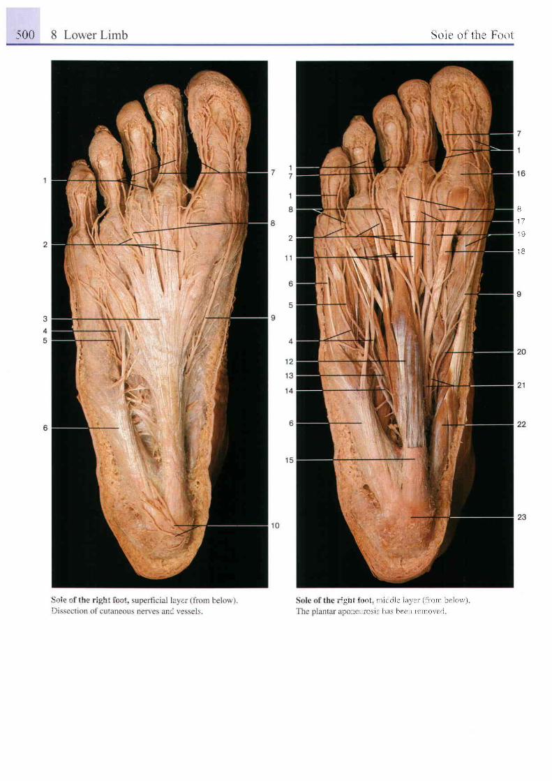
:500 Solc ol ' t i tu I t ' rut
B
t 7
1 9
1 B
\ i l t i " r . , : l i : j i , , r
i i l ' l t i t t l i r o t . n r t t l . i l t i l r r i r ( I r ( r r l r l r r ' i \ ) \ \ i
t , ' i : . " i l r ! ! \ t r l I t . i r c c n ! c n t i l \ ! ' r l
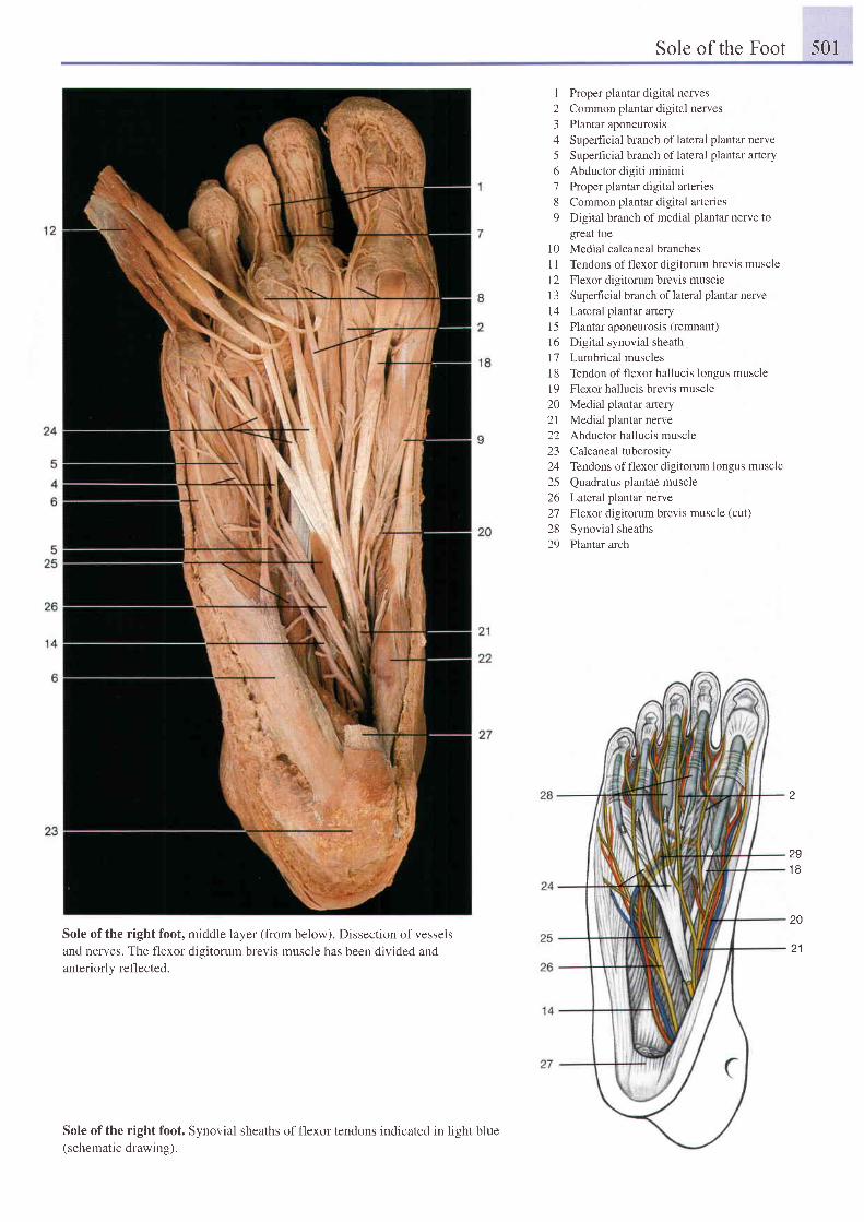
Sole of the Foot 501
I Proper plantar digital nerves2 Common plantar digital nerves3 Plantar aponeurosis4 Superficial branch oflateral plantar nerve5 Superficial branch oflateral plantar artery6 Abductor digiti minimi7 Proper plantar digital arteries8 Common plantar digital arteries9 Digital branch of medial plantar nerve to
great toe10 Medial calcaneal branches1l Tendons of flexor digitorum brevis muscle12 Flexor digitorum brevis muscle1 3 Superficial branch of lateral plantar nerve14 Lateral plantar artery15 Piantar aponeurosis (remnant)16 Digital synovial sheath17 Lumbrical muscles18 Tendon offlexor hallucis longus muscle19 Flexor hallucis brevis muscle20 Medial plantar arlery21 Medial plantar nerve22 Abductor hallucis muscle23 Calcanealtuberosity24 Tendons offlexor digitorum longus muscle25 Quadratus plantae muscle26 LaIeral plantar nerve27 Flexor digitorum brevis muscle (cut)28 Synovial sheaths29 Plantar arch
z
291 8
20
21Sole of the right foot, middle layer (from below). Dissection of vesselsand nerves. The flexor digitorum brevis muscle has been divided andanteriorly reflected.
Sole of the right foot. Synovial sheaths of flexor tendons indicated in light blue(schematic drawing).
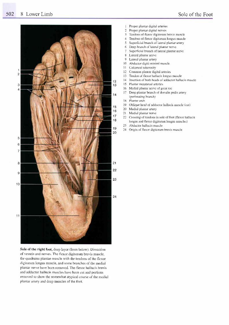
s02 8 Lower Limb Sole of the Foot
12345678o
l 01 1l 21 3
1 2 1 4
1 9 1 5l 6t 7
1 4
1 8l q
1 q) i
1 6 : :- 1 l
t t 2 21 8
z-)19 .rA
20
Proper plantar digital arteriesProper piantar digital nervesTendons of flexor digitorum brevis muscleTendons of flexor digitorum longus muscleSuperhcial branch of lateral plantar arteryDeep branch of lateral plantar nerveSuperficial branch of lateral plantar nerveLateral plantar nerveLateral plantar arteryAbductor digiti minimi muscleCalcaneal tuberosityCommon plantar digital artenesTendon of flexor hallucis longus muscleInsertion of both heads of adductor hallucis musclePlantar metatarsal arteriesMedial plantar nerve of great toeDeep plantar branch of dorsalis pedis artery(perforating branch)Plantar archOblique head of adductor hallucis muscle (cut)Medial plantar arteryMedial plantar nerveCrossing of tendons in sole of foot (flexor hallucislongus and flexor digitorum longus muscles)Abductor hallucis muscleOrigin of flexor digitorum brevis muscle
21
22
23
Sole of the right foot, deep layer (from below). Dissecrionof vessels and nerves. The flexor digitorum brevis muscle,the quadratus plantae muscle with the tendons of the flexordigitorum longus muscle, and some branches of the medialplantar nerve have been removed. The flexor hallucis brevisand adductor hallucis muscles have been cut and portionsremoved to show the somewhat atypical course of the medialplantar artery and deep muscles of the foot.
503
Index
Page numbers in bOld indicate Ampulla - of Luschka 114
main diSCussionS. - of anteriorsemicircularcanal 128f - of Magendie 94,103, 114- bony 129 - median,offourthventricle 94,103,114- - posterior 129 - nasal, anterior 5,45
A - ofductusdeferens 339f,342 - pelvic
ll - ofrectum 336f,354,357 - - inferior 434,438- ofuterinetube 355 f,362 - - superior 436
Abdominal cavity I f ,29ltf Anastomosis(-es) Apex- horizontal section 320f,324 - portocaval 303 - ofheadoffibula 440- midsagittalsection 313,322 - zygomaticolacrimal 68 - ofheart 252,256ff,281,281- parasagittalsection 323 Anatomicalsnuffbox 394,426 - oflung 248,271- segmentation 2,322 Aneurysm,abdominalpartofthe aorta 34'7 - ofsacrum 191Abdominal organs 306 ff Angle - of patella 441- midsagittal section 322 - inferior, of scapula 370 f,382 - of petrous part of temporalbone 27 , l2'l- position 292 - jugular, left 332 - ofurinarybladder 336,339,359- upper 301, 31 I tr - laterai, of scapula 188, 370 f Aponeurosis- - bloodsupply 315f - ofmandible 2l - bicipital 319,387f,423- vessels 302ff - ofribs 192,3'10 - ofexternalabdominalobliquemuscle 217,Abdominalwall 207,210ff - sphenoidal 29 363- anterior 293 - sternai 192f - palmar 388,423,426- - segments 205 - superior,ofscapula 371 - plantar 463,49'1,500- - surface anatomy 204f Anklejoint 432,443,448,450,495 - - longitudinalsectionthroughthefoot 451- horizontalsection 212ff - coronalsection 431 Apophyse 13- muscles 205 ff - ligaments 449 Appendix(-ces)- nerves 214,216 - longirudinalsection 451 - ofepididymis 341- posterior 318f,330tr Ansa - epiploicae 306- - peritoneal reflections from organs 318 - cervicalis 67,75,81,173, l'7'7,lElff - fibrosa ofliver 299- - peritonealrecesses 318 - - muscularbranch 175,181,266 - oftestis 341- - veins 279 - - roor - vermiform Zf,29l,302tr,306f,310- regions 217 - - - inferior 181,183 - - Head's arca 205- vessels 214,216 - - - superior 67,73,75,82,179,183 - - posit ion 2fAbductormuscles - subclaviaof sympathetictrunk 169 - - - variations 307- ofarm 382ff - thyroid,ofsympathetictrunk 169 Aqueduct- offoot 463 Anrihel ix 124 - cerebral 19,64f,69f,86,90,94,t02,103,- ofhand 388,390ff,394,4l6ff ,426,429 f Anti tragus 124 114,116,12l,13'1 f- ofthigh 454ff Anrum - ofcochlea 129Acetabulum 6,188,343,351,433,438ff,444 - mastoid l27f - ofthevesribule 21,128f- coronalsection 431 - pyloric 291,294 ArachnoidmaterAchill tendon 448,457 ff, 489 ff Anulus fibrosus 198 - cranial 84 f, 89, 92, l0'7, I18- bursa 451 Anus 2,350ff,356 - spinal 230,232,474- longitudinal section through the foot 451 Aorta 16 f Arbor vitae of cerebellum 105, I 16- surface anatomy 476 - abdominal 210,214,245,296,304f,308 f, ArchAcromion 6, 188 f, 370tr,378,382,384 f, 327,329,3j1 f - anrerior, ofatlas 89, 143,200,203
408 - - aneurysm 347 - aort ic 95,I62,169,I77,245,252f,256,Acousticcenters 141 - - horizontalsection 320 273,274,279ff,284Adductor muscles - - subtraction angiograph 328 - - in a newborn 2E9- ofarm 382ff - ascending 1j,187,252ff,255,266f,269, - - topography 275- offoot 463 272,284,396 - arterial, ofupper eyelid I42- ofthigh 342,350,452f,499 - - ofthefetus 2EE - costal 5,188,196,208,210,243,248,370Adhesio,interthalamic 86,109,114 - descending I'1 f,187,243,286 - ofcricoidcartilage 158Aircells - - ofthefetus 2EEf - iliopectineal 444f,453- ethmoidal 28,36,3Ef,41,44f,48,144 - inthediaphragm 283 - palatoglossal 147- mastoid 120,125,121,129 - mainbranches 281 - palatopharyngeal 147,163Ala see also Wing - thoracic 245, 2'19, 281, 332 - palmar 496 f- ofcentral lobuleofcerebellum 104 Aort icarchseeArch,aort ic - - deep 17,396,429f- ofvomer 45,48f Apefture - - superficial 17,396,423,426,428ffAmnion 361 - lateral,offourthventr icle 114 - plantar 11,467,50If
504 Index
Page numbers in bold indicate main discussions.
Arch- postenor- - ofatlas 98,200,239- - of axis 239- pubic 436- tendinous, ofsoleus muscle 457,458- venous- - dorsal- - - o f f o o t 4 6 8 , 4 9 2 t
surface anatomy 477- - jugular 110,172,378,398- vertebral l9I, 195, 199- zygomatic 20ff,33,52Area- bare,ofliver 299,318- calcarina 101,137- cortical- - acustic 101- - premotor 101- - reading comprehension 101- - somatomotor 101- - somatosensory 101- - visuosensory 101- cribriform, of renal papilla 326- offacialnerve 723- intercondylar- - anterior 440- - posterior 440- vestibular, superior 130Areola 290Arm see also Limb, upper- arteries 396f,414f- axialsection 424- coronal section 408- MRI scan 416- nerves 399f,414ff- reglon- - anteior 4l4f- - dorsal 403 ff- skeleton 5, 370 ff- surface anatomy- - anterior aspect 402- - dorsal aspect 401- veins 398- - superficial 402- vessels 414 ffArteriolae rectae of renal medulla
32'7 fArteriole- afferent 32'7- efferent 32'7Artery(-ies)- alveolar 16- - inferior 63,80, 165- - superior, posterior 63, 80- angular 77tf,142,168- appendicular 302, 304, 310
- arcuate 32'7,467- auricular, posterior 63,78, 80- axillary 17, 168, 186, 208, 387, 396f,411, 414- - main branches 396 f- basilar 31,'70, 86, 89, 93 ff, 98 f, 103, I 13,
116,14r- brachial 17 ,387 ,393,396, 405, 408, 410,
412, 414 f, 419- - main branches 397- ofbrain 71,92tr,113- - arteriogram 95- - central 92- - corlical 92- - MRI angiograph 95- - supply arcas 62,96- of bronchopulmonary segments 251- bucca l 63 ,78 ,80- of bulb of penis 35 1 ff- carotid- - common l '7,18,95,97 f ,152, 154,
168, 170, 186f,245,252,255 f,28t,396,398
- - - fetale 288 f- - external 63,'78, 82, 97, 152 f, 163, 16E fT,
I77 ff , lE2 ff ,186,396- - intemal 31,33,70,78,92tr,95 tr, 103,
tt3, 122, 124, t27 ,146,153,163 f, 168,1E6, 396
- - - angiogram 95 f- - - b r a n c h e s 9 4- cerebellar- - inferior- - - anterior 93f,96,99,103- - - posterior 93f,96,99, 116- - superior 93,96,99, 103- cerebral- - anterior 86,92ff,95 tr, 113, 118, 139- - - supply areas 96- - middle 92 f1,95 f , 9E f- - - parietal branches 94- - - supply areas 96- - - temporal branches 94- - posterior 92ft,95ff- - - supply areas 96- cervical- - ascending 168,170,396,412,414- - deep 168,237- - superhcial 168,170- - fansverse 162, 168,170,264,396, 414- - - deep branch 235- - - superficial branch 235- ciliar- - anterior 93,133f- - posterior 93- - - l o n g 1 3 4- - - short 133 f- colic
- - left 304,308- - middle 302 ff,305, 308 f, 316 I 328- * - anastomosis with the left colic artery
304, 308- - right 302,305,309- collateral- - middle 396,404- - radial 396,404- - ulnar- - - inferior 396,415, 420- - - s u p e r i o r 3 9 6 , 4 1 9- communicating- - anterior 93,98f- - posterior 93 ff,9E f- corona.ry 253 tr,262 f- - left 253 f, 259, 262, 2'70, 284, 286 t- - - anterior interventricular bftlch 263- - * circumflexbranch 254,263- - - diagonalbranch 253f,262- - - e n r a n c e 2 6 l- - - interventricular branch 253 f- - r ight 253f,257ff,261ff,287- - - branch of sinu-atrial node 262- - - posterior interventricular branch 262- - - right marginal branch 262- cortical, ftdrating 32'7- cystic 299,315f- deep- - ofclitoris 366- - ofpenis 344t,349,353- digital- - palmar- - - c o m m o n 3 9 6 , 4 2 3- - - proper, MRI scan 427- - - of thumb 426- - plantar- - - common 501 f- - - proper 501 f- dorsal- - of clitoris 366- - offoot 467,497- - - deep piantar branch 496 f- - ofpenis 339t,344,349,351ff- ofductusdeferens 341- eplgastflc- - inferior 209, 212 tr, 216, 218 f , 293, 333,
356- - - pubic&anch218,220- - superficial 209,216,220- - superior 206,209,214,216- ethmoidal 94- - anterior 134,143,146- - - nasal branches 146- - posterior 134- facial I7 , 63,77 fT,82,97 , 142, I52 f,
16E, 170, 173,175,178, 181, 183 fT,398
Index 505
Page numbers in bold indicate main discussions.
- - terminal arch 77 - - - branches in the male 345 - - occipital branch 170- - transverse 63,77,168,170 - iliolumbal 345 - ophthalmic 69,93f,99,132,134,141- femoral 8,17,214,216,218,342,348,361, - infra-orbital 63,142 - ovarian 329,360
452.467.4'79tr - insular 92 - - tubalbranch 360- - circumflex - intercostal 206,216,266 - palatine- - - la te ra l 467,480f - -pos ter io r 208,214,279t r - -ascend ing , tons i l la rb ranch 147
ascendingbranch467,481 - - - dorsalbranch 403 - - descending 63descendingbranch467,48l -- superior 168,281,396 -- greater 146
- - - m e d i a l 4 6 7 , 4 8 l - i n t e r o s s e o u s - - l e s s e r 1 4 6
ascendingbranch 481 - - anterior 396,41'7 - pallidostriate 92--- superficial 220 -- common 396 - pancreaticoduodenal-- crosssectionofthigh455 - -posterior 396,417 f -- inferior 317- - entering the adductor canal 452 - - recurrent 396 - - superror- - profunda 17 - interventricular,anterior 257,262,210 - - - anterior 316- fibular 467,468 - iridial 134 - - - posterior 316- gastric - jejunal 302fT,308 - perforating 329,467- - left 304,315 ff - labial - pericardiacophten\c 2'72- - - esophagealbranches 315,333 - - inferior 168 - perirenal 344- - right 304,3151T - - superior 168 - pharyngealascending 146,153,165- - short 3l5f - labyrinthine 93 - phrenic,inferior 315,329,333- gastroduodenal 296,304,315f - lacrimal 93 - plantar- - inferior 316 - laryngeal,superior 78,152,166,169 - - lateral 467,497,501f- gasrro-omental 315f - l ingual f46,152f,16E - - - superhcialbranch 501f- genicular - - deep 153 - - medial 467,501f- - descending 467 - macular,superior 134 - popliteal 17,448,458,467,487,491- - inferior - major,of thehumanbody 17 - princepspollicis 396,429--- lateral 467,487 f -malleolar, lateral,anterior49T -profunda- - - med ia l 467.487 f -masseter ic 63 .19 f - -b rach i i 396,403f ,406- -midd le 467,488 -max i l la ry63,80 ,82 ,97 f ,168 - - - anas tomos iswi thpos ter io rc i rcumf lex- - superior - - branchesofthenasalcavity 63 humeral artery 403f- - - la te ra l 467,487 f - -deeptempora lb ranches 63 - - femor is 467,48 I- - -med ia l 467,487 f - -p te rygo idbranches63 -o f thepterygo idcana l 63- gluteal - meningeal 31,88 - pudendal 353,482f- - inferior 344tr,361,482 - - anterior 31, 146 - - extemal 220,467- - - m u s c u l a r b r a n c h e s 4 8 3 - - m i d d l e 3 1 , 6 3 , 8 2 , 8 7 f , 1 2 6 - - i n t e m a l 3 4 4 t r , 3 4 9 , 3 5 1 , 3 5 3 , 3 6 0 , 3 6 1 ,- - superior 345,361,482 - - - frontalbranch 88 363,365,482- - -deepbranch483 - - -par ie ta lb ranch 88 -pu lmonary 16 ,24 '1 ,273t r ,286- helicine 349 - - posterior 31 - - left 246,249- hepatic - mesenteric 16 - - right 243,245,283- - common 296,3I5f - - inferior 281,304f,308,329,333 - radial 17,388,395f,414,417,419,426,- - proper 279,296,299,300,304,315fT - - superior 17,2E1,302ff,309,316f,328f, 429f- humeral'circumflex 331,333 - - dorsalcarpalbranch 418-- anterior 396 - -- branches 302 -- superficialpalmarbranch 396,426- - posterior 396,403,406 - - - in horizontal section 320 - rectal-- - anastomosiswiththeprofundabrachi i - -- relat iontopancreasandduodenum304 -- inferior 344,3511,363,483
ar te ry4O3f -meta tarsa l - -midd le345- ileal 302ff - - dorsal 467,497,502 - - superior 304,308- ileocolic 302ff - - plantar 467,502 - recurrent- iliac - musculophrenic 206,209,216 - - radial 396,419- - c i r c u m f l e x - n a s a l - - u l n a r 3 9 6- - - deep 209,216,467,480f - - dorsa l '18 ,146,168 - rena l281,326t r ,331,333- - - super f rc ia l209,216,218,467 - - in fe r io r ,o f re t ina 134 - -an ter io rbranch 328f- - common 17,281,308,310,331,333, - - superior,ofret ina 134 - - posteriorbranch 328
344f,360 - nasopalatine 146 - - superior 329- - - inhorizontalsection 320 - obturator 356.361.467 - retinal 134- - external 333,344,356 - occipital 63,77 f,81,85, 163,168,223, - - central 93,133f- - internal 332f,344ff,356,360 235f1,238,240f - ofroundligament 360
506 Index
Page numbers in bold indicate main discussions.
Artery(-ies) - thalamic 92 - posterior 3g5- sacral - thoracic 398 Axis 86, f54,159, 188,190, 195,200tr,238,- - lateral 345f - - highest 209,412 369- - middle 333,361 - - intemal 162,168,172,177,209,214, Azygosvenousarch 283- scapular 216,264fT,269,2i1,286,396- - circumflex 403,410,412 - - lareral 168,170,208,209,214,396,410,- - - anastomosis with the suprascapular 412
artery 406 - thoraco-acromial 168, 170,182,f85, 208, B- - descending 168,396,403,406 264,396,409,411f- - dorsal 413 - - acromialbranch 409 Back 221ff- scrotal,posterior 351 - - clavicularbranch 409 - innervation 226ff- segmental,anteriorinferior 328 - - pectoralbranch 409 - muscles 221ff- septal 143 - thoracodorsal 396,406,409f,415 - - intertransversalsystem 22I,225- sigmoid 304,308 - thyroid - - intrinsic 154,156,214,382- sphenopaiatine 63,80 - - inferior 162,168,177,396 - - - lateralcolumn 212- spinal - - superior 78,168ft 172,177,179,181, - - - medial cohtmn 212,225- - anterior 93,99,232,474 396 - - longtracts- - posterior 93,99,232,240 - tibial - - spinotransversalsystem 221- spiral,ofrenalpelvis 327 - - anterior 11,467f,4E8,491 - - transversospinalsystem 221,225- splenic 296,298,300,309,315f,328i - - posterior 17,457,467f,488,491 - nerves 226ff
333 - tympanic, anterior 63, 126 - - cutaneous 403- - pancreaticbranches 316 - ulnar 17,388,395f,415,419,429 f Bal l-and-socketioint 11,431- sternocleidomastoid 168 - umbilical 289,344 f Band(s)- striate 92 - - remnant 219,338,356 - longitudinal- subclavian 17,95,91, 177,182, f84 f, 187, - uterine 356,360 - - inferior, ofcruciform ligament 200 f
209,256,281,396,412 -- ovarianbranch 360 - - ofplantaraponeurosis 463- - lateral mammary branches 396 - vaginal 360 - septomarginal 258- - left 252f - vertebral 17,31,89,93tr,97 lT, 103, 154, - superior, of cruciformligament 200f- - - fetal 288 168f,177,2O0,232,237,240,414 Base- - mainbranches 396f - - muscularbranch 237 - ofcochlea 120,123.125.129- - nght 162,269 - vesical - offirstmetatarsalbone 442- subcortical 327,46'7 - - inferior 344 - ofmandible 52- submental 152,168 - - superior 345 - ofmetacarpalbone 376f- subscapuiar 396,412,415 ArticulationseeJoint - ofpatella 441- supra-orbiral r34, 142, 168 Atlas 53, 89, 98, 154, I 59, I 88 ff, 195, 200 ff, - of sacrum lgr, 434 f- suprarenal 240,369 - ofskull- - inferior 329 - articulationwiththedensof axis 200 - - cranial newe 37,64,70- - middle 329 Atrium - - inferior aspect 32 f- - superior 329 - left 2,252ff,256f,273,28'7 - - intemalaspecr 30!34- suprascapular 170,179,186,236,238,396, - - midsagittaisection 322 - - meningealarteries 31
403,405f,414 - nasal 144 - - ofanewborn 33.35- - anastomosiswithcircumflexscapular - right 243,245,252f,255ff,260,27r,273, - of stapes 128
artery 406 284,286 - ofurinarybladder 367- supratrochlear 93,134,142,168 - - musclefiberbundles26l Belly- sural 481 , 491 Auditory apparatus I22 ff _ anterior, of digastric muscle 60 f, 156 f,- tarsal, lateral 497 Auditoryparhway 131 150,161, I66,l ]3, l7i- temporal AJJicle 122,124 - - effect on the temporomandibular ioint- - deep 8o - ofatrium 56f- - inferior,ofretina 134 - - left 252,258,262 - frontal,ofoccipitofrontalismuscle 56,58- - middle 168 - - right 244,252,255,262,269 - occipital,ofoccipitofrontalismuscle 56,- - superf icial 16,63,71,78,80,82,153, - - - fetal 288 58
168, 170,398 Autonomicnervoussystem 14f,334f - posterior,ofdigastr icmuscle 56 160, 151,- - - anteriorauricularbranch 80 - cranialpart 14 156f,167,173- - - frontalbranch 79f,168 - ganglia 15 Bifurcationoftrachea 246,274f1,278- - - parietal branch 79, 168 - organization 14 f Bile duct(s)- - superior, of retina 134 Axillary fold - common 292,296f,299,3I7- testicular 218,329,34I,344,348 - anterior 385 - extraheoatic 316
Index 507
- - radiograph 297Bladder, urinary see Urinary bladderBlindness 139Body(-ies)- amygdaloid 109, 113, 115 f, 138- of axis 200- carotid 164 f- ciliary- - pars plana 133- - pars plicata 133- ofclitoris 362f,366- of epididymis 341- of fomix 108, 109- of gallbladder 297- geniculate 139- - lateral 109,137,139- - m e d i a l 1 1 5 , 1 3 1 , 1 3 7- o fhyo idbone 150,158- of incus 128- ofischium 433- oflumbarvertebra 189- mamil lary 65 f,86,94,102f,105, 109,
rrt, t3'l- of mandible 21 f,52- of maxilla 21 f- - nasal surface 42- of pancreas 297, 300, 316- perineal 353 f,362 f, 366- pineal 86,94,I02 I 108 ff, 115, 120- ofpubis 433- retrostemal 398- of rib 191 f , 19'7- of sphenoid 26- ofsternum 188 f, 192,264,370- of stomach 291, 294 f, 312, 316- of thoracic vertebra 189, I92- trapezoid 131- ofuterus 359- ofvertebra 6, 154, 189, l9lf,l97 f1,232- - median-sagittal section 198- vitreous 133,148Bone(s)- capirate 376 f, 395, 427- carpal 5,368,375 tT- - MRI scan 416- compact 8- coxal 188 f,432 f- - articular capsule 444- cuboid 443,495- - longitudinal section through the foot 451- cuneiform- - intermediate 442,495- - - longitudinal section through the foot
/ < 1
- - lateral 443,495- - medial 10,442,495,49'7- of digits see Phalanx(-ges)
- ethmoidal 20 tr,34 f, 38 fT, 4l tr- frontal 5, 20 ff, 28, 34 ff , 42, 44, 48- - orbital part 30, 132- - squamous part 28- hamate 376f,395,427- - MRI scan 427- hyoid 20,60f,73,86, 150 f, 155 ff , 161 f- - position 159- incisive 33,45- lacrimal 20 ff ,28,3'1 ,47,I32, 142- lunate 376f,42'7- - MRI scan 42'7- metacarpal 5,368,375 tT- - of a newborn 7- metacarpal I 375 ff, 380,427- - MRI scan 427- metacarpalv 380- metatarsal 1, 5, 432, 442 f, 495- - ofanewborn 7- metatarsal | 10,442- nasal 20 tr, 23, 37, 47, 144, 132- navicular 10, 442, 449, 495- - longitudinal section through the foot
451- occipital 5,23ff,27 tr,33 ff,37 ff,200- - basi larparl 33,201- - lateral part 33- - squamous parr 20,25,2'7,33,39fT- - - lower part 27- - - upper paft 2'.7- palatine 20, 28, 33, 31, 39 tr, 45, 48- parietal 5, 20 fT, 29, 33 ff, 37- pisiform 381,389, 392 ff- scaphoid 3761,421- - MRI scan 427- sesamoid 10- - longitudinal section through the foot 451- oftheskul l 20ff- sphenoidal 20 tr,25 f, 33 ff , 37 ff, 41 ff- - greater wing 20 ff ,25 f,38, 42,45,132- - lesser wing 20, 22, 25 f, 30, 36, 38, 42,
r32- spongy 8- structure 8 f- tarsal 5,432,442f- temporal 20ff,27 f,33ff,31- - longitudinal section 122 ff- - petromastoid part 125- - peFosquamous porlion 125- - petrous part 33 f, 3'1,122,16'7- - squamous parr 20f,2'7,35,125- - tympanic portion 20 f- trapezium 376 f, 395, 42'7- - MRI scan 427- trapezoid 376 f, 395, 421- - MRI scan 427- triquetrai 376f,427
Page numbers in bold indicate main discussions.
- - MRI scan 427- veftebral, sacral 6- zygomatic 5, 20 tr, 28, 33, 37, 45, 52, 132Border- anterior, of spleen 300- inferior, oflung 249- interosseous- - offibula 440- - ofribia 440- lateral, of scapula 370 f- medial- - ofscapula 371,382- - oftibia 440- posterior, of tibia 440- superior, of scapula 371Brachium, inferior 115, 131Brain 14 i 90 ff- arteries 92 ff- - central 92- - corlical 92- coronal section 1 16 f- crosssection 117- dissections 106 ff- divisions 91- horizontal section 1 18 ff- median section 91- veins 92- vesicles- - primary 91- - secondary 91Brain stem 67,114f,163- cranial nerves 73- definition 91Breast tissue 290Bronchus(-i) see also Lobe bronchus; see also
Main bronchus- segmental 246,278Bronchial rree 246 f , 250 f, 2'7 5Bronchopulmonary segments
246 f,250 f,2'75- arteries 251- veins 251Bulb- ofthe aorra 245,252,261,262- olfactory 31, 64 ff, 89, 93, 102, 105, 109,
r37 ,143- ofpenis 336f,339,340- of vestibule 354, 356, 362 ffBulla, ethmoidal 37Bundle- atrioventricular 261- of His 261- of Rasmussen 131- of Schiitz 109Bursa- omental 2,292,298- - horizontal section 311
s08 Index
Page numbers in bold indicate main discussions.
Bursa- omental- - midsagittal section 313,322- - posterior wall 315- - s i t e 3 1 1 , 3 1 3 , 3 1 9- synovial 13- - Achilles tendon 451- - suprapatellar 446 ff- - - extension ll
Bypass vessels 263
CCalcaneus 5. 443, 448 f . 495- longitudinal section 451- in the newborn 7
Calcar avis 106, 108, 112
Calvaria 19, 29, 88 f, 118, 241Calyx, renal- major 326- minor 326 f
Canal- adductor 452f,467- - entrance 480- anal 2,354- - midsagittal section 322- carotid 2'7, 33, 99, 125, 127, 164- centra l , of the spinal cord 86,91, 116- condylar 25,33- facial l25f,l29f- gaslric 294- hypoglossal 25, 33, 39, 201 f- i n c i s i ve i 3 . 42 .143 , 145 t- inguinal- - in the female 220- - in the male 2l'l ff- mandibular 72, 82 f, 165- - nerve 165- - vessels 165- musculotubal 129- nasolacrimal 47- nutrient 8- obturator 444,445- optic 25 f , 30, 34, 38, 44, 47, I32, 138- pterygoid 25,33,45- pudendal, entrance 483- sacral 6,434f ,43'7- ofSchlemm 133- semicircular- - anterior l23ff,I2Eff- - lateral l23ff,l27fT- - poster ior 124f ,128tr- spiral, ofcochlea 125- for the tendon ofperoneus longus muscle
465
- vertebral 2 f ,88, 188, 197,202,203,230ff ,
472 f- - MRI scan 473- - neonate 233- - sagittal section 232
Canaliculus- chordae tympani 125- cochlea;r 2'7- lacrimal- - inferior 142* - superior 142- mastoid 27, 125- forstapediusnerve 125
Canine 50
Capitulum of humerus 373, 37 5, 319, 393Capsula adiposa, perirenal 300,323 ff
Capsule- articular 1 I- - carpometacarpal joint of thumb 381- - e l lbow jo in l 379,393- - hipjotnt 444- - interphalangealjoint- - - o f f o o t 4 5 1
- - - o f h a n d 3 8 1
- - kneejoint 11,446f- - metatarsophalangealjoint 451- - shoulder jo int 10,378- - temporomandibularjoint 54f- external 116,119,120- fibrous, ofkidney 326- intemal 92,107 f ,111 lT, 115 f ,119,139
Carotid sinus nerve 164 f, 184
Carpal tunnel 389, 394 f , 429- axial section through the hand, 425
Carpals 5, 368,375 ff- MRI scan 416
Cartilage(-es)- alar- - greater 49,56,136- - lesser 49,56- articular- - ofcondylarprocess 54- - ofheadoft ib ia 10- arytenoid 158f f ,161- corniculate 15E, 16l- costal 5,189,192- cricoid 155 f, 158 ff, 161- nasal 49- - lateral 49- - septal 49,143- thyroid 15 l, 155 tr, 15E f, 161 f , I'72, l'17 f- tracheal 158 f
Cauda equina 2, 14, 230 fT, 470- horizontal section 320- midsagittal section 322
Cavity(-ies)- abdominal see Abdominal cavity
- articular- - elbow joint 425- - hipjoint 444- - kneejoint 449- - shoulderjoint 378- o fbody 2 f- glenoid 10, 188 f,370ff,378- o fhead 1 ,3 ,62 ,88- medullary, in the diaphysis 8- nasal 5, 22, 88, I43 ff, 147, 148- - arteries 146 f- - bones 144- - coronal section 62- - lateral wall l44f- - - i n n e r v a t i o n l 4 T- - nerves 146 f- oral 55, 88, 150 f, 163- - coronal section 55,62- - horizontal section 82- pelvic 358,436- - coronalsection 343,35'7,367- - horizontal section 367- of septumpellucidum 106, 120- thoracic 1,3- - divisions 243- - parasagittal section 323- - sagittal section 243- tympanic 73, 120, 122 fT, 125, 126 ff- - recess- - - h y p o t y m p a n i c l 2 8- - - r n f e r i o r I 2 T- uterine 361- ventricular, of brain- - see Ventricle, fourth- - see Venfticle, lateral- - see Ventricle, thirdCeatm 292,306f- horizontal section 320Center, tendinous,
ofperineum 353,362fCerebellum 14 f, 19, 66, 71, E6, 89 ff, 100,
104 f, ll3, 145Cerebrum 19, 100 lT- gray matter 1 18- lobes 100 ff- main cortical areas 10L- whitematter 118Cervix ofuterus 357,358 f,361- vaginal portion 358Chiasma, optic 65 f, 69 f, 86, 90 ff, 102, 135,
13E fChoana 33, 45,143,163Chondrocranium 22Chord, oblique 379Chorda(ae)- tendineae 253, 256, 258 f- tympani 62,72tr,'7'7,124f, 128,146, 153
lndex 509
Pase numbers in bold indicate main discussions.
- - extracranial parr 126 - Head's area 205 - - - in the adult 233- - intracranialpart 126 - sigmoid 2,291 f,306ff - - - intheneonate 233Chorion 361 - - coronal section 357 Cor see HeartChoroid 133 - - midsagittalsection 322,357 CordCircle - transverse 2f,245,291f,306ff - lateral,ofbrachialplexus 170,399,413,- arterial, ofWillis 93 f,98 f, 138 - - horizontal section 320 415- greater,ofiris 134 - - midsagittalsection 322 - medial,ofbrachialplexus 399,413,415- lesser, ofiris 134 Column(s) - posterior, ofbrachialplexus 399,413,415Circulation - of fomix 105, 107 tr, 115 f, ll9 f - spermatic 204,213 f,216tr,336,341f,- withintheheart 256 - renal 326 344,353,479f
hepaticportal 16 - vertebralseeVertebralcolumn - spinalseeSpinalcord- pulmonary 16 Commrssure - umbilicai 233,361Circulatory system 16 f - anterior 86, 90f,94, l}2f,lIlf,ll4f Comea 132f,135 f, 138, 148- direction ofthe blood flow 16 - offomix 106 Cornu see also Horn- fetal 288f - habenular 108f - greater,ofhyoidbone 61,6'1,I49f,158- - oxygengradient 289 - labiat,posterior 362 - inferior,ofthyroidcartilage 156,159- - shunts 288 Concha - lesser,ofhyoidbone 150,158Circumference,articular - ofauricle 124 - sacral 191- of radius 374 f - nasal - superior, of thyroid cartilage 15E f' 161- ofulna 374f - - rnferior 20,22,36,42,44,48,86,I44f, CoronaCistern(s) 141,246 - ofglanspenis 336,340,342- cerebellomedullary 85f,89,114,145,203, - - middle 22,33,36tr,86, - radiata 111f
230,240f 144f,246 Corpus- chiasmatic 85 - - superior 36 148,86,144f,246 - cal losum 19,62,85 f,90ff, 105, 10E- interpeduncular 85, 103 Conducting system ofthe hearl 267 - cavernosum- oflateralcerebralfossa 92 - leftlimb 261 - - ofclitoris 35E,362f- pontine 201 - rightlimb 261 - - penis 336ff,339f,349- subarachnoid 85 Condyle - spongiosumofpenis 336tr,339f,349Cistemachyli 18 - lateral CortexClaustrum 92,116,1I9f - - offemur 7,439,441,446f - cerebral 14,85Clava 115 - - - ax ia lsec t ion throughtheknee jo in t -o fk idney326
Clavicle l,5, 156 f, 188 f, 206ff,264,284, 499 - - glomeruli 327 f368 lT - - oftibia 440f,446 - striate l2l,l37f
- acromialend 369t378 - medial - oftemporallobe 116- articularfacet - - offemur 7,439,446 - visual 121,137f- - for acromion 369 - - - axial section through the kneejoint Cowper's glmrd 339 f,342- - for sternum 369 499 Cranial autonomic system 15- sternalend 369f - - oftibia 440.446 Crest- surfaceanatomy 204,401f - occipital 21,25,33,164,201f - frontal 28,30Cli toris 354tr,364,366 Confluenceofsinuses 64,E5,88,98,241 - ofgreatertubercle 373Clivus 25,27,30,38,89,203 Conjugate - i l iac 189,222f,433,435,437f,444- angle 19,37 - diagonal 434,438 - - cartilage 6Coccyx 5,189,191,193,195,351,433ff, - true 434,438 - - surface anatomy 476f
M4,455 Conjunctiva - infratemporal 21,25,38,52- surface anatomy 416 - oftheeyeball 133 - intertrochanteric 438f,444Cochlea l22ff,l27fl - patpebral I42 - lacrimal,posteior 22,47Colliculus(-i) Connections,intertendinous 390 - oflessertubercle 373- facial 115,164 Constrictor,inferior,ofpharynx 60f,I62, - nasal 45- inferior 67, 105, 108, 115 f, 131, 163 164tr - occipital- o fmidbra in 86 ,90 f , f02 f ,108f ,113,115, Conus - - ex temal 25
139 - arteriosus 258,271,286 - - intemal 30,42- seminal 338f - e'lasticus 156. 158 - sacral- superior 108,115f - medullaris 2,14,230,232, - - intermediate 191,434Colon 472f - - lateral 191.433f- ascending 2 f, 291 - - midsagittal section 322 - - median 191, 195, 433 f,43'7,472- descending 2,291 - - MRI scan 473 - sphenoidal 25- - horizontalsection 320 - - positioninthevertebral canal 472f,475 - transverse 123
510 Index
Pase numbers in bold indicate main discussions.
Crista- ga l i i 30 ,37 f , 49 ,143- terminalis 258
Crossing of tendons- in leg 460 f- insole 460f ,464f ,502
Crus- anterior, of stapes 128- ofclitoris 358, 362 f, 365- common 128 f- offornix 107- lateral, of superhcial inguinal ring 363- long, of incus 126, 126, 128- medial of superficial inguinal ring 363- penis 336,339,340- posterior, of stapes 128- short, of incus I26,l2E
Culmen 104
Cupula of cochlea 125, 129
Curvature- cervical 193- greater, of stomach 294 f,311 f- lesser, of stomach 294 fr3ll f- lumbar 193- thoracic 193
Cusp* anterior, of tricuspid valve 258 f- semilunar- * anterior,ofpulmonicvalve 259- - left- - - ofaortic valve 259- - - of pulmonicvalve 259- - posterior, of aofiic valve 259- - right- - - of aortic valve 259- - - ofpulmonicvalve 259- septal 259
DDeclive of vermis 104Decussation- of the pyramids 111, 115- of superiorcerebellarpeduncle i05Dens ofaxis 19,53, 86,89, 155, 165,19I,
195, 200 ffDentition 50- primary 50 f- secondary 50 fDesmocranium 22Diameter- of femalepelvis 434, 438- largest, ofpelvis 438- oblique 434- transverse 434
Diaphragm 2 f , 243 f , 255, 265 fT, 268, 278 ff,284 f
- changes in the position during respiration282
- horizontal section 320- midsagittal section 322- part- - costal 278,282t- - lumbar 232,282f- - sternal 278,283- pelvic 35I,356,364- * muscles 364- in situ 282- surface projection 3- urogenital 336- - in the female 364 ff- - in the male 353Diaphragma sellae 64Diaphysis 7- of femur 7 f, 439- of tibia 440 fDiarthrosis 11Diastole 260Diencephalon 9lDigestive system, organization 29 IDigitations, hippocampal 108 f, f 12Diploe 30, 84, 1 18Disc- articular- - of sternoclavicular joint 177, 245,37E- - of temporomandibular joint 54,79- f ibrocart i laginous see Meniscus- intervertebral 2, 5 f, I l, 89, 193, 195,
r97 ff, 201 , 203 , 232 , 245 , 438 , 47 2 f- - ofcervicalvertebralcolumn 155- - median section 357- - midsagittal section 322- - MRI scan 4'73- optic 134,138, 148Dorsum- of foot 492 ff- - nerves, cutaneous 492 f- - veins, cutaneous 492 f- ofhand- - nerves, cutaneous 418- - veins 418- sellae 25 f, 30, 36, 38, 140, 201Douglas pouch 354 fT, 35'7, 359 fDuct- cystic 296 f, 299 f , 315, 317- - spiral fold 29'7- ejaculatory 336tr,342- of epididymis 341- hepatic- - common 296f,299,317- - left 29'7- - right 297
- lymphatic, lght 111,332- nasofrontal 145- nasolacrimal 135, 142, 145- pancreatic 296 f, 300, 317- - accessory 297,300,31'7- parotid 54,58,77,77 f, l5I,153, 168- semicircular- - anterior 122,127- - lateral 122, l2'1- - posterior 122,127- submandibular 145, 151 ff- thoracic 16, 18, 170 tr,182,184,279,332Ductus* of Arantius 288 f- arteriosus 263,288f- Botalli see Ductus arteriosus- deferens 218,330,336 tr, 340 t 3M,349- endolymphaticus 129- venosus 288 fDuodenum 245, 291, 296, 3l'7- Head's area 205- horizontal section 320- midsagittal section 322- part- - descending 297,317- - horizontal 298,309,317- - superior 29I,31'7Dura mater 84- of the cranial caviry 86 ff, 99, 118, 133, 139- spinal'73, 75, 165, 198, 200, 23I f, 239,
24 t ,474
EEar- rnner 122,1281T,130- middle 122,1221T, 126 IT, 130- - walls 127 f- outer 122Elbow joint 368, 374 f, 379- afticular cavily 425- axes 393- axial section 425- effect of muscles of arm 386- ligaments 379- of a newborn 7Eminence- arcual€ 122- carpal, radral 394- collateral 107 f- iliopubic 433, 435, 436, 438- intercondylar 440 fEndometrium 367Epicardium 273Epicondyle
Index 511
- lateral- - of femur 373,439,441,446- - of humerus 379,390,393- - - surface anatomy 401- medial- - of femur 439,M7- - ofhumerus 373,379,387
Epididymis 218, 330, 336 f , 339, 341
Epiglot t is 19, 86, 89, 149 f ,153,155, f58 I161,246
Epiphysial line 8 f, 11- femur, distal 2148
- tibial 447 f
Epiphysial plate- cartilaginous 5- headofhumerus 378- remnants 8
Epiphysis E6,94,102 f, 108 f, 1r0, 115, 120- distal, of femur 7- head of femur 8- lateral, of sacrum 6
Equator oflens 133
Erb's point (Punctum nervosum) 7'7,178,226,
234 ,242 ,409
Esophagus 86, 150, 155 f, 163, 245,2'73,
218 f ,283,291,294- abdominal part 278,291,315- Head's area 205- thoracic part 278
Ethmoid see Bone, ethmoidal
Eustachian- tube see Tube, auditory- valve 288
Exostosis 380
Expiration- changes in the position- - ofthediaphragm 282- - ofthethoracic cage 282- effect of intercostal muscles 196
Extensor muscles- of arm 386- - ef fectontheelbowjoint 386- offoot 462- offorearm 390f- - surface anatomy 401- of leg 462f
Extremity, caudal, of caudate nucleus Il2 f, 115
Eye- anterior segment 134- fluorescent angiography 134- fundus 134
Eyebal l 13, 121,132 f ,138, 140,148- horizontal section 133- organization 133- sagittalsection 132- vascular tunic of bulb 133
Eyelid, upper, arterial arch 142
FFacet- articular, superior, of atlas 191, 200- inferior, for articulation with head ofrib
r9t t, 19'7- superior, for articulation with head ofrib
I9l f, 195, r97- articular, thyroidcartilage 160Falx- cerebelli 86- cerebri 14,64,67,70,85 lT, 118,120,241- inguinalis 218Fascia- ofadductor canal 452,467- beneath sartorius muscle 452 f,467 , 480 f- clavipectoral 409- cremasteric 341,349- crural 16,486,492f- - deep layer 468- - superficial layer 468- deep,ofpenis 348- of Gerota see Fascia, renal- inferior, of urogenital
diaphragm 350,364- lata 218,486- pectoral 290- pharyngobasilar 164, 167- renal- - anteriorlayer 324f- - posteriorlayer 324f- spe(natlc- - extemal 218,348- - internal 218,339,341,348- temporal 58,60,79- thoracolumbar 212, 214, 223, 226, 455- transversalis 210, 2II f, 22OFasciculus(-i)- longitudinal- - dorsal 109- - medial 116- mamillotegmental 109- mamillothalamic, of Vicq dAzyr 109 f- of Schiitz 109- transverse, of palmar aponeurosis 389Fastigium 104Fat- capsule, perirenal 300, 323 ff- pad- - buccal 168- - infrapatellr 447f,449Fatty tissue, perirenal 300,323 ffFemur 1 ,5 ,8 f f ,432,439- coronal section 431- diaphysis '7 f,438f- distal epiphysis 7- metaphysis 8
Page numbers in bold indicate main discussions.
- ofanewbom 7- ossification 7Fenestra- cochleae I25f, 128, 130- vestibuli I25, 129, 130Fibers- arcuate, cerebral ll2, 116- commissural 137- corticospinal 116- intercrural 217 f,363- pontine, transverse 116- radiating, of corpus callosum 106- transverse, ofpons 111,116- zonular 133Fibula 1, 5, l0 f, 432, 440 f- of a newborn 7Filum terminale 230 fT, 472, 47 5FimbriaCae)- hippocampal 108 f- ofuterine tube 355, 35E fFingers, ligaments 381Fissure- calcarine 19- horizontal- - ofcerebellum 101- - of lung 246,248f,267 f- longitudinal, of cerebrum 66, 100, I02,106- oblique, ofhng 244,246,248f,267 f- orbital- - inferior 22,47,132- - superior 22,25,30,38,47,132- petrotympanic 125Flexor muscles- of arm 386 f- - effectontheelbowjoint 386- offoot 460f- offorearm 388ff- - MRI scan 416- o fhand 388f- of leg 448, 451, 457, 460 f- ofthigh 456Flexure- colic- - left 284f- - righr 284- duodenojejunal 296, 306, 308 ffFlocculus ofcerebellum 66, 104, 1 15 fFluid, cerebrospinal 4'7 2- direction of flow 85, 114Fluorescent angiography ofthe eye 134Fold(s)- ary-epiglottic 150, 160, 163- axillary- - anterior 385- - posterior 385- duodenal- - inferior 310
512 Index
Page numbers in bold indicate main discussions
Fold(s)- duodenal- - superior 310- of gastric mucous membrane 294- gastropancreatic 312- ileocecal 310- interureteric 338, 354- iridial 134- recto-uterine 356,367- salpingopharyngeal 144 f- umbilical- - lateral 219,293,336- * medial 219,293,336,338- - median 219,293,338,355- vestibular 161- vocal 86,89, 155, 161,246Fontanelle- anterior 35- anterolateral 33,35- mastoid 33, 35- posterior 35- posterolateral 33,35- sphenoidal 33,35Foot 432- abductor muscles 463- adductor muscles 463- arteries 466- axis forinversion/eversion 451- coronal section 495- cross section 497- extensor muscles 462- flexor muscles 460 f- ligaments 449 ff- MRI scan 495- muscles 458 ff,463 ff- nerves 451- of a newborn,X-ray 7- sagittal section 451- skeleton 442 f- veins 468 fForamen(-ina)- cecum 28,30,149- epiploic 304,311 f,315, 318- - midsagittal section 322- ethmoidal- - anterior 28,47,132- - posterior 28,47,132- infra-orbital 22, 72, 142- infrapiriform 482- interventdcular 86, 94, 102 f, 107, lI4- intervertebral 193, 197 tr, 4'12- jugular 30,33,71,164- lacerum 26,30, 12'7,130, 164- Magendii 94- magnum 25,30,33,201f* mandibular 36,52- mastoid 27
- mental 20ff,52,72,83- nasal, ofnasalbone 47- nutrient 439- obturator 188 f,433,436 tr- omenta l 304,311 f ,315, 318- - midsagittal section 322- ovale 25 f, 30,33, 38, 130, 164, 288 f- palatine- - greater 33,3'7,45,164- - lesser 33,45,164- pai'elal 29- rotundum 25 f, 30, 34, 38, 45, 164- sacral- - anterior 434,438- - dorsal 191, 195, 433 f,437 f- sclauc- - greater 444,482f- - lesser 444,482- singulare 123- spinosum 25 f,30,38- stylomastoid 33,'14, 7'7, 127, 147, 164- supra-orbital 42, 44, 45- suprapiriform 482- transversarium 154, l9l, 200- vertebral 191- of Winslow see Foramen, epiploic- zygomaticofacial 28Forceps- major of corpus callosum 106- minor of corpus callosum 106Forearm 368- arteries 397- axial section 425- dorsal aspect 416 f- - nerves 417- - superficial veins 416- - vessels 417- extensor muscles 390 f- - deep layer 391- - superficiai layer 390- - surface anatomy 401- flexor muscles 388 ff- - deep layer 391- - MRI scan 416- - superficial layer 390- muscles 388 ff- nerves 416t,422f- skeleton 376 f- ventral aspe$ 422f- - nerves 422f- - vessels 422 fFormation, reticular I 16Fornix 86,90,102f- anterior, of vagina 351,359- columns 115- posterior, of vagina 357Fossa
- acetabular 433,445- axillary 385- - nerves 412- - vessels 412- canine 41,45,52- cerebellar 27- for cerebellar hemisphere 25- cercbral 27- for cerebral hemisphere 25- condylar 25- coronoid 373,379- cranial- - anterior 30 f- - - n e r v e s 3 1- - - v e s s e l s 3 l- - middle 30 t 140- - - nerves 31- - - v e s s e l s 3 l- - posterior 30 t 39- - - nerves 31- - - v e s s e l s 3 l- glenoidal 6- hypophysial (Sella turcica) 25 f,30,36,38,
49,64,88, r43, 167,20r* i l iac 433,435 f- infraspinous 6* infratemporal 52- inguinal- - latercl 293- - medial 293- intercondylar 7, 439, 441, 447- interpeduncular 66, 102, 105, 137- ischiorectal 343- jugular 27, 125, I27, 157- forlacrimal gland 28,47,52,132- mandibular 28, 33, 50, 54, 164- navicular 337- oval 258,283- popliteal 457,486tr- - nerves 486 f- - suface anatomy 476- - veins 468,486f- pterygopalatine 37- - cranial nerves 68- radial 373,379- rhomboid 67,73,75,115 t 163 f- of Rosenmtiller I44f- scaphoid 124- sublingual 52- submandibular 36,52- supraclavicular 157- supravesical 293- temporal 20,52- tiangular I24- trochlear 28Fovea
- centralis 134
Index 513
- of head of femur 8, 439, 445Frenulum- ofclitoris 363- ofileocecalvalve 310- vel i 115Frontal plane 3Fundus- of gallbladder 297,299- of stomach 291, 294 f,312- ofuterus 354
GGalea aponeurotica 58, 60,63,79, E4, 168,
234trGallbladder 291,296 ff,300, 311, 316 f- folds of mucous membrane 297- Head's arca 205- muscular coat 297Ganglion- cel iac 327,331,335- cervical- - inferior 185,335- - middle 162, 185,335- - superior 15, 67, 7 5, 146,162 tr, 167 f ,
1E3 ff, 335- cif iary 68f,72f,83,136, I39,l4l- geniculate 74, 77, 123 f, 127- impar 335- mesenteflc- - inferior 335- - superior 335- otic 73- pterygopalatine 72f- spinal 14, 73,'l 5, 99, 156, 229 tr, 239 f, 474- spiral 131- submandibular E2, 145, l5l- ofsympathetic trunk 15,280,472- trigeminal 3 1, 68 ff, 72 f , 140, 146Gaster see StomachGenital organs- female- - arteries 360 f- - extemal 362 f- - intemal 355 ff- - lymph vessels 361- male 336 ff- - external 340,348f,351- - vessels 348 fGenu- of corpus callosum f02f, 106 f, 109, 1i8 f- offacial nerve 74- of internal capsule 120Ginglymus 11Girdle, pelvic 432
Glabella 21 fGland(s)- accessory, male genital oryans 340,342- bulbo-urethral 336 I, 339 f, 342- lacrimal 68,140,148- mammary 209,290- - lymph nodes 290- palatine 55- parathyroid 167- parotid 58,'1'1, 152 f, 16'1, 170, 172, 178 f- pituitary 19, 37, 86,88, 12L, 139, 143, I4E- salivary 153- sublingual 149,152t- submandibular 77, 152 f, 157, 172, 174 f,
177, t79- suprarenal 17, 245, 300, 329, 331- - horizontal section 320- thyroid 154, l5'1, 167, 169, 176 f, 184 f,
243 t,265 ff- - blood vessels 177- vestibular, grearer 356, 360,362fGlans- of clitoris 358, 362 f, 365- penis 336 f, 339, 340, 342Globus pallidus 92, 112, 116, 120Glomerulus(-i) 327- cortrcal 327 f- juxtamedullary 328Goll 's nucleus 116Gomphosis 11Granulations, arachnoid, of Pacchioni E5, 100,
rt4Graymatter 15, 116Groove- of aortic arch 249- for auditory tube 33- ofazygos arch 249- deltopectoral 290, 387, 398, 408- ofesophagus 249- for greater petrosal nerve 30- lacrimal 28, 47, 52, I32- for middle meningeal arrcry 27,29, 49- mylohyoid 36,52f- nasolacrimal 39, 47- obturator 433- for occipital artery 33- for radial nerve 373- for the sigmoid sinus 27, 30, 130- ofsubclavianafiery 249- for the superior- - petrosal sinus 27, 30- - sagittal sirus 25,27 f,42- for tendon ofperoneus longus 443- for the transverse sinus 25,30,42- for ulnar nerve 373Growth plate see Epiphysial plateGubernaculum testis 341
Page numbers in bold indicate main discussions
Gyrus(i)- angular 101- cingulate 102 f, 105- dentate 108- frontal- - inferior 101- - middle 100 f- - superior 101- hippocampi 102, 108 f- long,of insula 111- occipitotemporal- - lateral 102- - medial 66, 102- orbital 66,102- parahippocampal 66- postcentral 100 ff- precentral 100 ff- rectus 66,102- short,of insula 111- ofstriatecortex 137- supracallosal 109- supramarginal 101- temporal- - inferior 101- - middle 101, 131- - superior 101,131- - transverse, ofHeschl 131
HHamulus- lacrrmal 47- ofhamatebone 377,394- pterygoid 25,37,164Hand- anteriorregion 422f- - neryes 4221,426tr- - vessels 422f,426tr- arteries 397,428f- arteiogram 429- axialscan 425- coronal section 42'7- - - MRI scan 427- dorsal region 418- - neryes 416,418- - veins 416- innervation 418,423- ligaments 380 f- longitudinal section 428- MRI scan 428- muscles 392 ff- - actions 395- ofanewborn,X-ray 7- synovial sheaths- - of extensortendons 390
514 Index
Pase numbers in bold indicate main discussions.
Hand- synovial sheaths- - offlexortendons 392- skeleton 5,375 tT
Handleof malleus 126, 128
Haversian lamellae 8
Head 1 ff, 5, 19 ff- arteries 168 ff
- ofepididymis 339,341- of femur 7, 438, 439, 444- - articular cartilage 444- - articular surface 8
- - horizontal section 367- - ossification center 7- - trajectorial lines 8- offibula 440f,446- of frrst metatarsal bone 442- horizontal section 55, 118 ff
- of humerus 10,372f,378,408- lateral
- oftalus 443,450- oftibia 440- transverse
- arteies 262f- bypass vessels 263- catheteizat\on 171- circulation 256
- direction ofbloodflow 256- - fetal circulation 288 f- function 260- Head's area 205- position 252,255- regional anatomy 268 ff
- - position 255- veins 262f
- bitemporal 139- homonymous 139
Hernia- femoral 218- inguinal 2 l8f
- aortic 282- esophageal 282,298- offacial canal 27- max i l la ry 36 f .42 .4E. 144- sacral 434,43'7- semilunar 145
- posterior, of lateral ventricle 106 ff, 112, ll4Humerus 5,368,372f,379- ofthenewbom 7
Ileum 291, 306 ffI l ium 5 f,433Impression(s)- cardiaca 249- for costoclavicular ligament 369- digitate 30- trigeminal 27,I22Incisor 50Incisure, angular, of stomach 295Inclinatio, pelvic 438Incus 20, 122 fT, 126, 128- articular facet of malleus 128Infundibulum 256- of pituitary gland 64 f,70, 87 , 102, 105,
1 1 1 , 1 1 5 f- ofuterine tube 355 f,358 f,362Inguinal ring- deep 218, 220, 293, 336, 338, 345- superficial 2lI, 217 f, 220, 348, 479- - crural fibers- - - lateral 217,220,363- - - m e d i a l 2 2 0 , 3 6 3Inner organs, position 2Inspiration- changes in the position- - ofthediaphragm 282- - ofthethoracic cage 282- effect of intercostal muscles 196Insula 92,94, 108, 119 f, 131Intersection, tendinous, of rectus abdominis
muscle 213 f,216,293Intertransvemal system 225Intestine, small see Small intestineIris 133 fIschium 5f,433,445Isthmus- ofomentalbursa 318- ofuterinetube 355
ra,
- - of adductor hallucis muscle 463 ff Hymen 357 f, 362- - of adductor pollicis muscle 430 Hypothalamus 19, 90 f, 103, I 10, 1 16- ofuina 374,380- ulnar, ofpronator teres muscle 421- veins 170 f,398
Hypothenar muscles 392 t, 395, 423
- of caudate nucleus 106 ff, 1 12, 1 15 f, 119 f Head's areas 205- coronalsection 149 Heart 2t, 16,243,252f[,266,28}tr,322 I
- - coronal section 343, 357,36'7,431 - conducting system 261
- humeral, pronator teres muscle 421 - valves 258 f
- - of gastrocnemius muscle 455 ff ,459, Helix 122, 124487 f,491 Hemianopsia
- - oftricepsbrachiimuscle 382,403 - binasal 139
- - ofbicepsbrachiimuscle 208,378 f, Hemisphere384 f,409,412 - o fb ra in 14 ,100t r
- - ofbicepsfemorismuscle 456,482 - ofcerebellum 64,108,104f- - oftriceps brachii muscle 382f,403 f Hepar see Liver
- - - surface anatomy 401- long
- of malleus 126, 128- of mandible 50,52- medial- - of gastrocnemius muscle 455 ff, 487 tr, Hiatus
491- - of triceps brachii muscle 382- - - surface anatomy 401- median sagittal section 86,88- of metacarpalbone 376f- oblique- - ofadductorhallucismuscle 463,465, Hilumof spleen 300
49'7 Hingejoint 431- - ofadductorpol l icismuscle 430 Hipbone 6,432f- ofpancreas 297,312- of radius 3'7 4, 3'79, 408- ofr ib 191,197- sagittal section 19- short- - ofbiceps brachii muscle
384 f, 409, 412- - of biceps femoris muscle 456- ofstapes 128- superficial. of flexor pollicis brevis muscle I l8
- ossifrcation 6Hipjoint 11f,432,438- arlicular cavity 444- artificial, X-ray 12- coronal section 431,444- ligaments 444 fHippocampus 55, 107 f, 138 fHorn see also Cornu- anterior, of lateral ventricle l06f,ll2,lI4, Jejunum 291 f,306tr
Joint(s) I , 10f f429 f - inferior,oflateralventricle l07t,II4,116 - acromioclavicular 368 tr,372,378
Index 515
- atlanto-axial 200 ff- - lateral 200 ff- - median 53,145, 146,200,202- arlanto-occipital 53, 154, 194,200tr- - articular capsule 200- axes of movement 11- ball-and-socket type 11, 431- biaxial 1 I- calcaneocuboid 443, 450- carpometacarpal 368, 375- - o f thumb 368,375,381- cartilaginous 11- costotransverse 191 f, 195,372- - articular facets 197- - firstrib 194- costovertebral 191 f, 195, 199- - articularfacets 197- - axis of movement 197- crico-arytenoid 158- cricothyroid 158- cuneonavicular 442- - longitudinal section 451- elbowseeElbowjoint- fibrous 11- hip see Hip joint- humeroradial 374 f- - axial section 425- humero-ulnar ll, 374 f, 408- - MRI scan 416- intercarpal 1 I- interphalangeal 1 1- - offoot 432,443- - - longitudinal section 451- - ofhand 368,3'75- intertarsal 11,451- knee see Kneejoint- metacarpophalangeal 368, 375- - ligament(s)- - - c o l l a t e r a l 3 8 l- - - p a l m a r 3 8 l- - of thumb 381- metatarsophalangeal 432, 443- - articularcapsule 451- - ofgreattoe 495- - longitudinal section 451- midcarpal 368,375- monaxial 11- multiaxial 11- radio-ulnar 11- - distal 375,3E1- - proximal 374 f- sacro-iliac 11, 432, 435 fr- shoulder see Shoulderjoint- sternoclaviculu 177, 245, 368 I 37E- subtalar 443,450- - longitudinal section 451- synovial 1 I
- - ellipsoidal 11- - general architecture 1 1- - gliding 11- talocalcaneonavicular 432, 443, 450, 495- - longitudinal section 451- tarsometatarsal 432, 443- - longitudinal section 45.- temporomandibular 50, 52, 54 f, 63, 120- - effectofthemasticatory muscles 57- - horizontal section 55- - ligaments 53,55- - sagittal section 54- tibiofibular 431 f,M0f,440f- - proximal 11- vertebral 195- zygapophysial 195Juga- alveolaria 41,45- cerebralia 28
KKerckring's fold 297Kidney 326 ff- arteries 328 f- coronai section 326- Head's uea 205- horizontal section 320- position 323 f- segments 326- vascular system 327Killian's tiangle 167Kneejoint 10f,432,44I- architecture 1 1- arteries 488- axial section 499- bones 441- capsule, articular ll, 446 f- cavity, articular 449- coronal section 431- frontal section 447- ligaments 446 f,449- MRI scan 12- ofanewbom 7- sagittal section 448Kyphosis, thoracic 193
LLabium- majus 220,356,362- - vessels 361- minus 354 ff, 358, 362 f
Page numbers in bold indicate main discussions.
Labrum, glenoidal 10, 378Labyrinth 129 fLaimer's triangle 167Lamina- affrxa l0'7 t,lls- ofaxis 200- of cricoid cartilage 158- pretracheal, of cervical fascia 174,179 f- prevertebral, of cervical fascta 174- superfrcial, ofcervical fascia 174,178- terminalis 91, 110- of thyroidcartilage 159Laryngopharynx 150, 155Larynx 150, 156, l'7'7, 203, 246 f, 291- cart i lages 15Ef,161- entrance 149,246- joints 158- mucous membrane 156- muscles 160- - intemal 160- nerves 162- position 159Leg 432- coronalsection 431- crosssection 491,498- extensor muscles 462, 494- flexormuscles 457- - deep 448,451,458,45E,460f,491- main veins 468- muscles 457 tf,490 f- - axial section 499- nerves 458,471,478f- - anterior aspect 492ff- - posterior aspect 489 ff- - segments 476 f- skeleton 5,440f- surface anatomy 476- - dorsal aspect 476- - ventral aspect 477- veins 468- - anastomoses 469- - anterior aspect 492f- - posterior aspect 489Lemniscus- lateral 131- medial 105,116Lens 121,132f,138,148- anteriorpole 133f- posteriorpole 133Lien see SpleenLigament(s)- alar 200f- anococcygeal 350,365- anterior, of malleus 126- anular, of proximal radio-ulnar joint 379- arcuate- - lateral 282
516 Index
Pase numbers in bold indicate main discussions.
Ligament(s)- arcuate- - medial 282f- bifurcate 450 f- broad, of uterus 358- calcaneofibular 449, 449- - plantar 450- carpal, radiating 380 f- carpometacarpal- - dorsal 380- - palmar 380- collateral- - fibular 10f,446f,449,458- - ofinterphalangealjoint 381- - of metacarpophalangeal joints 381- - radial- - - of elbow joint 379- - * o f w r i s t j o i n t 3 8 0- - tibial 10,446f- - ulnar- - - of elbow joint 379- - - o f w r i s t j o i n t 3 8 0- coraco-acromial 378- coronary, of liver 299- costotransverse 474- - lateral 199- - superior 19'7,198f- crico-arytenoid, posterior 158- cricothyroid 158- crucrate- - anterior 10f,446f1,499- - posterior 10f,446ff,499- cruciform- - inferior longitudinal band 200- - superior longitudinal band 200 f- cuneonavicular- - dorsal 450- - plantar 449 f- deltoidofankle 449f- - tibiocalcaneal part 450- - tibionavicular part 450- - tibiotalarpart 449f- denticulate 23I f, 240, 474- ofelbowjoint 379- falciform ofh er 243 f,268,278,293,
298 f,306 I 310 ff- - horizontal section 320- fundiform, of penis 212- gastrocolic 298,306 f,311 f,315- gastrosplenic 300, 311- ofhand 380f- of head of femtr 351, 444 f- - coronal section through the pelvic cavity
343,367- - horizontal section though the pelvic cavity
367- hepatoduodenal 299, 3lI f
- iliofemoral- - horizontalband 444f- - vertical band 444 f- iliolumbar 444 f- of inferior vena cava 299- inguinal 20'7, 2lI, 213 f , 216 fl, 220, 444 f ,
452 f, 479 f- intercarpal, deep 380- interfoveolar 218,293- interosseous, talocalcaneal 10, 450, 495- - longitudinal section through the foot 451- interspinal 198 f- intertransverse 197 f'f,225- intra-articular- - ofhead ofrlb 197,199- - ofkneejoint 10f,446tr,499- ischiofemoral 444 f- lateral, of temporomandibularjoint 54 f- longitudinal- - anterior 197f1,445- - posterior f98 f,201- medial, of ankle see Ligament, deltoid,
of ankle- meniscofemoral, posterior 10, 446- metacarpal- - palmar 380- - transvefse- - - d e e p 3 8 1- - - superficial 423, 426- of the ovary 355, 358 f- palmar- - ofinterphalangealjoint 381- - of metacarpophalangeal joint 381- palpebral- - lateral 142- - medial 142- patellar 446 ff, 452, 458- - axial section through the kneejoint 499- - surface anatomy 477- pisohamate 380 f- pisometacarpal 380 f- plantar, long 449 f,465- popliteal, oblique 458- pubofemoral 444 f- puboprostatic 342- pulmonary 249- radiate, of head of rib 197 , I99- radiocarpal- - dorsal 380- - palmar 380 f- round (Ligamentum teres)- - ofthe l ler 292f,298f,303 f, 306, 311,
3 t '7- - oftheuterus 220,355 f,358f,361,363- - - falty tissue 364- sacrococcygeal, dorsal, superficial 444- sacro-iliac
- - dorsal 444- - ventral 444f- sacrospinous 344, 444 f, 470- sacrotuberous 344, 353, 444 f, 483- scapular, transverse, superior 406- of shoulder 378- sphenomandibular 53,55, 153- splenorenal 311- stylohyoid 153- stylomandibular 53 ff- superior, of malleus 122- supraspinous 198 f, 224- suspensory- - of clitoris 363- - o fovary 355f ,358,360- - ofpenis 211,218,340- talocalcaneal, lateral 450- talohbular- - anterior 450- - posterior 449- talonavicular 451- tarsometatarsal- - dorsal 450 f- - plantar 449- thyro-epiglottic 158- thyrohyoid 158- - lateral 158- - median 157- tibiofibular- - anterior 450- - posterior 449- transverse- - ofat las 200f- - ofknee 446- tapezord 378- xiangular 299- ulnocarpal, palmar 380 f- umbilical- - lateral 356- - medial 330,356- - median 337,359- vocal 158, 160 f,291- of wrist 380fLigamentum- arteriosum 162,252f,256- flavum 198 f- nuchae 235,405- teres ofthe liver (round ligament) 292f,
298 f, 303 f , 306, 3tt, 317- - horizontal section of the abdominal cavity
320- venosum 299Limb- anterior, of internal capsule 120- lower 431 ff- - arteries 466 f- - MRI scan 431
Index 517
- - nerves 466f,470f,478f- - of anewbom,X-ray 7- - organization 432- - skeleton 5,432- - surface anatomy 476 f- posterior, of internal capsule 120- upper 368 ff- - arteries 396f,414f- - nerves 399f,414f- - of anewbom,X-ray 7- - skeleton 5,370ff- - surface anatomy 401 f- - - anterior aspect 402- - - dorsal aspect 401- - veins 398- - - s u p e r f i c i a l 4 0 2
Limbic system 109- main pathways 109Limbus of comea 134Limen insulae 111Line(s)- arcuate 213, 216, 293, 433, 435, 472- axillary 217- ofdirection 2f- ofGennari 137- gluteal- - anterior 433- - posterior 433,437- intermediate, of iliaccrest 435- intertrochanteric 438, 439, 445- median 217- midclavicular 217- mylohyoid 36,52f- nuchal- - inferior 25,21,33- - superior 25,27,33- oblique, of mandible 52- parasternal 217- pectineal, of femur 439- semilunar 216- soleal 440- sternal, lateral 2l'7- temporal- - infeior 20,44- - superior 20- transpyloric 217- transtubercular 217- transverse, of sacrum 434- trapezoid 369Linea- alba 204,206f,265- asperu 439- terminalis ofpelvis 358,434 ff,438Lingula- cerebellar 104- ofleftlung 249,268- of mandible 36, 52
- of sphenoid 25 f, 38Lip- acetabilar 444f- extemal, of iliac crest 433, 435- intemal, of iliac crest lI, 446 f- lateral, of linea aspera 439- lower 150- medial, of linea aspera 439Lwer 2 f, 243 t, 268, 284 ff , 291 f , 29E f- bloodsupply 315- Head's area 205- horizontal section 320- lobes 298 f- midsagittal section 322- position of omentalbursa 312- segmentation 299Lobe(s)- caudate, ofliver 2, 299,311 f,317- - horizontal section 320- - midsagittal section 322- frontal 65, 72, 85, 89 ff, 92, 100, 103, 1 18- - white matter 92- insular 92, 94, 108, Il9 f, 131- left, of liver 243, 278, 298 f, 306- lower- - of left lung 243 f,246,249,267 t,284- - ofr ightlung 244,246,249,267 f- of lung 249ff- - margins, surface projection on the
thoracic wall ?48- middle, of right lung 243 f, 249, 268 f- occipital 14, 65 ff, 70, 90 tr,92, 100 f, 103,
1 1 8- parietal 90,92,100, 118- pyramidal, ofthyroid gland 157, 16l,27I- quadrate, of liver 278, 299, 306- right, of liver 298 f- temporal 65,90, 92,100, 103- upper- - of left lung 243 t, 246,249, 267 f,284 f- - of right llung 243 f ,246,249,267 f ,284Lobe bronchus- inferior 251- - lefr 246- - righr 246- middle, right 246,251- superior 251- - left 246- - ight 246Lobule- of auricle 122,124- central,ofvermis 104- semilunar, inferior, of cerebellum 104Lobulus parietalis 101Loop ofthe internal carotid artery 96Lordosis, lumbar 193Lung 2, 243, 246 tr, 266 ff , 27 4, 284 tr
Paee numbers in bold indicate main discussions.
- left 246tr,249f- rrght 246ff,249f- segments 250 f- - arteries 251- - veins 251- veins 247Lymph node(s)- apical 290- axillary 18,290- - deep 411- - superficial 410- bronchopulmonary 275- cervrcal l72f- - deep 18,173, 180,332- - superf icial 172f,I75,181- iliac- - common 332- - external 361- - internal 18,361- infraclavicular 172 f- inguinal 18, 210, 361, 479- - superfrcial 218,220,479- jugulodigastric 172f- jugulo-omohyord 173, 332- lumbar 332,361- mediastinal 172,332- mesenteric 309- occipital 18,173- parastemal 265- paratracheal 275- parotid 18,173- - superficial 172- popliteal 18- posterior wall- - of abdominalcavrty 332- - ofthoracic cavity 332- retro-auricular 172 f- sacral 332,361- submandibular 18, 172 f, I19- submental 172 f- supraclavicular 172- tracheobronchial, superior 275Lymph trunk- bronchomediastinal 18, 332- descending 18- intestinal 18- jugular 18,l72f- lumbar- - left 335- - r ight 18,332,335- subclavian 173,332- - left 184- - right 18Lymph vessel(s) 16, 309- cervical l72f- iliac 335- posterior wal1
518 Index
Meatus- acoustlc
Page numbers in bold indicate main discussions.
Lymph vessel(s)- posterior wall- - of abdominal cavity 332- - ofthoracic cavity 332- inguinal 218,4'79
Lymphatic system 18
MMacula- lutea l 34- ofsaccule 129- ofutricle 129
Main bronchus- left 245, 246, 27 6, 2'78 f- right 246,280
Malleolus- lateral 440, 443, 449 f , 45'1, 495- - coronal section 431- - surface analomy 476f* - venous network 496- medial 440, 443, 450, 45'1, 495- - venous network 496Malleus 20,75,122tr. 126. 128
- frontal, ofparietal bone 29- infra-orbital 39 f- lambdoid, of occipitalbone 39- lateral, of scapula 370 f- mastoid, ofoccipitalbone 39- nasal, of frontal bone 28- occipital- - ofparietalbone 29- - oftemporalbone 27- parietal- - offrontalbone 28- - oftemporalbone 27- pupi l lary,of i r is 134- sagittal, ofparietalbone 29- sphenoidal, of temporal bone 27- squamous, ofparietalbone 29- superior,ofspleen 300- supra-orbital 2l f,28, 45Marshall's vein 262Massa intermedia 102, 120
- - extemal 20.27 f .72f.122.124.I29 Molar 50ff- - internal 30, 34, 130, 164, 16'l Mons pubis 365- inferior, ofnasalcavity 36,42,48,144 Muscle(s)- middle,ofnasal cavity 42,L44- superior, of nasal cavity 144
Mediastinal organs 27 4 ff
Mediastinum- anterior, main content 243- middle 269- - main content 243- posterior 274 ff- - main content 243- - organs 279 ff- supenor- - main content 243- - organs 281- testis 341
Medulla- ofkidney 326- - vasa rccta 32'7
- abductor- - digiti minimi 389, 392,394 f , 426, 429 f ,
463 f,501 f- - hal lucis 463.464,501 f- - pollicis- - - brevis 388,394, 418, 426, 429 f- - - longus 388, 390 ff,394, 416 t, 429 f
surface anatomy 401- adductor- - brevis 366,452f- - hallucis 464,502- - longus 8 ,366,452f ,480f- - - cross section of thigh 455- - magnus 8, 351, 365, 448,453,455 f,482- - - cross section of thigh 455- - minimus 453,456- - pol l icis 392,395,421,430
Caput obliquum 394Caput transversum 394
- - - MRI scan 427- - ofthigh 452f- - - c o r o n a l s e c t i o n 4 3 l
- - - superhciallayer 452- anconeus 390f,41'l- of the anterior body wall 205 ff- ofarm 382 ff,386 f- - dorsal 382 f- - ventral 384 f- articular, ofknee 447,452- ary-epiglottic 160- arytenoid 155, L61
- auricular, superior 77, 168- ofthe back see Back, muscles- biceps- - brachi i 38'7,391,393,408,419- - - a p o n e u r o s i s 4 l 9- - - surface anatomy 402 f- - femoris 8,455 f, 458,482,487 f- - - surface anatomy 476 f
- brachioradialis 387 I 390 f, 416,419- - surface anatomy 401- buccinator 54 ff, 58, 72, 18, 151, 165 fI- bulbospongiosus 350 ff, 362, 364- ciliary 133- coccygeus 351- constrictor
- - - cricopharyngealpart 166- - - thyropharyngeal parl 166- - middle, ofpharynx 61, 151, 153, 164 ff
- oblongata 14,19,31,67,71,86,89fi 105, - - - brevis11s, I 20, t63, 203, 230, 240
- - cross section 117Membrana tectoria 201
Membrane- intercostal, external 196, 208
Mandible 5, 19fl 33,3'1,52,150, 152, 159 - interosseous,of forearm 3'79tr,392f - - - deeolaver 453- alveolar part 22 - obturator 444 f
- costal 5, 188, 196, 208,210,243,248,3'70 Meninses 84 ff
Manubriumof sternum 155, 159, 177,188 f, - synovial 11192
Margin- anterior,oftibia 440- of comea 134
- thyrohyoid 158,162- tympanic 120,122tr, 126, 128- - movements 128- vasto-adductor 453,480 f
Meniscus(-i) 11- coronal section 431
- medial, of knee joint 10, 446 f
Mesencephalon see Midbrain
Mesenrery 2, 306 f , 310, 3 12, 357- midsagittal section 322
Meso-appendix 307,310
Mesocardium 273
Mesocolon- s igmoid 308,310,357
- - midsagittal section 322
Mesosalpinx 354,359
Mesotendon 13
Mesovarium 358 f
Metamerie 14
Metaphysis of femur 8
Metencephalon 9l
- median section I 10
Moderator band 258
- lateral,ofkneejoint 10,446fT,449 - - transverse 160
- transverse 2,306,308 f1,312,315 - brachial is 3E4 f,387, 389, 391
Midbrain 64,64f,69f,90f, 139,163 - - inferior,of pharynx 60f, 162,164fIMaxil la 5, 201T,23,28,33,37,39tT,45,48 - crosssecrion 117
lndex 519
Pase numbers in bold indicate main discussions
- - ofpharynx 83 - - ofthigh 452 - iliococcygeus 351- - superior,ofpharyngis 60f,151,164, - - ofthumb 391 - i l iocostal is 211,226
l66f - exra-ocular 135f - - horizontalsection 320- coracobrachialis 384f,387,411 - - action 135 - - cervicis 222,238- corrugator supercilii 58 - facial 58 f, 167 - - lumborum 222- cremaster 218,341,348f - - deeperlayer 58 - - thoracis 222f,236,405- crico-arytenoid - - sphincter-like 58 - iliopsoas 293,342,36'1,452f,480f- - lateral 160f - - superf icial iayer 58f - infrahyoid 60f '151,156f '166'174f,182,j84- - posterior 1601162 - fibers,oblique,ofstomach 295 - - innervation 183- cricothyroideus 160,162,166f,177 - flexor - infraspinatus 223,226,382f,403tr,406- deltoid 204,20'7,210,226, - - carpi - intercostal 189
3821,384f,403ff,404,408 --- radiai is388,389,392tr,419,423,429f -- effectoncostovertebral joints 196- - acromialpart 384,387 surfaceanatomy 402 - - external 196,204,208,222,264,405-- centralf ibers 384,387 -- - ulnaris 38S1392,394ff,419,423 -- innermost 206,474- -c lav icu la rpar t3E4f ,387 - -d ig i t im in imibrev is 389,392,394,463f - - in te rna l 196,206,208,264- - posteriorfibers 382,387 - - digitorum - interosseous 391,427- - scapu larpar1382,381 - - - b . "u i r 463t50 l - - dorsa l- - surface anatomy 401 f longitudinal section through the foot - - - of foot 463,495- depressor 45I - - - ofhand 394f- - angu l io r is 56 f ,58 ,7 '7 ,166f - - - longus 458,460f ,463f ,491 - - o f foo t464f- - lab i i in fe r io r is 56 t r ,167 - - -p ro fundus389,392,394,421,423, - -o fhand,MRlscan42T
- - supercilii 56,58 430 - - palmar 394 f- d igas t r i c - - - super f i c ia l i s 388f ,392f f ,419,421, - - p lan tar463- - anterior belly 54,7?, 81, 150, 152f,I56, 423,429 f - interspinai, lumbar 224
161' 166. 173.175.181 - - offorearm 388 f - intertransversar\ i 221,225- - intermediate tendon 150 - - hallucis - - lumbat 224f- - posteriorbel ly 78,81,151,153,156, - - - brevis 451,463f - ischiocavemosus 350f,364ff
16'7,113,181 - - - longus 451,458,460,463f,491 - - coronalsectionthroughthepelviccavity- erector spinae 222,382 - - ofhand 388f 343- - l a t e r a l - - p o l l i c i s - l a r y n g e a l l 6 0
- - - c o l u m n 2 l 2 - - - b r e v i s 4 2 6 - l a t i s s i m u s d o r s i 1 9 6 , 2 0 8 , 2 1 1 , 2 1 4 , 2 2 3 ,- - - tract 210 deeohead 394 226,234'382f '385'403f- - medial suoerhcial head 388. 394 - levator- - - c o l u m n 2 1 2 - - - l o n g u s 3 8 8 1 3 9 2 , 3 g 4 f f , 4 2 1 - - a n g u l i o r i s 5 6 , 5 8 , 1 6 7- - - tract 210 - gastrocnemius 448,458,491 - - ani 336,342,350f '353f,356,363ff- extensor - gemellus - - costarum 223'224f'241- - cispr - - inferior 455 f,483 - - labii superioris 56, 58, 167- - - radial is - - superior 455f,483 - - - alaequenasi 56,58,142
brevis 389 tr,394,41'7 - genioglossus 55, 86, 150 f, 155, 166 - - palpebrae superioris 13,132, I35 f
longus 387,390f,394,417 - geniohyoid 86,150ff,155,183 - - - aponeurosis 142- - -u lnar is 390f ,417 -g lu tea l - - scapu lae 182,222f ,224,235,238,241,- - digitiminimi 390f - - deeperlayer 454 382f,405f- - digitorum 390f,41'7 - - superficiallayer 454 - - velipalalini 62,122ff,145,167- - - brevis 459 - gluteus - longissimus 2ll- - - longus 459,462t ,491,493 - -max imus 223,225,350fT '363f f ,455f , - -cap i t i s 222,225- - - surfaceanatomy 40i 182 - - cervicis 222,236'238- - o f f ingers 390f - - - c rosssec t ionof th igh 455 - - thorac is 222f ,236,238- - o f fo rearm 390f - - - sur faceanuto-y476 - - -hor izon ta lsec t ion 320- - hallucis - - medius 431,455f,483 - longitudinal- - - b r e v i s 4 6 2 . 4 9 6 f - - - h o r i z o n t a l s e c t i o n 3 2 0 - - i n f e r i o r , o f t o n g u e 5 5 , 1 6 6
- - - l o n g u s 4 5 8 f , 4 6 2 f , 4 9 1 , 4 9 6 f - - m i n i m u s 4 5 5 , 4 8 3 - - s u p e r i o r , o f t o n g u e 5 5 , 8 6
- - ofhand 390 f - graci l is 8,350, 366, 452f,455,458,481, - longus- - indicis 390f,411 48'l - - capitis 60f' 154- - - s u r f a c e a n a t o m y 4 0 l - - c r o s s s e c t i o n o f t h i g h 4 5 5 - - c o l l i 1 5 4 , 1 ' 7 4- - pollicis - hyogiossus 56, 61, 150 ff, I 60, 166 f - lumbrical 394 f, 46t,463 f' 501- - - brevis 388,390f,394,416tr,429 - i l iacus 453 - masseter 54'56'58'7'7tr,152f,153' 157'- - - longus 390f,394,416t - - horizontalsection 320 165' 168' 178
520 Index
Page numbers in bold indicate main discussions.
Muscle(s) - - - abdominal pan 384,408 f major 222f,225,238- masseter - - - clavicularpart 178,182,384,408f minor 222f,225,238- -deep layer56 - - - s temocosta lpar r384,408f __ femor is g ,342,452,453,4g0f- -e f fec ton the temporomand ibu la r jo in t5T - - -sur faceanatomy 204,402 - - -c rosssec t ionof th igh455- - superficiallayer 56 - - minor 168, 182,2M,208,214t,264f, - - inferior 68, 132,135f- masticatory 56f 290,384f,409,411 - - lateral 13,68f, j5,r21,132,13sf,- - effect on the temporomandibularjoint 57 - perineus transverse 138 I 148- mental is 58,167 - - deep 338,350f,354,363tr - - medial 13,I32,135f,139,148- multifidus 2I1,223,224f - - - coronal section through the pelvic - - superior 13, 68 f, 132,135f,140- mylohyoid 57,60ff,77 f,81,86,150ff, caviry 343 - rhomboid
155ff ' 161,166,173,175,183 - - superf icial 350,363ff - - major 222f,225,234f,238,382f,38j,- nasalis 7'7,167 - peroneus 403,405- - alarpart 58 - - brevis 45j,459f,463,491,493,495 _ _ minor 235,239,3g2f- - transversepart 56,58,78 - - longus 45i,459f,463f,491,495 _ r isorius 5g- oblique - - tertius 459,49'1 - romrores 224,225- - abdominal - piriformis 455f,483 - sartorius 8,218,342,367,452f,455,457f,- - - external 196,207,209,211ff,216, - plantaris 456ft,48i,49t 480,497,491,499
264,384f -pop l i teus 448,45 j ,460 - -c rosssec t ionof rh igh455aponeurosis 217,363 - posteriorcervicalintertransversarii 225 - scalenus 156horizontalsection 320 - procerus 58 - - anterior 154,162,168,170,187,269,
- - - internal 204,210tr,213f,216,222 - pronaror 396- - inferior 68,132,135f - - quadratus 392,395,430 - - medius 154,162,182,187- - superior 13,681132,135f - - teres 387 - - posterior 154,162,192,1g7- obliquus capitis - - - humeral head 3gg _ semimembranosus g, 446, Mg,4ss tr,4g7 f- - inferior 222f,225,237f - - - insenionofradius 3E9,393 - - surface anatomy 476- -super io r223,225,237f -psoas _semisp ina l i s- obturator - - major 211,282,32j,357,45j - - capitis 222,225,23j f- -ex te rnus 342f ,367,453 - - -corona lsec t ion throughthepe lv ic __cerv ic is 20?. ,222f ,224f ,23 j f- - internus 13,342f,350,353 f,364,367, cavity 343 _ _ thoracis 225
455,455f,483 ---horizontalsection 320 - semitendinosus 8,448,452,455f,451f,- occipitofrontal 56,58 - - minor 453 492,497f- - frontalbelly 84, 168 - pterygoid - serrarus- - occipitalbelly 77,84,234ff - - lateral 53f,62,80 - - anterior 196,204,20'7ff,264,290,3E4f- omohyoid 60f,77,151 f' 154, 156t,172f, - - - effectonthetemporomandibularjoint - - posterior
1 7 8 f , 1 8 3 , 3 7 8 5 7 _ _ _ i n f e r i o r 2 2 2 , 2 2 6 , 4 0 5- - in fe r io rbe l l y 183 - -med ia l 54 ,s7 ,62 ,80 ,14 '1 ,153, r66 f - - - super io r 226,239,406- - superiorbelly 182 - pubococcygeus 351,367 - soleus 448,457tr,462,487f,491- opponens - puborectal 351 - - coronalsectionthroughtheleg 431- - digitiminimi 389,394 - pyramidal 213,367 - - rendinous arch 457- - pollicis 392,394,430 - quadratus - sphincterseealsoSphincter- o r b i c u l a r i s - - f e m o r i s 4 5 5 f , 4 8 3 - - a n a l- -ocu l i 57 f ,7 '7 ,79 , I67 - - lumborum 211,282,327,453 - - -ex temal 336f ,350f f ,356f ,362,364t r--- orbitalpart 56,58,168 - -- horizontalsection 320 coronalsectionthroughpelviccavity- - - palpebralparr 58 - - plantae 461,464,501 343- -or is 56 f ,58 ,77 ,150,167 - - - long i tud ina lsec t ion throughthe foo t - - - in te rna l 337,35 '7 ,362f- palmaris 451 coronal section through pelvic cavrty- - brevis 388,426 - quadricepsfemoris 447ff,458 343- - longus 388,419,423,426 - - surface anatomy 477 - - urethral--- surfaceanatomy 402 -rectus --- extemal 337f,345,367- papillary, ofheart 253 - - abdominis 206.209ft,216,293 - - - internal 337f- - anterior 258f,271,288 - - - horizontalsection 320 - spinal 221- -pos ter io r256,258,27 l - - - sheathseeRectusshearh _sp ina l i s thorac is 222f ,225- - septal 258 - - - surface anatomy 204 _ spinotransversal 221-pectineus 342,367,452,480f -- capit is - splenius- pectoral is - - -major237 --capit is 74,j '7,179,182,234,236tf,392,- - major 207f,211,214f,264f,290,3'18, - - - minor 237 405f
3E4f ,412 - - -posrer io r - -cerv ic is 226,236,239,405
Index 521
Page numbers in bold indicate main discussions.
- stapedius 127 - - - medialhead 382 - - superior 68,72- stemocleidomastoid 56,58,60,77 f,81, - - - surface anatomy 401 - - - anterior 83
154,156 f, 161, 163, 1'78f,I95,206f, - - surae, surface anatomy 476 - - - middle 73
234f,378,384f,405 - vastus - - - posterior 73'80,83- - clavicularhead 175 - - intermedius 8,452f,455 - anococcygeal 351,353,482- - stemal head 175 - - lateralis 8.342.452f.480f - auricular- - surfaceanatomy 401 - - - coronalsection 431 - - great 63, '77,80ff ' I '70,174,178ff ' 186,- sternohyoid 60f,7S,151, 154,156f,161, - - - cross sectionofthigh 455 229,234,236,398,406
172,175, 178f, 182f,264,378 - - medial is 8,452f,458,480f - - posterior 74'7'7 f- sternothyroid 60,151, 154,I56f,I75,I82f - - - cross sectionofthigh 455 - auriculotemporal 16,63,68,72f, '77 f,82,- styloglossus 57,60, 151, 153, 166 f - vertical, oftongue 86 168- stylohyoid 56 f, 60, 150 f, f56, 166, I75 - vocalis 160, 161 - - communicating branches to facial nerve- stylopharyngeal 67,167 - zygomaticus 80- subclavius 156f,177,208,384f,411,412 - - major 56,58,77tr,168 - axi l lary 399,403f,406,413tr- subscapularis 196,3?8,384f,387 - - minor 58,78 - buccal 63,68,72f- supinator 389,391,393,416f Muscularcoatofstomach 295 - cardiac,cervical- suprahyoid 60,15l,156f,166,175 Myelencephalon 91 - - inferior 335- supraspinatus 378,382t,407 Myocardium 257 - - middle 162- temporalis 54,56,60,'79f, 139 Myometrium 36I,367 - - superior 335- - effectonthetemporomandibularjoint 57 - cervicalfiansverse 63,'19,174,I7Ef,242,- temporoparietalis 58,78 408- tensor LT
- - communicating branch to cervical branch- - fasciae latae 452 f,455, 480 f 1\ of facial nerve 178- - - surface anatomy 471 - ciliary- - tympani 122f,126f,126 Nasopharynx 19,143,147,150,155,291 - - long 139- - veli palarini 60,62,167 - pharyngealrecess 147 - - short 68,72,83'139- teres Neck - cluneal- - major 223,225,234,382f,403f,406 - anatomical,ofhumerus 372f - - inferior 226,229,351ff,365,471,476,- - minor 382f,403f,406 - arteries 168ff 483f- ofthigh 452ff,479ff - offemur 7f,438,439 - - middle 226,229,476,482,484- - deep 453 - - spongybone 444 - - superior 223'226,229,476- thyro-arytenoid 160f - - trajectoriallines 8 - cranial 15,31,6411,99- thyrohyoid 60f, 151, f56t 160, 167,1'73, - ofgallbladder 297 - - base ofthe skull 31,64,70
l82f - horizontalsection 154, l '76,18'7,232 - - brainstem 73- tibialis - lymph nodes 172 f - - horizontal section through the head 70 f- - anterior 458f,462f,491 - lymphvessels 172f - - inferioraspectofthebrain 65- - - coronalsection 431 - ofmalleus 128 - - orbit 68f- - - surface anatomy 477 - median section 155 - cutaneous 16- - posterior 45E,460,491 - muscles 156f - - dorsal- - - longitudinal section through the foot - newborn 155 - - - intermedtate 496
4 5 1 - o r g a n i z a t i o n l 5 4 f - - - l a t e r a l 4 7 9 , 4 9 2 f
- ransverse - ofradius 374 - - - medial 468,489'496- - oftongue 55 - ofscapula 371 - - femoral- - oftrachea 160 - ofstapes 128 - - - lateral 214,218,333,470f,475ff,479- transversospinal 221,225 - surgical,ofhumerus 372f - - - posterior 226,351f,471,476,482- uansversus - oftalus 443 perinealbranch 353'366,482- - abdominis 204,206,210ff,216,293 - veins 170f,398 - - lateral- - rhoracis 206,264 Nerve(s) 16 - - - offorearm 399f,415f- trapezius 77,154,156,177,225f,234f, - abducent 31,64fT,68f, '71,73,93,99,I13, - - - lower,ofarm 399f'404
383,403ff 136,140 - - - superior 'ofarm 226,400'403t- - ascendingfibers 234,3E2,4O5 - accessory 31,33,63,65,71,73,75,81,93, - - medial- - descendingfibers 234,382,405 99,113,115,163 fT,167,179tr,184,225, - - - ofarm 186,399f '403 f,410,413- - transversefibers 234,3E2,405 235t,405f,413 - - - offorearm 399f,403f,410'413,415,- - surface anatomy 402 - - extracranialportion 67,73,164 419- triceps - - intracranial portion 67 - - perforating 483-- brachi i 382,387,389,391,393,404,408 - alveolar -- postenor- - - lonshead 383 - - in fe r io r 62 ,72 f ,75 ,80 ,83 ,146,153,165 - - - o fa rm 399f
522 Index
Page numbers in bold indicate main discussions.
Nerve(s)- cutaneous- - postenof- - - offorearm 399f,404,416- - sural- - - lateral 471,479,484,486- - - m e d i a l 4 8 4 , 4 8 7 , 4 8 9- digital- - dorsal 399f,493* - - p r o p r i i 4 0 0- - palmar, of thumb 400- - plantar- - - c o m m o n 5 0 l- - - proper 501 f- dorsal- - ofclitoris 365- - ofpenis 218,349,352f,470- ethmoidal anterior 72,I43- - internal nasal branches 147- facial 31, 33, 64 ft,l 1,'13 f, 76ff, 80, 87,
93, 99, 1 13, 123 f, 126 f, 146, 163 fi t 68,170
- - branch(es) 76, 168- - - buccal 74,7'1 f , l7E- - - cervical 74,77tr, l16, l '74, l '71,17Ef,
I 86, 378- - - communicating.to
auriculotemporal nerve 80- - - marginal mandibular 74,77 ft,l52,
1 7 8- - - temporal 74,'7'7 f- - - zygomatic 74,77 ff- femoral 2I4, 218, 293, 333, 348, 467,
470 ff ,415,479 fT- - cutaneous branch 217,467,471,479,
486- muscularbranch 481
- frontal 69,73,140- - lateral branch 140- genitofemoral 323, 333, 471 I, 47 5- - femoral branch 216, 348,471,479- - genital branch 216, 220,348,471,4i9- glossopharyngeal 31, 33, 65 fT, 71,73,75,
93,99, n3,151, 153, 162ft, t65- - lingual branch 75- gluteal- - inferior 482 ff- - superior 483- hypoglossal 31, 33, 63, 65 ff ,7 1,73, 75,
8l f ,99, 151 tT, 162, t64,165,166, 181 ff- - geniohyoidbranch 183- - intracranial portion 67- - lingual branch 75, 183- - omohyoidbranch 183- - thyrohyoidbranch 183- iliohypogastric 213, 216, 329 f , 333, 470 ff,
4'75
- - cutaneous branch 21 8, 471, 476 f, 479- ilio-inguinal 213 f, 216, 218 f, 220, 33 l,
333,348, 363, 470 tr,4'7 5- - cutaneous branches 477- infra-orbital 56, 68, 72 f , 79, 82, 135 f, 142- infratrochlear 73, 142
intercostal 15, 206, 2O8, 214, 216, 229, 230279 ff,474
- - branch(es)- - - c o l l a t e r a l 4 T 4- - - c u t a n e o u s 4 l 0
anterior 207 f , 214, 385, 400, 408,471lateral 196, 20'7 f, 214,385, 400,404, 47 |
- - - mammarian, medial 290- - - perforating, anterior 209- intercostobrachial 186, 196, 208, 290,
403 f,4t3- intermediate 64- lnterosseous- - anterior 399,417- - posterior 417 f- labial, posterior 365 f- lac r ima l 69 ,72 f ,136,140- laryngeal- - inferior 160, 163- - - s i n i s t e r 2 7 6- - recurrent 154,177- - - left 162, 266, 269, 271,274, 276,
2',78 f ,335- - - r ight 162,275,277,278,335- - superior 160,162,166- - - external branch 162- - - internalbranch 151, l62f- l ingua l 62 f ,72 f ,75 ,80 ,82 , 145 f ,151 f f
mandibular 3 I, 33, 68, 72 f,83, I4'1- masseteric 63, 68- maxillary 31, 68, 72 f, 83, 136, 147- median 168, 186,208,387 f, 393, 395 f,
399, 410 fi 413 ff, 419, 423, 429 f- - branches to pronator teres muscle 420- - cutaneousinnervation ofhand 423- - palmarbranch 399f,423,429- - roots 186,399- meningeal, recurrent 31- menta l 72 f ,79 ,83- musculocutaneous 186, 396, 399, 410 f,
413,4r5- mylohyoid '72t, '71 t,80f, 152, 183- nasociliary 68 f, 141- nasopalatine 143- obturator 333, 356, 467, 470 f, 475- - anteriorbranch 366,481- - cutaneousbranch 476f,479- - posteriorbranch 481- occipital
- - greater 74, 81,85, 223,226,229,230,234 ff, 237 f , 241, 404, 406
- - lesser'7'7, 81, 178 fT, lE4, 226, 229,234t r ,237 f ,405f
- - third 226,230,234ff,237,406- - - cutaneous branch 405- oculomotor 31, 64, 66, 68, 70,'72 f, 75, 87,
93,99, 102, t16, t37 , I40, 146, 148- - inferiorbranch 68,73, '75,136- - superiorbranch 136,141- olfactory 65,143,147- ophthalmic 31, 68 ff , 72f,83,136- optic 13, 31, 62, 64 fl, 68 f, 72, 87, 93, 99,
102, 111, t15, t2t,132 tr,137 f,140, 146,148 f
- - extracranial part 135- - intervaginal space 114- * intracranial part 73, 135 f- palatine 146- - greater 146 f- - lesser 146- parasympathetic 15, 334- pectoral- - lateral 208,409,412,415- - medial I77, 208, 214, 4O9, 412, 415- perineal 470- peroneal- - common 455, 45'7, 459, 467, 471, 484,
487,489- - - cutaneous branches 476 f- - deep 471,479,492f,497- - - cutaneous branches 477- - superficial 47I, 479, 484, 489, 493- - - cutaneous branch 477- - - intermediate dorsal cutaneous branch
479- - - lateral cutaneous branch 492f- - - medialcutaneousbranch 492f- - - medial dorsal cutaneous branch
400,419- petrosal- - deep 147- - greater 31,33, '7'1 ,123f,126f,147- - lesser 31,124,I26f- phrenic 162, 168, 170, I77, 185, 187, 269,
396,412,4t3- - left 269,271f,215ff,281- - r ight 266,269ff,272,275tr- plantar- - lateral 471,497,501f- - - deep branch 502- - - superficial branch 501 f- - medial 47l,5OI f- - - digital branch 501- of the pterygoid canal 146- pterygopalatine 68, 72 f- pudendal 344, 351, 353, 365 f, 470 f, 482 f
Index 523
Pase numbers in bold indicate main discussions.
- - perinealbranches 351ff,364f- radial 399, 403 t, 406, 410, 414 ff, 419- - deepbranch 393,399,414,416f- - dorsaldigitalbranches 418- - innervation of dorsal surfaces of hand
4 1 8- - main branches 399- - superfrcialbranch 3991 414f,416f- rectal, inferior 351 ft ,364f,470,483f- saphenous 467 f, 471, 479, 481, 486, 489,
492- - cutaneous branches 476 f- - entering the adductor canal 452- - infrapatellar branch 467, 471, 479, 486- scapular, dorsal 235, 4O3,406- sciatic 8, 342, 351, 471, 482 f, 484- - cross section of thigh 455- - muscular branches to hamstring muscles
484- scrotal- - anterior 47 I- - posterior 351ff,470- spinal 14f,229f- - cervical 66,71,99,154- - - dorsal root 154- - - v e n t r a l r a m u s 1 8 3- - - v e n t r a l r o o t l 5 4- - ramus(-i)- - - c o m m u n i c a n t e s 1 5- - - dorsal 15,223,229 tr, 405
lateral branches 226, 228 f , 231,403medial branches 226, 228 f , 231,403
- - - v e n t r a l 2 3 0- - root- - - d o r s a l 1 5 , 2 1 4 , 2 3 0 f- - - ventral 15,214- - sacral 231- - thoracal 400- splanchnic- - greater 15,279,327,335- - lesser 15,321,335- - lumbar 335- - pelvic 346,472- - sacral 335- subcostal 329, 333, 471, 4'75- - cutaneous branches 479- suboccipital 229, 238, 241- superficial 16- supraclavicular 18, 63,'71, l'70,186, 208,
) 1 4 ) 4 )
- - anterior 81- - lateral 81,174,178 ff , 383,400,403,
405,408- - medial 174,178,207,400,408- - middle 81,174, l7'7, l78ff,400,408
- supra-orbital 63,72,7'7, 136- - lateralbranch 69,73,142- - medialbranch 69,73,'79- suprascapular 170, 186, 403, 406, 414- supratrochlear 69, 82, I40, 142- sural 489,493- - cutaneousbranches 4'76,493- - lateral calcaneal branches 493- sympathetic 15,334- temporal, deep 68- thoracic, long 186, 196,208, 214,409,
4r2 f- thoracodorsal 186, 196, 208, 409, 413, 415- t ibial 448,455,457 f,467 f,471,484,
48'7 f- - muscularbranches 487,491- tr igeminal 3I,33,64 ff ,68 ff ,72f, '75,83,
87 , 99, 116, r24, 140, 146- - motor root 146- trochlear 31, 64 ff, 69 f,'12 f , 1 5, 99, 116,
t36 f,146, 164- - intracranial part 140- - intra-orbital part 140- ulnar 186,208, 382,395 f,399,405,410f,
413 fr,4r9- - common palmar digital branch 423- - deep branch 399- - dorsalbranch 399f,41'7,429- - lnnervatlon- - - of dorsal surfaces of hand 418- - - ofpalmarsurfaces ofhand 423- - palmar- - - b r a n c h 4 0 0 , 4 2 3- - - digital branches 400- - proper- - - dorsaldigitalbranches4lS- - - palmar digital branch 423- - superficialbanch 399,423- vagus 15 , 31 , 33 , 66 f t ,71 ,73 ,75 , 93 , 99 ,
113, 146, 152 ff , 162 ff ,165,16'.7,169,177,264,268 f,2'72,274
- - esophageal branches 278- - inferior cervical cardiac branch 276, 2'78- - leaving brain stem 73- - left 276ff,281,335,396- - right 276f,335- - superior cervical cardiac branch
276,278- vestibulocochlear 31, 64 ff,1 1,'73, 15, 8'7,
93,99,1t3,122 f, l31., 163 f- zygomatic 68,72fNervi erigentes 346, 472Nervous system, autonomic 14 f, 334 f- cranial part 14- ganglia 15- organization 14 fNetwork. venous
- dorsal, of hand 398, 416,418- - surface anatomy 401- around knee 486- at lateral malleolus 493
Neurocranium see Skull, cranial part
Nipple 210,290
Node- atrioventricular 261- sinu-atrial 260 f
Nodule of vermis 104
Notch- acetabular 433,436- angular, of stomach 294 f- cardiac, of left lung 248,268- cardial 294- frontal 42,45- interarytenoid 161- intertragic 124- jugular 192- mandibular 52, 54- mastoid 27, 33- pancreatic 297- pterygoid 25- radial, ofulna 374- scaptlar 372- sciatic- - greater 433,437f- - lesser 433,43'7- sphenopalatine 39,42- supra-orbital 22,28- tentorial 64,69f- thyroid, superior 159- trochlear 374- tympanic 125- ulnar,ofradius 374- vertebral- - inferior 191,197- - superior 191
Nucleus- caudate 62,92,113,115, 118 f , 139- cochlear- - dorsal 131- - ventral 131- cuneate, of Burdach 116- dentate 105- offacial nerve 74- gracilis of Goll 116- hypoglossal 116- hypothalamic 110- lentiform 108, 11L, 113, 115, ll9,139- of oculomotor nerve 116- - accessory 139- olivary,inferior 116- paraventricular 110- pulposus 198f,232- r ed 102 ,105 ,109 ,116- solitarius 116
524 Index
Page numbers in bold indicate main discussions.
Nucleus- subcortical llI ff, 120 f- vestibular 116
oOlecranon 375,379,382- fossa 373- surface anatomy 401Olfactory system, main pathways 109Olive 66- inferior 105, 113, 115, 131Omentum- greater 292, 298,306, 309- - midsagittal section 322- lesser 311 f- - midsagittalsection 322Opening- pharyngeal, of auditory tube E6, 143 ff, l4j,
359- saphenous 270,217 f,354,468,479Operculum 101 fOra serrata 133Orbit 5, 22,47,132 fl, 149- bones 132- cranial nerves 68 f- layers 140 f- sagittal section 132Organs- abdominal see Abdominal organs- pelvic- - nerves 346- - vessels 344 ff- refoperitoneal 323 ff- - horizontal section 325- thoracic 243ff,264tr- - horizontal section 244- - position 244 f- - regional anatomy264ff- - sagittalsection 243,245,322Orifice- internal, of uterus 361- ureteric 337 ff,354- urethral- - external- - - in the female 356,362fT- - - in the male 337- - internal 337 ff,343,354- vaginal 356, 358, 362 f,364 f- of vermiform appendix 310Oropharynx 19, 86, 145, 150, 155, 291Ossicles, auditory 20, 122 ff, 125, 128- movements 128Ossification 6Osteon 8
Ovary 354 ff,358 fT,362- position 3
PPacchionian granulations 85, 100, 1 14Palate- hard 19,55, 146, 150, L64, 246,291- osseum 45,143f- soft 62, 88 f, 143 f,146,150,246,291Pallidum 112Palm of hand 368, 37 5, 426 ffPalmar aponeurosis 388, 423,426Pancreas 2, 245, 291 f, 296 tr,300, 316 f- bloodsupply 315- ducts 296f,300f- Head's area 205- horizontal section 320- relation to superior mesenteric artery 304- midsagittal section 322Papilla(-ae)- circumvallate 149- duodenal- - greater 300,317- - - horizontal section 320- - lesser 296f,30O,317- filiform 149- foliate 149- fungiform 149- incisive 165- lacimal 142- renalis 326,327- sublingual 153Patel la 1, 10,432, Ul,447 f- surface anatomy 477Pecten pubis 433,435Pedicle- ofaxis 200- of arch of vertebra 1.91, l9'1Peduncle- cerebellar- - inferior 104 f, 115 f- - middle 104f, 173,115 1131- - superior 104 f, 1 13, 115- cerebral 66,69,75,102,105,110, 116, 131,
r37 tPelv is 1, 5, 188,432 f f- bones 433 f- coronal section 343,431- female 435 ff- - coronal section 357,367- - diameters 438- - horizontal section 367- - inclination 438- - midsagittal section 357
- horizontal section 338, 367,498- ligaments 444 f- male 435 ff- - horizont section 338- renal 323f,326f- - spiral arteies 32'7- skeleton 435 fPenis 218 ff,330- crosssection 339,348Perforated substance- anterior 66, 102, 105, 137- posterior 105,137Pericardium 1'7 0, 243 f , 25'1, 265 f, 268 f , 272 f,
275,28t f ,287Pericranium 84Perineum 351,366- in the female 354Periorbita 132Periosteum 8- canalis vefiebralis 232Peritoneum 292, 313, 322, 355- of posterior abdominal wall 310Pes- anserinus 452- hippocampi 108 fPhalanx(-ges)- distal- - offoot 443- - ofgreattoe 10,442,495- - ofhand 375,376f- - - capillary network 429- - - i n n e r v a t i o n 4 l 8- - ofthumb 376f,442- offoot 5,432,442f- - ofanewborn 7- ofhand 1, 5, 368, 375 f1,427 ff- - MRI scan 427- - ofanewborn 7- middle- - offoot 443- - ofhand 375ff- proximal- - offoot 443- - ofgreattoe 10,442,495- - ofhand 375fT,427- - of thumb 375tr,442Pharynx 164 ff- muscles 164,166f- nerves 165- vessels 165Piamater 84f,89,92,1O3, 107, 118- spinal 99,23IfPivotjoint 11Placenta 289,361Plane, nuchal 27,33Planesofthebody 2fPlantar aponeurosis 463, 497,5O0
Index 525
- longitudinal section through the foot 451
Plate- cribriform, of ethmoidal bone 19, 30,38,49- horizontal, of palatine bone 42, 45- orbital, of ethmoidal bone 21 f ,3E,41,4'7,
132- perpendicular- - ofethmoidalbone 3E,44,47,143- - ofpalatinebone 39,42,44- pterygoid- - lateral 21, 25, 33, 36 tr, 45, 60, 125, 164- - medial 25,33,36f,45,144,164- tarsal, superior 142Platysma muscle 55, 58 f, 77,174,385Pleura, costal 264 ff- margin anterior 265- surface projection 248Plexus- aorticorenal 335- autonomic 308- brachial 14,'77,97, 168, 170, 177, 181, 186,
264, 266 tr, 335, 396, 399, 406, 412 f'.f- - main branches 413- - relat iontothebloodvessels 186- - roots 413- carotid, internal 146- cel iac 279,32'7,335- cervical 14,75,97 f,170,177,181 f,184,
186f,409,4t2f- - cutaneous branches 174,I7E, 186, 208,
226,234,242- - muscularbranches 179- - relation to the blood vessels 186- choroid'7 5, 85, 9I, 112, l l4, 116, 118- - oflateralventricle 106f,115- - of third ventricle 86,94, 107, 119- dental- - rnferior 72- - superior 68,72- esophageal 275 tr, 279 tr, 335- facial, within parotid gland 76, 168- gastric 335- - anteior 279- hepat\c 279- hypogastric 331,335- - inferior 335,345f,472- - superior 335,360- lumbar 333,471,475- lumbosacral 14,470f- - main branches 471- pampiniform 218, 341, 345 t, 348- pelvic 346- pharyngeal 165- pudendal 471- sacral 229,470ff- tympanic 126- venous
- - prostatic 345,351- - vesicoproslatic 344,349- - vesico-uteine 354Plica circularis ofduodenum 297Pole- frontal 89- occipital 88 f- temporal 89Pons 66, 71,86, 89 f, 115 f,203Porta hepatis 299Portal triad 299Portion, vaginal, of cervix of uterus 354, 358 f- midsagittal section 322Pouch- recto-uterine 354 ft, 357, 359 f, 367- - midsagittal sectron 322- rectovesical 336,338- vesico-uterine 354 f1,357 f, 360, 367- - midsagittal section 322Premolar 50 ffPrepuce- ofclitoris 362,366- ofpenis 337,349Process(es)- alveolar- - of mandible 52- - ofmaxil la 21 f,37,39,45- articular- - inferior- - - of axis 200- - - o f v e r t e b r a l 9 l , l 9 1- - supenor- - - o f s a c r u m 4 3 3 f f , 4 3 7- - - o f v e r t e b r a 1 9 l , 1 9 7- ciliary 133- clinoid 64- - anterior 25t,30,36,38- - posterior 25 f,30,38- cochleariform 126 f- condylar, of mandible 20 f, 50, 52, 54- coracoid 6, 188 f,370f1,378,411- coronoid- - ofmandible 20f,52,54- - ofulna 374,379- costal 191 f, 195, 198 f- ethmoidal 48- frontal, of maxilla 2l f, 37, 39 f, 132, 144- intrajugular 39- jugular 25,39- lacrimal 48- lateral, of malleus 128- lenticular, ofincus 128- mamillary 191- mastoid 20f,33, 122- maxillary 48- muscular, of arytenoid cartilage 158- orbital, of palatine bone 39, 42, 132
Page numbers in bold indicate main discussions.
- palatine, of maxilla 33, 45, 48, I44- perpendicular, of palatine bone 49- pterygoid 21,33,127- pyramidal 39,42,45- sphenoidal 39,42- splnous- - of atlas 223- - of axis 53, 200, 202, 222 f, 225,237 t- - of cervical vertebra 154, 17'7,193- - oflumbarvertebra 193,472- - ofthoracicvertebra 193- - ofvertebra 191,195,197tr- styloid 28, 33, 53 tr, 60, 124, l5l, 164- - ofradius 374,376f,380f- - ofulna 374,376f,381- talar, posterior 443- transverse- - ofatlas 191,200tr- - ofaxis 191,200- - ofcervicalvertebra 154,17'7- - ofvertebra 195,197t- uncinate- - ofethmoidalbone 37- - ofpancreas 297- vocal, of arytenoid cartilage
158- xiphoid 189,192,244- zygomatlc- - offrontalbone 28,45- - of maxilla 39 f,45- - oftemporalbone 21fProcessus vaginalis, remnant 218 fProminence, utricular 128Promontory- sacral 2, 188 I 330,355 ff,
434 f,438,444,472- - coronal section 357- - midsagittalsection 322- of tympanic cavity 125 trPronation 393Prosencephalon 91Prostata 336 ff, 339, 342, 344 f- coronal section through 343- position 337Protuberance- mental 20tr,52- occipital- - external 21,25,201t- - internal 25,30Pubis 5 f,433,438Pulvinar of thalamus 1 15, 137Punctum- lacimal 142- nervosum 77, 178, 226, 234, 242, 409Purkinje fibers 261Putamen 92, 107 f , III f, 116, l2O, 139Pylorus 291 f . 296. 298. 315
526 Index
Page numbers in bold indicate main discussions.
Pyramid 122- of medulla 66- of vermis 104
a
- posteriorlayer 206,210,212ff,216,293 - - offoot 458,461
- costodiaphragmatic 248,265,282 - nuchal 157,234tr,404
Quadrangular space ofaxillary region 403 f - crural
RRadiation- acustic 131- optic 119, 120,137Radius 1, 5,368,374tr,379 ff- of a newborn 7Ramus- of ischium 437f- of mandible 52Raphe- fibrous, ofpharynx 167- mylohyoid 157- pterygomandibular 61, 166Recess
- duodenal- - inferior 318- - superior 310,31E- elliptical 129- epitympanic 125,128
- ileocecal 318- inferior,oftympanic cavity l2'1- infundibular 114, 138,148- intersigmoid 318- lateral, of fourth ventricle 1 16- mesenter ia l 318f- o p t i c 1 1 4 , 1 1 6- paracolic 318- pharyngeal 143 f, l4'1- pineal 114- piriform 163- retrocecal 318- retroduodenal 310
144,145- spherical 129- splenic, of lesser sac 318- superior, of lesser sac 3 1 8- suprapineal 114Rectum 2 f, 291, 322, 354 ffRectus sheath 204, 209, 213
Region- anal, in the male 352 f- axillar 409 ff- cervical 157- - anter ior I57, l74fT- - lateral 178 ff- - posterior 157,234fT
- oryanization 247
Retina 138 f- fluorescentangiography 134Retinaculum- extensor 390 f, 394, 416 fT- ofhand 390f,394,416f f
- - of hand 13, 389, 392 f,395, 423, 426
Rhombencephalon 91- cross section 1 17- median section 110
Rib(s) 1, 5, 187, 199, 369 f, 372- axis of movement 797- first 372- free 194,370- position, child 5- Irae 194
Ridge, supracondylar- lateral,ofhumerus 373- medial, of humerus 373
Rimaglottidis 55, 161, 187
Ring- inguinal see Inguinal ring- umbilical 207,213
Riolan's anastomosis 304 f
Roof of tympanic cavity 123
Root- filaments- - anterior,ofspinalnerves 4'14- - posterior, ofspinal newes 474- - of spinal nerves 75, 99, 239 tr, 472 fT- - - MRI scan 473- ofmesentery 306,318 f , 357- ofmeso-appendix 318- of mesosigmoid 318- ofpenis 293- of tongue l49f , I60f- of transverse mesocolon 312Rostrum, sphenoidal 25 f
Rugae, gastric 294
S
Sac
- lacrimal I42
- lesser 2,292,298- - horizontalsection 311- - midsagittal section 313,322- - posteriorwall 315- - s i t e 3 1 1 , 3 1 3 , 3 1 9
Saccus endolymphaticus 129Sacral autonomic system 15Sacrum 5 i 188 tr, 193,195,432tr- lateralpart 19L, 434f,438- ossif,rcation 6- surface anatomy 476Saddlejoint 11
- - anterior 492ff- - posterior 489 ff- cubital 419 ff- epigastric 217- facial- - deep 80,82- - superhcial 76 ff- g\fieal 482f- hypochondriac 217- hypogastric 217- lliac 217- infraclavicular 409- inguinal 217- - in the female 220- - lymph nodes 479- - in the male 21'7 ff- htmbar 27'/- malleolar, medial 468
- parapharyngeal 82 f, 151- retromandibular 8l f- retroperitoneal 324 f , 330 fI- - autonomic nervous system
334 f
- - lymph nodes 332- - lymph vessels 332- - parasagittal section 325- retropharyngeal 82f- sublingual 151- submandibular 81- umbilical 217- urogenital, in the male 352 f
Respiration- changes ofposition- - ofdiaphragm 282- - of thoracic cage 282* effect of intercostal muscles 196 Saccule 128 f
- hypotympanic,oftympaniccavity 128 - - horizontalsection 325
- spheno-ethmoidal, of Rosenmiiller Respiratory svstem 246 ff
- - inferioroffoot 459,462,495ff Sagittalplane 3- anteriorlayer 196,20'7,210tr,214,217, - - superioroffoot 459,462,496 Scalp 84f
220,264 - flexor - blood vessels 84
Index 527
Scapula 1, 3, 5 f, 10, 188, 368, 370 ff- of a newborn 7- ossification 6Scarring ofovary 359Sclera 121, 132 ff , 135 f, 138Scrotum 341Segment(s), bronchopulmonary 246, 250 f,
251,275,278Segmentation 1- ofbronchi 24'7 f,251- ofkidney 326- ofliver 299- of lung 249ff- ofspinal cord 475- of vertebral column 192 fSella turcica (hypophysial fossa) 25 f, 30,36,
38, 49, 64, 88, 143, 167, 201Septomarginal band 258Septum(-a)- intermuscular- - lateral 387, 390- - medial 382,387,392,419f- interventricular 256, 258 t- nasal 22,143,148- orbrtal 142- pellucidum 86,92,94,103, 106 fT, 109,
1 1 6 , 1 1 8 , 1 2 0- ofpenis 339f- oftestis 341,349- of the tongue 62Shaft- offemur 7f,438f- ofradius 374- ofrib l9lf,19'7- of tibia 440 fSheath(s)- carotid 154,174- fibrous- - ofdigits ofhand 389, 393 f,426,429 f- - oftoes 501- of rectus abdominis muscle
see Rectus sheath- synovial see Synovial sheath 501Shoulder- anterior aspect 408 ff- collateral circulation 406- dorsal aspect 403 ff- horizontal section 424- muscles 382ft,402tr- - dorsal 382 f- - ventral 383 f- regron- - anterior 408 f- - dorsal 403 ffShoulderjoint 10 f, 368,37E- articularcapsule 10, 378- articular cavity 378
- MRI scan 12- muscles 382 fT, 402 ff- ofanewborn 7- reglon- - anterior 408 f- - dorsal 403 ffShoulder girdle 188, 368 ff- bones 5,368 trShunts in the fetal circulat ion
system 288Sinus- aortic 261- caroticus 396- cavemous 62,E7,97, 146- coronary 257 f,258f,262,284,28'7 f- ethmoidal 121- frontal 19,37,48,86, l43ff- intercavernous 87- lactiferous 290- maxillary 37, 120, 144, l4E- - accessory opening 145- oblique, of pericardium 273- paranasal 144 f- - openings 145- petrosal, inferior 87- of pulmonary tnnk 252,259- renal 326- sagittal- - inferior 6'7,85,87,114,119- - superior 64,85ff,98,I l4, I l8f,241- sigmoid 87,98, 120,130,l7l- sphenoidal 19,36 f,49, 86, 8E, 89, l2l,
t43 fT,146,148- - opening 25- straight 64. E5fT, l2l . | ,45- tarsal 443- ftansverse 67, 87, 98, 121, 241- - pericardii 269,272* venous, dural 85 ffSkeleton 1,4 ff- appendicular 5- axial 5- ofthefoot 442f- offorearm 374f- ofhand 375ff- oftheleg 440- of thelowerlimb 431 ff- of thepelvic girdle 432- of shoulder girdle 368 ff- ofthorax 368ff- oftrunk 188ff- of the upper limb 368 ff- visceral, of the skull 22Skull- base see Base of skull- bones 20 ff- coronal section 62
Pase numbers in bold indicate main discussions.
- cranial part 1 f, 22,25ff- facial part 1 tr, 22,38fT- median section 36 f- ofanewborn 35- paramedian sagittal section 49Small intestine 2,291 f,306ff- Head's area 205- horizontal section 320,322- vessels 302 fSnuffbox, anatomical 394, 426Sole 463 fT,500 tT- ligaments 449- vessels 501- muscles 463 ff- - layers 464f,502- nerves 500 fSpace- epidural 232- quadrangular, ofaxillary region 403 f- subarachnoid 84 f,97, 133, 232, 241, 472- subdural 85,118,232- triangular, of axillary region 403 fSpeech area- motor,ofBroca 101- sensory, ofWernicke 101, 131Sphenoid see Bone, sphenoidalSphincter see also Muscle, sphincter- pyloic 294f- urethral- - extemal 337 f,345,367- - intemal 337 fSpinal cord 14 f, 86, 88, 90 f, 145, 154, 203,
230tr,233,239 tr,472tr- cervical 241,475- - enlargement 14- lumbar 14,475- - enlargement 14- meningeal coverings 232- MRI scan 473- in the newborn 233- root- - dorsal 232- - venral 232- segmentation 475- thoracal 241,475- vessels 232Spine- frontal 42,45- iliac- - anteflor- - - inferior 435 f- - - superior 189,216,218, 433, 435 f,
438,4M,480surface anatomy 477
- - posterlor- - - i n f e r i o r 1 8 8 , 4 3 3- - - superior 188,433,435,437
528 Index
Page numbers in bold indicate main discussions.
Spine- ischial 188,433,435 ff- mental 52- nasal 28- - anterior 20f,39,42,45- of scapula 6, 1 88, 370 tr,382, 403 f- of sphenoid 25- trochlear 22Spine-trochanter line 482Spine-tuber line 482Splanchnocranium see Skull,
facial partSpleen 2, 279, 300 f, 317 t, 331- bloodsupply 315- horizontal section 320- location 300Splenium ofcorpus callosum 65, 102 f, 106 ff,
1 t8 f , r37Squama- offrontalbone 44- of occipital bone 20, 25, 27, 33, 39 ff- of temporal bone 20 f, 27, 35, 125Stapes 20,122tr,127 fSternum 1 f, 5, 156, 264, 283, 322, 368, 370Stomach 2 f , 284 f, 291 f, 294 f- cardial parl 245,294f,333- circular muscle layer 295- Head's area 205- horizontal section 320- longitudinal muscle layer 295- midsagittal section 322- mucous membrane 294- muscular coat 295- in the newborn 289- position of omental bursa 312- pyloric paft 295,315StriaCae)- longitudinal- lateral 106 ff- medial 106 ff- medullaris 115, 131- - ofthalamus 102, 108,109- olfactory- - lateral 105,109, 137- - medial 105,109, 137- terminalis 105, 106fT, 115, 118Substance, perforated- anterior 66, 102, 105, 137- posterior 105,137Substantia nigra 65, 102, 105, 116,137 fSulcus- calcarine 102f, 105, 137- carotid 30,38- central 68,89 f, 100, 103- chiasmatic 25,30- cingulate l02f,lIl- coronary 252, 257, 260, 261
- ofcorpuscallosum 102- frontal, superior 100- infra-orbital 39 f,132- intertuberculr 373, 378- interventricular- - anterior 244,252,257,262,268- - posterior 252,257,262- lateral 89,100- - anteriorramus 100f- - ascending ramus 100 f* - horizontal ramus 101- - posteriorramus 101- lunate 100- malleolar, of tibia 440- median,oftongue 149- olfactory 66- orbital, of frontal lobe 66- parieto-occipital 102 f- postcentral 100 f- precentral 100 ff- temporal, inferior 66* terminalis 256,260f- - oftongue 149Supination 393Surface- antenor- - of patella 441- - ofradius 374f- - ofulna 375- anterolateral, of humerus 373- anteromedial, of humerus 373- articular- - calcaneal,oftalus 451- - inferior- - - o f t i b i a 4 4 0- - - o f v e r t e b r a l 9 l- - malleolar, of fibula 440- - ofnavicularbone 451- - navicular,oftalus 451- - ofpatella 441,447- - ofradius 374- - supenor- - - o f a x i s 1 9 I , 2 0 0- - - o f t i b i a 4 4 0 , 4 4 6- - - ofvertebra 154,191,197,199- - talar,ofcalcaneus 451- - ofvertebra 191- auricular 433- - of sacrum 191- costal- - oflung 249- - ofscapula 371- diaphragmatic- - ofliver 298- - of llung 249- dorsal, of scapula 370- gluteal,ofilium 437
- lateral,offibula 440- lunate, of acetabulum 433, 445- malleolar, lateral, of talus 443- orbital- - offrontalbone 28- - of maxilla 39 f- patellar, of femur 439, 441, 446 tf- pelvic, of sacrum 433- popliteal, of femur 439, 441, 456- postenor- - ofhumerus 373- - ofradius 374f- - oftibia 440- - ofulna 374- symphysial,ofpubis 433- temporal- - offrontalbone 44- - of greater wing of sphenoidalbone 42- visceral, of spleen 300Sustentaculum tali 443, 449Suture- coronal 20f1,29,35- ethmoidolacrimal 20- frontal 22,35- frontonasal 22- intermaxillary 22- internasal 22- lacrimomaxillary 20- lambdoid 20f,29,35- nasomaxillary 20 ff- of the newbom 35- occipitomastoid 20 f, 29- palatine- - median 33,45- - transverse 45- palatomaxillary 33- parietomastoid 20- sagittal 29,35- serrate I 1- oftheskul l 11- sphenofrontal 20 f- sphenosquamosal 21- squamomastoid 125- squamous 20 f- zygomaticomaxlll,ary 22Symmetry, bilateral ISympathetic trunk 14 f , 67, 7 5, 146, 154, 162,
164, 168, 185,229,279 ff ,334 f,470, 4'72- cervical pafi 174- left 266- lumbar part 335- ramus communicans 280- superior cardiac branch 335- whiterramus communicans 281Symphysis 11- pubic 2, 5, ll, 188 I 293, 322,354 f, 358,
435,436,438
Index 529
- - of extensor digitorum longus muscle 459 Thalamus 90 f, 102,105 tr, 109, 112, 116,
Synarthrosis 1 1Synchondrosis 11- spheno-occipital 27, 38Syndesmosis 11- tibiofibular 443,495Synovial sheath 13- common
- - offlexortendons 392f- of flexor tendons of foot 501- oftendon- - ofd ig i tsofhand 392- - of extensorhallucis longus muscle 459- - of flexorpollicis longus muscle 13,
392 f- - oftibialisanteriormuscle 459
Systole 260
TTaenia- ofchoroidplexus 108- coli 306- free- - ofascendingcolon 310- - ofcecum 307- omentalis 306Tail- of epididymis 341- of pancreas 297, 300, 312, 316Talus 10,443,448f,495- longitudinal section of foot 451- in the newborn 7Tarsal plate, superior 142Tarsa ls 1 ,5 ,432,442fTarsus 142Tectum of midbrain 94, 110, 145Telencephalon 91Tendon- anular, common 13,135,14I- ofbiceps brachii muscle 387- calcaneal 448,457 ff,489tr- - bursa 451- - longitudinal section through the foot
451- - surface anatomy 476- central, of diaphragm 2'7 5 t, 283, 329- conjoint 218- oflong head ofbiceps brachii muscle 10- sheath see Sheath, fibrous; see Synovial
sheath- of short head of biceps brachii muscle
387- of stapedius muscle 128- of superior oblique muscle 135 f
Tenon's space 132Tensor fasciae latae 452 f, 455Tentorium of cerebellum 14, 19, 64, 64, 67,
87 ,88 , 121Testis 2, 218, 330, 336 f , 339, 341, 4'79- Head's arca 205- investing layers 219, 341, 479
118 ff , 139Thenar muscles 392 t, 395, 423Thigh 432- anterior aspect 477 ff- CT scan 9- cutaneous newes 4'76 ff ,484- flexor muscles 456- horizontal section 498- MRI scan 9- muscles 452 ff- posterior aspect 4'76,484f- surface anatomy 476f- veins 478 fThoracic wall 204ff,264- arfiellor 204ff- arteries 209,216- dorsal (see also Back) 222tr- muscles 205 ff- nerves 214 ff- surface ar,atomy 204f- segments 205- surface anatomy 204 f- vessels 214 ffThorax 1 f,5,192 ff,369 ff- changes in the position during respiration
282- coronal section 284 f- horizontal secrion 244,286f- MRIscan 256,263- sagittal section 245Thymus 155, 233, 243, 266- remnants 245,398Tibia 1, 5, 10, 432, 440 f, 443- coronalsection 9,431- in the newborn 7- surface anatomy 477Tissue, fatty, orbital 132Tongue 83, 88, 151 ff, 163,166- innervation 151- muscle 55,62,86Tonsil- ofcerebellum 66,104- lingual 149,I49f- palatine 83,147,149- pharyngeal 144 fTooth(teeth) 50 ff- canine 50- deciduous 50- incisor 50
Page numbers in bold indicate main discussions.
- lower 50 ff- molar 50- permanent 50 f- premolar 50- upper 50 ffTubal elevation 143 fTrabeculae carneae 259Trachea 2, 154 tr, 158, 160, 177, 246 f, 266 fT,
269,272,276,279Tract- cerebellorubral 105- iliotibial 447, 452 f, 455 f, 480, 495- - surface anatomy 4'76f- olfactory 64 tr, 70, 73, 93, 99, 102 f, 105,
109,115, t37 ,14r ,143- - root- - - l a t e r a l l 0 2- - - m e d i a l 1 0 2- olivocochlear, of Rasmussen
1 3 1- optic 75, 102, 111,137 tr- pyramidal lll, ll3, 116- - lateral 105- spiral, foraminous 123Tractus solitarius 1 16Tragts 122,124Trajectorial lines, head of femur 8Transverse plane 3Transversospinal system 221, 225Triad,, portal 299Triangle- carcttd 157- cervical- - anterior 157- - lateral 157,173- - muscular 157- - posterior 157- deltopectoral 170 I 384,40E f- hypoglossal 115- lumbocostal 335- scalene l62,IE6f- stemocostal 283- submandibular 152, 157, 183- submental 157- suboccipital 237- supraclavicular 157Triangular space of axillary
region 403 fTrigone- ofbladder 337 ff,354- habenular 108,115- olfactory 66,115,13'7Trochanter- greater 8,4381,444- - coronal section 431- lesser 8,438f,4M- third 439
530 Index
Pase numbers in bold indicate main discussions
Trochlea- of humerus 373, 3'15, 379- peroneal, of calcaneus 443- of superior oblique muscle 13, 135 f- oftalus 443,449Trunk 1,5, 188 ff- brachiocephalic 162, 168 f , 252 f, 256,
266 f, 269, 2'7 | f , 27 5, 284, 396- celiac 279, 281, 283, 302, 314 f, 327 f , 329,
3 3 r , 3 3 3 , 3 3 5- - branches 314- of corpus callosum 102 f- costocervical 168- lumbosacral 470- lymph see Lymph trunk- mainlymphaticvessels 18- majorvessels 17- midsagittal section 2,322- pulmonary 245, 252 ff, 255, 256, 266, 273,
284- - ofthefetus 288- segments 189- skeleton 188 ff- thyrocervical 162, 168 f, I70, 177, 27 l,
396,406- vagal 335Tube- auditory 62, 120, 122 tr, 126 f , 130- uterine 354 fI, 359, 362- - coronal section through the pelvic cavity
35'7- - insert ionatfundusofuterus 355- - position 3Tuber- cinereum 102- frontal 35- parietal 29,35- of vermis 104Tubercle- antenor- - ofatlas 191,200- - of thalamus 108- - oftransverseprocess l54,l9l- articular 21,28, 50, 54- conoid 369- corniculate 160- cuneiform 160- genial 52- greater, ofhumerus 372f- infraglenoid 370 ff- intercondylar- - lateral 440- - medial 440,447- jugular 25,27,39- lesser, of humerus 373- olfactory 102- pharyngeal 25, 27, 33, 164
- posterior, of atlas 191, 200- pubic 433,435,438- ofr ib 191,197- supraglenoid 370 f- thyroid- - inferior 159* - superior 159Tuberculum sellae 38Tuberosity- calcaneal 443,449 f- deltoid 373- ofdistal phalanx- - of greatloe 442- - ofhand 376f- of fifth metatarsal bone 443, 463- ischial 188, 433, 436 tr,444,4'72- masseteric 52- maxillary 39 f- radial 374f,379- sacral 433 f- oftibia 440f,458- lulnar 374Tuber-trochanter line 482Tubules, seminiferous, convoluted 341Tunica- albuginea 341- - ofcorpusspongiosum 339- - ofcorporacavernosa 339- - oftest is 341,349- dartos 218,341- vaginalis testis 2, 218, 339, 34I- - parietallayer 347,349- - viscerallayer 341,349Tunnel- carpal 389,394f,429- axial section through the hand 425Tympanic ing 125
UUlna 1, 5,368,374 ff ,379 tr,408- in the newborn 7Umbilical rrng 207 ,213Umbilicus 2, 20'7, 210, 245, 293, 322, 354Uncus 103- hippocampi 102,108Urachus 289, 339, 355, 362- cord 219- remnant 338,356Ureter 3, 323 f , 326 f , 329, 332, 336 fl- abdominal pafi 323, 326, 330, 337, 354- Head's area 205- pelvic part 323,330- - in the female 354- - of themale 337
Urethra- female 322,354f,357- male 323, 336 ff- membranous 337 f,339 f,
i+z
- prostatic 336 ff, 339- spongy 336ff,339Urinary bladder 2, 323 f , 330, 342 ff , 362- base 367- of the female 354 ff, 359- - coronal section 357- Head's arca 205- ofmale 336ff- midsagittal section 322- mucous membrane 338,354- muscular coat 354- in the newborn 289Urinary organs- of the female 354- of the male 323, 330- position 323 fUrogenital system- female 354- male 336 ffUterus 2, 354 ff, 359- adnexa 359 ff- - drainage routes of lymph vessels 361- coronal section 357,367- full term 361- horizontal section 367- lymph vessels 361- midsagittal section 357- mucous membrane 359- position 2 f- sagittal seclion 322Utricle 128 f- prostatic 338Uvula 19, 86, I44, 147, I50, 163, 246, 291- ofbladder 338- of vermis 104
vVagina 354 f,358- coronal section 357- midsagittal section 322,357Vallecula of epiglottis 149Valve- aortic 243, 245, 253, 255 f , 258 f- atrioventricular- - left 255f,258f,287- - right 255f,258f,271- bicuspid 255 f, 258 f,287- offoramenovale 258- ileocecal 310, 320 f
Index 531
- gastric, left 303- ofpulmonarytrunk 255f,258f,271 - gastro-omenral 303
Pase numbers in bold indicate main discussions.
- - entering the adductor canal 452 - para-umbilical 303- perforating 16, 468 f, 489, 492- - ofCockett 468f- pericardiacophrenic 272- peroneal 468- plantar, lateral 49'7- popliteal 448, 458, 467, 48'7, 491- portal 16,296, 299,303, 315, 317- - in the newborn 289- posterior, of left ventricle 262- pudendal
- of inferior vena cava 288- mitral 255tr.258f.287
- tricuspid 255 f, 258 I, 27 IVasa recta ofrenal medulla 327Vein(s)- anastomotic- - inferior 92- - superior 89- angular 142,168,170- arcuate 327,468
- azygos 245, 216 t, 279, 332- basilic 398,402,410- - surface anatomy 402- brachial 398- - surface anatomy 402
- cardiac- - great 259- - small 262,270
410,4t6,4t8- - accessory 402,419- - - surface anatomy 401 f- - surface anatomy 401 f- cerebral 92- - grear 90,144- - inferior 89,92- - middle, superficial 92- - superior 89,92- circumflex, iliac, superficial
210 f,2I7,220,46E,479- colic, right 303
- - surface anaromy 402- cutaneous, superhcial 16- digital- - dorsal- - - o f f o o t 4 6 8
- - - o f h a n d 3 9 8
- - palmar 398- dorsal
353
- epigastric- - inferior 2I2f,218- - superf icial 210f,220,479- - superior 206,209
468,479 f- - cross section of thigh 455
- gluteal- - inferior 482- - superior 482- hemiazygos 279- - accessory 279,281,28'7- hepatic 16, 299, 303, 333- - segmentation of the
liver 299
- ileal 303- ileocolic 303- iliac- - common 360- - - horizontal section 320
- - posterior 208,279f1,403- interlobar 32'l- interlobular 327
- interventricular- - anterior 262,270- - posterior 262- jejunal 303- jugular
- labial- - inferior 170
- lacrimal 140- laryngeal, superior 78- lumbar, ascending 279- median, of forearm 402, 419- - surface anatomy 402- meningeal, middle 87- - frontal branch 88- - parietal branch 88
- - inferior 303
- metacarpal, dorsal- - offoot 468- - ofhand 398- nasal, superior, ofretina 134
- occipital 85, 170,398- ophthalmic, superior l4l
- - intemal 353,482- pulmonary 16 t,247, 2'74, 286 f- - left 245f,249,252f- - r ight 245,249,252f,283- rectal- - inferior 352- - superior 303- renal 17,326ff,331- - left 245,298- - right 320,333- retinal, central 133- retromandibular 152, 178- sacral, median 2'79- sapnenous- - grear 8, 210 I 2I4,217 f,342,348,
467 f,479,486,489- - - a n a s t o m o s e s 4 6 3- - smalf 468,484,486,489,492f
- - left 331- - right 331- splenic 245,303- subclavian 170 f, 177 , 182, 184, 1 87, 208,
255 , 264 f, 269, 281, 412- submental 152,170- superficial 16- supra-orbital 170- suprascapular 403- sural 487- temporal- - inferior, ofretina 134- - superf icial 16,77,79,168, 170, 398
- testicular 327, 346, 348- thalamostdatic 108, 115- thoracic- - internal 209,264 ff'286,398- - lateral 209,290,398- thoraco-epigastric 208,410 f- thymic 269 f- thyroid
- axillary 17, 186, 196, 214,398,411 - humeral, circumflex, posterior 403 - - extemal 210,218,220,46'7 f,479
- brachiocephalic 155,170,172,177,253, - - external 279,344,468255,265,266tr,27I,279,284,396,398 - - inlemal 279
- - ofthefetus 2EEf - infra-orbital 142- of bronchopulmonarysegments 251 - intercostal 266
- cephalic 170 ff, 182, 207 f,210 f,265,39E, - intermedian, of forearm 416
- - an ter io r 170,179f ,378,398,408 - - - anas tomoses 489- - extemal '77 f , 170, 1'72, 177 fT,264,268, - scapular, circumflex 403 f
398 - scrotal, posterior 351- - internal 17,33,81,L24,I27,154,170ff, - sigmoid 303
177 fr,187,255,264 ff ,268,398 - spermatic
- cubital,median 398,402,416,419 - - superior 170
- - deep,ofpenis 218,218,339,340,349, - mesenteric
- - superFrcial,ofpenis 344,348,479 - - superior 302ff,309,316,320 - - superior,ofretina 134
- facial 77,'19,142,152,170,178,398 - oblique, ofleftarritm 262- femoral 8,214,218,342,348,452.467. - obturator 356
s32 Index
Page numbers in bold indicate main discussions.
Vein(s)- thyroid- - inferior 177,181,187,269- - superior 170,1E1- tibial, posterior 468- umbilical 289- vertebral 154Velum, medullary, superior 104,115Vena- cava- - inferior 16 f , 210, 272, 277, 279, 299,
303 f,329,33t,333- - - entrance in right atrium 245- - - horizontal section 320- - - in the newborn 289- - superior 16, 169, 170, 177, 187, 244,
252 f, 255 f , 258, 269, 27 2, 27 9, 283 f ,396
- - - o f t h e f e t u s 2 8 8 f- - - i n t h e n e w b o r n 2 8 9- vorticosa 133,140Ventricle- fourth 19, '71,75, E6,90 i 94,I03f,114,
116,120- oflarynx 86,161- lateral 92, 108 I 116- - centralpart 114,118- teft 243 ff,252tr,255 t,2'13,284,287- - conducting system 261- - fetal circulatory system 288- ight 244, 252 tr, 255 f , 258, 269, 27 3- - conducting system 261- - fetal circulatory system 289- rhtud 86, 90f,94,102, 108, 114 ff,
l 1 9 fVermis ofcerebellum 64,70, 103 ff, 116Vertebra- articularfacets 191- cervical 5, 53, 154, 159, 183, 188 tr, 192 lT,
222,236,369
- general characteristics 191- lumbar 5, 188,190,369- processes- - articular 191- - muscular 191- prominens (C) 154,188 I 193,222,236- thoracic 5, 190, L92, 197, 369, 472- - ligaments 197,199Vertebral column I, 5, 188 tr, 192 tr,369- cervical part 1, 3, 154 f- joints connecting to the head 200 ff- ligaments 197 ff- lumbarparl 1,472f- - MRI scan 473- thoracic part, ligaments 198 fVesicle, seminal 336 ff,339 f,342,349Vessel(s)- coronary 253tr,262f- gastro-omental 311 f- lymphatic see Lymph vessel- renal 327- ofrenal capsile 327,329- retinal, fluorescentangiography 134- retroperitoneal 305Vestibular apparatus 122 trVestibule- oflarynx 149,16l- of lessersac 312- ofnasal cavity 144- of oral cavity 55, 150,291- ofvagina 354,362Vicq-dAzyr's bundle 109 fVidian canal 25, 33, 33, 45Vinculum(-a)- breve 394- offlexortendons 394- longum 394Viscerocranium see Skull, facial partVisual field 139Visual system, 3-D reconstruction 139
Vomer 20, 22 f , 33, 37, 45, 4E, 49, 143Vortex ofcardiac muscle frbers 257
wWhite matter 15Whiter ramus communicans of sympathetic
trunk 281Wing see also Ala- grcater, of sphenoid bone 20 tr, 25, 26, 38,
42 ,45- - maxillary surface 38- - orbital strtace 42,132- - temporal surtace 42- lesser, of sphenoid bone 20, 22, 25, 26, 30,
36,38,42- - orbital surface 132Wrist 5, 368, 375 ff- skeleton 5,368,375 lT- - MRI scan 416- transverse section 395Wrist joint 11, 368, 375- MRI scan 416
xXiphoid process 189, 192, 244
ZZonaorbictlais 444f
Zygomatic process- of frontal bone 28,45- of maxilla 39 f, 45- oftemporalbone 2l f
Recommended