REVISION AND NEW SPECIES OF VASOTREMA STUNKARD, 1926 (DIGENEA:
SCHISTOSOMATOIDEA): TURTLE BLOOD FLUKES OF NORTH AMERICAN SOFTSHELL
TURTLES (TESTUDINES: TRIONYCHIDAE: APALONE SPP.)
Jackson R. Roberts and Stephen A. Bullard
Aquatic Parasitology Laboratory, School of Fisheries, Aquaculture, and Aquatic Sciences, College of Agriculture, Auburn University, 203 Swingle Hall,Auburn, Alabama 36849. Correspondence should be sent to Jackson R. Roberts at: [email protected]
ABSTRACT: Gulf Coast spiny softshell turtles, Apalone spinifera aspera (Agassiz, 1857) (Testudines: Trionychidae) fromCanoe Lake (33847056.16 00N, 86829025.02 00W; Springville, Alabama) and Round Lake (32841050.91 00N, 87814030.39 00W;Perry Lakes State Park, Marion, Alabama), were infected by V. robustum Stunkard, 1928, Vasotrema longitestis Byrd,
1939, and Vasotrema rileyae n. sp. The new species differs from its congeners by having papillate suckers, a short testis,an ovary dextral to the oviduct, and a pre-ovarian genital pore that is lateral to the ventral sucker. We studied the newlycollected specimens and museum specimens of all congeners to revise the diagnosis of Vasotrema Stunkard, 1926 andredescribe and provide an updated dichotomous key to all species of the genus.
Extant softshell turtles (Testudines: Trionychidae) comprise 13
genera and 32 nominal species that range in the Nearctic,
Ethiopian, and Indomalayan realms (van Dijk et al., 2014; Guyer
et al., 2015). Softshell turtles are ‘‘good hosts’’ for turtle blood
flukes (Digenea: Schistosomatoidea; paraphyletic ‘‘Spirorchii-
dae’’; TBFs hereafter): 9 of 32 (28%) are infected by 18 TBFs
assigned to 5 genera (Coeuritrema Mehra, 1933; Enterohaemato-
trema Mehra, 1940; Cardiotrema Dwivedi, 1967; Hapalorhynchus
Stunkard, 1922; and Vasotrema Stunkard, 1926). These TBFs
range in the Nearctic and Indomalayan realms (Smith, 1997a,
1997b; Roberts et al., 2016a, 2016b). There are 4 extant Nearctic
trionychids: Gulf Coast smooth softshell turtle, Apalone calvata
(Webb, 1959), Florida softshell turtle, Apalone ferox (Schneider,
1783); midland softshell turtle, Apalone mutica (LeSueur, 1827);
and spiny softshell turtle, Apalone spinifera (LeSueur, 1827).
The 5 nominal species of Vasotrema (i.e., Vasotrema amydae
Stunkard, 1926 [type species]; Vasotrema attenuatum Stunkard,
1928; Vasotrema robustum Stunkard, 1928; Vasotrema longitestis
Byrd, 1939; and Vasotrema brevitestis Brooks and Mayes, 1975)
infect softshell turtles only. No record of a TBF infection exists
from Gulf Coast smooth softshell turtle. Despite these being
relatively commonly encountered parasites of North American
trionychids (Table I), few of these TBFs have been detailed since
their original description, type materials are generally poor, and
species- and genus-level anatomical details are indeterminate for
most of the taxa. In some instances, the original species
descriptions were sketchy and incomplete.
As part of an ongoing TBF survey in the southeastern United
States, we collected 3 TBF species from a few Gulf Coast spiny
softshell turtles, Apalone spinifera aspera (Agassiz, 1857), in
Alabama: V. robustum, V. longitestis, and a new species
resembling V. amydae. In addition to describing the new species,
we used these newly collected specimens plus examinations of all
extant type materials for all species of Vasotrema to revise the
genus, redescribe its nominal species, and produce an updated
dichotomous key for species of the genus.
MATERIALS AND METHODS
Four Gulf Coast spiny softshell turtles were collected with the
use of hoop nets baited with store-bought chicken liver and fish
on 25 June 2015 and 23 July 2015 from Canoe Lake
(33847 056.16 00N, 86829 025.02 00W), Springville, Alabama, and
Round Lake (32841050.91 00N, 87814030.39 00W), Perry Lakes State
Park, Marion, Alabama, respectively. Turtles were transported
alive to the laboratory in a cooler with pond water within an air-
conditioned vehicle cab, decapitated before necropsy, and
examined with the aid of 7.0 g/L sodium citrate saline solution
and a stereo-dissection microscope. Each host organ (brain, eye,
heart, lung, spleen, liver, intestine, mesentery, kidney, rectum) was
isolated in a glass container filled with saline. Portions of each
organ then were excised and macerated in a petri dish while being
viewed under high magnification with a Meiji Techno RZ (Meiji,
Saitama, Japan) dissection microscope until the entire organ had
been examined. The sediment from each petri dish and holding
container was then examined to gather TBFs that had crawled or
fallen from the excised organ/tissue. Living flukes were pipetted
from saline dishes, concentrated in a clean glass dish with saline,
rinsed in saline, pipetted onto glass slides, cover-slipped (only to
ensure the flukes remained flat; no pressure exerted on specimen
by coverslip), and killed with a 2-sec exposure to heat emitted
from a butane hand lighter. After heat killing, a few drops of
saline were applied to the edge of the coverslip before the
coverslip was lifted carefully with fine forceps and the fluke was
washed from the slide into a clean dish of 5% neutral buffered
formalin (n.b.f.). These flukes, intended for morphology, were
held in 5% n.b.f. until staining. Upon staining, specimens were
rinsed with distilled water, stained in Van Cleave’s hematoxylin
with several drops of Ehrlich’s hematoxylin, dehydrated with a
graded ethanol series, dehydrated in absolute EtOH and xylene,
cleared with clove oil, and permanently mounted in Canada
balsam.
Whole mounts were examined with the use of both Leica DM
2500 (Leica, Wetzlar, Germany) and Leica DMR microscopes,
both equipped with differential interference contrast (DIC).
Illustrations were made using both scopes equipped with drawing
tubes. Measurements were obtained with a calibrated ocular
micrometer (as straight lines along the course of each duct) and
Received 22 December 2016; revised 12 June 2017; accepted 20 June2017.
DOI: 10.1645/16-190
//TITAN/Production/p/para/live_jobs/para-103/para-103-05/para-103-05-18/layouts/para-103-05-18.3d � 14 September 2017 � 12:18 pm � Allen Press, Inc. � Customer MS# 16-190 Page 519
519
J. Parasitol., 103(5), 2017, pp. 519–540
� American Society of Parasitologists 2017
TABLEI.Host
andgeographic
locality
recordsandmuseum
specim
ensofVasotrem
aspp.
Turtle
host
Vasotrem
asp.
Sitein
host
Riverinelocality
Accessionno.
Reference
Apaloneferox
(Schneider,1783)
Vasotrem
aamydae
Stunkard,1926
(typespecies)
Blood(adult)
Nonespecified,Florida
AMNH
791*
Stunkard
(1926,1928)
Vasotrem
a
attenuatum
Stunkard,1928
Blood(adult)
Nonespecified,Florida
AMNH
806,807†
Stunkard
(1928)
Vasotrem
arobustum
Stunkard,1928
Nonespecified(adult)
Nonespecified,probably
Fort
Myers,Florida
USNM
37306‡
Wall(1951)
Lumen
ofheart,blood
vesselsofliver,lung,
intestine(adult)
LakeOkeechobee,Palm
Beach
County,Florida
HWML39326
Foster
etal.(1998)
Vasotrem
asp.
Nonespecified(adult)
Ochlockonee
River,LeonCounty,Florida
Nonespecified
Loftin
(1960)
Apalonemutica
(LeSueur,1827)
V.attenuatum
Blood(adult)
Nonespecified,Nebraska
Nonespecified
BrooksandMayes
(1975)
Vasotrem
a
brevitestisBrooks
andMayes,1975
Blood(adult)
MissouriRiver,site
(418310 22.3400N,
9688
0 7.3000W)2.4
km
south
ofBlair,
Nebraska
USNM
73817,
73818;HWML
20077
BrooksandMayes
(1975)
V.robustum
Nonespecified(adult)
Nonespecified(possibly
CumberlandRiver,
DavidsonCounty,Tennessee)
USNM
37306‡
Wall(1951)
Circulatory
system
(adult)
Nonespecified,Nebraska
Nonespecified
BrooksandMayes
(1975)
Apalonespinifera
(LeSueur,1827)
V.amydae(type
species)
Blood(adult)
Nonespecified,Indiana
AMNH
791*
Stunkard
(1926,1928)
Mesentericbloodvessels
(adult)
ReelfootLake(368210 12.2300N,898250 21.5000W),
Tennessee
USNM
9227
Byrd
(1939);Platt
and
Snyder
(2007);present
study
Blood(adult)
HuronRiver,Washtenaw
County,Michigan
Nonespecified
Wall(1951)
V.attenuatum
Blood(adult)
Nonespecified,Indiana
AMNH
806,807†
Stunkard
(1928)
Blood(adult)
Nonespecified,Nebraska
Nonereported
BrooksandMayes
(1975)
V.brevitestis
Blood(adult)
AtkinsonLake(428320 20.3600N,998’3.0400W),0.8
km
westofAtkinson,Nebraska
USNM
73819;
HWML
20076
BrooksandMayes
(1975)
Vasotrem
a
longitestisByrd,
1939
Arterialcirculation(adult)
ReelfootLake(368210 12.2300N,898250 21.5000W),
Tennessee
USNM
1321971;
HWML
31121
Byrd
(1939);Platt
and
Prestwood(1990)
V.robustum
Blood
Nonespecified,Indiana
AMNH
808,809
Stunkard
(1928)
Ventricle
ofheart
(adult)
ReelfootLake(368210 12.2300N,898250 21.5000W),
Tennessee
Nonespecified
Byrd
(1939)
Lumen
ofheart
andother
largebloodvessels
(adult)
HuronRiver,Washtenaw
County,Michigan
USNM
37306*
Wall(1951)
CumberlandRiver,DavidsonCounty,
Tennessee
Blood(adult)
Nonespecified,Nebraska
HWML20075
BrooksandMayes
(1975)
Nonespecified(adult)
NishnabotnaRiver,FloydCounty,Iowa
HWML45795
Snyder
(2004)
Apalonespinifera
aspera(A
gassiz,
1857)
V.longitestis
Mesentericbloodvessels
(adult)
CanoeLake(338470 56.1600N,868290 25.0200W),
Coosa
River,Springville,Alabama
USNM
1422437-
1422444
Presentstudy
//TITAN/Production/p/para/live_jobs/para-103/para-103-05/para-103-05-18/layouts/para-103-05-18.3d � 14 September 2017 � 12:18 pm � Allen Press, Inc. � Customer MS# 16-190 Page 520
520 THE JOURNAL OF PARASITOLOGY, VOL. 103, NO. 5, OCTOBER 2017
are herein reported in micrometers (lm) followed by their mean
and number measured in parentheses. For analyses including
newly collected vouchers and type materials (V. robustum and V.
longitestis), the voucher measurements are presented first, with the
type specimen measurements immediately following in brackets
(‘‘n/a’’ indicates not available because of poor specimen quality or
absence of feature). Turtle scientific names and taxonomic
authorities follow van Dijk et al. (2014) and Guyer et al. (2015).
Classification and anatomical terms for TBFs follow Roberts et
al. (2016a, 2016b, 2016c), and Yong et al. (2016; paired terminal
papillae).
Examined museum specimens were borrowed from the United
States National Museum Parasite Collection, Smithsonian
Institution (USNM), the American Museum of Natural History
(AMNH), and the Harold W. Manter Laboratory Collection
(HWML). Holotype and paratypes of the new species and newly
collected voucher specimens were deposited in the USNM. A
detailed list of examined specimens is provided in Table II.
DESCRIPTION
Vasotrema Stunkard, 1926 emended
(Figs. 1–20)
Diagnosis: Body dorsoventrally flattened (not cylindrical), 3–
203 longer than wide, aspinous; ventral body surface papillate.
Oral sucker spheroid, papillate or apapillate, spinous or aspinous;
paired terminal papillae present or absent; paired internal mouth
papillae present or absent. Ventral sucker papillate or apapillate.
Pharynx present, enveloping anterior extremity of esophagus.
Esophagus sinuous, extending posteriad approximately 1/10–1/4
of body length, having numerous lateral esophageal diverticula
and a single median esophageal diverticulum and associated
esophageal gland; lateral esophageal diverticula surrounding
esophagus for entire length, becoming larger and more numerous
posteriorly; median esophageal diverticulum dorsal to intestinal
bifurcation; esophageal gland surrounding esophagus from
posterior margin of pharynx to cecal bifurcation, strongly
basophilic, widest surrounding medial esophageal diverticulum
(Figs. 5, 6). Intestine inverse U-shaped, comprising paired ceca;
each cecum extending 1/2–3/4 of body length directly posteriad
and in parallel with body margin, terminating in posterior end of
body, smooth (lacking diverticula). Testis coiled or not, inter-
cecal. Vas deferens extending anteriad from and ventral to
anterior half of testis. External seminal vesicle present or
indistinct, pre-testicular, intercecal. Internal seminal vesicle
present. Cirrus sac pre-testicular. Cirrus straight. Ovary lobed
or not, intercecal, pre-testicular, between cirrus sac and testis.
Oviduct emerging from sinistral, dextral, or posterior margin of
ovary, extending posteriad or sinistrad. Oviducal seminal
receptacle comprising middle portion of oviduct, at level of or
posterior to ovary. Laurer’s canal intercecal, pre-testicular, pre-
ovarian or post-ovarian, extending anteriad or posteriad from
oviduct, opening dorsally. Vitellarium follicular, distributing from
cecal bifurcation to Manter’s organ. Ootype diminutive, pre-
testicular, intercecal, anterior to putative transverse vitelline duct.
Mehlis gland indistinct. Uterus short, accommodating a single
large egg. Metraterm short, at level of ovary or pre-gonadal.
Uterine pouch absent. Common genital pore opening on ventral
body surface at level of sinistral cecum, lacking suckers. ExcretoryTABLEI.Continued.
Turtle
host
Vasotrem
asp.
Sitein
host
Riverinelocality
Accessionno.
Reference
Mesentericbloodvessels
(adult)
RoundLake(328410 50.9100N,878140 30.3900W),
CahabaRiver,Perry
Lakes
State
Park,
Marion,Alabama
USNM
1422445,
1422446
Presentstudy
Vasotrem
arileyae
Roberts
and
Bullard,n.sp.
Mesentericbloodvessels
(adult)
CanoeLake(338470 56.1600N,868290 25.0200W),
Coosa
River,Springville,Alabama
Retained
by
Bullard
laboratory
Presentstudy
Mesentericbloodvessels
(adult)
RoundLake(328410 50.9100N,878140 30.3900W),
CahabaRiver,Perry
Lakes
State
Park,
Marion,Alabama
USNM
1422447-
1422451
Presentstudy
V.robustum
Mesentericbloodvessels
(adult)
CanoeLake(338470 56.1600N,868290 25.0200W),
Coosa
River,Springville,Alabama
USNM
1422436
Presentstudy
Mesentericbloodvessels
(adult)
RoundLake(328410 50.9100N,878140 30.3900W),
CahabaRiver,Perry
Lakes
State
Park,
Marion,Alabama
Specim
endestroyed
Presentstudy
Apalonesp.§
V.attenuatum
Nonespecified(adult)
Nonespecified
Nonespecified
Wall(1951)
*Stunkard
(1926,1928)did
notspecifyfrom
whichturtle
host
speciesthisspecim
encame.
†Stunkard
(1928)only
specified
onsomeslides
thehost
speciesfrom
whichthespecim
enscame.
‡Wall(1951)reported
infectionsofV.robustum
from
A.ferox,A.mutica,andA.spinifera.Hedepositedalllife
history
stages
(USNM
37306),butdid
notspecifyfrom
whichhost
themounted
voucherscame.
§Wall(1951)reported
aninfectionofV.attenuatum
butdid
notspecifytheinfected
tissue,
host
species,orlocality,anddid
notdepositavoucher
specim
en.
//TITAN/Production/p/para/live_jobs/para-103/para-103-05/para-103-05-18/layouts/para-103-05-18.3d � 14 September 2017 � 12:18 pm � Allen Press, Inc. � Customer MS# 16-190 Page 521
ROBERTS AND BULLARD—REVISION AND NEW SPECIES OF VASOTREMA STUNKARD, 1926 521
TABLEII.Turtle
bloodflukespecim
ensexamined
inthepresentstudy.
Vasotrem
asp.
Slidelabel
Accessionno.
No.
slides
Specim
enHost
Locality
Notes
Reference(s)
Vasotrem
aamydae
Stunkard,1926
Vasotrem
aamydae
AMNH
791
1Cotype
Notspecified(either
Apaloneferox
[Schneider,1783]or
Apalonespinifera
[LeSueur,1827])
Notspecified(either
FloridaorIndiana
river
drainage)
Twospecim
enson
slide;
specim
en
mountedcenteris
cotype
Stunkard
(1926,
1928)
Vasotrem
aattenuatum
Stunkard,1928
Vasotrem
aamydae
attenuatum
AMNH
806
17
Syntypes
Notspecified(either
A.
feroxorA.spinifera);
A.spiniferaforslides
806-1,2,12,16
Notspecified(either
FloridaorIndiana
river
drainage);
Indiana,forslides
806-1,2,12,16
Slides
806-1,2,12,
16haveApalone
spinifera(labeled
asAmyda
spinifera)
Stunkard
(1928)
Vasotrem
arobustum
Stunkard,1928
Originallabel:
Vasotrem
a
amydae ;
new
label:Vasotrem
a
robustum
AMNH
808
1Syntype
Notspecified(A
.
spinifera;see
Stunkard,1928)
Notspecified,Indiana
V.amydaeis
written,with
amydaescratched
out
Stunkard
(1928)
Vasotrem
alongitestis
Byrd,1939
Vasotrem
a
longitestis
USNM
1321971
1Holotype
A.spinifera(labeled
as
Amydaspinifera)
ReelfootLake
(368210 12.2300N,
898250 21.5000W),
Tennessee
Byrd
(1939)
Vasotrem
abrevitestis
BrooksandMayes,
1975
Vasotrem
a
brevitestis
HWML20076
4(loaned
2)
Paratypes
A.spinifera(labeled
as
Trionyxspiniferus)
AtkinsonLake
(428320 20.3600N,998
00 3.0400W),0.8
km
westofAtkinson,
Nebraska
Brooksand
Mayes
(1975)
HWML20077
6(loaned
3)
Paratypes
Apalonemutica
(LeSueur,1827)
(labeled
asTrionyx
muticus)
MissouriRiver,site
(418310 22.3400N,968
80 7.3000W)2.4
km
south
ofBlair,
Nebraska
Brooksand
Mayes
(1975)
//TITAN/Production/p/para/live_jobs/para-103/para-103-05/para-103-05-18/layouts/para-103-05-18.3d � 14 September 2017 � 12:18 pm � Allen Press, Inc. � Customer MS# 16-190 Page 522
522 THE JOURNAL OF PARASITOLOGY, VOL. 103, NO. 5, OCTOBER 2017
vesicle y-shaped; pore terminal. Manter’s organ present. Infecting
blood of North American trionychids.
Differential diagnosis: Body dorsoventrally flattened (not
cylindrical), aspinous. Oral sucker and pharynx present. Ventral
sucker present. Lateral esophageal diverticula surrounding
esophagus for entire length, becoming larger and more numerous
posteriorly; median esophageal diverticulum dorsal to intestinal
bifurcation. Testis intercecal. Ovary intercecal, pre-testicular,
between cirrus sac and testis. Vitellarium distributing from cecal
bifurcation to Manter’s organ. Ootype intercecal, anterior to
testis and putative transverse vitelline duct. Uterus short,
accommodating a single, large egg. Metraterm short, at level of
ovary or pre-gonadal. Uterine pouch absent. Common genital
pore opening on ventral body surface ventral to sinistral cecum,
lacking suckers. Manter’s organ present.
Type species: Vasotrema amydae Stunkard, 1926
Remarks
Stunkard (1926) proposed Vasotrema (misspelled therein as
‘‘Vasatrema,’’ which thereby is junior synonym of Vasotrema
(International Commission on Zoological Nomenclature, 2000) to
accommodate a new TBF infecting Florida softshell turtles and
spiny softshell turtles. Stunkard (1928) later named 2 additional
congeners and corrected the genus spelling, an error that he
attributed to ‘‘transcription error.’’ The 5 nominal species of
Vasotrema (V. amydae, V. attenuatum, V. robustum, V. longitestis,
V. brevitestis) infect softshell turtles (Trionychidae) only, and we
accept all of them as distinct taxa (see dichotomous key).
Recent molecular analyses indicate that Vasotrema is most
closely related to Spirorchis MacCallum, 1918 and Spirhapalum
Ejsmont, 1927 (Orelis-Ribeiro et al., 2014; Roberts et al., 2016a,
2016c). Vasotrema resembles these TBFs by having an oral
sucker, lateral esophageal diverticula, U-shaped ceca terminating
in the posterior body end, a putative transverse vitelline duct, and
a ventral and sinistral common genital pore (Roberts et al.,
2016c). Vasotrema can be differentiated from Spirorchis by having
a ventral sucker, a median esophageal diverticulum dorsal to the
cecal bifurcation, a single testis, and a pre-testicular ovary.
Spirorchis lacks a ventral sucker and has a median esophageal
diverticulum ventral to the cecal bifurcation (when present), a
testicular column comprising 4–11 testes, and a post-testicular
ovary. Vasotrema differs from Spirhapalum by having a single
testis and a pre-testicular ovary. Spirhapalum has a pre-ovarian
testicular column comprising 4–12 testes with a post-ovarian testis
as well. Further comparisons of Vasotrema and Spirhapalum are
impossible without obtaining type materials because their original
descriptions are incomplete (Ejsmont, 1927; Rohde et al., 1968).
We herein revise the generic diagnosis for Vasotrema to include
features associated with spines, papillae, pharynx, esophageal
diverticula, vitellarium, and uterus. Some of these features were
misinterpreted or omitted from previous concepts of the genus
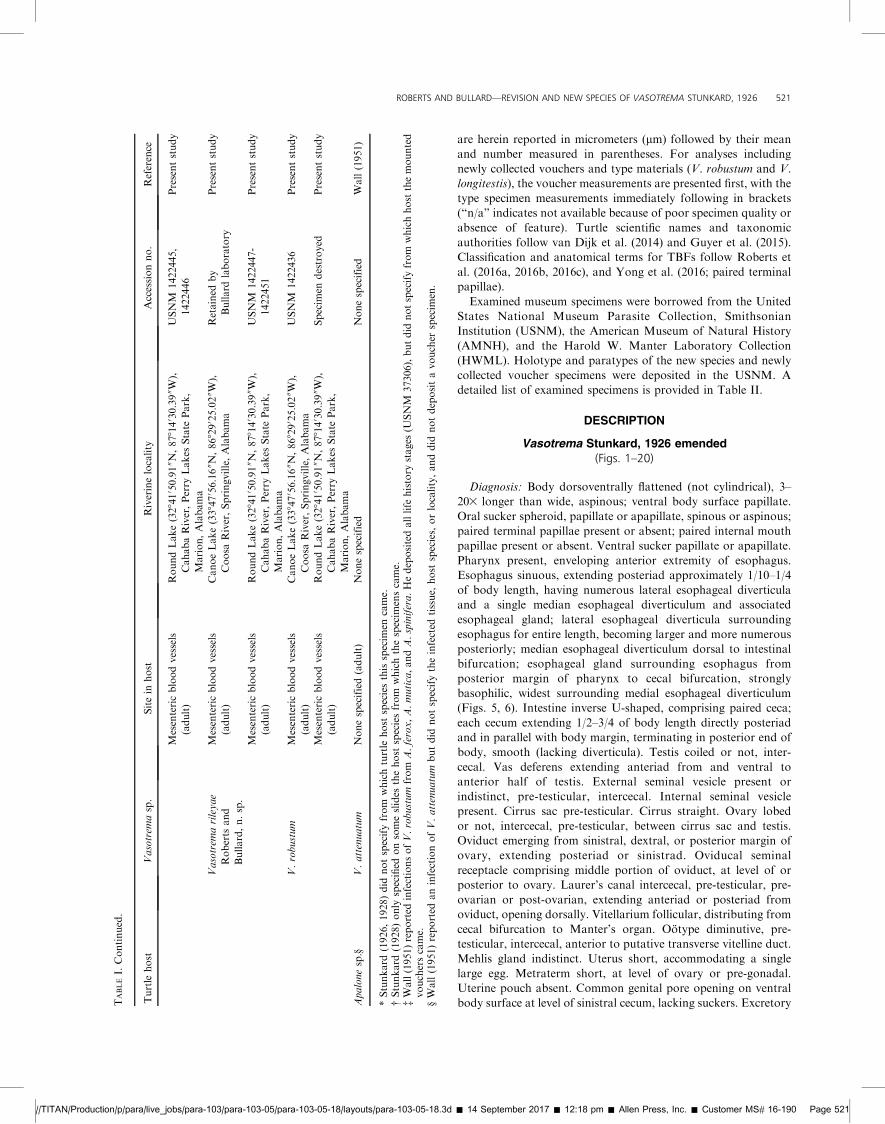
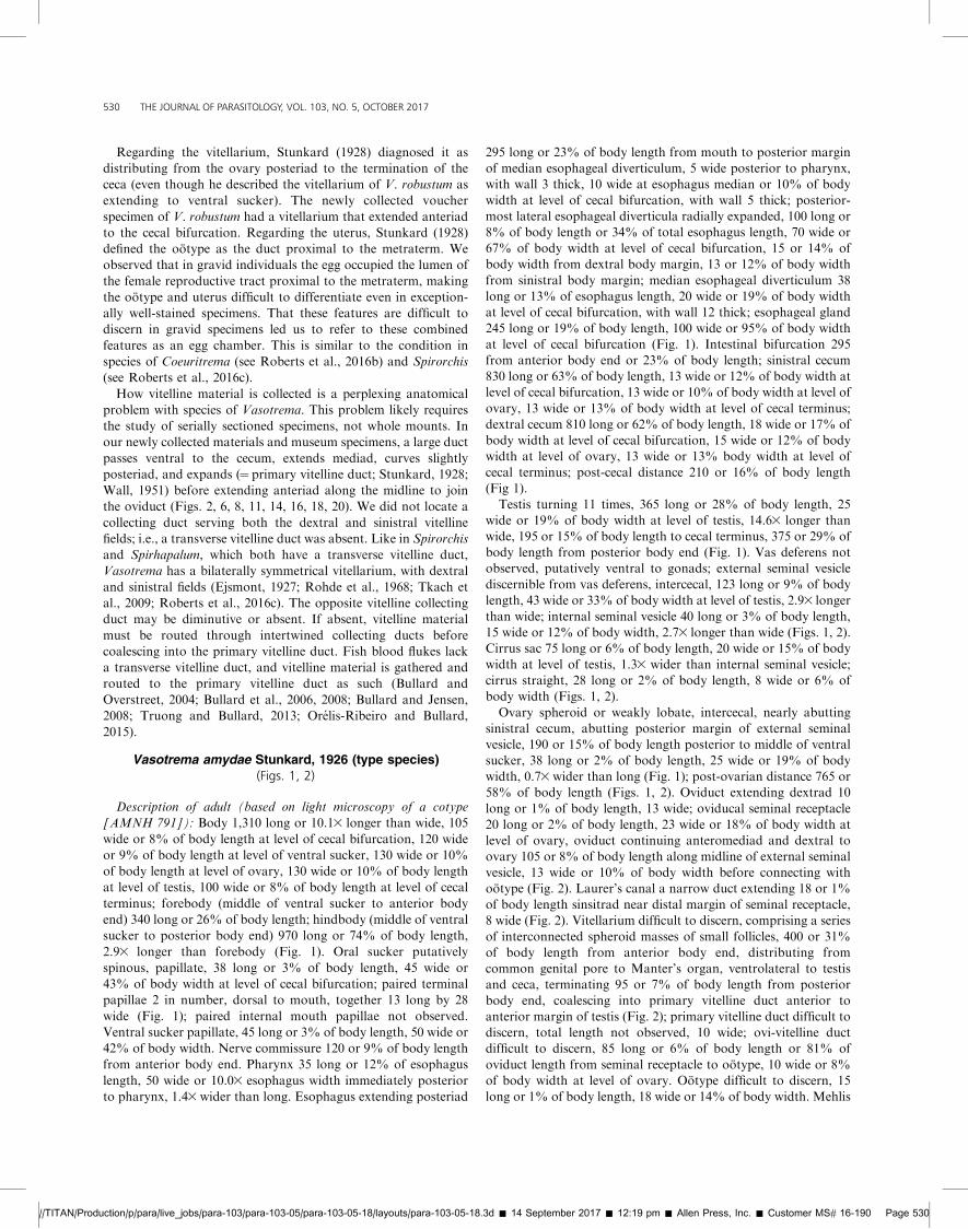
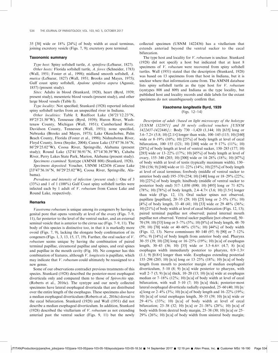
FIGURES 1, 2. Vasotrema amydae Stunkard, 1926 (cotype, AMNHColl. No. 791) from spiny softshell turtle, Apalone spinifera (LeSueur,1827) (Testudines: Trionychidae), or Florida softshell turtle, Apalone ferox(LeSueur, 1827), from a river in Indiana or Florida. Scale values besidebars. (1) Body (dorsal view) showing oral sucker (os), paired terminalpapillae (tp), pharynx (ph), nerve commissure (nc), esophagus (es),esophageal gland (eg), median esophageal diverticulum (med), sinistralcecum (sc), ventral sucker (vs), dextral cecum (dc), common genital pore
(cgp), cirrus sac (cs), uterus (ut), external seminal vesicle (esv), ovary (ov),vitellarium (vr), testis (ts), cecal terminus (ct), Manter’s organ (Mo),excretory vesicle (ev), and excretory pore (ep). (2) Genitalia (dorsal view)showing cirrus (ci), metraterm (mt), internal seminal vesicle (isv), ootype(oo), ovi-vitelline duct (ovt), oviduct (od), oviducal seminal receptacle(osr), Laurer’s canal (Lc), primary vitelline duct (vt), and putative vasdeferens (pv).
//TITAN/Production/p/para/live_jobs/para-103/para-103-05/para-103-05-18/layouts/para-103-05-18.3d � 14 September 2017 � 12:19 pm � Allen Press, Inc. � Customer MS# 16-190 Page 523
ROBERTS AND BULLARD—REVISION AND NEW SPECIES OF VASOTREMA STUNKARD, 1926 523

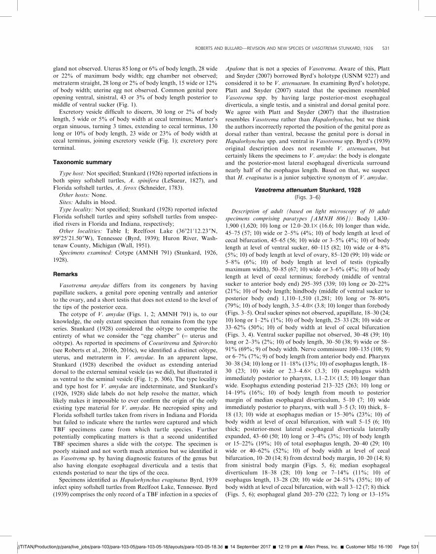
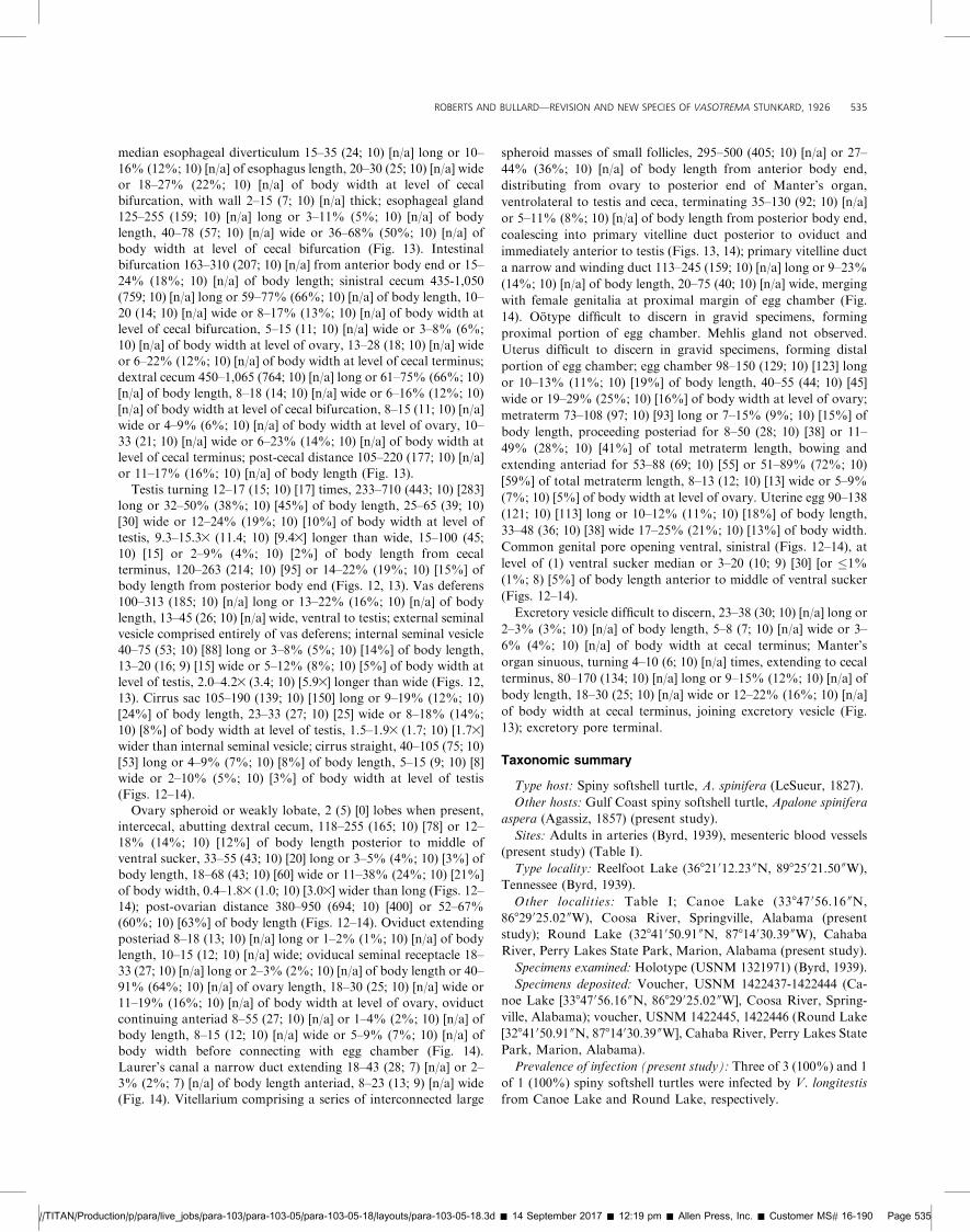
FIGURES 3–6. Vasotrema attenuatum Stunkard, 1928 (syntype, AMNH Coll. No. 806-4) from spiny softshell turtle, Apalone spinifera (LeSueur, 1827)(Testudines: Trionychidae), or Florida softshell turtle, Apalone ferox (LeSueur, 1827), from a river in Indiana or Florida. (3) Body (dorsal view) showingoral sucker (os), ventral sucker (vs), common genital pore (cgp), ovary (ov), testis (ts), and cecal terminus (ct). Scale value beside bar; dashed line indicatesbody segments illustrated at higher magnification in Figures 4 and 5. (4) Anterior portion of body (dorsal view) showing pharynx (ph), esophagus (es), nervecommissure (nc), esophageal gland (eg), median esophageal diverticulum (med), sinistral cecum (sc), dextral cecum (dc), ventral sucker (vs), cirrus sac (cs),common genital pore (cgp), metraterm (mt), cirrus (ci), uterus (ut), internal seminal vesicle (isv), ovary (ov), Laurer’s canal (Lc), oviduct (od), oviducalseminal receptacle (osr), vas deferens (vd), vitelline duct (vt), and testis (ts). (5) Posterior portion of body (dorsal view) vitellarium (vr), cecal terminus (ct),Manter’s organ (Mo), excretory vesicle (ev), and excretory pore (ep). (6) Genitalia (dorsal view) showing ootype (oo) and ovi-vitelline duct (ovt).
//TITAN/Production/p/para/live_jobs/para-103/para-103-05/para-103-05-18/layouts/para-103-05-18.3d � 14 September 2017 � 12:19 pm � Allen Press, Inc. � Customer MS# 16-190 Page 524
524 THE JOURNAL OF PARASITOLOGY, VOL. 103, NO. 5, OCTOBER 2017
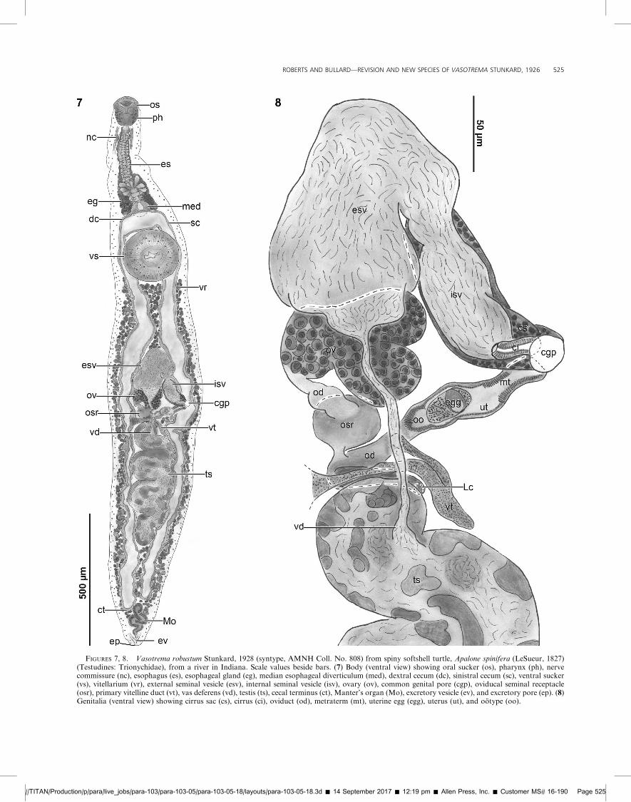
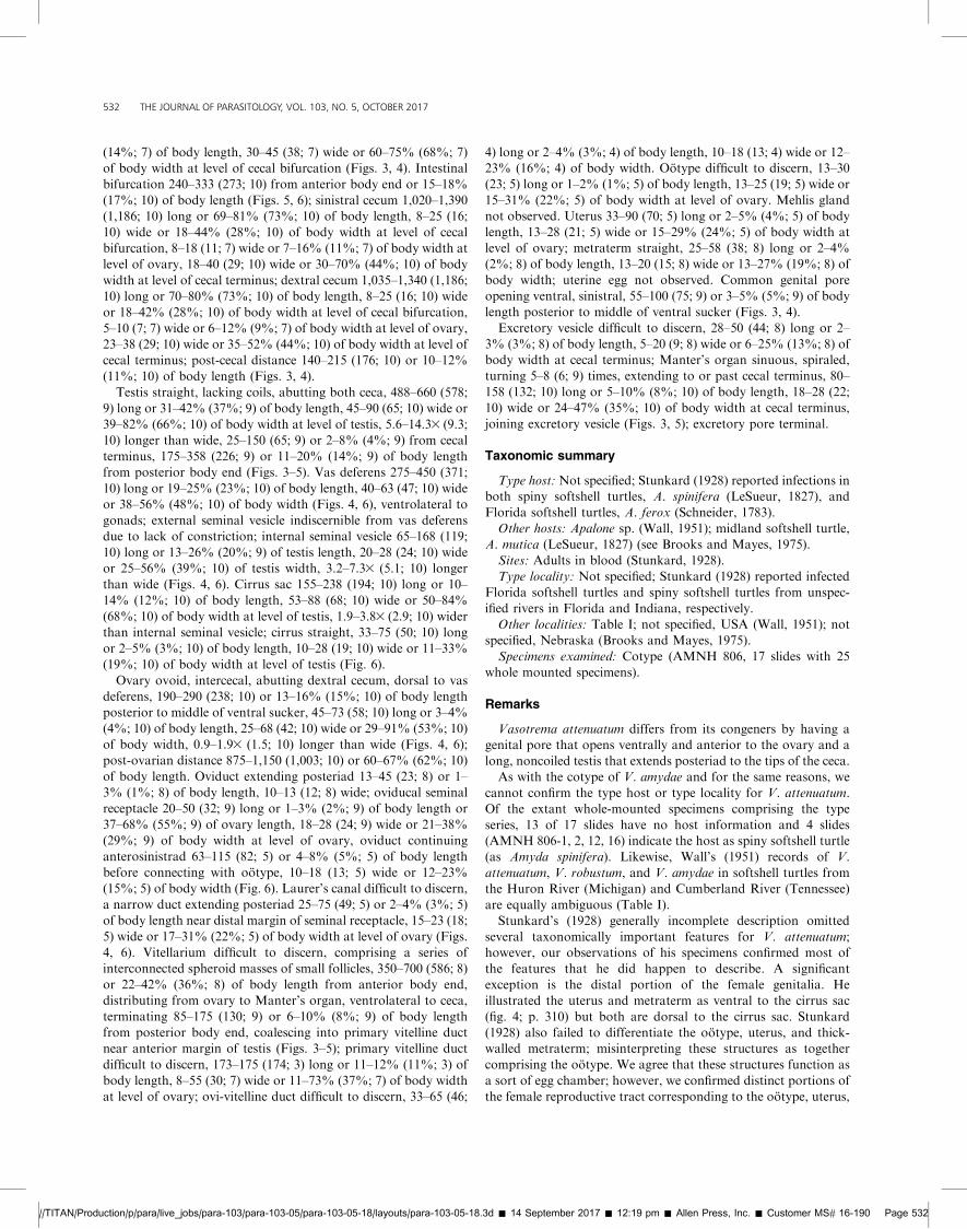
FIGURES 7, 8. Vasotrema robustum Stunkard, 1928 (syntype, AMNH Coll. No. 808) from spiny softshell turtle, Apalone spinifera (LeSueur, 1827)(Testudines: Trionychidae), from a river in Indiana. Scale values beside bars. (7) Body (ventral view) showing oral sucker (os), pharynx (ph), nervecommissure (nc), esophagus (es), esophageal gland (eg), median esophageal diverticulum (med), dextral cecum (dc), sinistral cecum (sc), ventral sucker(vs), vitellarium (vr), external seminal vesicle (esv), internal seminal vesicle (isv), ovary (ov), common genital pore (cgp), oviducal seminal receptacle(osr), primary vitelline duct (vt), vas deferens (vd), testis (ts), cecal terminus (ct), Manter’s organ (Mo), excretory vesicle (ev), and excretory pore (ep). (8)Genitalia (ventral view) showing cirrus sac (cs), cirrus (ci), oviduct (od), metraterm (mt), uterine egg (egg), uterus (ut), and ootype (oo).
//TITAN/Production/p/para/live_jobs/para-103/para-103-05/para-103-05-18/layouts/para-103-05-18.3d � 14 September 2017 � 12:19 pm � Allen Press, Inc. � Customer MS# 16-190 Page 525
ROBERTS AND BULLARD—REVISION AND NEW SPECIES OF VASOTREMA STUNKARD, 1926 525
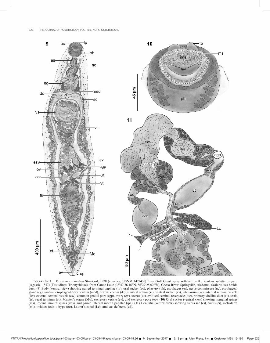
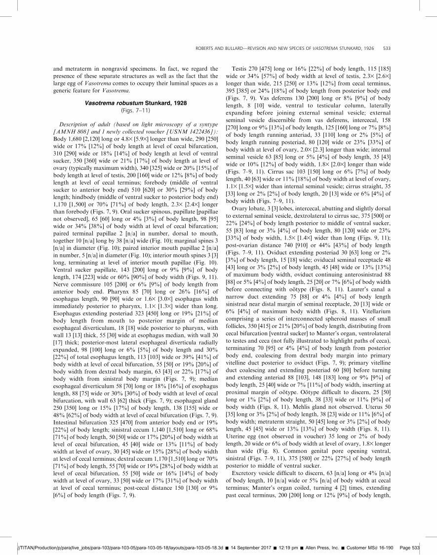
FIGURES 9–11. Vasotrema robustum Stunkard, 1928 (voucher, USNM 1422436) from Gulf Coast spiny softshell turtle, Apalone spinifera aspera(Agassiz, 1857) (Testudines: Trionychidae), from Canoe Lake (33847056.16 00N, 86829025.02 00W), Coosa River, Springville, Alabama. Scale values besidebars. (9) Body (ventral view) showing paired terminal papillae (tp), oral sucker (os), pharynx (ph), esophagus (es), nerve commissure (nc), esophagealgland (eg), median esophageal diverticulum (med), dextral cecum (dc), sinistral cecum (sc), ventral sucker (vs), vitellarium (vr), internal seminal vesicle(isv), external seminal vesicle (esv), common genital pore (cgp), ovary (ov), uterus (ut), oviducal seminal receptacle (osr), primary vitelline duct (vt), testis(ts), cecal terminus (ct), Manter’s organ (Mo), excretory vesicle (ev), and excretory pore (ep). (10) Oral sucker (ventral view) showing marginal spines(ms), internal mouth spines (ims), and paired internal mouth papillae (ipa). (11) Genitalia (ventral view) showing cirrus sac (cs), cirrus (ci), metraterm(mt), oviduct (od), ootype (oo), Laurer’s canal (Lc), and vas deferens (vd).
//TITAN/Production/p/para/live_jobs/para-103/para-103-05/para-103-05-18/layouts/para-103-05-18.3d � 14 September 2017 � 12:19 pm � Allen Press, Inc. � Customer MS# 16-190 Page 526
526 THE JOURNAL OF PARASITOLOGY, VOL. 103, NO. 5, OCTOBER 2017
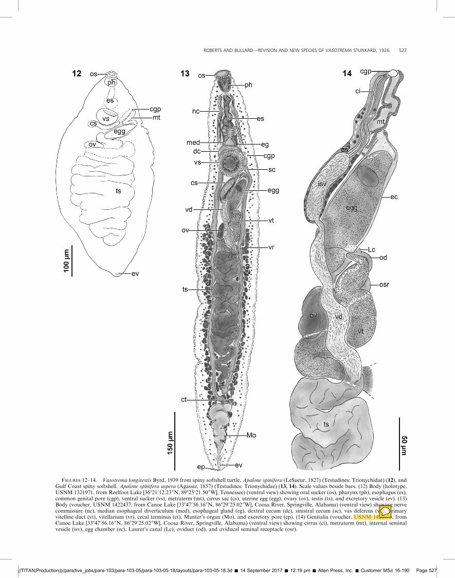
FIGURES 12–14. Vasotrema longitestis Byrd, 1939 from spiny softshell turtle, Apalone spinifera (LeSueur, 1827) (Testudines: Trionychidae) (12), andGulf Coast spiny softshell, Apalone spinifera aspera (Agassiz, 1857) (Testudines: Trionychidae) (13, 14). Scale values beside bars. (12) Body (holotype,USNM 1321971, from Reelfoot Lake [36821012.23 00N, 89825021.50 00W], Tennessee) (ventral view) showing oral sucker (os), pharynx (ph), esophagus (es),common genital pore (cgp), ventral sucker (vs), metraterm (mt), cirrus sac (cs), uterine egg (egg), ovary (ov), testis (ts), and excretory vesicle (ev). (13)Body (voucher, USNM 1422437, from Canoe Lake [33847056.16 00N, 86829025.02 00W], Coosa River, Springville, Alabama) (ventral view) showing nervecommissure (nc), median esophageal diverticulum (med), esophageal gland (eg), dextral cecum (dc), sinistral cecum (sc), vas deferens (vd), primaryvitelline duct (vt), vitellarium (vr), cecal terminus (ct), Manter’s organ (Mo), and excretory pore (ep). (14) Genitalia (voucher, USNM 1422473, fromCanoe Lake [33847056.16 00N, 86829025.02 00W], Coosa River, Springville, Alabama) (ventral view) showing cirrus (ci), metraterm (mt), internal seminalvesicle (isv), egg chamber (ec), Laurer’s canal (Lc), oviduct (od), and oviducal seminal receptacle (osr).
//TITAN/Production/p/para/live_jobs/para-103/para-103-05/para-103-05-18/layouts/para-103-05-18.3d � 14 September 2017 � 12:19 pm � Allen Press, Inc. � Customer MS# 16-190 Page 527
ROBERTS AND BULLARD—REVISION AND NEW SPECIES OF VASOTREMA STUNKARD, 1926 527
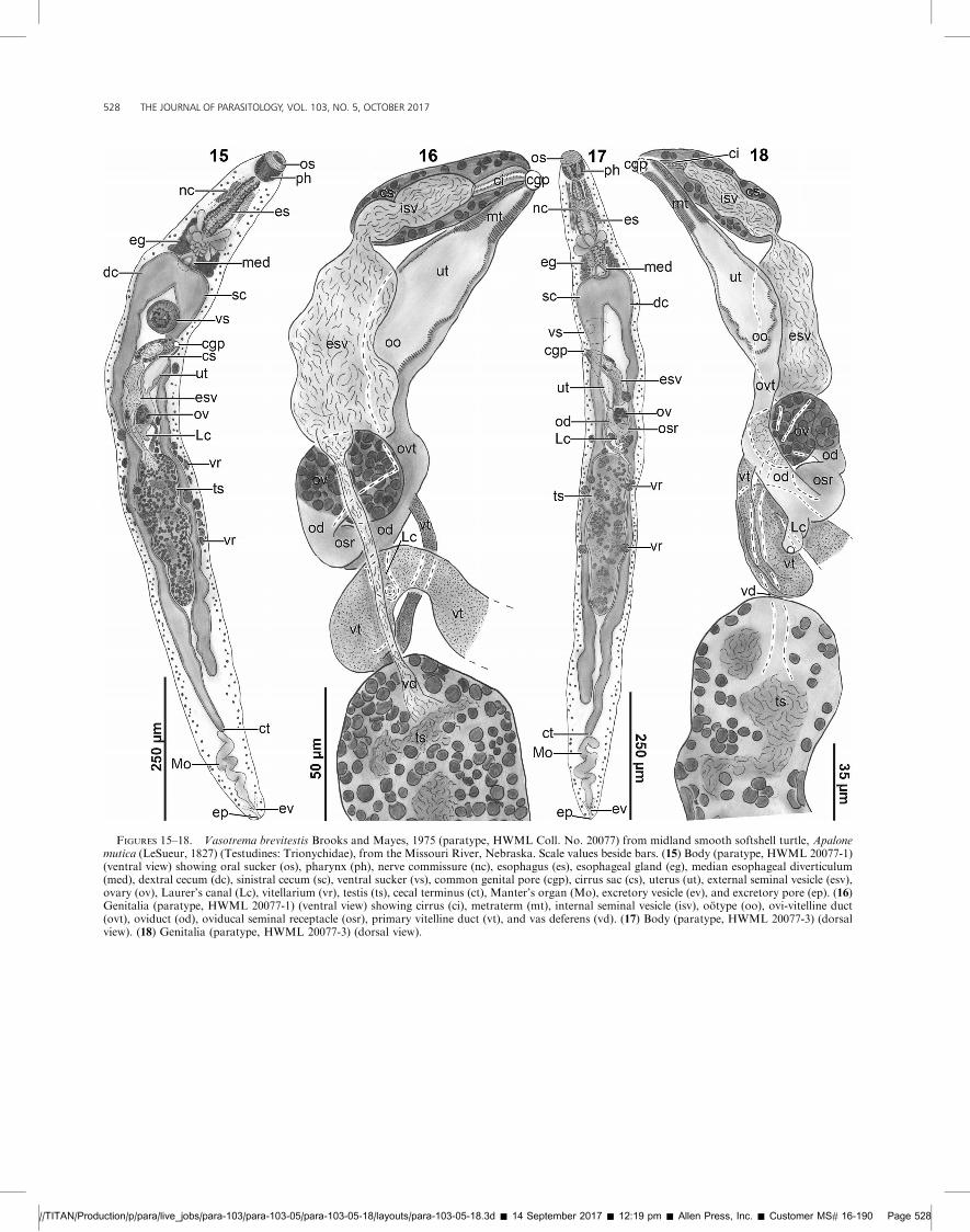
FIGURES 15–18. Vasotrema brevitestis Brooks and Mayes, 1975 (paratype, HWML Coll. No. 20077) from midland smooth softshell turtle, Apalonemutica (LeSueur, 1827) (Testudines: Trionychidae), from the Missouri River, Nebraska. Scale values beside bars. (15) Body (paratype, HWML 20077-1)(ventral view) showing oral sucker (os), pharynx (ph), nerve commissure (nc), esophagus (es), esophageal gland (eg), median esophageal diverticulum(med), dextral cecum (dc), sinistral cecum (sc), ventral sucker (vs), common genital pore (cgp), cirrus sac (cs), uterus (ut), external seminal vesicle (esv),ovary (ov), Laurer’s canal (Lc), vitellarium (vr), testis (ts), cecal terminus (ct), Manter’s organ (Mo), excretory vesicle (ev), and excretory pore (ep). (16)Genitalia (paratype, HWML 20077-1) (ventral view) showing cirrus (ci), metraterm (mt), internal seminal vesicle (isv), ootype (oo), ovi-vitelline duct(ovt), oviduct (od), oviducal seminal receptacle (osr), primary vitelline duct (vt), and vas deferens (vd). (17) Body (paratype, HWML 20077-3) (dorsalview). (18) Genitalia (paratype, HWML 20077-3) (dorsal view).
//TITAN/Production/p/para/live_jobs/para-103/para-103-05/para-103-05-18/layouts/para-103-05-18.3d � 14 September 2017 � 12:19 pm � Allen Press, Inc. � Customer MS# 16-190 Page 528
528 THE JOURNAL OF PARASITOLOGY, VOL. 103, NO. 5, OCTOBER 2017
and descriptions of its species. Regarding spines, Wall (1951) and
Brooks and Mayes (1975) reported that tegumental body spines
were present on V. robustum and V. brevitestis, respectively;
however, we found no evidence of a tegumental spine on any
species of Vasotrema. Regarding sucker papillae, none had
previously been detailed in any species of TBF (Platt, 2002) but
we describe several types: marginal papillae of the oral sucker and
ventral sucker (Figs. 1, 7, 9, 10, 13, 19), paired terminal papillae
dorsal to the mouth (Figs. 1, 9, 10, 19), and paired papillae within
the mouth cavity (Fig. 10). Voucher specimens of Hapalotrema
mehrai Rao, 1976, from Thomas R. Platt’s collection indicate that
Hapalotrema Looss, 1899 may have a papillate ventral sucker also
(J. R. Roberts and S. A. Bullard, unpubl. data). These specimens
have a ventral sucker with large spinose crenulations similar to
the marginal papillae described herein. Morphologically similar
papillae are present in some genera of fish blood flukes (Digenea:
Aporocotylidae; Bullard and Overstreet, 2004; Truong and
Bullard, 2013; Yong et al., 2016). As these structures are present
in phylogenetically unrelated taxa (Orelis-Ribeiro et al., 2014),
they likely evolved independently. Perhaps these papillae function
to allow the fluke to sense their location within the turtle’s
vascular system or to sense the presence of other flukes.
Regarding the pharynx, Stunkard (1926, 1928) and others
(Byrd, 1939; Wall, 1951; Brooks and Mayes, 1975; Platt, 2002)
diagnosed Vasotrema as lacking a pharynx. As in species of
Spirorchis, Unicaecum Stunkard, 1925, Coeuritrema, and Barack-
trema Roberts, Platt, and Bullard, 2016, we suspect that previous
workers misinterpreted the pharynx as a component of the oral
sucker because it is immediately dorsal to the oral sucker (Roberts
et al., 2016a, 2016b, 2016c). Regarding the esophageal diverticula,
comprising numerous lateral diverticula plus a median divertic-
ulum dorsal to the cecal bifurcation, previous descriptions
detailed the posterior-most diverticula only. The diverticula
emanating laterally from the anterior portion of the esophagus
are considerably smaller and more difficult to delineate from the
esophageal gland; which envelops the esophagus and its
diverticula. Similar to Spirorchis, the diverticula surround the
esophagus for its entire length and become larger and more
numerous posteriorly (Figs. 5, 6; Roberts et al., 2016c). Regarding
the median esophageal diverticulum, Stunkard (1928) emphasized
that he did not observe a ‘‘poche [pouch]’’ at the esophageal–cecal
junction as he did in Spirorchis spp. (Stunkard, 1923). Whereas
the median esophageal diverticulum identified in Vasotrema spp.
is similar to, and probably homologous to, that described in
Spirorchis spp. (Roberts et al., 2016c), it differs by being dorsal to,
rather than ventral to, the cecal bifurcation. Our results herein
confirmed that the median esophageal diverticulum is present in
all species of Vasotrema; however, it is evidently lacking in some
species of Spirorchis (i.e., Spirorchis elegans Stunkard, 1923; see
Stunkard, 1923; Platt, 1993; Roberts et al., 2016c).
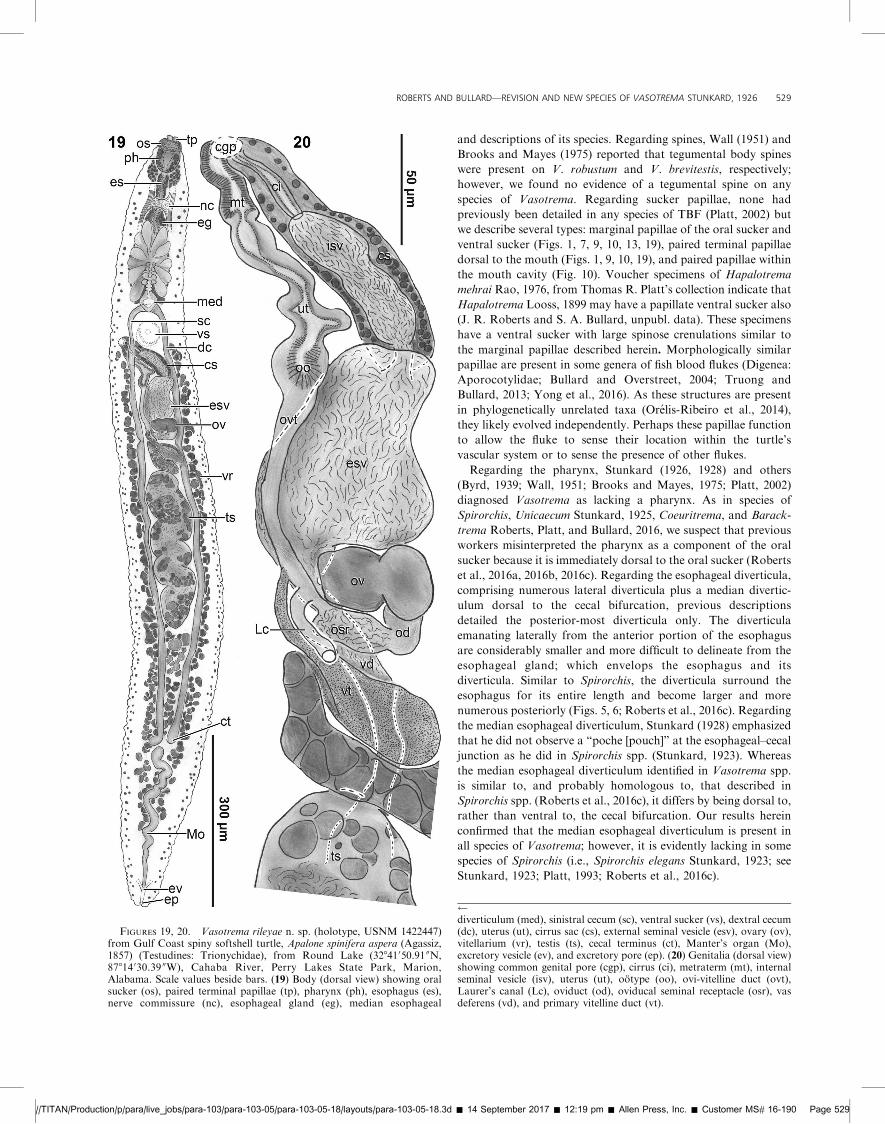
FIGURES 19, 20. Vasotrema rileyae n. sp. (holotype, USNM 1422447)from Gulf Coast spiny softshell turtle, Apalone spinifera aspera (Agassiz,1857) (Testudines: Trionychidae), from Round Lake (32841050.91 00N,87814 030.39 00W), Cahaba River, Perry Lakes State Park, Marion,Alabama. Scale values beside bars. (19) Body (dorsal view) showing oralsucker (os), paired terminal papillae (tp), pharynx (ph), esophagus (es),nerve commissure (nc), esophageal gland (eg), median esophageal
diverticulum (med), sinistral cecum (sc), ventral sucker (vs), dextral cecum(dc), uterus (ut), cirrus sac (cs), external seminal vesicle (esv), ovary (ov),vitellarium (vr), testis (ts), cecal terminus (ct), Manter’s organ (Mo),excretory vesicle (ev), and excretory pore (ep). (20) Genitalia (dorsal view)showing common genital pore (cgp), cirrus (ci), metraterm (mt), internalseminal vesicle (isv), uterus (ut), ootype (oo), ovi-vitelline duct (ovt),Laurer’s canal (Lc), oviduct (od), oviducal seminal receptacle (osr), vasdeferens (vd), and primary vitelline duct (vt).
//TITAN/Production/p/para/live_jobs/para-103/para-103-05/para-103-05-18/layouts/para-103-05-18.3d � 14 September 2017 � 12:19 pm � Allen Press, Inc. � Customer MS# 16-190 Page 529
ROBERTS AND BULLARD—REVISION AND NEW SPECIES OF VASOTREMA STUNKARD, 1926 529
Regarding the vitellarium, Stunkard (1928) diagnosed it as
distributing from the ovary posteriad to the termination of the
ceca (even though he described the vitellarium of V. robustum as
extending to ventral sucker). The newly collected voucher
specimen of V. robustum had a vitellarium that extended anteriad
to the cecal bifurcation. Regarding the uterus, Stunkard (1928)
defined the ootype as the duct proximal to the metraterm. We
observed that in gravid individuals the egg occupied the lumen of
the female reproductive tract proximal to the metraterm, making
the ootype and uterus difficult to differentiate even in exception-
ally well-stained specimens. That these features are difficult to
discern in gravid specimens led us to refer to these combined
features as an egg chamber. This is similar to the condition in
species of Coeuritrema (see Roberts et al., 2016b) and Spirorchis
(see Roberts et al., 2016c).
How vitelline material is collected is a perplexing anatomical
problem with species of Vasotrema. This problem likely requires
the study of serially sectioned specimens, not whole mounts. In
our newly collected materials and museum specimens, a large duct
passes ventral to the cecum, extends mediad, curves slightly
posteriad, and expands (¼ primary vitelline duct; Stunkard, 1928;
Wall, 1951) before extending anteriad along the midline to join
the oviduct (Figs. 2, 6, 8, 11, 14, 16, 18, 20). We did not locate a
collecting duct serving both the dextral and sinistral vitelline
fields; i.e., a transverse vitelline duct was absent. Like in Spirorchis
and Spirhapalum, which both have a transverse vitelline duct,
Vasotrema has a bilaterally symmetrical vitellarium, with dextral
and sinistral fields (Ejsmont, 1927; Rohde et al., 1968; Tkach et
al., 2009; Roberts et al., 2016c). The opposite vitelline collecting
duct may be diminutive or absent. If absent, vitelline material
must be routed through intertwined collecting ducts before
coalescing into the primary vitelline duct. Fish blood flukes lack
a transverse vitelline duct, and vitelline material is gathered and
routed to the primary vitelline duct as such (Bullard and
Overstreet, 2004; Bullard et al., 2006, 2008; Bullard and Jensen,
2008; Truong and Bullard, 2013; Orelis-Ribeiro and Bullard,
2015).
Vasotrema amydae Stunkard, 1926 (type species)(Figs. 1, 2)
Description of adult (based on light microscopy of a cotype
[AMNH 791]): Body 1,310 long or 10.13 longer than wide, 105
wide or 8% of body length at level of cecal bifurcation, 120 wide
or 9% of body length at level of ventral sucker, 130 wide or 10%
of body length at level of ovary, 130 wide or 10% of body length
at level of testis, 100 wide or 8% of body length at level of cecal
terminus; forebody (middle of ventral sucker to anterior body
end) 340 long or 26% of body length; hindbody (middle of ventral
sucker to posterior body end) 970 long or 74% of body length,
2.93 longer than forebody (Fig. 1). Oral sucker putatively
spinous, papillate, 38 long or 3% of body length, 45 wide or
43% of body width at level of cecal bifurcation; paired terminal
papillae 2 in number, dorsal to mouth, together 13 long by 28
wide (Fig. 1); paired internal mouth papillae not observed.
Ventral sucker papillate, 45 long or 3% of body length, 50 wide or
42% of body width. Nerve commissure 120 or 9% of body length
from anterior body end. Pharynx 35 long or 12% of esophagus
length, 50 wide or 10.03 esophagus width immediately posterior
to pharynx, 1.43wider than long. Esophagus extending posteriad
295 long or 23% of body length from mouth to posterior margin
of median esophageal diverticulum, 5 wide posterior to pharynx,
with wall 3 thick, 10 wide at esophagus median or 10% of body
width at level of cecal bifurcation, with wall 5 thick; posterior-
most lateral esophageal diverticula radially expanded, 100 long or
8% of body length or 34% of total esophagus length, 70 wide or
67% of body width at level of cecal bifurcation, 15 or 14% of
body width from dextral body margin, 13 or 12% of body width
from sinistral body margin; median esophageal diverticulum 38
long or 13% of esophagus length, 20 wide or 19% of body width
at level of cecal bifurcation, with wall 12 thick; esophageal gland
245 long or 19% of body length, 100 wide or 95% of body width
at level of cecal bifurcation (Fig. 1). Intestinal bifurcation 295
from anterior body end or 23% of body length; sinistral cecum
830 long or 63% of body length, 13 wide or 12% of body width at
level of cecal bifurcation, 13 wide or 10% of body width at level of
ovary, 13 wide or 13% of body width at level of cecal terminus;
dextral cecum 810 long or 62% of body length, 18 wide or 17% of
body width at level of cecal bifurcation, 15 wide or 12% of body
width at level of ovary, 13 wide or 13% body width at level of
cecal terminus; post-cecal distance 210 or 16% of body length
(Fig 1).
Testis turning 11 times, 365 long or 28% of body length, 25
wide or 19% of body width at level of testis, 14.63 longer than
wide, 195 or 15% of body length to cecal terminus, 375 or 29% of
body length from posterior body end (Fig. 1). Vas deferens not
observed, putatively ventral to gonads; external seminal vesicle
discernible from vas deferens, intercecal, 123 long or 9% of body
length, 43 wide or 33% of body width at level of testis, 2.93 longer
than wide; internal seminal vesicle 40 long or 3% of body length,
15 wide or 12% of body width, 2.73 longer than wide (Figs. 1, 2).
Cirrus sac 75 long or 6% of body length, 20 wide or 15% of body
width at level of testis, 1.33 wider than internal seminal vesicle;
cirrus straight, 28 long or 2% of body length, 8 wide or 6% of
body width (Figs. 1, 2).
Ovary spheroid or weakly lobate, intercecal, nearly abutting
sinistral cecum, abutting posterior margin of external seminal
vesicle, 190 or 15% of body length posterior to middle of ventral
sucker, 38 long or 2% of body length, 25 wide or 19% of body
width, 0.73wider than long (Fig. 1); post-ovarian distance 765 or
58% of body length (Figs. 1, 2). Oviduct extending dextrad 10
long or 1% of body length, 13 wide; oviducal seminal receptacle
20 long or 2% of body length, 23 wide or 18% of body width at
level of ovary, oviduct continuing anteromediad and dextral to
ovary 105 or 8% of body length along midline of external seminal
vesicle, 13 wide or 10% of body width before connecting with
ootype (Fig. 2). Laurer’s canal a narrow duct extending 18 or 1%
of body length sinsitrad near distal margin of seminal receptacle,
8 wide (Fig. 2). Vitellarium difficult to discern, comprising a series
of interconnected spheroid masses of small follicles, 400 or 31%
of body length from anterior body end, distributing from
common genital pore to Manter’s organ, ventrolateral to testis
and ceca, terminating 95 or 7% of body length from posterior
body end, coalescing into primary vitelline duct anterior to
anterior margin of testis (Fig. 2); primary vitelline duct difficult to
discern, total length not observed, 10 wide; ovi-vitelline duct
difficult to discern, 85 long or 6% of body length or 81% of
oviduct length from seminal receptacle to ootype, 10 wide or 8%
of body width at level of ovary. Ootype difficult to discern, 15
long or 1% of body length, 18 wide or 14% of body width. Mehlis
//TITAN/Production/p/para/live_jobs/para-103/para-103-05/para-103-05-18/layouts/para-103-05-18.3d � 14 September 2017 � 12:19 pm � Allen Press, Inc. � Customer MS# 16-190 Page 530
530 THE JOURNAL OF PARASITOLOGY, VOL. 103, NO. 5, OCTOBER 2017
gland not observed. Uterus 85 long or 6% of body length, 28 wideor 22% of maximum body width; egg chamber not observed;
metraterm straight, 28 long or 2% of body length, 15 wide or 12%
of body width; uterine egg not observed. Common genital pore
opening ventral, sinistral, 43 or 3% of body length posterior to
middle of ventral sucker (Fig. 1).
Excretory vesicle difficult to discern, 30 long or 2% of body
length, 5 wide or 5% of body width at cecal terminus; Manter’sorgan sinuous, turning 3 times, extending to cecal terminus, 130
long or 10% of body length, 23 wide or 23% of body width at
cecal terminus, joining excretory vesicle (Fig. 1); excretory pore
terminal.
Taxonomic summary
Type host: Not specified; Stunkard (1926) reported infections in
both spiny softshell turtles, A. spinifera (LeSueur, 1827), andFlorida softshell turtles, A. ferox (Schneider, 1783).
Other hosts: None.
Sites: Adults in blood.
Type locality: Not specified; Stunkard (1928) reported infectedFlorida softshell turtles and spiny softshell turtles from unspec-
ified rivers in Florida and Indiana, respectively;
Other localities: Table I; Reelfoot Lake (36821 012.23 00N,
89825021.50 00W), Tennessee (Byrd, 1939); Huron River, Wash-
tenaw County, Michigan (Wall, 1951).
Specimens examined: Cotype (AMNH 791) (Stunkard, 1926,
1928).
Remarks
Vasotrema amydae differs from its congeners by havingpapillate suckers, a genital pore opening ventrally and anterior
to the ovary, and a short testis that does not extend to the level of
the tips of the posterior ceca.
The cotype of V. amydae (Figs. 1, 2; AMNH 791) is, to our
knowledge, the only extant specimen that remains from the type
series. Stunkard (1928) considered the ootype to comprise the
entirety of what we consider the ‘‘egg chamber’’ (¼ uterus andootype). As reported in specimens of Coeuritrema and Spirorchis
(see Roberts et al., 2016b, 2016c), we identified a distinct ootype,
uterus, and metraterm in V. amydae. In an apparent lapse,
Stunkard (1928) described the oviduct as extending anteriad
dorsal to the external seminal vesicle (as we did), but illustrated itas ventral to the seminal vesicle (Fig. 1; p. 306). The type locality
and type host for V. amydae are indeterminate, and Stunkard’s
(1926, 1928) slide labels do not help resolve the matter, which
likely makes it impossible to ever confirm the origin of the only
existing type material for V. amydae. He necropsied spiny and
Florida softshell turtles taken from rivers in Indiana and Floridabut failed to indicate where the turtles were captured and which
TBF specimens came from which turtle species. Further
potentially complicating matters is that a second unidentified
TBF specimen shares a slide with the cotype. The specimen is
poorly stained and not worth much attention but we identified itas Vasotrema sp. by having diagnostic features of the genus but
also having elongate esophageal diverticula and a testis that
extends posteriad to near the tips of the ceca.
Specimens identified as Hapalorhynchus evaginatus Byrd, 1939
infect spiny softshell turtles from Reelfoot Lake, Tennessee. Byrd
(1939) comprises the only record of a TBF infection in a species of
Apalone that is not a species of Vasotrema. Aware of this, Platt
and Snyder (2007) borrowed Byrd’s holotype (USNM 9227) and
considered it to be V. attenuatum. In examining Byrd’s holotype,
Platt and Snyder (2007) stated that the specimen resembled
Vasotrema spp. by having large posterior-most esophageal
diverticula, a single testis, and a sinistral and dorsal genital pore.
We agree with Platt and Snyder (2007) that the illustration
resembles Vasotrema rather than Hapalorhynchus, but we think
the authors incorrectly reported the position of the genital pore as
dorsal rather than ventral, because the genital pore is dorsal in
Hapalorhynchus spp. and ventral in Vasotrema spp. Byrd’s (1939)
original description does not resemble V. attenuatum, but
certainly likens the specimens to V. amydae: the body is elongate
and the posterior-most lateral esophageal diverticula surround
nearly half of the esophagus length. Based on that, we suspect
that H. evaginatus is a junior subjective synonym of V. amydae.
Vasotrema attenuatum Stunkard, 1928(Figs. 3–6)
Description of adult (based on light microscopy of 10 adult
specimens comprising paratypes [AMNH 806]): Body 1,430–
1,900 (1,620; 10) long or 12.0–20.13 (16.6; 10) longer than wide,
45–75 (57; 10) wide or 2–5% (4%; 10) of body length at level of
cecal bifurcation, 45–65 (56; 10) wide or 3–5% (4%; 10) of body
length at level of ventral sucker, 60–115 (82; 10) wide or 4–8%
(5%; 10) of body length at level of ovary, 85–120 (99; 10) wide or
5–8% (6%; 10) of body length at level of testis (typically
maximum width), 50–85 (67; 10) wide or 3–6% (4%; 10) of body
length at level of cecal terminus; forebody (middle of ventral
sucker to anterior body end) 295–395 (339; 10) long or 20–22%
(21%; 10) of body length; hindbody (middle of ventral sucker to
posterior body end) 1,110–1,510 (1,281; 10) long or 78–80%
(79%; 10) of body length, 3.5–4.03 (3.8; 10) longer than forebody
(Figs. 3–5). Oral sucker spines not observed, apapillate, 18–30 (24;
10) long or 1–2% (1%; 10) of body length, 25–33 (28; 10) wide or
33–62% (50%; 10) of body width at level of cecal bifurcation
(Figs. 3, 4). Ventral sucker papillae not observed, 30–48 (39; 10)
long or 2–3% (2%; 10) of body length, 30–50 (38; 9) wide or 58–
91% (69%; 9) of body width. Nerve commissure 100–135 (108; 9)
or 6–7% (7%; 9) of body length from anterior body end. Pharynx
30–38 (34; 10) long or 11–18% (13%; 10) of esophagus length, 18–
30 (23; 10) wide or 2.3–4.63 (3.3; 10) esophagus width
immediately posterior to pharynx, 1.1–2.13 (1.5; 10) longer than
wide. Esophagus extending posteriad 213–325 (263; 10) long or
14–19% (16%; 10) of body length from mouth to posterior
margin of median esophageal diverticulum, 5–10 (7; 10) wide
immediately posterior to pharynx, with wall 3–5 (3; 10) thick, 8–
18 (13; 10) wide at esophagus median or 15–30% (23%; 10) of
body width at level of cecal bifurcation, with wall 5–15 (6; 10)
thick; posterior-most lateral esophageal diverticula laterally
expanded, 43–60 (50; 10) long or 3–4% (3%; 10) of body length
or 15–22% (19%; 10) of total esophagus length, 20–40 (29; 10)
wide or 40–62% (52%; 10) of body width at level of cecal
bifurcation, 10–20 (14; 8) from dextral body margin, 10–20 (14; 8)
from sinistral body margin (Figs. 5, 6); median esophageal
diverticulum 18–38 (28; 10) long or 7–14% (11%; 10) of
esophagus length, 13–28 (20; 10) wide or 24–51% (35%; 10) of
body width at level of cecal bifurcation, with wall 3–12 (7; 8) thick
(Figs. 5, 6); esophageal gland 203–270 (222; 7) long or 13–15%
//TITAN/Production/p/para/live_jobs/para-103/para-103-05/para-103-05-18/layouts/para-103-05-18.3d � 14 September 2017 � 12:19 pm � Allen Press, Inc. � Customer MS# 16-190 Page 531
ROBERTS AND BULLARD—REVISION AND NEW SPECIES OF VASOTREMA STUNKARD, 1926 531
(14%; 7) of body length, 30–45 (38; 7) wide or 60–75% (68%; 7)
of body width at level of cecal bifurcation (Figs. 3, 4). Intestinal
bifurcation 240–333 (273; 10) from anterior body end or 15–18%
(17%; 10) of body length (Figs. 5, 6); sinistral cecum 1,020–1,390
(1,186; 10) long or 69–81% (73%; 10) of body length, 8–25 (16;
10) wide or 18–44% (28%; 10) of body width at level of cecal
bifurcation, 8–18 (11; 7) wide or 7–16% (11%; 7) of body width at
level of ovary, 18–40 (29; 10) wide or 30–70% (44%; 10) of body
width at level of cecal terminus; dextral cecum 1,035–1,340 (1,186;
10) long or 70–80% (73%; 10) of body length, 8–25 (16; 10) wide
or 18–42% (28%; 10) of body width at level of cecal bifurcation,
5–10 (7; 7) wide or 6–12% (9%; 7) of body width at level of ovary,
23–38 (29; 10) wide or 35–52% (44%; 10) of body width at level of
cecal terminus; post-cecal distance 140–215 (176; 10) or 10–12%
(11%; 10) of body length (Figs. 3, 4).
Testis straight, lacking coils, abutting both ceca, 488–660 (578;
9) long or 31–42% (37%; 9) of body length, 45–90 (65; 10) wide or
39–82% (66%; 10) of body width at level of testis, 5.6–14.33 (9.3;
10) longer than wide, 25–150 (65; 9) or 2–8% (4%; 9) from cecal
terminus, 175–358 (226; 9) or 11–20% (14%; 9) of body length
from posterior body end (Figs. 3–5). Vas deferens 275–450 (371;
10) long or 19–25% (23%; 10) of body length, 40–63 (47; 10) wide
or 38–56% (48%; 10) of body width (Figs. 4, 6), ventrolateral to
gonads; external seminal vesicle indiscernible from vas deferens
due to lack of constriction; internal seminal vesicle 65–168 (119;
10) long or 13–26% (20%; 9) of testis length, 20–28 (24; 10) wide
or 25–56% (39%; 10) of testis width, 3.2–7.33 (5.1; 10) longer
than wide (Figs. 4, 6). Cirrus sac 155–238 (194; 10) long or 10–
14% (12%; 10) of body length, 53–88 (68; 10) wide or 50–84%
(68%; 10) of body width at level of testis, 1.9–3.83 (2.9; 10) wider
than internal seminal vesicle; cirrus straight, 33–75 (50; 10) long
or 2–5% (3%; 10) of body length, 10–28 (19; 10) wide or 11–33%
(19%; 10) of body width at level of testis (Fig. 6).
Ovary ovoid, intercecal, abutting dextral cecum, dorsal to vas
deferens, 190–290 (238; 10) or 13–16% (15%; 10) of body length
posterior to middle of ventral sucker, 45–73 (58; 10) long or 3–4%
(4%; 10) of body length, 25–68 (42; 10) wide or 29–91% (53%; 10)
of body width, 0.9–1.93 (1.5; 10) longer than wide (Figs. 4, 6);
post-ovarian distance 875–1,150 (1,003; 10) or 60–67% (62%; 10)
of body length. Oviduct extending posteriad 13–45 (23; 8) or 1–
3% (1%; 8) of body length, 10–13 (12; 8) wide; oviducal seminal
receptacle 20–50 (32; 9) long or 1–3% (2%; 9) of body length or
37–68% (55%; 9) of ovary length, 18–28 (24; 9) wide or 21–38%
(29%; 9) of body width at level of ovary, oviduct continuing
anterosinistrad 63–115 (82; 5) or 4–8% (5%; 5) of body length
before connecting with ootype, 10–18 (13; 5) wide or 12–23%
(15%; 5) of body width (Fig. 6). Laurer’s canal difficult to discern,
a narrow duct extending posteriad 25–75 (49; 5) or 2–4% (3%; 5)
of body length near distal margin of seminal receptacle, 15–23 (18;
5) wide or 17–31% (22%; 5) of body width at level of ovary (Figs.
4, 6). Vitellarium difficult to discern, comprising a series of
interconnected spheroid masses of small follicles, 350–700 (586; 8)
or 22–42% (36%; 8) of body length from anterior body end,
distributing from ovary to Manter’s organ, ventrolateral to ceca,
terminating 85–175 (130; 9) or 6–10% (8%; 9) of body length
from posterior body end, coalescing into primary vitelline duct
near anterior margin of testis (Figs. 3–5); primary vitelline duct
difficult to discern, 173–175 (174; 3) long or 11–12% (11%; 3) of
body length, 8–55 (30; 7) wide or 11–73% (37%; 7) of body width
at level of ovary; ovi-vitelline duct difficult to discern, 33–65 (46;
4) long or 2–4% (3%; 4) of body length, 10–18 (13; 4) wide or 12–23% (16%; 4) of body width. Ootype difficult to discern, 13–30
(23; 5) long or 1–2% (1%; 5) of body length, 13–25 (19; 5) wide or
15–31% (22%; 5) of body width at level of ovary. Mehlis gland
not observed. Uterus 33–90 (70; 5) long or 2–5% (4%; 5) of body
length, 13–28 (21; 5) wide or 15–29% (24%; 5) of body width atlevel of ovary; metraterm straight, 25–58 (38; 8) long or 2–4%
(2%; 8) of body length, 13–20 (15; 8) wide or 13–27% (19%; 8) of
body width; uterine egg not observed. Common genital pore
opening ventral, sinistral, 55–100 (75; 9) or 3–5% (5%; 9) of body
length posterior to middle of ventral sucker (Figs. 3, 4).
Excretory vesicle difficult to discern, 28–50 (44; 8) long or 2–
3% (3%; 8) of body length, 5–20 (9; 8) wide or 6–25% (13%; 8) ofbody width at cecal terminus; Manter’s organ sinuous, spiraled,
turning 5–8 (6; 9) times, extending to or past cecal terminus, 80–
158 (132; 10) long or 5–10% (8%; 10) of body length, 18–28 (22;
10) wide or 24–47% (35%; 10) of body width at cecal terminus,
joining excretory vesicle (Figs. 3, 5); excretory pore terminal.
Taxonomic summary
Type host: Not specified; Stunkard (1928) reported infections inboth spiny softshell turtles, A. spinifera (LeSueur, 1827), and
Florida softshell turtles, A. ferox (Schneider, 1783).
Other hosts: Apalone sp. (Wall, 1951); midland softshell turtle,
A. mutica (LeSueur, 1827) (see Brooks and Mayes, 1975).
Sites: Adults in blood (Stunkard, 1928).
Type locality: Not specified; Stunkard (1928) reported infected
Florida softshell turtles and spiny softshell turtles from unspec-
ified rivers in Florida and Indiana, respectively.
Other localities: Table I; not specified, USA (Wall, 1951); not
specified, Nebraska (Brooks and Mayes, 1975).
Specimens examined: Cotype (AMNH 806, 17 slides with 25
whole mounted specimens).
Remarks
Vasotrema attenuatum differs from its congeners by having agenital pore that opens ventrally and anterior to the ovary and a
long, noncoiled testis that extends posteriad to the tips of the ceca.
As with the cotype of V. amydae and for the same reasons, we
cannot confirm the type host or type locality for V. attenuatum.
Of the extant whole-mounted specimens comprising the type
series, 13 of 17 slides have no host information and 4 slides(AMNH 806-1, 2, 12, 16) indicate the host as spiny softshell turtle
(as Amyda spinifera). Likewise, Wall’s (1951) records of V.
attenuatum, V. robustum, and V. amydae in softshell turtles from
the Huron River (Michigan) and Cumberland River (Tennessee)
are equally ambiguous (Table I).
Stunkard’s (1928) generally incomplete description omitted
several taxonomically important features for V. attenuatum;however, our observations of his specimens confirmed most of
the features that he did happen to describe. A significant
exception is the distal portion of the female genitalia. He
illustrated the uterus and metraterm as ventral to the cirrus sac
(fig. 4; p. 310) but both are dorsal to the cirrus sac. Stunkard
(1928) also failed to differentiate the ootype, uterus, and thick-walled metraterm; misinterpreting these structures as together
comprising the ootype. We agree that these structures function as
a sort of egg chamber; however, we confirmed distinct portions of
the female reproductive tract corresponding to the ootype, uterus,
//TITAN/Production/p/para/live_jobs/para-103/para-103-05/para-103-05-18/layouts/para-103-05-18.3d � 14 September 2017 � 12:19 pm � Allen Press, Inc. � Customer MS# 16-190 Page 532
532 THE JOURNAL OF PARASITOLOGY, VOL. 103, NO. 5, OCTOBER 2017
and metraterm in nongravid specimens. In fact, we regard the
presence of these separate structures as well as the fact that the
large egg of Vasotrema comes to occupy their luminal spaces as a
generic feature for Vasotrema.
Vasotrema robustum Stunkard, 1928
(Figs. 7–11)
Description of adult (based on light microscopy of a syntype
[AMNH 808] and 1 newly collected voucher [USNM 1422436]):
Body 1,680 [2,120] long or 4.83 [5.93] longer than wide, 290 [250]
wide or 17% [12%] of body length at level of cecal bifurcation,
310 [290] wide or 18% [14%] of body length at level of ventral
sucker, 350 [360] wide or 21% [17%] of body length at level of
ovary (typically maximum width), 340 [325] wide or 20% [15%] of
body length at level of testis, 200 [160] wide or 12% [8%] of body
length at level of cecal terminus; forebody (middle of ventral
sucker to anterior body end) 510 [620] or 30% [29%] of body
length; hindbody (middle of ventral sucker to posterior body end)
1,170 [1,500] or 70% [71%] of body length, 2.33 [2.43] longer
than forebody (Figs. 7, 9). Oral sucker spinous, papillate [papillae
not observed], 65 [60] long or 4% [3%] of body length, 98 [95]
wide or 34% [38%] of body width at level of cecal bifurcation;
paired terminal papillae 2 [n/a] in number, dorsal to mouth,
together 10 [n/a] long by 38 [n/a] wide (Fig. 10); marginal spines 3
[n/a] in diameter (Fig. 10); paired interior mouth papillae 2 [n/a]
in number, 5 [n/a] in diameter (Fig. 10); interior mouth spines 3 [3]
long, terminating at level of interior mouth papillae (Fig. 10).
Ventral sucker papillate, 143 [200] long or 9% [9%] of body
length, 174 [223] wide or 60% [90%] of body width (Figs. 9, 11).
Nerve commissure 105 [200] or 6% [9%] of body length from
anterior body end. Pharynx 85 [70] long or 26% [16%] of
esophagus length, 90 [90] wide or 1.63 [3.03] esophagus width
immediately posterior to pharynx, 1.13 [1.33] wider than long.
Esophagus extending posteriad 323 [450] long or 19% [21%] of
body length from mouth to posterior margin of median
esophageal diverticulum, 18 [18] wide posterior to pharynx, with
wall 13 [13] thick, 55 [30] wide at esophagus median, with wall 30
[17] thick; posterior-most lateral esophageal diverticula radially
expanded, 98 [100] long or 6% [5%] of body length and 30%
[22%] of total esophagus length, 113 [103] wide or 39% [41%] of
body width at level of cecal bifurcation, 55 [50] or 19% [20%] of
body width from dextral body margin, 63 [43] or 22% [17%] of
body width from sinistral body margin (Figs. 7, 9); median
esophageal diverticulum 58 [70] long or 18% [16%] of esophagus
length, 88 [75] wide or 30% [30%] of body width at level of cecal
bifurcation, with wall 63 [62] thick (Figs. 7, 9); esophageal gland
250 [350] long or 15% [17%] of body length, 138 [155] wide or
48% [62%] of body width at level of cecal bifurcation (Figs. 7, 9).
Intestinal bifurcation 325 [470] from anterior body end or 19%
[22%] of body length; sinistral cecum 1,140 [1,510] long or 68%
[71%] of body length, 50 [50] wide or 17% [20%] of body width at
level of cecal bifurcation, 45 [40] wide or 13% [11%] of body
width at level of ovary, 30 [45] wide or 15% [28%] of body width
at level of cecal terminus; dextral cecum 1,170 [1,510] long or 70%
[71%] of body length, 55 [70] wide or 19% [28%] of body width at
level of cecal bifurcation, 55 [50] wide or 16% [14%] of body
width at level of ovary, 33 [50] wide or 17% [31%] of body width
at level of cecal terminus; post-cecal distance 150 [130] or 9%
[6%] of body length (Figs. 7, 9).
Testis 270 [475] long or 16% [22%] of body length, 115 [185]
wide or 34% [57%] of body width at level of testis, 2.33 [2.63]
longer than wide, 215 [250] or 13% [12%] from cecal terminus,
395 [385] or 24% [18%] of body length from posterior body end
(Figs. 7, 9). Vas deferens 130 [200] long or 8% [9%] of body
length, 8 [10] wide, ventral to testicular column, laterally
expanding before joining external seminal vesicle; external
seminal vesicle discernible from vas deferens, intercecal, 158
[270] long or 9% [13%] of body length, 125 [160] long or 7% [8%]
of body length running anteriad, 33 [110] long or 2% [5%] of
body length running posteriad, 80 [120] wide or 23% [33%] of
body width at level of ovary, 2.03 [2.3] longer than wide; internal
seminal vesicle 63 [85] long or 5% [4%] of body length, 35 [43]
wide or 10% [12%] of body width, 1.83 [2.03] longer than wide
(Figs. 7–9, 11). Cirrus sac 103 [150] long or 6% [7%] of body
length, 40 [63] wide or 11% [18%] of body width at level of ovary,
1.13 [1.53] wider than internal seminal vesicle; cirrus straight, 35
[33] long or 2% [2%] of body length, 20 [13] wide or 6% [4%] of
body width (Figs. 7–9, 11).
Ovary lobate, 3 [3] lobes, intercecal, abutting and slightly dorsal
to external seminal vesicle, dextrolateral to cirrus sac, 375 [500] or
22% [24%] of body length posterior to middle of ventral sucker,
55 [83] long or 3% [4%] of body length, 80 [120] wide or 23%
[33%] of body width, 1.53 [1.43] wider than long (Figs. 9, 11);
post-ovarian distance 740 [910] or 44% [43%] of body length
(Figs. 7–9, 11). Oviduct extending posteriad 30 [63] long or 2%
[3%] of body length, 15 [18] wide; oviducal seminal receptacle 48
[43] long or 3% [2%] of body length, 45 [48] wide or 13% [13%]
of maximum body width, oviduct continuing anterosinistrad 88
[88] or 5% [4%] of body length, 25 [20] or 7% [6%] of body width
before connecting with ootype (Figs. 8, 11). Laurer’s canal a
narrow duct extending 75 [88] or 4% [4%] of body length
sinistrad near distal margin of seminal receptacle, 20 [13] wide or
6% [4%] of maximum body width (Figs. 8, 11). Vitellarium
comprising a series of interconnected spheroid masses of small
follicles, 350 [415] or 21% [20%] of body length, distributing from
cecal bifurcation [ventral sucker] to Manter’s organ, ventrolateral
to testes and ceca (not fully illustrated to highlight paths of ceca),
terminating 70 [95] or 4% [4%] of body length from posterior
body end, coalescing from dextral body margin into primary
vitelline duct posterior to oviduct (Figs. 7, 9); primary vitelline
duct coalescing and extending posteriad 60 [80] before turning
and extending anteriad 88 [103], 148 [183] long or 9% [9%] of
body length, 25 [40] wide or 7% [11%] of body width, inserting at
proximal margin of ootype. Ootype difficult to discern, 25 [50]
long or 1% [2%] of body length, 38 [33] wide or 11% [9%] of
body width (Figs. 8, 11). Mehlis gland not observed. Uterus 50
[35] long or 3% [2%] of body length, 38 [23] wide or 11% [6%] of
body width; metraterm straight, 50 [45] long or 3% [2%] of body
length, 45 [45] wide or 13% [13%] of body width (Figs. 8, 11).
Uterine egg (not observed in voucher) 35 long or 2% of body
length, 20 wide or 6% of body width at level of ovary, 1.83 longer
than wide (Fig. 8). Common genital pore opening ventral,
sinistral (Figs. 7–9, 11), 375 [580] or 22% [27%] of body length
posterior to middle of ventral sucker.
Excretory vesicle difficult to discern, 63 [n/a] long or 4% [n/a]
of body length, 10 [n/a] wide or 5% [n/a] of body width at cecal
terminus; Manter’s organ coiled, turning 4 [2] times, extending
past cecal terminus, 200 [200] long or 12% [9%] of body length,
//TITAN/Production/p/para/live_jobs/para-103/para-103-05/para-103-05-18/layouts/para-103-05-18.3d � 14 September 2017 � 12:19 pm � Allen Press, Inc. � Customer MS# 16-190 Page 533
ROBERTS AND BULLARD—REVISION AND NEW SPECIES OF VASOTREMA STUNKARD, 1926 533
35 [38] wide or 18% [24%] of body width at cecal terminus,joining excretory vesicle (Figs. 7, 9); excretory pore terminal.
Taxonomic summary
Type host: Spiny softshell turtle, A. spinifera (LeSueur, 1827).
Other hosts: Florida softshell turtle, A. ferox (Schneider, 1783)
(Wall, 1951; Foster et al., 1998); midland smooth softshell, A.
mutica (LeSueur, 1827) (Wall, 1951; Brooks and Mayes, 1975);
Gulf coast spiny softshell, Apalone spinifera aspera (Agassiz,
1857) (present study).
Sites: Adults in blood (Stunkard, 1928), heart (Byrd, 1939;present study), mesenteric blood vessels (present study), and other
large blood vessels (Table I).
Type locality: Not specified; Stunkard (1928) reported infected
spiny softshell turtles from an unspecified river in Indiana.
Other localities: Table I; Reelfoot Lake (36821 012.23 00N,
89825021.50 00W), Tennessee (Byrd, 1939); Huron River, Wash-
tenaw County, Michigan (Wall, 1951); Cumberland River,
Davidson County, Tennessee (Wall, 1951); none specified,
Nebraska (Brooks and Mayes, 1975); Lake Okeechobee, PalmBeach County, Florida (Foster et al., 1998); Nishnabotna River,
Floyd County, Iowa (Snyder, 2004); Canoe Lake (33847056.16 00N,
86829025.02 00W), Coosa River, Springville, Alabama (present
study); Round Lake (32841050.91 00N, 87814030.39 00W), Cahaba
River, Perry Lakes State Park, Marion, Alabama (present study).
Specimens examined: Syntype (AMNH 808) (Stunkard, 1928).
Specimens deposited: Voucher, USNM 1422436 (Canoe Lake
[33847056.16 00N, 86829025.02 00W], Coosa River, Springville, Ala-bama).
Prevalence and intensity of infection (present study): One of 3
(33%) and 1 of 1 (100%) Gulf Coast spiny softshell turtles were
infected each by 1 adult of V. robustum from Canoe Lake and
Round Lake, respectively.
Remarks
Vasotrema robustum is unique among its congeners by having a
genital pore that opens ventrally at level of the ovary (Figs. 7–9,11), far posterior to the level of the ventral sucker, and an external
seminal vesicle that is anterior to the cirrus sac (Figs. 7–9, 11). The
body of this species is distinctive too, in that it is markedly more
ovoid (Figs. 7, 9), lacking the elongate body confirmation of its
congeners (Figs. 1, 3, 13, 15, 17, 19). Further, the oral sucker of V.robustum seems unique by having the combination of paired
terminal papillae, circumoral papillae and spines, and oral spines
and papillae in the mouth cavity (Fig. 10). No congener has this
combination of features, although V. longitestis is papillate, which
may indicate that V. robustum could ultimately be reassigned to a
new genus.
Some of our observations contradict previous treatments of thisspecies. Stunkard (1928) described the posterior-most esophageal
diverticula only and compared them to those of Spirorchis spp.
(Roberts et al., 2016c). The syntype and our newly collected
specimens have lateral esophageal diverticula that are distributed
over the entire length of the esophagus. These specimens also have
a median esophageal diverticulum (Roberts et al., 2016c) dorsal tothe cecal bifurcation. Stunkard (1928) and Wall (1951) did not
describe a median esophageal diverticulum. In addition, Stunkard
(1928) described the vitellarium of V. robustum as not extending
anteriad past the ventral sucker (Figs. 9, 11) but the newly
collected specimen (USNM 1422436) has a vitellarium that
extends anteriad beyond the ventral sucker to the cecal
bifurcation.
The type host and locality for V. robustum is unclear. Stunkard
(1928) did not specify a host but indicated that at least 8
specimens of V. robustum were recovered from spiny softshell
turtles. Wall (1951) stated that the description (Stunkard, 1928)
was based on 13 specimens from that host in Indiana, but it is
unclear where that information came from. The AMNH database
lists spiny softshell turtle as the type host for V. robustum
(syntypes 808 and 809) and Indiana as the type locality, but
published host and locality records and slide labels for the extant
specimens do not unambiguously confirm that.
Vasotrema longitestis Byrd, 1939(Figs. 12–14)
Description of adult (based on light microscopy of the holotype
[USNM 1321971] and 10 newly collected vouchers [USNM
1422437-1422446]): Body 730 –1,420 (1,144; 10) [635] long or
3.6–7.23 (5.8; 10) [2.13] longer than wide, 100–145 (115; 10) [160]
wide or 8–19% (10%; 10) [25%] of body length at level of cecal
bifurcation, 100–155 (121; 10) [180] wide or 9–17% (11%; 10)
[28%] of body length at level of ventral sucker, 150–285 (177; 10)
[285] wide or 13–22% (17%; 10) [45%] of body length at level of
ovary, 155–340 (203; 10) [300] wide or 14–28% (18%; 10) [47%]
of body width at level of testis (typically maximum width), 130–
250 (156; 9) [160] wide or 11–22% (14%; 10) [25%] of body length
at level of cecal terminus; forebody (middle of ventral sucker to
anterior body end) 195–370 (254; 10) [140] long or 18–29% (22%;
10) [22%] of body length; hindbody (middle of ventral sucker to
posterior body end) 517–1,050 (890; 10) [495] long or 71–82%
(78%; 10) [78%] of body length, 2.4–4.73 (3.6; 10) [3.53] longer
than wide (Figs. 12, 13). Oral sucker spines not observed,
papillate [papillate], 20–35 (29; 10) [25] long or 2–5% (3%; 10)
[4%] of body length, 33–48 (41; 10) [33] wide or 29–48% (36%;
10) [21%] of body width at level of cecal bifurcation (Figs. 12, 13);
paired terminal papillae not observed; paired internal mouth
papillae not observed. Ventral sucker papillate [not observed], 50–
70 (59; 10) [53] long or 5–7% (5%; 10) [8%] of body length, 48–70
(59; 10) [70] wide or 40–60% (51%; 10) [44%] of body width
(Figs. 12, 13). Nerve commissure 80–140 (97; 9) [90] or 7–12%
(9%; 9) [14%] of body length from anterior body end. Pharynx
30–55 (39; 10) [28] long or 16–25% (19%; 10) [n/a] of esophagus
length, 30–43 (36; 10) [35] wide or 3.5–6.63 (4.7; 8) [n/a]
esophagus width immediately posterior to pharynx, 0.9–1.33
(1.1; 9) [0.83] longer than wide. Esophagus extending posteriad
155–298 (205; 10) [n/a] long or 13–25% (18%; 10) [n/a] of body
length from mouth to posterior margin of median esophageal
diverticulum, 5–10 (8; 9) [n/a] wide posterior to pharynx, with
wall 2–7 (5; 9) [n/a] thick, 10–20 (13; 10) [n/a] wide at esophagus
median or 7–18% (12%; 10) [n/a] of body width at level of cecal
bifurcation, with wall 5–10 (7; 10) [n/a] thick; posterior-most
lateral esophageal diverticula radially expanded, 25–60 (40; 10) [n/
a] long or 2–4% (3%; 10) [n/a] of body length and 16–22% (19%;
10) [n/a] of total esophagus length, 30–55 (39; 10) [n/a] wide or
29–41% (33%; 10) [n/a] of body width at level of cecal
bifurcation; 23–38 (32; 10) [n/a] or 21–38% (28%; 10) [n/a] of
body width from dextral body margin, 25–38 (30; 10) [n/a] or 25–
29% (26%; 10) [n/a] of body width from sinistral body margin;
//TITAN/Production/p/para/live_jobs/para-103/para-103-05/para-103-05-18/layouts/para-103-05-18.3d � 14 September 2017 � 12:19 pm � Allen Press, Inc. � Customer MS# 16-190 Page 534
534 THE JOURNAL OF PARASITOLOGY, VOL. 103, NO. 5, OCTOBER 2017
median esophageal diverticulum 15–35 (24; 10) [n/a] long or 10–
16% (12%; 10) [n/a] of esophagus length, 20–30 (25; 10) [n/a] wide
or 18–27% (22%; 10) [n/a] of body width at level of cecal
bifurcation, with wall 2–15 (7; 10) [n/a] thick; esophageal gland
125–255 (159; 10) [n/a] long or 3–11% (5%; 10) [n/a] of body
length, 40–78 (57; 10) [n/a] wide or 36–68% (50%; 10) [n/a] of
body width at level of cecal bifurcation (Fig. 13). Intestinal
bifurcation 163–310 (207; 10) [n/a] from anterior body end or 15–
24% (18%; 10) [n/a] of body length; sinistral cecum 435-1,050
(759; 10) [n/a] long or 59–77% (66%; 10) [n/a] of body length, 10–
20 (14; 10) [n/a] wide or 8–17% (13%; 10) [n/a] of body width at
level of cecal bifurcation, 5–15 (11; 10) [n/a] wide or 3–8% (6%;
10) [n/a] of body width at level of ovary, 13–28 (18; 10) [n/a] wide
or 6–22% (12%; 10) [n/a] of body width at level of cecal terminus;
dextral cecum 450–1,065 (764; 10) [n/a] long or 61–75% (66%; 10)
[n/a] of body length, 8–18 (14; 10) [n/a] wide or 6–16% (12%; 10)
[n/a] of body width at level of cecal bifurcation, 8–15 (11; 10) [n/a]
wide or 4–9% (6%; 10) [n/a] of body width at level of ovary, 10–
33 (21; 10) [n/a] wide or 6–23% (14%; 10) [n/a] of body width at
level of cecal terminus; post-cecal distance 105–220 (177; 10) [n/a]
or 11–17% (16%; 10) [n/a] of body length (Fig. 13).
Testis turning 12–17 (15; 10) [17] times, 233–710 (443; 10) [283]
long or 32–50% (38%; 10) [45%] of body length, 25–65 (39; 10)
[30] wide or 12–24% (19%; 10) [10%] of body width at level of
testis, 9.3–15.33 (11.4; 10) [9.43] longer than wide, 15–100 (45;
10) [15] or 2–9% (4%; 10) [2%] of body length from cecal
terminus, 120–263 (214; 10) [95] or 14–22% (19%; 10) [15%] of
body length from posterior body end (Figs. 12, 13). Vas deferens
100–313 (185; 10) [n/a] long or 13–22% (16%; 10) [n/a] of body
length, 13–45 (26; 10) [n/a] wide, ventral to testis; external seminal
vesicle comprised entirely of vas deferens; internal seminal vesicle
40–75 (53; 10) [88] long or 3–8% (5%; 10) [14%] of body length,
13–20 (16; 9) [15] wide or 5–12% (8%; 10) [5%] of body width at
level of testis, 2.0–4.23 (3.4; 10) [5.93] longer than wide (Figs. 12,
13). Cirrus sac 105–190 (139; 10) [150] long or 9–19% (12%; 10)
[24%] of body length, 23–33 (27; 10) [25] wide or 8–18% (14%;
10) [8%] of body width at level of testis, 1.5–1.93 (1.7; 10) [1.73]
wider than internal seminal vesicle; cirrus straight, 40–105 (75; 10)
[53] long or 4–9% (7%; 10) [8%] of body length, 5–15 (9; 10) [8]
wide or 2–10% (5%; 10) [3%] of body width at level of testis
(Figs. 12–14).
Ovary spheroid or weakly lobate, 2 (5) [0] lobes when present,
intercecal, abutting dextral cecum, 118–255 (165; 10) [78] or 12–
18% (14%; 10) [12%] of body length posterior to middle of
ventral sucker, 33–55 (43; 10) [20] long or 3–5% (4%; 10) [3%] of
body length, 18–68 (43; 10) [60] wide or 11–38% (24%; 10) [21%]
of body width, 0.4–1.83 (1.0; 10) [3.03] wider than long (Figs. 12–
14); post-ovarian distance 380–950 (694; 10) [400] or 52–67%
(60%; 10) [63%] of body length (Figs. 12–14). Oviduct extending
posteriad 8–18 (13; 10) [n/a] long or 1–2% (1%; 10) [n/a] of body
length, 10–15 (12; 10) [n/a] wide; oviducal seminal receptacle 18–
33 (27; 10) [n/a] long or 2–3% (2%; 10) [n/a] of body length or 40–
91% (64%; 10) [n/a] of ovary length, 18–30 (25; 10) [n/a] wide or
11–19% (16%; 10) [n/a] of body width at level of ovary, oviduct
continuing anteriad 8–55 (27; 10) [n/a] or 1–4% (2%; 10) [n/a] of
body length, 8–15 (12; 10) [n/a] wide or 5–9% (7%; 10) [n/a] of
body width before connecting with egg chamber (Fig. 14).
Laurer’s canal a narrow duct extending 18–43 (28; 7) [n/a] or 2–
3% (2%; 7) [n/a] of body length anteriad, 8–23 (13; 9) [n/a] wide
(Fig. 14). Vitellarium comprising a series of interconnected large
spheroid masses of small follicles, 295–500 (405; 10) [n/a] or 27–
44% (36%; 10) [n/a] of body length from anterior body end,
distributing from ovary to posterior end of Manter’s organ,
ventrolateral to testis and ceca, terminating 35–130 (92; 10) [n/a]
or 5–11% (8%; 10) [n/a] of body length from posterior body end,
coalescing into primary vitelline duct posterior to oviduct and
immediately anterior to testis (Figs. 13, 14); primary vitelline duct
a narrow and winding duct 113–245 (159; 10) [n/a] long or 9–23%
(14%; 10) [n/a] of body length, 20–75 (40; 10) [n/a] wide, merging
with female genitalia at proximal margin of egg chamber (Fig.
14). Ootype difficult to discern in gravid specimens, forming
proximal portion of egg chamber. Mehlis gland not observed.
Uterus difficult to discern in gravid specimens, forming distal
portion of egg chamber; egg chamber 98–150 (129; 10) [123] long
or 10–13% (11%; 10) [19%] of body length, 40–55 (44; 10) [45]
wide or 19–29% (25%; 10) [16%] of body width at level of ovary;
metraterm 73–108 (97; 10) [93] long or 7–15% (9%; 10) [15%] of
body length, proceeding posteriad for 8–50 (28; 10) [38] or 11–
49% (28%; 10) [41%] of total metraterm length, bowing and
extending anteriad for 53–88 (69; 10) [55] or 51–89% (72%; 10)
[59%] of total metraterm length, 8–13 (12; 10) [13] wide or 5–9%
(7%; 10) [5%] of body width at level of ovary. Uterine egg 90–138
(121; 10) [113] long or 10–12% (11%; 10) [18%] of body length,
33–48 (36; 10) [38] wide 17–25% (21%; 10) [13%] of body width.
Common genital pore opening ventral, sinistral (Figs. 12–14), at
level of (1) ventral sucker median or 3–20 (10; 9) [30] [or �1%(1%; 8) [5%] of body length anterior to middle of ventral sucker
(Figs. 12–14).
Excretory vesicle difficult to discern, 23–38 (30; 10) [n/a] long or
2–3% (3%; 10) [n/a] of body length, 5–8 (7; 10) [n/a] wide or 3–
6% (4%; 10) [n/a] of body width at cecal terminus; Manter’s
organ sinuous, turning 4–10 (6; 10) [n/a] times, extending to cecal
terminus, 80–170 (134; 10) [n/a] long or 9–15% (12%; 10) [n/a] of
body length, 18–30 (25; 10) [n/a] wide or 12–22% (16%; 10) [n/a]
of body width at cecal terminus, joining excretory vesicle (Fig.
13); excretory pore terminal.
Taxonomic summary
Type host: Spiny softshell turtle, A. spinifera (LeSueur, 1827).
Other hosts: Gulf Coast spiny softshell turtle, Apalone spinifera
aspera (Agassiz, 1857) (present study).
Sites: Adults in arteries (Byrd, 1939), mesenteric blood vessels
(present study) (Table I).
Type locality: Reelfoot Lake (36821012.23 00N, 89825021.50 00W),
Tennessee (Byrd, 1939).
Other localities: Table I; Canoe Lake (33847 056.16 00N,
86829025.02 00W), Coosa River, Springville, Alabama (present
study); Round Lake (32841050.91 00N, 87814030.39 00W), Cahaba
River, Perry Lakes State Park, Marion, Alabama (present study).
Specimens examined: Holotype (USNM 1321971) (Byrd, 1939).
Specimens deposited: Voucher, USNM 1422437-1422444 (Ca-
noe Lake [33847056.16 00N, 86829025.02 00W], Coosa River, Spring-
ville, Alabama); voucher, USNM 1422445, 1422446 (Round Lake
[32841050.91 00N, 87814030.39 00W], Cahaba River, Perry Lakes State
Park, Marion, Alabama).
Prevalence of infection (present study): Three of 3 (100%) and 1
of 1 (100%) spiny softshell turtles were infected by V. longitestis
from Canoe Lake and Round Lake, respectively.
//TITAN/Production/p/para/live_jobs/para-103/para-103-05/para-103-05-18/layouts/para-103-05-18.3d � 14 September 2017 � 12:19 pm � Allen Press, Inc. � Customer MS# 16-190 Page 535
ROBERTS AND BULLARD—REVISION AND NEW SPECIES OF VASOTREMA STUNKARD, 1926 535
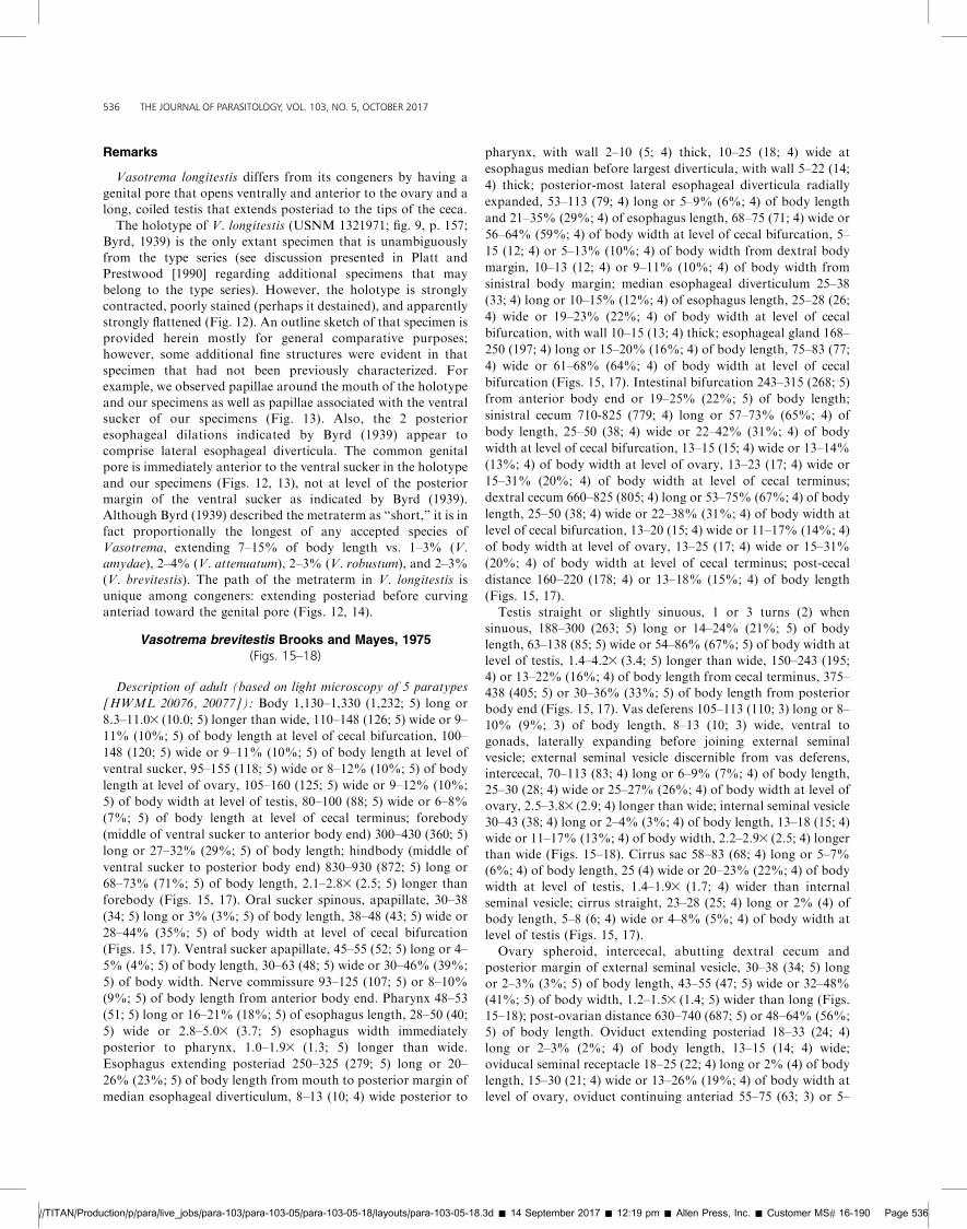
Remarks
Vasotrema longitestis differs from its congeners by having a
genital pore that opens ventrally and anterior to the ovary and a
long, coiled testis that extends posteriad to the tips of the ceca.
The holotype of V. longitestis (USNM 1321971; fig. 9, p. 157;
Byrd, 1939) is the only extant specimen that is unambiguously
from the type series (see discussion presented in Platt and
Prestwood [1990] regarding additional specimens that may
belong to the type series). However, the holotype is strongly
contracted, poorly stained (perhaps it destained), and apparently
strongly flattened (Fig. 12). An outline sketch of that specimen is
provided herein mostly for general comparative purposes;
however, some additional fine structures were evident in that
specimen that had not been previously characterized. For
example, we observed papillae around the mouth of the holotype
and our specimens as well as papillae associated with the ventral
sucker of our specimens (Fig. 13). Also, the 2 posterior
esophageal dilations indicated by Byrd (1939) appear to
comprise lateral esophageal diverticula. The common genital
pore is immediately anterior to the ventral sucker in the holotype
and our specimens (Figs. 12, 13), not at level of the posterior
margin of the ventral sucker as indicated by Byrd (1939).
Although Byrd (1939) described the metraterm as ‘‘short,’’ it is in
fact proportionally the longest of any accepted species of
Vasotrema, extending 7–15% of body length vs. 1–3% (V.
amydae), 2–4% (V. attenuatum), 2–3% (V. robustum), and 2–3%
(V. brevitestis). The path of the metraterm in V. longitestis is
unique among congeners: extending posteriad before curving
anteriad toward the genital pore (Figs. 12, 14).
Vasotrema brevitestis Brooks and Mayes, 1975(Figs. 15–18)
Description of adult (based on light microscopy of 5 paratypes
[HWML 20076, 20077]): Body 1,130–1,330 (1,232; 5) long or
8.3–11.03 (10.0; 5) longer than wide, 110–148 (126; 5) wide or 9–
11% (10%; 5) of body length at level of cecal bifurcation, 100–
148 (120; 5) wide or 9–11% (10%; 5) of body length at level of
ventral sucker, 95–155 (118; 5) wide or 8–12% (10%; 5) of body
length at level of ovary, 105–160 (125; 5) wide or 9–12% (10%;
5) of body width at level of testis, 80–100 (88; 5) wide or 6–8%
(7%; 5) of body length at level of cecal terminus; forebody
(middle of ventral sucker to anterior body end) 300–430 (360; 5)
long or 27–32% (29%; 5) of body length; hindbody (middle of
ventral sucker to posterior body end) 830–930 (872; 5) long or
68–73% (71%; 5) of body length, 2.1–2.83 (2.5; 5) longer than
forebody (Figs. 15, 17). Oral sucker spinous, apapillate, 30–38
(34; 5) long or 3% (3%; 5) of body length, 38–48 (43; 5) wide or
28–44% (35%; 5) of body width at level of cecal bifurcation
(Figs. 15, 17). Ventral sucker apapillate, 45–55 (52; 5) long or 4–
5% (4%; 5) of body length, 30–63 (48; 5) wide or 30–46% (39%;
5) of body width. Nerve commissure 93–125 (107; 5) or 8–10%
(9%; 5) of body length from anterior body end. Pharynx 48–53
(51; 5) long or 16–21% (18%; 5) of esophagus length, 28–50 (40;
5) wide or 2.8–5.03 (3.7; 5) esophagus width immediately
posterior to pharynx, 1.0–1.93 (1.3; 5) longer than wide.
Esophagus extending posteriad 250–325 (279; 5) long or 20–
26% (23%; 5) of body length from mouth to posterior margin of
median esophageal diverticulum, 8–13 (10; 4) wide posterior to
pharynx, with wall 2–10 (5; 4) thick, 10–25 (18; 4) wide at
esophagus median before largest diverticula, with wall 5–22 (14;
4) thick; posterior-most lateral esophageal diverticula radially
expanded, 53–113 (79; 4) long or 5–9% (6%; 4) of body length
and 21–35% (29%; 4) of esophagus length, 68–75 (71; 4) wide or
56–64% (59%; 4) of body width at level of cecal bifurcation, 5–
15 (12; 4) or 5–13% (10%; 4) of body width from dextral body
margin, 10–13 (12; 4) or 9–11% (10%; 4) of body width from
sinistral body margin; median esophageal diverticulum 25–38
(33; 4) long or 10–15% (12%; 4) of esophagus length, 25–28 (26;
4) wide or 19–23% (22%; 4) of body width at level of cecal
bifurcation, with wall 10–15 (13; 4) thick; esophageal gland 168–
250 (197; 4) long or 15–20% (16%; 4) of body length, 75–83 (77;
4) wide or 61–68% (64%; 4) of body width at level of cecal
bifurcation (Figs. 15, 17). Intestinal bifurcation 243–315 (268; 5)
from anterior body end or 19–25% (22%; 5) of body length;
sinistral cecum 710-825 (779; 4) long or 57–73% (65%; 4) of
body length, 25–50 (38; 4) wide or 22–42% (31%; 4) of body
width at level of cecal bifurcation, 13–15 (15; 4) wide or 13–14%
(13%; 4) of body width at level of ovary, 13–23 (17; 4) wide or
15–31% (20%; 4) of body width at level of cecal terminus;
dextral cecum 660–825 (805; 4) long or 53–75% (67%; 4) of body
length, 25–50 (38; 4) wide or 22–38% (31%; 4) of body width at
level of cecal bifurcation, 13–20 (15; 4) wide or 11–17% (14%; 4)
of body width at level of ovary, 13–25 (17; 4) wide or 15–31%
(20%; 4) of body width at level of cecal terminus; post-cecal
distance 160–220 (178; 4) or 13–18% (15%; 4) of body length
(Figs. 15, 17).
Testis straight or slightly sinuous, 1 or 3 turns (2) when
sinuous, 188–300 (263; 5) long or 14–24% (21%; 5) of body
length, 63–138 (85; 5) wide or 54–86% (67%; 5) of body width at
level of testis, 1.4–4.23 (3.4; 5) longer than wide, 150–243 (195;
4) or 13–22% (16%; 4) of body length from cecal terminus, 375–
438 (405; 5) or 30–36% (33%; 5) of body length from posterior
body end (Figs. 15, 17). Vas deferens 105–113 (110; 3) long or 8–
10% (9%; 3) of body length, 8–13 (10; 3) wide, ventral to
gonads, laterally expanding before joining external seminal
vesicle; external seminal vesicle discernible from vas deferens,
intercecal, 70–113 (83; 4) long or 6–9% (7%; 4) of body length,
25–30 (28; 4) wide or 25–27% (26%; 4) of body width at level of
ovary, 2.5–3.83 (2.9; 4) longer than wide; internal seminal vesicle
30–43 (38; 4) long or 2–4% (3%; 4) of body length, 13–18 (15; 4)
wide or 11–17% (13%; 4) of body width, 2.2–2.93 (2.5; 4) longer
than wide (Figs. 15–18). Cirrus sac 58–83 (68; 4) long or 5–7%
(6%; 4) of body length, 25 (4) wide or 20–23% (22%; 4) of body
width at level of testis, 1.4–1.93 (1.7; 4) wider than internal
seminal vesicle; cirrus straight, 23–28 (25; 4) long or 2% (4) of
body length, 5–8 (6; 4) wide or 4–8% (5%; 4) of body width at
level of testis (Figs. 15, 17).
Ovary spheroid, intercecal, abutting dextral cecum and
posterior margin of external seminal vesicle, 30–38 (34; 5) long
or 2–3% (3%; 5) of body length, 43–55 (47; 5) wide or 32–48%
(41%; 5) of body width, 1.2–1.53 (1.4; 5) wider than long (Figs.
15–18); post-ovarian distance 630–740 (687; 5) or 48–64% (56%;
5) of body length. Oviduct extending posteriad 18–33 (24; 4)
long or 2–3% (2%; 4) of body length, 13–15 (14; 4) wide;
oviducal seminal receptacle 18–25 (22; 4) long or 2% (4) of body
length, 15–30 (21; 4) wide or 13–26% (19%; 4) of body width at
level of ovary, oviduct continuing anteriad 55–75 (63; 3) or 5–
//TITAN/Production/p/para/live_jobs/para-103/para-103-05/para-103-05-18/layouts/para-103-05-18.3d � 14 September 2017 � 12:19 pm � Allen Press, Inc. � Customer MS# 16-190 Page 536
536 THE JOURNAL OF PARASITOLOGY, VOL. 103, NO. 5, OCTOBER 2017
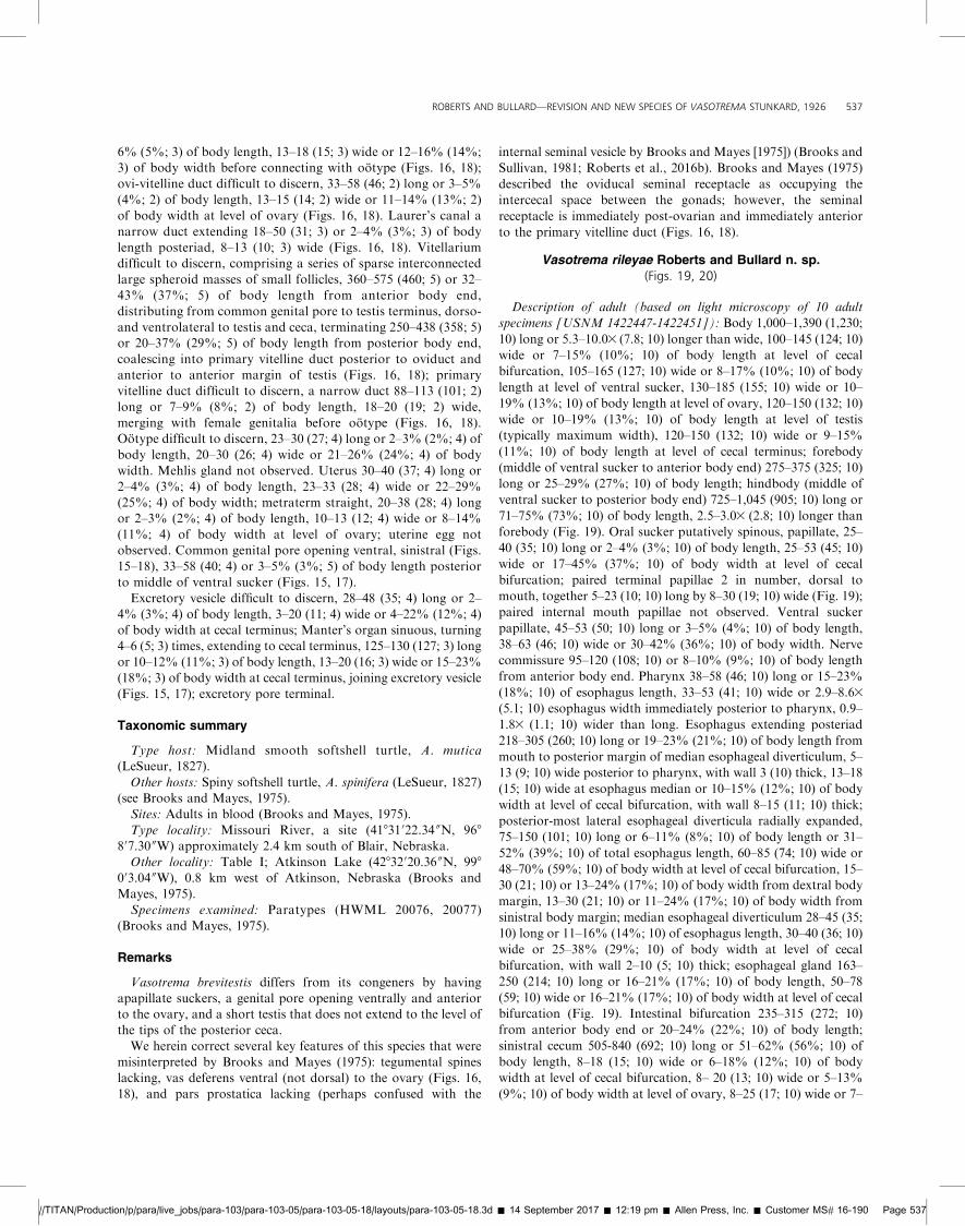
6% (5%; 3) of body length, 13–18 (15; 3) wide or 12–16% (14%;3) of body width before connecting with ootype (Figs. 16, 18);
ovi-vitelline duct difficult to discern, 33–58 (46; 2) long or 3–5%
(4%; 2) of body length, 13–15 (14; 2) wide or 11–14% (13%; 2)
of body width at level of ovary (Figs. 16, 18). Laurer’s canal a
narrow duct extending 18–50 (31; 3) or 2–4% (3%; 3) of body
length posteriad, 8–13 (10; 3) wide (Figs. 16, 18). Vitellariumdifficult to discern, comprising a series of sparse interconnected
large spheroid masses of small follicles, 360–575 (460; 5) or 32–
43% (37%; 5) of body length from anterior body end,
distributing from common genital pore to testis terminus, dorso-
and ventrolateral to testis and ceca, terminating 250–438 (358; 5)or 20–37% (29%; 5) of body length from posterior body end,
coalescing into primary vitelline duct posterior to oviduct and
anterior to anterior margin of testis (Figs. 16, 18); primary
vitelline duct difficult to discern, a narrow duct 88–113 (101; 2)
long or 7–9% (8%; 2) of body length, 18–20 (19; 2) wide,
merging with female genitalia before ootype (Figs. 16, 18).Ootype difficult to discern, 23–30 (27; 4) long or 2–3% (2%; 4) of
body length, 20–30 (26; 4) wide or 21–26% (24%; 4) of body
width. Mehlis gland not observed. Uterus 30–40 (37; 4) long or
2–4% (3%; 4) of body length, 23–33 (28; 4) wide or 22–29%
(25%; 4) of body width; metraterm straight, 20–38 (28; 4) longor 2–3% (2%; 4) of body length, 10–13 (12; 4) wide or 8–14%
(11%; 4) of body width at level of ovary; uterine egg not
observed. Common genital pore opening ventral, sinistral (Figs.
15–18), 33–58 (40; 4) or 3–5% (3%; 5) of body length posterior
to middle of ventral sucker (Figs. 15, 17).
Excretory vesicle difficult to discern, 28–48 (35; 4) long or 2–
4% (3%; 4) of body length, 3–20 (11; 4) wide or 4–22% (12%; 4)of body width at cecal terminus; Manter’s organ sinuous, turning
4–6 (5; 3) times, extending to cecal terminus, 125–130 (127; 3) long
or 10–12% (11%; 3) of body length, 13–20 (16; 3) wide or 15–23%
(18%; 3) of body width at cecal terminus, joining excretory vesicle
(Figs. 15, 17); excretory pore terminal.
Taxonomic summary
Type host: Midland smooth softshell turtle, A. mutica(LeSueur, 1827).
Other hosts: Spiny softshell turtle, A. spinifera (LeSueur, 1827)(see Brooks and Mayes, 1975).
Sites: Adults in blood (Brooks and Mayes, 1975).
Type locality: Missouri River, a site (41831 022.34 00N, 968
807.30 00W) approximately 2.4 km south of Blair, Nebraska.
Other locality: Table I; Atkinson Lake (42832020.36 00N, 998
003.04 00W), 0.8 km west of Atkinson, Nebraska (Brooks and
Mayes, 1975).
Specimens examined: Paratypes (HWML 20076, 20077)
(Brooks and Mayes, 1975).
Remarks
Vasotrema brevitestis differs from its congeners by having
apapillate suckers, a genital pore opening ventrally and anteriorto the ovary, and a short testis that does not extend to the level of
the tips of the posterior ceca.
We herein correct several key features of this species that were
misinterpreted by Brooks and Mayes (1975): tegumental spines
lacking, vas deferens ventral (not dorsal) to the ovary (Figs. 16,
18), and pars prostatica lacking (perhaps confused with the
internal seminal vesicle by Brooks and Mayes [1975]) (Brooks and
Sullivan, 1981; Roberts et al., 2016b). Brooks and Mayes (1975)
described the oviducal seminal receptacle as occupying the
intercecal space between the gonads; however, the seminal
receptacle is immediately post-ovarian and immediately anterior
to the primary vitelline duct (Figs. 16, 18).
Vasotrema rileyae Roberts and Bullard n. sp.(Figs. 19, 20)
Description of adult (based on light microscopy of 10 adult
specimens [USNM 1422447-1422451]): Body 1,000–1,390 (1,230;
10) long or 5.3–10.03 (7.8; 10) longer than wide, 100–145 (124; 10)
wide or 7–15% (10%; 10) of body length at level of cecal
bifurcation, 105–165 (127; 10) wide or 8–17% (10%; 10) of body
length at level of ventral sucker, 130–185 (155; 10) wide or 10–
19% (13%; 10) of body length at level of ovary, 120–150 (132; 10)
wide or 10–19% (13%; 10) of body length at level of testis
(typically maximum width), 120–150 (132; 10) wide or 9–15%
(11%; 10) of body length at level of cecal terminus; forebody
(middle of ventral sucker to anterior body end) 275–375 (325; 10)
long or 25–29% (27%; 10) of body length; hindbody (middle of
ventral sucker to posterior body end) 725–1,045 (905; 10) long or
71–75% (73%; 10) of body length, 2.5–3.03 (2.8; 10) longer than
forebody (Fig. 19). Oral sucker putatively spinous, papillate, 25–
40 (35; 10) long or 2–4% (3%; 10) of body length, 25–53 (45; 10)
wide or 17–45% (37%; 10) of body width at level of cecal
bifurcation; paired terminal papillae 2 in number, dorsal to
mouth, together 5–23 (10; 10) long by 8–30 (19; 10) wide (Fig. 19);
paired internal mouth papillae not observed. Ventral sucker
papillate, 45–53 (50; 10) long or 3–5% (4%; 10) of body length,
38–63 (46; 10) wide or 30–42% (36%; 10) of body width. Nerve
commissure 95–120 (108; 10) or 8–10% (9%; 10) of body length
from anterior body end. Pharynx 38–58 (46; 10) long or 15–23%
(18%; 10) of esophagus length, 33–53 (41; 10) wide or 2.9–8.63
(5.1; 10) esophagus width immediately posterior to pharynx, 0.9–
1.83 (1.1; 10) wider than long. Esophagus extending posteriad
218–305 (260; 10) long or 19–23% (21%; 10) of body length from
mouth to posterior margin of median esophageal diverticulum, 5–
13 (9; 10) wide posterior to pharynx, with wall 3 (10) thick, 13–18
(15; 10) wide at esophagus median or 10–15% (12%; 10) of body
width at level of cecal bifurcation, with wall 8–15 (11; 10) thick;
posterior-most lateral esophageal diverticula radially expanded,
75–150 (101; 10) long or 6–11% (8%; 10) of body length or 31–
52% (39%; 10) of total esophagus length, 60–85 (74; 10) wide or
48–70% (59%; 10) of body width at level of cecal bifurcation, 15–
30 (21; 10) or 13–24% (17%; 10) of body width from dextral body
margin, 13–30 (21; 10) or 11–24% (17%; 10) of body width from
sinistral body margin; median esophageal diverticulum 28–45 (35;
10) long or 11–16% (14%; 10) of esophagus length, 30–40 (36; 10)
wide or 25–38% (29%; 10) of body width at level of cecal
bifurcation, with wall 2–10 (5; 10) thick; esophageal gland 163–
250 (214; 10) long or 16–21% (17%; 10) of body length, 50–78
(59; 10) wide or 16–21% (17%; 10) of body width at level of cecal
bifurcation (Fig. 19). Intestinal bifurcation 235–315 (272; 10)
from anterior body end or 20–24% (22%; 10) of body length;
sinistral cecum 505-840 (692; 10) long or 51–62% (56%; 10) of
body length, 8–18 (15; 10) wide or 6–18% (12%; 10) of body
width at level of cecal bifurcation, 8– 20 (13; 10) wide or 5–13%
(9%; 10) of body width at level of ovary, 8–25 (17; 10) wide or 7–
//TITAN/Production/p/para/live_jobs/para-103/para-103-05/para-103-05-18/layouts/para-103-05-18.3d � 14 September 2017 � 12:19 pm � Allen Press, Inc. � Customer MS# 16-190 Page 537
ROBERTS AND BULLARD—REVISION AND NEW SPECIES OF VASOTREMA STUNKARD, 1926 537
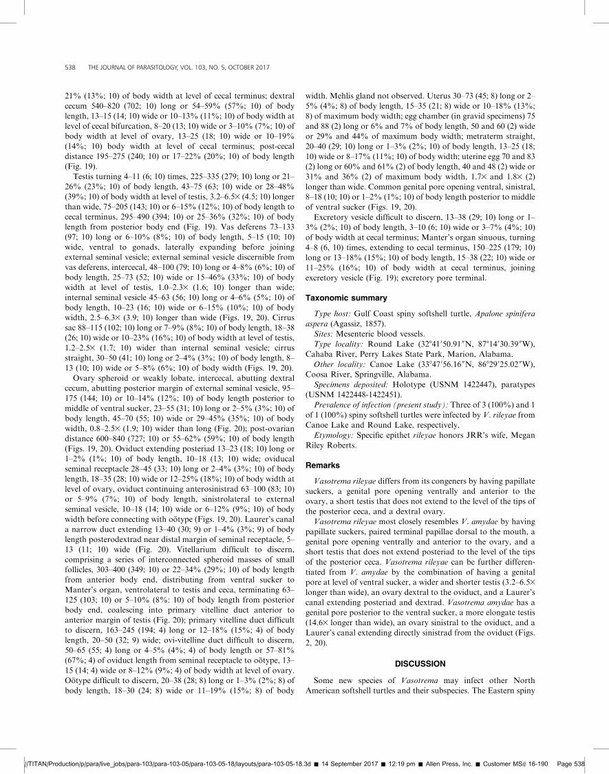
21% (13%; 10) of body width at level of cecal terminus; dextral
cecum 540–820 (702; 10) long or 54–59% (57%; 10) of body
length, 13–15 (14; 10) wide or 10–13% (11%; 10) of body width at
level of cecal bifurcation, 8–20 (13; 10) wide or 3–10% (7%; 10) of
body width at level of ovary, 13–25 (18; 10) wide or 10–19%
(14%; 10) body width at level of cecal terminus; post-cecal
distance 195–275 (240; 10) or 17–22% (20%; 10) of body length
(Fig. 19).
Testis turning 4–11 (6; 10) times, 225–335 (279; 10) long or 21–
26% (23%; 10) of body length, 43–75 (63; 10) wide or 28–48%
(39%; 10) of body width at level of testis, 3.2–6.53 (4.5; 10) longer
than wide, 75–205 (143; 10) or 6–15% (12%; 10) of body length to
cecal terminus, 295–490 (394; 10) or 25–36% (32%; 10) of body
length from posterior body end (Fig. 19). Vas deferens 73–133
(97; 10) long or 6–10% (8%; 10) of body length, 5–15 (10; 10)
wide, ventral to gonads, laterally expanding before joining
external seminal vesicle; external seminal vesicle discernible from
vas deferens, intercecal, 48–100 (79; 10) long or 4–8% (6%; 10) of
body length, 25–73 (52; 10) wide or 15–46% (33%; 10) of body
width at level of testis, 1.0–2.33 (1.6; 10) longer than wide;
internal seminal vesicle 45–63 (56; 10) long or 4–6% (5%; 10) of
body length, 10–23 (16; 10) wide or 6–15% (10%; 10) of body
width, 2.5–6.33 (3.9; 10) longer than wide (Figs. 19, 20). Cirrus
sac 88–115 (102; 10) long or 7–9% (8%; 10) of body length, 18–38
(26; 10) wide or 10–23% (16%; 10) of body width at level of testis,
1.2–2.53 (1.7; 10) wider than internal seminal vesicle; cirrus
straight, 30–50 (41; 10) long or 2–4% (3%; 10) of body length, 8–
13 (10; 10) wide or 5–8% (6%; 10) of body width (Figs. 19, 20).
Ovary spheroid or weakly lobate, intercecal, abutting dextral
cecum, abutting posterior margin of external seminal vesicle, 95–
175 (144; 10) or 10–14% (12%; 10) of body length posterior to
middle of ventral sucker, 23–55 (31; 10) long or 2–5% (3%; 10) of
body length, 45–70 (55; 10) wide or 29–45% (35%; 10) of body
width, 0.8–2.53 (1.9; 10) wider than long (Fig. 20); post-ovarian
distance 600–840 (727; 10) or 55–62% (59%; 10) of body length
(Figs. 19, 20). Oviduct extending posteriad 13–23 (18; 10) long or
1–2% (1%; 10) of body length, 10–18 (13; 10) wide; oviducal
seminal receptacle 28–45 (33; 10) long or 2–4% (3%; 10) of body
length, 18–35 (28; 10) wide or 12–25% (18%; 10) of body width at
level of ovary, oviduct continuing anterosinistrad 63–100 (83; 10)
or 5–9% (7%; 10) of body length, sinistrolateral to external
seminal vesicle, 10–18 (14; 10) wide or 6–12% (9%; 10) of body
width before connecting with ootype (Figs. 19, 20). Laurer’s canal
a narrow duct extending 13–40 (30; 9) or 1–4% (3%; 9) of body
length posterodextrad near distal margin of seminal receptacle, 5–
13 (11; 10) wide (Fig. 20). Vitellarium difficult to discern,
comprising a series of interconnected spheroid masses of small
follicles, 303–400 (349; 10) or 22–34% (29%; 10) of body length
from anterior body end, distributing from ventral sucker to
Manter’s organ, ventrolateral to testis and ceca, terminating 63–
125 (103; 10) or 5–10% (8%: 10) of body length from posterior
body end, coalescing into primary vitelline duct anterior to
anterior margin of testis (Fig. 20); primary vitelline duct difficult
to discern, 163–245 (194; 4) long or 12–18% (15%; 4) of body
length, 20–50 (32; 9) wide; ovi-vitelline duct difficult to discern,
50–65 (55; 4) long or 4–5% (4%; 4) of body length or 57–81%
(67%; 4) of oviduct length from seminal receptacle to ootype, 13–
15 (14; 4) wide or 8–12% (9%; 4) of body width at level of ovary.
Ootype difficult to discern, 20–38 (28; 8) long or 1–3% (2%; 8) of
body length, 18–30 (24; 8) wide or 11–19% (15%; 8) of body
width. Mehlis gland not observed. Uterus 30–73 (45; 8) long or 2–
5% (4%; 8) of body length, 15–35 (21; 8) wide or 10–18% (13%;
8) of maximum body width; egg chamber (in gravid specimens) 75
and 88 (2) long or 6% and 7% of body length, 50 and 60 (2) wide
or 29% and 44% of maximum body width; metraterm straight,
20–40 (29; 10) long or 1–3% (2%; 10) of body length, 13–25 (18;
10) wide or 8–17% (11%; 10) of body width; uterine egg 70 and 83
(2) long or 60% and 61% (2) of body length, 40 and 48 (2) wide or
31% and 36% (2) of maximum body width, 1.73 and 1.83 (2)
longer than wide. Common genital pore opening ventral, sinistral,
8–18 (10; 10) or 1–2% (1%; 10) of body length posterior to middle
of ventral sucker (Figs. 19, 20).
Excretory vesicle difficult to discern, 13–38 (29; 10) long or 1–
3% (2%; 10) of body length, 3–10 (6; 10) wide or 3–7% (4%; 10)
of body width at cecal terminus; Manter’s organ sinuous, turning
4–8 (6, 10) times, extending to cecal terminus, 150–225 (179; 10)
long or 13–18% (15%; 10) of body length, 15–38 (22; 10) wide or
11–25% (16%; 10) of body width at cecal terminus, joining
excretory vesicle (Fig. 19); excretory pore terminal.
Taxonomic summary
Type host: Gulf Coast spiny softshell turtle, Apalone spinifera
aspera (Agassiz, 1857).
Sites: Mesenteric blood vessels.
Type locality: Round Lake (32841050.91 00N, 87814030.39 00W),
Cahaba River, Perry Lakes State Park, Marion, Alabama.
Other locality: Canoe Lake (33847056.16 00N, 86829025.02 00W),
Coosa River, Springville, Alabama.
Specimens deposited: Holotype (USNM 1422447), paratypes
(USNM 1422448-1422451).
Prevalence of infection (present study): Three of 3 (100%) and 1
of 1 (100%) spiny softshell turtles were infected by V. rileyae from
Canoe Lake and Round Lake, respectively.
Etymology: Specific epithet rileyae honors JRR’s wife, Megan
Riley Roberts.
Remarks
Vasotrema rileyae differs from its congeners by having papillate
suckers, a genital pore opening ventrally and anterior to the
ovary, a short testis that does not extend to the level of the tips of
the posterior ceca, and a dextral ovary.
Vasotrema rileyae most closely resembles V. amydae by having
papillate suckers, paired terminal papillae dorsal to the mouth, a
genital pore opening ventrally and anterior to the ovary, and a
short testis that does not extend posteriad to the level of the tips
of the posterior ceca. Vasotrema rileyae can be further differen-
tiated from V. amydae by the combination of having a genital
pore at level of ventral sucker, a wider and shorter testis (3.2–6.53
longer than wide), an ovary dextral to the oviduct, and a Laurer’s
canal extending posteriad and dextrad. Vasotrema amydae has a
genital pore posterior to the ventral sucker, a more elongate testis
(14.63 longer than wide), an ovary sinistral to the oviduct, and a
Laurer’s canal extending directly sinistrad from the oviduct (Figs.
2, 20).
DISCUSSION
Some new species of Vasotrema may infect other North
American softshell turtles and their subspecies. The Eastern spiny
//TITAN/Production/p/para/live_jobs/para-103/para-103-05/para-103-05-18/layouts/para-103-05-18.3d � 14 September 2017 � 12:19 pm � Allen Press, Inc. � Customer MS# 16-190 Page 538
538 THE JOURNAL OF PARASITOLOGY, VOL. 103, NO. 5, OCTOBER 2017
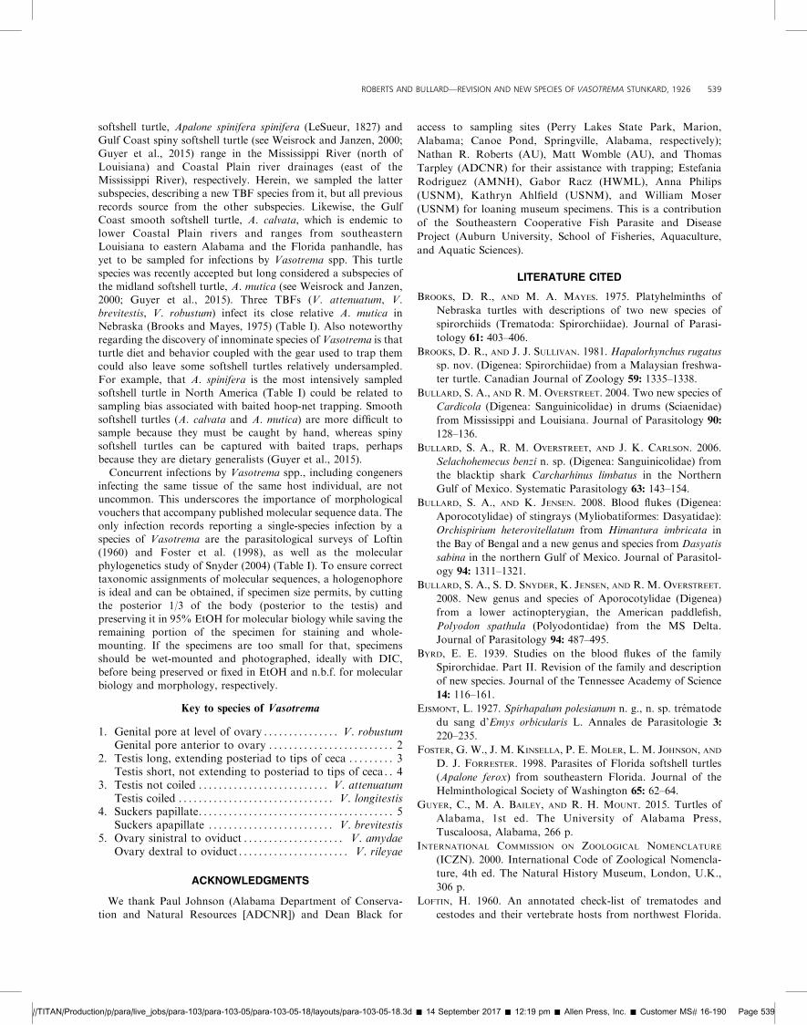
softshell turtle, Apalone spinifera spinifera (LeSueur, 1827) andGulf Coast spiny softshell turtle (see Weisrock and Janzen, 2000;Guyer et al., 2015) range in the Mississippi River (north of
Louisiana) and Coastal Plain river drainages (east of theMississippi River), respectively. Herein, we sampled the lattersubspecies, describing a new TBF species from it, but all previous
records source from the other subspecies. Likewise, the GulfCoast smooth softshell turtle, A. calvata, which is endemic to
lower Coastal Plain rivers and ranges from southeasternLouisiana to eastern Alabama and the Florida panhandle, hasyet to be sampled for infections by Vasotrema spp. This turtle
species was recently accepted but long considered a subspecies ofthe midland softshell turtle, A. mutica (see Weisrock and Janzen,2000; Guyer et al., 2015). Three TBFs (V. attenuatum, V.
brevitestis, V. robustum) infect its close relative A. mutica inNebraska (Brooks and Mayes, 1975) (Table I). Also noteworthyregarding the discovery of innominate species of Vasotrema is that
turtle diet and behavior coupled with the gear used to trap themcould also leave some softshell turtles relatively undersampled.
For example, that A. spinifera is the most intensively sampledsoftshell turtle in North America (Table I) could be related tosampling bias associated with baited hoop-net trapping. Smooth
softshell turtles (A. calvata and A. mutica) are more difficult tosample because they must be caught by hand, whereas spinysoftshell turtles can be captured with baited traps, perhaps
because they are dietary generalists (Guyer et al., 2015).Concurrent infections by Vasotrema spp., including congeners
infecting the same tissue of the same host individual, are not
uncommon. This underscores the importance of morphologicalvouchers that accompany published molecular sequence data. The
only infection records reporting a single-species infection by aspecies of Vasotrema are the parasitological surveys of Loftin(1960) and Foster et al. (1998), as well as the molecular
phylogenetics study of Snyder (2004) (Table I). To ensure correcttaxonomic assignments of molecular sequences, a hologenophoreis ideal and can be obtained, if specimen size permits, by cutting
the posterior 1/3 of the body (posterior to the testis) andpreserving it in 95% EtOH for molecular biology while saving theremaining portion of the specimen for staining and whole-
mounting. If the specimens are too small for that, specimensshould be wet-mounted and photographed, ideally with DIC,
before being preserved or fixed in EtOH and n.b.f. for molecularbiology and morphology, respectively.
Key to species of Vasotrema
1. Genital pore at level of ovary . . . . . . . . . . . . . . . V. robustumGenital pore anterior to ovary . . . . . . . . . . . . . . . . . . . . . . . . . 2
2. Testis long, extending posteriad to tips of ceca . . . . . . . . . 3Testis short, not extending to posteriad to tips of ceca. . 4
3. Testis not coiled . . . . . . . . . . . . . . . . . . . . . . . . . . V. attenuatumTestis coiled . . . . . . . . . . . . . . . . . . . . . . . . . . . . . . . V. longitestis
4. Suckers papillate. . . . . . . . . . . . . . . . . . . . . . . . . . . . . . . . . . . . . . . 5Suckers apapillate . . . . . . . . . . . . . . . . . . . . . . . . . V. brevitestis
5. Ovary sinistral to oviduct . . . . . . . . . . . . . . . . . . . . V. amydaeOvary dextral to oviduct . . . . . . . . . . . . . . . . . . . . . . V. rileyae
ACKNOWLEDGMENTS
We thank Paul Johnson (Alabama Department of Conserva-
tion and Natural Resources [ADCNR]) and Dean Black for
access to sampling sites (Perry Lakes State Park, Marion,
Alabama; Canoe Pond, Springville, Alabama, respectively);
Nathan R. Roberts (AU), Matt Womble (AU), and Thomas
Tarpley (ADCNR) for their assistance with trapping; Estefania
Rodriguez (AMNH), Gabor Racz (HWML), Anna Philips
(USNM), Kathryn Ahlfield (USNM), and William Moser
(USNM) for loaning museum specimens. This is a contribution
of the Southeastern Cooperative Fish Parasite and Disease
Project (Auburn University, School of Fisheries, Aquaculture,
and Aquatic Sciences).
LITERATURE CITED
BROOKS, D. R., AND M. A. MAYES. 1975. Platyhelminths of
Nebraska turtles with descriptions of two new species of
spirorchiids (Trematoda: Spirorchiidae). Journal of Parasi-
tology 61: 403–406.
BROOKS, D. R., AND J. J. SULLIVAN. 1981. Hapalorhynchus rugatus
sp. nov. (Digenea: Spirorchiidae) from a Malaysian freshwa-
ter turtle. Canadian Journal of Zoology 59: 1335–1338.
BULLARD, S. A., AND R. M. OVERSTREET. 2004. Two new species of
Cardicola (Digenea: Sanguinicolidae) in drums (Sciaenidae)
from Mississippi and Louisiana. Journal of Parasitology 90:
128–136.
BULLARD, S. A., R. M. OVERSTREET, AND J. K. CARLSON. 2006.
Selachohemecus benzi n. sp. (Digenea: Sanguinicolidae) from
the blacktip shark Carcharhinus limbatus in the Northern
Gulf of Mexico. Systematic Parasitology 63: 143–154.
BULLARD, S. A., AND K. JENSEN. 2008. Blood flukes (Digenea:
Aporocotylidae) of stingrays (Myliobatiformes: Dasyatidae):
Orchispirium heterovitellatum from Himantura imbricata in
the Bay of Bengal and a new genus and species from Dasyatis
sabina in the northern Gulf of Mexico. Journal of Parasitol-
ogy 94: 1311–1321.
BULLARD, S. A., S. D. SNYDER, K. JENSEN, AND R. M. OVERSTREET.
2008. New genus and species of Aporocotylidae (Digenea)
from a lower actinopterygian, the American paddlefish,
Polyodon spathula (Polyodontidae) from the MS Delta.
Journal of Parasitology 94: 487–495.
BYRD, E. E. 1939. Studies on the blood flukes of the family
Spirorchidae. Part II. Revision of the family and description
of new species. Journal of the Tennessee Academy of Science
14: 116–161.
EJSMONT, L. 1927. Spirhapalum polesianum n. g., n. sp. trematode
du sang d’Emys orbicularis L. Annales de Parasitologie 3:
220–235.
FOSTER, G. W., J. M. KINSELLA, P. E. MOLER, L. M. JOHNSON, AND
D. J. FORRESTER. 1998. Parasites of Florida softshell turtles
(Apalone ferox) from southeastern Florida. Journal of the
Helminthological Society of Washington 65: 62–64.
GUYER, C., M. A. BAILEY, AND R. H. MOUNT. 2015. Turtles of
Alabama, 1st ed. The University of Alabama Press,
Tuscaloosa, Alabama, 266 p.
INTERNATIONAL COMMISSION ON ZOOLOGICAL NOMENCLATURE
(ICZN). 2000. International Code of Zoological Nomencla-
ture, 4th ed. The Natural History Museum, London, U.K.,
306 p.
LOFTIN, H. 1960. An annotated check-list of trematodes and
cestodes and their vertebrate hosts from northwest Florida.
//TITAN/Production/p/para/live_jobs/para-103/para-103-05/para-103-05-18/layouts/para-103-05-18.3d � 14 September 2017 � 12:19 pm � Allen Press, Inc. � Customer MS# 16-190 Page 539
ROBERTS AND BULLARD—REVISION AND NEW SPECIES OF VASOTREMA STUNKARD, 1926 539
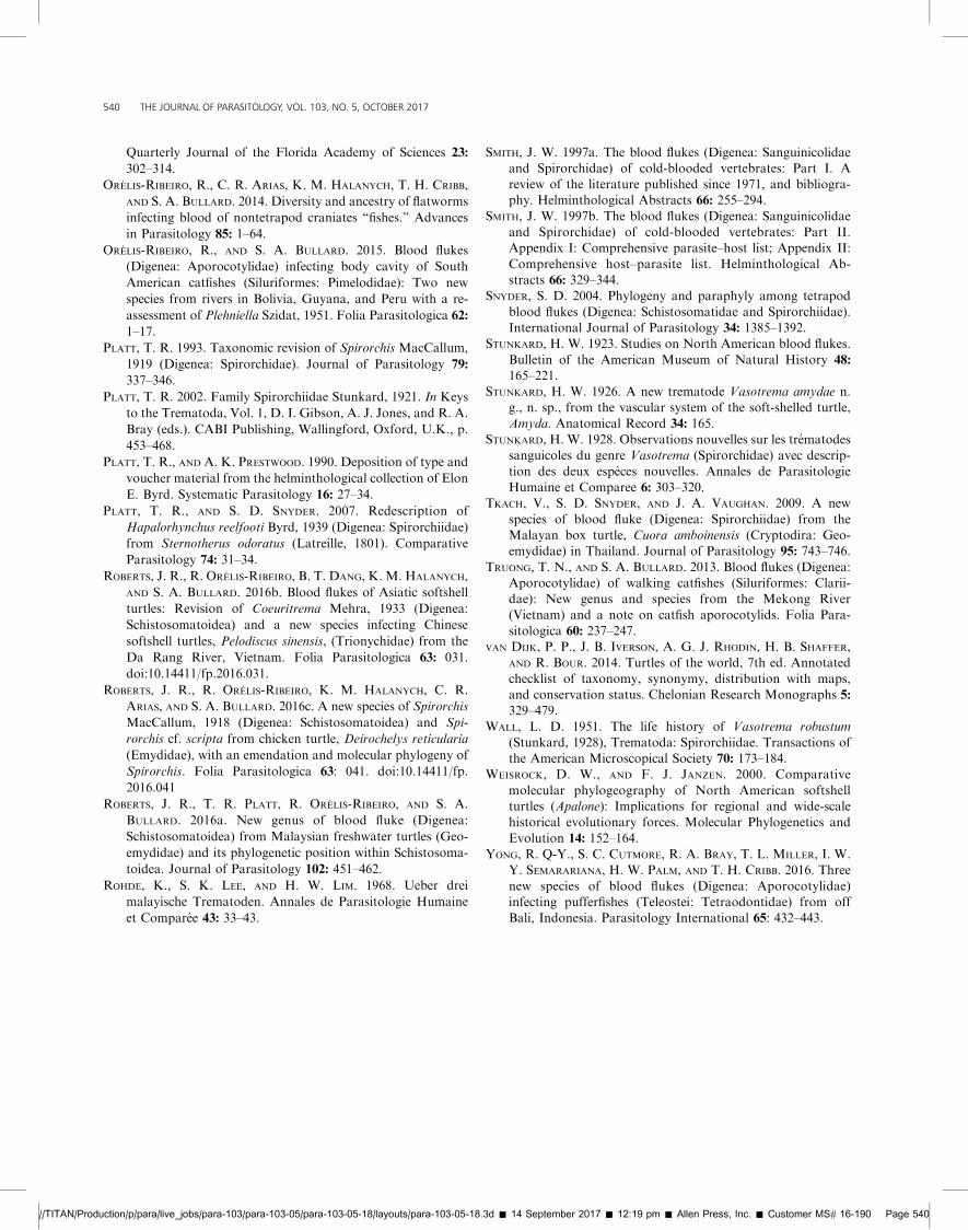
Quarterly Journal of the Florida Academy of Sciences 23:
302–314.
ORELIS-RIBEIRO, R., C. R. ARIAS, K. M. HALANYCH, T. H. CRIBB,
AND S. A. BULLARD. 2014. Diversity and ancestry of flatworms
infecting blood of nontetrapod craniates ‘‘fishes.’’ Advances
in Parasitology 85: 1–64.
ORELIS-RIBEIRO, R., AND S. A. BULLARD. 2015. Blood flukes
(Digenea: Aporocotylidae) infecting body cavity of South
American catfishes (Siluriformes: Pimelodidae): Two new
species from rivers in Bolivia, Guyana, and Peru with a re-
assessment of Plehniella Szidat, 1951. Folia Parasitologica 62:
1–17.
PLATT, T. R. 1993. Taxonomic revision of Spirorchis MacCallum,
1919 (Digenea: Spirorchidae). Journal of Parasitology 79:
337–346.
PLATT, T. R. 2002. Family Spirorchiidae Stunkard, 1921. In Keys
to the Trematoda, Vol. 1, D. I. Gibson, A. J. Jones, and R. A.
Bray (eds.). CABI Publishing, Wallingford, Oxford, U.K., p.
453–468.
PLATT, T. R., AND A. K. PRESTWOOD. 1990. Deposition of type and
voucher material from the helminthological collection of Elon
E. Byrd. Systematic Parasitology 16: 27–34.
PLATT, T. R., AND S. D. SNYDER. 2007. Redescription of
Hapalorhynchus reelfooti Byrd, 1939 (Digenea: Spirorchiidae)
from Sternotherus odoratus (Latreille, 1801). Comparative
Parasitology 74: 31–34.
ROBERTS, J. R., R. ORELIS-RIBEIRO, B. T. DANG, K. M. HALANYCH,
AND S. A. BULLARD. 2016b. Blood flukes of Asiatic softshell
turtles: Revision of Coeuritrema Mehra, 1933 (Digenea:
Schistosomatoidea) and a new species infecting Chinese
softshell turtles, Pelodiscus sinensis, (Trionychidae) from the
Da Rang River, Vietnam. Folia Parasitologica 63: 031.
doi:10.14411/fp.2016.031.
ROBERTS, J. R., R. ORELIS-RIBEIRO, K. M. HALANYCH, C. R.
ARIAS, AND S. A. BULLARD. 2016c. A new species of Spirorchis
MacCallum, 1918 (Digenea: Schistosomatoidea) and Spi-
rorchis cf. scripta from chicken turtle, Deirochelys reticularia
(Emydidae), with an emendation and molecular phylogeny of
Spirorchis. Folia Parasitologica 63: 041. doi:10.14411/fp.
2016.041
ROBERTS, J. R., T. R. PLATT, R. ORELIS-RIBEIRO, AND S. A.
BULLARD. 2016a. New genus of blood fluke (Digenea:
Schistosomatoidea) from Malaysian freshwater turtles (Geo-
emydidae) and its phylogenetic position within Schistosoma-
toidea. Journal of Parasitology 102: 451–462.
ROHDE, K., S. K. LEE, AND H. W. LIM. 1968. Ueber drei
malayische Trematoden. Annales de Parasitologie Humaine
et Comparee 43: 33–43.
SMITH, J. W. 1997a. The blood flukes (Digenea: Sanguinicolidaeand Spirorchidae) of cold-blooded vertebrates: Part I. Areview of the literature published since 1971, and bibliogra-phy. Helminthological Abstracts 66: 255–294.
SMITH, J. W. 1997b. The blood flukes (Digenea: Sanguinicolidaeand Spirorchidae) of cold-blooded vertebrates: Part II.Appendix I: Comprehensive parasite–host list; Appendix II:
Comprehensive host–parasite list. Helminthological Ab-stracts 66: 329–344.
SNYDER, S. D. 2004. Phylogeny and paraphyly among tetrapod
blood flukes (Digenea: Schistosomatidae and Spirorchiidae).International Journal of Parasitology 34: 1385–1392.
STUNKARD, H. W. 1923. Studies on North American blood flukes.
Bulletin of the American Museum of Natural History 48:
165–221.STUNKARD, H. W. 1926. A new trematode Vasotrema amydae n.
g., n. sp., from the vascular system of the soft-shelled turtle,
Amyda. Anatomical Record 34: 165.STUNKARD, H. W. 1928. Observations nouvelles sur les trematodes
sanguicoles du genre Vasotrema (Spirorchidae) avec descrip-
tion des deux especes nouvelles. Annales de ParasitologieHumaine et Comparee 6: 303–320.
TKACH, V., S. D. SNYDER, AND J. A. VAUGHAN. 2009. A new
species of blood fluke (Digenea: Spirorchiidae) from theMalayan box turtle, Cuora amboinensis (Cryptodira: Geo-emydidae) in Thailand. Journal of Parasitology 95: 743–746.
TRUONG, T. N., AND S. A. BULLARD. 2013. Blood flukes (Digenea:Aporocotylidae) of walking catfishes (Siluriformes: Clarii-dae): New genus and species from the Mekong River(Vietnam) and a note on catfish aporocotylids. Folia Para-
sitologica 60: 237–247.VAN DIJK, P. P., J. B. IVERSON, A. G. J. RHODIN, H. B. SHAFFER,
AND R. BOUR. 2014. Turtles of the world, 7th ed. Annotated
checklist of taxonomy, synonymy, distribution with maps,and conservation status. Chelonian Research Monographs 5:329–479.
WALL, L. D. 1951. The life history of Vasotrema robustum(Stunkard, 1928), Trematoda: Spirorchiidae. Transactions ofthe American Microscopical Society 70: 173–184.
WEISROCK, D. W., AND F. J. JANZEN. 2000. Comparative
molecular phylogeography of North American softshellturtles (Apalone): Implications for regional and wide-scalehistorical evolutionary forces. Molecular Phylogenetics and
Evolution 14: 152–164.YONG, R. Q-Y., S. C. CUTMORE, R. A. BRAY, T. L. MILLER, I. W.
Y. SEMARARIANA, H. W. PALM, AND T. H. CRIBB. 2016. Three
new species of blood flukes (Digenea: Aporocotylidae)infecting pufferfishes (Teleostei: Tetraodontidae) from offBali, Indonesia. Parasitology International 65: 432–443.
//TITAN/Production/p/para/live_jobs/para-103/para-103-05/para-103-05-18/layouts/para-103-05-18.3d � 14 September 2017 � 12:19 pm � Allen Press, Inc. � Customer MS# 16-190 Page 540
540 THE JOURNAL OF PARASITOLOGY, VOL. 103, NO. 5, OCTOBER 2017
Recommended