Research ArticleStudy of the Performance of the Organic Extracts ofChenopodium ambrosioides for Ag Nanoparticle Synthesis
Luis M. Carrillo-López,1 Ramón M. Soto-Hernández,2
Hilda A. Zavaleta-Mancera,2 and Alfredo R. Vilchis-Néstor3
1Facultad de Zootecnia y Ecologıa, CONACYT-Universidad Autonoma de Chihuahua, 31453 Chihuahua, CHIH, Mexico2Botanica, Colegio de Postgraduados, 56230 Texcoco, MEX, Mexico3Centro Conjunto de Investigacion en Quımica Sustentable UAEM-UNAM, 50200 Toluca, MEX, Mexico
Correspondence should be addressed to Luis M. Carrillo-Lopez; [email protected]
Received 21 April 2016; Accepted 7 August 2016
Academic Editor: Xuping Sun
Copyright © 2016 Luis M. Carrillo-Lopez et al. This is an open access article distributed under the Creative Commons AttributionLicense, which permits unrestricted use, distribution, and reproduction in any medium, provided the original work is properlycited.
There are many ways to obtain metal nanoparticles: biological, physical, and chemical ways and combinations of these approaches.Synthesis assisted with plant extracts has been widely documented. However, one issue that is under discussion refers tothe metabolites responsible for reduction and stabilization that confine nanoparticle growth and prevent coalescence betweennanoparticles in order to avoid agglomeration/precipitation. In this study, Ag nanoparticles were synthesized using organicextracts of Chenopodium ambrosioides with different polarities (hexane, dichloromethane, and methanol). Each extract wasphytochemically characterized to identify the nature of the metabolites responsible for nanoparticle formation. With methanolextract, the compounds responsible for reducing and stabilizing silver nanoparticle were associated with the presence of phenoliccompounds (flavonoids and tannins), while, with dichloromethane and hexane extracts, the responsible compounds were mainlyterpenoids. Large part of the reducing activity of secondary metabolites in C. ambrosioides is closely related to compounds withantioxidant capacity, such as phenolic compounds (flavone glycoside and isorhamnetin), which are the main constituents ofthe methanol extracts. Otherwise, terpenoids (trans-diol, 𝛼-terpineol, monoterpene hydroperoxides, and apiole) are the centralmetabolites present in dichloromethane and hexane extracts.
1. Introduction
Nanoparticles, with their different morphologies and sizes,have been of growing interest because of their properties,among which is their mass/volume ratio, which is differentfrom that of matter in bulk. Chemical and physical methodsof synthesis are the traditional methods, but their disadvan-tage is the use of chemicals that are toxic for the environment.Biological methods include the use of plants as well as otherorganisms such as bacteria and fungi. Biological synthesisis a relatively easy and versatile technique, quite attractivefor medical applications, because nanoparticle formationemploys natural molecules that are abundant in plants. Thereducing properties of plant extracts and their constituents,such as flavanones and terpenoids, play a crucial role in thereduction process of metallic ions to nanoparticles [1].
C. ambrosioides is an odorous ruderal arvensis nativeof South America and Mexico and naturalized in hot andtemperate regions of the Old World [2]. In Mexico, itsdistribution includes 29 of the country’s 31 states [3]. Theextensive uses of the genus Chenopodium in traditionalmedicine have resulted in considerable chemical analysesof the plants and their active principles. Phytochemicalresearch on the genus Chenopodium has come up withcompounds with a vast variety of structural patterns such asprimary metabolites (carbohydrates, amino acids, nonpolarconstituents, proteins, aromatic cytokinins, and hormones)and secondary metabolites (flavonoids, saponins, terpenes,sterols, and alkaloids).
Titrimetric trials have been used for preliminaryevaluation of the reducing potential of different medicinalplant species. Eucalyptus camaldulensis has a KOE (amount
Hindawi Publishing CorporationJournal of NanomaterialsVolume 2016, Article ID 4714162, 13 pageshttp://dx.doi.org/10.1155/2016/4714162
2 Journal of Nanomaterials
in mg of KMnO4used to oxidize 1mg of dry extract) of 2.6
and Pelargonium roseum with a KOE of 3.29. Both specieshad high reducing potential. For this reason, their methanolextracts were used for metal nanoparticle synthesis. Thereducing ability of the extracts was significantly higher thanthat of Azadirachta indica, used as the control (KOE = 1.2).E. camaldulensis and P. roseum produced gold nanoparticlesof 1.25–17.5 nm and 2.5–27.5 nm, with average sizes of 5.5 nmand 7.5 nm, respectively [4]. Au nanoparticles producedwith extracts of Magnolia Kobus were coated with proteinsand other biomolecules such as terpenoids with functionalgroups of amine, aldehydes, carboxyl, and alcohols thatplay a vital role in nanoparticle synthesis [5]. A morespecific study on C. ambrosioides detailed the followingmetabolites: hexoses (glucose), amino acids (alanine, glycine,valine, and leucine), phenolics (flavonoids such as flavoneglycoside, kaempferol), sterols, monoterpenoids (b-myrcene,cis-b-ocimene, nerol, geraniol, citronellyl acetate, limonene,𝛼-terpinene, 𝛼-terpinolen, b-phellandrene, p-cymene,carvacrol, thymol, trans-pinocarveol, 𝛼-terpineol, carvone,pinocarvone, piperitone, trans-pinocarveylhydroperoxide(TPCHP), ascaridole, isoascaridole, dihydroascaridole,caryophyllene oxide,𝛿3-carene,𝛿4-carene, camphor, apiole,and chenopanone), sesquiterpenoids (𝛽-caryophyllene and𝛾-curcumene), and carotenoid terpenoids (𝛼-carotene andb-carotene) [6]. The principal components in the essentialoil from C. ambrosioides are products of monoterpenoids(C10) and sesquiterpenoids (C15), mainly ascaridole, inconcentrations up to 70%, as well as limonene, trans-pinocarveyl, aritasone, 𝛽-pinene, myrcene, phellandrene,camphor, and 𝛼-terpineol. Some peroxide oxygenatedalcohols were isolated from C. ambrosioides by extractionwith n-hexane-EtOH-MeOH (1 : 1 : 1), and its structures wereelucidated by NMR [7–9].
A study of phenolic compounds in C. ambrosioides foundthirty-five compounds [10], eight of which were phenolicacids (derived from hydroxycinnamic acid). Among these,five compounds were derivates of p-coumaric acid.The otherthreewere identified as derivates of ferulic acid.The rest of thephenolic compounds are flavones and flavonols,most of thembeing derivates of quercetin and kaempferol. The flavonoidswere the most abundant phenolic compounds (quercetin46.98% and kaempferol 45.91%), followed by phenolic acids(6.58%). The phenolic compounds are known by their highantioxidant activity [11, 12]. Their role as antioxidants couldfollow several mechanisms, including hydrogen donationreactions, metal chelation and overregulation, or protectionof antioxidant defense (levels of intracellular glutathione).The aqueous extracts of C. ambrosioides are rich in polyphe-nols. The chemical composition in lipophilic compounds(fatty acids and tocopherols) was also analyzed by Barros et al.[10]. More than 26 fatty acids were identified and quantified.Polyunsaturated fatty acids predominated over saturated andmonounsaturated ones. The 𝛼-linolenic and linoleic acidscontributed to the high levels of polyunsaturated fatty acids(68.44%) [13].
Biosynthesis of nanoparticles from plants seems to be avery effective method of developing a rapid, clean, nontoxic,and ecofriendly technology. Furthermore, phytosynthesis
is truly a “green” synthesis route in comparison to otherknown methods of nanoparticle synthesis. It is evident frommany reports that plants have been exploited successfullyfor rapid extracellular biosynthesis of metal nanoparticles[14, 15]. The potential of plants is yet to be utilized in fullthrottle for synthesizing metallic nanoparticles. Plants areknown to harbor a broad range of metabolites. However,more profound studies are required to understand the roleof these metabolites during the nanoparticle formation andstabilization. Few scientific reports on biogenic synthesis arefocused on this topic [16–18], knowledge of which is requiredto obtain nanoparticles within a narrow size range and tohave accurate morphology control. For this reason, this studyused organic extracts ofC. ambrosioides of different polarities,which were characterized phytochemically to determine thecompounds responsible for Ag nanoparticle synthesis. More-over, quantity, size, and morphology of the particles formedwere studied.
2. Materials and Methods
2.1. Procedure for Obtaining Organic Extracts. Chenopodiumambrosioides grows spontaneously in greenhouse chrysan-themum beds in Texcoco, Mexico. One hundred gramsof mature leaves was harvested, washed three times withdeionized water, and sonicated for 15min to eliminate dustand soil. Fifty grams was cut into 0.5 × 0.5 cm squareswith stainless steel scissors previously disinfected with 95%ethanol and placed in a hermetically sealed container toperform the extractions with organic solvents. The rest ofthe material was dried in an oven at 62∘C for 48 h, groundin an agate mortar, and sifted through a stainless steel 20-mesh screen (20 threads per inch) to homogenize particlesize. This constituted the dry plant material (DPM). Fivegrams of DPM and 50 g of fresh plant material (FPM) wereweighed andplaced separately in hermetically sealed contain-ers containing 40mL and 250mL hexane, respectively. Threeextractions were performed during 48 h for each organicsolvent used: hexane, dichloromethane, and methanol. Eachextract was evaporated in a Buchi Rotavapor R-114� until thecrude extract yields of each plant material were obtained.
2.2. Preliminary Analysis of Secondary Metabolites in CrudeExtracts. A preliminary analysis was done to detect thepresence of secondary metabolites in the crude extracts. Forthe test, 1 g of DPMwas placed in 10mL solvent and 10 g FPMin 100mL solvent (hexane, dichloromethane, or methanol).A total of six extracts were counted. These were sonicated intriplicate for 15min and filtered through Whatman number40 filter paper. They were later placed in test tubes forthe preliminary tests, which were conducted following themethodology reported byHarborne [19], addingDragendorffreagents (alkaloids), Liebermann-Burchard (triterpenes), 3%FeCl3(phenolic compounds), Folin andNaOH(tannins), and
magnesium tape and HCl (flavonoids) to 1mL extract.
2.3. Thin-Layer Chromatography of Crude Extracts. Positivetests in the preliminary results of secondary metabolites
Journal of Nanomaterials 3
were subjected to chromatographic analysis. The extracts(methanol, dichloromethane, and hexane) were concentratedin a 5mL beaker. Four chromatographic chambers weresoakedwith the following elutionmedia: three chamberswithethyl acetate :methanol (7 : 3) for flavonoids, phenol com-pounds, and tannins and one chamber with hexane : ethylacetate (8 : 2) for terpenoids. The extracts were applied to thechromatograph plate with silica gel, and the chromatographdeveloped. The plates were left to dry and observed with UVlight; finally, it was sprayed with the chromogen agent.
2.4. Resuspension of Organic Extracts. The crude extractsobtained from DPM and FPM were resuspended in 250mLdeionized water. For the crude extracts of dichloromethaneand hexane, 0.1% dimethyl sulfoxide was used to enhanceplantmaterial dissolution. Resuspension consisted of dissolv-ing each crude extract using a system of continuous shakingwith amagnet and a conventional hot plate at 45∘C.The crudeextracts in dichloromethane were centrifuged for 5min at3500 rpm to eliminate undissolved plant matter.
2.5. Preparation of Silver Nanoparticles. The reagent AgNO3
was acquired from Sigma-Aldrich� of Mexico. The aqueoussolution of 10mM AgNO
3was prepared following Carrillo-
Lopez et al. [20]. The colloidal solutions of nanoparticlesconsisted of 15mL vials containing 1mL resuspended crudeextract, 5mL 10mM AgNO
3, and 9mL deionized water.
Bioreduction was carried out at 95∘C until a change in colorwas observed.
2.6. UV-Vis Spectrophotometry. Bioreduction ofAg+ ions intonanoparticles in solution was monitored through a Perkin-Elmer Lambda 40 UV-Vis spectrophotometer. Color changeindicated nanoparticle formation; colloidal silver nanopar-ticle solutions have a yellowish color associated with theplasmon resonance phenomena. Bioreduction finalizedwhenabsorbance of the plasmon remained constant.
2.7. Infrared Spectroscopy (FTIR). To remove all residualbiomass that did not form part of the nanoparticle coating,the samples were centrifuged at 10000 rpm for 5min.Thepre-cipitate was discarded and the supernatant was redispersedin 10mL deionized water. The samples were lyophilized foranalysis by FTIR spectroscopy with a FTIR Bruker Tensor27 spectrometer. Scanning was done in the range of 500 to4000 cm−1 at a resolution of 1 cm−1.
2.8. Transmission Electron Microscopy (TEM). Samples forTEM characterization were obtained from AgNP solutions,when absorbance stabilized. Copper grids (mesh 200) werecoated with collodion (2% in amyl acetate) and carbon; then4 𝜇L of the sample was placed on the grid and allowed to dryin a Petri dish. Particles were observed with a transmissionelectron microscope (JEM-2010 JEOL) operated at 120 kV.Images were analyzed using ImageJ (Scion Corporation, ver-sion 1.45) software to estimate size distribution of synthesizedparticles.
Table 1: Yields of C. ambrosioides organic extracts.
Extract Yield (%)Methanol/dry plant material 9.966Methanol/fresh plant material 1.519Dichloromethane/dry plant material 1.211Dichloromethane/fresh plant material 0.237Hexane/dry plant material 2.690Hexane/fresh plant material 0.524
3. Results and Discussion
3.1. Organic Extract Yield. Sousa et al. [21] reported extractyields obtained from dry C. ambrosioides shoots with 2.4%hexane, 2.94% dichloromethane, 5.4% ethyl acetate, and5.53% ethanol. Taking the dry plant material as the reference,the highest extract yield was obtained in methanol (Table 1),similar to that obtained by Sousa et al. [21] in ethanol.Yield obtained with hexane was also similar to that reportedby Sousa et al. [21]. The lowest yield was obtained withdichloromethane, contrasting with Sousa et al. [21], whofound a higher extract yield with dichloromethane than withhexane. Yield is higher in dry extracts because contact areawith the solvent is larger when plant material is ground to asmaller particle size.
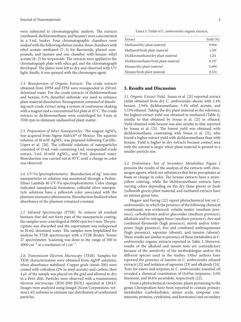
3.2. Preliminary Test of Secondary Metabolites. Figure 1presents the results of the analysis of the extracts with chro-mogen agents, which are substances that form precipitates orfoam or change in color. The hexane extracts have a straw-yellow coloring, while the dichloromethane extracts havevarying colors depending on the dry (lime green) or fresh(yellowish green) plant material, and methanol extracts havean intense green tone.
Hegazy and Farrag [22] report phytochemical test on C.ambrosioides, in which the presence of the following chemicalconstituents was evidenced: volatile matter (medium pres-ence), carbohydrates and/or glucosides (medium presence),alkaloids and/or nitrogen bases (medium presence), free andcombined flavonoids (high presence), sterols and/or triter-penes (high presence), free and combined anthraquinones(high presence), saponins (absent), and tannins (absent).These results are similar to presence of thesemetabolites inC.ambrosioides organic extracts reported in Table 2. However,results of the alkaloid and tannin tests are contradictorybecause of the sensitivity of the methodologies and/or thedifferent species used in the studies. Other authors havereported the presence of tannins in C. ambrosioides ethanolextracts [21] and isolation of saponins [23] and alkaloids [24].Tests for esters and terpenes in C. ambrosioides essential oilrevealed a chemical constitution of 14.6%𝛼-terpinene, 3.6%limonene, and 18.6% ascaridole, respectively [22].
From a phytochemical viewpoint, plants pertaining to thegenus Chenopodium have been reported to contain primarymetabolites (carbohydrates, amino acids, nonpolar con-stituents, proteins, cytokinins, and hormones) and secondary
4 Journal of Nanomaterials
Table 2: Preliminary analysis of secondary metabolites in C. ambrosioides organic extracts.
Extracts Alkaloids Terpenoids PC7 Tannins Flavonoids SaponinsHS1 − + − − −
HF2 − + − − −
DS3 − ++ − + −
DF4 − ++ − + −
MS5 − +++ +++ ++ ++ −
MF6 − +++ ++ +++ ++ −1Hexane-dry plant material. 2Hexane-fresh plant material. 3Dichloromethane-dry plant material. 4Dichloromethane-fresh material. 5Methanol-dry plantmaterial. 6Methanol-fresh plant material. 7Phenolic compounds.
(a)
1 2 3 4 5 6
(b)
1 2 3 4 5 6
(c)
1 2 3 4 5 6
(d)
1 2 3 4 5 6
(e)
1 2 3 4 5 6
(f)
Figure 1: Qualitative analysis of secondary metabolites in C. ambrosioides. ((a) and (b)) Organic extracts obtained, (c) alkaloids, (d)triterpenes, (e) phenolic compounds, and (f) tannins; 1: hexane-dry plant material, 2: hexane-fresh plant material, 3: dichloromethane-dryplant material, 4: dichloromethane-fresh plant material, 5: methanol-dry plant material, and 6: methanol-fresh plant material.
metabolites (flavonoids, saponins, terpenes, sterols, and alka-loids). Figure 1(c) illustrates the color changes that occurredin the evaluated extracts. There was no brown precipitate,an indicator of the presence of alkaloids, confirming thenegative results of the test for alkaloids in Table 2. Figure 1(d)shows a pink-purple coloring, which reveals the presenceof triterpenes in the extracts. The intense coloring in thematerials in methanol indicates an abundance of this type ofmetabolites (+++ in Table 2). Kokanova-Nedialkova et al. [6]report a vast wealth of monoterpenoids and sesquiterpenoidsin C. ambrosioides, including 𝛽-caryophyllene, 𝛾-curcumene,b-myrcene, cis-b-ocimene and trans-isomer, nerol, geraniol,citronellyl acetate, limonene, 𝛼-terpinene, 𝛼-terpinolen, andb-phellandrene. Figure 1(e) shows the color change to violet,indicating the presence of phenolic compounds, testingpositive for extracts in methanol and negative for the extractsin hexane and dichloromethane. Figure 1(f) presents thetypical blue coloring of extracts rich in tannins, which is
evident in the materials in methanol (particularly methanol-fresh material). The presence of flavonoids is evident in theextracts in methanol (Table 2). The presence of numerousflavonoids, such as flavone glycoside, kaempferol, kaempferoldiglycoside, and kaempferol triglycoside, has been reportedin C. ambrosioides[6]. A preliminary qualitative analysis of C.ambrosioides extracts obtained by Sousa et al. [21] revealedthe presence of phenolic compounds, tannins, flavonoids andsteroids for the ethanol extract, and flavonoids and steroidsfor the extract in dichloromethane.
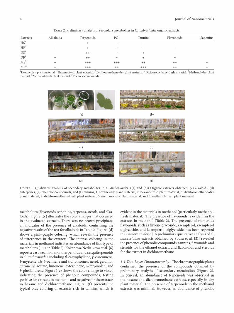
3.3.Thin-LayerChromatography. Thechromatographicplatesconfirmed the presence of the compounds obtained bypreliminary analysis of secondary metabolites (Figure 2).In general, an abundance of terpenoids was observed inthe hexane and dichloromethane extracts, especially in dryplant material. The presence of terpenoids in the methanolextracts was minimal. However, an abundance of phenolic
Journal of Nanomaterials 5
T H T D T M
P M TA D F MTA M
Figure 2: Thin-fine layer chromatography of organic C. ambrosioides extracts. T: terpenoids, P: phenolic compounds, TA: tannins, F:flavonoids, H: hexane, D: dichloromethane, and M: methanol. On each chromatography plate, left: dry plant material and right: fresh plantmaterial.
compounds was observed in the methanol extracts, partic-ularly flavonoids and tannins.
Nine classes of secondary metabolites were detectedin C. ambrosioides chloroform extracts (sterols and ter-penes, anthraquinones), ethanol (saponins), water (tannins),methanol (alkaloids), and acetone (flavonoids).The testswerepositive for terpenoids, sterols, saponins, tannins, alkaloids,flavonoids, phenols, and volatile oils [25].
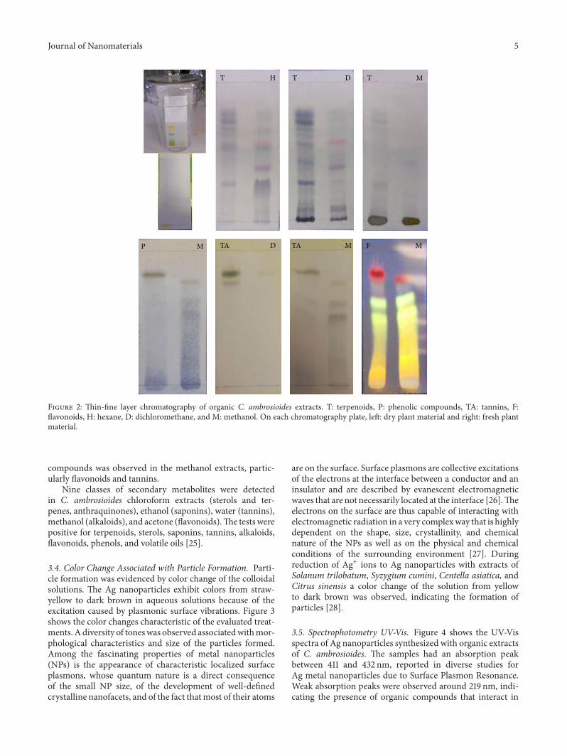
3.4. Color Change Associated with Particle Formation. Parti-cle formation was evidenced by color change of the colloidalsolutions. The Ag nanoparticles exhibit colors from straw-yellow to dark brown in aqueous solutions because of theexcitation caused by plasmonic surface vibrations. Figure 3shows the color changes characteristic of the evaluated treat-ments. A diversity of toneswas observed associatedwithmor-phological characteristics and size of the particles formed.Among the fascinating properties of metal nanoparticles(NPs) is the appearance of characteristic localized surfaceplasmons, whose quantum nature is a direct consequenceof the small NP size, of the development of well-definedcrystalline nanofacets, and of the fact thatmost of their atoms
are on the surface. Surface plasmons are collective excitationsof the electrons at the interface between a conductor and aninsulator and are described by evanescent electromagneticwaves that are not necessarily located at the interface [26].Theelectrons on the surface are thus capable of interacting withelectromagnetic radiation in a very complexway that is highlydependent on the shape, size, crystallinity, and chemicalnature of the NPs as well as on the physical and chemicalconditions of the surrounding environment [27]. Duringreduction of Ag+ ions to Ag nanoparticles with extracts ofSolanum trilobatum, Syzygium cumini, Centella asiatica, andCitrus sinensis a color change of the solution from yellowto dark brown was observed, indicating the formation ofparticles [28].
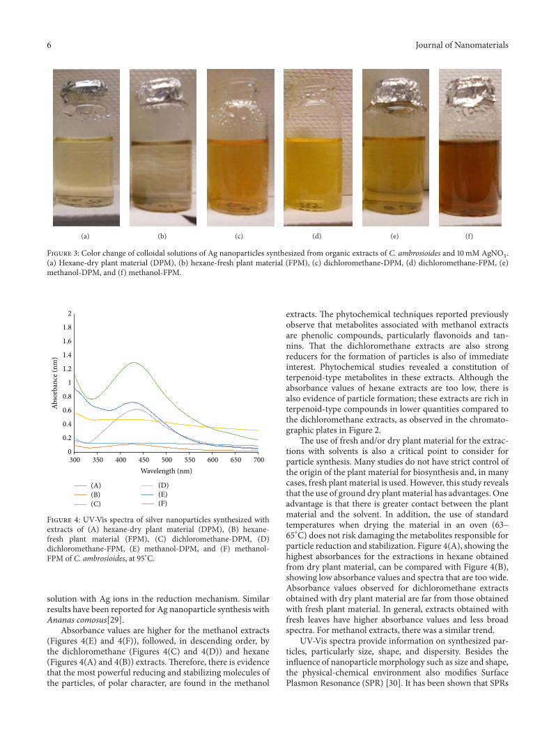
3.5. Spectrophotometry UV-Vis. Figure 4 shows the UV-Visspectra of Ag nanoparticles synthesized with organic extractsof C. ambrosioides. The samples had an absorption peakbetween 411 and 432 nm, reported in diverse studies forAg metal nanoparticles due to Surface Plasmon Resonance.Weak absorption peaks were observed around 219 nm, indi-cating the presence of organic compounds that interact in
6 Journal of Nanomaterials
(a) (b) (c) (d) (e) (f)
Figure 3: Color change of colloidal solutions of Ag nanoparticles synthesized from organic extracts of C. ambrosioides and 10mM AgNO3.
(a) Hexane-dry plant material (DPM), (b) hexane-fresh plant material (FPM), (c) dichloromethane-DPM, (d) dichloromethane-FPM, (e)methanol-DPM, and (f) methanol-FPM.
0
0.2
0.4
0.6
0.8
1
1.2
1.4
1.6
1.8
2
300 350 400 450 500 550 600 650 700
Abso
rban
ce (n
m)
Wavelength (nm)
(A)(B)(C)
(D)(E)(F)
Figure 4: UV-Vis spectra of silver nanoparticles synthesized withextracts of (A) hexane-dry plant material (DPM), (B) hexane-fresh plant material (FPM), (C) dichloromethane-DPM, (D)dichloromethane-FPM, (E) methanol-DPM, and (F) methanol-FPM of C. ambrosioides, at 95∘C.
solution with Ag ions in the reduction mechanism. Similarresults have been reported for Ag nanoparticle synthesis withAnanas comosus[29].
Absorbance values are higher for the methanol extracts(Figures 4(E) and 4(F)), followed, in descending order, bythe dichloromethane (Figures 4(C) and 4(D)) and hexane(Figures 4(A) and 4(B)) extracts. Therefore, there is evidencethat the most powerful reducing and stabilizing molecules ofthe particles, of polar character, are found in the methanol
extracts. The phytochemical techniques reported previouslyobserve that metabolites associated with methanol extractsare phenolic compounds, particularly flavonoids and tan-nins. That the dichloromethane extracts are also strongreducers for the formation of particles is also of immediateinterest. Phytochemical studies revealed a constitution ofterpenoid-type metabolites in these extracts. Although theabsorbance values of hexane extracts are too low, there isalso evidence of particle formation; these extracts are rich interpenoid-type compounds in lower quantities compared tothe dichloromethane extracts, as observed in the chromato-graphic plates in Figure 2.
The use of fresh and/or dry plant material for the extrac-tions with solvents is also a critical point to consider forparticle synthesis. Many studies do not have strict control ofthe origin of the plant material for biosynthesis and, in manycases, fresh plant material is used. However, this study revealsthat the use of ground dry plant material has advantages. Oneadvantage is that there is greater contact between the plantmaterial and the solvent. In addition, the use of standardtemperatures when drying the material in an oven (63–65∘C) does not risk damaging the metabolites responsible forparticle reduction and stabilization. Figure 4(A), showing thehighest absorbances for the extractions in hexane obtainedfrom dry plant material, can be compared with Figure 4(B),showing low absorbance values and spectra that are too wide.Absorbance values observed for dichloromethane extractsobtained with dry plant material are far from those obtainedwith fresh plant material. In general, extracts obtained withfresh leaves have higher absorbance values and less broadspectra. For methanol extracts, there was a similar trend.
UV-Vis spectra provide information on synthesized par-ticles, particularly size, shape, and dispersity. Besides theinfluence of nanoparticle morphology such as size and shape,the physical-chemical environment also modifies SurfacePlasmon Resonance (SPR) [30]. It has been shown that SPRs
Journal of Nanomaterials 7
shift if the dielectric properties of the surrounding media arechanged. In particular, the SPRs in a medium with 𝑛 > 1are red-shifted with respect to those in vacuum [26]. Metalnanoparticles with a smaller number of faces and moreacute vertices create resonances in a broader spectrum ofwavelengths. When a nanoparticle is truncated, the principalabsorption is displaced to blue, secondary resonances over-lap, and, therefore, the total width increases to half of themaximum. For dodecahedral particles, truncation exhibitsthe same change to blue, but width decreases, possiblybecause the secondary resonances no longer exist when thenumber of faces increases [26]. Figure 4(B) shows widespectra for the different concentrations of silver evaluated,suggesting that they are large polydisperse particles withtruncated shapes. Although broad spectra are not observedin spectra (E), Figure 4 shows that the shape of the spectrais not symmetrical; the left side is cut off, and, therefore,the information of the particles they provide is that they aremore polyhedral in shape and less truncated but polydisperse.Figures 4(C), 4(E), and 4(F) present spectra with symmetricalshapes and acute angles. For this reason, it would be expectedthat the synthesized particles possess little polydispersity andare small and polyhedral-shaped, desirable characteristics fornanoparticle synthesis.
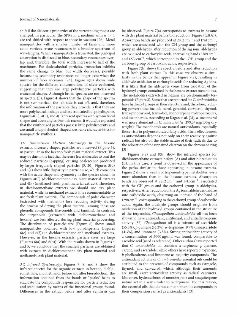
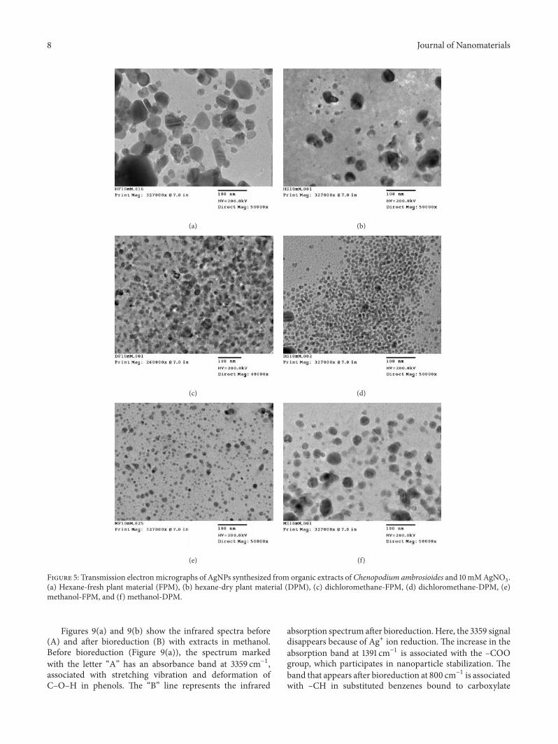
3.6. Transmission Electron Microscopy. In the hexaneextracts, diversely shaped particles are observed (Figure 5),in particular in the hexane-fresh plant material extract. Thismay be due to the fact that there are fewmolecules to coat thereduced particles (capping) causing coalescence producedby larger irregularly shaped particles [5, 20]. Figures 5(d)and 5(e) show little disparity in particle size, which coincideswith the acute shape and symmetry in the spectra shown inFigures 4(C) (dichloromethane-dry plant material extract)and 4(F) (methanol-fresh plant material extract). Therefore,in dichloromethane extracts we should use dry plantmaterial, while in methanol extracts it is recommendable touse fresh plant material. The compounds of polar character(extracted with methanol) lose reducing activity duringthe process of drying the plant material; among these arephenolic compounds (flavonoids and tannins). In contrast,the terpenoids (extracted with dichloromethane andhexane) are less affected during plant material processing.The distribution of particle size (Figure 6) shows smallnanoparticles obtained with low polydispersity (Figures6(c) and 6(f)) in dichloromethane and methanol extracts.However, in the hexane extracts, particle sizes are large(Figures 6(a) and 6(b)). With the results shown in Figures 4and 5, we conclude that the smallest particles are obtainedwith extracts in dichloromethane-dry plant material andmethanol-fresh plant material.
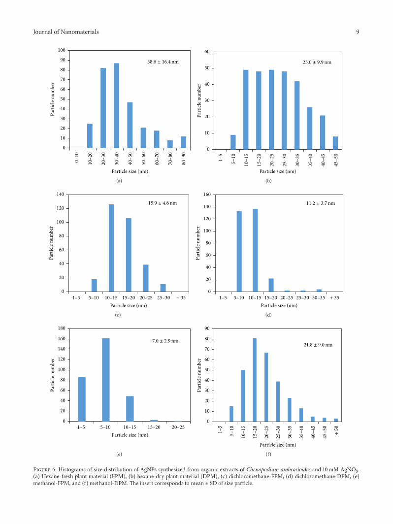
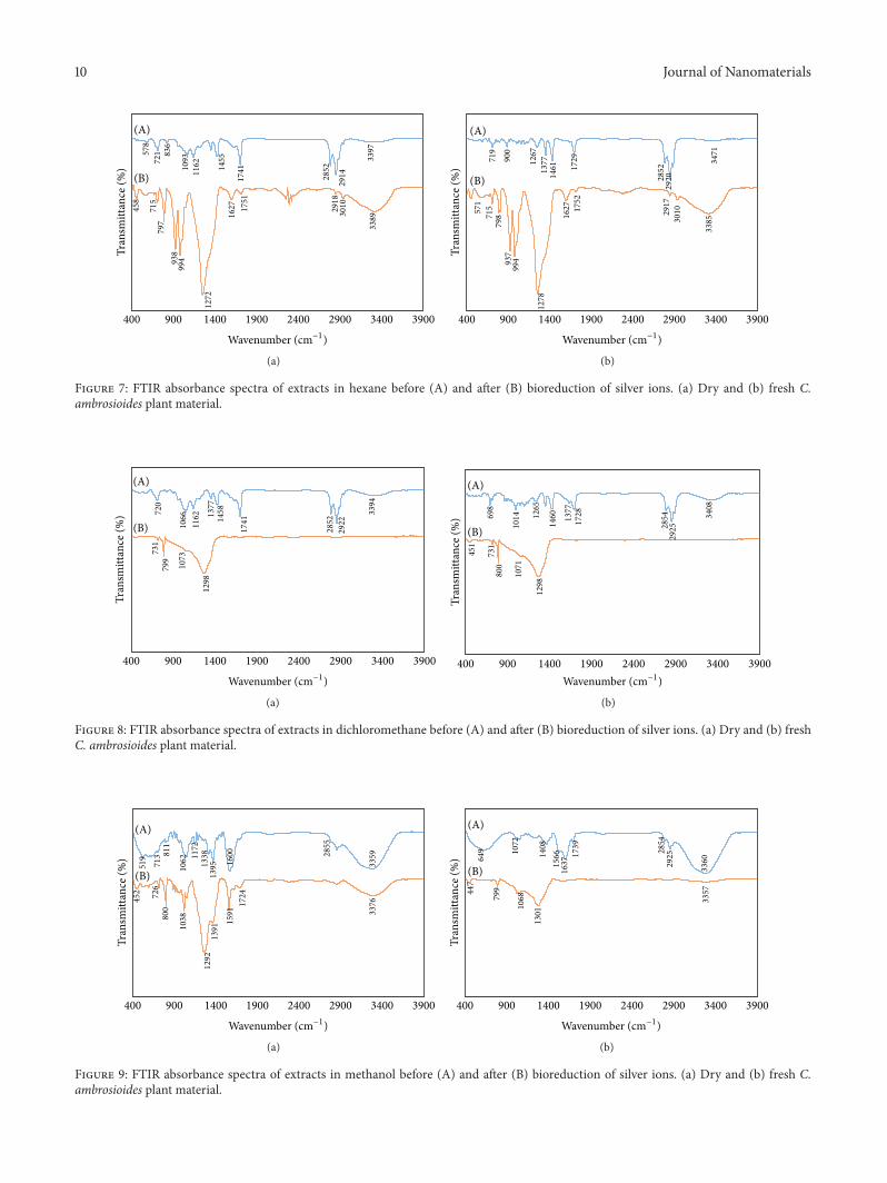
3.7. Infrared Spectroscopy. Figures 7, 8, and 9 show theinfrared spectra for the organic extracts in hexane, dichlo-romethane, andmethanol, before and after bioreduction.Theinformation obtained from the bands or “peaks” helps toelucidate the compounds responsible for particle reductionand stabilization by means of the functional groups found.Differences in the extract before and after synthesis can
be observed. Figure 7(a) corresponds to extracts in hexanewith dry plant material before bioreduction (Figure 7(a)(A)).Absorption bands are produced at 2852 cm−1 and 1741 cm−1,which are associated with the CH group and the carbonylgroup in aldehydes; after reduction of the Ag ions, aldehydesare oxidized to carboxylic acids, increasing bands 3389 cm−1and 1272 cm−1, which correspond to the –OH group and thecarbonyl group of carboxylic acids, respectively.
Figure 7(b) shows the spectra before and after reductionwith fresh plant extract. In this case, we observe a simi-larity in the bands that appear in Figure 7(a), resulting inaldehyde oxidation to carboxylic acids for reducing Ag ions.It is likely that the aldehydes come from oxidation of thehydroxyl groups contained in the hexane extract metabolites.The metabolites extracted in hexane are predominantly ter-penoids (Figure 2). Some that are reported forC. ambrosioideshave hydroxyl groups in their structure and, therefore, reduc-ing power; these include nerol, geraniol, carvacrol, thymol,trans-pinocarveol, trans-diol, monoterpene hydroperoxides,and tocopherols. According to Kagan et al. [31], 𝛼-tocopherolwas more abundant in C. ambrosioides (199.37mg/100 g dryweight). The tocopherols are natural antioxidants, especiallythose rich in polyunsaturated fatty acids. Their effectivenessas antioxidants depends not only on their reactivity againstradicals but also on the stable nature of their radicals due tothe relocation of the unpaired electron on the chromane ring[31].
Figures 8(a) and 8(b) show the infrared spectra fordichloromethane extracts before (A) and after bioreduction(B). In this case, a trend is observed in the appearance ofthe peaks similar to those appearing in hexane extracts.Figure 2 shows a wealth of terpenoid-type metabolites, evenmore abundant than in the hexane extracts. Absorptionbands are observed at 2852 cm−1 and 1741 cm−1, associatedwith the CH group and the carbonyl group in aldehydes,respectively. After reduction of the Ag ions, aldehydes oxidizeinto carboxylic acids, observing an increase of the band of1298 cm−1, corresponding to the carbonyl group of carboxylicacids. Again, the aldehyde groups should originate fromoxidation of the hydroxyl groups contained in the structureof the terpenoids. Chenopodium ambrosioides oil has beenshown to have antioxidant, antifungal, and antiaflatoxigenicactivity [32]. Chenopodium oil is a mixture of ascaridole(55.3%), p-cymene (16.2%), 𝛼-terpinene (9.7%), isoascaridole(4.3%), and limonene (3.8%). Strong antioxidant activity ofa concentration of 3000 𝜇g/mL was found, comparable toascorbic acid (used as reference).Other authors have reportedthat C. ambrosioides oil contains 𝛼-terpinene, p-cymene,carene, and ascaridole, while others have reported 𝛼-pinene,b-phellandrene, and limonene as majority compounds. Theantioxidant activity of C. ambrosioides essential oils could beattributed to the presence of compounds such as estragole,thymol, and carvacrol, which, although their amountsare small, exert antioxidant activity as radical capturers.Moreover, hydrocarbons of monoterpene and sesquiterpenenature act in a way similar to 𝛼-terpinene. For this reason,the essential oils that do not contain phenolic compounds intheir composition can act as antioxidants [33].
8 Journal of Nanomaterials
(a) (b)
(c) (d)
(e) (f)
Figure 5: Transmission electronmicrographs of AgNPs synthesized from organic extracts of Chenopodium ambrosioides and 10mMAgNO3.
(a) Hexane-fresh plant material (FPM), (b) hexane-dry plant material (DPM), (c) dichloromethane-FPM, (d) dichloromethane-DPM, (e)methanol-FPM, and (f) methanol-DPM.
Figures 9(a) and 9(b) show the infrared spectra before(A) and after bioreduction (B) with extracts in methanol.Before bioreduction (Figure 9(a)), the spectrum markedwith the letter “A” has an absorbance band at 3359 cm−1,associated with stretching vibration and deformation ofC–O–H in phenols. The “B” line represents the infrared
absorption spectrum after bioreduction.Here, the 3359 signaldisappears because of Ag+ ion reduction. The increase in theabsorption band at 1391 cm−1 is associated with the –COOgroup, which participates in nanoparticle stabilization. Theband that appears after bioreduction at 800 cm−1 is associatedwith –CH in substituted benzenes bound to carboxylate
Journal of Nanomaterials 9
Particle size (nm)
0
10
20
30
40
50
60
70
80
90
100Pa
rtic
le n
umbe
r
38.6 ± 16.4 nm
0–10
10–2
0
20–3
0
30–4
0
40–5
0
50–6
0
60–7
0
70–8
0
80–9
0(a)
Particle size (nm)
0
10
20
30
40
50
60
Part
icle
num
ber
25.0 ± 9.9 nm
1–5
5–10
10–1
5
15–2
0
20–2
5
25–3
0
30–3
5
35–4
0
40–4
5
45–5
0
(b)
1–5 5–10 10–15 15–20 20–25 25–30 + 35Particle size (nm)
0
20
40
60
80
100
120
140
Part
icle
num
ber
15.9 ± 4.6nm
(c)
1–5 5–10 10–15 15–20 20–25 25–30 30–35 + 35Particle size (nm)
0
20
40
60
80
100
120
140
160Pa
rtic
le n
umbe
r11.2 ± 3.7nm
(d)
1–5 5–10 10–15 15–20 20–25Particle size (nm)
0
20
40
60
80
100
120
140
160
180
Part
icle
num
ber
7.0 ± 2.9 nm
(e)
1–5
5–10
10–1
5
15–2
0
20–2
5
25–3
0
30–3
5
35–4
0
40–4
5
45–5
0
+ 50
Particle size (nm)
0
10
20
30
40
50
60
70
80
90
Part
icle
num
ber
21.8 ± 9.0 nm
(f)
Figure 6: Histograms of size distribution of AgNPs synthesized from organic extracts of Chenopodium ambrosioides and 10mM AgNO3.
(a) Hexane-fresh plant material (FPM), (b) hexane-dry plant material (DPM), (c) dichloromethane-FPM, (d) dichloromethane-DPM, (e)methanol-FPM, and (f) methanol-DPM.The insert corresponds to mean ± SD of size particle.
10 Journal of Nanomaterials
3397
291428
52
174114
55
116210
93836
72157
8
338930
102918
1751
1627
1272
99493
879
771
5
458
Tran
smitt
ance
(%)
(A)
(B)
900 1400 1900 2400 2900 3400 3900400
Wavenumber (cm−1)
(a)
Tran
smitt
ance
(%)
(A)
(B)
1729
146113
771267
3471
2920
900
719
2852
1278
1627 17
52
2917
3010
3385
571
715
798
937
994
900 1400 1900 2400 2900 3400 3900400
Wavenumber (cm−1)
(b)
Figure 7: FTIR absorbance spectra of extracts in hexane before (A) and after (B) bioreduction of silver ions. (a) Dry and (b) fresh C.ambrosioides plant material.
3394
2922
1741
285214
581377
1162
106672
0
1298
799
731
1073
Tran
smitt
ance
(%)
(A)
(B)
900 1400 1900 2400 2900 3400 3900400
Wavenumber (cm−1)
(a)
400 900 1400 1900 2400 2900 3400 3900
3408
2854
2925
17281377
1460
800
731
451
1014
1071
698
1265
1298
Tran
smitt
ance
(%)
(A)
(B)
Wavenumber (cm−1)
(b)
Figure 8: FTIR absorbance spectra of extracts in dichloromethane before (A) and after (B) bioreduction of silver ions. (a) Dry and (b) freshC. ambrosioides plant material.
335928
55
1600
139513
381172811
106271
3
1591
519
1391
800
1292
726
452
1038
337617
24
Tran
smitt
ance
(%)
(A)
(B)
900 1400 1900 2400 2900 3400 3900400
Wavenumber (cm−1)
(a)
1739 2854
1301
1072
649 1408
1566
1637 29
25
3360
447
799
1068 33
57
Tran
smitt
ance
(%)
(A)
(B)
900 1400 1900 2400 2900 3400 3900400
Wavenumber (cm−1)
(b)
Figure 9: FTIR absorbance spectra of extracts in methanol before (A) and after (B) bioreduction of silver ions. (a) Dry and (b) fresh C.ambrosioides plant material.
Journal of Nanomaterials 11
groups. Bioreduction studies with Chenopodium album havereported participation of carbonyl groups in the Ag+ ionreduction process and carboxylate ions as stabilization agentsin nanoparticles synthesized over time [34]. In contrast, someauthors report that –OH groups participate in the reductionprocess, oxidizing the –OH groups to carbonyl groups, andcarbonyl and carboxylate groups are involved in particlestabilization [34, 35].
Many authors name flavones, terpenoids, and polysac-charides as responsible for bioreduction. In our study withC. ambrosioides, the disappearance of the –OH group afterbioreduction makes it evident that this functional group isresponsible for reduction of silver ions [35]. Phytochemicalcharacterization of C. ambrosioides methanol found that theprincipal metabolites were phenolic compounds (flavonoidsand tannins) and a smaller proportion of terpenoids.
Barros et al. [10] evaluated bioactivity and characterizedhydrophilic and lipophilic compounds of C. ambrosioides.The bioactive properties include antioxidant and antitumoractivity and hepatotoxicity. The aqueous extracts are moreactive against free radicals, bleaching𝛽-carotene, and inhibit-ing TBARS (inhibition of lipid peroxidation by decreas-ing thiobarbituric acid reactive substances) than extractsin methanol. They also have higher reducing activity. C.ambrosioides essential oils from leaves also show antioxi-dant activity. Aqueous extracts of C. ambrosioides did notexhibit antitumor potential, but the extracts in methanol did.Organic acids, such as citric acid, have been reported topossess antioxidant activity [36].
Yoosaf et al. [37] found that a molecule should haveat least two hydroxyl groups in ortho- or para-position toproduce ion reduction. The dihydroxyl compounds of gallicacid oxidize to a quinone, and the nanoparticles formedstabilize with interaction of the carboxylic acid group.
Rodrıguez-Leon et al. [38] found that the Ag+ ionreducing agents were the organic molecules of epicatechinand epicatechin gallate, found by nuclearmagnetic resonancein extracts of R. hymenosepalus. The reducing mechanisminvolves abstraction of hydrogen because of the OH groupsin polyphenol molecules.
In the methanol extracts, the phenolic compoundswith hydroxyl groups include flavone glycosides, quercetin,kaempferol, and their glycosides and isorhamnetin, in par-ticular, flavone glycoside, which has two hydroxyl groupsin its structure. The terpenoids found in the extracts inhexane and dichloromethane that have two hydroxyl groupsinclude monoterpene hydroperoxides, apiole, 𝛼-terpineol,and related derivates, trans-diol, (−)-(1R, 2S, 3S, 4S)-1,2,3,4,-tetrahydroxy-p-menthane, avenasterol, and 𝛼-spinasterol.
Conventional phytochemical tests conducted on dryCitrus sinensis plant material produced positive resultsfor alkaloids, tannins, flavonoids, and steroids. C. sinen-sis and Syzygium cumini (positive for flavonoids) producehigher absorbance values in UV-Vis spectrophotometry thanSolanum trilobatum (positive for alkaloids, tannins, andsteroids) and Centella asiatica (positive for tannins andsteroids). This indicates that flavonoids are the metaboliteswith greater Ag+ ion reducing power and particle stabiliza-tion for these cases [28, 37].
4. Conclusions
This is the first study to elucidate the organic compounds ofChenopodium ambrosioides that participate in bioreductionof Ag nanoparticles. The strategy was based on the studyof biosynthesis in extracts of different polarities (high withmethanol, mediumwith dichloromethane, and low with hex-ane).The extractedmetabolites were assessed and the organiccompounds were analyzed before and after bioreduction(FTIR technique). The extracts in methanol, rich in phenoliccompounds (flavonoids and tannins), have greater reduc-ing and stabilizing activity for Ag nanoparticle synthesis.Nevertheless, the terpenoids present in the dichloromethaneand hexane extracts also have reducing activity, though inlower proportion. Large part of the reducing activity of thesecondary metabolites in C. ambrosioides is favored by thecompounds that have antioxidant capacity, including the phe-nolic compounds (flavone glycoside and isorhamnetin) andterpenoids (trans-diol,𝛼-terpineol, and derivatives,monoter-pene hydroperoxides and apiole). Dry plant material is moreefficient in the production of Ag nanoparticles when workingwith hexane and dichloromethane extracts. However, whenworking with methanol, fresh plant material should be used.
Competing Interests
The authors declare that there is no conflict of interestsregarding the publication of this manuscript.
References
[1] S. S. Shankar, A. Rai, A. Ahmad, andM. Sastry, “Rapid synthesisof Au, Ag, and bimetallic Au core-Ag shell nanoparticles usingNeem (Azadirachta indica) leaf broth,” Journal of Colloid andInterface Science, vol. 275, no. 2, pp. 496–502, 2004.
[2] N. Yadav, N. Vasudeva, S. Singh, and S. K. Sharma, “Medicinalproperties of genus Chenopodium Linn.,” Indian Journal ofNatural Products and Resources, vol. 6, no. 2, pp. 131–134, 2007.
[3] R. Villase and G. Espinosa, Catalogo de malezas de Mexico,Consejo Nacional Consultivo Fitosanitario, Fondo de CulturaEconomica, Mexico City, Mexico, 1998.
[4] N. Ramezani, Z. Ehsanfar, F. Shamsa et al., “Screening ofmedicinal plant methanol extracts for the synthesis of goldnanoparticles by their reducing potential,” Zeitschrift fur Natur-forschung B, vol. 63, no. 7, pp. 903–908, 2008.
[5] J. Y. Song and B. S. Kim, “Biological synthesis of bimetallicAu/Ag nanoparticles using Persimmon (Diopyros kaki) leafextract,” Korean Journal of Chemical Engineering, vol. 25, no. 4,pp. 808–811, 2008.
[6] Z. Kokanova-Nedialkova, P. T. Nedialkov, and S. D. Nikolov,“The genus Chenopodium: phytochemistry, ethnopharmacol-ogy and pharmacology,” Pharmacognosy Reviews, vol. 3, no. 6,pp. 280–306, 2009.
[7] M. A. Johnson and R. Croteau, “Biosynthesis of ascari-dole: iodide peroxidase-catalyzed synthesis of a monoterpeneendoperoxide in soluble extracts of Chenopodium ambrosioidesfruit,”Archives of Biochemistry and Biophysics, vol. 235, no. 1, pp.254–266, 1984.
12 Journal of Nanomaterials
[8] A. A. Ahmed, “Highly oxygenated monoterpenes from Cheno-podium ambrosioides,” Journal of Natural Products, vol. 63, no.7, pp. 989–991, 2000.
[9] F. Kiuchi, Y. Itano, N. Uchiyama et al., “Monoterpene hydroper-oxides with trypanocidal activity from Chenopodium ambro-sioides,” Journal of Natural Products, vol. 65, no. 4, pp. 509–512,2002.
[10] L. Barros, E. Pereira, R. C. Calhelha et al., “Bioactivity andchemical characterization in hydrophilic and lipophilic com-pounds of Chenopodium ambrosioides L.,” Journal of FunctionalFoods, vol. 5, no. 4, pp. 1732–1740, 2013.
[11] E. Gonzalez de Mejıa, Y. S. Song, C. I. Heck, and M. Ramırez-Mares, “Yerba mate tea (Ilex paraguariensis): phenolics, antiox-idant capacity and in vitro inhibition of colon cancer cellproliferation,” Journal of Functional Foods, vol. 2, no. 1, pp. 23–34, 2010.
[12] O. R. Pereira, R. I. R. Macıas, M. J. Perez, J. J. G. Marın, andS. M. Cardoso, “Protective effects of phenolic constituents fromCytisus multiflorus, Lamium album L. and Thymus citriodoruson liver cells,” Journal of Functional Foods, vol. 5, no. 3, pp. 1170–1179, 2013.
[13] J. Whelan, “The health implications of changing linoleic acidintakes,” Prostaglandins, Leukotrienes and Essential Fatty Acids,vol. 79, no. 3–5, pp. 165–167, 2008.
[14] K. Quester, M. Avalos-Borja, and E. Castro-Longoria, “Biosyn-thesis andmicroscopic study ofmetallic nanoparticles,”Micron,vol. 54-55, pp. 1–27, 2013.
[15] M. S. Akhtar, J. Panwar, and Y.-S. Yun, “Biogenic synthesisof metallic nanoparticles by plant extracts,” ACS SustainableChemistry & Engineering, vol. 1, no. 6, pp. 591–602, 2013.
[16] M. S. Akhtar, M. K. Swamy, A. Umar, and A. A. Al Sahli,“Biosynthesis and characterization of silver nanoparticles frommethanol leaf extract of cassia didymobotyra and assessmentof their antioxidant and antibacterial activities,” Journal ofNanoscience and Nanotechnology, vol. 15, no. 12, pp. 9818–9823,2015.
[17] M. R. Chitsazi, H. Korbekandi, G. Asghari, R. Bahri Najafi, A.Badii, and S. Iravani, “Synthesis of silver nanoparticles usingmethanol and dichlorometane extracts of Pulicaria gnaphalodes(Vent.) Boiss,”Artificial Cells, Nanomedicine, and Biotechnology,vol. 44, no. 1, pp. 328–333, 2014.
[18] F. Azizinezhad, Z. Z. Nasrollahi, and S. Sadrnezhaad, “Synthesisof the silver nanoparticles with the using of camomile plant,”European Journal of Experimental Biology, vol. 4, no. 2, pp. 124–127, 2014.
[19] A. Harborne, Phytochemical Methods: A Guide to Modern Tech-niques of Plant Analysis, Springer, Amsterdam, Netherlands,1984.
[20] L. M. Carrillo-Lopez, H. A. Zavaleta-Mancera, A. Vilchis-Nestor et al., “Biosynthesis of silver nanoparticles usingChenopodium ambrosioides,” Journal of Nanomaterials, vol.2014, Article ID 951746, 9 pages, 2014.
[21] Z. L. Sousa, F. F. de Oliveira, A. O. da Conceicao et al., “Biologi-cal activities of extracts from Chenopodium ambrosioides Lineuand Kielmeyera neglecta Saddi,” Annals of Clinical Microbiologyand Antimicrobials, vol. 11, pp. 20–27, 2012.
[22] A. Hegazy and H. Farrag, “Allelopathic potential of Chenopo-diumambrosioides on germination and seedling growth of some
cultivated and weed plants,” Global Journal of Biotechnology &Biochemistry, vol. 2, no. 1, pp. 1–9, 2007.
[23] G. Gupta and M. Behari, “Chemical investigation ofChenopodium ambrosioides,” Journal of Indian ChemicalSociety, vol. 49, no. 3, pp. 317–319, 1972.
[24] A. Hasseb, B. Singh, A. Khan, and S. Saxena, “Evaluationof nematicidal property in certain alkaloid bearing plants,”Geobios, vol. 5, pp. 116–118, 1978.
[25] S. Okhale, H. Egharevba, E. Ona, and O. Kunle, “Phytochem-icaland proximate analyses and thin layer chromatographyfingerprinting of the aerial part of Chenopodium ambrosioidesLinn. (Chenopodiaceae),” Journal of Medicinal Plants Research,vol. 6, no. 12, pp. 2289–2294, 2012.
[26] C.Noguez, “Surface plasmons onmetal nanoparticles: the influ-ence of shape and physical environment,” Journal of PhysicalChemistry C, vol. 111, no. 10, pp. 3806–3819, 2007.
[27] D. Cruz, M. Rodrıguez, J. Lopez, V. Herrera, A. Orive, and A.Creus, “Metallic nanoparticles and surface plasmons: a deeprelationship,” Avances en Ciencias e Ingenierıa, vol. 3, no. 2, pp.67–78, 2012.
[28] P. Logeswari, S. Silambarasan, and J. Abraham, “Ecofriendlysynthesis of silver nanoparticles from commercially availableplant powders and their antibacterial properties,” Scientia Iran-ica, vol. 20, no. 3, pp. 1049–1054, 2013.
[29] N. Ahmad and S. Sharma, “Green synthesis of silver nanopar-ticles using extracts of Ananas comosus,” Green and SustainableChemistry, vol. 02, no. 04, pp. 141–147, 2012.
[30] C. Novo, A. M. Funston, I. Pastoriza-Santos, L. M. Liz-Marzan,and P. Mulvaney, “Influence of the medium refractive indexon the optical properties of single gold triangular prisms on asubstrate,” The Journal of Physical Chemistry C, vol. 112, no. 1,pp. 3–7, 2008.
[31] V. E. Kagan, A. I. Kuzmenko, A. A. Shvedova et al., “Directevidence for recycling of myeloperoxidase-catalyzed phenoxylradicals of a vitamin E homologue, 2,2,5,7,8-pentamethyl-6-hydroxy chromane, by ascorbate/dihydrolipoate in living HL-60 cells,” Biochimica et Biophysica Acta (BBA)—General Sub-jects, vol. 1620, no. 1–3, pp. 72–84, 2003.
[32] R. Kumar, A. K. Mishra, N. K. Dubey, and Y. B. Tripathi,“Evaluation of Chenopodium ambrosioides oil as a potentialsource of antifungal, antiaflatoxigenic and antioxidant activity,”International Journal of Food Microbiology, vol. 115, no. 2, pp.159–164, 2007.
[33] B. Jaramillo, E. Duarte, andW.Delgado, “Bioactividad del aceiteesencial de Chenopodium ambrosioides colombiano,” RevistaCubana de Plantas Medicinales, vol. 17, no. 1, pp. 54–64, 2012.
[34] A. Dhar Dwivedi and K. Gopal, “Biosynthesis of silver and goldnanoparticles using Chenopodium album leaf extract,” Colloidsand Surfaces A: Physicochemical and Engineering Aspects, vol.369, no. 1–3, pp. 27–33, 2010.
[35] J. Huang, Q. Li, D. Sun et al., “Biosynthesis of silver and goldnanoparticles by novel sundried Cinnamomum camphora leaf,”Nanotechnology, vol. 18, no. 10, Article ID 105104, 2007.
[36] A. R. Hras, M. Hadolin, Z. Knez, and D. Bauman, “Comparisonof antioxidative and synergistic effects of rosemary extract with𝛼-tocopherol, ascorbyl palmitate and citric acid in sunfloweroil,” Food Chemistry, vol. 71, no. 2, pp. 229–233, 2000.

Journal of Nanomaterials 13
[37] K. Yoosaf, B. I. Ipe, C. H. Suresh, and K. G. Thomas, “Insitu synthesis of metal nanoparticles and selective naked-eyedetection of lead ions from aqueous media,” The Journal ofPhysical Chemistry C, vol. 111, no. 34, pp. 12839–12847, 2007.
[38] E. Rodrıguez-Leon, R. Iniguez-Palomares, R. E. Navarroet al., “Synthesis of silver nanoparticles using reducingagents obtained from natural sources (Rumex hymenosepalusextracts),” Nanoscale Research Letters, vol. 8, no. 1, pp. 1–9, 2013.

Submit your manuscripts athttp://www.hindawi.com
ScientificaHindawi Publishing Corporationhttp://www.hindawi.com Volume 2014
CorrosionInternational Journal of
Hindawi Publishing Corporationhttp://www.hindawi.com Volume 2014
Polymer ScienceInternational Journal of
Hindawi Publishing Corporationhttp://www.hindawi.com Volume 2014
Hindawi Publishing Corporationhttp://www.hindawi.com Volume 2014
CeramicsJournal of
Hindawi Publishing Corporationhttp://www.hindawi.com Volume 2014
CompositesJournal of
NanoparticlesJournal of
Hindawi Publishing Corporationhttp://www.hindawi.com Volume 2014
Hindawi Publishing Corporationhttp://www.hindawi.com Volume 2014
International Journal of
Biomaterials
Hindawi Publishing Corporationhttp://www.hindawi.com Volume 2014
NanoscienceJournal of
TextilesHindawi Publishing Corporation http://www.hindawi.com Volume 2014
Journal of
NanotechnologyHindawi Publishing Corporationhttp://www.hindawi.com Volume 2014
Journal of
CrystallographyJournal of
Hindawi Publishing Corporationhttp://www.hindawi.com Volume 2014
The Scientific World JournalHindawi Publishing Corporation http://www.hindawi.com Volume 2014
Hindawi Publishing Corporationhttp://www.hindawi.com Volume 2014
CoatingsJournal of
Advances in
Materials Science and EngineeringHindawi Publishing Corporationhttp://www.hindawi.com Volume 2014
Smart Materials Research
Hindawi Publishing Corporationhttp://www.hindawi.com Volume 2014
Hindawi Publishing Corporationhttp://www.hindawi.com Volume 2014
MetallurgyJournal of
Hindawi Publishing Corporationhttp://www.hindawi.com Volume 2014
BioMed Research International
MaterialsJournal of
Hindawi Publishing Corporationhttp://www.hindawi.com Volume 2014
Nano
materials
Hindawi Publishing Corporationhttp://www.hindawi.com Volume 2014
Journal ofNanomaterials
Recommended