Turkish Journal of Physiotherapy and Rehabilitation; 32(3)
ISSN 2651-4451 | e-ISSN 2651-446X
www.turkjphysiotherrehabil.org 3888
REPRESENTATION OF YOGA POSTURES BASED ON JOINT ANGLE
VALUES ALONG WITH RANGE OF MOTION LIMITATIONS OF JOINTS TO
CLASSIFY TIME SERIES YOGA ASANAS
Ponmozhi Chezhiyan1, Anisha M2, N. Vigneshwari3 1Assistant Professor, Department of Computer Applications, Kalasalingam Academy of Research and
Education, [email protected] 2Assistant Professor, Department of Biomedical Engineering, Kalasalingam Academy of Research and
Education, [email protected] 3Assistant Professor, Department of Biomedical Engineering, Kalasalingam Academy of Research and
Education.
ABSTRACT
Yoga has been considered as a complementary therapy as its practice will help in aligning, strengthening the
structure of the body, also used to maintain balance of human body. It plays important role in reducing Lower
Back Pain (LBP), and used as a mechanism to enhance the muscle balance that ensures spinal flexibility. Yoga
asanas include postures like sitting, standing, and simple movements like backward and forward bend, twists.
Some yoga asana include supine position. This paper proposes a method to classify yoga asana postures based
on joint angles. Not every joint is involved in all asanas. Set of joints that contributes a lot for a particular asana
and their range of motion has studied in order to classify asanas based on the joints involved. Applicability
using BVH and softmax classification has been discussed.
I. INTRODUCTION
Gravitation is the Universal mechanism which affects all object both living as well as non-living. Motion or
movement of any object is influenced by the biological system of the object and the environment. The branch of
study which gives insights on the changes in the molecular level to the system level of an object by mechanics
while moving is called Biomechanics [10]. Proper body movement can be done only when there is a coordinated
interaction of bones, muscles and joints in the body [11]. Thus, it becomes essential to understand and represent
them in the proper methodology so that injury can be prevented, abnormality in human body structure can be
corrected, it can be used in rehabilitation and sometime to heal the pain.
For instance, older individuals who are having less hip flexors and reduced plantar flexors will have gait features
such as walking slowly with shorter step length [26]. We use these gait features to identify whether muscle strength
is reduced, range of motion of joints, instability in postures [27].
Yoga is suggested as a means to strengthen the bones, to improve proper balance of the structure of body, align the
joints properly and hence to maintain and improve stability of the body during activities. Too be fit, human body
has to work in all the three planes namely sagittal plane, frontal (coronal) plane and transverse plane. The primary
aim of yoga asanas is to make body movements in all the three planes. Most of the yoga asanas have postures like
standing, sitting, bending forward and backward, hand balances, twists etc. In every posture, the individual joints
and segments may be rotated, or shifted to particular angle value.
Yoga also improves the gait functions [28], joint flexibility [29], range of Motion [30] and most importantly the
isokinetic and isometric muscle strength. Some of the yoga asanas have been used as an alternative to medication
for issues like lower-back pain [1]. Flexibility in hip movements, spinal cord is important in reduction of lowering
back pain [2],[3], some of the yoga asana ensure this flexibility.
Describing motion patterns in terms of human body’s biomechanics especially in terms of joint angles will be useful
in analysing gait and daily living activities of human beings. Describing yoga movements in terms of joint angles
Turkish Journal of Physiotherapy and Rehabilitation; 32(3)
ISSN 2651-4451 | e-ISSN 2651-446X
www.turkjphysiotherrehabil.org 3889
would help to understand how every yoga posture has to be completed, so that self-assisted yoga practice can be
done as effective as if practiced with yoga teachers. Also, to evaluate the benefits of yoga for various ailments,
identifying the biomechanical motion patterns of every yoga posture [31] has to be known.
II. BACKGROUND
2.1 Orientation & direction of joint movements
Human body parts and the movement joints are always described relative to the anatomical position. Anatomical
position is standing upright; keeping arms on the sides of the body and palms has to face forward. Anatomical
position is also termed as anterior and toward or back of the body is called as Posterior direction.
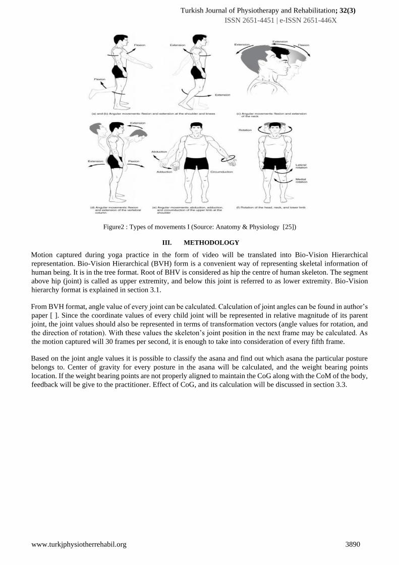
2.2 Body movement types
Biomechanics studies the forces acting on the human body and the reactive forces generated by the body, the
deciding factors of the movement of every human body parts, movement type and the degree of movement of any
joint depends on the structural type of the joint. Every joint in human body is capable of performing four possible
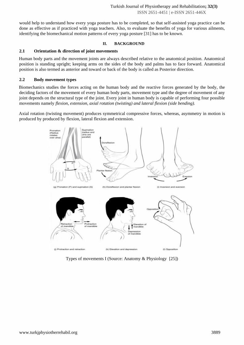
movements namely flexion, extension, axial rotation (twisting) and lateral flexion (side bending).
Axial rotation (twisting movement) produces symmetrical compressive forces, whereas, asymmetry in motion is
produced by produced by flexion, lateral flexion and extension.
Types of movements I (Source: Anatomy & Physiology [25])
Turkish Journal of Physiotherapy and Rehabilitation; 32(3)
ISSN 2651-4451 | e-ISSN 2651-446X
www.turkjphysiotherrehabil.org 3890
Figure2 : Types of movements I (Source: Anatomy & Physiology [25])
III. METHODOLOGY
Motion captured during yoga practice in the form of video will be translated into Bio-Vision Hierarchical
representation. Bio-Vision Hierarchical (BVH) form is a convenient way of representing skeletal information of
human being. It is in the tree format. Root of BHV is considered as hip the centre of human skeleton. The segment
above hip (joint) is called as upper extremity, and below this joint is referred to as lower extremity. Bio-Vision
hierarchy format is explained in section 3.1.
From BVH format, angle value of every joint can be calculated. Calculation of joint angles can be found in author’s
paper [ ]. Since the coordinate values of every child joint will be represented in relative magnitude of its parent
joint, the joint values should also be represented in terms of transformation vectors (angle values for rotation, and
the direction of rotation). With these values the skeleton’s joint position in the next frame may be calculated. As
the motion captured will 30 frames per second, it is enough to take into consideration of every fifth frame.
Based on the joint angle values it is possible to classify the asana and find out which asana the particular posture
belongs to. Center of gravity for every posture in the asana will be calculated, and the weight bearing points
location. If the weight bearing points are not properly aligned to maintain the CoG along with the CoM of the body,
feedback will be give to the practitioner. Effect of CoG, and its calculation will be discussed in section 3.3.

Turkish Journal of Physiotherapy and Rehabilitation; 32(3)
ISSN 2651-4451 | e-ISSN 2651-446X
www.turkjphysiotherrehabil.org 3891
IV. PROPOSED SYSTEM DESIGN
Figure 3: System design
1. Steps of the asanas are described based on angle variations from the starting position
2. We classify the asanas into 5 types based on its base support. For this study we have taken only 5 asanas each
belongs to one group.
3. From the video formart_to_bhv will generate BHV file for the corresponding image. The x,y coordinate values
of the joints can then be extracted , joint angles are calculated (only 20 joint angles as specified in the bhv
format has been used for calculation). This will create the data to be classified.
4. Calculations for variations from the expected positions will be detected and intimated.
4.1 General Yoga description
Every yoga asana will start from a particular posture and move to various postures with variation in the positions
of segments or joints and will be ended with a particular posture. The time elapsed between every posture will also
be in a particular range.
Starting positions specifies the body part which is (are) on the ground. Through those points the force generated
because of body weight are transmitted down to the earth. Most of the time, these parts will be legs or pelvis. If we
use our hands for transmitting weights then we can say that as arm support. Starting positions of any asana will be
from any one of the following:
S.No Starting Position Weight-bearing Part
1 Standing Supported on legs
2 Seated Supported on base of pelvis
3 Kneeling Supported on knees, shins and tops of feet
4 Supine Back of the body as a support
5 Prone Front surface of the body as a support
Table 1: postures and weight bearing parts of human body
Find the posture variation
and give feed back
Video of Asana
Representation in BHV Format
Separate Joints X and Y coordinates
Create the parameters for classification
Pass it to Classi_Yoga, find the asana
Turkish Journal of Physiotherapy and Rehabilitation; 32(3)
ISSN 2651-4451 | e-ISSN 2651-446X
www.turkjphysiotherrehabil.org 3892
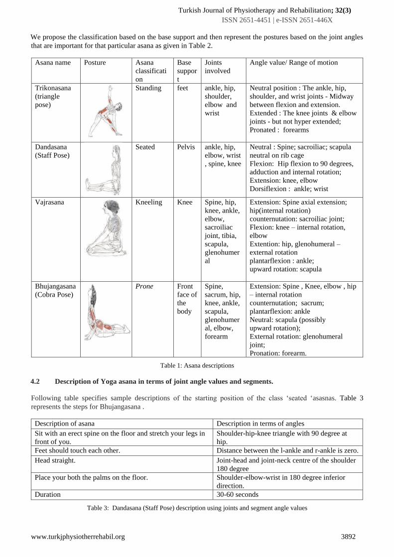
We propose the classification based on the base support and then represent the postures based on the joint angles
that are important for that particular asana as given in Table 2.
Asana name Posture Asana
classificati
on
Base
suppor
t
Joints
involved
Angle value/ Range of motion
Trikonasana
(triangle
pose)
Standing feet ankle, hip,
shoulder,
elbow and
wrist
Neutral position : The ankle, hip,
shoulder, and wrist joints - Midway
between flexion and extension.
Extended : The knee joints & elbow
joints - but not hyper extended;
Pronated : forearms
Dandasana
(Staff Pose)
Seated Pelvis ankle, hip,
elbow, wrist
, spine, knee
Neutral : Spine; sacroiliac; scapula
neutral on rib cage
Flexion: Hip flexion to 90 degrees,
adduction and internal rotation;
Extension: knee, elbow
Dorsiflexion : ankle; wrist
Vajrasana
Kneeling Knee Spine, hip,
knee, ankle,
elbow,
sacroiliac
joint, tibia,
scapula,
glenohumer
al
Extension: Spine axial extension;
hip(internal rotation)
counternutation: sacroiliac joint;
Flexion: knee – internal rotation,
elbow
Extention: hip, glenohumeral –
external rotation
plantarflexion : ankle;
upward rotation: scapula
Bhujangasana
(Cobra Pose)
Prone Front
face of
the
body
Spine,
sacrum, hip,
knee, ankle,
scapula,
glenohumer
al, elbow,
forearm
Extension: Spine , Knee, elbow , hip
– internal rotation
counternutation; sacrum;
plantarflexion: ankle
Neutral: scapula (possibly
upward rotation);
External rotation: glenohumeral
joint;
Pronation: forearm.
Table 1: Asana descriptions
4.2 Description of Yoga asana in terms of joint angle values and segments.
Following table specifies sample descriptions of the starting position of the class ‘seated ‘asasnas. Table 3
represents the steps for Bhujangasana .
Description of asana Description in terms of angles
Sit with an erect spine on the floor and stretch your legs in
front of you.
Shoulder-hip-knee triangle with 90 degree at
hip.
Feet should touch each other. Distance between the l-ankle and r-ankle is zero.
Head straight. Joint-head and joint-neck centre of the shoulder
180 degree
Place your both the palms on the floor. Shoulder-elbow-wrist in 180 degree inferior
direction.
Duration 30-60 seconds
Table 3: Dandasana (Staff Pose) description using joints and segment angle values

Turkish Journal of Physiotherapy and Rehabilitation; 32(3)
ISSN 2651-4451 | e-ISSN 2651-446X
www.turkjphysiotherrehabil.org 3893
Bhujangasana (Cobra pose) Steps
Description of asana Description in terms of angles
Step 1:
Lie prone on the ground Shoulder-hip-knee 180 degree.
forehead touching the floor Head neck straight line.
legs together Distance between the l-ankle and r-ankle is zero.
hands by the side of thighs l-shoulder:l-arm:lwrist 180 degree
r-shoulder:r-arm:rwrist 180 degree
Step 2:
Fold the hands at elbows and place the palms by the side of
the shoulders. Thumbs under armpit. Tip of the fingers not
crossing the shoulder line.
l-shoulder:l-elbow:lwrist acute angle;
lwrist(x) = lshoulder(x);
Step 3:
Slowly raise the head, neck and shoulders. Shoulders should
be shrugged backwards.
In distal direction; torso- shoulders-neck acute
angle in counter-clock direction;
Step 4:
Raise the trunk up to the navel region. Raise the chin as high
as possible.
Hip-shoulder-knee > 90 degree.Shoulder-centre-
neck-head acute angle.
Table 4: Bhujangasana (Cobra pose) description using joints and segment angle values

Turkish Journal of Physiotherapy and Rehabilitation; 32(3)
ISSN 2651-4451 | e-ISSN 2651-446X
www.turkjphysiotherrehabil.org 3894
4.3 Classification of yoga asanas based on angle
Figure 4: Asana classification
Shoulder – hip-knee angle
=180 degree
=90 degree
Standing, Prone, Spine, kneeling
Sitting
Hip-knee-foot
=180 degree <=90 degree
Standing, prone, Spine,
kneelin
g
Y value of toe & y value of
shoulders
Both are
equal
Both are not
equal
Spine Prone,
standing
Y value of foot & y value
of shoulders
Both are equal Both are not equal
prone standing

Turkish Journal of Physiotherapy and Rehabilitation; 32(3)
ISSN 2651-4451 | e-ISSN 2651-446X
www.turkjphysiotherrehabil.org 3895
4.4 Representation of postures
With the above said representation, We will represent every posture based on the joint angle values and segmental
values.
Difficulties in pose estimation are due to the factors like
1. variability in human physique
2. complexity of human skeletal structure,
3. high dimensionality of the pose
3D postures can be estimated by joint or angular values among skeletal image. The bio-mechanics of joints angular
values and range of motion (ranges depending on other group of joints involved) can be used as constraints. For
example, knee joints cannot be rotated beyond 180 degrees, where as neck can rotate 360 degrees, The constraint
mobility of joints reduces the information to be stored for a posture.
Because of the bio-mechanics of joints, the search space may also be limited. The search will begin from the root
joint. In yoga, the asanas are grouped based on their initial or starting postures. Every asana will move from its
initial to a final posture with time varying postures for some period of time.
We can represent knowledge about human body such as dependencies and relations, composition processes of
joints in the pose, including kinematics, symmetry, motor coordination in BHV format itself.
4.4.1 Capturing & representing Human postures
All the motion capturing devices collects values of important or key joints of the objects being tracked. Values of
these joints are recorded for a period of time, and they are translated into 3-Dimensional digital representation.Key
joints selected will represent the orientation of the objects too. The two kinds of representation that commonly used
by many motion capturing companies are ASCII and binary.
Decoding and understanding the information is easy in the case of ASCII format. In this paper, we use the BioVision
Hierarchy format, which is a ASCII based text format.
Keywords that will be used in this format to represent the objects, in this case the human body, and to describe the
manipulations to be done on the key joints to actualize motion are:
• skeleton – the whole human body
• bone – It is the smallest entity or segment used to represent a skeleton.To produce animation or motion,
individual translation and the orientation changes will be applied to these entities.Bones are associated with
mesh of vertices, which represents a particular part in the skeleton in hierarchical to produce the whole
structure.
• Degrees of freedom – This part is used to specify the transformation details of the skeletal joints such as
orientation change (rotation), positional change(translation) and the scale change during motion. In BVH
format, these details are given as channels. Executing the channel data will produce the required posture
change.
• Frame – Frame corresponds to one posture. Many number of frames may be needed for an animation, Each
frame will have the channel details of every bone to be implemented to produce a posture.
4.4.2 Human joints hierarchy (in BVH format)
We are representing skeleton having joint_hips as center as that of given in [12]. We have selected BVH format for
representing asana postures because aBiovision Hierarchy file format is used to describe relationship among joints
human body and it considers hips as centre.
Turkish Journal of Physiotherapy and Rehabilitation; 32(3)
ISSN 2651-4451 | e-ISSN 2651-446X
www.turkjphysiotherrehabil.org 3896
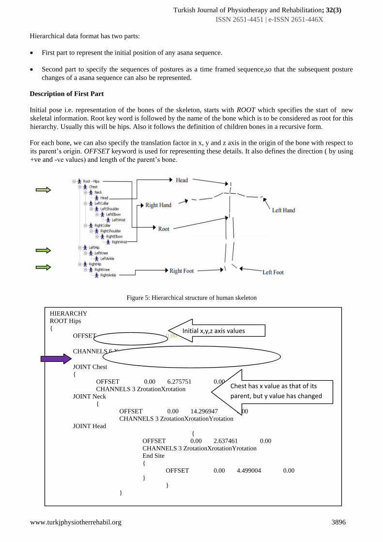
Hierarchical data format has two parts:
• First part to represent the initial position of any asana sequence.
• Second part to specify the sequences of postures as a time framed sequence,so that the subsequent posture
changes of a asana sequence can also be represented.
Description of First Part
Initial pose i.e. representation of the bones of the skeleton, starts with ROOT which specifies the start of new
skeletal information. Root key word is followed by the name of the bone which is to be considered as root for this
hierarchy. Usually this will be hips. Also it follows the definition of children bones in a recursive form.
For each bone, we can also specify the translation factor in x, y and z axis in the origin of the bone with respect to
its parent’s origin. OFFSET keyword is used for representing these details. It also defines the direction ( by using
+ve and -ve values) and length of the parent’s bone.
Figure 5: Hierarchical structure of human skeleton
HIERARCHY ROOT Hips { OFFSET 0.00 0.00 0.00 CHANNELS 6 XpositionYpositionZpositionZrotationXrotationYrotation JOINT Chest { OFFSET 0.00 6.275751 0.00 CHANNELS 3 ZrotationXrotation
JOINT Neck { OFFSET 0.00 14.296947 0.00 CHANNELS 3 ZrotationXrotationYrotation JOINT Head
{ OFFSET 0.00 2.637461 0.00 CHANNELS 3 ZrotationXrotationYrotation End Site { OFFSET 0.00 4.499004 0.00 }
} }
Initial x,y,z axis values
Chest has x value as that of its
parent, but y value has changed

Turkish Journal of Physiotherapy and Rehabilitation; 32(3)
ISSN 2651-4451 | e-ISSN 2651-446X
www.turkjphysiotherrehabil.org 3897
Figure 6 : Skeleton description of First part in BVH format
Figure 6 is a representation of the initial pose of an asana. The hip joint has been considered as root and its position
in x, y and z axis is 0,0,0 respectively. Joint chest, left hip, right hip are the children of this joint(which is indicated
by green arrow in figure 5). The offset of the chest in x axis is not changed means that it is located as that of its
parent hips, where as its y value is 6.2 higher than hips.
The information related to the degrees of freedom can be given after the keyword CHANNELS. Underwhich, we
may give translation in all the three directions and rotation in all the axis. We may omit translation or rotation
whichever is not needed. We need to provide number of channel. (For example, number 6 after channel in the
indicated line in figure 6). We need to give the correct order of rotation as this will be the order of representation
of values given in the motion part of the file. Figure 7 gives the visual display of the file in bhvhacker software.
Figure 7: Display in the bhv viewing software
• Description of Second Part
This part is used to specify the number of frames in sequence to create animation. Keyword MOTION denotes the
starting of the second part. We can specify the duration of a single frame which is used to specify the amount of
time the current posture should maintain. Other details that we will specify in this section are number of frames in
the data, frame rate etc. which is followed by the channel data to be used to calculate the transformation matrix.
Based on the channel data, a composite matrix will be created. The matrix manipulation will be in the order
specified in equation 1.1:
𝑀 = 𝑇 ∗ 𝑅 ∗ 𝑆 (1.1)
where T,R,S represents Translation Matrix, Rotation Matrix and Scaling Matrix respectively.
Since rotations will be applied to a vertex based on the axis for vertex there may be three matrices namely Rx , Ry
, Rz for the axis of X, Y and Z respectively. Hence, it is necessary to create a composite rotational matrix. R in
equation represents the composite matrix of rotational matrices in the order as specified in equation 1.2
𝑅 = 𝑅𝑥 ∗ 𝑅𝑦 ∗ 𝑅𝑧 (1.2)
This transformation matrix will be applied to the points representing the particular bone as in equation 1.3
𝐽′ = 𝐽 ∗ 𝑀 (1.3)
Turkish Journal of Physiotherapy and Rehabilitation; 32(3)
ISSN 2651-4451 | e-ISSN 2651-446X
www.turkjphysiotherrehabil.org 3898
Transformation data will be provided as that of the hierarchy of the human skeleton. So, the joint J may be applied
with the transformation matrix as that of equation 1.3. But this effect should pass on to the parent of this joint J ,
and to its parent and so on till the root. We term this as local transformation and the repeated application till the
root may be termed as global transformation Mfwhich may be represented as in equation 1.4
𝑀𝑓 = ∏𝑛𝑖=0 𝑀𝑖 (1.4)
Classi_yoga
Input : f1: Posture file represented in the form of BHV , f2: sequence of postures performed in BHV format (data
to be classified).
Output: classified asana name
Step 1: Calculate the angles values of every joints and segments
Step 2: Compare them and classify (as specified in the diagram 3)
Step 4: use the motion section of the file 1 ; repeat step 1 and 2
Step 5: if any deviation in the sequence find the difference in terms of angle values.
Step 6: Give Feed back
V. IMPLEMENTATION
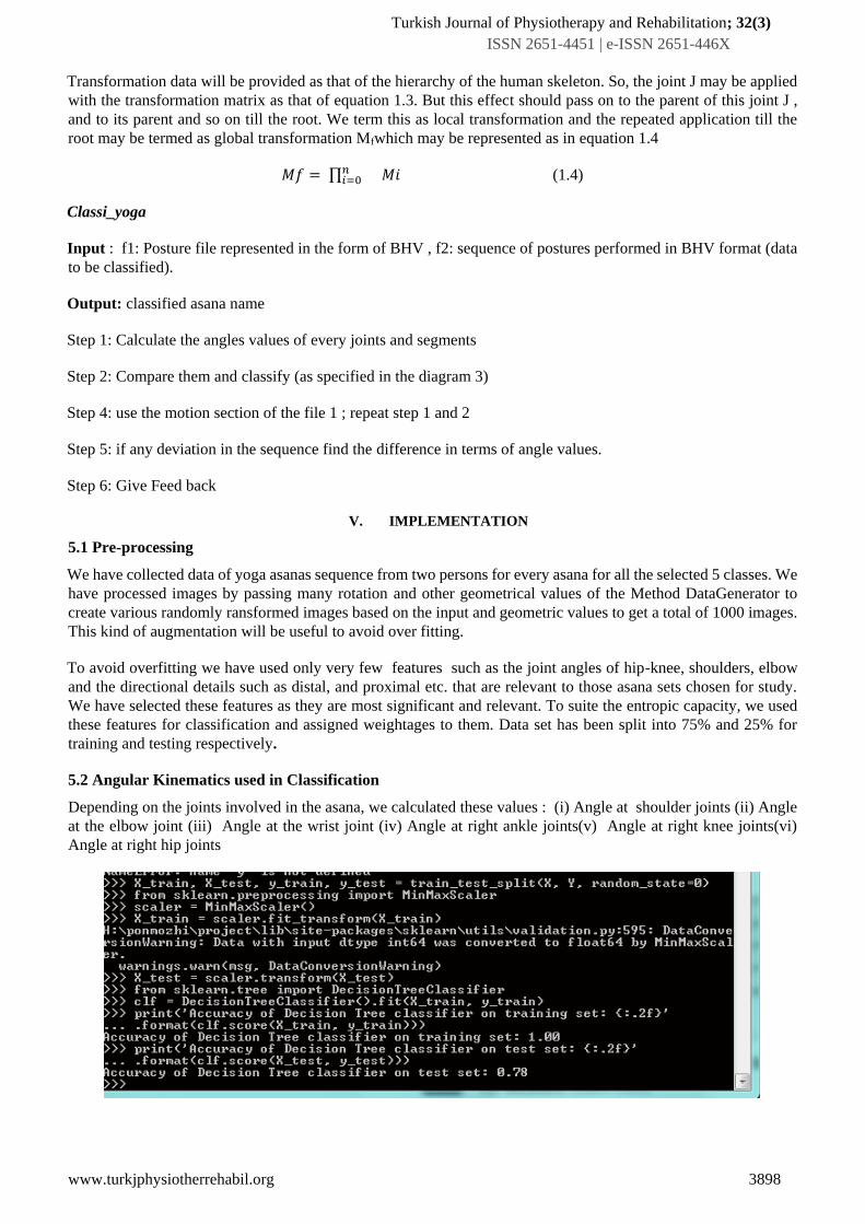
5.1 Pre-processing
We have collected data of yoga asanas sequence from two persons for every asana for all the selected 5 classes. We
have processed images by passing many rotation and other geometrical values of the Method DataGenerator to
create various randomly ransformed images based on the input and geometric values to get a total of 1000 images.
This kind of augmentation will be useful to avoid over fitting.
To avoid overfitting we have used only very few features such as the joint angles of hip-knee, shoulders, elbow
and the directional details such as distal, and proximal etc. that are relevant to those asana sets chosen for study.
We have selected these features as they are most significant and relevant. To suite the entropic capacity, we used
these features for classification and assigned weightages to them. Data set has been split into 75% and 25% for
training and testing respectively.
5.2 Angular Kinematics used in Classification
Depending on the joints involved in the asana, we calculated these values : (i) Angle at shoulder joints (ii) Angle
at the elbow joint (iii) Angle at the wrist joint (iv) Angle at right ankle joints(v) Angle at right knee joints(vi)
Angle at right hip joints
Turkish Journal of Physiotherapy and Rehabilitation; 32(3)
ISSN 2651-4451 | e-ISSN 2651-446X
www.turkjphysiotherrehabil.org 3899
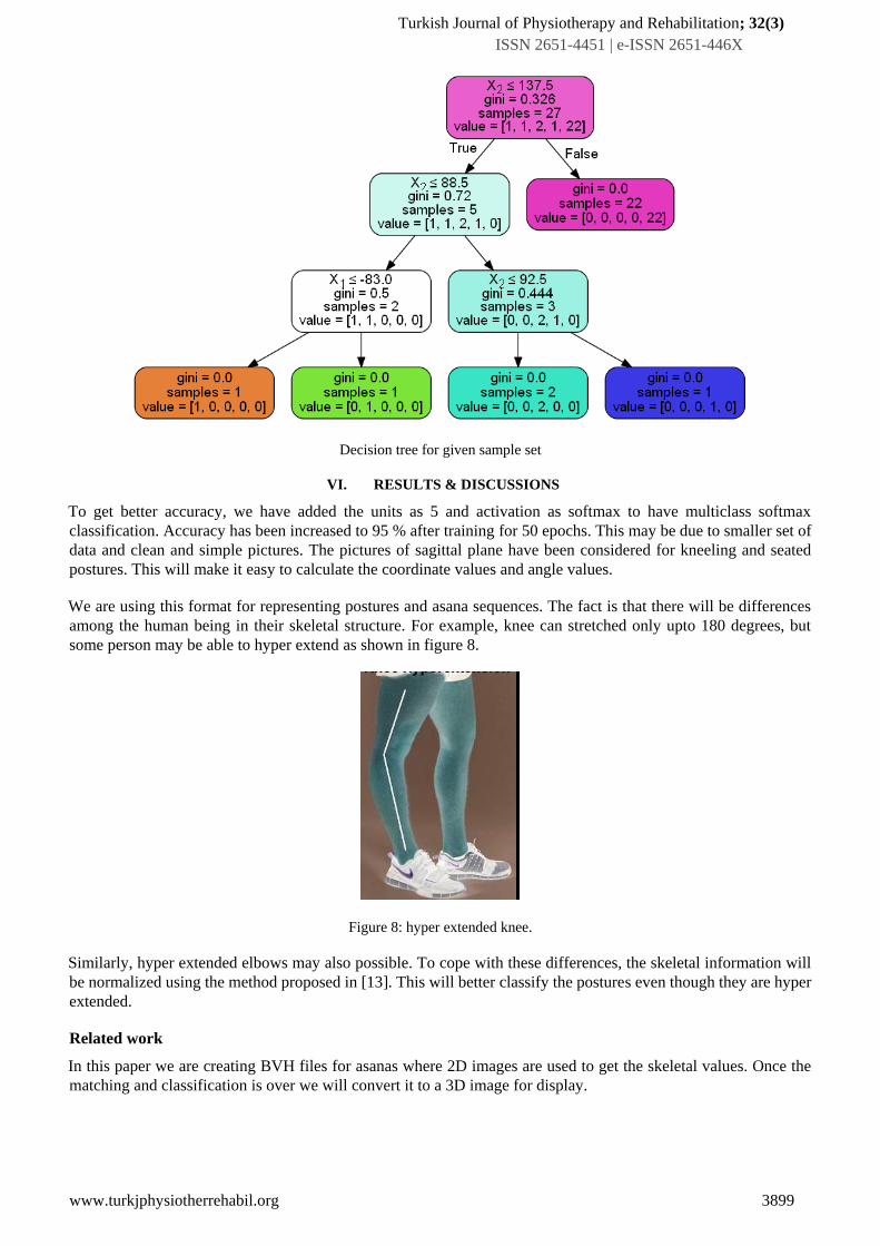
Decision tree for given sample set
VI. RESULTS & DISCUSSIONS
To get better accuracy, we have added the units as 5 and activation as softmax to have multiclass softmax
classification. Accuracy has been increased to 95 % after training for 50 epochs. This may be due to smaller set of
data and clean and simple pictures. The pictures of sagittal plane have been considered for kneeling and seated
postures. This will make it easy to calculate the coordinate values and angle values.
We are using this format for representing postures and asana sequences. The fact is that there will be differences
among the human being in their skeletal structure. For example, knee can stretched only upto 180 degrees, but
some person may be able to hyper extend as shown in figure 8.
Figure 8: hyper extended knee.
Similarly, hyper extended elbows may also possible. To cope with these differences, the skeletal information will
be normalized using the method proposed in [13]. This will better classify the postures even though they are hyper
extended.
Related work
In this paper we are creating BVH files for asanas where 2D images are used to get the skeletal values. Once the
matching and classification is over we will convert it to a 3D image for display.

Turkish Journal of Physiotherapy and Rehabilitation; 32(3)
ISSN 2651-4451 | e-ISSN 2651-446X
www.turkjphysiotherrehabil.org 3900
In order to do the process of 2d to 3d matching features such as edge direction histogram[14],shape
context[15],SIFT descriptors[16]. Technique of getting 3d details from 2d colorimgae [17],and using machine
learning technique such as end-to-end deep architectures [18] are also utilized.
Machine learning approach such as supervised learning [19][20][21] to identify discerning features, using the
structure of known objects to infer poses [22][23], inferring postures based on joint angles and segments[ 24] are
some of the approaches used to get 2d details from 3d images.
VII. CONCLUSION
This study propose a method of representing yoga asanas in terms of the joints involved in that asana. The
biomechanics of the joints are considered. Based on the joint angle values between the involved joints asana
classification is done. The data taken is very small, which may not be useful to justify the classification accuracy.
In future more sample data set will be used to ensure and whether we can classify even more number of aanas based
on other joints too.
REFERENCE
1. Cramer H, Lauche R, Haller H, Dobos G. A systematic review and meta-analysis of yoga for low back pain. Clin J Pain. 2013;29:450–60.
2. Williams K, Abildso C, Steinberg L, Doyle E, Epstein B, Smith D, et al. Evaluation of the effectiveness and efficacy of Iyengar yoga therapy on chronic low back pain. Spine (Phila Pa 1976) 2009;34:2066–76.
3. Galantino ML, Bzdewka TM, Eissler-Russo JL, Holbrook ML, Mogck EP, Geigle P, et al. The impact of modified Hatha yoga on chronic low back
pain: A pilot study. AlternTher Health Med. 2004;10:56–9.
4. Vakos JP, Nitz AJ, Threlkeld AJ, Shapiro R, Horn T. Electromyographic activity of selected trunk and hip muscles during a squat lift. Effect of
varying the lumbar posture. Spine (Phila Pa 1976) 1994;19:687–95
5. Nashner LM, McCollum G. The organization of human postural movements: A formal basis and experimental synthesis. Behav Brain Sci 1985;8:135-72.
6. Gatev P, Thomas S, Kepple T, Hallett M. Feedforward ankle strategy of balance during quiet stance in adults. J Physiol 1999;514:915-28.
7. Barnett C, Napier J. The axis of rotation at the ankle joint in man.Its influence upon the form of the talus and the mobility of the fibula. J Anat 1952;
86(Pt 1): 1.
8. Nigg B, Fisher V, Ronsky J. Gait characteristics as a function of age and gender. Gait Posture 1994; 2: 213-20.
9. Chopra S, Rouhani H, Assal M, Aminian K, Crevoisier X.Outcome of unilateral ankle arthrodesis and total anklereplacement in terms of bilateral gait
mechanics. J Orthop ResMar 2014; 32: 377-84.
10. Tung-Wu Lu, Chu-Fen Chang. Biomechanics of human movement and its clinical applications. Kaohsiung Journal of Medical Sciences (2012) 28,
S13-S25.
11. Watkins J. Structure and function of the musculoskeletal system. Champaign: Human Kinetics; 1999.
12. D.Wu,L.Shao,Leveraging hierarchical parametric networks for skeletal joints based action segmentation and recognition, in:Proceedings of Con ference on Computer Vision and Pattern Recognition(CVPR),IEEE,Colum- bus, Ohio.2014,pp.724–731.
13. C. Hecker, B. Raabe, R.W. Enslow, J. DeWeese, J. Maynard, K. van Prooijen, Real-time motion retargeting to highly varied user-created
morphologies, ACM Trans. Graph. 27 (3) (2008) 27, http://dx.doi.org/10.1145/ 1399504.1360626
14. G. Shakhnarovich, P. A. Viola, and T. J. Darrell. Fast pose estimation with parameter-sensitive hashing. In ICCV, 2003
15. G. Mori and J. Malik. Recovering 3D human body configurations using shape contexts. TPAMI, 28(7):1052–1062, July 2006
16. L. F. Bo, C. Sminchisescu, A. Kanaujia, and D. N. Metaxas. Fast algorithms for large scale conditional 3D prediction. In CVPR, pages 1–8, 2008
17. G. Rogez and C. Schmid. Mocap-guided data augmentation for 3D pose estimation in the wild. In NIPS, 2016
18. G. Pavlakos, X. Zhou, K. G. Derpanis, and K. Daniilidis.Coarse-to-fine volumetric prediction for single-image 3D human pose. In CVPR, 2017.
19. D. Eigen and R. Fergus. Predicting depth, surface normals and semantic labels with a common multi-scale convolutional architecture. In ICCV, 2015.
20. F. Liu, C. Shen, and G. Lin. Deep convolutional neural fields for depth estimation from a single image. In CVPR, pages 5162–5170. IEEE Computer Society, 2015.
21. .A. Popa, M. Zanfir, and C. Sminchisescu. Deep Multitask Architecture for Integrated 2D and 3D Human Sensing. In CVPR, 2017
22. . F. Bogo, A. Kanazawa, C. Lassner, P. Gehler, J. Romero,and M. J. Black. Keep it SMPL: Automatic estimation of 3d human pose and shape from a single image. In ECCV, 2016.], 55. X. Zhou, M. Zhu, S. Leonardos, and K. Daniilidis. Sparse representation for 3d shape estimation: A convex
relaxation approach. TPAMI, 99(1), 2016
23. X. Zhou, M. Zhu, S. Leonardos, K. G. Derpanis, and K. Daniilidis. Sparseness meets deepness: 3d human pose estimation from monocular video. In CVPR, 2016
24. X. Zhou, X. Sun, W. Zhang, S. Liang, and Y. Wei. Deep kinematic pose regression. In ECCV Workshops, 2016
25. Types of Body Movements Edited and Revised by Lindsay M. Biga, Sierra Dawson, Amy Harwell, Robin Hopkins, Joel Kaufmann, Mike LeMaster,
Philip Matern, Katie Morrison-Graham, Devon Quick, Jon Runyeon Art edited and created by Leeah Whittier
26. Judge, J. O., Ounpuu, S., & Davis, R. B., 3rd. (1996). Effects of age on the biomechanics and physiology of gait. Clin Geriatr Med, 12(4), 659-678
27. Teixeira-Salmela, L. F., Nadeau, S., McBride, I., & Olney, S. J. (2001).Effects of muscle strengthening and physical conditioning training on
temporal, kinematic and kinetic variables during gait in chronic stroke survivors. J Rehabil Med, 33(2), 53-60.
28. DiBenedetto, M., Innes, K. E., Taylor, A. G., Rodeheaver, P. F., Boxer, J. A., Wright, H. J., & Kerrigan, D. C. (2005). Effect of a gentle Iyengar yoga
program on gait in the elderly: An exploratory study. Archives of Physical Medicine and Rehabilitation, 86(9), 1830-1837
29. Roland, K. P., Jakobi, J. M., & Jones, G. R. (2011). Does Yoga Engender Fitness in Older Adults? A Critical Review. Journal of Aging & Physical Activity, 19(1), 62-79.
30. Tran, M. D., Holly, R. G., Lashbrook, J., & Amsterdam, E. A. (2001). Effects of Hatha Yoga Practice on the Health-Related Aspects of Physical Fitness. PrevCardiol, 4(4), 165-170.
31. Omkar, S. N., Mour, M., & Das, D. (2011). A mathematical model of effects on specific joints during practice of the Sun Salutation - A sequence of yoga postures. Journal of Bodywork and Movement Therapie, 15(2), 201-208.
Recommended