DNA Replication
Replication of DNA
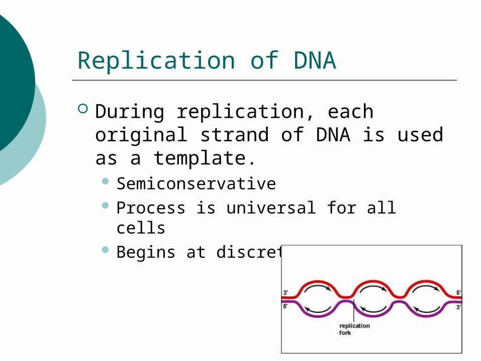
During replication, each original strand of DNA is used as a template. Semiconservative Process is universal for all cells Begins at discrete points
Replication Process
Late 1950s, John Cairns observed structures in E. coli during DNA replication – later called replication forks Prokaryotes – circular DNA Occur at discrete point proceeds both
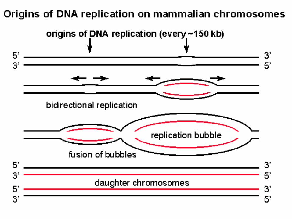
directions Eukaryotes – linear, multiple starting
points (100-150 Kb) proceeds both directions (bubbles)
In a nut shell!!
DNA replication must overcome physical and biochemical obstacles:1. Replication site must be located2. Aggregation of proper enzymes3. Unwinding of the double helix4. Relief of twisting strain on portions of
the helix farther away5. Complementary nucleotides must be
put in place and linked to form new strand
6. Errors must be checked and corrected
Prokaryote DNA replication
E. coli Origin of replication contains unique sequence of
base pairs which an initiator protein binds. Three AT rich 13-mer repeats and four 9-mer
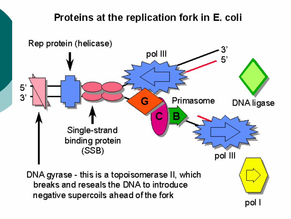
repeats Attraction of helicases (enzymes) that interrupt
base pairing into short single strands. Other binding proteins attach to maintain the
single strand separation Adjacent to the single strand region another
enzyme called DNA gyrase cleaves and reforms the sugar-phosphate backbone of the double stranded helix, which relieves the strain.
Each strand grows in opposite directions due to 5’ to 3’ polarity
End of this bubble is called the replication fork
As replication occurs the helicase molecules are pushed towards the fork and further separates the double strand
Priming and Elongation
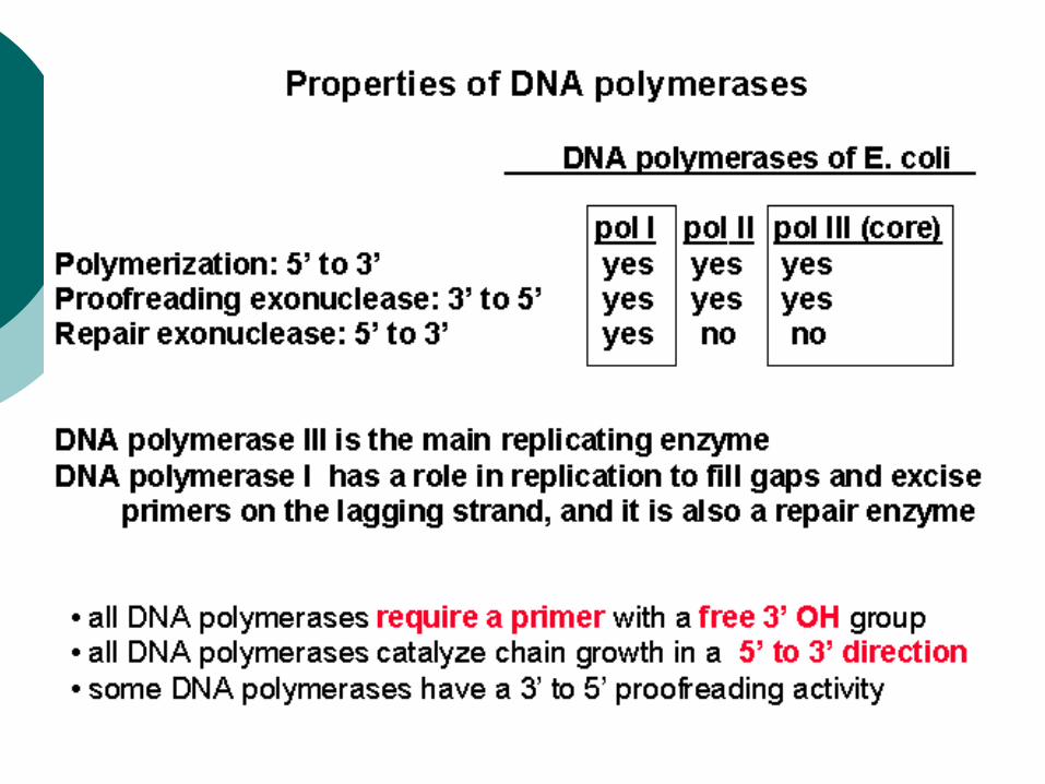
3 enzymes are used for replication in E. coli Pol III catalyzes both leading and
lagging strand elongation and proofreads for errors and replaces mismatches.
Pol I digests the RNA nucleotides and replaces with DNA nucleotides as well as proofreads and replaces errors.
Pol II mostly repairs replication errors.
Free nucleotides are added to the 3’ of the OH group of the last nucleotide in the growing chain.
The 3’ OH of the last nucleotide attacks the high-energy triphosphate group at the 5’ position of the free nucleotide, splitting off two phosphates and forming a covalent bond to the innermost phosphate.
This binds the new nucleotide to the existing chain.
Interesting facts: Incorporation of dideoxynucleotides which are missing the 3 OH end shuts down replication
Anticancer drugs Antiviral drugs Sanger sequencing
How does a nucleotide get added?
Elongation
Can’t start with DNA polymerases because it requires an existing chain with 3’ OH end and that is located on the newly replicated strand. RNA priming called primase Primase places a short sequence of RNA
nucleotides into position at the origin of replication which is complementary to the 3’ end of the single-stranded portion of the template at that point. DNA polymerase then adds nucleotide to the RNA’s 3’ OH.
Continuous elongation is leading strand Discontinuous is lagging strand
Every new section primase creates a new RNA primer and 3’ OH end to start the DNA elongation. These are Okazaki fragments.
Each short fragment only last a bit until the ribonucleotides are digested and replaced with DNA nucleotides then ligated by DNA ligase.
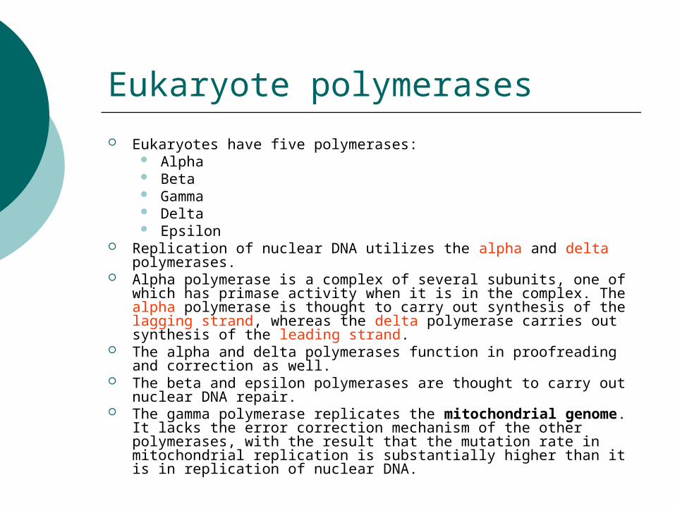
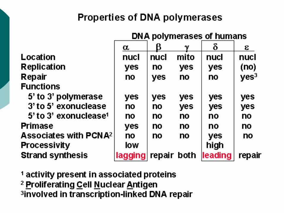
Eukaryote polymerases Eukaryotes have five polymerases:
Alpha Beta Gamma Delta Epsilon
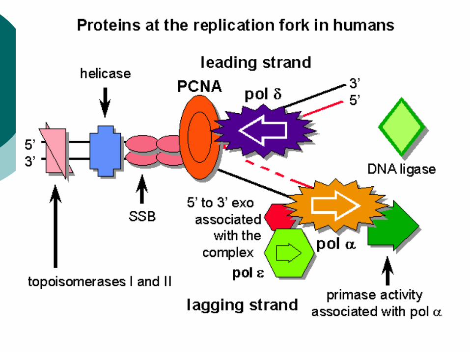
Replication of nuclear DNA utilizes the alpha and delta polymerases.
Alpha polymerase is a complex of several subunits, one of which has primase activity when it is in the complex. The alpha polymerase is thought to carry out synthesis of the lagging strand, whereas the delta polymerase carries out synthesis of the leading strand.
The alpha and delta polymerases function in proofreading and correction as well.
The beta and epsilon polymerases are thought to carry out nuclear DNA repair.
The gamma polymerase replicates the mitochondrial genome. It lacks the error correction mechanism of the other polymerases, with the result that the mutation rate in mitochondrial replication is substantially higher than it is in replication of nuclear DNA.
Telomeres and Telomerase
First discovered by Elizabeth Blackburn and Carol Greider in 1985
Structure discovered by Scott Cohen in 2007 Linear chromosomes have ends – telomeres
Can reach up to 15,000 base pairs Looses 25-200 base pairs per division Critical length = apoptosis
Telomerase Reverse Transcriptase (TERT) 1131 AA
Telomerase RNA (TERC) 451 nucleotide remains as RNA Template region = 3’-CAAUCCCAAUC-5’
Template for the 2 strands cannot be primed at the last nucleotides because there is no further DNA on which to build.
Telomeric DNAs designed to avoid this problem.
TERT wraps around TERC TERT uses TERC to add 5’-TTAGGG to
the 3’ strand of the chromosome
Transcription
Differences between DNA-RNA replication
RNA 5-10% by weight, DNA 1% by weight Ribonucleotides are used Extra hydroxyl group in RNA makes it unstable
-> more susceptible to hydrolysis -> easily degraded
Uracil replaces thymine RNA:DNA hybrid duplex product eventually
unravels and RNA is released. RNA polymerase is used to link nucleotides. The product is a single-stranded species.
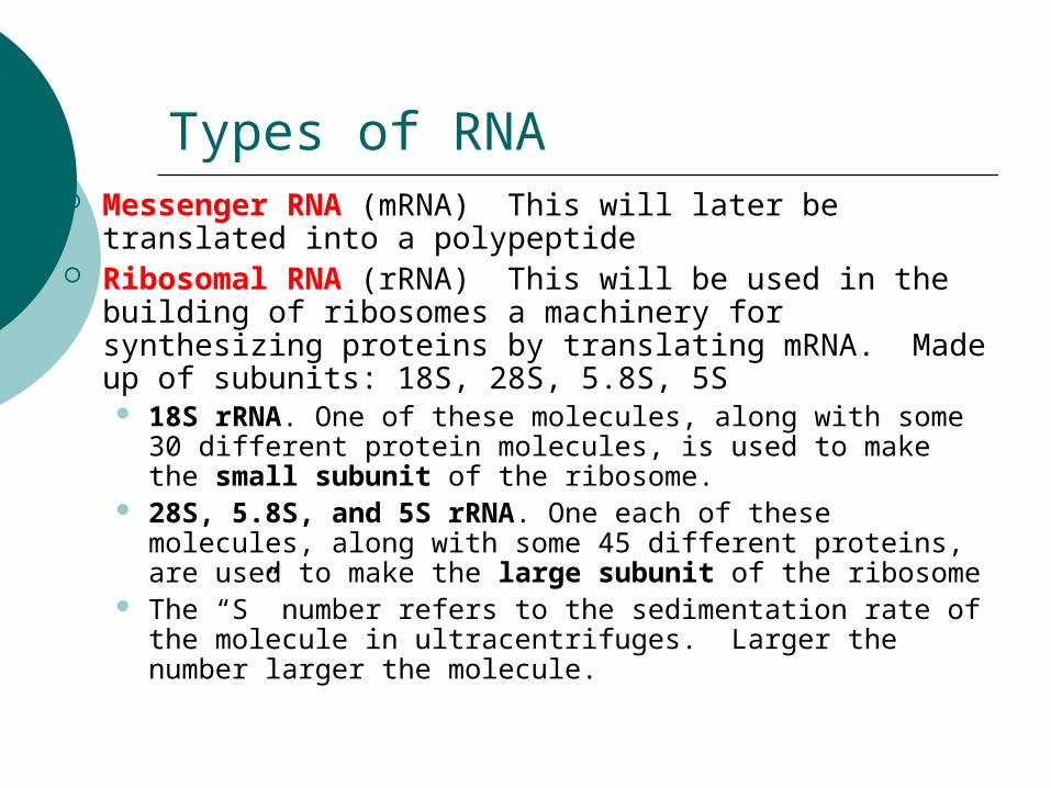
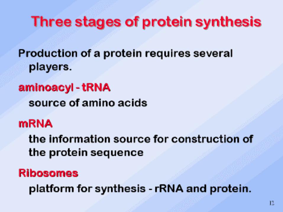
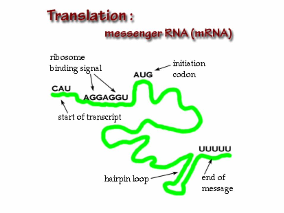
Types of RNA Messenger RNA (mRNA) This will later be translated
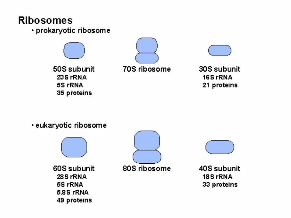
into a polypeptide Ribosomal RNA (rRNA) This will be used in the building
of ribosomes a machinery for synthesizing proteins by translating mRNA. Made up of subunits: 18S, 28S, 5.8S, 5S 18S rRNA. One of these molecules, along with some 30
different protein molecules, is used to make the small subunit of the ribosome.
28S, 5.8S, and 5S rRNA. One each of these molecules, along with some 45 different proteins, are used to make the large subunit of the ribosome
The “S” number refers to the sedimentation rate of the molecule in ultracentrifuges. Larger the number larger the molecule.
The RNA polymerases
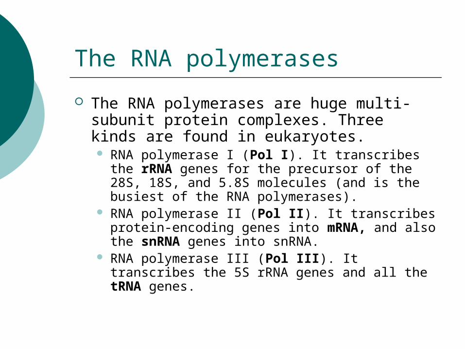
The RNA polymerases are huge multi-subunit protein complexes. Three kinds are found in eukaryotes. RNA polymerase I (Pol I). It transcribes the
rRNA genes for the precursor of the 28S, 18S, and 5.8S molecules (and is the busiest of the RNA polymerases).
RNA polymerase II (Pol II). It transcribes protein-encoding genes into mRNA, and also the snRNA genes into snRNA.
RNA polymerase III (Pol III). It transcribes the 5S rRNA genes and all the tRNA genes.
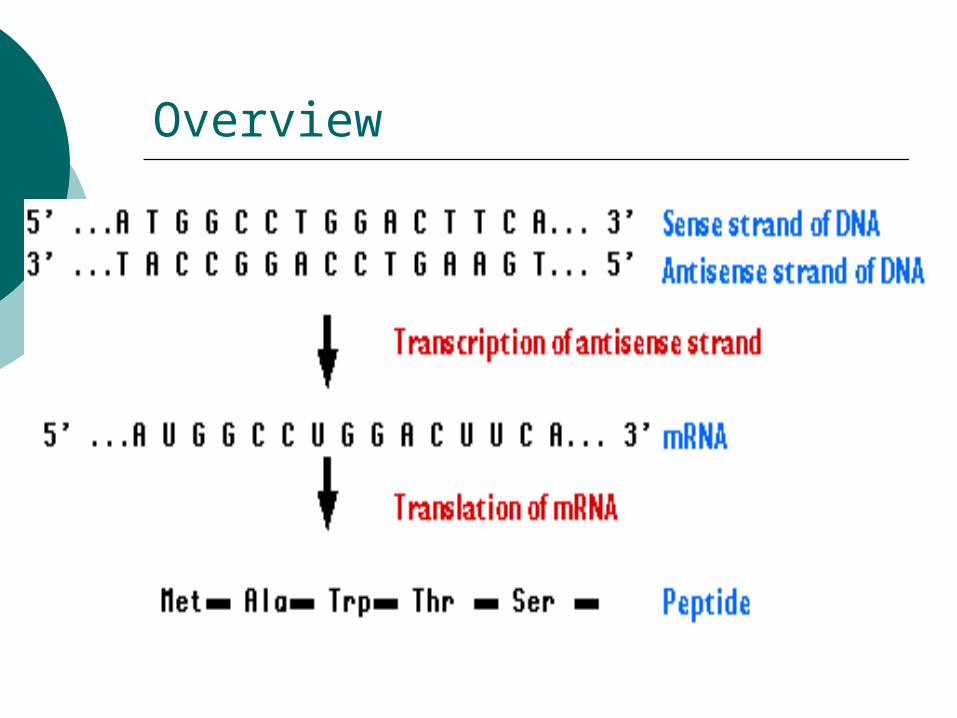
Overview
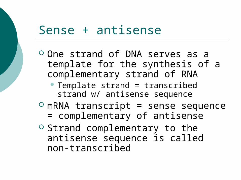
Sense + antisense
One strand of DNA serves as a template for the synthesis of a complementary strand of RNA Template strand = transcribed strand w/
antisense sequence mRNA transcript = sense sequence =
complementary of antisense Strand complementary to the
antisense sequence is called non-transcribed
Gene Regulation in Eukaryotes
Some of these called “house keeping genes” are expressed in all cells and all the time. They are important in routine metabolic functions such as breathing etc..
Some are expressed when cell enters a particular pathway of differentiation
Some are expressed all the time after they are differentiated. Ex. Plasma cell – antibody production
Some are only turned on when other signals arrives, such as hormone may turn something on or off
How is gene expression regulated
Altering the rate of transcription. Most widely used strategy. Altering RNA procession within the
nucleus Altering stability of mRNA (RNA
interference) Altering efficiency of ribosomes
translates the mRNA into a polypeptide
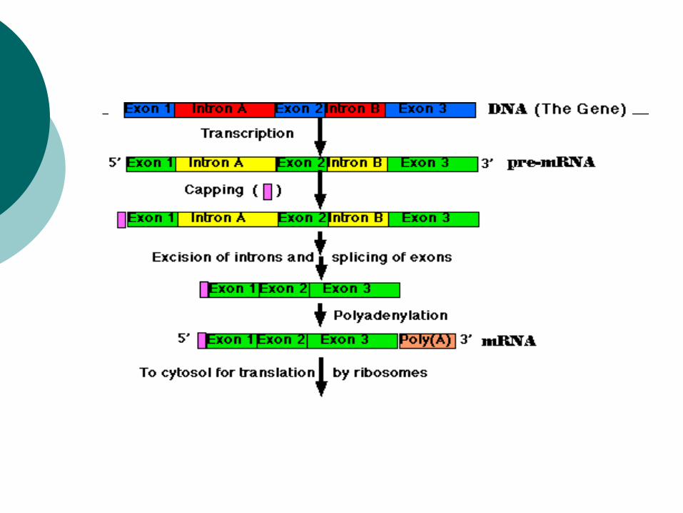
Components of Protein coding genes
Exons – sequence encodes the polypeptide Introns – that will be removed from the
mRNA before translation occurs A transcription start site A promoter
The basal or core promoter located within 40 bp of the start site
An upstream promoter, 200 bp further upstream Enhancers Silencers
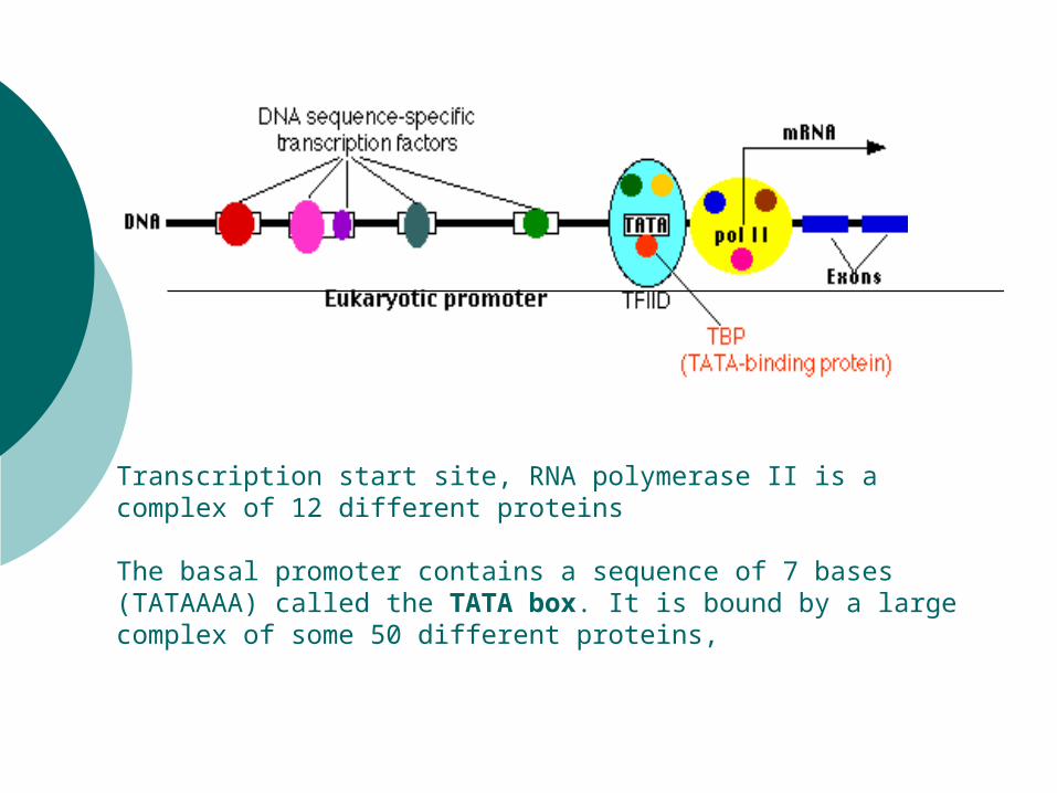
Transcription start site, RNA polymerase II is a complex of 12 different proteins
The basal promoter contains a sequence of 7 bases (TATAAAA) called the TATA box. It is bound by a large complex of some 50 different proteins,
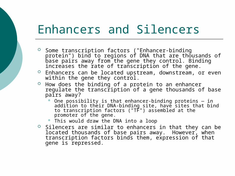
Enhancers and Silencers Some transcription factors ("Enhancer-binding protein")
bind to regions of DNA that are thousands of base pairs away from the gene they control. Binding increases the rate of transcription of the gene.
Enhancers can be located upstream, downstream, or even within the gene they control.
How does the binding of a protein to an enhancer regulate the transcription of a gene thousands of base pairs away?
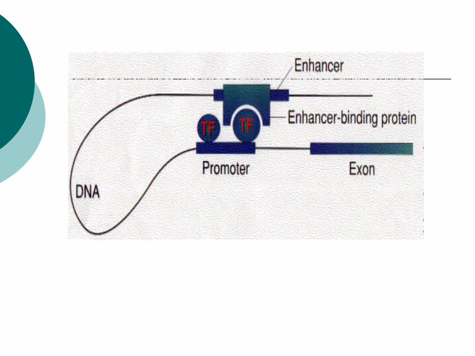
One possibility is that enhancer-binding proteins — in addition to their DNA-binding site, have sites that bind to transcription factors ("TF") assembled at the promoter of the gene.
This would draw the DNA into a loop Silencers are similar to enhancers in that they can be
located thousands of base pairs away. However, when transcription factors binds them, expression of that gene is repressed.
Insulators
Located between the Enhancers and promoter Silencers and promoter
Adjacent genes or clusters of adjacent genes.
Their function is to prevent a gene from being influenced by the activation (or repression) of its neighbors.
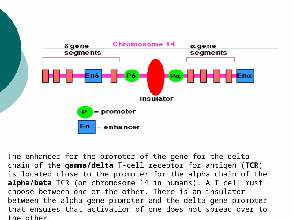
The enhancer for the promoter of the gene for the delta chain of the gamma/delta T-cell receptor for antigen (TCR) is located close to the promoter for the alpha chain of the alpha/beta TCR (on chromosome 14 in humans). A T cell must choose between one or the other. There is an insulator between the alpha gene promoter and the delta gene promoter that ensures that activation of one does not spread over to the other.
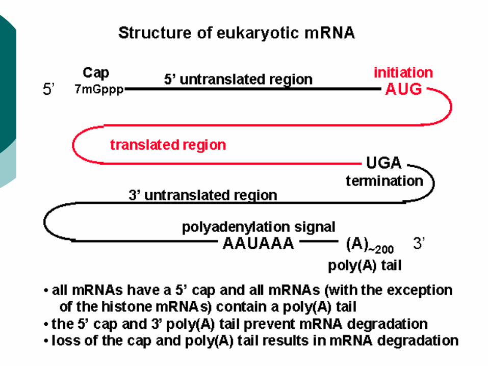
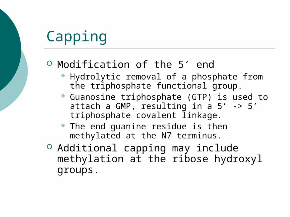
Capping
Modification of the 5’ end Hydrolytic removal of a phosphate from the
triphosphate functional group. Guanosine triphosphate (GTP) is used to attach
a GMP, resulting in a 5’ -> 5’ triphosphate covalent linkage.
The end guanine residue is then methylated at the N7 terminus.
Additional capping may include methylation at the ribose hydroxyl groups.
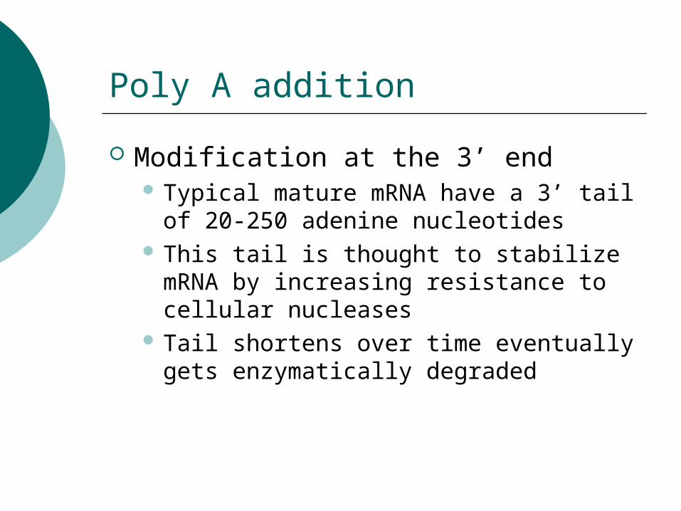
Poly A addition
Modification at the 3’ end Typical mature mRNA have a 3’ tail of
20-250 adenine nucleotides This tail is thought to stabilize mRNA by
increasing resistance to cellular nucleases
Tail shortens over time eventually gets enzymatically degraded
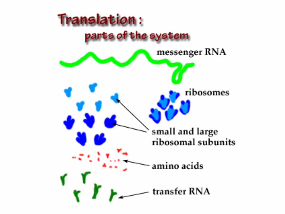
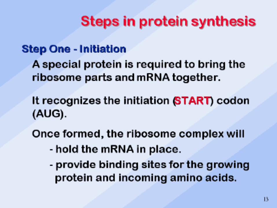
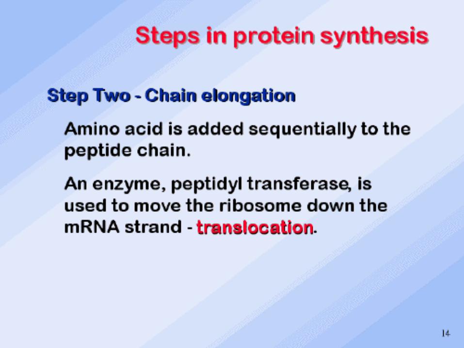
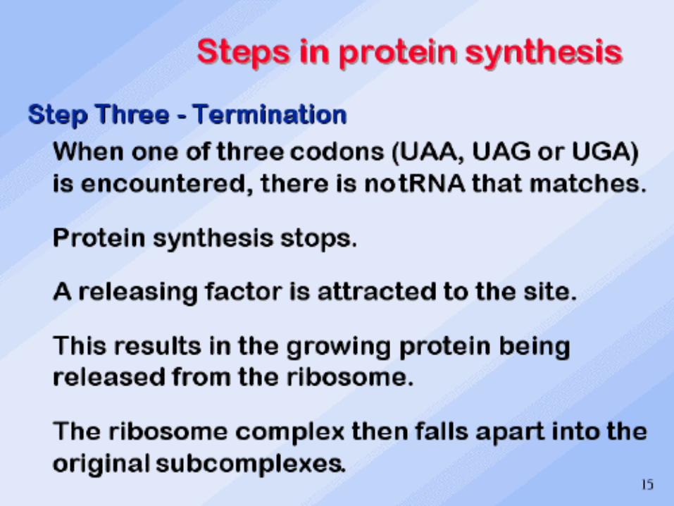
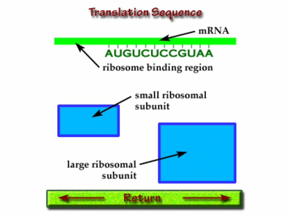
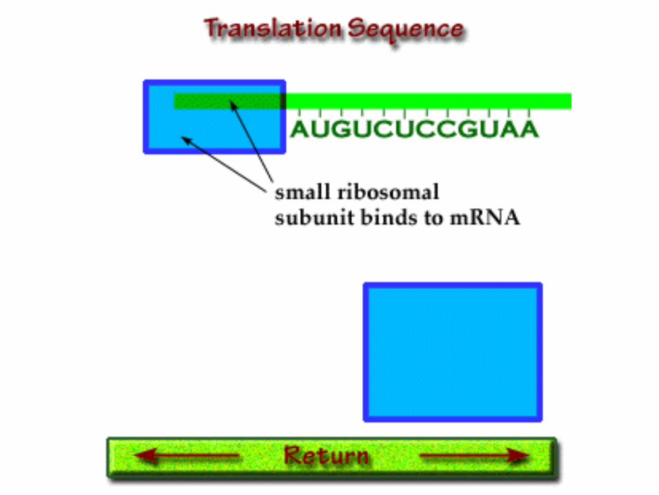
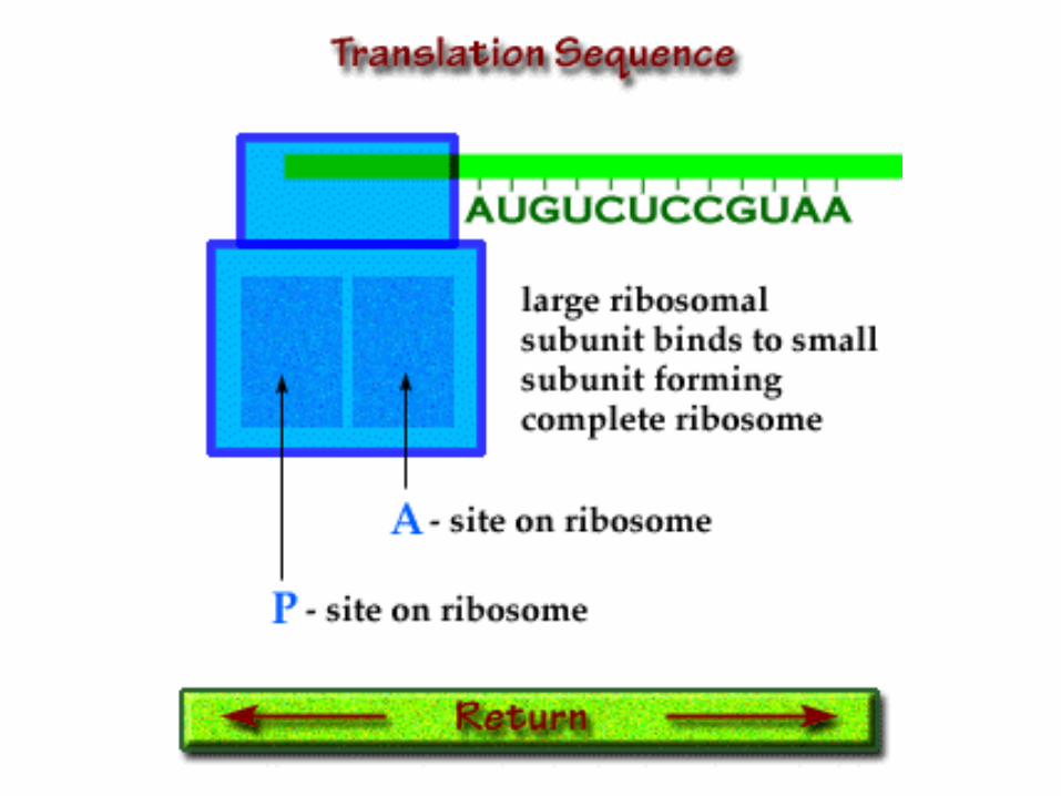
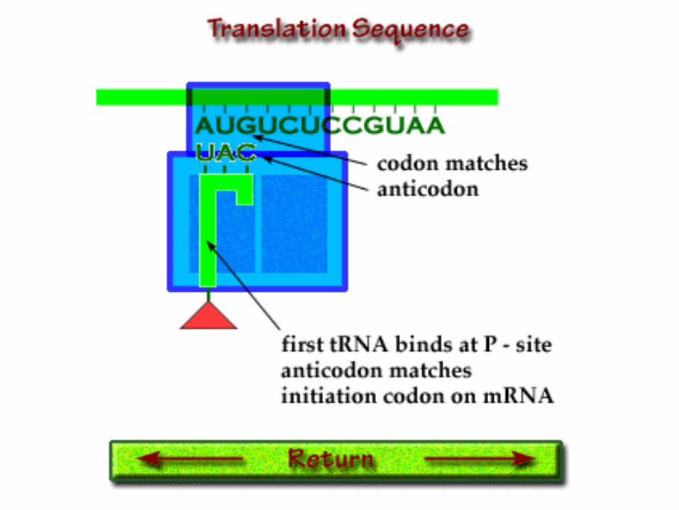
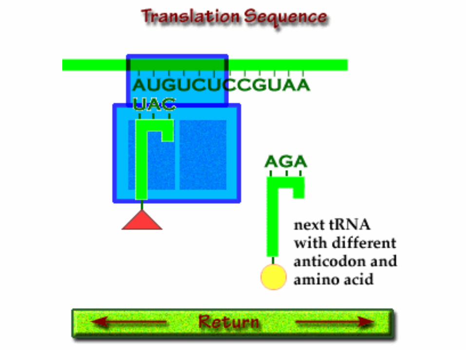
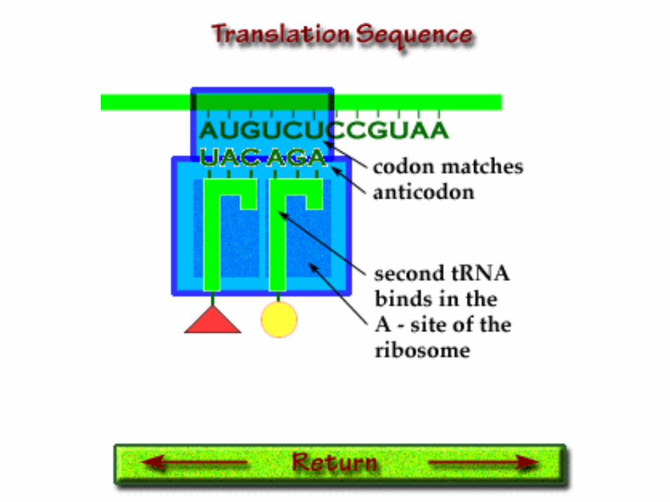
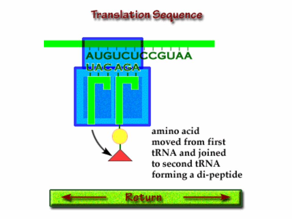
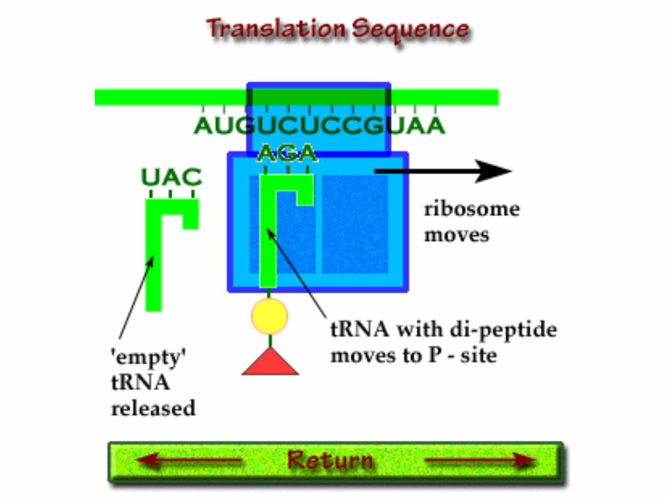
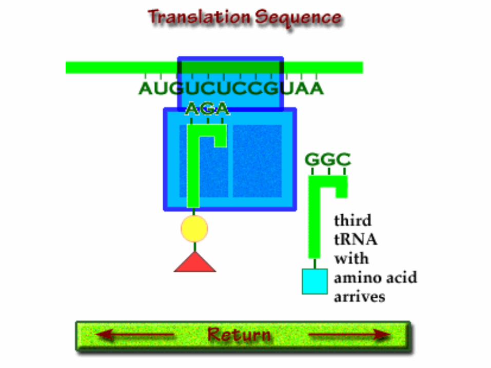
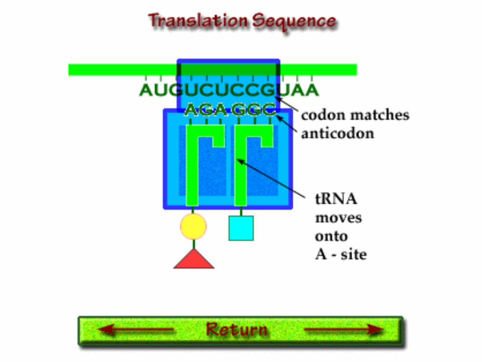
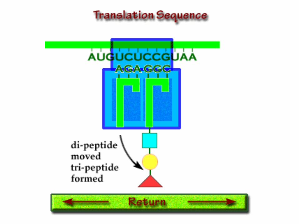
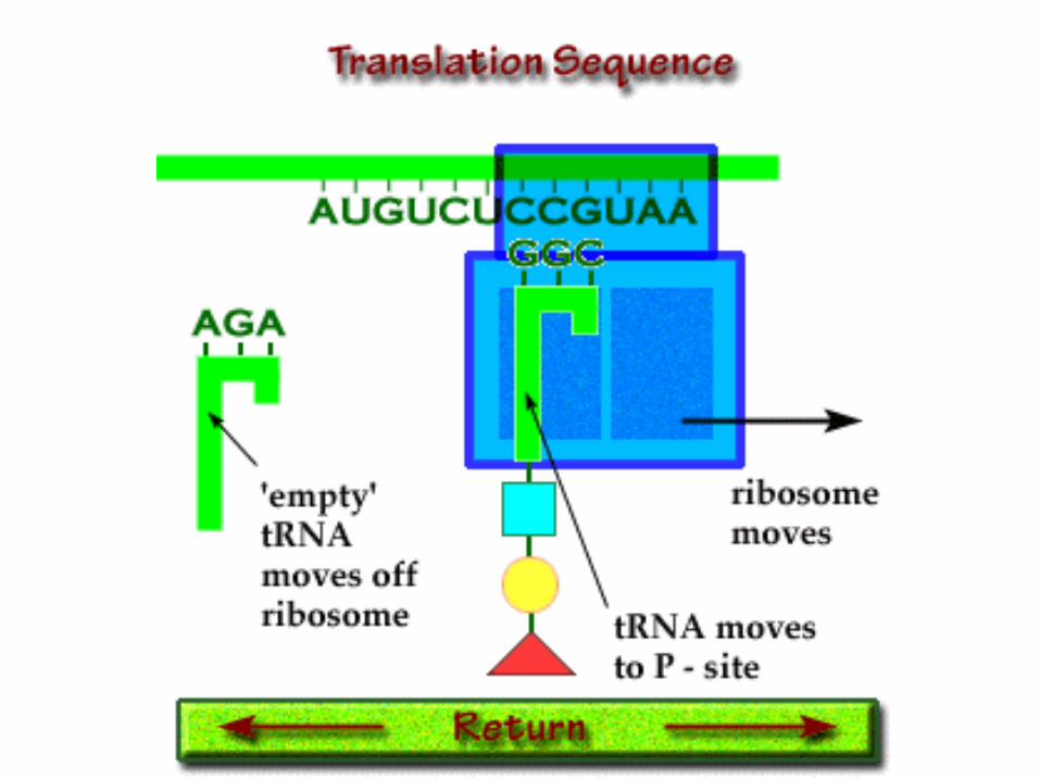
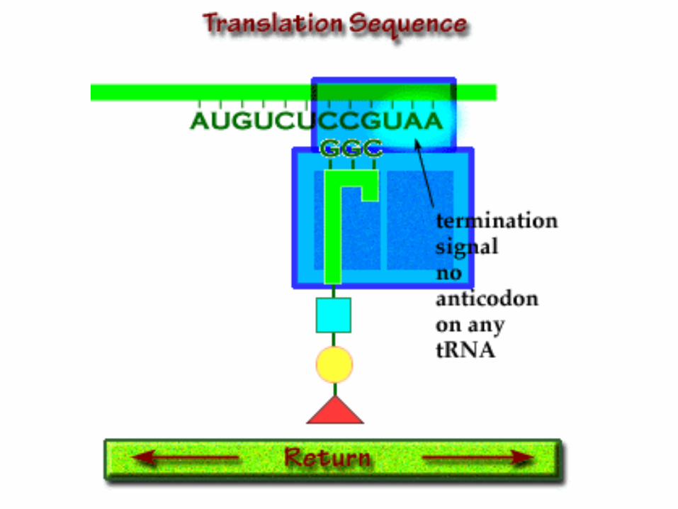
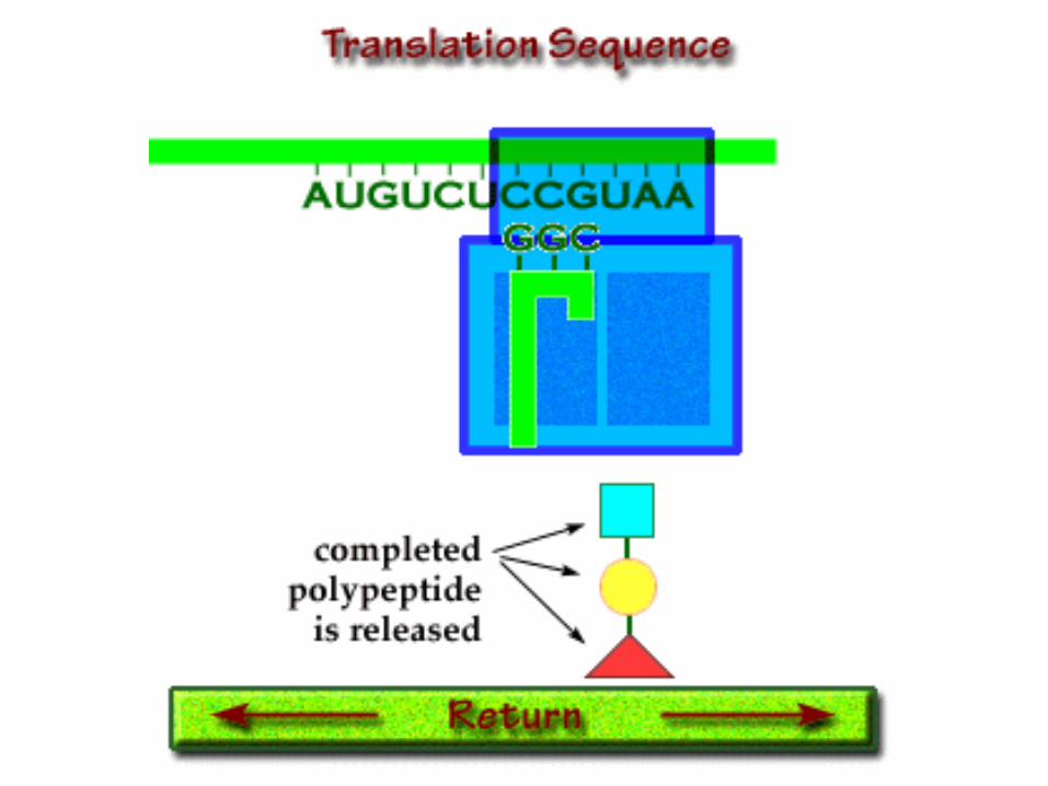
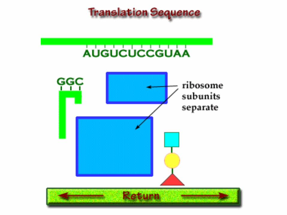
Translation
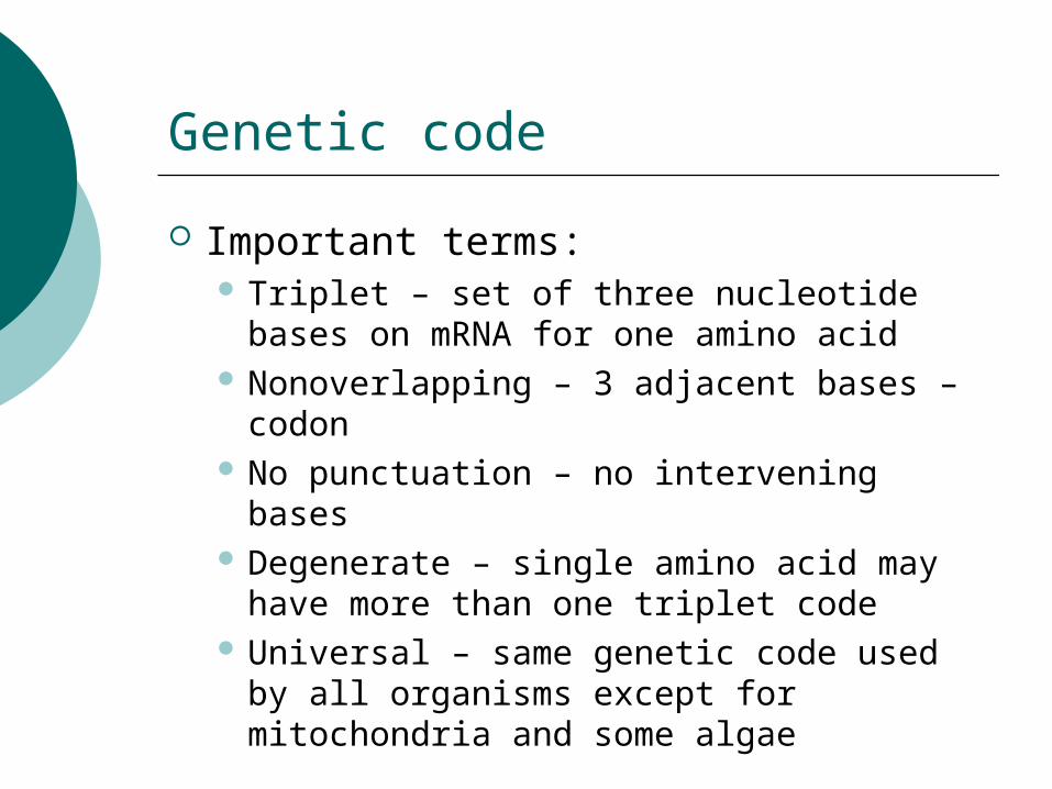
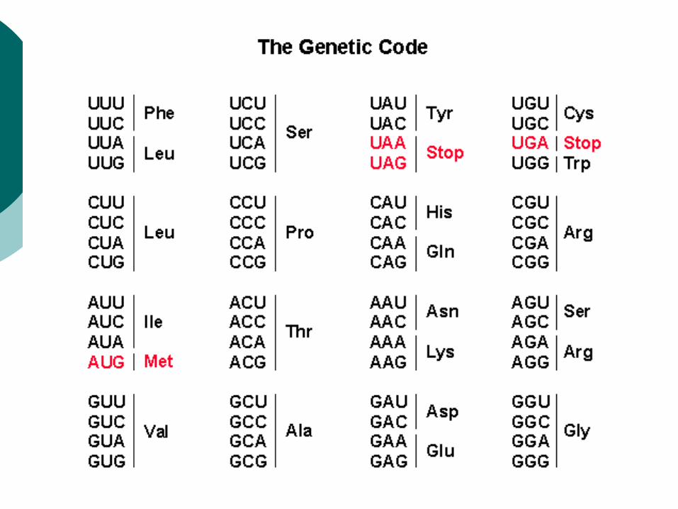
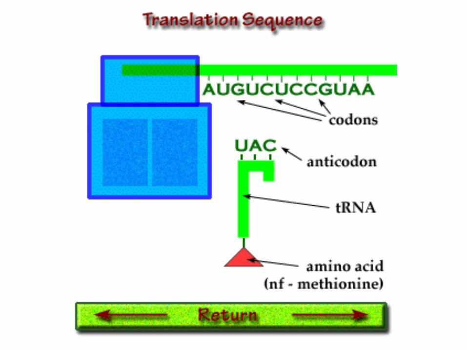
Genetic code
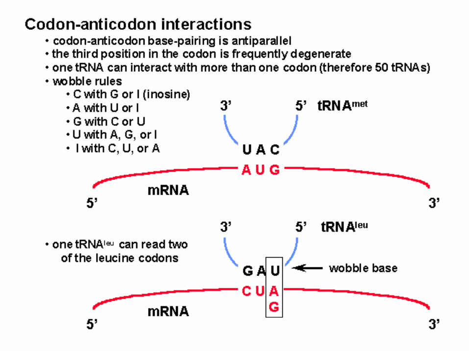
Important terms: Triplet – set of three nucleotide bases
on mRNA for one amino acid Nonoverlapping – 3 adjacent bases –
codon No punctuation – no intervening bases Degenerate – single amino acid may
have more than one triplet code Universal – same genetic code used by
all organisms except for mitochondria and some algae
tRNA structure
• One of the smallest types of RNA consisting of 74-93 nucleotides
• They carry the 20 amino acid types with at least one tRNA for each
• Binds to a single amino acid at one end• Bring proper amino acid to the right spot on
the mRNA-ribosome complex.
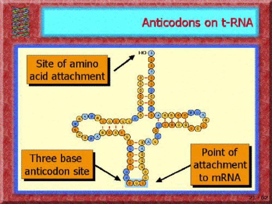
• Amino acids are linked to tRNAs by aminoacyl-tRNA synthetases
• These enzymes are able to recognize both the correct tRNA and the corresponding amino acid.
Recommended