Pulmonary Medicine - Dr. Gary Mumaugh 1
Pulmonary Medicine Section 9
Classic Respiratory Symptoms
Dyspnea
Cough
Hemoptysis
Chest pain Dyspnea
Three forms o Intense need to breath heavily and rapidly
Anxiety attacks, low O2 in air, high CO2 in blood Can occur after vigorous exercise
o Increased efforts of breathing with increased airflow resistance Asthma, croup, bronchitis
o Chest tightness Asthma, croup, pneumothorax, pleural effusion, pneumonia
Specific manifestations of dyspnea o Tachypnea – rapid respiratory rate o Hyperventilation – greater respiratory effort o Apnea – no breathing o Orthopnea – SOB when recumbent o PND – paroxysmal nocturnal dyspnea
Awakening from sleep with orthopnea
Treatment o Treatment determined of what condition is
causing dyspnea
Diagnosis of dyspnea o Airway problems
Foreign body, upper airway obstruction o Parenchymal lung disease – interstitial lung disease
ARDS, pneumonia, interstitial fibrosis o Pulmonary vascular disease
Pulmonary embolism, pulmonary hypertension o Pleural diseases
Pneumothorax, pleural effusion o Neuromuscular or chest wall diseases
Myasthenia gravis, severe scoliosis o Cardiovascular o Increased respiratory drive
Hyperthyroidism, pregnancy o Psychological reasons
Anxiety, panic attacks, psychosomatic disorders

Pulmonary Medicine - Dr. Gary Mumaugh 2
Cough
Differential diagnosis of cough o Airway irritants – smoke, dust, atmospheric irritants o Aspiration of foreign bodies – food, oral secretions, gastric secretions o Airway disease and URI – most common cause
Tracheitis (croup), bronchitis, bronchiectasis, pneumonia
o Interstitial disease within the lung tissue Pneumonia, lung abscess, pulmonary
fibrosis o Congestive heart failure
Productive cough o Produces clear, mucoid or grey phlegm
Nonproductive cough o Viral pneumonia, bronchitis, chest cold, flu (dry
cough) Hemoptysis
Coughing up blood
Differential diagnosis o Airway diseases
Bronchitis, bronchiectasis, bronchiogenic carcinoma
o Interstitial diseases Tuberculosis, lung abscess after
pneumonia o Vascular diseases
Left ventricular failure, pulmonary embolism
Chest pain
Chest pain does not start in the lungs, because there is not pain fibers in the lung tissue
Differential diagnosis o Pleuritis o Pneumothorax o Pleural effusion o Pulmonary embolism extending to the lung surface o Abscess or hematoma under the diaphragm
Ruptured appendicitis, ovarian cysts, gallbladder, tubal pregnancy
o Lupus lung o Heart disease or mediastinal disease

Pulmonary Medicine - Dr. Gary Mumaugh 3
Pulmonary Diagnostic Tools
Physical exam o Inspection – chest size, shape, symmetry, strength, atrophy, ease of
respiration, intercostal retraction o Palpation – depth of excursion, chest wall compliance o Percussion – top and bottom of chest bilateral o Ascultation – the main part of the examination
Clubbing o Unknown cause, seen in pulmonary disease o Finger clubbing is characterized by enlarged fingertips and a loss of the
normal angle at the nail bed
Chest x-ray
CT scans – good for interstitial diseases
MRI scans – good for mediastinal and hilar diseases
Lung scans
Pulmonary angiograms
Bronchoscopy
Pulmonary function tests o Lung volumes – shows the size of various parts in the lung o Flow rates – if the flow in the airways is impeded or not o Diffusing capacity – how fast the gas transfers o Arterial blood gases

Pulmonary Medicine - Dr. Gary Mumaugh 4
Normal Chest X-Ray
CHF Chest X-Ray

Pulmonary Medicine - Dr. Gary Mumaugh 5
Asthma
Incidence
o In the last decade the incidence of asthma has increased by 1/3 o 20 million people in the US o 6 million children and 14 million adults
Children under 16 and adults over 65 are more prone
Asthma is a process that affects the airways with excessive mucus production, bronchial muscle contraction, and swelling causing obstruction.
o During an asthma attack, spasms in the muscles and bronchi constrict, impeding the outward passage of stale air. Sufferers can get starved for air with coughing, wheezing and chest tightness.
o The basic cause is hyper-responsiveness of the airways to perceived noxious substances inhaled
o Recently, asthma has been found to be a chronic inflammatory process with the prior symptoms.
o Most of the research has been aimed at determining what might trigger asthma responses and what to avoid.
Risk Factors and Triggers o It is thought that asthma starts in childhood and may be due to interaction of
several factors o Asthma symptoms can be triggered by several factors o Allergies, viral or sinus infections, exercise, medications, foods, anxiety,
heredity o Reflux disease
stomach acid flowing back up the esophagus o Air pollutants
tobacco, wood smoke, chemicals, and ozone
Pulmonary Medicine - Dr. Gary Mumaugh 6
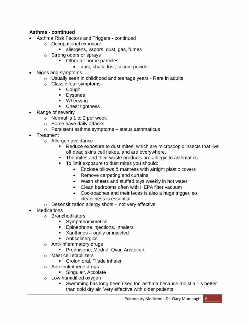
Asthma - continued
Asthma Risk Factors and Triggers - continued o Occupational exposure
allergens, vapors, dust, gas, fumes o Strong odors or sprays
Other air borne particles
dust, chalk dust, talcum powder
Signs and symptoms o Usually seen in childhood and teenage years - Rare in adults o Classic four symptoms
Cough Dyspnea Wheezing Chest tightness
Range of severity o Normal is 1 to 2 per week o Some have daily attacks o Persistent asthma symptoms – status asthmaticus
Treatment o Allergen avoidance
Reduce exposure to dust mites, which are microscopic insects that live off dead skins cell flakes, and are everywhere.
The mites and their waste products are allergic to asthmatics. To limit exposure to dust mites you should:
Enclose pillows & mattress with airtight plastic covers
Remove carpeting and curtains
Wash sheets and stuffed toys weekly in hot water
Clean bedrooms often with HEPA filter vacuum
Cockroaches and their feces is also a huge trigger, so cleanliness is essential
o Desensitization allergy shots – not very effective
Medications o Bronchodilators
Sympathomimetics Epinephrine injections, inhalers Xanthines – orally or injected Anticolinergics
o Anti-inflammatory drugs Prednisone, Medrol, Qvar, Aristocort
o Mast cell stabilizers Crolon oral, Tilade inhaler
o Anti-leukotriene drugs Singulair, Accolate
o Low humidified oxygen Swimming has long been used for asthma because moist air is better
than cold dry air. Very effective with older patients.

Pulmonary Medicine - Dr. Gary Mumaugh 7
COPD
Includes chronic bronchitis and emphysema
Bronchitis – chronic cough, sputum production and thickening of bronchial passages
Emphysema destroys the lung and enlarges the alveoli air spaces
Incidence o 16 million Americans, 110,000 death per year
Etiology o Smoking o Genetic predisposition o Multiple lung infections in children o Heavy exposure to environmental and industrial pollution o Cystic fibrosis
How COPD develops o Smoking causes increased mucus production and bronchial inflammation o Nicotine paralyzes the mucociliary escalator
Mucociliary escalator traps mucus, bacteria, irritants o Nicotine blocks protein inhibitors which will eventually dissolve the alveoli
Pulmonary Medicine - Dr. Gary Mumaugh 8
COPD - continued
Pathophysiology o Involves all four parts of the respiratory tract
Bronchi Bronchioles Alveoli Parenchyma
Specific Pathophysiology o Increased resistance to airflow o Loss of elastic recoil o Decreased expiratory flow rate o Alveolar walls frequently break because of the increased resistance of air
flows o The hyper inflated lungs flatten the curvature of the diaphragm and enlarge
the rib cage o The altered configuration of the chest cavity places the respiratory muscles,
including the diaphragm, at a mechanical disadvantage and impairs their force-generating capacity
o Consequently, the metabolic work of breathing increases, and dyspnea increases
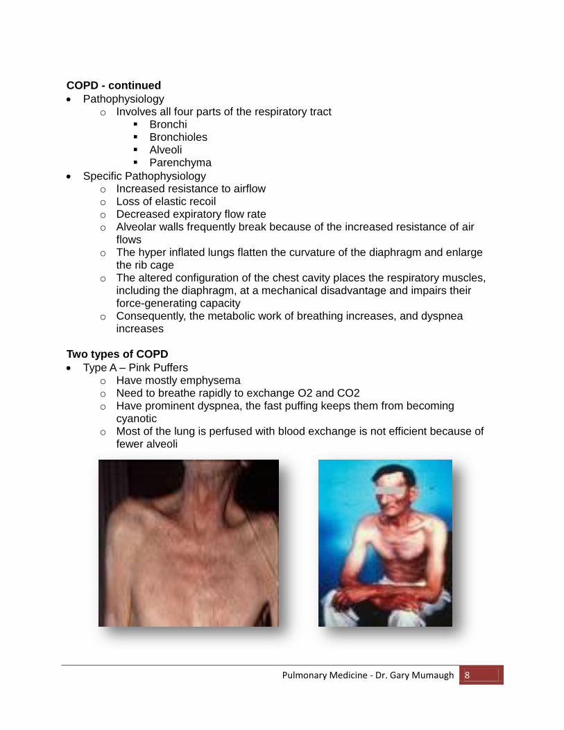
Two types of COPD
Type A – Pink Puffers o Have mostly emphysema o Need to breathe rapidly to exchange O2 and CO2 o Have prominent dyspnea, the fast puffing keeps them from becoming
cyanotic o Most of the lung is perfused with blood exchange is not efficient because of
fewer alveoli

Pulmonary Medicine - Dr. Gary Mumaugh 9
COPD - continued
Type B – Blue Bloaters o Have mostly chronic bronchitis with bronchiolar obstruction and non-
ventilated alveoli o Results in shunting of cyanotic blood away from the area where there is no air
in the lungs o Results in pulmonary hypertension which leads to heart failure with peripheral
swelling o Severe dyspnea with any exertion
Diagnosis of COPD
Smoker with hacking cough, sputum and dyspnea
Type A – thin, dorsal kyphosis, clubbing, pigeon breast (pectus carinatum) or funnel chest (pectus excavatum)
Type B – obese, swollen appearance, cyanotic
X-ray findings o Large lung volumes hyperlucent, flat diapgragm, increased AP diameter
Pulmonary function tests o Airway obstruction and decrease, air trapping
Blood gases o Type A – normal blood gases o Type B – marked hypoxemia and CO2 retention
Treatment of COPD o Bronchodilators o Antibiotics o Corticosteroids o Supplemental oxygen therapy o Chest physiotherapy to lose secretions o Surgery to remove diseased lung tissue o Lung transplantation

Pulmonary Medicine - Dr. Gary Mumaugh 10

Pulmonary Medicine - Dr. Gary Mumaugh 11
Bronchiectasis
Pathophysiology o Irreversible dilation of part of the bronchial tree o Caused by chronic infection of bronchi & bronchioles o Chronic bronchial infection causes a dilatation of the air passages which are
trapped with muco-purulent material o Caused by slow-growing bacteria and fungi
S & S o Chronic deep hacking cough o Copious amounts of foul-spelling pus sputum o Frequent attacks of pneumonia
Diagnosis o Localized rales and coarse ronchi o Appears similar to COPD with clubbing o Normal blood gases o History of chronic infection o CT scan confirms the diagnosis
Treatment o Antibiotics – ciprofloxacin o Bronchopulmonary drainage
Bending over, almost standing on head, to get the mucus up and out o Bronchodilators
Cystic Fibrosis
Inherited disease that causes thick, sticky mucus to build up in the lungs and digestive tract
The most common type of chronic lung disease in children and young adults o 1 in every 3,300 – most children and teenagers o May result in early death
S & S o Pneumonitis, bronchiectasis, lung abscesses, pancreatic insuffieciency
Diagnosis o Established by the sweat electrolyte test
Treatment

Pulmonary Medicine - Dr. Gary Mumaugh 12
Pulmonary fibrosis
Referred to as interstitial lung diseases
Causes inflammation and fibrosis of the connective tissue between the alveoli
Most common causes o Environmental causes – inhaled dusts, asbestosis, silicosis, glass makers,
construction workers o Antigens – hypersensitivity pneumonitis o Drugs – Methotrexate o Radiation injury o Other diseases – sarcoidosis, RA o Mimicking disorders similar presentation but vastly different
CHF, pneumocystis or viral pneumonia, carcinomatosis
Pathology of interstitial lung disease o Inflammation of the alveolar wall and inter-alveolar spaces o Fibrous scarring o Granuloma formation o End stage leads to a mass of scar tissue with contraction and the formation of
cystic areas
Pulmonary Medicine - Dr. Gary Mumaugh 13
Pulmonary fibrosis - continued
Impairment of pulmonary function o Decreased lung volume o Decreased compliance (stiff lungs) o Impairment of diffusion o Decreased gas exchange o Shunting and spasm of pulmonary arteries o Heart failure resulting from pulmonary hypertension
S & S of pulmonary fibrosis o Obvious dyspnea o Chronic nonproductive cough o Clubbing o Mild cyanosis
Diagnosis of pulmonary fibrosis o CT scan is confirmatory
Specific diseases that can cause pulmonary fibrosis o Silicosis – disease of glass makers, sand blasters, rock miners and stone
cutters Takes 20 years to develop
o Pneumoconiosis – coal miner’s disease Severe lung fibrosis with hypoxia
o Asbestosis – leads to 3 distinct diseases Bronchiogenic carcinoma Mesothelioma of lung (cancer of lung pleura) Interstitial fibrosis – takes 20 years to develop
o Drug-induced pulmonary fibrosis – chemotherapy
Treatment of pulmonary fibrosis o Very little effective care o Oxygen 24 / 7 o Corticosteroids

Pulmonary Medicine - Dr. Gary Mumaugh 14

Pulmonary Medicine - Dr. Gary Mumaugh 15
Pulmonary Embolism
Occurs when a blood clot is from the deep venous system travels to the lungs o Usually involves veins of legs, arms and pelvis (pregnancy)
Three conditions are put you at risk o Increased coagulation of blood
Stress, surgery, injury, heart attack, severe illness o Stasis or stagnation of blood flow
Seen in conditions of immobility such as prolonged bed rest long car rides of plane flights in cramped position
o Damage to vessel wall or venous valves Stasis-induced phlebitis, soft-tissue injury, bad ankle sprain
Pathophysiology o Pulmonary infarction of distal tissues occurs in a small number of cases o Hemorrhage and edema of tissues distal to the clot is more common o Vasoconstriction of pulmonary blood vessels occurs
This causes a release of serotonin an vasoconstrictive amines which cause more constriction
o Low blood pH causes even more constriction o Right sided heart failure followed by left sided blood flow followed by syncope
and sudden death
S & S o Sudden dyspnea o Pleuritic chest pain with hemoptysis o Can have syncope followed by death
Diagnosis o Normal chest x-ray o Perfusion lung scan shows absence of perfusion to involved arteries o Pulmonary arteriography – “gold standard” o Contrast CT o Decreased blood gases and increased pH
Treatment o tPA – tissue plasminogen activator if potentially life threatening embolism o Complete bed rest o Anticoagulation with heparin in ICU o Coumadin anticoagulation for six months o Vena caval filter surgery
PE prophylaxis o Most common secondary cause of hospital deaths o Lower extremity anti-embolism device with compression during surgery are
after heart attack or sever illness o Low dose heparin during surgery o Graduated compression support hose for patients with deep venous
insufficiency

Pulmonary Medicine - Dr. Gary Mumaugh 16
Pleural effusion
Caused by inflammation of pleura
S & S o Dyspnea o Pleuritic chest pain
Physical findings o Decreased breath sounds
Pleural friction rub
Differential diagnosis o Pleural effusion with have exudate or transudate o Hemothorax from trauma will have blood o Empyema becomes a giant abscess of pus
Diagnosis o Chest x-rays o Ultrasound o Thoracentesis – needle aspiration
Treatment o Treat the cause o Drain off fluid o May need pleurodesis – surgical breakup of adhesions
Pneumothorax
Air in the pleural space that collapses all or part of the lung o The pressure of the air against the lung causes it to give way, often leading to
mild to severe chest pain and shortness of breath o Lung collapses in proportion to the amount of air that leaks into your chest
cavity o Although the entire lung can collapse, a partial collapse is much more
common
Causes o Trauma o Growing tumor blocking a major airway o An infection o Inhaled foreign object.
S & S o If a small amount of air enters the pleural space, there may be a few signs or
symptoms o Even a minimally collapsed lung is likely to cause some chest pain o When the lung has collapsed 25 percent or more:
Sudden, sharp chest pain on the same side as the affected lung Dyspnea Chest tightness Tachycardia
Physical exam o Asymmetrical breath sounds o Decreased sounds over pneumothorax

Pulmonary Medicine - Dr. Gary Mumaugh 17
Pneumothorax - continued
Diagnosis o Chest x-ray is pathognomonic
Complications of pneumothorax o Hypoxemia o Respiratory failure o Cardiac arrest o Shock
Treatment o Bedrest and observation if collapse is less then 25 % o Chest tube if collapse over 25 %
Suction continued for up to 3 days
Thoracotomy needed if lung does not re-inflate
Mediastinal Diseases
Mediastinal masses o Thyoma, germ cell tumor, lymphoma, thyroid enlargement, carcinoma,
pericardial cysts, neurogenic tumors
S & S o Half of masses have no symptoms and the growth is found incidental on
another examination o The other half of the patients have chest pain, dyspnea, nonproductive cough
and facial edema
Diagnosis o Lateral chest x-ray o CT scan o Mediastinoscopy
Pulmonary Medicine - Dr. Gary Mumaugh 18
Disorders of Ventilatory Control
Hyperventilation – rapid breathing o Occurs with neurological diseases of the cortex such as meningitis,
encephalitis, strokes o Can also occur with panic attacks
Hypoventilation o Occurs with neurological diseases of the medulla such as encephalitis
Cheyne-Stokes breathing o Cyclic pattern of increasing and decreasing respiration leading to apnea o Seen in neurological disorders and CHF
Sleep apnea syndrome o During REM sleep there is a collapse of soft tissues with periods of apnea
ARDS - Acute Respiratory Distress Syndrome
Life threatening process with breakdown of alveolar cell membranes leading to pulmonary edema
Causes o Aspiration of gastric contents or water (near drowning) o Toxic gas inhalation o Severe diffuse pneumonia o Sepsis, shock, sever trauma, head trauma o Intravascular coagulation o Fat embolism o Multiple transfusions o Pancreatitis o Drug reactions
Called heroin pulmonary edema
ARDS pathology o Damage to alveolar epithelial cells leads to a buildup of interstitial and
alveolar fluid leading to alveolar collapse o This leads to increased vascular resistance, decreased lung compliance and
buildup of hyaline membranes o The thickening of the membranes leads to pulmonary fibrosis
S & S o Dyspnea, tachypnea, tachycardia, rales, hypoxia
Treatment o Aggressive treatment of the cause (sepsis, shock, etc.) o Corticosteroids o Support of impaired gas exchange
Prognosis o Mortality 50 %

Pulmonary Medicine - Dr. Gary Mumaugh 19
Pulmonary Red Flags
Severe asthma attacks
New onset of dyspnea or worsening dyspnea
All cases of chest pain
Upper airway disease
Pulmonary embolism
Pneumothorax
Pleural effusion with dyspnea
Pneumonitis
Cheyne-Stokes breathing
Acute onset of any breathing problems
Acute respiratory distress Pulmonary sub-acute red flags
New onset of asthma
New cough lasting for more than one week
Hemoptysis
COPD if not currently under care
Bronchiectasis
Cystic fibrosis
Pulmonary fibrosis
Pulmonary effusion
History or S & S of mediastinal disease
Sleep apnea
Recommended