1
Medicine & Science in Sports & Exercise ; IF : 4.43 (accepté le 8 Mai 2013)
Prolonged mental exertion does not alter neuromuscular
function of the knee extensors
Authors: Benjamin Pageaux1,2
, Samuele M. Marcora1, Romuald Lepers
2
1 Endurance Research Group, School of Sport & Exercise Sciences, University of Kent at
Medway, Chatham Maritime, Kent ME4 4AG, UK
2 Laboratoire INSERM U1093, Université de Bourgogne, Faculté des Sciences du Sports –
UFR STAPS, Dijon, France
Correspondence to: Dr Romuald Lepers
Laboratoire INSERM U1093
Faculté des Sciences du Sports
Université de Bourgogne
BP 27877
21078 Dijon Cedex
France
Email address: [email protected]
Phone: +33.3.8039.67.60
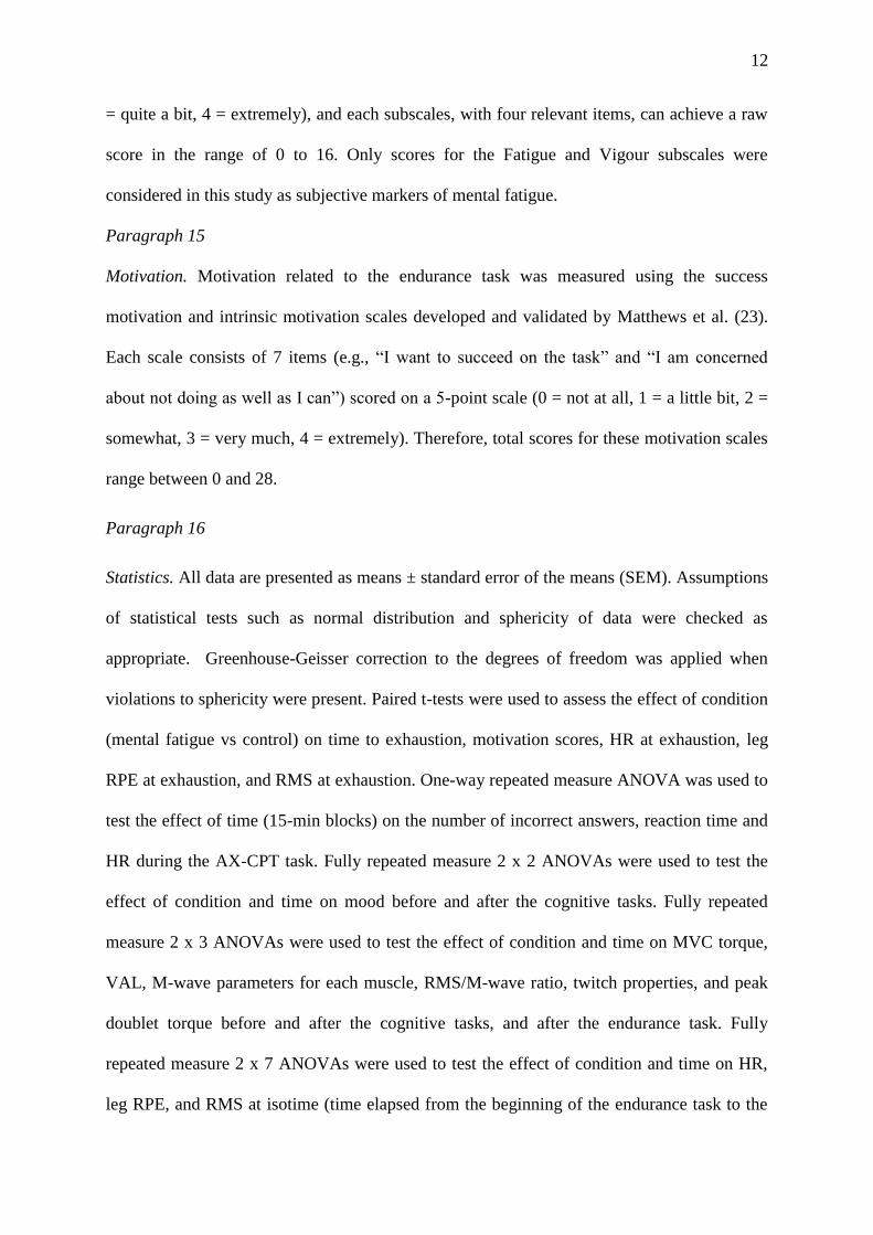
Fax: +33.3.80.39.67.02
Running Head: Mental fatigue and neuromuscular function
Conflict of Interest and Source of Funding: None

2
ABSTRACT
Purpose: The aim of this study was to test the hypotheses that prolonged mental exertion i)
reduces maximal muscle activation and ii) increases the extent of central fatigue induced by
subsequent endurance exercise. Methods: Neuromuscular function of the knee extensor
muscles was assessed in 10 male subjects in two different conditions: i) before and after
prolonged mental exertion leading to mental fatigue; ii) before and after an easy cognitive
task (control). Both cognitive tasks lasted 90 min and were followed by submaximal
isometric knee extensor exercise until exhaustion (endurance task), and a third assessment of
neuromuscular function. Results: Time to exhaustion was 13 ± 4% shorter in the mental
fatigue condition (230 ± 22s) compared to the control condition (266 ± 26 s) (P<0.01).
Prolonged mental exertion did not have any significant effect on maximal voluntary
contraction (MVC) torque, voluntary activation level (VAL) and peripheral parameters of
neuromuscular function. A similar significant decrease in MVC torque (mental fatigue
condition: -26.7 ± 5.7%; control condition: -27.6 ± 3.3%, P<0.001), VAL (mental fatigue: -
10.6 ± 4.3%; control condition: - 11.2 ± 5.2%, P<0.05) and peripheral parameters of
neuromuscular function occurred in both conditions following the endurance task. However,
mentally fatigued subjects rated perceived exertion significantly higher during the endurance
task compared to the control condition (P < 0.05). Conclusion: These findings provide the
first experimental evidence that i) prolonged mental exertion does not reduce maximal
muscle activation and ii) does not increase the extent of central fatigue induced by subsequent
endurance exercise. The negative effect of mental fatigue on endurance performance seems to
be mediated by higher perception of effort rather than impaired neuromuscular function.
Keywords: Perception of effort, muscle activation, mental fatigue, peripheral fatigue, central
fatigue, endurance performance.
3
INTRODUCTION
Paragraph 1
Prolonged mental exertion is well known to induce mental fatigue, a
psychobiological state characterized by subjective feelings of ‗‗tiredness‘‘ and ‗‗lack of
energy‘‘ (3). The negative effects of mental fatigue on cognitive performance are well
established and include impairments in attention, action monitoring, and cognitive control
(e.g. 3, 37). On the contrary, the effects of mental fatigue on physical performance have been
scarcely investigated. In 1906, Mosso (25) reported that two of his colleagues did poorly in a
muscle fatigue test performed after delivering long physiology lectures and viva
examinations. More recently, Bray et al. (5, 6) showed that performing a demanding
cognitive task before or between isometric contractions significantly reduces the endurance
and strength of isolated upper limb muscles. However, in these studies, mental exertion was
not prolonged enough to induce subjective feelings of mental fatigue. Furthermore,
neuromuscular function was assessed with EMG, a method that does not provide a valid
measure of maximal voluntary activation of muscle (15). Therefore, the link between
prolonged mental exertion and the central component of muscle fatigue is still unclear.
Marcora et al. (21) conducted the first experimental study on the effect of prolonged mental
exertion on endurance performance during dynamic whole-body exercise. These investigators
induced mental fatigue in a group of healthy and fit subjects using a prolonged demanding
cognitive task performed for 90 min, and found a significant reduction in time to exhaustion
during subsequent high-intensity cycling exercise. However, the physiological mechanisms
underlying the negative effect of prolonged mental exertion on endurance performance are
currently unknown. Marcora et al. (21) did not find any effect of mental fatigue on the
cardiovascular, respiratory and metabolic responses to high-intensity cycling exercise.
Motivation related to the time to exhaustion test was also unaffected by mental fatigue. In this
4
study, the only factor that could explain a premature exhaustion was the higher perception of
effort experienced by mentally fatigued subjects during high-intensity cycling exercise.
According to the psychobiological model of endurance performance, exhaustion is not caused
by muscle fatigue (20), i.e. by the failure of the fatigued neuromuscular system to produce the
force/power required by the endurance task despite a maximal voluntary effort. On the
contrary, it is proposed that exhaustion results from a conscious decision to disengage from
the endurance task. In highly motivated subjects this effort-based decision is taken when
perception of effort is maximal and continuation of the endurance task seems impossible.
Paragraph 2
Although this explanation is plausible, Marcora et al. (21) did not measure
neuromuscular function. Therefore, a reduction in maximal muscle activation or an increase
in the extent of central fatigue induced by endurance exercise may also explain the negative
effect of mental fatigue on endurance performance. Central fatigue is an exercise-induced
reduction in the capacity of the central nervous system (CNS) to fully recruit the active
muscles (muscle activation) during a maximal voluntary contraction (MVC), and occurs at
both spinal and/or supraspinal level (15). Central fatigue is thought to negatively affect
endurance performance (1) and several authors have proposed a strong link between mental
and central fatigue (e.g. 5, 11, 26). Because supraspinal fatigue seems to occur in brain areas
upstream of the primary motor cortex (34), it is plausible that prolonged mental exertion can
alter maximal muscle activation and, thus, impair endurance performance.
Paragraph 3
The main aim of the present study was to test experimentally this hypothetical link
between mental fatigue, maximal muscle activation and central fatigue. Specifically, we
hypothesized that prolonged mental exertion leading to mental fatigue i) would reduce
maximal muscle activation and ii) would increase the extent of central fatigue induced by

5
subsequent endurance exercise. We tested these two main hypotheses by measuring maximal
muscle activation of the knee extensor muscles before and after prolonged mental exertion,
and immediately after subsequent submaximal isometric contraction of the knee extensor
muscles until exhaustion (endurance task). Additionally, we hypothesized that prolonged
mental exertion would reduce endurance performance via a higher perception of effort during
the endurance task.
METHODS
Paragraph 4
Subjects and Ethical Approval. Ten physically active male adults (age: 22 ± 2 yr, height: 177
± 6 cm, weight: 70 ± 8 kg) volunteered to participate in this study. None of the subjects had
any known mental or somatic disorder. Each subject gave written informed consent prior to
the study. Experimental protocol and procedures were approved by the local Ethics
Committee of the Faculty of Sport Sciences, University of Burgundy in Dijon. All subjects
were given written instructions describing all procedures related to the study but were naive
of its aims and hypotheses. Participants believed that the study was on the effects of two
different cognitive activities (a computerized task and watching a movie) on the
neuromuscular responses to an endurance task. To ensure high motivation during the
cognitive and endurance tasks, a reward (ticket to a professional sport event) was given to the
best performances in both the cognitive and endurance tasks. At the end of the last session,
subjects were debriefed and asked not to discuss the real aims of the study with other
participants. The study conformed to the standards set by the World Medical Association
Declaration of Helsinki ―Ethical Principles for Medical Research Involving Human Subjects‖
(2008).
Paragraph 5

6
Experimental Protocol. Subjects visited the laboratory on three different occasions. During
the first visit, subjects were familiarised with the laboratory and the experimental procedures.
During the second and third visit, subjects performed either a mental fatigue task or a control
task (see Cognitive Tasks for more details) in a randomized and counterbalanced order. After
the cognitive task, subjects performed submaximal isometric knee extensor exercise until
exhaustion (see Endurance Task for more details). Neuromuscular function of the knee
extensor muscles was tested before and after the cognitive task, and after the subsequent
endurance task. Mood was assessed before and after the cognitive task, whilst motivation was
measured before the subsequent endurance task (Fig. 1). For more details see Neuromuscular
Function Tests and Psychological Questionnaires.
Paragraph 6
Each participant completed all three visits over a period of 3 weeks with a minimum
of 72 hours recovery period between visits. All participants were given instructions to sleep
for at least 7 hours, refrain from the consumption of alcohol, and not to practise vigorous
physical activity the day before each visit. Participants were also instructed not to consume
caffeine and nicotine at least 3 hours before testing, and were asked to declare if they had
taken any medication or had any acute illness, injury or infection.
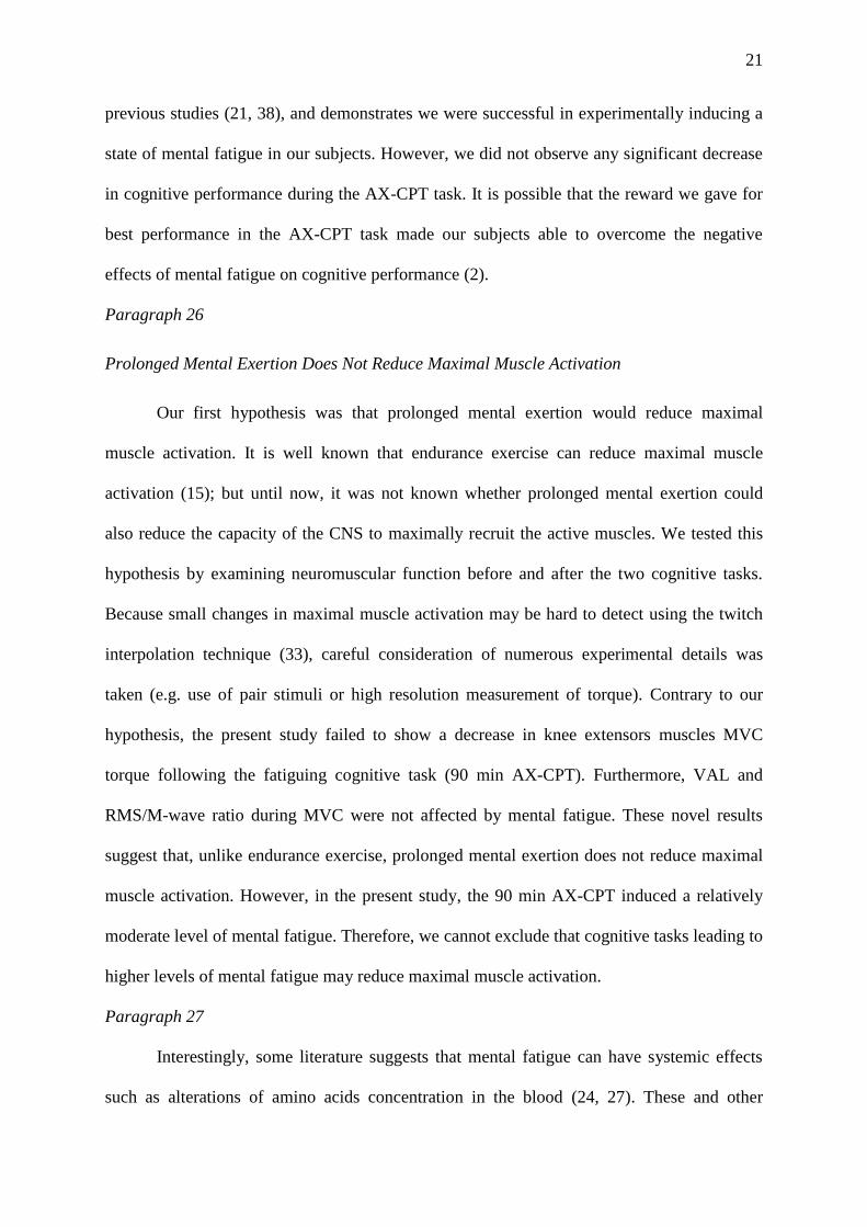
Figure 1. Graphical overview of the protocol for one session.
Order and timing was the same for each subject and each session. Q = psychological questionnaires, CT =
cognitive task, MVC = maximal voluntary contraction

7
Paragraph 7
Cognitive Tasks
Mental Fatigue Task. Mental fatigue was induced by asking the subject to perform the AX-
Continuous Performance Test (AX-CPT) (8) for 90 min on a personal computer. In this
cognitive task, sequences of letters were visually presented one at time in a continuous
fashion on a computer screen with black background. All letters were presented centrally, for
duration of 300 ms in 24-point uppercase Helvetica font. Each letter was followed by a 1200
ms interval, for a total of a 4500 ms delay between the presentation of cue and probe stimuli.
Participants sat in front of the computer screen and were instructed to press the keyboard
space bar on target trials and the control button otherwise. Any missed or incorrect response
activated a beep sound from two speakers as a prompt to increase speed and accuracy. To
further increase engagement in the mental fatigue task, a ticket for a professional sporting
event was given as a prize for the best performance. Feedback on performance was presented
on the computer screen every 30 minutes as a percentage of the maximum possible score.
Performance was scored automatically by the computer on the basis of correct responses and
response time. Target trials were defined as a cue-probe sequence in which the letter A (in
red) appeared as a cue and the letter X (in red) as the probe. To increase task difficulty, two
white distractor letters (except A, K, X or Y) were presented between the cue and probe (in
white). All other cue-probe sequences served as invalid cues and non-target probes. Letter
sequences were presented in pseudorandom order, such that target (AX) trials occurred with
70 % and non-target trials occurred with 30 % frequency.
Paragraph 8
Control Task. The non-fatiguing cognitive task consisted of watching ―Earth‖, a documentary
following the migration paths of four animal families (Alastair Fothergill and Mark Linfield,
2007), for 90 min on the same computer. During both cognitive tasks, heart rate (HR) was
8
recorded continuously using a HR monitor (Polar RS400, Polar Electro Oy, Kempele,
Finland).
Paragraph 9
Endurance Task. To evaluate endurance performance, subjects performed one prolonged
submaximal isometric contraction of the knee extensor muscles until exhaustion. Equipment
and subject position was similar to that used for mechanical recordings during the
neuromuscular function tests. A target value of 20% MVC torque was chosen. The MVC
before the cognitive task was used to calculate the target torque. Visual feedback of the
torque exerted during the endurance task was clearly displayed on a computer screen located
one meter in front of the subject. Torque feedback was represented as a horizontal line and
subjects were required to reach an upper target line fixed at the target level. The endurance
task terminated when torque fell below the required target value for more than 3 s despite
strong verbal encouragement (exhaustion) given by a research assistant blind to the nature of
the cognitive task previously performed by the subject. Endurance performance was
measured as time to exhaustion. Subjects were not aware of time during the endurance task,
and they were made aware of their times to exhaustion after the study was completed.
Perception of effort defined as ―the conscious sensation of how hard, heavy, and strenuous
exercise is‖ (18) was measured using the 15 points rating of perceived exertion (RPE) scale
(4). Standardized explanations of the scale were given to each subject before the warm-up.
Briefly subjects were asked to rate how hard they were driving their leg during the endurance
task. Leg RPE was assessed every 20 s. HR and electromyographic (EMG) signal (see
Electromyographic recordings) for the knee extensor muscles were continuously recorded
during the endurance task. HR was calculated for consecutive sampling intervals of 20 s.
Paragraph 10
Neuromuscular Function Tests
9
Electrical stimulation. Both single and double (100 Hz frequency) stimulation were used for
assessment of neuromuscular function. Transcutaneous electrically-evoked contractions of
the knee extensor muscles were induced by using a high-voltage (maximal voltage 400 V)
constant-current stimulator (model DS7 modified, Digitimer, Hertfordshire, UK). The
femoral nerve was stimulated using a monopolar cathode ball electrode (0.5 cm diameter)
pressed into the femoral triangle by the same experimenter during all tests. The site of
stimulation producing the largest resting twitch amplitude and compound muscle action
potential (M-Wave) was located and was marked on the skin so that it could be repeated
reliably before and after the cognitive task, and after the endurance task. The anode was a 50
cm² (10 × 5 cm) rectangular electrode (Compex SA, Ecublens, Switzerland) located in the
gluteal fold opposite the cathode. The optimal intensity of stimulation (i.e. that which
recruited all knee extensors motor unit) was considered to be reached when an increase in the
stimulation intensity did not induce a further increase in the amplitude of the twitch torque
and of the peak-to-peak amplitude of the knee extensors compound muscle action potentials
(M-waves). The stimulus duration was 1 ms and the interval of the stimuli in the doublet was
10 ms. Once the optimal intensity was found, 130% of this intensity was used and kept
constant throughout the session for each subject. The supramaximal intensities ranged from
60 to 140 mA. Methodology and supramaximal intensities are according to previous studies
(e.g. 29, 30).
Paragraph 11
Mechanical recordings. Mechanical parameters were recorded using a Biodex isokinetic
dynamometer (Biodex Medical Systems Inc., New York, USA). The axis of the dynamometer
was aligned with the knee axis, and the lever arm was attached to the shank with a strap.
Extraneous movement of the upper body was limited by two crossover shoulder harnesses
and a belt across the abdomen. Neuromuscular function tests were performed with the right
10
leg at a knee joint angle of 90° of flexion (0° = knee fully extended) and a hip angle of 90°.
The following parameters were analysed from the twitch response (average of 3 single
stimulation interspaced by 3 s): peak twitch (Tw), time to peak twitch (contraction time, Ct)
and half-relaxation time. The peak torque of the doublet (potentiated doublet, 5 s after the
MVC) was also analysed. MVC torque was considered as the peak torque attained during the
MVC. Voluntary activation level (VAL) during the MVC was estimated according to the
following formula:
VAL = 1-superimposed doublet amplitude
potentiated doublet amplitude
æ
èç
ö
ø÷ ´ 100
MVC
nstimulatioat MVC corresponding to Strojnik and Komi correction (4) was used if the
stimulation appears not at the MVC torque value. All VAL calculations were performed for a
MVCat stimulation between 95 and 100% MVC in order to ensure reliability of measurement.
Mechanical signals were digitized on-line at a sampling frequency of 1 kHz using a
computer, and stored for analysis with commercially available software (Acqnowledge 4.1
for MP Systems, Biopac Systems Inc., Goleta, USA). Timing of stimulation could be found
in Figure 1.
Paragraph 12
Electromyographic recordings. EMG of the vastus lateralis (VL) and rectus femoris (RF)
muscles was recorded with pairs of silver chloride circular (recording diameter of 10 mm)
surface electrodes (Swaromed, Nessler Medizintechnik, ref 1066, Innsbruck, Austria) with an
interelectrode (centre-to-centre) distance of 20 mm. Recording sites were then carefully
adjusted by eliciting the greatest M-wave amplitude for each muscle at a given intensity via
femoral nerve stimulation at the beginning of each testing session. Low resistance between
the two electrodes (< 5kΩ) was obtained by shaving the skin, and dirt were removed from the
skin using alcohol swabs. The reference electrode was attached to the patella of the left knee.

11
Myoelectrical signals were amplified with a bandwidth frequency ranging from 1 Hz to 5
kHz (common mode rejection ratio = 110 dB; impedance input = 1000 MΩ; gain = 1000 for
RF and 500 for VL), digitized on-line at a sampling frequency of 2 kHz using a computer,
and stored for analysis with commercially available software (Acqnowledge 4.1 for MP
Systems, Biopac Systems Inc., Goleta, USA). The root mean square (RMS), a measure of
EMG amplitude, was automatically calculated with the software.
Paragraph 13
Peak-to-peak amplitude and duration of the M-waves were analysed for VL and RF
muscles with the average of the three trials used for analysis. EMG amplitude of VL and RF
muscles during the knee extensors MVC was quantified as the RMS for a 0.5 s interval at
peak torque (250 ms interval either side of the peak torque). Maximal RMS values for VL
and RF muscles were then normalized by the M-wave peak-to-peak amplitude for the
respective muscles, in order to obtain RMS/M-wave ratio. This normalization procedure
accounted for peripheral influences including neuromuscular propagation failure and changes
in impedance from the EMG recordings. RMS EMG was calculated for consecutive sampling
intervals of 20 s during the endurance task for both VL and RF. The RMS EMG during
endurance task was normalized to the RMS EMG determined during the MVC precognitive
task.
Paragraph 14
Psychological Questionnaires
Mood. The Brunel Mood Scale (BRUMS) developed by Terry et al. (36) was used to quantify
current mood (―How do you feel right now?‖) before and after the cognitive tasks. This
questionnaire contains 24 items (e.g., ―angry, uncertain, miserable, tired, nervous, energetic‖)
divided into six respective subscales: anger, confusion, depression, fatigue, tension, and
vigor. The items are answered on a 5 point scale (0 = not at all, 1 = a little, 2 = moderately, 3
12
= quite a bit, 4 = extremely), and each subscales, with four relevant items, can achieve a raw
score in the range of 0 to 16. Only scores for the Fatigue and Vigour subscales were
considered in this study as subjective markers of mental fatigue.
Paragraph 15
Motivation. Motivation related to the endurance task was measured using the success
motivation and intrinsic motivation scales developed and validated by Matthews et al. (23).
Each scale consists of 7 items (e.g., ―I want to succeed on the task‖ and ―I am concerned
about not doing as well as I can‖) scored on a 5-point scale (0 = not at all, 1 = a little bit, 2 =
somewhat, 3 = very much, 4 = extremely). Therefore, total scores for these motivation scales
range between 0 and 28.
Paragraph 16
Statistics. All data are presented as means ± standard error of the means (SEM). Assumptions
of statistical tests such as normal distribution and sphericity of data were checked as
appropriate. Greenhouse-Geisser correction to the degrees of freedom was applied when
violations to sphericity were present. Paired t-tests were used to assess the effect of condition
(mental fatigue vs control) on time to exhaustion, motivation scores, HR at exhaustion, leg
RPE at exhaustion, and RMS at exhaustion. One-way repeated measure ANOVA was used to
test the effect of time (15-min blocks) on the number of incorrect answers, reaction time and
HR during the AX-CPT task. Fully repeated measure 2 x 2 ANOVAs were used to test the
effect of condition and time on mood before and after the cognitive tasks. Fully repeated
measure 2 x 3 ANOVAs were used to test the effect of condition and time on MVC torque,
VAL, M-wave parameters for each muscle, RMS/M-wave ratio, twitch properties, and peak
doublet torque before and after the cognitive tasks, and after the endurance task. Fully
repeated measure 2 x 7 ANOVAs were used to test the effect of condition and time on HR,
leg RPE, and RMS at isotime (time elapsed from the beginning of the endurance task to the
13
last measurement before exhaustion of the shortest performance). Significant main effects of
time and significant interactions were followed up with Bonferonni tests as appropriate.
Significance was set at 0.05 (2-tailed) for all analyses, which were conducted using the
Statistical Package for the Social Sciences, version 19 for Mac OS X (SPSS Inc., Chicago,
IL, USA).
RESULTS
Paragraph 17
Manipulation Checks. Heart rate decreased over time in both conditions (P<0.001) but it was
significantly higher in the mental fatigue condition (73 ± 1 beat/min) compared to the control
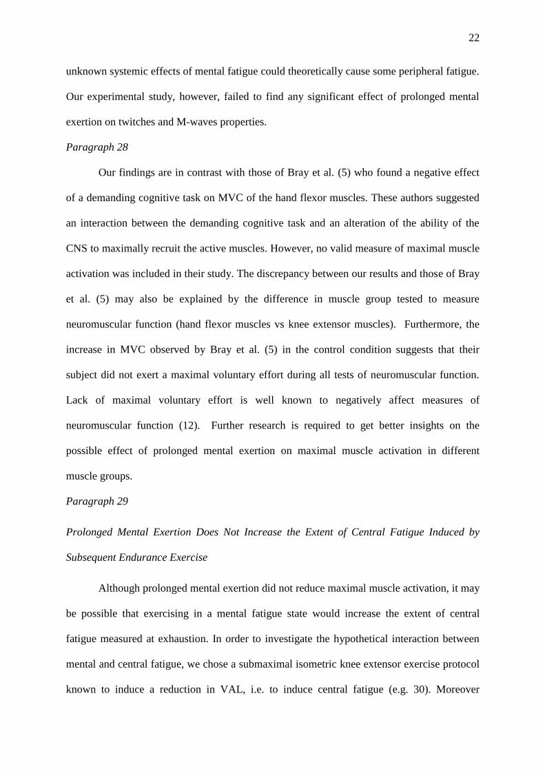
condition (69 ± 1 beat/min) (P=0.004) (Fig. 2B). The number of incorrect responses (Fig. 2C)
and reaction time (Fig. 2D) did not change significantly over time during the AX-CPT task.
Paragraph 18
The mood questionnaire revealed a significant decrease in Vigour after both the AX-CPT
task (9.0 ± 0.9 to 6.5 ± 0.9) and the control task (9.7 ± 0.6 to 7.1 ± 0.7) (P=0.003) with no
significant difference between conditions. However, there was a significant interaction for the
subjective fatigue (P=0.033). Follow-up tests demonstrated that Fatigue increased
significantly only after the AX-CPT task (P=0.007) with no significant change after the
control task (Fig. 2A).

14
Figure 2. Markers of mental fatigue.
A. Effect of cognitive tasks on self-reported fatigue. B. Heart rate during both cognitive tasks. C. Number of
incorrect responses during the mental fatigue task. D. Reaction time during the mental fatigue tasks. CT =
cognitive task. $$ Significant main effect of condition (P<0.01). ** Significant condition x time interaction
(P<0.01). ). ### Significant main effect of time (P < 0.001). Data are presented as means ± SEM
Paragraph 19
Effects of Mental Fatigue on Intrinsic and Success Motivation. There were no significant
differences between conditions in intrinsic motivation (mental fatigue condition 16.5 ± 1.3,
control condition 16.5 ± 1.1, P=1.000) and success motivation (mental fatigue condition 18.8
± 1.6, control condition 15.8 ± 1.8, P=0.111).
15
Paragraph 20
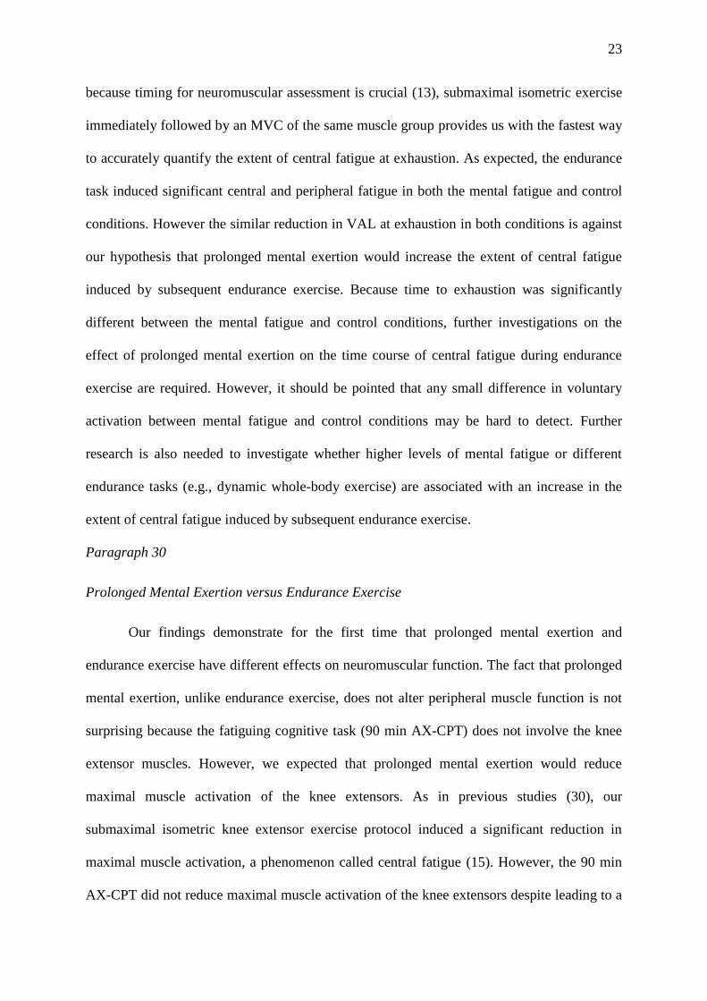
Effects of Mental Fatigue on Time to exhaustion, HR, EMG amplitude, and Perception of
Effort during the Endurance Task. Time to exhaustion (Fig. 3A) was 13 ± 4 % shorter in the
mental fatigue condition compared to the control condition (P=0.008). Individual times to
exhaustion were shorter in the mental fatigue condition compared to the control condition in
8 out of 10 subjects (Fig. 3B). Heart rate (Fig. 3D) increased significantly during the
endurance task (P<0.001) with no significant differences between conditions at both isotime
and exhaustion. EMG amplitude (RMS/RMS pre-cognitive task MVC) of the VL muscle (Fig
3C) increased significantly during the endurance task (P=0.003) with no significant
difference between conditions at isotime. At exhaustion, however, VL EMG amplitude
tended to be higher in the control condition (52.8 ± 6.8 %) compared to the mental fatigue
condition (41.5 ± 5.9 %) (P = 0.095). Leg RPE (Fig. 3E) increased significantly during the
endurance task (P<0.001) and it was significantly higher in the mental fatigue condition
compared to the control condition (P = 0.045), without interaction effect (P = 0.353). Leg
RPE at exhaustion was not significantly different between conditions.
16
Figure 3. Effects of cognitive tasks on time to exhaustion, and physiological and perceptual responses
during the endurance task.
A. Mean effect of mental fatigue on time to exhaustion. B. Individual effect of mental fatigue on time to
exhaustion. C. Root mean square (RMS) EMG of the vastus lateralis (VL) muscle during the endurance task.
Values are expressed as a percentage of the maximal value before the cognitive task. D. Heart rate (HR) during
the endurance task. E. Leg rating of perceived exertion (RPE) during the endurance task
17
$ Significant main effect of condition (P<0.05). $$ Significant main effect of condition (P<0.01). ## significant
main effect of time (P<0.01). ### Significant main effect of time (P<0.001). Data are presented as means ±
SEM
Paragraph 21
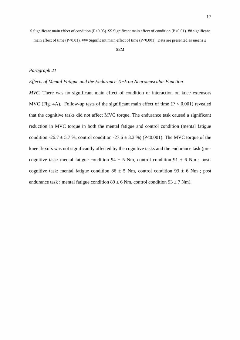
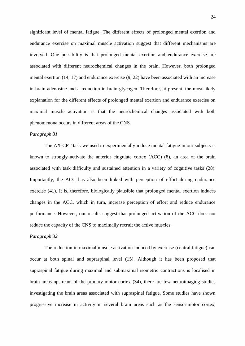
Effects of Mental Fatigue and the Endurance Task on Neuromuscular Function
MVC. There was no significant main effect of condition or interaction on knee extensors
MVC (Fig. 4A). Follow-up tests of the significant main effect of time (P < 0.001) revealed
that the cognitive tasks did not affect MVC torque. The endurance task caused a significant
reduction in MVC torque in both the mental fatigue and control condition (mental fatigue
condition -26.7 ± 5.7 %, control condition -27.6 ± 3.3 %) (P<0.001). The MVC torque of the
knee flexors was not significantly affected by the cognitive tasks and the endurance task (pre-
cognitive task: mental fatigue condition 94 ± 5 Nm, control condition 91 ± 6 Nm ; post-
cognitive task: mental fatigue condition 86 ± 5 Nm, control condition 93 ± 6 Nm ; post
endurance task : mental fatigue condition 89 ± 6 Nm, control condition 93 ± 7 Nm).
18
Figure 4. Effects of cognitive tasks and endurance task on central and peripheral parameters of
neuromuscular function.
A. Maximal voluntary contraction (MVC) torque of the knee extensors (KE). B. Peak torque of the doublet. C.
Peak twitch (Tw). D. Voluntary activation level (VAL). E. Root mean square (RMS)/Mmax (M-wave) ratio of
the vastus lateralis (VL) muscle. Values are expressed as a percentage of baseline values (pre cognitive task
values). CT = cognitive task, ET = endurance task.# Significant main effect of time (P<0.05). ## Significant
main effect of time (P<0.01). ### Significant main effect of time (P < 0.001). Data are presented as means ±
SEM
19
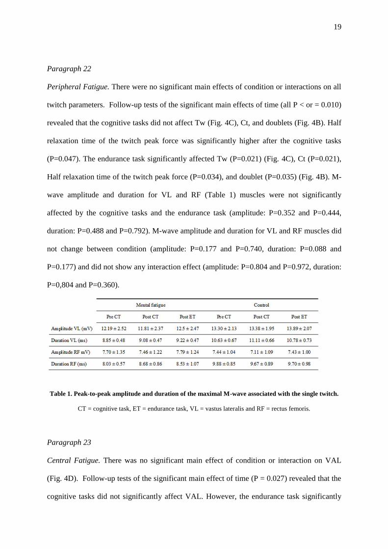
Paragraph 22
Peripheral Fatigue. There were no significant main effects of condition or interactions on all
twitch parameters. Follow-up tests of the significant main effects of time (all P < or = 0.010)
revealed that the cognitive tasks did not affect Tw (Fig. 4C), Ct, and doublets (Fig. 4B). Half
relaxation time of the twitch peak force was significantly higher after the cognitive tasks
(P=0.047). The endurance task significantly affected Tw (P=0.021) (Fig. 4C), Ct (P=0.021),
Half relaxation time of the twitch peak force (P=0.034), and doublet (P=0.035) (Fig. 4B). M-
wave amplitude and duration for VL and RF (Table 1) muscles were not significantly
affected by the cognitive tasks and the endurance task (amplitude: P=0.352 and P=0.444,
duration: P=0.488 and P=0.792). M-wave amplitude and duration for VL and RF muscles did
not change between condition (amplitude: P=0.177 and P=0.740, duration: P=0.088 and
P=0.177) and did not show any interaction effect (amplitude: P=0.804 and P=0.972, duration:
P=0,804 and P=0.360).
Table 1. Peak-to-peak amplitude and duration of the maximal M-wave associated with the single twitch.
CT = cognitive task, ET = endurance task, VL = vastus lateralis and RF = rectus femoris.
Paragraph 23
Central Fatigue. There was no significant main effect of condition or interaction on VAL
(Fig. 4D). Follow-up tests of the significant main effect of time (P = 0.027) revealed that the
cognitive tasks did not significantly affect VAL. However, the endurance task significantly
20
reduced VAL (P=0.024). Similarly, there was no significant main effect of condition or
interaction on RMS/M-wave ratio of the RF and VL (Fig. 4E) muscles. Follow-up tests of
the significant main effects of time (all P < 0.009) revealed that the cognitive tasks did not
affect RMS/M of the RF and VL muscles. However, RMS/M decreased significantly after the
endurance task for both the RF (P < 0.001) and VL (P=0.010) muscles.
DISCUSSION
Paragraph 24
The main aim of the present study was to test the hypotheses that prolonged mental exertion
leading to mental fatigue i) would reduce maximal muscle activation and ii) would increase
the extent of central fatigue induced by subsequent endurance exercise. Contrary to our
hypotheses, this study demonstrates that prolonged mental exertion does not lead to any
impairment in neuromuscular function. In accordance with previous findings (21), the
negative effect of prolonged mental exertion on endurance performance seems to be mediated
by the higher perception of effort experienced by mentally fatigued subjects during the
endurance task.
Paragraph 25
Prolonged Mental Exertion and Mental Fatigue
The higher HR observed during the AX-CPT task compared to watching a movie confirms
the demanding nature of this cognitive task. In fact, an increase in HR and other
cardiovascular changes are associated with exertion of effort during cognitive tasks (32).
Given its demanding nature, it is not surprising that 90-min of the AX-CPT task induced a
significant increase in subjective feelings of fatigue. This effect is in accordance with
21
previous studies (21, 38), and demonstrates we were successful in experimentally inducing a
state of mental fatigue in our subjects. However, we did not observe any significant decrease
in cognitive performance during the AX-CPT task. It is possible that the reward we gave for
best performance in the AX-CPT task made our subjects able to overcome the negative
effects of mental fatigue on cognitive performance (2).
Paragraph 26
Prolonged Mental Exertion Does Not Reduce Maximal Muscle Activation
Our first hypothesis was that prolonged mental exertion would reduce maximal
muscle activation. It is well known that endurance exercise can reduce maximal muscle
activation (15); but until now, it was not known whether prolonged mental exertion could
also reduce the capacity of the CNS to maximally recruit the active muscles. We tested this
hypothesis by examining neuromuscular function before and after the two cognitive tasks.
Because small changes in maximal muscle activation may be hard to detect using the twitch
interpolation technique (33), careful consideration of numerous experimental details was
taken (e.g. use of pair stimuli or high resolution measurement of torque). Contrary to our
hypothesis, the present study failed to show a decrease in knee extensors muscles MVC
torque following the fatiguing cognitive task (90 min AX-CPT). Furthermore, VAL and
RMS/M-wave ratio during MVC were not affected by mental fatigue. These novel results
suggest that, unlike endurance exercise, prolonged mental exertion does not reduce maximal
muscle activation. However, in the present study, the 90 min AX-CPT induced a relatively
moderate level of mental fatigue. Therefore, we cannot exclude that cognitive tasks leading to
higher levels of mental fatigue may reduce maximal muscle activation.
Paragraph 27
Interestingly, some literature suggests that mental fatigue can have systemic effects
such as alterations of amino acids concentration in the blood (24, 27). These and other
22
unknown systemic effects of mental fatigue could theoretically cause some peripheral fatigue.
Our experimental study, however, failed to find any significant effect of prolonged mental
exertion on twitches and M-waves properties.
Paragraph 28
Our findings are in contrast with those of Bray et al. (5) who found a negative effect
of a demanding cognitive task on MVC of the hand flexor muscles. These authors suggested
an interaction between the demanding cognitive task and an alteration of the ability of the
CNS to maximally recruit the active muscles. However, no valid measure of maximal muscle
activation was included in their study. The discrepancy between our results and those of Bray
et al. (5) may also be explained by the difference in muscle group tested to measure
neuromuscular function (hand flexor muscles vs knee extensor muscles). Furthermore, the
increase in MVC observed by Bray et al. (5) in the control condition suggests that their
subject did not exert a maximal voluntary effort during all tests of neuromuscular function.
Lack of maximal voluntary effort is well known to negatively affect measures of
neuromuscular function (12). Further research is required to get better insights on the
possible effect of prolonged mental exertion on maximal muscle activation in different
muscle groups.
Paragraph 29
Prolonged Mental Exertion Does Not Increase the Extent of Central Fatigue Induced by
Subsequent Endurance Exercise
Although prolonged mental exertion did not reduce maximal muscle activation, it may
be possible that exercising in a mental fatigue state would increase the extent of central
fatigue measured at exhaustion. In order to investigate the hypothetical interaction between
mental and central fatigue, we chose a submaximal isometric knee extensor exercise protocol
known to induce a reduction in VAL, i.e. to induce central fatigue (e.g. 30). Moreover
23
because timing for neuromuscular assessment is crucial (13), submaximal isometric exercise
immediately followed by an MVC of the same muscle group provides us with the fastest way
to accurately quantify the extent of central fatigue at exhaustion. As expected, the endurance
task induced significant central and peripheral fatigue in both the mental fatigue and control
conditions. However the similar reduction in VAL at exhaustion in both conditions is against
our hypothesis that prolonged mental exertion would increase the extent of central fatigue
induced by subsequent endurance exercise. Because time to exhaustion was significantly
different between the mental fatigue and control conditions, further investigations on the
effect of prolonged mental exertion on the time course of central fatigue during endurance
exercise are required. However, it should be pointed that any small difference in voluntary
activation between mental fatigue and control conditions may be hard to detect. Further
research is also needed to investigate whether higher levels of mental fatigue or different
endurance tasks (e.g., dynamic whole-body exercise) are associated with an increase in the
extent of central fatigue induced by subsequent endurance exercise.
Paragraph 30
Prolonged Mental Exertion versus Endurance Exercise
Our findings demonstrate for the first time that prolonged mental exertion and
endurance exercise have different effects on neuromuscular function. The fact that prolonged
mental exertion, unlike endurance exercise, does not alter peripheral muscle function is not
surprising because the fatiguing cognitive task (90 min AX-CPT) does not involve the knee
extensor muscles. However, we expected that prolonged mental exertion would reduce
maximal muscle activation of the knee extensors. As in previous studies (30), our
submaximal isometric knee extensor exercise protocol induced a significant reduction in
maximal muscle activation, a phenomenon called central fatigue (15). However, the 90 min
AX-CPT did not reduce maximal muscle activation of the knee extensors despite leading to a
24
significant level of mental fatigue. The different effects of prolonged mental exertion and
endurance exercise on maximal muscle activation suggest that different mechanisms are
involved. One possibility is that prolonged mental exertion and endurance exercise are
associated with different neurochemical changes in the brain. However, both prolonged
mental exertion (14, 17) and endurance exercise (9, 22) have been associated with an increase
in brain adenosine and a reduction in brain glycogen. Therefore, at present, the most likely
explanation for the different effects of prolonged mental exertion and endurance exercise on
maximal muscle activation is that the neurochemical changes associated with both
phenomenona occurs in different areas of the CNS.
Paragraph 31
The AX-CPT task we used to experimentally induce mental fatigue in our subjects is
known to strongly activate the anterior cingulate cortex (ACC) (8), an area of the brain
associated with task difficulty and sustained attention in a variety of cognitive tasks (28).
Importantly, the ACC has also been linked with perception of effort during endurance
exercise (41). It is, therefore, biologically plausible that prolonged mental exertion induces
changes in the ACC, which in turn, increase perception of effort and reduce endurance
performance. However, our results suggest that prolonged activation of the ACC does not
reduce the capacity of the CNS to maximally recruit the active muscles.
Paragraph 32
The reduction in maximal muscle activation induced by exercise (central fatigue) can
occur at both spinal and supraspinal level (15). Although it has been proposed that
supraspinal fatigue during maximal and submaximal isometric contractions is localised in
brain areas upstream of the primary motor cortex (34), there are few neuroimaging studies
investigating the brain areas associated with supraspinal fatigue. Some studies have shown
progressive increase in activity in several brain areas such as the sensorimotor cortex,
25
supplementary motor areas, frontal cortex, and the insular cortex during submaximal
fatiguing exercise (16, 31, 39, 40). However, it is not clear whether the concept of central
fatigue is meaningful during submaximal muscle contractions (35). In fact, these changes in
cerebral activity during submaximal fatiguing exercise are likely to reflect brain adaptations
to compensate for spinal and/or peripheral muscle fatigue rather than mechanisms of
supraspinal fatigue. To the best of our knowledge, only van Duinen et al. (39) have
investigated the brain areas associated with supraspinal fatigue by measuring their activity
during MVCs performed before and after fatiguing exercise. These authors showed a
significant decrease in activity of the supplementary motor areas and, to a lesser extent, in
parts of the paracentralgyrus, right putamen, and in a small cluster of the left parietal
operculum. The fact that central fatigue was not associated with changes in ACC activity
suggests that the brain areas affected by prolonged mental exertion and endurance exercise
are different.
Paragraph 33
Furthermore, we have to consider that the neurochemical changes induced by
prolonged mental exertion are likely to be confined to the brain, whilst some of the
neurochemical changes leading to central fatigue may also occur at spinal level (15).
Therefore, the different effects of prolonged mental exertion and endurance exercise on
maximal muscle activation could be explained by i) the different brain areas affected by
prolonged mental exertion and endurance exercise, and ii) the spinal alterations likely to
occur during endurance exercise but not during prolonged mental exertion..
Paragraph 34
26
Mental Fatigue, Perceived Exertion and the Psychobiological Model of Endurance
Performance
Finally, the present results provide experimental evidence that higher perception of
effort induced by prolonged mental exertion is not associated with lower muscle activation
before exercise. In fact, the higher perception of effort experienced by mentally fatigued
subjects occurs despite no reduction of maximal muscle activation before the endurance task,
and similar extent of central fatigue at exhaustion in the mental fatigue and control
conditions. However, the increase in perception of effort occurring over time during the
endurance task in both conditions may be caused, at least in part, by the central and
peripheral fatigue induced by endurance exercise. In fact, in the presence of significant
muscle fatigue, an increase in central motor command is required to maintain the same
submaximal force. Because the sensory signal for perception of effort is the corollary
discharge of the central motor command, the increase in central motor command required to
overcome muscle fatigue is reflected in a significant increase in perception of effort (10, 19).
A previous study (21) suggests that the higher perception of effort experienced by mentally
fatigued subjects during the endurance task may be due to altered central processing of
sensory signals. However, further research is required to understand the neurophysiological
mechanisms underlying the negative effect of mental fatigue on perception of effort during
endurance exercise.
Paragraph 35
Similar to previous findings on the effect of mental fatigue on endurance performance
during dynamic whole-body exercise (21), we found that mental fatigue significantly reduces
time to exhaustion during submaximal isometric knee extensor exercise. These results
suggest that mental fatigue has a negative effect on endurance performance regardless of the
type of contraction and muscle mass active during endurance exercise.
27
Paragraph 36
A plausible explanation for the negative effect of mental fatigue on endurance
performance is provided by the psychobiological model of endurance performance (20) based
on Motivational Intensity Theory (7). This model postulates that exhaustion is a form of task
disengagement that occurs when subjects perceive the task as being impossible to complete
despite their maximal effort, or when the effort required by the task exceeds the upper limit
of what people are willing to do (potential motivation). Accordingly, a reduction in time to
exhaustion can occur either because of an increase in perception of effort or a reduction in
potential motivation. In accordance to a previous study (21), we did not measure any negative
effect of mental fatigue on intrinsic and success motivation related to the endurance task.
Therefore, the only mechanism that can explain the negative effect of prolonged mental
exertion on time to exhaustion is the higher perception of effort experienced by mentally
fatigued subjects during the endurance task. As leg RPE increased similarly over time in both
conditions, mentally fatigued subjects reached their maximal level of perceived exertion and
disengaged from the endurance task earlier than in the control condition.
Paragraph 37
Conclusions and Perspectives
The present study provides the first experimental evidence that prolonged mental exertion
does not alter neuromuscular function measured as maximal muscle activation and central
fatigue induced by subsequent endurance exercise. These findings suggest that prolonged
mental exertion and endurance exercise affect different areas of the CNS. Future studies on
brain and endurance performance should investigate the specific mechanisms of mental
fatigue and central fatigue without making the wrong assumption that these two phenomena
are two different aspects of the same central alterations. Because perception of effort is the
most likely mediator of the negative effect of mental fatigue on endurance performance,

28
further studies are required to investigate the neurophysiological alterations associated with
the higher perception of effort experienced by mentally fatigued subjects during endurance
exercise. On a more practical perspective, the present study suggests that the negative impact
of mental fatigue on physical performance is limited to endurance and may not have a
negative impact on performance of short maximal voluntary efforts such as sprint or jump.
ACKNOWLEDGEMENTS
The authors thank Cyril Sirandre and Yves Ballay for their technical advices and
support.
CONFLICT OF INTEREST
The results of the present study do not constitute endorsement by ACSM. No conflict
of interest, financial or otherwise is declared by the authors. No funding was received for this
study.
29
REFERENCES
1. Amann M. Central and peripheral fatigue: interaction during cycling exercise in
humans. Med Sci Sports Exerc. 2011;43(11):2039-45.
2. Baumeister RF, Vohs KD. Self-Regulation, Ego Depletion, and Motivation
. Soc Personal Psychol Compass. 2007;1(1):115-28.
3. Boksem MA, Tops M. Mental fatigue: costs and benefits. Brain Res Rev.
2008;59(1):125-39.
4. Borg G. Borg's Perceived exertion and pain scales. Champaign, IL: Human Kinetics;
1998, viii, 104 p. p.
5. Bray SR, Graham JD, Martin Ginis KA, Hicks AL. Cognitive task performance
causes impaired maximum force production in human hand flexor muscles. Biol
Psychol. 2012;89(1):195-200.
6. Bray SR, Martin Ginis KA, Hicks AL, Woodgate J. Effects of self-regulatory strength
depletion on muscular performance and EMG activation. Psychophysiology.
2008;45(2):337-43.
7. Brehm JW, Self EA. The intensity of motivation. Annu Rev Psychol. 1989;40:109-31.
8. Carter CS, Braver TS, Barch DM, Botvinick MM, Noll D, Cohen JD. Anterior
cingulate cortex, error detection, and the online monitoring of performance. Science.
1998;280(5364):747-9.
9. Davis JM, Zhao Z, Stock HS, Mehl KA, Buggy J, Hand GA. Central nervous system
effects of caffeine and adenosine on fatigue. Am J Physiol Regul Integr Comp
Physiol. 2003;284(2):R399-404.
10. de Morree HM, Klein C, Marcora SM. Perception of effort reflects central motor
command during movement execution. Psychophysiology. 2012;(49):1242–53.
11. Di Giulio C, Daniele F, Tipton CM. Angelo Mosso and muscular fatigue: 116 years
after the first Congress of Physiologists: IUPS commemoration. Adv Physiol Educ.
2006;30(2):51-7.
12. Enoka RM. Mechanisms of muscle fatigue: Central factors and task dependency. J
Electromyogr Kinesiol. 1995;5(3):141-9.
13. Froyd C, Millet GY, Noakes TD. The Development of Peripheral Fatigue and Short-
Term Recovery During Self-Paced High-Intensity Exercise. J Physiol. 2012.
14. Gailliot MT. Unlocking the Energy Dynamics of Executive Functioning Linking
Executive Functioning to Brain Glycogen. Perspect Psychol Sci. 2008;3(4):Adv
Physiol EducBiol Psychol245-63.
15. Gandevia SC. Spinal and supraspinal factors in human muscle fatigue. Physiol Rev.
2001;81(4):1725-89.
16. Liu JZ, Shan ZY, Zhang LD, Sahgal V, Brown RW, Yue GH. Human brain activation
during sustained and intermittent submaximal fatigue muscle contractions: an FMRI
study. J Neurophysiol. 2003;90(1):300-12.
17. Lorist MM, Tops M. Caffeine, fatigue, and cognition. Brain Cogn. 2003;53(1):82-94.
18. Marcora SM. Effort: perception of. In: G EB editor. Encyclopedia of Perception2009.
19. Marcora SM, Bosio A, de Morree HM. Locomotor muscle fatigue increases
cardiorespiratory responses and reduces performance during intense cycling exercise
independently from metabolic stress. Am J Physiol Regul Integr Comp Physiol.
2008;294(3):R874-83.
20. Marcora SM, Staiano W. The limit to exercise tolerance in humans: mind over
muscle? Eur J Appl Physiol. 2010;109(4):763-70.
21. Marcora SM, Staiano W, Manning V. Mental fatigue impairs physical performance in
humans. J Appl Physiol. 2009;106(3):857-64.
30
22. Matsui T, Soya S, Okamoto M, Ichitani Y, Kawanaka K, Soya H. Brain glycogen
decreases during prolonged exercise. J Physiol. 2011;589(Pt 13):3383-93.
23. Matthews G, Campbell S, Falconer S. Assessment of motivational states in
performance environments. . Proc Annu Meeting Human Factors Ergonomics Soc.
2001;(45):906-10.
24. Mizuno K, Tanaka M, Nozaki S et al. Mental fatigue-induced decrease in levels of
several plasma amino acids. J Neural Transm. 2007;114(5):555-61.
25. Mosso A. Fatigue. London: Swan Sonnenschein & Co. Ltd; 1906, 334 p.
26. Newsholme EA, Blomstrand E, Ekblom B. Physical and mental fatigue: metabolic
mechanisms and importance of plasma amino acids. Br Med Bull. 1992;48(3):477-95.
27. Nozaki S, Tanaka M, Mizuno K et al. Mental and physical fatigue-related
biochemical alterations. Nutrition. 2009;25(1):51-7.
28. Paus T. Primate anterior cingulate cortex: where motor control, drive and cognition
interface. Nat Rev Neurosci. 2001;2(6):417-24.
29. Place N, Lepers R, Deley G, Millet GY. Time course of neuromuscular alterations
during a prolonged running exercise. Med Sci Sports Exerc. 2004;36(8):1347-56.
30. Place N, Maffiuletti NA, Ballay Y, Lepers R. Twitch potentiation is greater after a
fatiguing submaximal isometric contraction performed at short vs. long quadriceps
muscle length. J Appl Physiol. 2005;98(2):429-36.
31. Post M, Steens A, Renken R, Maurits NM, Zijdewind I. Voluntary activation and
cortical activity during a sustained maximal contraction: an fMRI study. Hum Brain
Mapp. 2009;30(3):1014-27.
32. Richter M, Friedrich A, Gendolla GH. Task difficulty effects on cardiac activity.
Psychophysiology. 2008;45(5):869-75.
33. Shield A, Zhou S. Assessing voluntary muscle activation with the twitch interpolation
technique. Sports Med. 2004;34(4):253-67.
34. Taylor JL, Allen GM, Butler JE, Gandevia SC. Supraspinal fatigue during intermittent
maximal voluntary contractions of the human elbow flexors. J Appl Physiol.
2000;89(1):305-13.
35. Taylor JL, Gandevia SC. A comparison of central aspects of fatigue in submaximal
and maximal voluntary contractions. J Appl Physiol. 2008;104(2):542-50.
36. Terry PC, Lane AM, Fogarty GJ. Construct validity of the Profile of Mood States —
Adolescents for use with adults. Psychol Sport Exerc. 2003;4:125-39.
37. van der Linden D, Frese M, Meijman TF. Mental fatigue and the control of cognitive
processes: effects on perseveration and planning. Acta Psychol (Amst).
2003;113(1):45-65.
38. van der Linden D, Massar SA, Schellekens AF, Ellenbroek BA, Verkes RJ. Disrupted
sensorimotor gating due to mental fatigue: preliminary evidence. Int J Psychophysiol.
2006;62(1):168-74.
39. van Duinen H, Renken R, Maurits N, Zijdewind I. Effects of motor fatigue on human
brain activity, an fMRI study. NeuroImage. 2007;35(4):1438-49.
40. van Duinen H, Renken R, Maurits NM, Zijdewind I. Relation between muscle and
brain activity during isometric contractions of the first dorsal interosseus muscle. Hum
Brain Mapp. 2008;29(3):281-99.
41. Williamson JW, McColl R, Mathews D, Mitchell JH, Raven PB, Morgan WP.
Hypnotic manipulation of effort sense during dynamic exercise: cardiovascular
responses and brain activation. J Appl Physiol. 2001;90(4):1392-9.

31
Recommended