PRIMER OF NECK AND BACK
INJURIES FOR AIIORNEYS
AND STAFF 11lird Edition
by Gerald A. Schwartz, B.S., Pharmacy, J.D.
Virginia Trial Lawyers Association
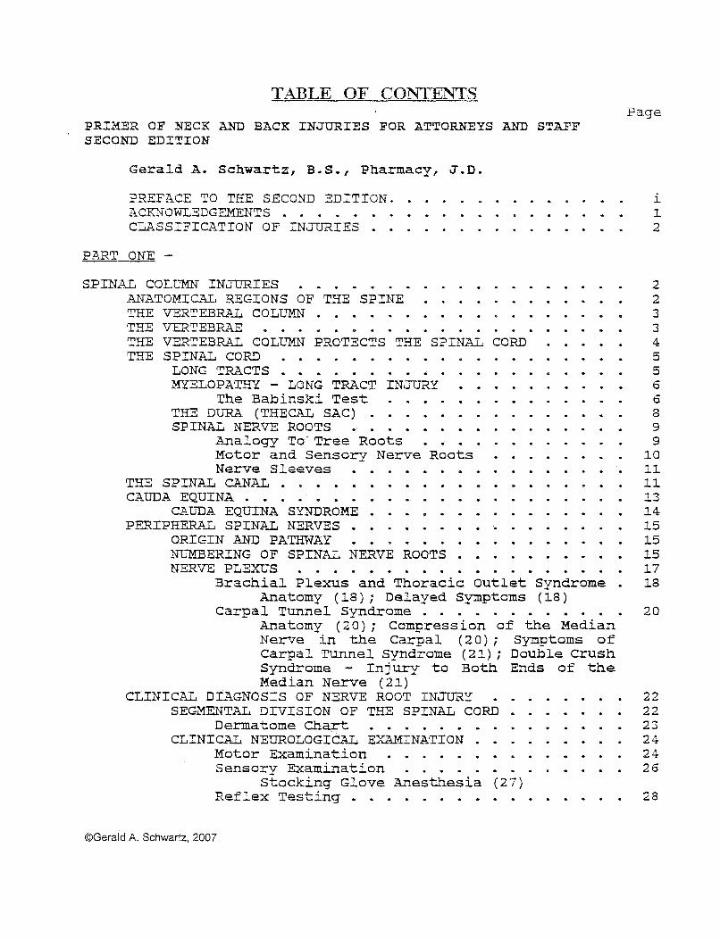
TABLE OF CONTENTS Page
PRIMER OF NECK AND BACK INJURIES FOR ATTORNEYS AND STAFF SECOND EDITION
Gerald A. Schwartz, B.S., Pharmacy, J.D.
PREFACE TO THE SECOND EDITION .. ACKNOWLEDGEMENTS . . . . . . . . CLASSIFICATION OF INJURIES . .
i 1. 2
PART ONE -
SPINAL COLUMN INJURIES • . . . . . . . . . . . . . . . 2 ANATOMI~~ REGIONS OF THE SPINE . . . . . . . . . 2 THE VERTEBRAL COLUMN . . . . . . . . . . . . . 3 THE VERTEBRAE . . . . . • . . . . . . . . . . . . 3 THE VERTEBRAL COLUMN PROTECTS THE SPINAL CORD . . . . 4 THE SPINAL CORD . . . . . . . . . . . . . . . . • . 5
LONG TRACTS . . . . . • . . . . . . . . . 5 MYELOPATHY - LONG TRACT INJURY . • . . . . . . . 6
The Babinski Test . • • . • . • . . . . 6 THE DURA (THECAL SAC) . . . . . . . . . . . • . 8 SPINAL NERVE ROOTS . . . . . . . . . . . 9
Analogy To Tree Roots . . • . . • . 9 Motor and Sensory Nerve Roots . . . 1.0 Nerve Sleeves • • . . . • . . . 11.
THE SPINAL CANAL . . . . . . . . . . . . 11. CAUDA EQUINA . • . . · . . . . . . . . . . . . . . . 1.3
CAUDA EQUINA SYNDROME . . . . . . . . . . . . 1.4 PERIPHERAL SPINAL NERVES . . . . . . . . . . . . . 15
ORIGIN AND PATHWAY . . . . . . . 1.5 NUMBERING OF SPINAL NERVE ROOTS . . . . • . . 1.5 NERVE PLEXUS . . . . . . . . . . • . . . . . . . 1. 7
Brachial Plexus and Thoracic outlet Syndrome . 1.8 Anatomy (1.8); Delayed Symptoms (18)
Carpal Tunnel Syndrome . . . • . . • . • . . 20 Anatomy (20); Compression of the Median Nerve in the Carpal ( 2 0) ; Symptoms of Carpal Tunnel Syndrome (21.); Double crush Syndrome - Injury to Both Ends of the Median Nerve (21.)
CLINICAL DIAGNOSIS OF NERVE ROOT INJURY . . . 22 SEGMENTAL DIVISION OF THE SPINAL CORD . . . • . . 22
Dermatome Chart • • • . . • • . . • . . 23 CLINICAL NEUROLOGICAL EXAMINATION . . . . 24
Motor Examination . • . . . . • . . 24 Sensory Examination . . • . • . . 26
Stocking Glove Anesthesia (27) Reflex Testing . • . . . . • • • . . . . • . 28
©Gerald A. Schwartz, 2007
THE
EL~LES INVOLVING SPECIFIC NERVE ROOT LEVELS . The Fifth Cervical Nerve Root,(C-5) ..... The Fourth Lumbar Nerve Root (L4) The Fifth Lumbar Nerve Root (LS) ...... . The First Sacral Level (S-1) ... . Dermatomes of the Lumbosacral Nerve Roots
FUNCTIONAL UNIT OF THE SPINE . . . . . . . .
30 30 31 32 33 35 36
ANATOMY OF Al~ INTERVERTEBRAL DISC . 37 37 NUMBERING . . . . . . . . . .
ANNULUS FIBROSUS (OUTSIDE FIBROUS RING OF A DISC) 38
NUCLEUS PULPOSUS (JELLY-LIKE CENTER OF DISC) 39
BULGING, PROTRUDED AND HERNIATED DISCS 40
PATHOLOGICAL CHANGES IN THE NUCLEUS PULPOSUS 42
SITES OF DISC HERNIATION . . . . . . . . . . . . . . 43 ANATOMY OF THE ANTERIOR AND POSTERIOR LONGITUDINAL
LI G.AMENT S . . . . . . . . . . . . 4 3 ILLUSTRATIONS OF DISC HERNIATIONS . . . . . . . . . . . 45 DISC HERNIATION AND SYMPTOMS . . . . . . . . . . • . 46
RADICULAR SYMPTOMS RESULT FROM COMPRESSION OF THE NERVE ROOT . . . • • • . . . . . . . . . . . . 4 6
DIAMETER OF THE SPINAL CANAL . . . . . . . . . • . 47 PATIENTS WITH ASYMPTOMATIC DISC HERNIATION MAY
DEVELOP SYMPTOMS IN LATER YEARS . . . • . 48 DISC HERNIATION COMPRESSES THE LOWER ROOT . . . 48
RADICULOPATHY AND RADICULITIS . . • . . COUGHING, SNEEZING AND BOWEL STRAINING
49 50
SCIATICA (IRRITATION OF THE SCIATIC NERVE) . . . . 51 ANATOMY . • . . . . . . • . • . . . . . . . . . 51 SYMPTOMS OF SCIATICA . . . . . . • . . . . . . . . . 52 ANATOMICAL BASIS FOR SCIATIC STRETCH TESTS . . • . . 53 THE STRAIGHT LEG RAISING TEST . . . . . . . 54
PERFORMING THE TEST . . . . . . . . . . . . . . . . 54 POSITIVE TEST . . . . . . . . . . . . . . . . . . . 55 THE VARIATION - SEATED STRAIGHT LEG-RAISING TEST 56
OSTEOARTHRITIS OF THE SPINE . . . . • . . . . . . . . . . . . 58 HYPERTROPHIC DEGENERATIVE CHANGES . . . . . . . . . 58 DEGENERATIVE DISC DISEASE - THE AGING PROCESS . 59 PEOPLE WITH DEGENERATIVE DISC DISEASE OFTEN HAVE NO
SYMPTOMS • . . . • . . • . . . . . . . . . 61 TRAUMA PRECIPITATES SYMPTOMS OF PREEXISTING,
ASYMPTOMATIC OSTEOARTHRITIS . • . . . . . 61 ANALOGY OF OSTEOARTHRITIS OF THE SPINE TO A GRAY
HAIR AND A MATCH . . . . . . . . . . . . . . . 64
MINOR IMPACT CASE - USE PREEXISTING OSTEOARTHRITIS AS AN OFFENSE • . . . . . . . . . . • . . Closing Argument - Analogy to Eggs and Golf
Balls . . . . . . . . . . . . . . . . TRAUMA ITSELF MAY CAUSE OSTEOARTHRITIS • o • •
FACET JOINT - OSTEOARTHRITIS o • • • • • • •
SPINAL STENOSIS . . . • . . . . . . . • . . . •
SPONDYLOLYSIS AND SPONDYLOLISTHESIS o • • • • • • • • • •
DEFECT IN THE PARS INTERARTICULARIS . • . PREEXISTING CONDITION OFTEN WITH NO SYMPTOMS . THE ROLE OF TRAUMA IN SPONDYLOLYSIS AND
SPONDYLOLISTHESIS . . • . • • . • • . • . • . • • .
PART TWO -
65
65 66 67 69
71 72 73
73
SOFT TISSUE INJURIES • • • . • . • • • . • • • . • • . . 75 MECHANISM OF INJURY - FORCED HYPEREXTENSION-HYPERFLEXION
MOTION . • • . . • • . • • . • • . • • 7 5 REAR-END COLLISIONS • • • • . • • . . • . . 75 HEAD-ON COLLISIONS • • • • • . . . • . • • 76 SIDE COLLISIONS . • . • • . • • . • . • . . • . 76 THE SUDDEN STOP • o • • • • • • • • 76
FACTORS AFFECTING THE SEVERITY OF INJURY 77 EQUATING BODILY INJURY WITH THE EXTENT OF PROPERTY
DAMAGE CONDEMNED • • . • • • • . • • . . • 78 THE POSITION OF THE HEAD AT IMPACT . • . . 78 ACCELERATION OF THE CAR BEING STRUCK • • . . • 79 SURPRISE COLLISIONS AND BRACING . • . • 80 PARAMETERS OF THE PLAINTIFF'S VEHICLE • . • • . 80
Energy Absorbing Built to Collapse Vehicles . • . . . . . • . . . . 8 o
Head Restraints • . • . • • . . . . . 81 Seatbacks • . • . . . . . . . 82 Seatbelts • • . . • . . • . . 82 Human Factors . . . 0 • • • • • • 83
CATEGORIES OF SOFT TISSUE INJURIES . . • . . . . . . 83 INJURIES TO LIGAMENTS, MUSCLES AND FASCIA •. o 83
Spinal Ligaments . . . . . . . • • . . . . 83 Ligaments Hold the Spine in Place (83); Interspinal Ligament (84); Ligamentum Flavum (The Yellow Ligament) (84); The Anterior and Posterior Longitudinal (84); Diagram of the Spinal Ligaments (85); Injury to the Spinal Ligaments (86)
MUSCLES AND FASCIA o • • • • • • • • • • • • • • 88 FASCIA . . • . • • . . • . . • • • . • • . 88 PARASPINAL MUSCLES . . o • • • • • • • o • 88 TRAPEZIUS MUSCLES . . • • • • • • • 88 STERNOCLEIDOMASTOID MUSCLE . • . . 89 SPLENIUS CAPITIS MUSCLE • • . . . . • • o • • • 89 INJURY TO MUSCLE AND FASCIA . • . o 89 MYOFASCIAL PAIN SYNDROME (MFPS) 90

SPRAIN AND STRAIN INJURIES . . . DEFINITION . . . . . . . .
Reference to Levels of the Spine and Specific
91 91
Muscles . . . . . . . . . . . 92 Strain and Sprain are Distinct Injuries . . . 92
HEALING: "ONCE A SCAR, ALWAYS A SCAR" . . . . 92 SYMPTOMS OF STRAIN AND SPRAIN INJURIES . . . . 93
Onset . . . . . . . . . . . . . . . . • . 93 Referred Pain o • o • • • • • • • • • • • 94 Muscle Spasm, Stiffness and Decreased Range of
Motion . . • . . . . . . . . . . . . 95 Headache . . . . . . . . . . . . . . . . . 96
Muscle Tension Headache (96); Vascular Headaches (96); Psychogenic (Emotional) Headache ( 9 7)
BLOOD VESSEL INJURY . . . . . . . . . . . . . . . . 97 Common Symptoms Resulting From Injury to the
Vertebral Artery . . . . o • 98 SYMPATHETIC NERVES . . . . . . . . . . 99
Anatomy of Sympathetic Nerves . . . . . . 99 Function of sympathetic Nerves . . • . 99
Blue Arms and Cold Hands (100); Symptoms Resulting From Injuries to the Sympathetic Nerves (101)
BRAIN CONCUSSION (POST-CONCUSSION SYNDROME) . . . . 102 INJURY TO THE ESOPHAGUS AND TMJ JOINT . . . . . . • 103
NAMES OF HYPEREXTENSION-HYPERFLEXION INJURIES . . . 103 DIFFUSING THE WORD "WHIPLASH" . . . . . . . . . . . 104
EXAMINATION FINDINGS OF HYPEREXTENSION-HYPERFLEXION INJURIES . . . . . . . . • . . . . . . . . 105 PLAIN X-RAY FINDINGS . . . . . . . . . 105
Loss (Straightening) of the Normal Lordotic curve . . . . . . . . . . . . . . 105 The Three Curves of the Spine (105); Paraspinal Muscle Spasm Causes Loss (Straightening) of the Lordotic Curve (106); Comparison of Normal and Straightened Lordotic Curve (106)
Reversal of the Lordotic Curve . . . . . • 108 False Positive (+) Results . . . . . . . . . . 109
Routine Spinal X-ray Series (109); Positioning of the Neck Causes False Positive (109)
False Negative (-) Results . • . . . . . . 110 The Negative X-Ray . . . . . . . . . . 110
PHYSICAL EXAMINATION . . . . . . . 111 Muscle Spasm . • . . • . . . . . . . . • . 111
Range of Motion (ROM) Examination (112) SUBJECTIVE COMPLAINTS vs. OBJECTIVE FINDINGS . 114
Cross-examination of the IME Doctor . . . o • 114 SOFT TISSUE INJURIES RESULT IN PERMANENT
RESIDUALS . . • . o • • • • • • • • • • • • • 115 PERMANENT RESIDUALS FROM SOFT TISSUE INJURIES
REMAIN AFTER SETTLEMENT ......... 117
HOW TO GET FAVORABLE MEDICAL ARTICLES INTO EVIDENCE. . . . . . . . . . . . . . . . . . . . l18
PART THREE -
FIBROMYALGIA . . . . . . . . . . . INTRODUCTION . . . . . . . . . . SYMPTOMS OF FIBROMYALGIA . . . . . . . . . . . . THE AMERICAN COLLEGE OF RHEUMATOLOGY'S CRITERIA FOR
DIAGNOSING FIBROMYALGIA . . . . . . . • . . PHYSICAL EXAMINATION (THE 18 TENDER POINTS) LOCATION OF THE 18 TENDER POINTS . . . . . . LAB TESTS .. . . . . . . . . CAUSATION . . . . TREATMENT . . . . . . .
PART FOUR -
S~~y OF COMMONLY USED DIAGNOSTIC TESTS PLAIN X-RAYS . . . . • DISCOGRAPHY , . .. • . . . . . . - . NEEDLE EMG AND NCV TESTING . SURFACE EMG TESTING . . . . . MYELOGRAM ... CAT SCAN • . •. M:RI • • •
PART FIVE -
. l20
. l20
. l21
. 121
. 122 • 123 . 124 • 124 . 126
• 127 . 127 . 127 . 128 . 129 - l.29 . 131 - 131
DRUGS USED TO TREAT NECK AND BACK INJURIES . . . . • . . 13 3 INTRODUCTION . . . . . . . . .. . . . . 13 3 CLASSIFICATION OF DRUGS . . . . . . . - . . . . . . 133
BASED UPON ACTION . .. . 13 3 BASED UPON GROOP FORMULA . . .. . . .. . . . 13 3 DRUG NAMES o· • • • • • • • • • •. • • • 13 4
Haw Drugs are Nal!led . .. . . . • • 13 5 ANALGESICS . • . . o •• • • • • • • • • • • 13 6
INTRODUCTION . . . . . . . . . . . 13 6 NARCOTIC ANALGESICS . . . . . . . . . 136
Sister Morphine, Brother Codeine and Little Vinegar - Enter the Morphine Narcotics The Morphine Group . . . • . . . . . . . . The Morphine Narcotic Analgesics . . . The Methadone and Meperidine (Demerol) Group Methadone and Meperidine (Demerol) Narcotic
Analgesics . . . . . . NON-NARCOTIC ANALGESICS . . . . . .
THE SALICYLATE GROUP . . . ACETAMINOPHEN (TYLENOL) NSAIDs . • . . . . ANTI-DEPRESSANTS
ANTI-INFLAMMATORY DRUGS
0 136 . 137 . 138
139
. 139
. 140
. 140 141
. 141
. 141 142
STEROIDAL ANTI-INFLAMMATORY DRUGS DISCOVERY OF THE STEROIDS . . . . THE HORMONES OF THE ADRENAL CORTEX THE THREE CLASSES OF STERIODS . . . SIDE-EFFECTS OF ANTI-INFLAMMATORY STEROIDS SYNTHETIC STERIODAL ANTI-INFLAMMATORY DRUGS
NSAIDS .. • . . . . . " . "' . . . . . . . . . . . GENERAL . . . . . . • . • . . . o • • • • o
NON-STERIODAL ANTI-INFLAMMATORY DRUGS . . . MUSCLE RELAXANTS . . o • • • • • • • o • o o
MUSCLE RELAXANT DRUGS . • o • • • • o o o o
ANT I-DEPRESSANTS • o o o • o o • o o • • • o o
ANTI -DEPRESSANT DRUGS . o •· • • • o o o o
ANTI-ANXIETY DRUGS . • o o • o o
ANTI-ANXIETY DRUGS o • • • o • o • o • o
SLEEPING PILLS . . o • o • o • • • • o o o o
BENZODIAZEPHINE "SLEEPING PILLS" • . • . . . . o
• 142 . 142 0 142 0 142 0 144 0 144 • 145 . 145 . 146 • 146 . 147 • 148 . 149 0 150 0 150 0 151 • 151
PRIMER OF NECK AND BACK INJURIES FOR ATTORNEYS AND STAFF
THIRD EDITION, 2007
By:
GERALD A. SCHWARTZ, B.S., PHARMACY, J.D. Alexandria, Virginia
PREFACE TO THE THIRD EDITION
This third edition again is intended to be a primer on neck and back injuries for
persons who have never handled a personal injury claim. Complex anatomy is
described in simple, everyday language. Analogies have been used whenever possible
to simplify medical concepts. After reading this outline, it is hoped that the reader will
have a working knowledge of the anatomy of the neck and back in order to properly
evaluate a neck and back injury case; to effectively interpret medical reports, x-ray, MRI
and CT scan reports; and to effectively conduct direct and cross-examination of a
medical expert.
Some parts of the first edition (November, 1992) were revised in the second
edition in 1998. New material in the second edition included a discussion of "How to
Get Favorable Medical Articles Into Evidence" (p. 118); Part Three, Fibromyalgia (pp.
120-126); and Part Five, Drugs Used to Treat Neck and Back Injuries (pp. 133-151 ).
New material in the third edition (2007) includes an update on Drugs Used to Treat
Neck and Back Injuries (p. 133-151) and Discography (p. 127).
(i)

ACKNOWLEDGMENTS
The author wishes to thank the following publishers for granting permission to
use their copyrighted drawings and material: Matthew Bender, publisher of Proving
Medical Diagnosis and Prognosis, multi-volume work by Houts and Marmor, and the
multi-volume Trauma series (Figure Nos. 3, 4, 7, 8, 11 C, 168, 19, 20, 22, 24, 25, 26, 28,
29, 31, 33, 34, 38, 39, 41, 42 and 43); The Michie Company, publisher of Low Back
Pain, 2d Ed. (Figure Nos. 15, 17, 18, 27, 30 and 328) and Neck Pain, 2d Ed., by
Wiesel, et al; and Handling Soft Tissue Cases a two-volume set by Preiser and Preiser
(Figure Nos. 1, 2, 6, 9, 11A, 11 B, 23, 32A, 35 and 37); ATLA publisher of Anatomy of a
Personal Injury Lawsuit, 3d Ed., edited by James S. Rogers (Figure Nos. 5, 10, 11, 12,
13, 14, 16A and 40).
-1-

CLASSIFICATION OF INJURIES
Injuries to the neck and back are classified in many ways.
This outline will classify these injuries simply into three
categories: PART ONE - Symptoms resulting from injuries to the
spinal column and its contents; PART TWO - Hyperextension-Hyper-
flexion soft tissue injuries; PART THREE - Fibromyalgia.
PART ONE
I. SPINAL COLUMN IN.IDRIES
A. ANATOMICAL REGIONS OF THE SPINE.
The spine is divided into four regions: cervical (neck) ,
thoracic (dorsal - upper back) , lumbar (low back) , and sacral
(fused).
FIGURE l ©See p. (ii)
-2-
CERVICAL Cl - C7
THORACIC Tl - Tl2
LUMBAR Ll - 15
SACRAL (fused)
B. THE VERTEBRAL CQL!Jfy!N".
The vertebral column (spine) is composed of individual bony
vertebrae, hinged together by stacking one vertebrae on top · of
another. Each vertebrae is assigned to a region and labeled with
a number starting from top to bottom. For example, the cervical
vertebrae are numbered consecutively from top to bottom, from Cl
through C7; the thoracic vertebrae are numbered consecutively from
Tl through Tl2; and the lumbar vertebrae are similarly numbered
from Ll through LS. The five sacral vertebrae (referred to as the
sacrum) are 'tused together into one large bone. The junction
between the last lumbar vertebrae (L5) and the first sacral
vertebrae (the sacrum) is called the lumbosacral junction or
lumbosacral joint and is abbreviated LS. Physicians often refer to
the low back as the lumbosacral spine. Figure 1.
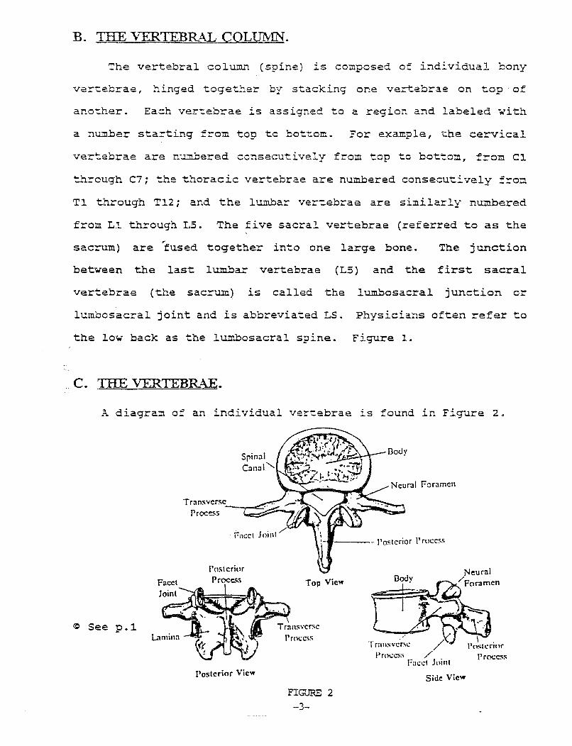
C. THE VERTEBR-ill.
A diagram of an individual vertebrae is found in Fi~~re 2.
Facet
~ See p.l
Spinal Canal'-...
1 . / f-acet o111l
Posterior Proce.o;s
Posterior View
---.. Posterior I' roccss
Top View
FIG"JRE 2 -3-
Tran.svcr~c
l'rocc:;s
Side View
The spinal canal is formed by stacking one vertebrae on top of
another around the cylindrical spinal cord. The piece of bone in
the individual vertebrae, ;.·hich surround the spinal cord, l.S
referred to as the neural a.=ch (neural referring to nerves and arch
referring to an archway).
Lamina is a piece of bone found in the back of the vertebrae
and forms the back of the neural arch, the "archway" to the cord.
In performing an operation called a n laminectomy", the surgeon must
first remove the bony lamina to operate en the spinal cord and the
nerve roots.
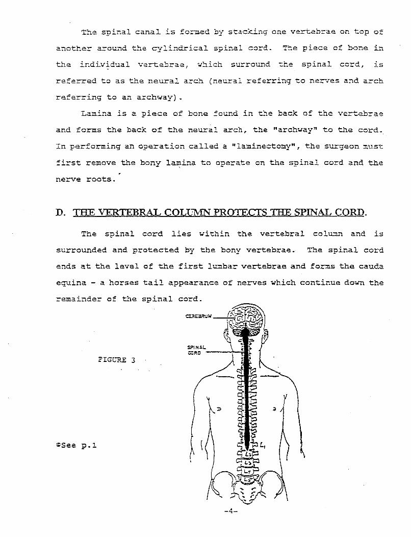
D. THE VERTEBRAL COLITMN" PROTECTS THE SPINAL CORD.
The spinal cord lies within the vertebral colwnn and is
surrounded and protected by the bony vertebrae. The spinal cord
ends at the level of the first lumbar vertebrae and forms the cauda
equina - a horses tail appearance of nerves which continue down the
remainder of the spinal cord.
FIGURE 3
COSee p.~
-4-

E. THE SPINAL CORD:
1.. LONG TRACTS:
The spinal cord is cylindrical measuring 26" long -with an
average width of 5/8". The spinal cord is an extension of the
brain and is analogous to a telephone cable with ascending and
descending long tracts of wires. The ascending tracts conduct
impulses to the brain, and the descending tracts carry impulses
away from the brain. The brain controls voluntary movements·by
sending and receiving nerve messages along the spinal cord through
these long tracts called the pvramidal and extrapyramidal tracts.
Damage to the long tracts in the spinal cor'd is called
mvelooathy. Figure 4A and 4B is a cross-section of the spinal cord
showing pyramidal and extrapyramidal ascending and descending long
tracts inside the spinal cord.
---1· ( I FIGURE ..4A
<eSee p.l.
-5-
~::r.iE---- oec:uss.no,. OP I'TI~IOS
,t.-;--:::J~--- OlltC:T I"''U. ...
llf::::~:=;:::1 TlAC:T
FIGURE 4B
2. MYELOPATHY - LONG TRACT INJURY:
Compression of the spinal cord produces a condition
called myelopathy (myel = spinal cord; apathy = disease) .
Compression of the cord can be caused by many events, including a
large central disc herniation pressing against the spinal cord; or
a preexisting osteophyte present on the vertebrae striking the
spinal cord during a rear-end collision with resulting
hyperextension flexion of the spine.
If the spinal cord is compressed, damage can be done to the
"long tracts" present inside the spinal cord (pyramidal and
extrapyramidal tracts)
If the spinal cord is compressed in the cervical area 1 the
condition is known as cervical myelopathy. Symptoms include
peculiar sensations in the hands with clumsiness and weakness,
especially in the legs with hyperactive reflexes and some
spasticity. The clinical picture often presents a patchy
distribution of symptoms.
Patients with long tract damage to the spinal cord
(myelopathy), will show a classic finding, on physical exam- a
positive (+) Babinski test - described in the next section.
(a) THE BABINSKI TEST:
This is a simple test developed over 100 years ago by a
neurologist named Babinski. A positive test suggests an upper
motor neuron injury in the pyramidal tract, one of the long tracts
sending impulses to the voluntary muscles from the cerebral cortex
of the brain down the spinal cord. A positive Babinski sign in an
-6-
infant (under 18 months of age) is insignificant since the infant's
central nervous system is not fully developed.
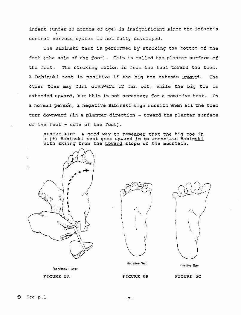
The Babinski test is performed by stroking the bottom of the
foot (the sole of the foot) . This is called the plantar surface of
the foot. The stroking motion is from the heel toward the toes.
A Babinski test is positive if the big toe extends upward. The
other toes may curl downward or fan out, while the big toe is
extended upward, but this is not necessary for a positive test. In
a normal person, a negative Babinski sign results when all the toes
turn downward (in a plantar direction - toward the plantar surface
of the foot sole of the foot) .
MEMORY AID: A good way to remember that the big toe in a (+) Babinski test goes upward is to associate Babinski with skiing from the unward slope of the mountain.
fi?r ru~ -~ I ~~
/~- ~ ,' ' ~ f /,;(
r \ '( I / '!\ •· (( r
~. '? ~ I p \. ,. ; ' ( ~ ) 1 ... '' '
C~9 ~)C) 1\ • . \\ I /"'\
~-(:
Q9 Babinski Test
FIGURE SA
<S> See p.l
\ I i /
u Negative iest
FIGURE 5B
-7-
(
\ I
~sitive iest
FIGURE SC
3. THE DURA (THECAL SAC):
The spinal cord is covered and protected by the dura,
also called the meninges, which consists of three layers from
inside out as follows: (1) inside layer - pia mater (latin for
sof.t mother since this layer is soft); (2) middle layer - arachnoid
(arachnoid meaning spider since this layer looks like a spider
web); and (3) outer layer - dura mater (latin for hard mother since
this layer is tough and finrous). The dura mater is also referred
to as the thecal sac (not to be confused with fecal sac which is
the bowel). Infection of the meninges is called meningitis.
The space between the first layer, pia mater, and the second
layer, arachnoid, is called the subarachnoid space (under the
arachnoid) . The subarachnoid space contains the cerebrospinal
fluid (CSF) that surrounds the brain and spinal cord. A spinal tap
consists of removing cerebrospinal fluid (CSF) from the
subarachnoid space; while a myelogram test consists of injecting a
contrast dye in this space to outline the spinal cord and its nerve
roots.
PRACTICE NOTE - "INDENTING THE THECAL SAC": The outside membrane covering the spinal cord and its nerve roots is the thecal sac, which is often referred to in MRI reports. For example, the radiologist often states "the lesion is indenting the thecal sac" - meaning that the space-occupying lesion such as a protruded disc, a protruding osteophyte, etc., is indenting the thecal sac of the spinal cord.
-8-
~See p.~
/ Vcn•rn! s~.r~l
Rout
4. SPINAL N~RVE ROOTS:
FIGURE 6
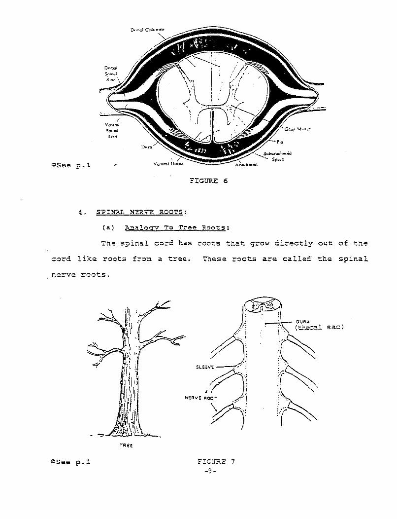
(a) Analogy To Tree Roots:
The spinal cord has roots that grow directly out of the
cord like roots f~om a tree.
nerve roots.
<:lSee p.~
These roots are called the spinal
FIGURE 7 -9-
sac)
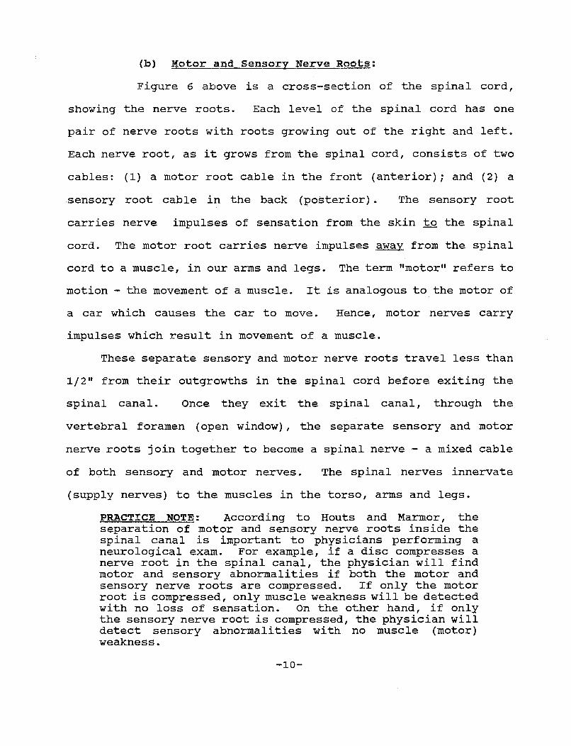
(b) Motor and Sensory Nerve Roots:
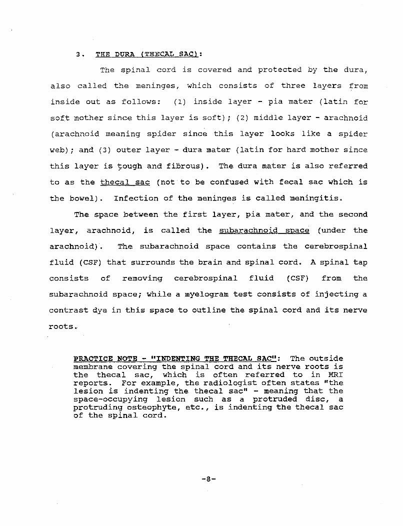
Figure 6 above is a cross-section of the spinal cord,
showing the nerve roots. Each level of the spinal cord has one
pair of nerve roots with roots growing out of the right and left.
Each nerve root, as it grows from the spinal cord, consists of two
cables: (1) a motor root cable in the front (anterior); and (2) a
sensory root cable in the back (posterior) . The sensory root
carries nerve impulses of sensation from the skin to the spinal
cord. The motor root carries nerve impulses away from the spinal
cord to a muscle, in our arms and legs. The term "motor" refers to
motion - the movement of a muscle. It is analogous to the motor of
a car which causes the car to move. Hence, motor nerves carry
impulses which result in movement of a muscle.
These separate sensory and motor nerve roots travel less than
1/2" from their outgrowths in the spinal cord before exiting the
spinal canal. Once they exit the spinal canal, through the
vertebral foramen (open window), the separate sensory and motor
nerve roots join together to become a spinal nerve - a mixed cable
of both sensory and motor nerves. The spinal nerves innervate
(supply nerves) to the muscles in the torso, arms and legs.
PRACTICE NOTE: According to Houts and Marmor, the separation of motor and sensory nerve roots inside the spinal canal is important to physicians performing a neurological exam. For example, if a disc compresses a nerve root in the spinal canal, the physician will find motor and sensory abnormalities if both the motor and sensory nerve roots are compressed. If only the motor root is compressed, only muscle weakness will be detected with no loss of sensation. On the other hand, if only the sensory nerve root is compressed, the physician will detect sensory abnormalities with no muscle (motor) weakness.
-10-
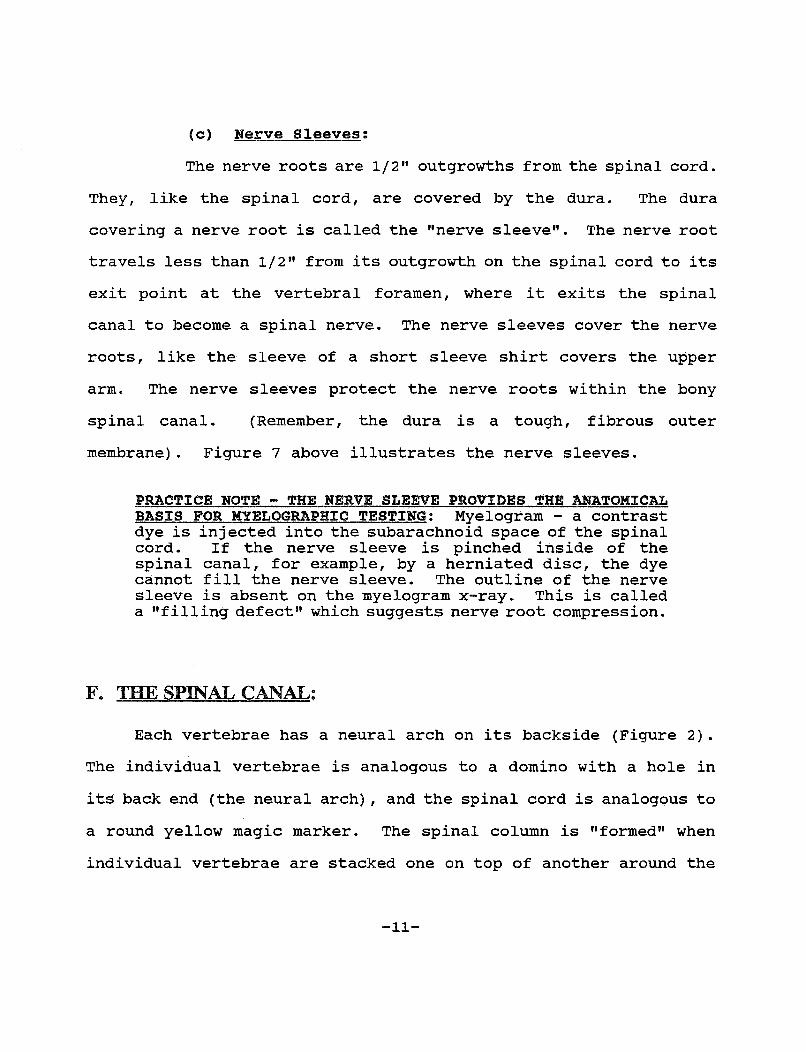
(c) Nerve Sleeves:
The nerve roots are 1/2" outgrowths from the spinal cord.
They, like the spinal cord, are covered by the dura. The dura
covering a nerve root is called the "nerve sleeve". The nerve root
travels less than 1/2" from its outgrowth on the spinal cord to its
exit point at the vertebral foramen, where it exits the spinal
canal to become a spinal nerve. The nerve sleeves cover the nerve
roots, like the sleeve of a short sleeve shirt covers the upper
arm. The nerve sleeves protect the nerve roots within the bony
spinal canal. (Remember, the dura is a tough, fibrous outer
membrane). Figure 7 above illustrates the nerve sleeves.
PRACTICE NOTE - THE NERVE SLEEVE PROVIDES THE ANATOMICAL BASIS FOR MYELOGRAPHIC TESTING: Myelogram - a contrast dye is injected into the subarachnoid space of the spinal cord. If the nerve sleeve is pinched inside of the spinal canal, for example, by a herniated disc, the dye cannot fill the nerve sleeve. The outline of the nerve sleeve is absent on the myelogram x-ray. This is called a "filling defect" which suggests nerve root compression.
F. THE SPINAL CANAL:
Each vertebrae has a neural arch on its backside (Figure 2).
The individual vertebrae is analogous to a domino with a hole in
its back end (the neural arch) , and the spinal cord is analogous to
a round yellow magic marker. The spinal column is "formed" when
individual vertebrae are stacked one on top of another around the
-11-
cylindrical spinal cord - like stacking individual dominoes with a
hole in it around a magic marker. In most people, the vertebral
neural arches form a "loose fit" around the spinal cord, like
stacked horse shoes around a metal stake. A canal is formed around
the spinal cord by the neural arch of each vertebrae. The spinal
canal contains the neural (pertaining to nerve) elements which are
the spinal cord and the spinal nerve roots, before they exit the
canal. The outside column formed by stacking the bony vertebrae
around the spinal cord is called the spinal column (or the spine)
and is a protective bony casing for the spinal cord.
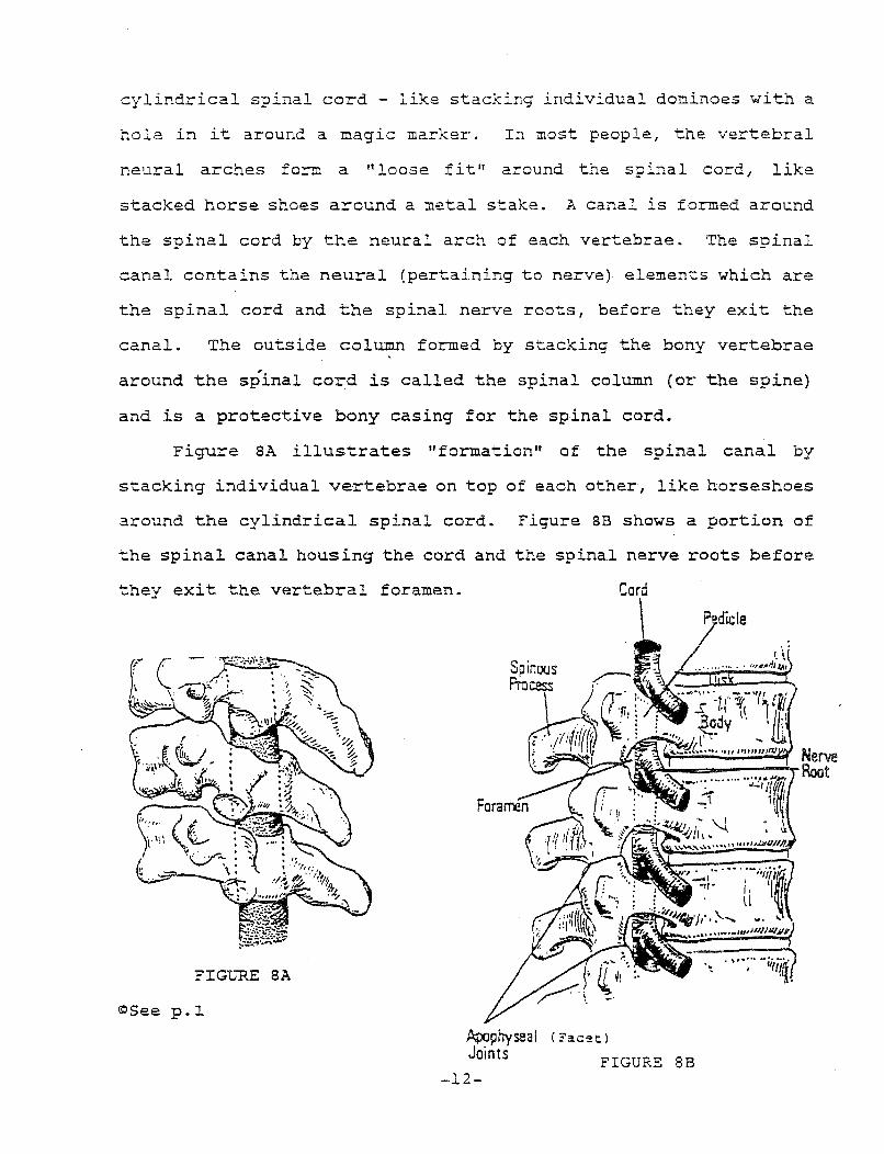
Figure SA illustrates "formation" of the spinal canal by
stacking individual vertebrae on top of each other, like horseshoes
around the cylindrical spinal cord. Figure 8B shows a portion of
the spinal canal housing the cord and the spinal nerve roots before
they exit the vertebral foramen.
FIGURE SA
<OSee p.l Apophyseal Joints
-12-
Cord
(Facet:.)
FIGURE 8B

?RACTICE NOTE: Mechanical coopression and ir~itation of the spinal nerve root as it exits the spinal canal through the bony foramen will result in radiating pain down the course of the nerve and is called radiculopathy. This concept is the key to understanding nerve root injury.
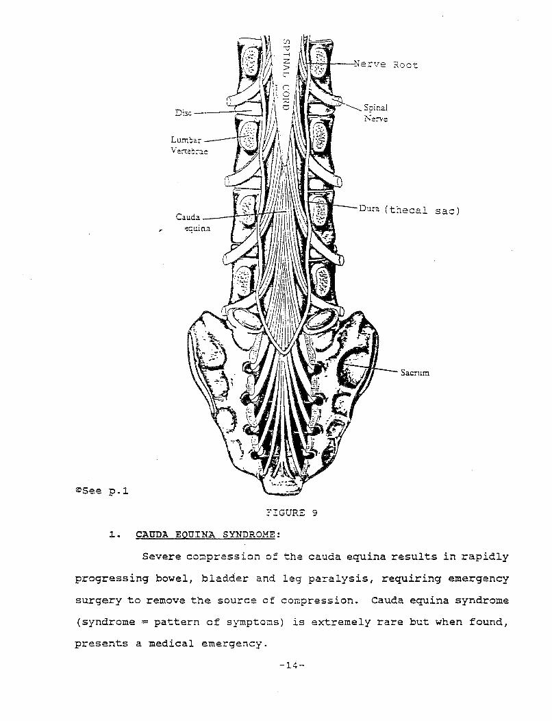
G. CAliDA EQUINA:
The vertebral column is longer than the spinal cord (Figure
3) . The splnal cord ends at the level of the first lumbar
vertebrae (L1). At this level, the spinal cord sends out nerve
fibers which run down the remainder of the vertebral canal, all the
way to the bottom of the sacrum. These nerve fibers look like a
horse's tail when viewed from the back. Accordingly, this group of
nerves is named cauda equina (horses tail). The thecal sac (outer
dura membrane) of the cauda equina is firmly anchored to the bottom
of the vertebral column. Figure 9 shows the cauda equina housed
within a portion of the spinal column.
PRACTICE NOTE: A spinal tap and a myelogram are usually performed at a level below Ll to avoid damage to the spinal cord which ends at the Ll level in the average person. In some people, the spinal cord ends further down near the L2 level. The needle is inserted into the subarachnoid space which surrounds the cauda equina and is filled with cerebrospinal fluid.
-13-
Dura (thecal sac)
Sacrum
~See p.l
FIGURE 9
~. CAUDA EQUINA SYNDROME:
Severe co~pression of the cauda equina results in rapidly
progressing bowel, bladder and leg paralysis, requiring emergency
surgery to remove the source of compression. Cauda equina syndrome
(syndrome = pattern of symptons) is extremely rare but when found,
presents a medical emergency.
-14-
H. PERIPHERAL SPIN"AL NERVES:
1. ORIGIN AND PATHWAY:
Peripheral spinal nerves are formed from the spinal nerve
roots at the point where the nerve root exits the spinal canal
through the vertebral foramen (open window) (Figure 8B above). The
vertebral foramen is also referred to as the neural (pertaining to
nerves) foramen. The spinal nerves innervate (supply nerves) to
muscles in the; arms and l'egs - in the periphery, hence the name
peripheral nerves. They are also referred to as peripheral spinal
nerves since they originate from the nerve roots in the spinal
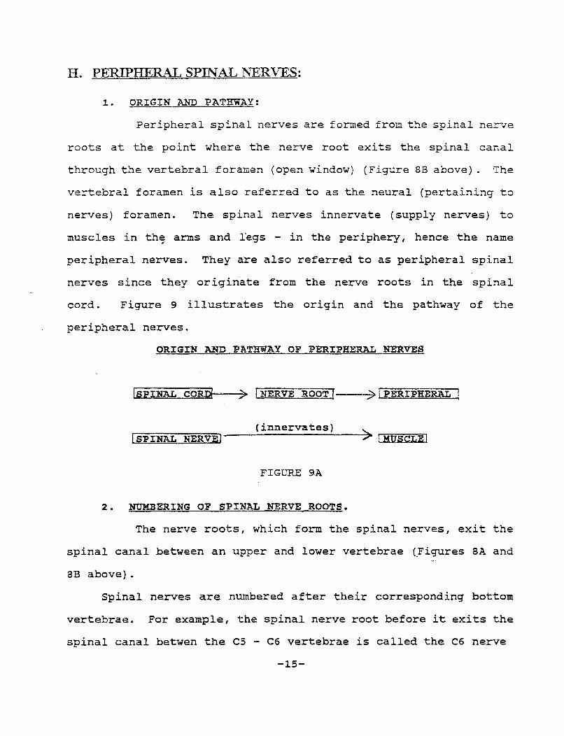
cord. Figure 9 illustrates the origin and the pathway of the
peripheral nerves.
ORIGIN AND PATHWAY OF PERIPHERAL NERVES
!SPINAL CORd > !NERVE ROOT!----->~IPERIPHERAL
(innervates) l SPINAL NERVE! __ _,;_ _____ ;__ __ >-?> 1 MUSCLE]
FIGURE 9A
2. NUMBERING OF SPINAL NERVE ROOTS.
The nerve roots, which form the spinal nerves, exit the
spinal canal between an upper and lower vertebrae (Figures SA and
8B above).
Spinal nerves are numbered after their corresponding bottom
vertebrae. For example, the spinal nerve root before it exits the
spinal canal betwen the cs - C6 vertebrae is called the C6 nerve
-15-
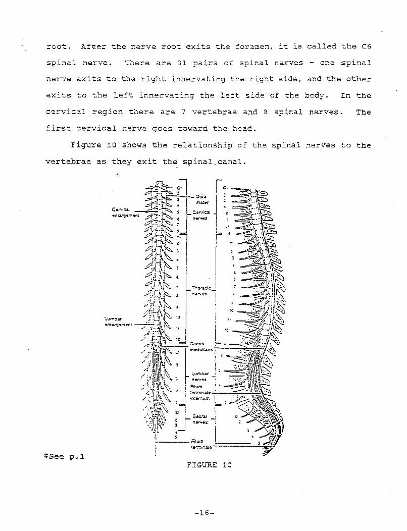
root. After the nerve root exits the foramen, it is called the C6
spinal nerve. There are 31 pairs of spinal nerves - one spinal
nerve exits to the right innervating the right side, and the other
exits to the left innervating the left side of the body. In the
cervical region there are 7 vertebrae and 8 spinal nerves. The
first cervical nerve goes toward the head.
Figure 10 shows the relationship of the spinal nerves to the
vertebrae as they exit the spinal_canal.
<eJSee p.l FIGURE 10
-16-
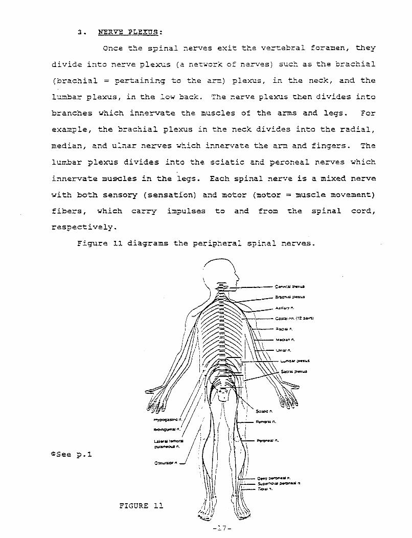
3. NERVE PLEXUS:
Once the spinal nerves exit the ve~ebral foramen, they
divide into nerve plexus (a network of nerves) such as the brachial
(brachial = pertaining to the arm) plexus, in the neck, and the
lumbar plexus, in the lew back. The nerve plexus then divides . .... ~n .... o
branches which innervate the muscles of the arms and legs. For
example, the brachial plexus in the neck divides into the radial,
median, and ulnar nerves which innervate the arm and fingers. The
lumbar plexus divides into the sciatic and peroneal nerves which
innervate muscles in the legs. Each spinal nerve is a mixed nerve
with both sensory (sensation) and motor (motor = muscle movement)
fibers, which carry impulses to and from the spinal cord,
respectively.
Figure 11 diagrams the peripheral spinal nerves.
~See p.1
FIGURE ll
-17-
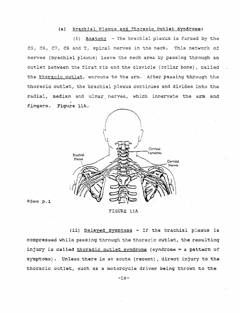
(a) Brachial Plexus and Thoracic outlet svndrome:
(i) Anatomy - The brachial plexus is formed by the
CS, C6, C7, C8 and T, spinal nerves in the neck. This network of
nerves (brachial plexus) leave the neck area by passing through an
outlet between the first rib and the clavicle (collar bone), called
the thoracic outlet, enroute to the arm. After passing through the
thoracic outlet, the brachial plexus continues and divides into the
radial, median and ulnar, nerves, which innervate the arm and
fingers. Figure llA.
~See p.l
FIGURE llA
(ii) Delayed Symptoms - If the brachial plexus is
compressed while passing through the thoracic outlet, the resulting
injury is called thoracic outlet svndrome (syndrome = a pattern of
symptoms) • Unless there is an acute (recent), direct injury to the
thoracic outlet, such as a motorcycle driver being thrown to the
-18-

ground, symptoms may take weeks or months to develop. For example,
if the long, thin scalene muscles which cover the cervical spine
and attach to the first rib are torn (as in a cervical sprain),
scar tissue formed by the injured muscle during the healing
process, can compress the nerves that run betwen them, causing
irritation of the brachial plexus.
In addition, if the collar bone or first rib are fractured,
the slowly developing callus formation (overgrowth of bone at the
fracture site)., can also compress the nerves of the brachial plexus
producing a delayed onset of symptoms.
Figure llB shows the relationship of the scalene muscles to
the brachial plexus .
Longus Colli
~See p.l
('~ I \ \
.. /\ \ \
FIGURE llB
The thoracic outlet is in the costo-clavicular space (costa = rib; clavicular = clavical) - the
space between the 1st rib and the clavicle (collar bone)
-19-
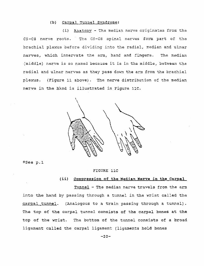
(b) Carpal Tunnel Svndrome:
(i) Anatomv - The median nerve originates from the
CS-C8 nerve roots. The C5-C8 spinal nerves form part of the
brachial plexus before dividing into the radial, median and ulnar
nerves, which innervate the arm, hand and fingers. The median
(middle) nerve is so named because it is in the middle, between the
radial and ulnar nerves as they pass down the arm from the brachial
plexus. (Figure 11 above). The nerve distribution of the median
nerve in the nand is illustrated in Figure llC.
\ \
~See p.l
FIGURE llC
(ii) Compression of the Median Nerve in the Carcal
Tunnel - The median nerve travels from the arm
into the hand by passing through a tunnel in the wrist called the
carnal tunnel. (Analogous to a train passing through a tunnel) .
The top of the carpal tunnel consists of the carpal bones at the
top of the wrist. The bottom of the tunnel consists of a broad
ligament called the carpal ligament (ligaments hold bones
-20-
together). Localized swelling from trauma, such as by striking
extended hands on the dashboard or by holding the steering wheel
tightly during an auto collision, takes up space in the carpal
tunnel, compressing the median nerve which is "just passing
through" the tunnel, enroute to the hand and fingers.
(iii) symptoms of carpal Tunnel syndrome - symptoms
resulting from carpal tunnel syndrome, such as pain, numbness,
tingling and w~akness, follow the nerve distribution of the median
nerve into the hand and fingers, as noted in Figure llC.
Patients with carpal tunnel syndrome often complain of being
awakened at night from pain and often drop objects due to the
weakness of the hand. Symptoms of carpal tunnel syndrome are often
delayed in onset from weeks to months following trauma. Positive
(+) EMG testing is often diagnostic for carpal tunnel syndrome;
however, a patient may have a 11 subclinical" carpal tunnel syndrome,
which is not picked-up on the EMG until a later date.
(iv) Double crush Syndrome - Injury to Both Ends of
the Median Nerve - A patient may injure both ends
of the median nerve in the same auto collision. This is called
"double crush syndrome" since the nerve was injured twice - once at
each end. For example, he may injure the C6 nerve root in the neck
(the origin of the median nerve) and at the same time sustain
injury to the median nerve as it passes through the carpal tunnel,
by striking his extended·hands against the dashboard at the time of
-21-
impact. Since the C6 nerve root innervates the entire arm, down to
the fingers, the treating physician must be careful to diagnose two
separate injuries to the median nerve - at the C6 nerve root level
in the neck, and at the carpal tunnel level in the wrist.
PRACTICE NOTE: During the initial interview, it is important to ask the client if he was either holding the steering wheel tightly or struck his extended hands against the dashboard at the time of impact. This initial history may later help prove that the carpal tunnel syndrome was post-traumatic 1 and a result of the auto collision, especially in delayed onset cases where causationris vigorously contested.
L CLINICAL DIAGNOSIS OF NERVE ROOT IN.UJRY:
l. SEGMENTAL DIVISION OF THE SPINAL CORD:
The spinal cord is divided into segments (cervical,
thoracic, lumbar and sacral) . Nerve roots exit the spinal cord at
specific subsegments (levels), such as the sixth cervical (C6)
level; the fifth lumbar (LS) level; or the first sacral (Sl) level.
Each subsection of the spinal cord sends nerve impulses to a
specific address a specific muscle or muscle group called
myotomes and supplies the feeling of sensation (touch, pain and
temperature) to specific addresses in the skin called dermatomes.
The peripheral spinal nerves are analogous to cables sending
impulses along the length of the cable. Each nerve root is
"programmed" to innervate a specific muscle ( s) (myotome) and a
specific area of the skin (dermatome) • The nerve impulse from a
specific subsection of the spinal cord always follows the ~
pathway to the same muscle and to the same area of the skin.
-22-
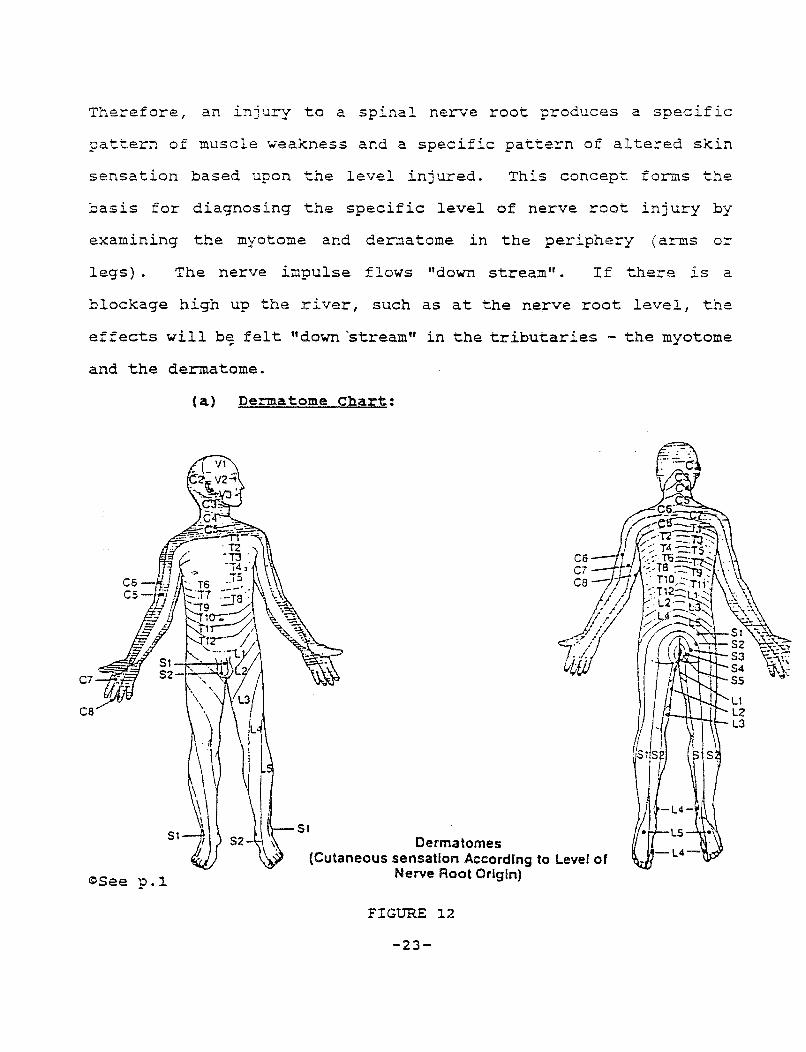
Therefore, an injury to a spinal nerve root produces a specific
pattern of muscle weakness and a specific pattern of altered skin
sensation based upon the level injured. This concept forms the
basis for diagnosing the specific level of nerve root injury by
examining the myotome and dermatome in the periphery (arms or
legs) . The nerve impulse flows "down stream". If there is a
blockage high up the river, such as at the nerve root level, the
effects will b~ felt 11 down 'stream" in the tributaries - the myotome
and the dermatome.
(a) Dermatome Chart:
C7
ca
51
~See p.l
Derma tomes (Cutaneous sensation According to Level of
Nerve Root Origin)
FIGURE 12
-23-
2. CLINICAL NEUROLOGICAL EXAMINATION:
A clinical examination refers to a physical exam
performed by a physician observing the patient's responses to
testing. The basis for the neurological exam consists of a muscle
(motor) examination, a reflex examination, and a sensory
examination. An easy way to remember these three parts of the
neurological examination is to use the abbreviation "MRS" (Motor,
Beflex, Sensor~)-
The clinical exam involving motor, reflex and sensory testing
gives the physician "clues" to a specific diagnosis. Abnormal
tests only indicate an injury somewhere along the course of the
nerve impulse from the brain to the muscle. Abnormal test results
can be caused by a tumor, by a spinal nerve root compressed by a
herniated disc or a large osteophyte, or by an injury to the muscle
being tested, to name a few.
Physicians often form an initial differential diagnosis (a
laundry list of possible diagnoses). Further testing, hopefully,
will rule out ( abbrev. "rIo") each possibility on the list,
resulting in a specific diagnosis.
(a) Motor Examination - Motor refers to a muscle or to
a nerve whose function results in movement. For example, motor
nerves innervate muscle resulting in movement. The term lS
analogous to a motor of a car - it is the motor that produces
movement. In the human body, muscles produce movement; hence the
term motor examination - refers to an examination of the peripheral
muscles such as the calf, biceps, thigh muscles, etc.
-24-
Muscle testing is pa=formed on individual muscles to test fo=
muscle strength and weakness. The patient is asked to contract a
specific muscle r,.,·i th all his night. The physician resists the
patient's effort by applying a directly opposite force - pushing
with his hands - in the opposite direction.
During physical examination, muscle strength is graded as
either 5, 4, 3, 2, 1 or 0, with 5 being normal. Anything less than
5 is abnormal. For example, a notation in a medical report which
states, "muscle testing of the biceps muscle was 5+ (or sometimes
referred to as "+++++") " indicates no muscle weakness - normal
muscle strength.
The test for muscle weakness must be conducted by comparing
the strength of the muscle in both the right and left extremity.
The results of muscle testing, at times, vary from examination
to examination. On one day, the motor exam can show weakness, and
on anothe~ day it can be normal. In addition, the time of day or
the patient's activities can effect the exam. For example, the
patient who has had a hard day of physical labor, who is examined
in the late afternoon, may have slightly different findings than if
he were examined the first thing in the morning. Therefore, it is
important for muscle testing to be conducted over several
examinations.
The physician, performing a motor exam, also observes and
measures specific muscles for atrophv (decreases in size of the
muscle due to non-use or weakness).
-25-
(b) sensory Examination - Each spinal nerve contains
motor fibers and sensory fibers - a mixed cable. Each dermatome in
the skin receives sensations of pain, touch and temperature, and
relays these sensations along sensory fibers backup to the specific
level of the spinal cord which innervates the dermatome. By way of
analogy, feelings of sensation are relayed back up-stream from the
skin to the main river in the spinal cord. An injury to the spinal
nerve root, in the spinal canal, will interfere with the sense of
sensation - pain, touch and temperature - in the skin corresponding
to the dermatome pattern of the injured spinal nerve root.
Identification of the involved dermatome leads to a diagnosis of
the specific level of nerve root injury in the spinal canal.
The sensation of pain is tested by pricking the patient's skin
with a pin or by a pinwheel. In response, the patient will tell
the doctor each time he feels a pinprick. The sensation of light
touch is examined by touching the patient's skin with a cotton swab
and recording the patient's response of what he feels.
The results of the sensory testing with a pinprick for pain
and a cotton swab for touch are recorded as either normal,
increased, decreased, altered or absent. Physicians use the
following terminology:
1. Increased sensitivity - Hyperesthesia (hyper = increased, and esthesia from the Greek = sensation)
2. Decreased sensitivity - Hypesthesia (hyp from hypo = decreased, and esthesia from the Greek = sensation)
3. Altered sensation - Dysesthia (dys = dysfunction, and esthesia from the Greek = sensation)
-26-
4. Absent (no sensation; numbness) Anes-chesia (an absent, and esthesia from the Greek = sensation)
Paresthesia (para from the Greek = beyond, and esthesia from
the Greek= sensation) describes a patient's complaints of abnormal
sensations in the arm or leg, such as burning, tingling, and
numbness. A history of paresthesia from the patient is consistent
with nerve root irritation.
Like musc!e testing, sensory testing is positive when there is
a difference between one extremity compared to the other.
A patient who has consumed analgesics (pain relieving drugs)
or alcohol may give a false negative test result since he may not
feel the full extent of the pinprick because of these drugs.
i. Stocking Glove Anesthesia - If a patient's sensory
exam reveals sensory loss, such as numbness surrounding a body part
as the arm, hand or leg - like a glove fitting over a hand or a
stacking fitting over a leg - the patient's sensory loss does not
fallow the dermatome pattern and is called nan-anatomic - stacking
anesthesia or stocking glove anesthesia. In addition, a patient's
sensory lass may be on one side of the body only. Medical
terminology used to describe symptoms on only one-half of the body
contain the prefix "hemi" (meaning half). For example, if a
patient experienced abnormal sensations, such as numbness and
tingling, only on the right side of his body, this is called
"hemiparesthesia". Again, this is non-anatomic, and like stocking
glove anesthesia is suggestive of a functional condition (an
-27-
emotional overlay) or conversion hysteria.
Physicians label symptoms resulting from an emotional (psychological) response
as "functional" and symptoms resulting from actual physical injury, such as compression
of a nerve root, as "organic" or somatic" (soma from the Greek= body as distinguished
from the mind).
Conversion hysteria (also called post-traumatic neurosis) is an involuntary
response by the patient, which is often caused by trauma. The patient's psychological
response to the trauma is so overwhelming that it cannot be controlled and the patient's
mind which "converts" emotional symptoms to physical symptoms which mimic the
symptoms of nerve root irritation, i.e., pain radiating down an extremity, sensory loss,
muscle weakness. However, as noted, these symptoms do not follow the well
recognized dermatome and myotome patterns and are termed non-anatomic.
PRACTICE NOTE: The terms "stocking glove anesthesia" and "nonanatomic pattern" are red flags for conversion hysteria or for an emotional overlay - the patient's emotional symptoms overlay and superimpose physical pain.
(c) Reflex Testing- Reflex testing is another procedure which helps identify the
specific level of nerve root damage in the spinal cord. The test is performed by the phy-
sician striking a deep tendon with his reflex hammer (tendons attach bone to muscle).
-28-
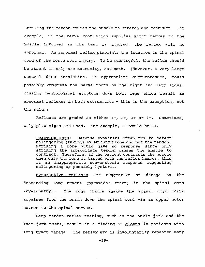
Striking the tendon causes the muscle to stretch and contract. For
example, if the nerve root which supplies motor nerves to the
muscle involved in the test is injured, the reflex will be
abnormal. An abnormal reflex pinpoints the location in the spinal
cord of the nerve root injury. To be meaningful, the reflex should
be absent in only one extremity, not both. (However, a very large
central disc herniation, in appropriate circumstances, could
possibly compress the ne~e roots on the right and left sides,
causing neurological symptoms down both legs which result in
abnormal reflexes in both extremities - this is the exception, not
the rule.)
Reflexes are graded as either 1+, 2+, 3+ or 4+. Sometimes,
only plus signs are used. For example, 2+ would be ++.
PRACTICE NOTE: Defense examiners often try to detect malingering (faking) by striking bone and not the tendon. Striking a bone would give no response since only striking the appropriate tendon causes the muscle to contract. Therefore, if the patient contracts the muscle when only the bone is tapped with the reflex hammer, this is an inappropriate non-anatomic response suggesting malingering o~ possibly hysteria.
Hyperactive reflexes are suggestive of damage to the
descending long tracts (pyramidal tract) in the spinal cord
(myelopathy) . The long tracts inside the spinal cord carry
impulses from the brain down the spinal cord via an upper motor
neuron to the spinal nerves.
Deep tendon reflex testing, such as the ankle jerk and the
knee jerk tests, result in a finding of clonus in patients with
long tract damage. The reflex arc is involuntarily repeated many
-29-
times producing jerking of the extremity at a rapid rate.
Hyperactive reflexes are graded++++ (4+).
5. EXAMPLES INVOLVING SPECIFIC NERVE ROOT LEVELS:
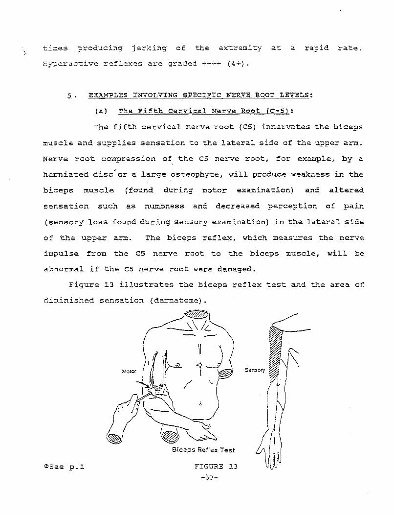
(a) The Fifth Cervical Nerve Root (C-5):
The fifth cervical nerve root (CS) innervates the biceps
muscle and supplies sensation to the lateral side of the upper arm.
Nerve root compression of the CS nerve root, for exaJD.ple, by a
herniated disc'or a large osteophyte, will produce weakness in the
biceps muscle (found during motor examination) and altered
sensation such as numbness and decreased perception of pain
(sensory loss found during sensory examination) in the lateral side
of the upper arm. The biceps reflex, which measures the nerve
impulse from the CS nerve root to the biceps muscle, will be
abnormal if the CS nerve root were damaged.
Figure 23 illustrates the biceps reflex test and the area of
diminished sensation (dermatome).
~See p.2
Biceps Reflex Test
FIGURE 13 -30-
~
~~·· ~
Sen>o<y n j\J I
~[ v
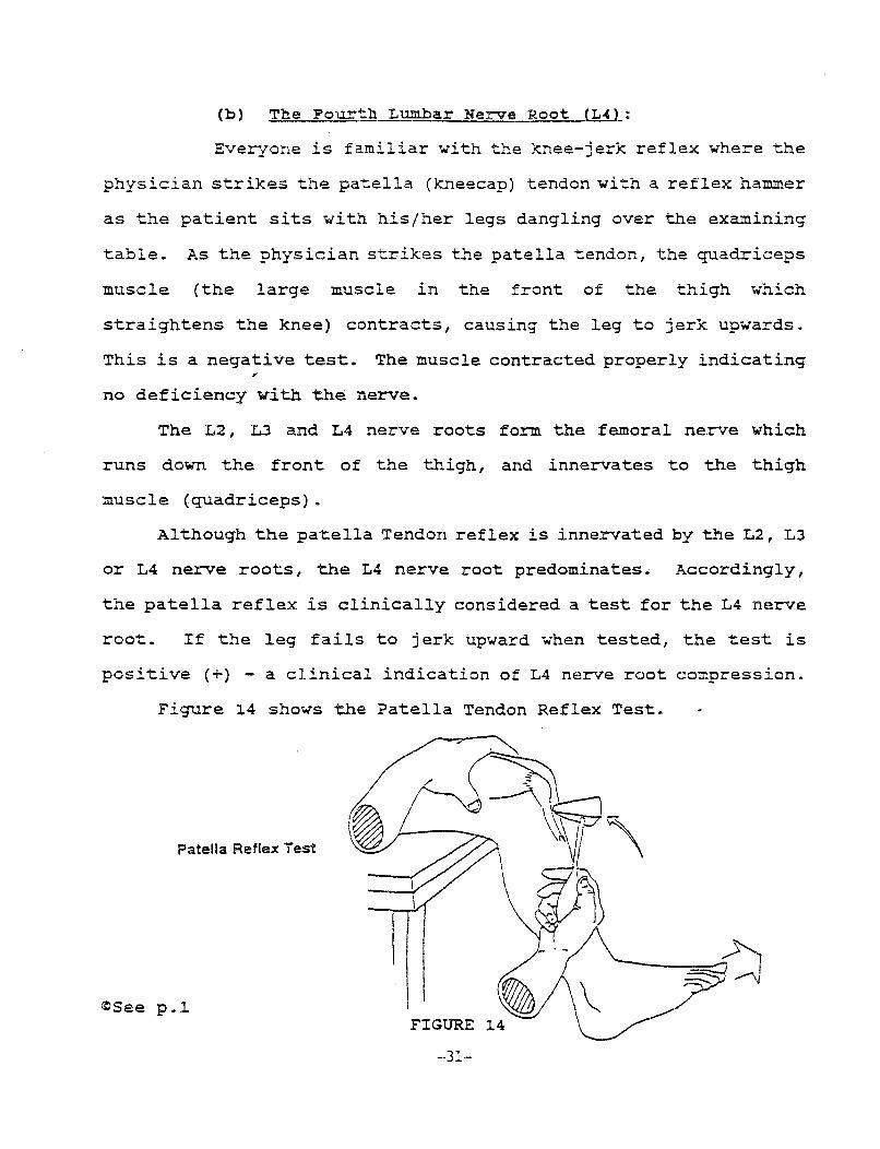
{b) The Fourth Lumbar Nerve Root (L4):
Everyone is familiar with ~~e knee-jerk reflex where the
physician strikes the patella (kneecap) tendon with a reflex hammer
as the patient sits with his/her legs dangling over the examining
table. As the physician strikes the patella tendon, the quadriceps
muscle (the large muscle in the front of the thigh which
straightens the knee) contracts, causing the leg to jerk upwards.
This is a negative test. The muscle contracted properly indicating
no deficiency with the nerve.
The L2, L3 and L4 nerve roots form the femoral nerve which
runs down the front of the thigh, and innervates to the thigh
muscle (quadriceps).
Although the patella Tendon reflex is innervated by the L2, L3
or L4 nerve roots, the L4 nerve root predominates. Accordingly,
the patella reflex is clinically considered a test for the L4 nerve
root. If the leg fails to jerk upward when tested, the test is
positive (+) - a clinical indication of L4 nerve root compression.
Figure 14 shows the Patella Tendon Reflex Test.
Patella Reflex Test
~See p.l FIGURE
-31-
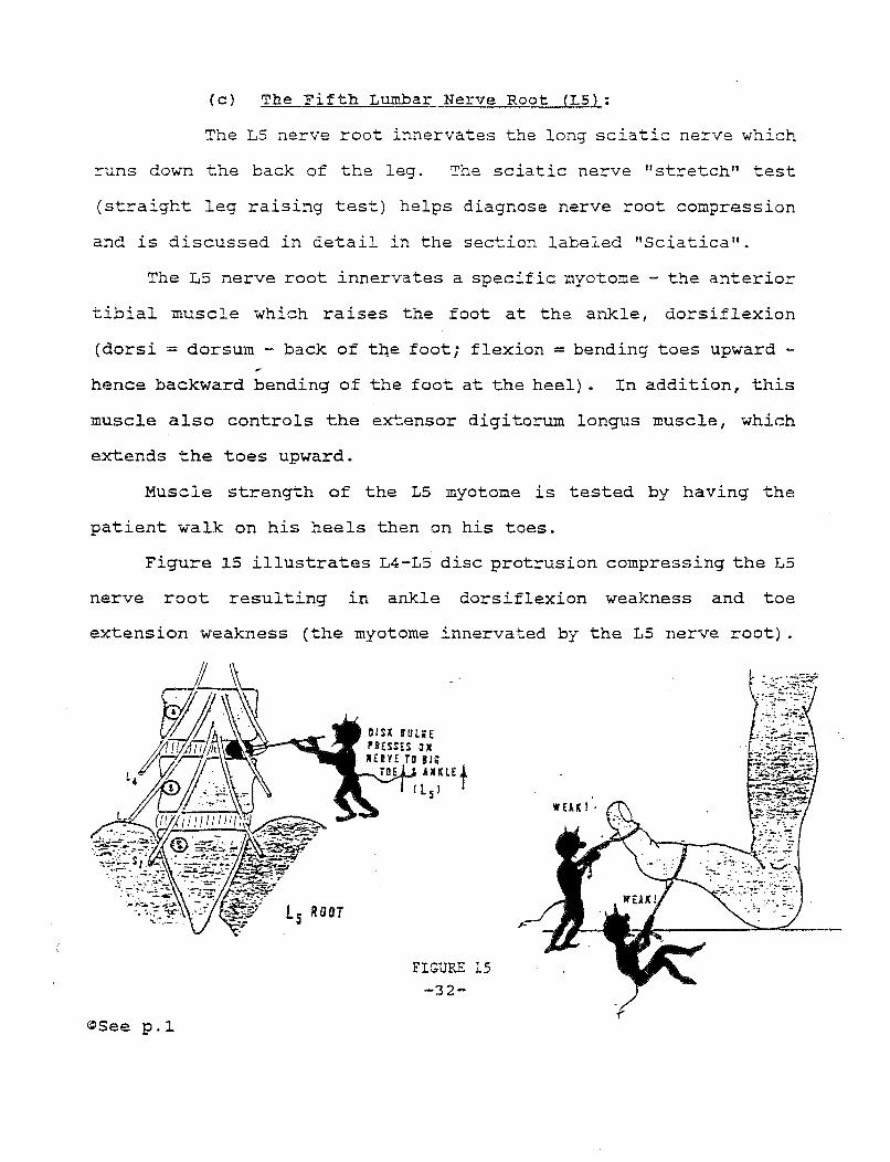
(c) The Fifth Lumbar Nerve Root (L5):
The L5 nerve root innervates the long sciatic nerve which
runs down the back of the leg. The sciatic nerve "stretch" test
(straight leg raising test) helps diagnose nerve root compression
and is discussed in detail in the section labeled "Sciatica".
The L5 nerve root innervates a specific myotome - the anterior
tibial muscle which raises the foot at the ankle, dorsiflexion
(dorsi = dorsum - back of tne foot; flexion = bending toes upward -
hence backward bending of the foot at the heel) . In addition, this
muscle also controls the extensor digitorum longus muscle, which
extends the toes upward.
Muscle strength of the LS myotome is tested by having the
patient walk on his heels then on his toes.
Figure l5 illustrates L4-L5 disc protrusion compressing the L5
nerve root resulting in ankle dorsiflexion weakness and toe
extension weakness (the myotome innervated by the L5 nerve root) .
t 5 Raar
~See p.l
IHH SULliE P'liESSES ON IIEHE TO 11;
AlULEt c l s)
FIGURE 15 -32-
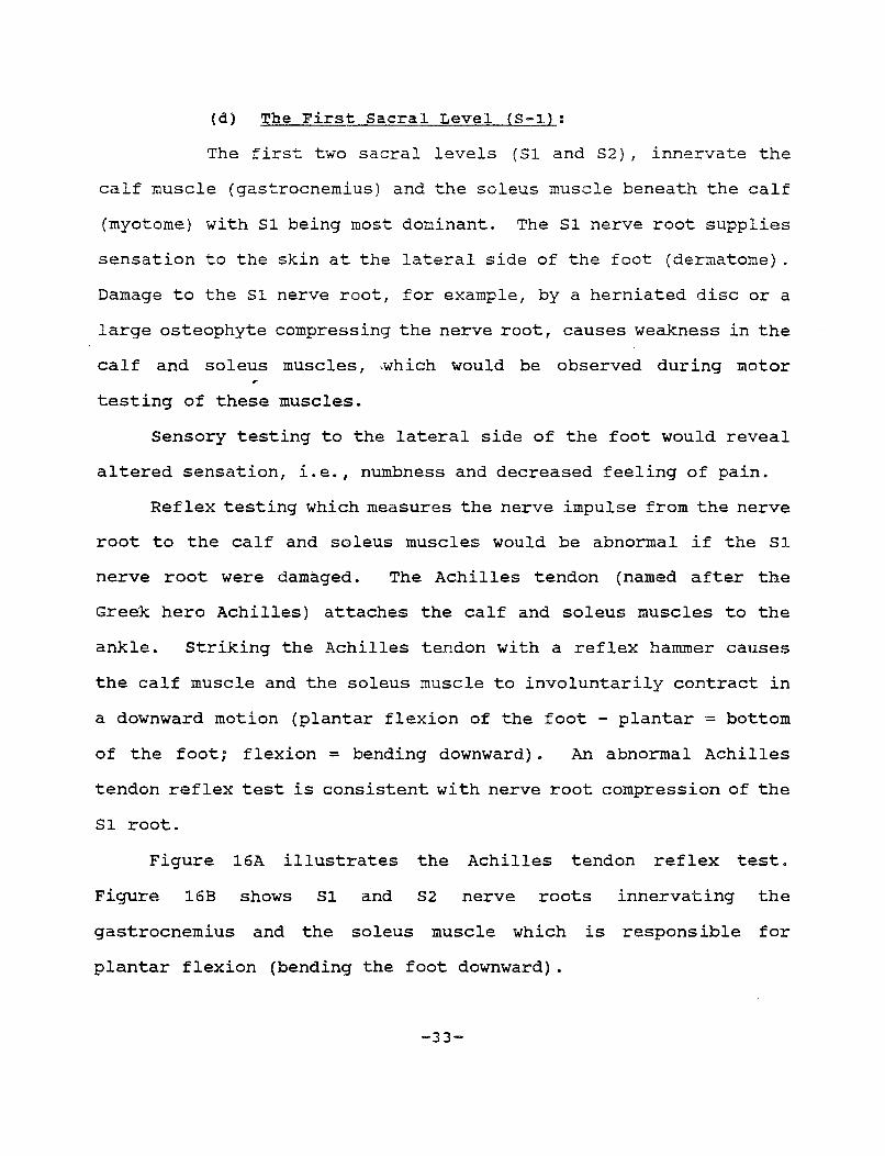
(d) The First Sacral Level {S-1}:
The first two sacral levels (Sl and S2), innervate the
calf muscle (gastrocnemius) and the soleus muscle beneath the calf
(myotome) with Sl being most dominant. The Sl nerve root supplies
sensation to the skin at the lateral side of the foot (dermatome) .
Damage to the Sl nerve root, for example, by a herniated disc or a
large osteophyte compressing the nerve root, causes weakness in the
calf and soleus muscles, ,which would be observed during motor
testing of these muscles.
Sensory testing to the lateral side of the foot would reveal
altered sensation, i.e., numbness and decreased feeling of pain.
Reflex testing which measures the nerve impulse from the nerve
root to the calf and soleus muscles would be abnormal if the Sl
nerve root were damaged. The Achilles tendon (named after the
Greek hero Achilles) attaches the calf and soleus muscles to the
ankle. Striking the Achilles tendon with a reflex hammer causes
the calf muscle and the soleus muscle to involuntarily contract in
a downward motion (plantar flexion of the foot - plantar = bottom
of the foot; flexion= bending downward). An abnormal Achilles
tendon reflex test is consistent with nerve root compression of the
Sl root.
16A illustrates Achilles tendon reflex test. Figure
Figure 16B shows Sl and
the
S2 nerve roots innervating
responsible
the
for gastrocnemius and the soleus muscle which is
plantar flexion (bending the foot downward) .
-33-
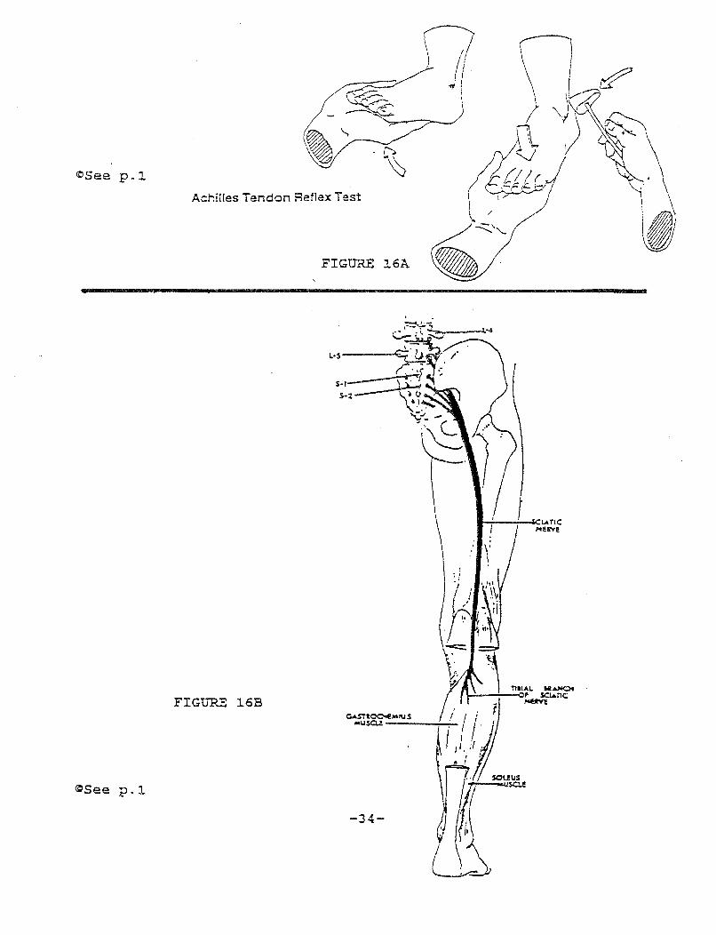
~See p.l
~See p.l
Reflex Test Achilles Tendon
FIGURE 16A
FIGURE 16B
-34-
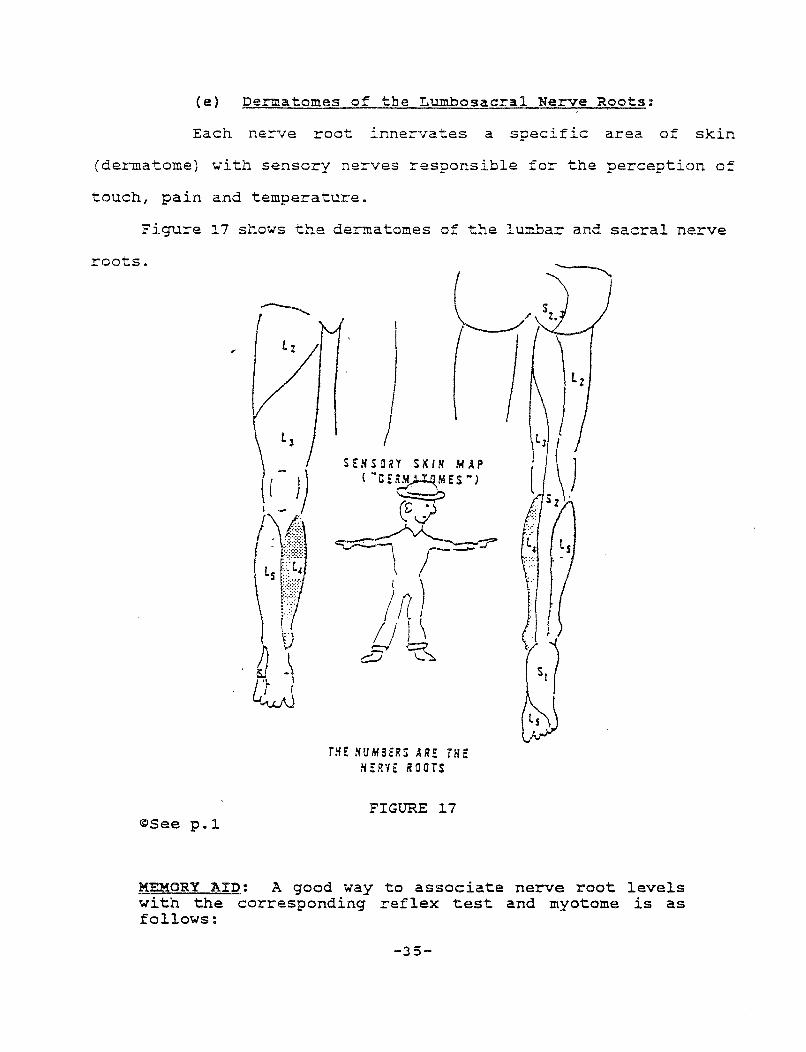
(e) Dermatomes of the Lumbosacral Nerve Roots:
Each nerve root innervates a specific area of skin
(dermatome) with sensory nerves responsible for the perception of
touch, pain and temperature.
Figure 17 shows the dermatomes of the lumbar and sacral nerve
roots.
~See p.l
SENSORY SI<IN MAP {~CE~M MESN)
THE NUMHliS HE THE N!R'IE ROOTS
FIGURE 17
MEMORY AID: A good way to associate nerve root levels with the corresponding reflex test and myotome is as follows:
-35-
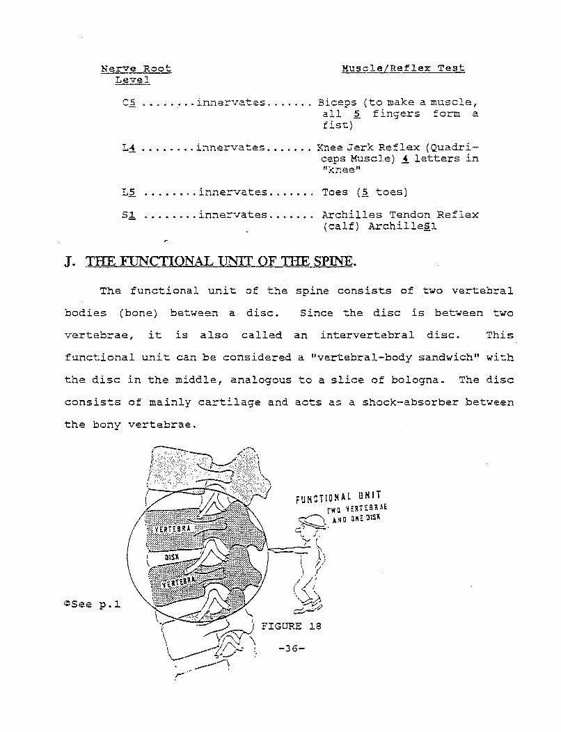
Nerve Root Level
Muscle/Reflex Test
C.2_ ........ innervates ....... Biceps (to make a muscle, all .2_ fingers form a fist)
L1. ........ innervates ....... Knee Jerk Reflex (Quadri-ceps Muscle) .! letters in "knee"
L~ ........ innervates ....... Toes (.2. toes)
S~ ........ innervates ....... Archilles Tendon Reflex (calf) Archille~l
J. THE FlJNCTIONAL UNIT OF THE SPINE.
The functional unit of the spine consists of two vertebral
bodies (bone) between a disc. Since the disc is between two
vertebrae, it is also called an intervertebral disc. This
functional unit can be considered a "vertebral-body sandwich" with
the disc in the middle, analogous to a slice of bologna. The disc
consists of mainly cartilage and acts as a shock-absorber between
the bony vertebrae.
<:lSee p.l
FIGURE 18
-36-
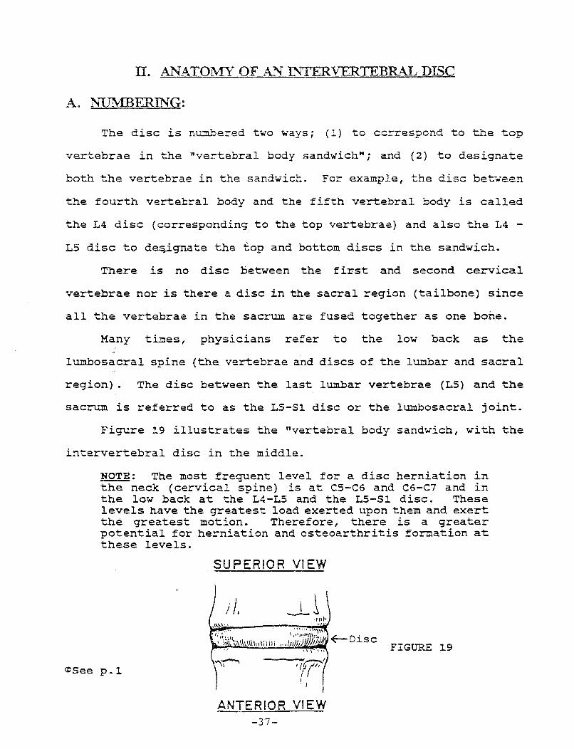
A. I'llJMBERING:
The disc is numbered two ways; (1) to correspond to ~~e top
vertebrae in the "vertebral body sandwich"; and (2) to designate
both the vertebrae in the sandwich. For example, the disc between
the fourth vertebral body and the fifth vertebral body is called
the L4 disc (corresponding to the top vertebrae) and also the L4 -
LS disc to de~gnate the top and bottom discs in the sandwich.
There is no disc between the first and second cervical
vertebrae nor is there a disc in the sacral region (tailbone) since
all the vertebrae in the sacrum are fused together as one bone.
Many times, physicians refer to the low back as the
lumbosacral spine (the vertebrae and discs of the lumbar and sacral
region) • The disc between the last lumbar vertebrae (LS) and the
sacrum is referred to as the LS-Sl disc or the lumbosacral joint.
Figure 19 illustrates the "vertebral body sandwich, with the
intervertebral disc in the middle.
NOTE: The most frequent level for a disc herniation in the neck (cervical spine) is at CS-C6 and C6-C7 and in the low back at the L4-L5 and the LS-Sl disc. These levels have the greatest load exerted upon them and exert the greatest motion. Therefore, there is a greater potential for herniation and osteoarthritis formation at these levels.
~See p .1
SUPERIOR VIEW
•f?' . I
ANTERIOR VIEW -37-
~Disc FIGURE 19
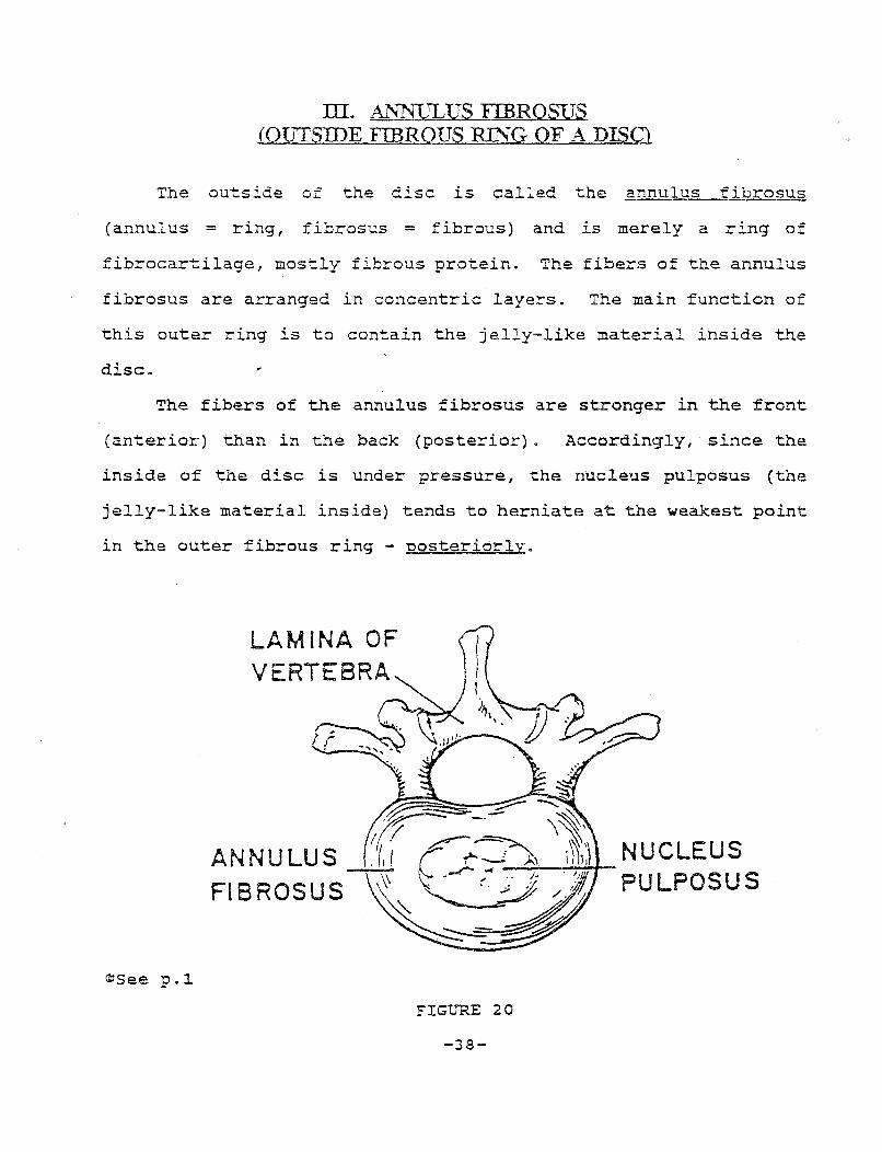
III. AL'lliULUS FIBROSUS (QJITSIDE FIBROUS RING OF A DISO
The outside of the disc is called the annulus fibrosus
(annulus = ring, fibrosus = fibrous) and .!.S merely a ring of
fibrocartilage, mostly fibrous protein. The fibers of the annulus
fibrosus are arranged in concentric layers. The main function of
this outer ring is to contain the jelly-like material inside the
disc.
The fibers of the annulus fibrosus are stronger in the front
(anterior) than in the back (posterior). Accordingly, since the
inside of the disc is under pressure, the nucleus pulposus (the
jelly-like material inside) tends to herniate at the weakest point
in the outer fibrous ring - oosteriorly.
©See p.l.
LAMINA OF VERTEBRA
ANNULUS FIBROSUS
FIGURE 20
-38-
Damage to the annulus fibrosus (the outer ring of the disc)
may cause rupture of the annulus fibrosus resulting ln disc
herniation by allowing the jelly-like center of the disc, which is
under pressure, to push out the confines of its annular outside
ring. In many cases, the patient sustains injury to the outside
annular ring without immediate disc herniation. Over time, with
normal wear and tear, the annulus fibrosus will deteriorate and
lose it's capa~ity to hold'the jelly-like material inside the disc
resulting in disc rupture ·(herniation) at a later date.
PRACTICE NOTE: To explain causation to the jury, an analogy to a tire may be used. The outside rim of a tire (equivalent to the annulus fibrosus) hits the curb causing unnoticeable damage to the outside of the tire. With normal road use of the tire, several weeks later, the tire will blow and the inside air will escape through the damaged outer ring, causing the tire to flatten. Similarly, initial damage to the annulus fibrosus, with time, can result in a deterioration of this outer ring. The inside jelly-like material which, like the tire is also under pressure, now breaks through the fibers of the annulus fibrosus causing disc herniation.
The inside center of the disc is called the nucleus pulposus
which consists of collagen fibers meshed in a jelly-like substance
with a high water content. The inside of the disc is semi-gelatin-
ous. As we get older, the percentage of water in the center of the
disc decreases and the disc is said to "dry out11 which reduces the
height of the disc space. The disc space is the space between the
-39-
upper and lower vertebrae of the "disc sandwich" and is occupied by
the disc. As the disc loses water, the size of the disc decreases,
causing the disc space to also narrow. The function of the disc is
to act as a shock absorber. Loss of water decreases the ability of
the nucleus pulposus to perform this function. The "drying out" of
the inside of the disc, with advancing age, is the beginning stage
of degenerative disc disease with the resulting formation of
osteoarthritis (bony overgrowths (spurs) on the vertebral bodies). ,.
An intervertebral disc is like a jelly doughnut. The inside
of the disc is similar to the inside of the doughnut. The annulus
fibrosus, the circular (annular) ring which holds the jelly inside
the disc, is similar to the outside tough layer of the jelly
doughnut which holds the jelly inside. Rupture of the annulus
fibrosus results in the inside jelly-like nucleus pulposus
extruding out of the disc - just like the rupture of the outside
layer of the jelly doughnut - with the inside jelly extruding out
of the doughnut.
V. BULGING, PROTRUDED AND HERNIATED DISCS
Since the jelly-like nucleus pulposus inside the center of the
disc is under pressure, it will bulge out or protrude at the
weakest spot in its outer fibrous ring, the annulus fibrosus.
A disc is herniated (ruptured) if the jelly-like nucleus
pulposus breaks through fibers of the annul us f ibrosus. The fibers
of the annulus fibrosus have thus ruptured, allowing the semi-
gelatinous nucleus pulposus out, like the jelly inside a doughnut
-40-
rupturing through the outside layer. A herniated disc is often
called an extruded disc because, like a tube of toothpaste, the
jelly is extruded outside its tube; hence the name extruded disc.
A herniated disc is also referred to as a "orolapsed disc".
(Prolapsed meaning out of position). Lastly, a herniated disc is
called a "slipoed disc" since the disc is said to slip out of
place.
When a p~ece of the 'jelly-like nucleus pulposus, which has
ruptured through the outer annulus fibrosus ring, breaks-off into
the spinal canal, disc material is said to have sequestered. This
type of disc is called a sequestered disc.
If the jelly-like nucleus pulposus does not break through the
annulus fibrosus, the disc has not herniated. However, the jelly
inside of the disc may bulge or protrude out at the weakest spot of
the annulus fibrosus. Some physicians use these terms interchange-
ably. However, generally, a protruded disc is one where the
nucleus pulposus not only bulges out, but actually pinches a nerve
root. A bulging disc and protruded disc differ from a herniated
disc in that the former have not ruptured through the outer annular
ring; the inner jelly material of the nucleus pulposus is still
contained within the annulus fibrosus even though it bulges out.
PRACTICE NOTE: A herniated disc is often abbreviated in medical records as "HNP 11 which stands for herniated nucleus pulposus since it is the nucleus pulposus which herniates through its outer annulus fibrosus ring.
-4~-
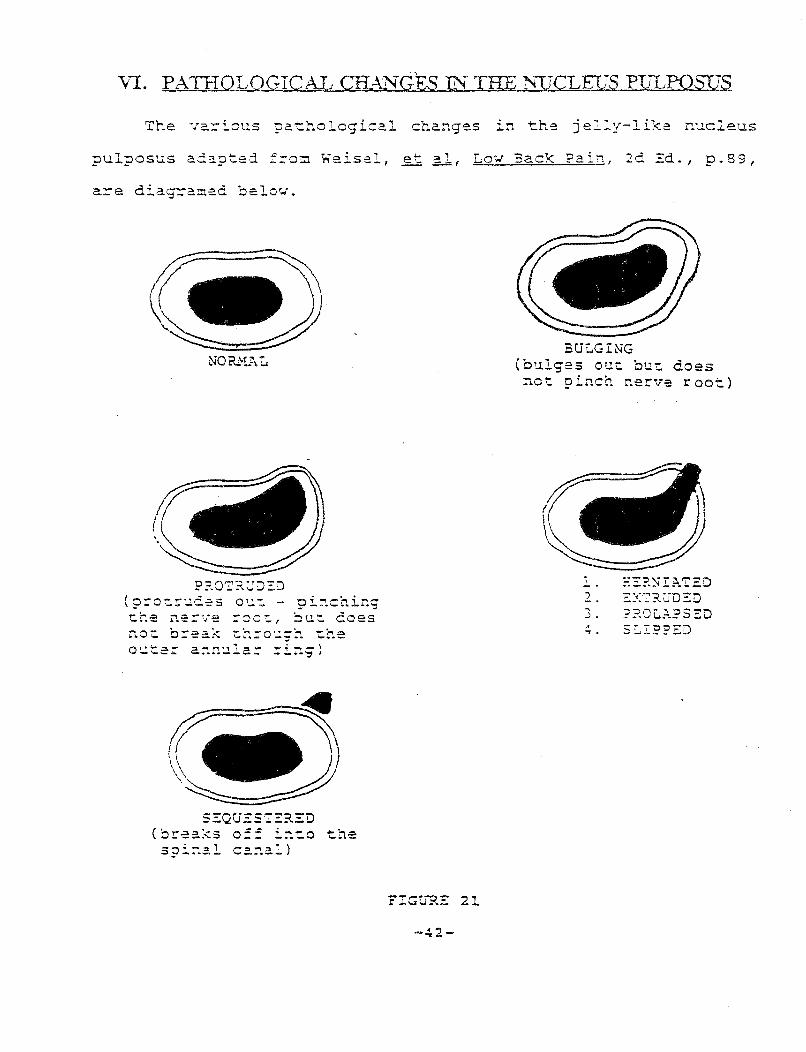
v!. PATHOLOGICAL CR~~GEs lli THE ~{UCLEUS PD""LPOSUS
The va=ious pathological changes in the jelly-like nucleus
pulposus adapt:ed f:-·::J::::J. Weisel, et: al, Lo~o~ Back Pai", 2d Ed., p.89,
a=e diagra~ed belo~o~.
P?.O'I'~\.:'J:::J
( ?:"0':.!:~ ... : .. =.-?:s o 1·- - ?i::"!.chir:.g
che ne=ve =oc~, ~ut does no~ b=ea~ ~h=o~~h ~he o~t== an~ula= =i~g}
s:::QUEST:::?..=::D (~rea~s o== ~~~o the spi::al ca::.a:)
FIGtJ"?.E 2~
BULGING (bulges ou~ but does net 9i~ch ~erve root)
1 ::::?.~IAT2D 2. ::::<-:?-;JD'SD 3. ??.OC..:l..PS2D .. .....
Vll. SITES OF DISC HER~TION:
A disc may herniate in the front (anterior) or in the back
(posterior) . In addition, the disc may herniate at the midline
(central disc herniation) or to the side (lateral disc herniation) .
The term lateral is used in medicine to refer to "the side" as
opposed to the midline. An analogy can be made to a football pass
- a pass to the side is a lateral pass.
The anat9my of the 'annulus fibrosus and the anterior and
posterior longitudinal ligaments generally determine the site of
disc herniation, i.e., anterior, posterior lateral, central.
A. ANAT01\1Y OF THE ANfERIQR AND POSTERIOR LONGITUDINAL LIGAMENfS:
The anterior and posterior longitudinal ligaments run up and
down the spinal column covering the front and back of the vertebrae
and discs, respectively. These long ligaments are fixed to the
vertebrae and discs and hold them in place. The anterior
longitudinal ligament covers the front end of the annulus fibrosus
and the posterior longitudinal ligament covers the back.
For a disc to herniate, the inside jelly of the nucleus
pulposus must break through two barriers: (1) the outer annular
ring of the disc; and (2) the longitudinal ligament covering the
outside of the disc.
Most discs herniate posteriorly (in the back) not anteriorly
(front side). The reason for this is twofold: (1) the annulus
-43-
fibrosus is anatomically weaker in the back than in the front; and
(2) the anterior longitudinal ligament is anatomically broader and
stronger than the narrower and weaker posterior longitudinal
ligament. Since the disc is under constant pressure, like the
inside of a pressure cooker, inside disc material (nucleus
pulposus) will herniate at the weakest point of its outer ring, and
at the weakest point of the ligament covering its outer ring -
posteriorly. The thinner and weaker ligament 1 the posterior
longitudinal ligament, ~s rhomboid in shape. Therefore 1 the
weakest spot in the posterior longitudinal ligament is not in the
center, but to the side (laterally) . The majority of disc
herniations are found in the posterior lateral position. (In the
back of the disc to the right or left side) .
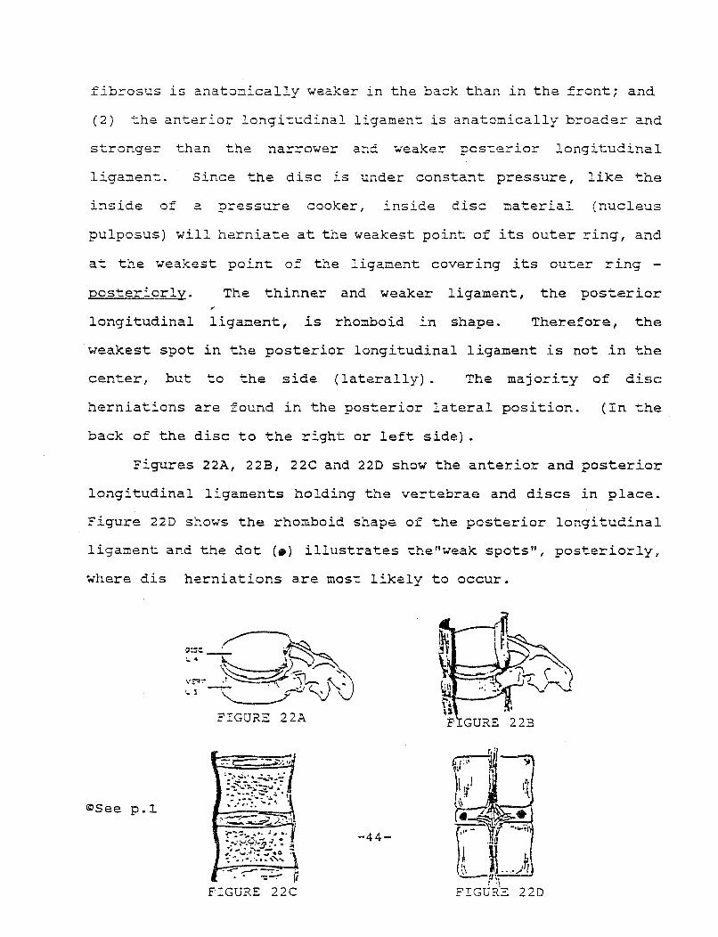
Figures 22A, 22B, 22C and 220 show the anterior and posterior
longitudinal ligaments holding the vertebrae and discs in place.
Figure 220 shows the rhomboid shape of the posterior longitudinal
ligament and the dot (•) illustrates the"weak spots", posteriorly,
where dis herniations are most likely to occur.
~See p.l
-44-
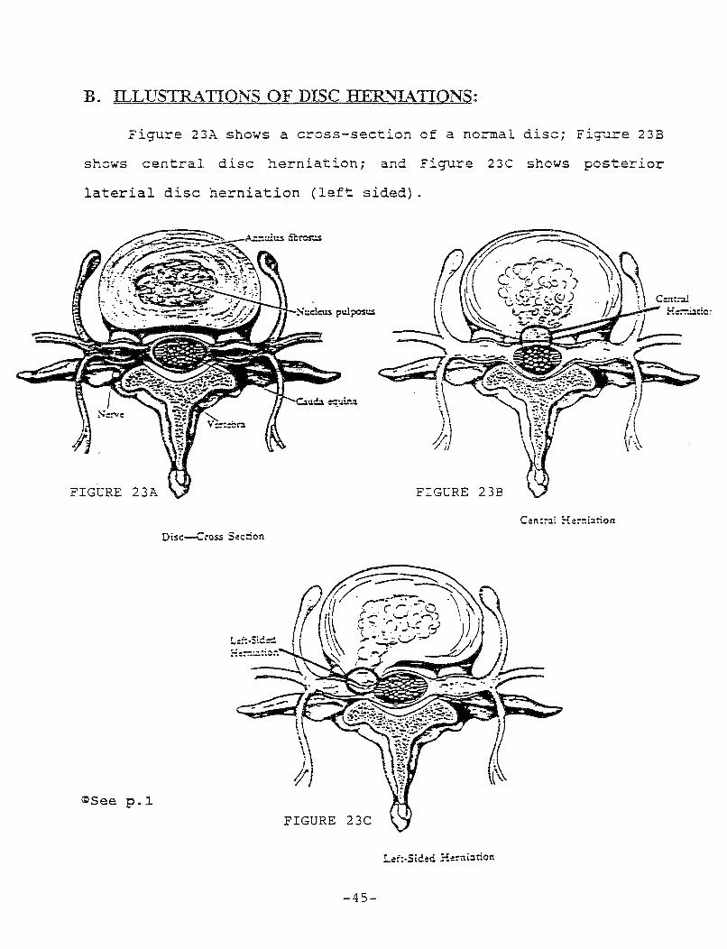
B. ILLUSTRATIONS OF DISC HER~"LATIONS:
Figure 23A shows a cross-section of a normal disc; Figure 23B
shows central disc herniation; and Figure 23C shows posterior
laterial disc herniation (left sided).
FIGURE 23A FIGURE 23B
Disc--Cross S<!c::ion
~See p.l FIGURE 23C
-45-
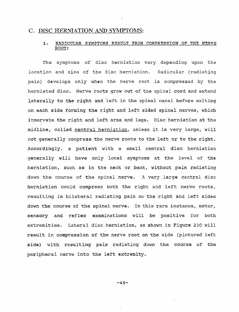
C. DISC HERNIATION AND SY1\1PTOMS:
1. RADICULAR SYMPTOMS RESULT FROM COMPRESSION OF THE NERVE ROOT:
The symptoms of disc herniation vary depending upon the
location and size of the disc herniation. Radicular (radiating
pain) develops only when the nerve root is compressed by the
herniated disc. Nerve roots grow out of the spinal cord and extend '
laterally to the right and left in the spinal canal before exiting
on each side forming the right and left sided spinal nerves, which
innervate the right and left arms and legs. Disc herniation at the
midline, called central herniation, unless it is very large, will
not generally compress the nerve roots to the left or to the right.
Accordingly, a patient with a small central disc herniation
generally will have only local symptoms at the level of the
herniation, such as in the neck or back, without pain radiating
down the course of the spinal nerve. A very large central disc
herniation could compress both the right and left nerve roots,
resulting in bilateral radiating pain on the right and left sides
down the course of the spinal nerve. In this rare instance, motor,
sensory and reflex examinations will be positive for both
extremities. Lateral disc herniation, as shown in Figure 23C will
result in compression of the nerve root on the side (pictured left
side) with resulting pain radiating down the course of the
peripheral nerve into the left extremity.
-46-
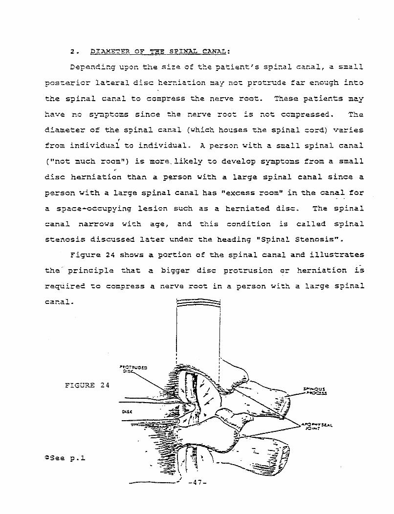
Depending upon the size of the patient" s spinal canal, a small
posterior lateral disc herniation may not protrude far enough into
the spinal canal to compress the nerve root. These patients may
have no S;{'l!tptoms since the nerve root is not compressed. The
diameter of the spinal canal (~hich houses the spinal cord) varies f
from individual to individual. A person with a small spinal canal
("not much room") is more,likely to develop symptoms from a small
disc herniation than a person with a large spinal canal since a
person vith a large spinal canal has "excess room" in the canal for
a space-occupying lesion such as a herniated disc. The spinal
canal narrows W"ith age, and this condition is called spinal
stenosis discussed later under the heading "Spinal Stenosis••.
Figure 24 shows a portion of the spinal canal and illustrates
the principle that a bigger disc protrusion or herniation is
required to compress a nerve root in a person with a large spinal
canal. -
FIGURE 24
G:See p.~
; _____ ... -47-
3. PATIENTS liiTTli ASYMPTOMATIC DISC KERNIATION MAY DEVELOP SYMPTOMS IN LJ\TER YEARS:
As discused, disc herniation may not result in radicular
symptoms unless the nerve root is compressed.
In later years, a patient ~ith an asymptomatic disc herniation
may develop radiating pain if the herniated disc material becomes
larger and/ or the diameter of the spinal canal narrows with
resulting nerve root compression.
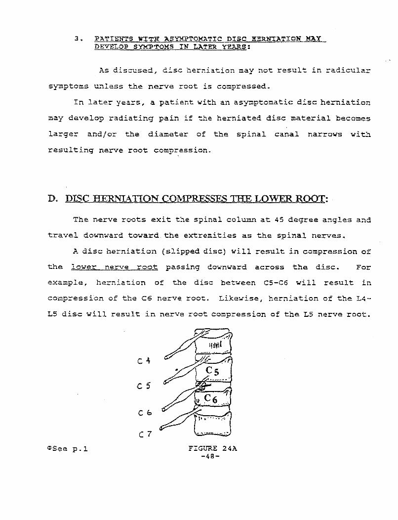
D. DISC HERNIATION COMPRESSES 'I'HE LOWER ROOT:
The nerve roots exit the spinal column at 45 degree angles and
travel downward toward the extremities as the spinal nerves.
A disc herniation (slipped disc) will result in compression of
the lower nerve root passing downward across the disc. For
example, herniation of the disc between CS-C6 will result in
compression of the C6 nerve root. Likewise, herniation of the L4-
LS disc will result in nerve root compression of the LS nerve root.
c5
Cb
C7 ~Seep.~
-..... ·--·. -·.·
FIGURE 24A -48-

Vill. RADICQLOPATHY A.1\ffi RADICULITJS
Whenever a protruded or herniated disc compresses (pinches) a
nerve root, pain and paresthesia (abnormal sensations such as
tingling or numbness) are felt by the patient "down stream" into
the arm or leg along the course of the spinal nerve which is
pinched.
For example, a disc compressing the seventh cervical nerve
root (C-7) will produce radiating pain along the course of the
spinal nerve into the arm. This radiating pain (radiating downward
from the nerve root along the course of the nerve} is called
radicular pain since it originates from the spinal nerve root.
("radicular"= spinal nerve root; hence radicular pain).
Physicians call the medical condition resulting from
compression (pinching) of a spinal nerve root a radiculooathy
(radicular = spinal nerve root; apathy = disease) . Pain and
paresthesia (numbness, burning and tingling) follow the course of
the spinal nerve. Radiating pain down the arm from compression of
a cervical nerve root is called cervical radiculopathy; and
radiating pain down the leg from compression of a lumbar nerve root
~s called lumbar radiculopathy.
Radiculopathy may result from many "space occupying lesions"
(conditions that take up space in the spinal canal}, such as a
protruded or herniated disc, a large osteophyte (bony spur), or a
tumor.
-49-

Radiculitis (radicular = spinal nerve root; itis = inflam-
mation) is a medical condition resulting from inflammation of a
spinal nerve root with pain and paresthesia along the course of the
spinal nerve. A nerve root may be inflamed without being pinched,
such as from being stretched (neuropraxia) or from surrounding
tissue swelling, both resulting from the trauma of an auto
collision.
A. COUGHING, SNEEZING AND BOWEL STRAINING:
Increased pressure is placed on a "space occupying lesion"
such as a herniated disc, in the vertebral canal, when the patient
coughs, sneezes or strains his bowels. The increased pressure
further compresses the spinal nerve root resulting in pain "down
stream", along the course of the compressed spinal nerve.
Reproduction of symptoms of nerve root compression by
sneezing, coughing or straining is the basis of the Valsalva test.
This is a simple test where the patient is asked to bear down and
exert pressure in a manner similar to a bowel movement while
holding his breath. A positive test results in reproduction of
nerve root symptoms either radiating down the arm (cervical
radiculopathy) or down the leg (lumbar radiculopathy).
PRACTICE NOTE: In medical reports, physicians will often note, "the patient denies increased pain with sneezing or coughing. 11 The physician, in reciting the patient's history, is describing a negative (-) Valsalva maneuver which is one piece of evidence ruling out nerve root irritation.
-50-
IX. SCIATICA CIRRITATION OF THE SCIATIC NERVEl
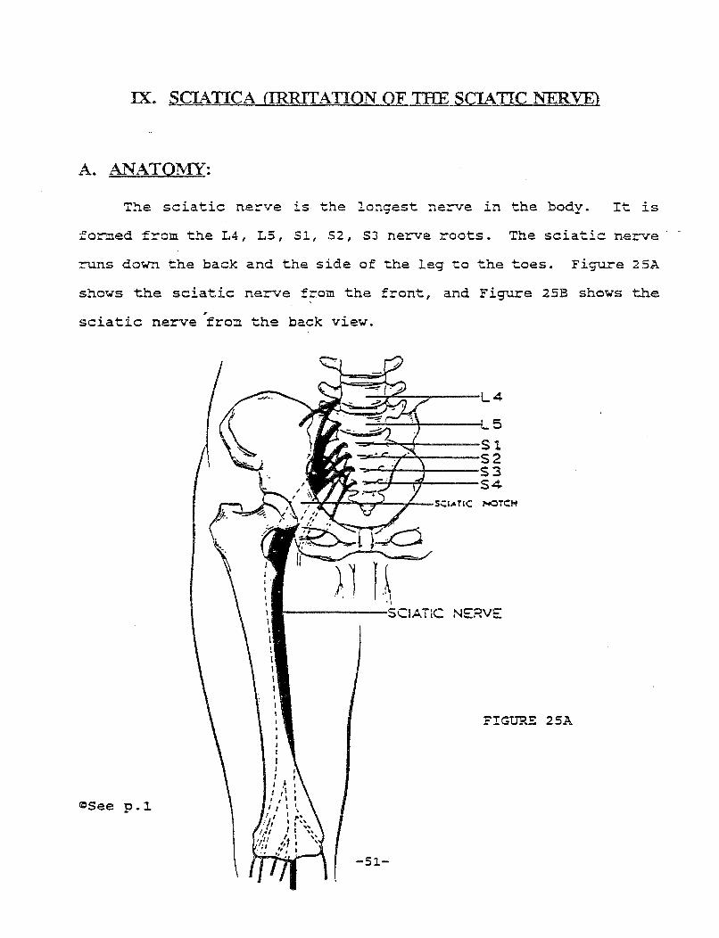
A. ANATO"MY:
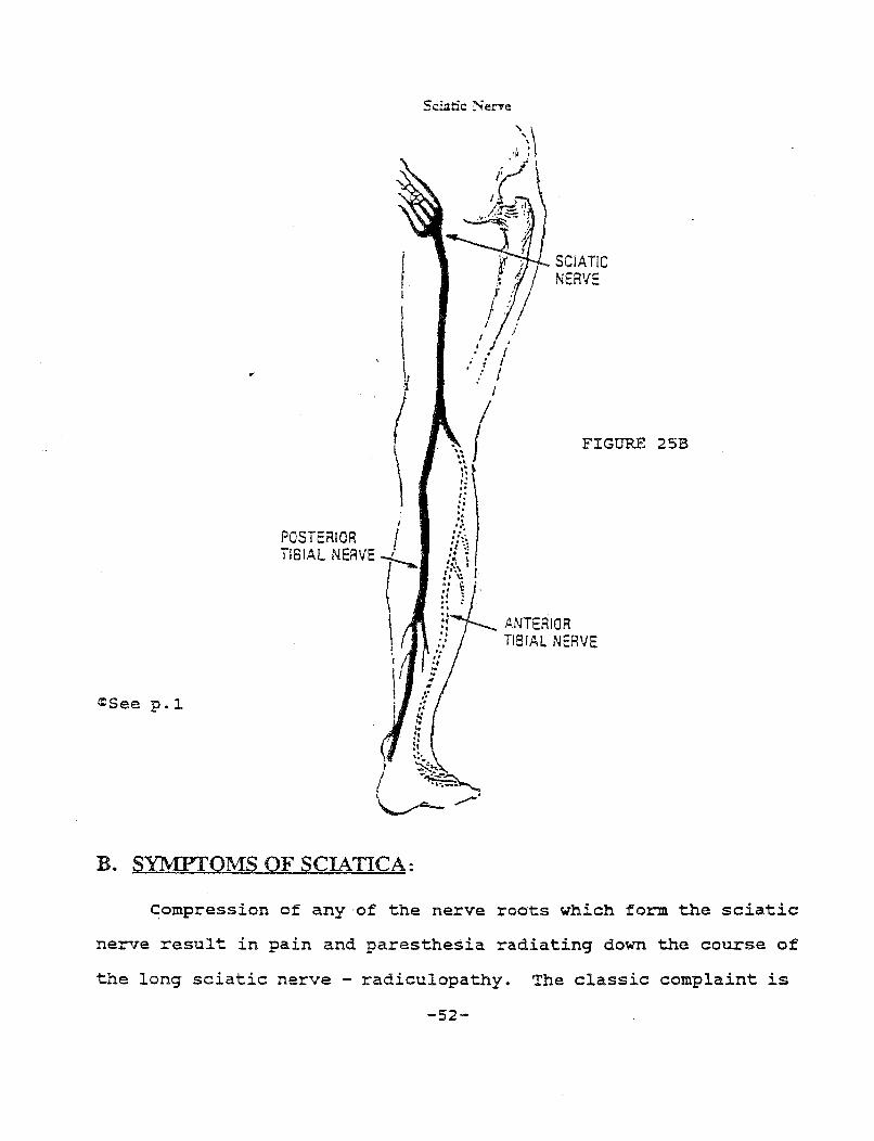
The sciatic ne~ve is the longest nerve in the body. It is
formed from the L4, L5, Sl, S2, SJ nerve roots. The sciatic n~rve ·
runs down the back and the side of the leg to the toes. Figure 25A
shows the sciatic nerve from the front, and Figure 2SB shows the ,.
sciatic nerve from the back view.
~See p.~
\ I I I l I I I I
4
NOTCH
-------SCIATiC NERVE
FIGURE 2SA
-51-
POSTERIOR I TIBIAL NERVE -/-_
<esee p. 1
B. SYI\1PTOMS OF SCIATICA:
Sciatic Nene
I: .. .. '• .. ... . . ·
:;;~ ... . ~ ~ ·:\" • r fi 1;· .. : ... .. . , •• . , ..
SCIATIC NERVE
FIGURE 25B
ANTERIOR TIBIAL NERVE
Compression of any·of the nerve roots which form the sciatic
nerve result in pain and paresthesia radiating down the course of
the long sciatic nerve - radiculopathy. The classic complaint is
-52-

pain radiating down the buttock, back of the thigh, down the side
of the leg and down the back of the leg, generally at least below
the knee.
Radiculopathy involving the sciatic nerve is given a special
name in honor of the body's longest nerve - sciatica. A detailed
discussion of sciatica - its symptoms, examination findings, causes
and treatment is found in Attachment No. 4 - Kahanovitz, "Sciatica:
Verifying the Diagnosis, Offering Relief", J. Musculoskeletal
Medicine, Jan. 1998, pages 51-58.
C. ANATOMICAL BASIS FOR SCIATIC STRETCH TESTS:
Sciatic tension tests, such as the straight leg-raising test
(SLR), the Lasegue's test, and the Milgram test, puts the sciatic
nerve "on stretch", and are universally performed during physical
examination of the low back as an aid in diagnosing sciatica.
The sciatic nerve is elastic. If any of the nerve roots,
which form the sciatic nerve, is compressed, the distance the
sciatic nerve must travel from the spinal canal down the leg to the
toes is increased.
The tension tests put the sciatic nerve on "stretch" by taking
up the normal "slack" in the long sciatic nerve which increases the
compression and tension on an already compressed and irritated
nerve root. The result is reproduction of radiating pain along the
course of the sciatic nerve - a positive (+) test. A normal
sciatic nerve (non-irritated) does not cause pain when put under
stretch by these tests.
-53-

<!:)See p.~
) "
t ' \ \
~ A
t r.; ~-
r' i
~ / I
\ ) .. \\. /;. \
t. \
~ .;. \: •' !
.. ....;· ~
~'.'
FIGURE 26
Diagrammctie representction of the c:oncept of tne sciatic: nerYe .. on stretc:n" wnic:h produc:es "hyperirritcbi!ity" end pain. (A) Normal nerve in relaxed po-sition. (B) The spoc:e-oc:c:upying lesion (X) develops at the nerve's sourc:e increasing the distance tne inelastic: nerve must travel. (C) Dorsal extension (upwcrd bending of the ankle iaint) ploc:es the tout, "nyperirrtable" nerve "on stretc:n," whic:h c:ouses pain. These pnysic:cl phenomena ore the physiologic:cl and onotomic:cl base for many of the low boc:k diagnostic: maneuvers.
D. THE STRAIGfff LEG RAISIN"G TEST:
1. PERFORMING THE TEST:
The straight leg-raising test is performed with the
patient lying on his back (supine) on the examining table. The
physician simply lifts the patient's leg off of the table, flexing
-54-
the hip in the direction of the patient's head. The test is
positive (+) if the patient reports radiating pain down the course
of the sciatic nerve before a 90 degree angle is reached. The
physician records the angle at which the sciatica is reproduced,
e.g., 60 degrees. Generally, the more severe a patient's
condition, the lower the angle is required to reproduce the sciatic
pain. The opposite (normal) leg is also tested and will also
result in a positive straight leg-raising test in many cases with
sciatica reproduced in the involved leg - a contralateral straight
leg-raising test.
2. POSITIVE TEST:
A positive sciatic stretch test, such as a straight leg-
raising test is an indication of nerve root compression in the
nerve roots which form the sciatic nerve. As previously noted, a
disc can be herniated yet cause no nerve root compression.
Symptoms of sciatica will not be experienced unless the nerve root
is actually compressed by the herniated disc. A patient with a
herniated disc which does not compress the nerve root will
generally experience only localized low back pain (or be free from
symptoms) and will have a negative straight leg-raising test.
However, later in life, the inside disc material from the nucleus
pulposus may become larger and actually press upon the nerve root
producing sciatica with a positive straight leg-raising test.
The physician must distinguish a positive straight leg-raising
test from a tight hamstring muscle (the thigh muscle in back of the
-55-
thigh when bends the knee) . A tight hamstring muscle can restrict
the angle the patient can raise hisjher leg due to muscle spasm.
The straight leg-raising test can be further defined to rule out
pain from a tight hamstring muscle by additional maneuver of
stretching the foot upward (dorsiflexion) toward the patient's head
while the patient's leg is already off of the examining table and
flexed. In addition, the patient's neck may be flexed forward by
bending the chin toward -the chest. Both maneuvers exert an
additional "stretch" on the sciatic nerve confirming that a
positive straight leg-raising test is the result of nerve root
irritation and not a tight hamstring muscle.
The result of the straight leg-raising test usually vary from
one office examination to the next depending on periods of
decreased (remission)
(exacerbation) symptoms.
symptoms and periods of increased
In addition, the results of the straight
leg-raising test may be altered if a patient taking analgesics
(pain relievers) since his threshold for experiencing sciatic pain
may be increased, i.e., the test may be positive only by raising
the leg at a higher angle in order to reproduce the sciatic pain.
3. THE VARIATION - SEATED STRAIGHT LEG-RAISING TEST:
A variation of the straight leg-raising test is done with
the patient seated at the edge of the examining table with his legs
dangling off the edge instead of lying supine.
The seated straight leg-raising test is performed by the
physician who raises the patient's leg upward to the horizontal
-56-

position by straightening the patient's knee. The sciatic nerve is
thus stretched.
PRACTICE NOTE: Defense examinations (IME) often have the patient perform both standard (supine) straight leg-raising test and the seated straight leg-raising test to observe discrepancies between both tests in an effort to rule-out malingering (faking).
FIGURE 27
<C'iSee p.~
-57-
LIFT lf f:.lll
X. OSTEOARTIIRITIS OF THE SPINE
A. HYPERTROPIDC DEGENERATIVE CHANGES:
Osteoarthritis (osteo = bone) of the spine involves
"hypertrophic degenerative changes" and can occur at different
places in the vertebral column. The term "hypertrophic
degenerative changes 11 refers simply to an overgrowth of bone on the
vertebrae, and is used synonymously with osteoarthritis.•
Osteophytes, spur like projections that look like the spurs of a
cowboy boot, develop.
NOTE: Rheumatoid arthritis should not be confused with osteoarthritis. Rheumatoid arthritis of the spine involves systemic inflammatory conditions usually effecting several joints, including the knee joint, joints in the hand and the spine. Environmental factors trigger rheumatoid arthritic conditions in genetically predisposed persons. A patient with rheumatoid arthritis experiences classic symptoms of severe pain and stiffness in the morning which improves later in the day with activity. In contrast, osteoarthritis results from the overgrowth of bone, and is either wear and tear or traumatic arthritis. Osteoarthritis usually is localized to specific areas, and is not systemic (affecting the whole body) .
*Strictly speaking, arthritis can only occur at a synovial joint. Although the facet joint is the only synovial joint in the vertebral column, the term osteoarthritis is loosely used by physicians to describe excess bony growth at any location in the vertebral column whether or not a true synovial joint.
-58-

~. DEGENERATIVE DISC DISEASE - THE AGING PROCESS:
As we age, our vertebrae and discs degenerate. An analogy can
be made to a new car which slowly ages and degenerates with
increased mileage.
have full height,
While we are young, our intervertebral discs
the annulus fibrosus is firm and the nucleus
pulposus is tense, and the outside annular ring is thick and
functioning; our foramen are equivalent to big open windows; our
spinal canal has sufficient "breathing room" for the spinal cord
and the nerve roots; our facet joints are smooth with well oiled
joint cartilages gliding freely one over the other; and our spinal
ligaments are vibrant, elastic and strong.
The aging process begins with the intervertebral disc. The
disc acts as a shock absorber, distributing stress between the
vertebrae above and below it. The inside nucleus pulposus is able
to act as a shock absorber because of its high water content. As
we age, the nucleus pulposus slowly loses water and gradually
"dries out". The outside annular ring (annulus fibrosus) slowly
degenerates by losing its elasticity and slowly develops fissures.
The intervertebral disc space and disc height (the space occupied
by the disc sandwiched between the upper and lower vertebrae)
becomes smaller. As the disc space gradually collapses, stress is
placed on the posterior facet joints and ligaments which are not
suited to handle the excessive stress. Degenerative, hypertrophic
changes develop at the anterior (front) side of the vertebral body.
Overgrowth of bone (hypertrophic degenerative changes) occur. Bony
-59-
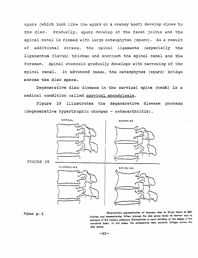
spurs (which look like the spurs on a cowboy boot) develop close to
the disc. Gradually, spurs develop at the facet joints and the
spinal canal is rimmed with large osteophytes (spurs). As a result
of additional stress, the spinal ligaments (especially the
ligamentum flavum) thicken and encroach the spinal canal and the
foramen. Spinal stenosis gradually develops with narrowing of the
spinal canal. In advanced cases, the osteophytes (spurs) bridge
across the disc space.
Degenerative disc disease in the cervical spine (neck) is a
medical condition called cervical spondylosis.
Figure 28 illustrates the degenerative disease process
(degenerative hypertrophic changes osteoarthritis).
\ FIGURE 28
<:JSee p.~
NOIHI.ll..
Oiagrc::mafio: nt;:~rnenl\'lrion or .:hc:nges saen in X-ray ,;_, af disk injuries and ostaoarri'uiris.. When injurttd, me ais.li: spac:e 11tnds 1'0 ncti"'''W due teo
c:alloc::u of me nuc:leus 01ul~asus. Osteophytes or spurs de .. elop ar me ed~;~n e~f me .. .,~,;brc::l body. In old O:::MS, me cnreophyTeS may aeuolly brid<;• CIC:I'OSS m~ disk spoc:e.
-60-
2. PEOPLE WITH DEGENERATIVE DISC DISEASE OFTEN HAVE NO SYMPTOMS:
People with degenerative disc disease (osteoarthritis)
often have no symptoms whatsoever. For example, almost all people
over 55 have asymptomatic cervical spondylosis (degenerative disc
disease of the cervical spine) which is only detected if plain x-
rays are taken. People with cervical spondylosis can go throughout
life with no symptoms. Wiesel, et al., in their text book, Neck
Pain, 2d Ed., at page 50-51, states, "· .. In cadaver studies, almost
everyone over the age of 55 will show evidence of cervical
spondylosis. Obviously, not all of these patients are symptomatic.
(Friedenberg, et al.*), have emphasized the lack of correlation
between radiographs of the cervical spine and symptoms.
... Radiographs (AP, lateral, oblique) of the cervical spine in
cervical spondylosis show varying degrees of changes. These
include disc space narrowing, osteophytosis, foraminal narrowing,
degenerative changes of the facets, or instability. As previously
discussed, these findings do not correlate with symptoms ... ".
(emphasis added).
3. TRAUMA PRECIPITATES SYMPTOMS OF PREEXISTING, ASYMPTOMATIC OSTEOARTHRITIS:
Osteoarthritis of the spine is characterized by
hypertrophic degenerative changes - the overgrowth of bone
*"Deneration - Disc Disease of the Cervical Spine", JBJS 45A, 1171-1178, 1963)
-61-
resulting 1.n bony spur-like projections. The spur-like projections
are sharp like the spur on a cowboy boot. These osteophytic spurs
are located on the vertebrae near the vertebral foramen, the window
where the nerve root exits from the spinal canal. As noted, many
people have severe osteoarthritis of the spine with no symptoms
whatsoever.
The hyperextension - hyperflexion motion (extending the neck
backward and forward beyond its normal limits) as seen in auto
collisions can cause a sharp pointed spur to strike a nerve root in
the spine precipitating symptoms. In addition, the trauma can
result in compression of a preexisting osteoarthritic spur against
a nerve root. Trauma, as seen in hyperextension-hyperflexion
injuries, causes stretching and tearing of the spinal ligaments.
The surrounding microhemorrhage and tissue swelling can precipitate
the symptoms of osteoarthritis in an asymptomatic person.
Professor Hoppenfeld, in his textbook Orthopedic Neurology, A
Diagnostic Guide to Neurologic Levels, p. 8, (1977) describes the
effects of an auto collision on the cervical spine as follows:
"Patients frequently develop neck pain after automobile accidents
which cause the cervical spine to whip back and forth (whiplash) or
twist. The resulting injury may stretch an individual nerve root,
cause a nerve root to impinge upon an osteoarthritic spur, or
produce a herniated disc •.. " (emphasis added) •
-62-
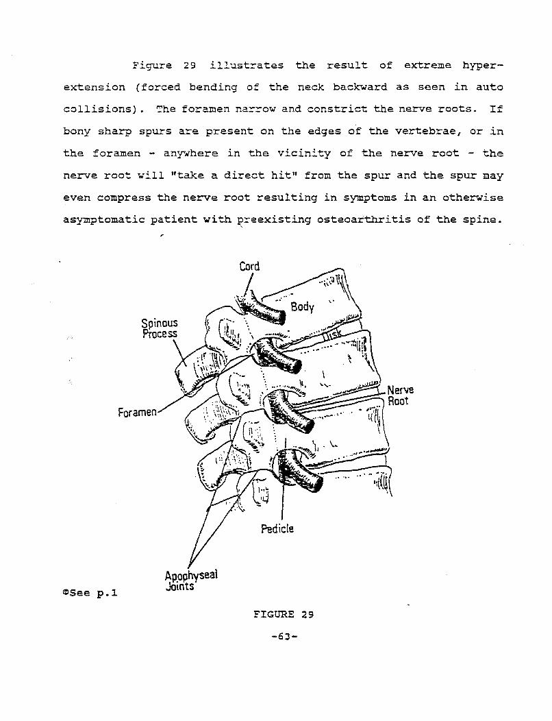
Figure 29 illustrates the result of extreme hyper-
extension (forced bending of the neck backward as seen in auto
collisions) . The foramen narrow and constrict the nerve roots. If
bony sharp spurs are present on the edges of the vertebrae, or in
the foramen - anywhere in the vicinity of the nerve root - the
nerve root will "take a direct hit" from the spur and the spur may
even compress the nerve root resulting in symptoms in an otherwise
asymptomatic patient with ~reexisting osteoarthritis of the spine.
Foramen
<rJSee p.l. Ap.ophyseal J()mts
Pedicle
FIGURE 29
-63-

4. ANALOGY OF OSTEOARTHRITIS OF THE SPINE TO A GRAY HAIR AND A MATCH:
The jury should understand that degenerative osteoarthritis,
even in severe cases, can produce no symptoms. The aging process
in the spine can be painless like the aging process involving our
hair turning gray with no symptoms. Trauma from an auto
collision can precipitate symptoms like striking a match. The
phosphorus is already present at the tip of the match. Trauma from
the friction strike, lights up the match causing symptoms.
Similarly, trauma from an auto collision lights up symptoms in
asymptomatic, preexisting osteoarthritis of the spine. The
defendant takes the plaintiff as he finds her and is responsible
for precipitation and aggravation of the symptoms.
PRACTICE NOTE - CROSS-EXAMINATION OF THE DEFENSE 11 IME11
PHYSICIAN: The defense, many times, will allege that the patient's symptoms are solely the result of a preexisting osteoarthritis of the spine. The defense examiner must admit that patients, even with severe degenerative osteoarthritis are often asymptomatic. The following is a sample cross-examination of the defense doctor which illustrates this point:
Q: Doctor, in your own practice, you have at times seen patients with severe osteoarthritis of the spine, as shown on x-ray, who had no symptoms whatsoever just before a traumatic injury, such as an auto collision?
A: Yes.
Q: Doctor, isn't it well known in medicine that many patients who have severe degenerative osteoarthritis of the spine, often have no symptoms whatsoever?
A: Yes.
Q: Doctor, isn't it also well known in medicine that patients with severe degenerative osteoarthritis of the spine, as shown on x-rays, may go throughout their lifetime with no symptoms?
-64-
A: Yes.
Q: Doctor, before this auto wreck, Mr. Plaintiff had no history whatsoever of neck or back pain.
A: Yes, that is correct.
Q: Doctor, would you agree that the precipitating cause of the plaintiff's pain (severe osteoarthritis) was the auto collision of November 5, 1997?
A: Yes.
5. MINOR IMPACT CASE - USE PREEXISTING OSTEOARTHRITIS AS AN OFFENSE:
Jurors have a difficult time believing that a substantial
injury can result from a minor impact collision. In a patient with
preexisting, asymptomatic osteoarthritis, the defense will argue
that the patient's injury is a result of this preexisting condition
and not the minor impact collision. This defense should be used as
an offense on behalf of the plaintiff. An elderly person with
asymptomatic osteoarthritis is more likely to sustain a significant
neck injury from a minor impact then would a healthy football
player, 19 years of age. In addition, the soft tissue injury to
the plaintiff's neck, ligaments and muscles will take much longer
to heal, and may even be permanent because of the activation of the
plaintiff's preexisting asymptomatic osteoarthritis. (Remember:
The defendant takes the plaintiff as he finds him.)
(a) Closing Argument - Analogy to Eqqs and Golf Balls:
The "eggshell skull" doctrine can be illustrated to the
jury, during closing argument, by asking a rhetorical question
using an analogy to "eggs and golf balls".
-65-
CLOSING ARGUMENT: Assume, this case were about a
farmer taking eggs to the market. Enroute, he is rear-
ended, and all the eggs fall out of the truck and break
as they hit the ground. No one would have any trouble
awarding the farmer damages for the cost of his eggs. Is
it a defense to say "the farmer should have been carrying
golf balls"? Similarly, is it a defense to say that Mr.
Plaintiff should have had the spine of a 19 year old boy?
6. TRAUMA ITSELF MAY CAUSE OSTEOARTHRITIS:
Trauma from an auto collision can itself cause
hypertrophic degenerative changes (osteoarthritis) in the spine.
The trauma from an auto collision results in hyperextension,
hyperflexion of the spine with resulting tearing and stretching of
ligaments. Microhemorrhaging and swelling in the tissues
surrounding the vertebrae develop - causing the bony vertebrae to
react by overgrowing bone - hypertrophic spur formation. It is
important to obtain plain x-ray films taken on the day of the
accident in the emergency room and compare these with x-ray films
taken at a later date. It normally takes at least 4 - 6 months for
degenerative changes to develop in the spine as a result of trauma.
In addition, trauma can accelerate an already existing degenerative
condition. A comparison of the x-rays, CT scans and MRI films at
different time intervals is essential to document the development
of osteoarthritis where none previously existed and to document the
acceleration of a preexisting osteoarthritic condition. For
-66-
example, in one case the author tried, the initial emergency room
x-ray report found "minimal degenerative changes" i whereas six
months later, the x-ray report noted "moderate degenerative
changes" in the same area of the cervical spine. The trawna of the
auto collision had accelerated the preexisting osteoarthritis of
the spine.
B. FACET .JOINT- OSTEOARTHRITIS: (
In the back of each vertebrae are two sets of facets (a smooth
flat surface of bone) which 11 hook-up 11 with the facets of vertebral
bodies directly above and below when the vertebrae are stacked
together, one on top of the other, to form the vertebral colwnn.
When the facets hook-up, they form facet joints (also called
apophyseal joints). A joint is defined as a place in the body
where two bone surfaces meet. This "meeting place" is also called
an articulation and the joint surfaces are called articular
surfaces. The surfaces of the facets are smooth and flat. Facet
joints are true synovial joints (the only synovial joints in the
vertebral column), since it is lined by a thin synovial membrane
which produces a 11 gliding oil" called synovial fluid. Synovial
fluid is present in all synovial joints, such as the knee, and
helps the bones of the joint glide over each other. Synovial fluid
also lubricates the joint surfaces preventing wear and tear
breakdown. The facet joints glide over each other and permit
motion of the spine, especially bending forward (flexion) and
backward (extension).
-67-
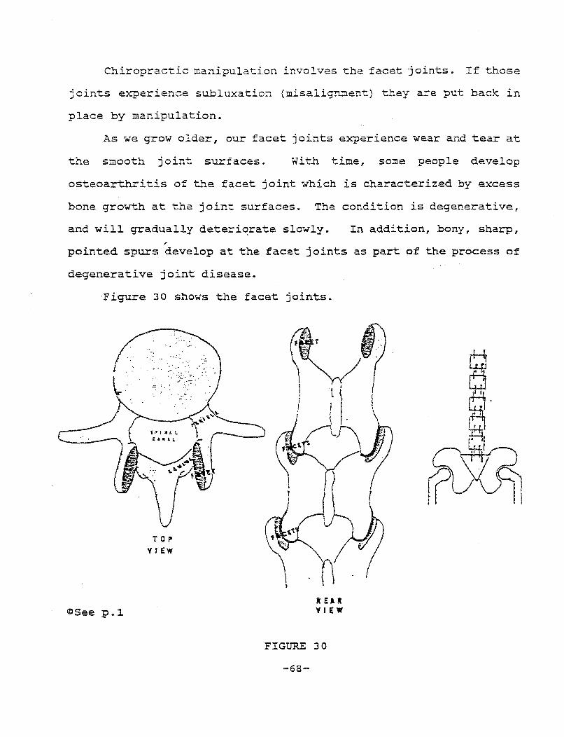
Chiropractic manipulation involves the facet joints. If those
joints experience subluxation (misalignment) they are put back in
place by manipulation.
As we grow older, our facet joints experience wear and tear at
the smooth joint surfaces. With time, some people develop
osteoarthritis of the facet joint which is characterized by excess
bone growth at the joint surfaces. The condition is degenerative,
and will gradually deteriqrate slowly. In addition, bony, sharp, ,.
pointed spurs develop at the facet joints as part of the process of
degenerative joint disease.
·Figure 30 shows the facet joints.
~See p.l.
TOP VIEW
X £A R Y I EW
FIGURE 30
-68-
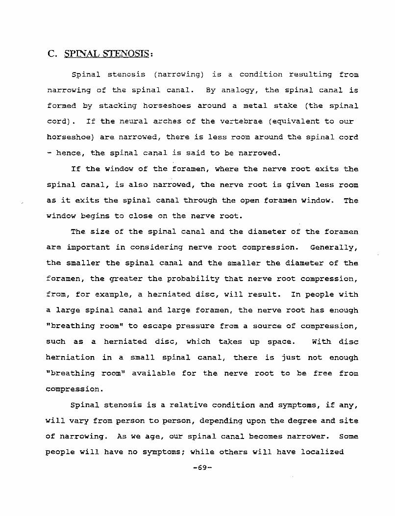
C. SPIN"AL STENOSIS:
Spinal stenosis (narrowing) is a condition resulting from
narrowing of the spinal canal. By analogy, the spinal canal is
formed by stacking horseshoes around a metal stake (the spinal
cord). If the neural arches of the vertebrae (equivalent to our
horseshoe) are narrowed, there is less room around the spinal cord
hence, the spinal canal is said to be narrowed.
If the window of the foramen, where the nerve root exits the
spinal canal, is also narrowed, the nerve root is given less room
as it exits the spinal canal through the open foramen window. The
window begins to close on the nerve root.
The size of the spinal canal and the diameter of the foramen
are important in considering nerve root compression. Generally,
the smaller the spinal canal and the smaller the diameter of the
foramen, the greater the probability that nerve root compression,
from, for example, a herniated disc, will result. In people with
a large spinal canal and large foramen, the nerve root has enough
"breathing room" to escape pressure from a source of compression,
such as a herniated disc, which takes up space. With disc
herniation in a small spinal canal, there is just not enough
"breathing room" available for the nerve root to be free from
compression.
Spinal stenosis is a relative condition and symptoms, if any,
will vary from person to person, depending upon the degree and site
of narrowing. As we age, our spinal canal becomes narrower. Some
people will have no symptoms; while others will have localized
-69-
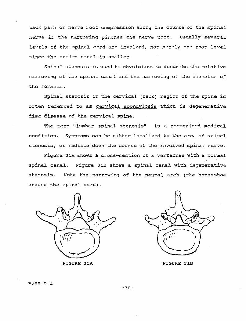
back pain or nerve root compression along the course of the spinal
nerve if the narrowing pinches the nerve root. Usually several
levels of the spinal cord are involved, not merely one root level
since the entire canal is smaller.
Spinal stenosis is used by physicians to describe the relative
narrowing of the spinal canal and the narrowing of the diameter of
the foramen.
Spinal stenosis in the cervical (neck) region of the spine is
often referred to as cervical snondylosis which is degenerative
disc disease of the cervical spine.
The term "lumbar spinal stenosis" is a recognized medical
condition. Symptoms can be either localized to the area of spinal
stenosis, or radiate down the course of the involved spinal nerve.
Figure 31A shows a cross-section of a vertebrae with a normal
spinal canal. Figure 31B shows a spinal canal with degenerative
stenosis. Note the narrowing of the neural arch (the horseshoe
around the spinal cord).
FIGURE 31A FIGURE 31B
~See p.l -70-
XI. SPONDYLOLYIS AND SPONDYLOLISTHESIS ('!'HE SPONDY BROTHERS)
Soondvlolvis is derived from the Greek (spondylos = vertebrae
and lysis dissolution) since this condition represents a
flattening of a vertebral body ~ithout any forward displacement.
Soondvlolisthesis, on the other hand, involves forward displacement
- sliooage of one vertebrae on another. The bottom vertebrae is a
shelf for the top vertebrae. Spondylolisthesis is derived from the
Greek (spondylosis = ver~ebrae, olisthanein = to slip).
Spondylolisthesis is found mainly at the lumbosacral joint
~here the L-5 vertebrae slips forward (like the Leaning Tower of
Pisa) on the S-1 vertebral body.
MEMORY AID: An easy ~ay to remember that spondylolisthesis involves slippage of a vertebrae is to think of a graduate student failing his thesis exam -"slipped up on his thesis"!
Figure 32A compares spondylolyis and spondylolisthesis. Note
the slippage of the LS vertebrae on the first sacral vertebrae
~hich is the classic finding in spondylolisthesis.
sho~s Grade 4 spondylolisthesis.
~See p .1 Spondylolysis
?:.ln !nc~r..lrticul::ui:.
-71- Spondylolisth~is
Figure 32B
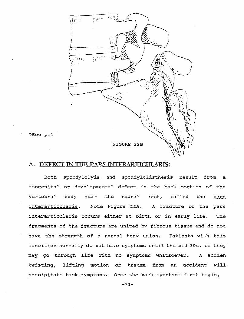
©See p.~
\ . \. . .. dill'
I \\' l''.'
FIGURE 32B
A. DEFECT IN THE PARS INTERARTICtJLARIS:
Both spondylolyis and spondylolisthesis result from a
congenital or developmental defect in the back portion of the
vertebral body near the neural arch, called the pars
interarticularis. Note Figure 32A. A fracture of the pars
interarticularis occurs either at birth or in early life. The
fragments of the fracture are united by fibrous tissue and do not
have the strength of a normal bony union. Patients with this
condition normally do not have symptoms until the mid 30s, or they
may go through life with no symptoms whatsoever. A sudden
twisting, lifting motion or trauma from an accident will
precipitate back symptoms. Once the back symptoms first begin,
-72-

they will continue on a recurring basis with periods of remission.
The symptoms resulting from these conditions are normally localized
to the low back unless the slipped vertebrae of spondylolisthesis
pushes on the nerve root causing radiating pain along the course of
the spinal nerve.
B. PREEXISTING CONDIDON OFTEN WITH NO SYMPTOMS:
Many peopl;_e with spondylolisthesis, even past middle age, have
no symptoms. Weisel, et al., in their text Low Back Pain, 2d Ed.,
p. 71, state, "At this point, it should be reemphasized that in some
people, even severe displacement is asymptomatic and gives rise to
no disability. It is not uncommon to pick-up a previously
unrecognized spondylolisthesis on a routine gastrointestinal
radiological study of a 50 year old patient."
C. THE ROLE OF TRAUMA IN SPONDYLOLYSIS AND SPONDYLOLISTHESIS:
A patient with spondylolysis or spondylolisthesis is an
"eggshell skull" plaintiff with an already preexisting unstable
back from the condition itself. A traumatic event, such as an auto
collision, can trigger or precipitate the symptoms of spondylolysis
or spondylolisthesis in an asymptomatic patient. (The symptoms can
also develop without any noticeable acute trauma) .
A patient with preexisting spondylolisthesis with only
localized low back pain may develop nerve root irritation
following trauma if the "slipped forward" vertebrae is pushed into
-73-
a nerve root. Spondylolisthesis is measured by grades 1-4,
depending upon the degree of slippage of the fifth lumbar
vertebrae. Trauma from an auto collision can cause the vertebrae
to slip forward even more. For example, trauma may push a Grade 1
condition to Grade 2.
In addition, trauma can convert a spondylolyis (which involves
a defect of the pars interarticularis, but with no slippage of one
vertebrae on another) i~to a spondylolisthesis by causing a
slippage of the top vertebrae.
It is possible to sustain an acute fracture of the pars
interarticularis as a result of trauma but this is rare. The
defense will claim that the fracture of the pars interarticularis
is a preexisting condition. If there are no prior x-rays, the only
way to prove the recent fracture is with a bone scan within three
months of the acute injury, which will light-up at the site of the
fracture if the fracture is recent. (A "hot spot" resulting from
the accumulation of the radioactive bone scan dye).
-74-
PART TWO
I. SOFT TISSUE IN.UJRIES
Soft tissue injuries result from forced hyperextension-
hyperflexion motion caused by auto collisions. Injuries to the
following soft tissues are discussed in this section*:
• Ligaments, muscle and fascia -- strain and sprain injuries and myofascial pain syndrome (MPS) .
• Blood vessels (vertebral and carotid artery) --vasoconstriction with decreased blood flow to the brain.
• Sympathetic nerves -- nerve stimulation and irritation.
• Brain -- post-concussion syndrome.
A. MECHANISM OF INJURY - FORCED HYPEREXTENSION-HYPERFLEXION MOTION.
In all auto collisions, the head and neck are forced in the
direction of the striking car.
1. REAR-END COLLISIONS:
Rear-end collisions result in a sudden acceleration of the
body forward, while the head and neck are forcefully hyperextended
(backward) followed by a recoil of forced hyperflexion (forward) .
*Fibromyalgia is discussed in Part III
-75-
2. HEAD-ON COLLISIONS:
Head-on collisions result in the head and neck being forced
forward in hyperextension followed by a recoil of hyperflexion
(backward) .
3. SIDE COLLISIONS:
In side collisions, the head and neck are thrown to the side
towards the striking car and then recoils in the opposite
direction.
4. THE SODDEN STOP:
In sudden stops, the body is accelerated forward.
In a rear-end collision, the defense attorney will attempt to
prove that a sudden stop caused the plaintiff's neck injury rather
than the rear-end collision. If the plaintiff testifies that his
head and neck were "pushed forward causing my head to strike the
windshield", the laws of physics support the defense attorney's
contention.
If the cause of the plaintiff's injury were solely from the
rear-end collision, the plaintiff's head and neck would first be
thrown backward {hyperextension) in the direction of the striking
~~ followed by recoil (hyperflexion) forward. Since the
plaintiff testified that his head and neck were pushed forward
(without first being thrown backward), the plaintiff's own
testimony establishes the cause of his striking the windshield to
be the sudden stop.
-76-
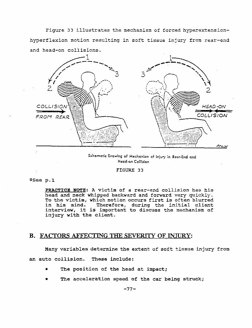
Figure 33 illustrates the mechanism of forced hyperextension-
hyperflexion motion resulting in soft tissue injury from rear-end
and head-on collisions. 1
FROM
~See p.l
Schem:tic Drr::win; or Med":c::'lism or ln[ury in Reor-E:~d c:nd He:::d-<:~n Collision
FIGURE 33
PRACTICE NOTE: A victim of a rear-end collision has his head and neck whipped backward and forward very quickly. To the victim, which motion occurs first is often blurred in his mind. Therefore, during the initial client interview, it is important to discuss the mechanism of injury with the client.
B. FACTORS AFFECTING THE SEVERITY OF INJURY:
Many variables determine the extent of soft tissue injury from
an auto collision. These include:
• The position of the head at impact;
• The acceleration speed of the car being struck;
-77-
Vehicle parameters, such as:
Being built to collapse (energy absorbing);
Head restraints;
Seatbacks;
Seatbelts and shoulder harnesses.
Human factors, such as the condition of the plaintiff
prior to impact.
1. EQUATING BODI:LY INJURY Wl:TB THE EXTENT OF PROPERTY DAMAGE CONDEMNED:
The practice of equating bodily injury with the extent of
property damage to the autos has been condemned by physicians and
engineers. For example, Drs. Foreman and Croft, in their textbook,
Whiplash Injuries (1988), p. 62, state:
"The notion that one can calculate or predict the type or extent of soft tissue damage sustained by occupants of a vehicle merely by calculating the G forces produced in the vehicle by the resultant collision should clearly be laid to rest. The oractice of calculating bodil~ in;ury to the victim by estimating the cost of auto damage is naive at best and should be condemned ... "(emphasis added) .
Similarly, Drs. Carroll et al., "J. Musculoskeletal Medicine"
(June 1992) p.lOO state:
"The amount of damages to the automobile bears little relation to the force applied to the cervical spine of the occupants ..• "
2. THE POSITION OF THE HEAD AT IMPACT:
It is well known that a victim whose head is partially
rotated and caught off balance will incur a severe injury even from
a minor impact. More severe injuries result from oblique angle
-78-
collisions, where a driver is hit from the left rear corner while
turning left. (The driver's head is rotated at impact).
Drs. Foreman and Croft, "Whiplash Injuries", (1988) p.68,
explain the physiological basis for the increased severity of
injury:
"When the head is rotated 45°, the amount of extension that the spine is capable of is decreased by 50 percent. This results in increased compressive load at the facet joint ....
The intervertebral foramen is also small on the side of rotation and lateral flexion, and its contents are more vulnerable to injury ... "
See also, Carroll et al., J. Musculoskeletal Medicine (June
1992) 97, 109, Attachment No. 1.
3. ACCELERATION OF THE CAR BEING STRUCK:.
The acceleration of the plaintiff's car upon being struck
is a factor in determining the extent of soft tissue injury since
the rate of acceleration forward determines the rate of
hyperextension of the neck following impact. Professors Wiesel,
et. al., in their text, Neck Pain, 2d Edition (1992), p. 292,
discuss acceleration as a factor in causing injury:
" .... it becomes apparent that the degree of injury depends on the force applied and inertia rather than the speed of the striking vehicle. The faster the struck vehicle is accelerated forward, the more rapid the hyperextension of the neck and the greater the injury. Therefore, a car struck by a large truck or loaded bus travelling 5-7 mph will be accelerated forward as much as if struck by a small car travelling in a much greater velocity. Also, how rapidly the struck car is accelerated forward will depend on road and traffic conditions. If the struck car is essentially bumper to bumper with the car in front, it cannot be catapulted forward, then a strike from the rear might result in
-79-
significant damage to the struck car, from both forward and back collisions but the struck car might not be accelerated forward making the resulting injury to the driver slight. In this case, the damage to the car might be severe. If, however, the struck car were on an icy street with no cars in front, a rear end collision would accelerate the struck car at a high rate causing very little demonstrable damage to the car but a severe hyperextension cervical injury to the driver ... " (emphasis added) .
Other factors affecting the acceleration of the plaintiff's
car at impact and hence the rate of hyperextension are whether the
brakes were on at the. time of impact and the speed of the
plaintiff's vehicle at the time it was struck.
The faster the plaintiff is travelling at time of impact, the
greater will be his acceleration. A greater acceleration results
in a greater rate of hyperextension with increased soft tissue
injury.
4. SURPRISE COLLISIONS AND BRACING:
"Surprise" rear-end collisions result in greater soft
tissue injury than collisions where the victim is aware of an
impending crash and has braced himself. Bracing for the collision
decreases the extent of the forced hyperextension and recoil
hyperflexion forces which cause injury to the soft tissues.
5. PARAMETERS OF THE PLAINTIFF'S VEHICLE:
(a) Energy Absorbing - Built to Collapse Vehicles:
The force (energy) from an auto collision is either
absorbed by the car or by its occupants. In older cars which are
"built like a tank," little property damage is done to the car with
most of the force of impact absorbed by its occupants. Modern
-so-
safety engineering has now designed cars that are "built to
collapse,". The car absorbs the energy of impact rather than the
occupants. For example, Volvo has advertised the safety of its
cars by "collapsing on impact" - absorbing the forces which would
otherwise have been transmitted to the occupant - thereby providing
a safer car.
Drs. Foreman and Croft, in "Whiplash Injuries", p. 67,
describe the secret of safety engineering as follows: ,.
" .•• The driver climbs out of the wreckage with only minor injuries. The secret of this incredible crashworthiness is built into the car - this intentional piece-meal dis-integration absorbs nearly all the energy in the crash. Thus, a car that is built to collapse with impact (within safe limits) is safer than one that is not ..•. "
(b) Head Restraints:
Head restrains are designed to limit injury resulting
from the forceful hyperextension (backward) motion of the head and
neck at the time of impact. If the force of the backward
hyperextension phase is reduced, so will the force of the
hyperflexion - with less injury to the muscles and ligaments of the
neck.
In 1969, head restraints became mandatory. Unfortunately,
head restraints have become "headrests" with the initial purpose of
safety becoming second place to comfort. If the head restraint is
not adjusted properly, increased neck injury can result. For
example, if the head restraint is adjusted downward, it acts as a
fulcrum, especially with tall drivers, causing greater injury to
the ligaments and muscles of the neck.
-81-
(c) Seatbacks:
Seatbacks, like head restraints, if properly designed and
used, decrease the rate of forced hyperextension and its recoil
hyperflexion during impact. Proper seat adjustment and the degree
of "spring" in the seatback directly influence the extent of
injury. For example, a seatback which is designed like a "steel
spring" would increase the intensity of the recoil hyperflexion in
a rear-end collision.
Engineering studies have shown that controlled seatback
collapse at the time of impact reduces the severity of soft tissue
injuries. Accordingly, cars having this type of seatback design
result in less severe neck injuries.
(d) Seatbelts:
The severity of soft tissue injury is affected by the use
of seatbelts, head restraints, and shoulder straps. Wiesel, et al.,
Neck Pain, 2d Edition, pg. 13, describes this relationship.
"The influence of the seatbelts on acute neck injuries is dependent on other factors - the most important of which is the presence or absence of seat head restraints. If head restraints are present, the use of seatbel ts alone result in fewer neck injuries. Without head restraints, the use of seatbelts alone appear to slightly increase the incidence of neck injuries. Combining seat belts with a shoulder strap reduces the incidence of acute neck injuries even without head restraints. The combined use of seatbelt, shoulder strap and head restraint significantly reduces the number of acute neck injuries."
Some vehicles contain shoulder harnesses which lock at a
distance from the occupant's body for extra comfort. During
impact, the slack in this type of shoulder harness allows the
-82-
occupant's body to travel a g=eater distance, with potentiation of
forward angular rotation, resulting in more severe neck injury had
no slack been present.
(e) Human Factors:
People, whose neck structure is less resilient will
suffer greater soft tissue injury than others with nore resilient,
:c:lastic muscles and ligaments. Drs. Foreman and Croft, in their
0xt Whiplash Injuries (1988), p. 67, describe this physiological
~riance affecting the extent of soft tissue injury:
"Certain physiological variances in people will signifi-cantly affect the amount of damage they sustain in a collision. This is why, as is often seen in practice, an occupant in a crash may be injured, while another may escape with no apparent injury. With an increase in age, the elasticity of the tissues decreases. Range of motion in the cervical spine also decreases. In both cases, the potential fer injury is increased because the neck is less resilient ... 11
In addition, a plaintiff, who has asymptomatic degenerative
osteoarthritis of the spine, is more likely to sustain injury in a
minor impact collision than a younger person in his twenties with
no osteoarthritis of the spine.
C. CATEGORIES OF SOFT TISSUE IN.UJRIES:
l. INJURIES TO LIGAMENTS, MUSCLES AND FASCIA:
(a) Spinal Ligaments:
(i) Ligaments Hold the Spine in Place - The spine
is essentially a stacked "bag of bones." Without a binder (glue),
the vertebral column would topple like a high stack of dominos.
-83-
The spinal ligaments are the glue holding each vertebrae in the
vertebral column in place. The ligaments essentially prevent the
vertebrae from 11 slipping around. 11 (Remember that the ligaments
hold bone to bone. In the spine, the ligaments hold vertebrae to
vertebrae) .
(ii) Interspinal Ligament - Each vertebrae has
projections on,.its back side called a "process." We can feel these
projections by placing our fingers in the middle of our back.
These projections are also called the spinous process (spinous = spine) . The interspinous ligament {between the spine) , also called
the spinal ligament, hold the spinous processes together.
This interspinal ligament is illustrated in Figures 34 and 35.
(iii) Ligamentum Flavum (The Yellow Ligament}
Ligamentum Flavum (flavum = yellow) bind the lamina (the back of
neural arch) of one vertebrae to another. (Remember: In the aging
process of the disc, the ligamentum flavum thickens and begins to
encroach the spinal canal and the foramen.
(iv) The Anterior and Posterior Longitudinal
Ligaments (The Long Ligaments) The
anterior and posterior longitudinal ligaments run the entire length
of the vertebral column in the front (anterior) and in the back
(posterior) . As noted, these ligaments are attached to the
vertebrae and discs and actually cover the outside of the annulus
-84-
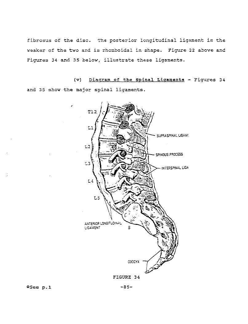
fibrosus of the disc. The posterior longitudinal ligament is the
weaker of the two and is rhomboidal in shape. Figure 22 above and
Figures 34 and 35 below, illustrate these ligaments.
{v) Diagram of the Spinal Ligaments - Figures 34
and 35 show the major spinal ligaments.
a:>See p.l.
COCCYX
FIGURE 34
-85-
SPINOUS P!=IOCQS
!NiE.=\S?!NAL LlG.ol-
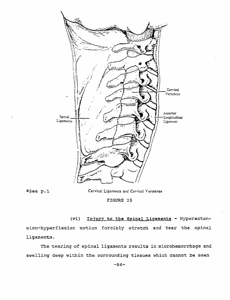
Ante::ior • -· - Longin:dina!
,._"'""--..,. ..... ~ Lipment
i '
~See p.l. Cervic:1l Lig:unencs :1nd Cervic:1! Verteb.-ae
FIGURE 35
(vi) Injury to the Spinal Ligaments - Hyperexten-
sion-hyperflexion motion forcibly stretch and tear the spinal
ligaments.
The tearing of spinal ligaments results in microhemorrhage and
swelling deep within the surrounding tissues which cannot be seen
-86-
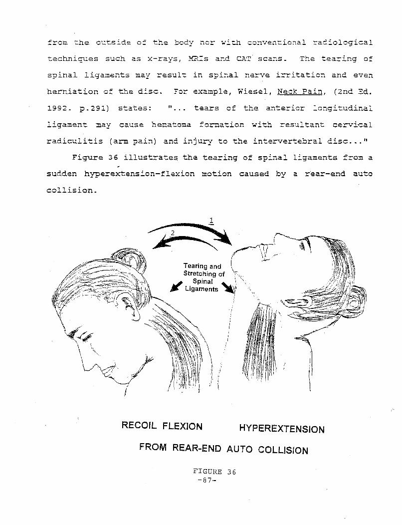
from the outside of the body nor with conventional radiological
techniques such as x-rays, MRis and CAT scans. The tearing of
spinal ligaments may result in spinal nerve irritation and even
herniation of the disc. For example, Wiesel, Neck Pain, (2nd Ed.
1992. p.291) states: " tears of the anterior longitudinal
ligament may cause hematoma formation with resultant cervical
radiculitis (arm pain) and injury to the intervertebral disc ... "
Figure 36 illustrate~ the tearing of spinal ligaments from a
sudden hyperextension-flexion motion caused by a r·ear-end auto
collision.
Tearing and Stretching of
J Spinal ~ Ligaments
(
RECOIL FLEXION HYPEREXTENSION
FROM REAR-END AUTO COLLISION
FIGURE 36 -87-

B. IVfiJSCLES M"'D FASCIA:
J.. FASCIA:
Muscles and fascia lay on top of the spinal ligaments.
Muscles are enclosed in a band of tough, fibrous tissue called
fascia. (Fascia from the Latin meaning band). In essence, the
muscle is "in the bag" - held together in a bag of fascia.
2 • PARASPINAL MUSCLES:
The paraspinal muscles (para = beside) - also called the
paraspinous muscles - lay on top of the spinal ligaments. These
muscles protect the spine, and assist with movement. Figure 39B
shows the para~pinal muscles. On top of the paraspinal muscles lay
fascia and the exterior muscles of the neck and back, such as
scalene muscles, shown in Figure l.l.B above. The paraspinal muscles
in the neck are termed the cervical paraspinal muscles, and in the
low back the lumbar paraspinal muscles.
3. TRAPEZIUS MUSCLES:
The trapezius muscle is named after its shape as a trapezoid
(four-sided) . The trapezius muscle is a large muscle arising from
the back of the head and the cervical and thoracic vertebrae. The
trapezius extends to the collar bone and the shoulder blades. It
helps raise and straighten the shoulder. Since the trapezius is a
large muscle, some physicians label various portions as Tl., T2, T3,
and T4.
-88-
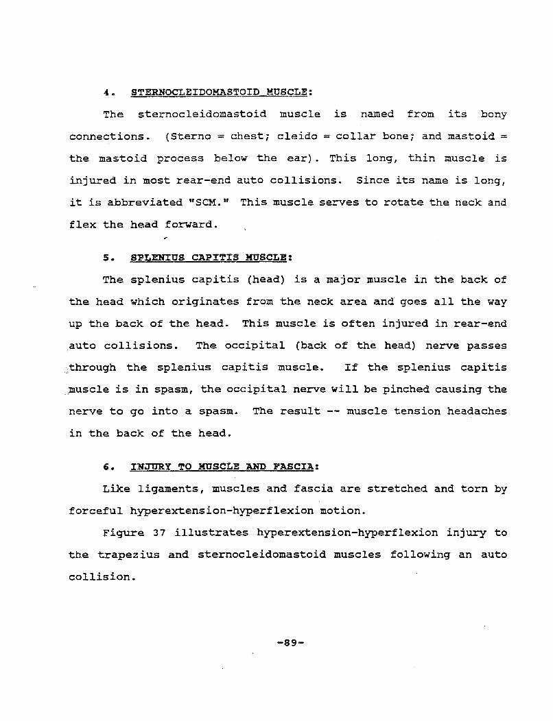
4. STERNOCLEIDOMASTOID MUSCLE:
The sternocleidomastoid muscle is named from its bony
connections. (Sterno = chest; cleido = collar bone; and mastoid = the mastoid process below the ear) . This long, thin muscle is
injured in most rear-end auto collisions. Since its name is long,
it is abbreviated "SCM. 11 This muscle serves to rotate the neck and
flex the head forward.
5. SPLENIUS CAPITIS MUSCLE:
The splenius capitis (head) is a major muscle in the back of
the head which originates from the neck area and goes all the way
up the back of the head. This muscle is often injured in rear-end
auto collisions. The occipital (back of the head) nerve passes
through the splenius capitis muscle. If the splenius capitis
muscle is in spasm, the occipital nerve will be pinched causing the
nerve to go into a spasm. The result -- muscle tension headaches
in the back of the head.
6. INJURY TO MUSCLE AND FASCIA:
Like ligaments, muscles and fascia are stretched and torn by
forceful hyperextension-hyperflexion motion.
Figure 37 illustrates hyperextension-hyperflexion injury to
the trapezius and sternocleidomastoid muscles following an auto
collision.
-89-
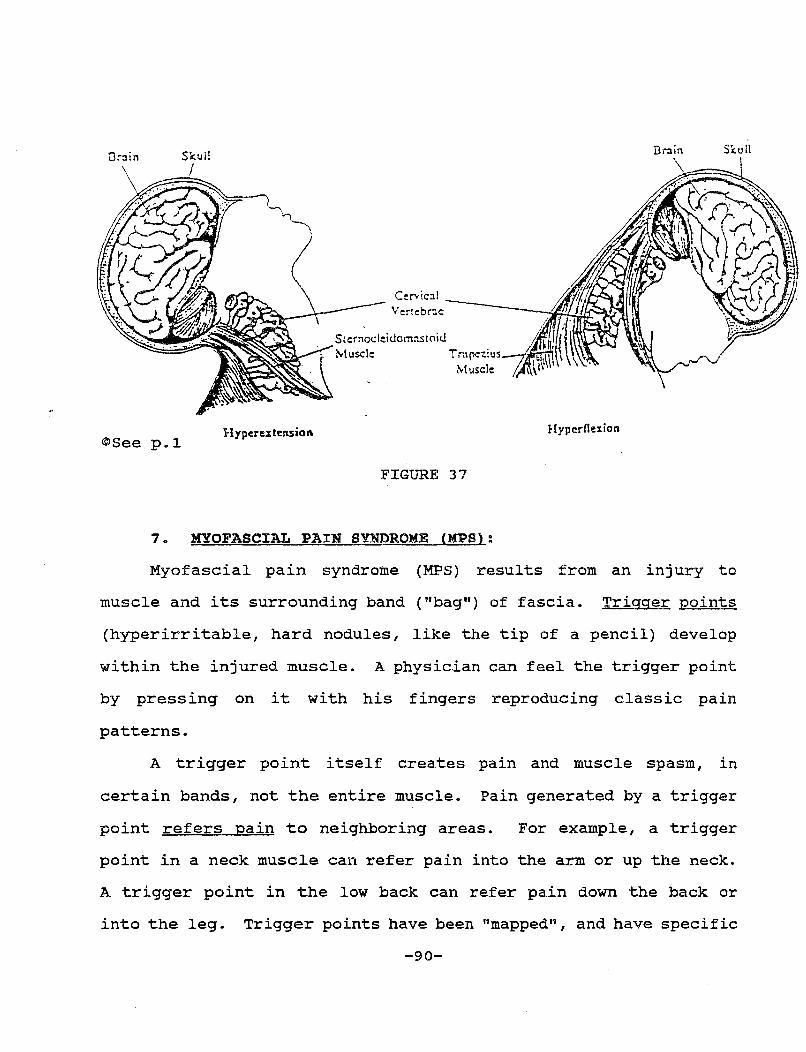
Drain Skull SS:ull
I
Cervical
@See p.l Hyperextension Hyperfle:tion
FIGURE 37
7. MYOFASCIAL PAIN SYNDROME (MPS):
Myofascial pain syndrome (MPS) results from an injury to
muscle and its surrounding band ("bag") of fascia. Trigger points
(hyperirritable, hard nodules, like the tip of a pencil) develop
within the injured muscle. A physician can feel the trigger point
by pressing on it with his fingers reproducing classic pain
patterns.
A trigger point itself creates pain and muscle spasm, in
certain bands, not the entire muscle. Pain generated by a trigger
point refers pain to neighboring areas. For example, a trigger
point in a neck muscle can refer pain into the arm or up the neck.
A trigger point in the low back can refer pain down the back or
into the leg. Trigger points have been "mapped", and have specific
-90-
patterns of referred pain. This concept is the key to understand-
ing the symptoms of myofascial pain syndrome.
Trigger points are treated with injections of a local
anesthetic alone or mixed with an anti-inflammatory steroid,
followed with vigorous stretching during physical therapy to break
up the trigger point. The International Rehabilitation Medicine
Association's Monograph on Myofascial Pain Syndrome is attached as
Exhibit No. 6.
C. SPRAIN AND STRAIN INJURIES:
1. DEFINITION:
Hyperextension-hyperflexion injuries to ligaments and
muscles are called strains and sprains. Professor Ruth Jackson, in
her textbook, The Cervical Syndrome, 4th Ed. (1978) pp. 86-87
defines these terms as follows*:
• Strain - the stretching of ligaments and muscles
• Sprain - the tearing of ligaments and muscles.
These injuries are also referred to as "myoligamentous strain
or sprain." (Myo =muscle, Ligamentous= ligament).
*Other physicians define a "strain" as a stretch or tear injury to a ligament; and a "sprain" as a stretch or tear injury to a muscle.
-91-
MEMORY AID: A good way to result in stretching is letters of STrain with STretching.
remember that strain injuries to associate the first two the first two letters of
(a) Reference to Levels of the Spine and specific
Muscles:
Strain and sprain injuries are subdivided according to
the four regions of the spine. For example, if ligaments and/or
muscles of the,neck are strained the injury is called a cervical
strain. If ligaments andjor muscles of the low back are torn the
injury is called a lumbar sprain or a lumbosacral (LS) sprain.
If specific muscles are injured, some physicians will specify
the muscle in the diagnosis. For example a strain of the
trapezius.
(b) strain and Sprain are Distinct Injuries:
Strain and sprain injuries are distinct injuries
resulting from forced hyperextension-hyperflexion motion. Although
these terms have been used interchangeably, Dr. Jackson in her
textbook, The Cervical Syndrome, 4th Ed. (1978) p. 86 states:
"Injuries of ligamentous, capsular and muscular struc-tures are often referred to as strains and sprains. These two words are used interchangeably but erroneously, to indicate the type of injury."
2. HEALING: 11 0NCE A SCAR, ALWAYS A SCAR":
Torn ligaments and muscles heal by developing scar
tissue. Scar tissue is permanent. Houts and Marmor use the term
"Once a scar always a scarn to describe this permanent change from
healthy tissue to scar tissue.
-92-
Heal thy tissue is elastic while scar tissue is inelastic.
Scar tissue shrinks during the healing process and looks knotted.
Scar tissue is abnormal and can place stress on muscles and nerves
causing further injury.
The scarring process varies from person to person; some people
develop heavy thick scars; others develop thinner, finer scars.
This concept explains why some patients develop permanent residuals
from sprain injuries while,others do not.
The formation of permanent scar tissue from sprain injuries is
well recognized in medicine. For example, Professor Jackson, in
The Cervical Syndrome, 4th Ed. (1978) p. 87 states:
" sprains of the ligaments and capsules heal within 6-8 weeks by formation of scar tissue, which is less elastic and less functional than normal tissue. Varying degrees of residual alteration of their functional capacity is inevitable."
3 • SYMPTOMS OF STRAIN AND SPRAIN INJURIES:
Common symptoms of strain and sprain injuries include:
1. Pain;
2. Muscle spasm;
3. Stiffness;
4. Decreased range of motion; and
5 • Headaches .
(a) Onset:
Symptoms of strain and sprain injuries usually develop
12-24 hours after trauma. For example, Wiesel et al., Neck Pain,
2nd Ed. (1992) p. 293 state: "usually, the driver is often unaware
-93-
that he has been injured. He suffers little discomfort at the
scene of the accident and often does not wish to go to the
hospital. Later that evening or the next day, 12 to 14 hours after
the accident, the patient begins to feel stiffness in his neck.
Pain at the base of the neck increases and is made worse by head
and neck movements. Soon, any movement of the head or neck causes
excruciating pain .... "
PRACTICE ~OTE: Defense attorneys min1m1ze a plaintiff's injury by pointing out that the plaintiff did not report an injury at the scene nor went to an emergency room. In addition, emergency room records may not reflect all de-veloping soft tissue injuries since symptoms in all areas may not be present. For example, a plaintiff may present at the emergency room only with neck pain. Back pain may not begin until the next morning. The treating physician should explain to the jury that delayed onset of symptoms is expected in strain and sprain injuries.
(b) Referred Pain:
Pain associated with neck and back strains and sprains is
often referred to nearby areas. For example, pain is often
referred from the neck to the following areas: the shoulder blade
(scapula); to the back of the shoulder; to the chest (often
referred to cervical angina pectoris); and to the back of the head
(the occipital region). (From the Latin pertaining to the occiput
back of the head) .
Referred pain is distinguished from radicular (radiating pain)
since referred pain results from a direct injury to ligaments and
muscles whereas radicular pain results from compression of a spinal
nerve root, with pain radiating down the course of the nerve into
the arms or the legs.
-94-
Patients with strains and sprains may also experience
radicular pain since forced hyperextension-hyperflexion motion can
(1) stretch nerve roots (neuropraxia); (2) stretch and tear
ligaments with resulting bleeding and swelling deep within the
surrounding tissues which can irritate spinal nerves; ( 3) scar
tissue from healing spinal ligaments or muscles can further
irritate spinal nerves; and (4) cause a sharp pre-existing
osteoarthritic,spur to either strike or compress a nerve root.
(c) Muscle Spasm, Stiffness and Decreased Range of Motion:
Muscle spasm is an involuntary contraction of muscle to
prevent further pain by "splinting" the injured area. Involuntary
muscle spasm attempts to prevent movement which would increase
pain. The result of muscle spasm is stiffness and decreased range
of motion.
As discussed, ligaments bind and protect the spinal column.
Stretching or tearing of these ligaments results in involuntary,
reflex spasm of the paraspinal muscles, which cover the spinal
ligaments. The result is immobilization of the spinal column and
its surrounding ligaments. Muscle spasm has in effect placed a
"protective splint" around the spinal column to prevent increased
pain and further injury.
Severe, continuous muscle spasm of the neck muscles cause the
head to tilt to one side. This condition is called torticollis,
also known as "wryneck." (Latin for tortus = twisted; collum = neck) .
-95-
(d) Headache:
The most common types of headache from cervical strain
and sprain injuries, according to Houts and Marmor, are muscle
tension (spasm headache) ; vascular headache and psychogenic
headache (originated by the mind).
( i) Muscle Tension Headache Muscle tension
headaches are the result of muscle spasms. For example, as noted,
fibers of the occipital nerve pass through the splenius capitis
muscle, which runs from the neck all the way up the back of the
head. Neck (cervical) strains and sprains often cause the splenius
capitis muscle to go into spasm and become tense. Spasm of the
splenius capitis muscle "pinches and tightens the occipital nerve"
running between its muscle fibers, causing a muscle tension
headache.
This condition is often referred to as occipital neuralgia
(occipital = occipital nerve in the back of the head; neuralgia =
pain along the course of the nerve).
A muscle tension headache is often called a basilar headache
since it occurs at the base of the head (the occipital region).
(ii) Vascular Headaches - Vascular (blood vessels)
headaches results from disrupted blood flow to the brain. Forced
hyperextension-hyperflexion motion can reduce blood flow of the
vertebral artery as it u snakes" it way through the cervical
vertebrae to the brain. Figure 38.
A classic symptom of vascular headache is a "throbbing
headache." The throbbing follows the beating of the heart.
-96-
(iii) Psychogenic (Emotional) Headache Neck
strain and sprain injuries produce physical pain. A person's life
may be interrupted by the trauma of an auto collision resulting in
emotional stress. The emotional stress can either cause muscle
tension headache or aggravate one by generating additional muscle
spasm creating a vicious cycle: muscle tension headache produces
emotional str~ss which produces muscle spasm which causes
additional emotional distress which produces additional muscle
spasm.
4. BLOOD VESSEL INJURY:
The vertebral artery supplies the back half of the brain with
its blood supply. The vertebral artery 11 snakes" through and around
the cervical vertebrae in route to the brain. The front half of
the brain receives its blood supply from the common carotid and
internal carotid arteries. Blood vessels are considered soft
tissues because they are "soft" compared to hard tissue, such as
bone and cartilage. Injury to the vertebral artery is also called
a "vertebrobasilar vascular" injury (vertebra = vertebrae; basilar
= base of the head; vascular = blood vessel) .
The vertebral artery is particularly vulnerable to being
stretched or compressed during forced hyperextension-hyperflexion
motion causing the artery to go into spasm with resulting
vasoconstriction.
-97-
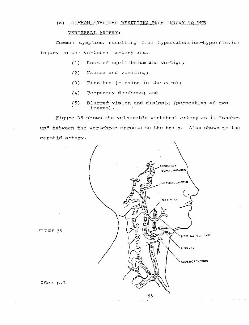
(a) COMMON SYMPTOMS RESULTING FROM INJURY TO THE
VERTEBRAL ARTERY:
Common symptoms resulting from hyperextension-hyperflexion
injury to the vertebral artery are:
(1)
( 2)
(3)
(4)
(5) ,.
Loss of equilibrium and vertigo;
Nausea and vomiting;
Tinnitus (ringing in the ears);
Temporary deafness; and
Blurred vision and diplopia (perception of two images).
Figure 38 shows the vulnerable vertebral artery as it "snakes
up" between the vertebrae enroute to the brain. Also shown is the
carotid artery.
FIGURE 38
~See p.l
-98-
5. SYMPATHETIC NERVES:
(a) Anatomy of sympathetic Nerves:
Sympathetic nerves are part of the autonomic nervous
symptom. The central nervous system (CNS) consists of the brain,
spinal cord,
discussed in
and spinal nerves (the cranial nerves are not
this outline) which innervate skeletal muscles
resulting in movement. Unlike the CNS, the autonomic nervous
system regulates the body!s internal environment by innervating
involuntary muscle. The automonic nervous symptom innervates the
smooth muscle and glands in the following areas:
1.
2.
3.
4.
Eye
Ear
Larynx (voice box)
Lungs
5.
6.
7.
Blood vessels
Skin
GI tract
These sympathetic nerves originate from the spinal cord below
the level of CS where they form chains of nerves. The sympathetic
nerves are vulnerable to injury from forced hyperextension-
hyperflexion motion.
(b) Function of Sympathetic Nerves:
The autonomic nervous system is divided into (1)
sympathetic nerves and (2) parasympathetic nerves (para = opposite), each providing equal and opposite responses to smooth
muscle and glands thereby providing the body with balance. For
example, sympathetic nerves to the iris of the eye cause the pupil
to enlarge (dilate) while parasympathetic nerves cause the pupil to
-99-
become smaller (constrict). Paralysis of the sympathetic nerve to
the iris causes the pupil to constrict. (Horner's Syndrome, named
after it's discoverer).
The sympathetic nervous system aids the body in an emergency
prepares us for "Fight" and "Flight". Thus, stimulation of
sympathetic nerves dilate the eye; increases the heart rate;
constricts arteries where blood is not needed in an emergency, such
as the skin and the in"t:estines, while dilating the coronary ,.
arteries which supplies blood to the heart-where it is needed; the
skin is contracted and the sweat glands are stimulated. The
parasympathetic nerve stimulation has the opposite effect
preparing the body for relaxation such as constricting the eye;
decreasing the heart rate; dilating arteries which supply blood to
the skin and intestines.
(i) Blue Arms and Cold Hands Victims of
hyperextension-hyperflexion injury may report their extremities
turn blue and cold. The basis for this change is injury to the
sympathetic nervous system causing stimulation of the nerve.
Arteries which carry blood into the skin receive sympathetic
and parasympathetic nerve supply. Sympathetic nerves cause these
arteries to constrict (vasoconstriction) (vaso =blood vessels);
while the corresponding parasympathetic nerve causes the artery to
dilate with increased blood flow to the skin.
Coldness in an extremity, with the skin turning blue, is
caused by the vasoconstriction of the sympathetic nerve with
decreased blood flow to the skin. Since the blood flow to the skin
-100-
is decreased, the skin will turn blue and skin temperature will
become colder. Blue skin is called "cyanotic." (The term cyanotic
is derived from the effects of cyanide poisoning -- a lack of
oxygenated blood to the tissues) .
(ii) Symptoms Resulting From Injuries to the
Sympathetic Nerves - Forced hyperextension-
hyperflexion injuries to the SYinpathetic nerves cause either ,
stimulation or irritation of the nerve resulting in the following
recognized symptoms:
A. The Eye.
1. Dilation of the pupil.
2. Constriction of the pupil resulting from paralysis
of the sympathetic nerve (Horner's Syndrome).
3. Blurred vision.
4. Pain behind the eye.
5. Excessive tearing.
B. The Ear.
1. Loss of equilibrium and balance.
2. Vertigo, dizziness and nausea.
3. Tinnitus (ringing in the ears).
4. Temporary deafness.
c. The Larynx (Voice Box).
1. Hoarseness.
-101-
D. The Skin.
1. Cold and blue.
6. BRAIN CONCUSSION (POST-CONCUSSION SYNDROME):
The brain consists of very soft tissue having the
consistency of Jell-0. Imagine a 10 inch diameter Jell-0 mold
surrounded by two bowls -- one on top and one on the bottom. You
have just pict';red the brain housed in the skull! What effect will
a sudden, violent, forced hyperextension-hyperflexion motion have
on the Jell-0 mold or brain? At a minimum, it will be "shaken up"
which is the definition of a concussion a "shaking of the
brain." Physical impact with a fixed object, such as the head
striking the windshield is not necessary. The shaking of the brain
has already occurred by the very force generated by the
hyperextension-hyperflexion motion.
Post-concussion syndrome is a medical condition often seen
following forced hyperextension-hyperflexion motion to the neck and
head with a well recognized set of symptoms (syndrome).
Wiesel, et al., Neck Pain, 2nd Ed. {1992) p. 293 describes
post-concussion syndrome resulting from an auto collision:
"Patients receiving a whiplash injury of the neck can also suffer
from a concussion. Mechanical deformation of the brain occurs
during the acceleration-deceleration phase of the injury and
concussion can occur without the head striking anything. This can
account for transient loss of consciousness, as well as post-
concussion syndrome or headache, photophobia, mild transient
-102-
confusion, tinnitus (ringing in the ears), fatigue, and transient
difficulty with concentration."
7 • INJURY TO THE ESOPHAGUS AND TMJ JOINT.
Forced hyperextension-hyperflexion injury may injure the
esophagus (food pipe) resulting in difficulty swallowing, dysphagia
(dys = dysfunction, phagia = to swallow) .
Forced hyperextension-hyperflexion also causes injury to
larynx (voice box) causing hoarseness.
During the hyperextension phase, the driver's head is forcibly
thrown backwards, with the mouth opening, which may damage the
temporomandibular joint {TMJ). The TMJ joint refers to the joint
formed where the temporal bone meets the mandible in the jaw.
D. NAMES OF HYPEREXTENSION-HYPERFLEXION INJURIES:
Different names have been used interchangeably over the years
to describe soft tissue injuries resulting from forced
hyperextension-hyperflexion motion. These include:
(1) Hyperextension-hyperflexion injuries;
(2) Hyperextension-flexion injuries;
(3) Acceleration-Deceleration injuries
(4) Strain and sprain injuries;
(5) Myoligamentous injuries;
(6) Soft tissue injuries;
(7) Myofascial pain syndrome (MPS);
(8) Cervical syndrome; and
(9) Whiplash injuries.
-103-
Thetermshyperextension-hyperflexion, hyperextension-flexion,
acceleration-deceleration, and whiplash injury refer to the
mechanism of injury when the neck is forcibly thrown backward and
forward, for example, during an auto collision.
"Whiplash11 can be used offensively by equating the forces of
hyperextension-hyperflexion motion with the force generated by the
cracking of a large, long "bullwhip".
1. DIFFUSING THE WORD "WHIPLASH":
Jurors have negative feelings toward a plaintiff when the
defense attorney calls his injury a "whiplash". The negative
connotation of a "whiplash injury" must be diffused beginning with
voir dire and ending with the closing argument. The following is
a sample method of taking the sting out of a "whiplash injury. 11
VOIR DIRE:
Q: Has anyone injured hisjher neck before?
A: Mrs Jones: I have.
Q: Did anyone call your injury a "whiplash?"
A: Yes.
Q: Were you offended?
A: Yes, because my neck really hurt.
CLOSING ARGUMENT (Plaintiff's Rebuttal):
"Whiplash! . . Whiplash!. The defense attorney has
called this injury a "whiplash" -- not once -- but seven
times during his closing argument. He is trying to plant
the "seeds of prejudice" in your mind. Is this fair to
Mr. Plaintiff? .•.• 11
-104-
E. EXAMINATION FINDINGS OF HYPEREXTENSION-
HYPERFLEXION INJURIES:
1. PLAIN X-RAY FINDINGS:
(a) Loss (Straightening) of the Normal Lordotic Curve:
(i) The Three Curves of the Spine - The vertebrae
are not "stacked" in a straight line, one on top of the other. If
they were, motion would be painful and limited. As a result, our
spine (vertebral column) is curved in three places:
(1) The Cervical Spine (Neck): The cervical curve is convex.
( 2) The Thoracic Spine (Mid-Back) : The curve in the thoracic
spine is with the convexity backwards.
(3) The Lumbar Spine (Low Back): The curve in the lumbar
spine is convex. The convexing curve of the low back
balances the convexity curve of the neck.
The physicians call the normal curvature of the spine
lordosis. Abnormal curvature of the spine is termed "loss of
lordosis." For example, if the normal cervical and lumbar curves
are abnormal, physicians refer to this condition as "loss of
cervical lordosis" and "loss of lumbar lordosis", respectively.
Figure 39a shows the normal curves of the spine.
-105-
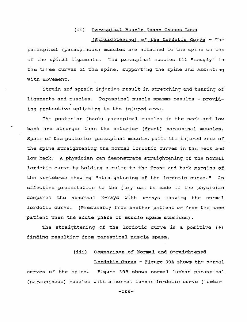
{ii) Paraspinal Muscle Spasm Causes Loss
{Straightening) of the Lordotic curve - The
paraspinal (paraspinous) muscles are attached to the spine on top
of the spinal ligaments. The paraspinal muscles fit "snugly'' in
the three curves of the spine, supporting the spine and assisting
with movement.
Strain and sprain injuries result in stretching and tearing of
ligaments and muscles. Paraspinal muscle spasms results - provid-
ing protective splinting to the injured area.
The posterior (back) paraspinal muscles in the neck and low
back are stronger than the anterior (front) paraspinal muscles.
Spasm of the posterior paraspinal muscles pulls the injured area of
the spine straightening the normal lordotic curves in the neck and
low back. A physician can demonstrate straightening of the normal
lordotic curve by holding a ruler to the front and back margins of
the vertebrae showing ''straightening of the lordotic curve." An
effective presentation to the jury can be made if the physician
compares the abnormal x-rays with x-rays showing the normal
lordotic curve. {Presumably from another patient or from the same
patient when the acute phase of muscle spasm subsides).
The straightening of the lordotic curve is a positive (+)
finding resulting from paraspinal muscle spasm.
(iii} Comparison of Normal and Straightened
Lordotic curve - Figure 39A shows the normal
curves of the spine. Figure 39B shows normal lumbar paraspinal
{paraspinous) muscles with a normal lumbar lordotic curve {lumbar
-106-
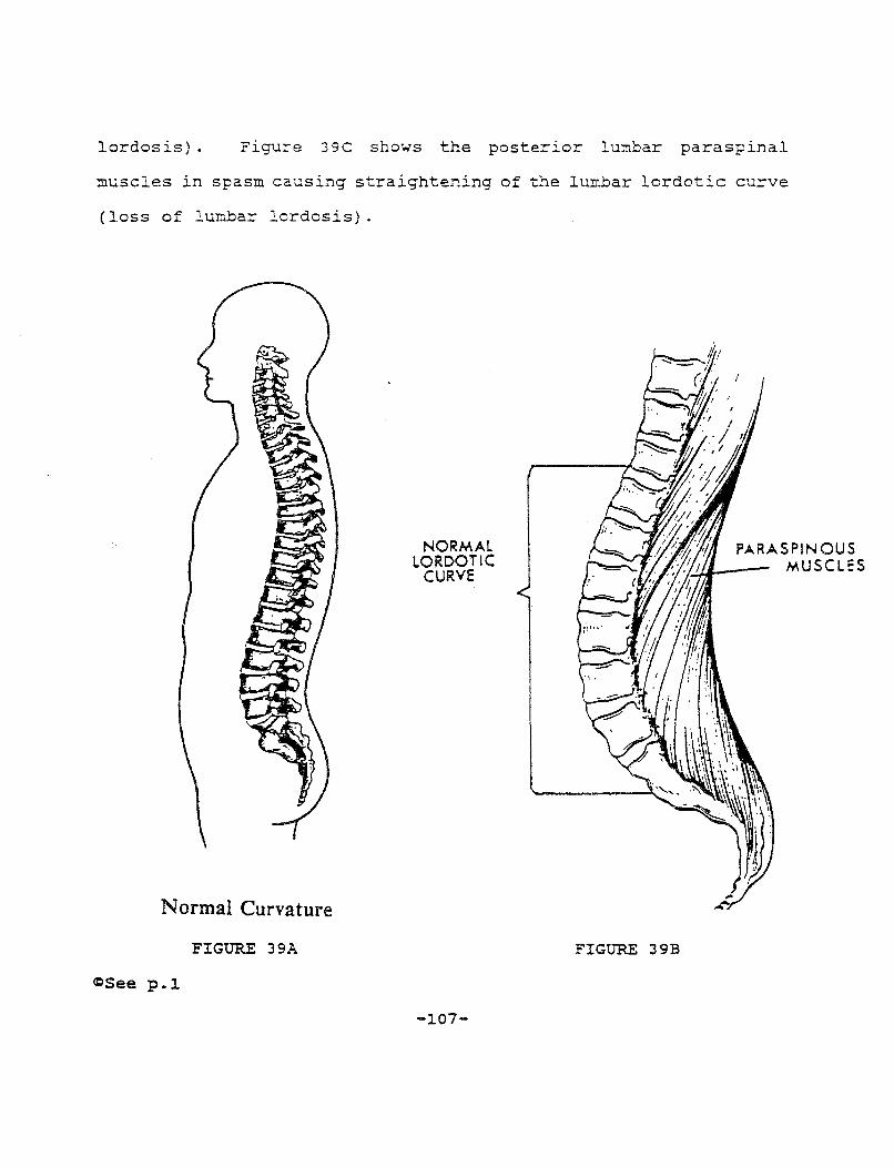
lordosis) . Figure 39C shows the posterior lumbar paraspinal
muscles in spasm causing straightening of the lumbar lordotic curve
(loss of lumbar lordosis).
Normal Curvature
FIGURE 39A
<=See p.l
NORMAL LORDOTIC
CURVE
-107-
FIGURE 39B
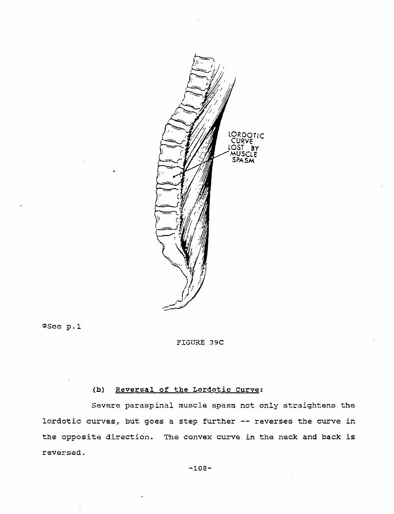
@See p.l
FIGURE 39C
lORDOTIC CURVE
lOST BY MUSCLE SPASM
(b) Reversal of the Lordotic curve:
Severe paraspinal muscle spasm not only straightens the
lordotic curves, but goes a step further -- reverses the curve in
the opposite direction. The convex curve in the neck and back is
reversed.
-108-

(c) False Positive (+) Results:
( i) Routine soinal x-ray Series - The routine
spinal x-ray series consists of ( 1) A-P (anterior - posterior)
views (front to back view); (2) Right and left oblique views; and
(3) Lateral views (side views).
Each of the three standard "views" can be taken from three
positions: (1) the neutral position; (2) the flexion position with
the head and neck flexed toward the chest; and (3) the extension
position the head and neck are extended backward.
(ii) Positioning of the Neck causes False Positive -
Lateral views of spine show straightening of the lordotic curve.
As noted above, the lateral views are taken in the neutral, flexion
and extension positions.
In the flexion position (with the head and neck flexed toward
the chest), the normal lordotic curve in the neck is reduced, but
does not completely straighten. Exception: Positioning, a patient
with a long slender neck, for a lateral view of the spine, in the
flexion position can itself result in loss of normal lordotic
curvature. However, lateral x-rays taken in the neutral and
extension position (head backwards) will not result in
straightening of the lordotic curve by merely positioning the
patient on the x-ray table.
Practice Note: The plaintiff should obtain all three lateral views of cervical spine x-rays. Loss of the normal lordotic curve in the flexion position could be the result of positioning a patient with a slender long neck. However, loss of the lordotic curve in the other
-109-
two views (neutral and extension) will prove that the loss of lordosis is as a result of muscle spasm, not merely positioning the patient on the x-ray table as may be the case with the lateral x-ray in the flexion position only.
(d) False Negative (-) Results:
Loss (straightening) of the normal lordotic curve of the
spine is the result of paraspinal muscle spasm. If a patient is
taking muscle relaxant medication, a false negative (-) result may
occur.
(e) The Negative X-Ray:
Except for straightening of the lordotic curve, plain x-
rays are generally negative in hyperextension-hyperflexion soft
tissue injury cases. X-rays are designed to show "bone", not "soft
tissues". The spinal ligaments, paraspinal muscles, intervertebral
discs, nerve roots, spinal nerves, blood vessels, and sympathetic
nerves -- all types of soft tissue -- will not show up on plain x-
rays.
Practice Note: The positive use of the negative x-ray. The plaintiff's attorney should vividly demonstrate to the jury that plain x-rays do not show damage to soft tissues by having the medical expert draw on the x-ray itself the soft tissues, such as the spinal ligaments, paraspinal muscles, the intervertebral discs, the blood vessels, including the vertebral artery, the nerve roots and spinal nerves, as well as the sympathetic nerves. The jury must be shown, on the x-ray, the vulnerability of the these soft tissues to injury, with a forced hyperextension-hyperflexion motion. The negative x-ray thus becomes a positive plaintiff's exhibit. An excellent example of this courtroom technique is found in Houts and Marmor, Proving Medical Diagnosis and Prognosis, Vol. 3, Section 6~. ~4 entitled, "Redirect Examination of Plaintiff's Medical Witness Who Reported 'Negative X-rays' of Plaintiff".
-~10-
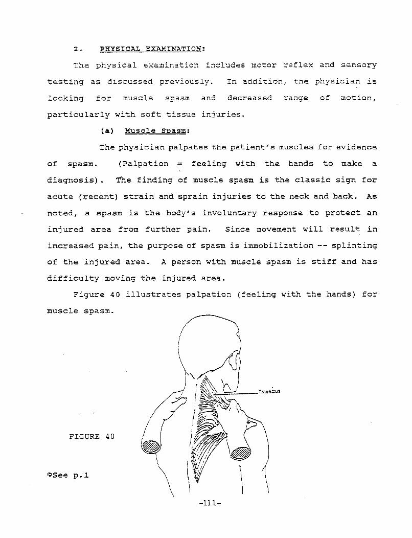
2. PHYSICAL EXAMINATION:
The physical examination includes motor reflex and sensory
testing as discussed previously. In addition, the physician is
looking for muscle spasm and decreased range of motion,
particularly with soft tissue injuries.
(a) Muscle Spasm:
The physician palpates the patient's muscles for evidence
of spasm. (Palpation = feeling with the hands to make a
diagnosis} . The finding of muscle spasm is the classic sign for
acute (recent} strain and sprain injuries to the neck and back. As
noted, a spasm is the body's involuntary response to protect an
injured area from further pain. Since movement will result in
increased pain, the purpose of spasm is immobilization -- splinting
of the injured area. A person with muscle spasm is stiff and has
difficulty moving the injured area.
Figure 40 illustrates palpation (feeling with the hands) for
muscle spasm.
I FIGURE 40 ~
<r~See p.~
-111-
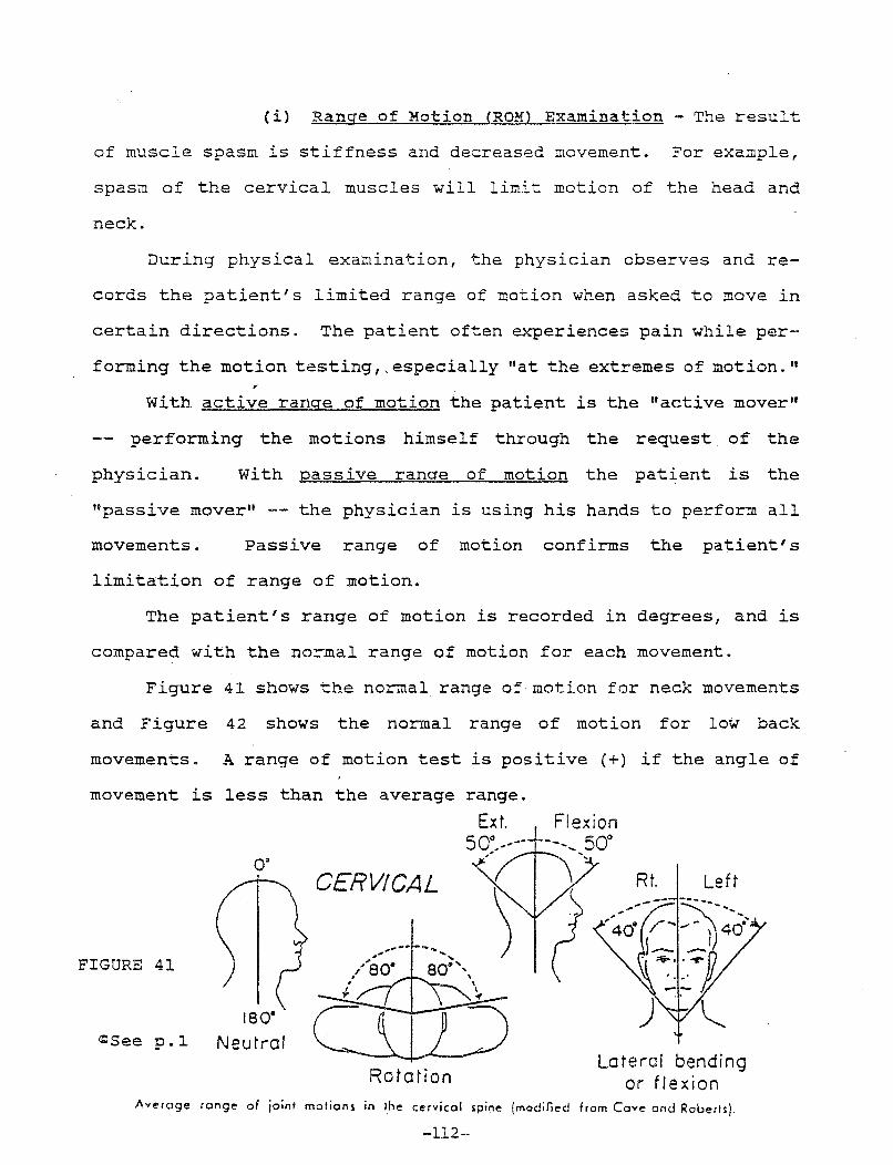
(i) Range of Motion (ROM) Examination - The result
of muscle spasm is stiffness and decreased movement. For example,
spasm of the cervical muscles will limit motion of the head and
neck.
During physical examination, the physician observes and re-
cords the patient's limited range of motion when asked to move in
certain directions. The patient often experiences pain while per-
forming the motion testing,, especially "at the extremes of motion."
With active range of motion the patient is the "active mover"
performing the motions himself through the request of the
physician. With passive range of motion the patient is the
"passive mover" -- the physician is using his hands to perform all
movements. Passive range of motion confirms the patient's
limitation of range of motion.
The patient's range of motion is recorded in degrees, and is
compared with the normal range of motion for each movement.
Figure 41 shows the normal range of motion for neck movements
and Figure 42 shows the normal range of motion for low back
movements. A range of motion test is positive (+) if the angle of
movement is less than the average range.
o·
FIGURE 41
180. t:lSee p.l Neutral
CERVICAL
Ext. j_ Flexion 50°/'-- --, ... 50°
,' .......
Rt. Left
,. ..... --- -- ......... ,./'so· 80::''-,
.;
Rotation Lateral bending
or flexion Averc1ge range of joint motions in the cervical spine (modified from Cove onc.J Roberts).
-112-

~See p.l.
Range of Motion
I I
I I
i .r I l '1
~ I -rl
I .. ).
\I 3 4 \I 5 . ~ \. I
T • Fle.-:ion. or forwc:~d. bending of the lumber S.:)ine. The cveroc;e l'lormcl Re.-:ion is cbout 90 degrees. 2. Extension. or bc::kwcrd bending. of the lumber s;line is c:lso recorded in the r::nc;e of bc:::::k motio" Tne cvercc;e ede,sio" is c:bout T 0 degrees. 3 & J, Lc:teral. or side bending is measured in the lumbar J!=line. lne c:vercc;e lateral bend is about 1 0 degrees. S Rotation, or ,._isrinc; the Sl=li"e· is mec:sured c:nd recorded cs the degree of shoulder motion. lne average ro~::tiort avercges cbout 90 ciec;rees.
FIGURE 42
-ll.3-
3. SUBJECTIVE COMPLAINTS vs. OBJECTIVE FINDINGS:
The first part of every medical examination consists of the
"history" where the patient describes to the doctor his complaints
of pain. Based upon the patient's symptoms, findings on physical
examination and results of appropriate tests, the physician makes
a diagnosis. The history is important in making the diagnosis --
it is so important that medical students are taught the maxim:
"listen to the patient, he· is telling you the diagnosis." ,.
The plaintiff's complaints of pain, provided in the history,
are termed subjective complaints. Objective findings are those
findings actually observed by the physician, such as feeling muscle
spasm with his own hands.
Usually, after the initial acute phase, the objective findings
will be minimal.
(a) cross-examination of the IME Doctor:
The cornerstone of the defense of a soft tissue injury case is
to use the "IME doctor" to point out to the jury the difference
between subjective complaints and objective findings.
The following is a sample cross-examination "debunking" an IME
doctor on subjective complaints vs. objective findings:
Q: Doctor, a patient's history is important in arriving at a diagnosis. Isn't that true?
A: Yes.
Q: Doctor, a history consists of the patient telling you his subjective complaints?
A: Yes.
Q: In your own practice do you rely upon the patient's history in forming your diagnosis?
-114-
A: Yes.
Q: Doctor, assume that a patient gave you a history of severe crushing chest pain radiating into the arm after shoveling snow. This patient could be suffering from an acute heart attack. Isn't that true?
A: Yes.
Q: Doctor, isn't this a medical emergency?
A: Yes.
Q: Doctor, assume that I just cut my finger on a piece of paper a paper cut. This would be an objective finding wouldn't it?
A: Yes.
Q: But the chest pain - that would be a subjective complaint?
A: Yes.
Q: Requiring immediate admission to the emergency room?
A: Yes.
Q: Thank you doctor, I have no further questions.
4. SOFT TISSUE INJURIES RESULT IN PRRMANENT RESIDUALS:
The medical literature verifies that soft tissue injuries
leave permanent residuals. For example, Drs. Gargan and Bannister
at the University of Bristol followed 43 patients with soft tissue
neck injuries for 8 to 12 years after the initial injury. Eighty-
eight percent of the patient's had been in rear-end auto
collisions. The results verifying permanent residuals are reported
in Gargan and Bannister, "Long Term Prognosis of Soft Tissue
Injuries of the Neck," J. Bone and Joint Surgery, Vol. 72-B, No.
5 Sept. 1.990: -115-
The Severity of Symptoms: Patients were subdivided into four groups on the basis of their symptoms. Group A (12%) were free of any discomfort and considered that they had made a complete recovery from their accident. (88% had not fully recovered). Group B (48%) were left with mild residuals which did not interfere with their work or leisure activities. Group C (28% complained of intrusive symptoms which handicapped work and leisure activities and caused them to seek relief by frequent intermittent use of analgesic, orthoses or physiotherapy. Group D (12%) suffered from severe problems and had iost their jobs, relied continually on orthoses or analgesics, and had undergone repeated medical consultations.
Dr. Gargan's and Dr. Bannister's long term study showed 28% of the patients had
residual symptoms which were intrusive, and 12% of the patients had severe residual
symptoms 10.8 years after the initial auto collision.
In an NIH sponsored study, Dr. Mason Hohl followed 146 patients, 5 years after their
auto collision. No patient had pre-existing osteoarthritis. Symptomatic recovery (no residual
problems) occurred only in 57% of patients (43% still had residual symptoms 5 years after
the collision). Dr. Hohl's article appeared in the December 1974 issue of The Journal of
Bone and Joint Surgery.
Medical school professors, Drs. Carroll, et al., "J. Musculoskeletal Medicine", June
1992, p. 97-114, reported similar findings of long-term permanent residuals in patients
involved in auto collisions:
" ... Long-term studies show that aches and pains with no evident physical cause persist in 20% - 45% of patients with significant whiplash injuries. Few of these patients have radicular pain. Roentgenographic (x-ray) studies show that degenerative problems develop after injury in 39% of patients. By comparison, degenerative changes develop over a comparable time in only 6% of the general population over age 30. Thus, it would seem apparent that whiplash injuries predispose patients to cervical degenerative osteoarthritis.
-116-
Hohl found. that t?e following early symptoms or signs were assoc~a~ed w~th long-term disability: persistent upper extr~mlty paln and numbness; interscapular pain; a sharp cervlcal curve reversal; and one level of localized restricted c~rvical motion. Men had a higher incidence of symp~omatlc. recovery. Degenerative changes were more common ln patlents who had lost consciousness. A most important finding was that whether or not litigation pro~eeded did.not appear to influence recovery among the patlents studled." (emphasis added).
5. PERMANENT RESIDUALS FROM SOFT TISSUE INJURIES REMAIN AFTER SETTLEMENT:
Studies, in the medical literature, point out that patients
with soft tissue injuries continue to experience permanent symptoms
even after an injury case settles.
For example, Drs. Gargan and Bannister, in their article cited
above, reviewed the p:pior medical literature and their own studies
of patients 8 to 12 years after the date of injury and concluded
--- thaE permanent res"iduals of· soft tissues remain ellen after settle-
ment,, stating:: "McNab (1964) and Norris and Watt (1983) recorded that symptoms remained unchanged in those whose claims had been settled. The fact that symptoms do not resolve even after 10 years supports the conclusion of these authors."
Dr. Hohl, in his article in The Journal of Bone and Joint
Surgery, December, 1974, found that 50% of his patients who made no
claim, were still experiencing residual symptoms five years after
their auto collision, stating: "44 of the 146 patients in the
series had no legal claim.... Only 50% of these 44 patients became
asymptomatic."
In addition, Drs. Carroll, et. al., "J. Musculoskeletal
Medicine", June 1992, p. 97-114, reported similar results stating:
" Research indicates that neither litigation nor personality disorder has any great bearing on the duration or intensity of a whiplash victims' symptoms."
-117-

6.. '.ROW TO GET FAVORABLE MEDICAL ARTICLES INTO EVIDENCE:·
Federal Rule of Evidence 8 0:3 ( l8) entitled, "Learned Treatises n
and Virginia Code §8.0l-40l.l, third paragraph*, (l994 amendment)
provide the answer:
"To the extent called to the attention of an expert witness upon cross-examination or relied upon by the expert witness in direct examination, statements contained in published treatises, periodicals or pamphlets on a subject of history, medicine or other science or art, established as a reliable authority by testimony or by stipulation shall not be excluded as hearsay. If admitted, the statements may be read into evidence but may not be received as exhibits. If the state-ments are to be introduced through an expert witness upon direct examination, copies of the statements shall be provided to opposing parties thirty days prior to trial unless otherwise ordered by the court. " (emphasis· added) ..
The last sentence requiring 30 days notice is not present in
Federal Rule of Evidence 803(l8). In addition, the Federal Rule
uses slightly different wording to achieve the same result as Code
§8.0l-40l.~, at line 8 above:
11 ••• the testi!!lony or admission of the witness or by other expert. testimony or by judicial notice ... "
The Virginia Supreme Court, in Weinberg v. Given, 252 Va. 22l,
476 S.E.2d 502 (1996), held Virginia Code §401.1, third paragraph,
"permits the hearsay content of such. articles to be read into the
record as· substantive evidence ..• "
Following Weinberg v. Given, an expert witness can read
favorable statements from medical articles directly into evidence
"A Guide to Evidence in Virginia, Rule 707(a) entitled, "Use of Learned Treatises with Experts"
-118-
provided the expert states (1) the article is reliable; and (2) the
article is relied upon by the expert (the expert agrees with the
statements in the article).
The Supreme Court, in Weinberg v. Given, 252 Va. 221, at page
223, approved the following foundation for the admissibility of
statements from medical articles:
"Dr. Partridge also identified several articles that he
believed were reliable and authoritative. For example, Dr.
Partridge gave the following testimony:
"Q: And Doctor, are you familiar with the article entitled 'Oral Contraception: Past, Present and Future Perspectives' by Daniel Mishell? An again, this would be in the International Journal of Fertility, 1991. Are you familiar with that, sir?
A: Yes.
Q. Is that a reliable authority in the field as of 1991 on this issue?
A. Yes.
Q. And Doctor, going right to the last page, to the conclusion, 'Today 85 percent of prescriptions of [oral contraception] written in the United States specify formulations containing 30 to 35 micrograms of ethinyl estradiol, the lowest possible dose required to achieve optimal contraceptive [efficacy] with minimal risk of adverse effects. These new compounds do not appear to have any adverse cardiovascular effects and can be safely used by healthy, nonsmoking, premenopausal women until age 45 or older.' Did I read that correctly, sir?
A: You Did.
Q. Did you agree with that?
A. Yes."
-119-
PART THREE
FIBROMY ALGIA
A. INTRODUCTION
Fibromyalgia, (also called fibromyalgia syndrome - FMS, FS or
FM), is a medically recognized soft tissue rheumatic condition
involving pain in fibrous tissue and muscle. Fibrous tissue is
tissue that contains fibers such as (1) fascia, (2) tough tendons
that connect muscle to bone, and (3) ligaments which connect bone
to bone. Fascia is a sheath that encloses and holds muscle
together - like a strong "baggie".
The term fibromyalgia literally means pain from fibrous tissue
and muscle ("fibro" referring to fibrous tissue; "my" referring to
muscle; and "algia" meaning pain).
Myalgia is different from fibromyalgia since myalgia involves
only simple
fibromyalgia
muscle pain. Myositis is also different from
since it involves inflailllllation of muscle ("myo"
meaning muscle; "itis" meaning inflammation).
The term "fibrositis" had been used interchangeably with
"fibromyalgia" by physicians. In recent years, the term
"fibrositis" has lost favor since studies have shown that patients
suffering from fibromyalgia do not exhibit muscle inflammation.
-120-
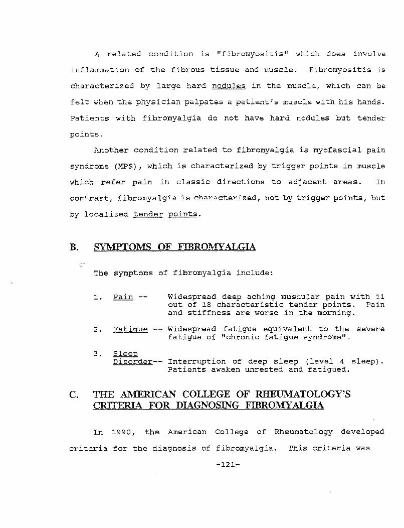
A related condition is "fibromyositis" which does involve
inflammation of the fibrous tissue and muscle. Fibromyositis is
characterized by large hard nodules in the muscle, which can be
felt when the physician palpates a patient:s muscle with his hands.
Patients with f ibromyalgia do not have hard nodules but tender
points.
Another condition related to fibromyalgia is myofascial pain
syndrome (MPS) , which is characterized by trigger points in muscle
which refer pain in classic directions to adjacent areas. In
contrast; fibromyalgia is characterized, not by trigger points, but
by localized tender points.
B. SY1V!PTOMS OF FIBROI\ITALGLJ\
The symptoms of fibromyalgia include:
1. Pain Widespread deep aching muscular pain with 11 out of 18 characteristic tender points. Pain and stiffness are worse in the morning.
2. Fatigue -- Widespread fatigue equivalent to the severe fatigue of "chronic fatigue syndrome".
3. Sleep Disorder-- Interruption of deep sleep (level 4 sleep) .
Patients awaken unrested and fatigued.
C. THE AMERICAN COLLEGE OF RHEUMATOLOGY'S CRITERIA FOR DIAGNOSING FIBROMYALGIA
In 1990, the American College of Rheumatology developed
criteria for the diagnosis of fibromyalgia. This criteria was
-121-
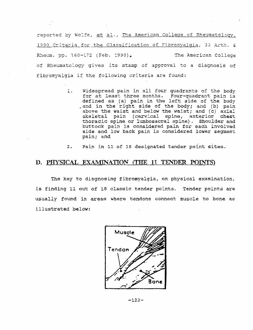
reported by Wolfe, et al., The American College of Rheumatology 1
1990 Criteria for the Classification of Fibromyalgia, 33 Arth. &
Rheum. pp. 160-172 (Feb. 1990)* The American College
of Rheumatology gives its stamp of approval to a diagnosis of
fibromyalgia if the following criteria are found:
1. Widespread pain in all four quadrants of the body for at least three months. Four-quadrant pain is defined as (a) pain in the left side of the body
, and in the, right side of the body; and (b) pain above the waist and below the waist; and (c) axial skeletal pain (cervical spine, anterior chest thoracic spine or lumbosacral spine) . Shoulder and buttock pain is considered pain for each involved side and low back pain is considered lower segment pain; and
2. Pain in ll of l8 designated tender point sites.
D. PHYSICAL EXAMINATION (THE 11 TENDER POINTS)
The key to diagnosing fibromyalgia, on physical examination,
is finding ll out of l8 classic tender points. Tender points are
usually found in areas where tendons connect muscle to bone as
illustrated below:
-l22-
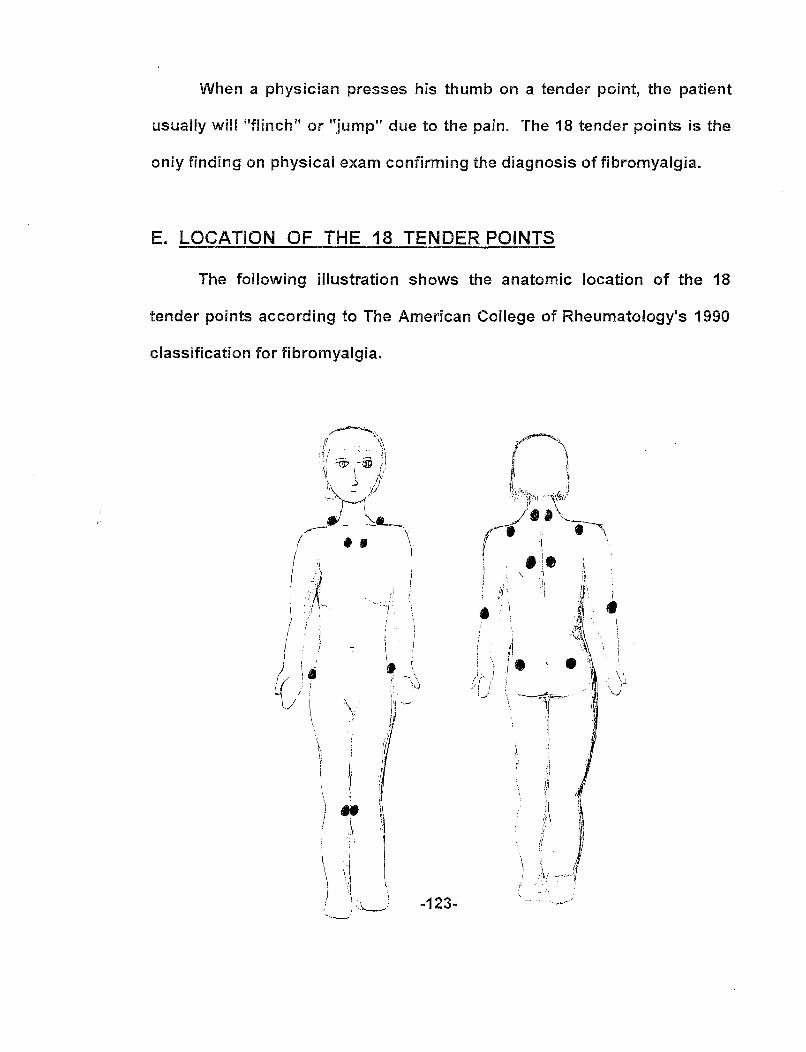
When a physician presses his thumb on a tender point, the patient
usually will "flinch" or "jump" due to the pain. The 18 tender points is the
oniy finding on physicai exam confirming the diagnosis of fibromyalgia.
E. LOCATION OF THE 18 TENDER POINTS
The following illustration shows the anatomic location of the 18
tender points according to The American College of Rheumatology's 1990
classification for fibromyalgia.
I J
I .
i \. ~j v \
\ r • • . 1 . .-...,\ . \j
I' .
)J ·-
/ r !' I•
li
-123-
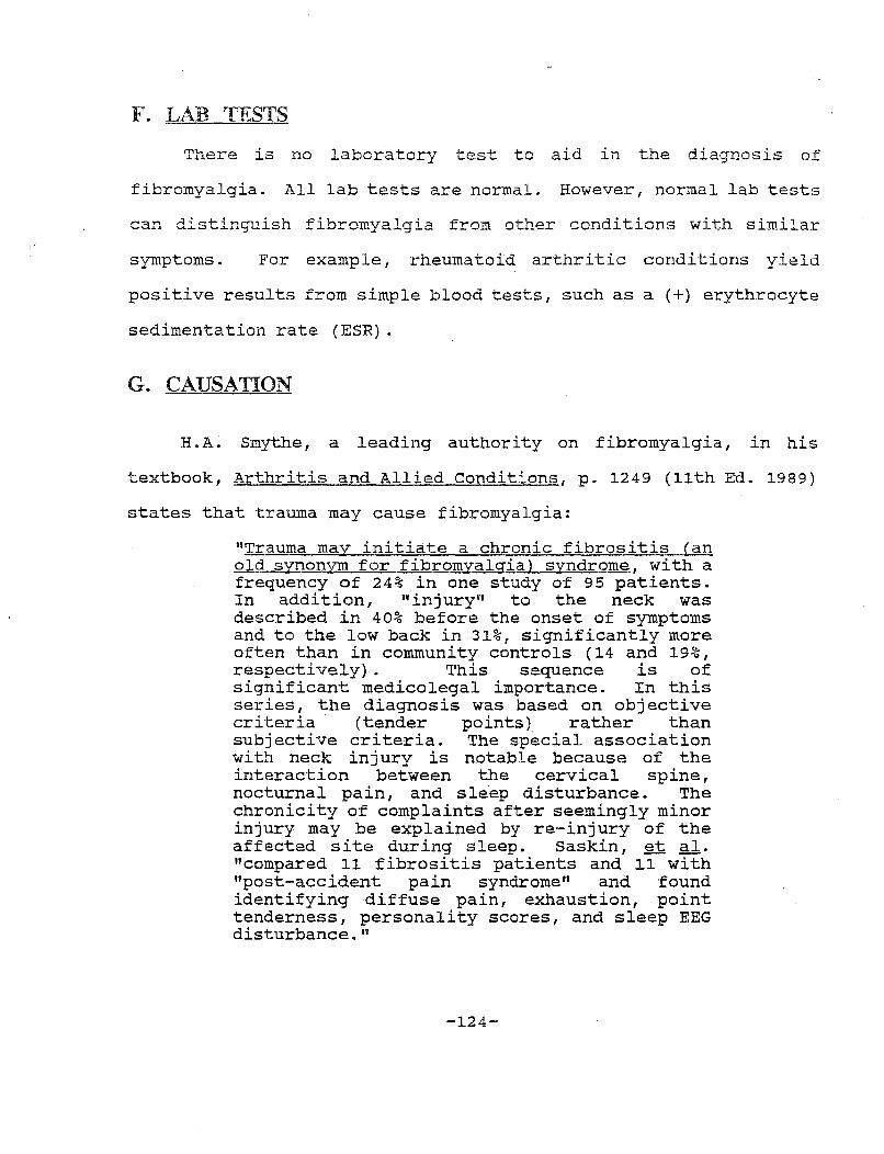
F. LAB TESTS
There is no laboratory test to aid in the diagnosis of
fibromyalgia. All lab tests are normal. However 1 normal lab tests
can distin~Jish fibromyalgia from other conditions with similar
symptoms. For example 1 rheumatoid arthritic conditions yield
positive results from simple blood tests, such as a (+) erythrocyte
sedimentation rate (ESR) .
G. CAUSATION
H.A. Smythe, a leading authority on fibromyalgia, in his
textbook, Arthritis and Allied Conditions, p. 1249 (11th Ed. 1989)
states that trauma may cause fibromyalgia:
11 Trauma may initiate a chronic fibrositis (an old synonym for fibromyalgia) syndrome, with a frequency of 24% in one study of 95 patients. In addition, "injury" to the neck was described in 40% before the onset of symptoms and to the low back in 31%, significantly more often than in community controls (14 and 19%, respectively) . This sequence is of significant medicolegal importance. In this series, the diagnosis was based on objective criteria (tender points) rather than subjective criteria. The special association with neck injury is notable because of the interaction between the cervical spine, nocturnal pain, and sleep disturbance. The chronicity of complaints after seemingly minor injury may be explained by re-injury of the affected site during sleep. Saskin, et al. "compared 11 fibrositis patients and 11 with "post-accident pain syndrome" and found identifying diffuse pain, exhaustion, point tenderness, personality scores, and sleep EEG disturbance."
-124-
Greenfield, et al., in their article "Reactive Fibromyalgia
Syndrome", 35 Arth. & Rheum., pp. 678-681 (June 1992), found that
patients who developed reactive fibromyalgia (as a reaction to a
prior precipitating event, such as trauma from an auto collision,
elective surgery, or a non-traumatic medical illness) had greater
disability than patients who developed primary fibromyalgia
(fibromyalgia caused without a prior precipitating event).
Post-Traumatic Fibromyalgia Syndrome (PTFS) is well recognized
in the medical literature. For example, Thomas J. Romano, M.D.,
Ph.D., a Rheumatologist, teaching at thew. Virginia University
School of Medicine, in his article, "Clinical Experiences With
Post-Traumatic Fibromyalgia Syndrome", W. Va. Medical Journal, Vol.
86, No. 5, pp. 198-202, (May 1990) defines PTFS as, "It occurs in
the setting of an acute injury, after which signs and symptoms of
fibromyalgia syndrome (FS) appear. . .• Often, the diagnosis is
delayed. In this series the mean length of time time from the date
of injury to date of diagnosis of PTFS was 17.3 months .•. "
In addition, G. W. Waylonis et al. , from the Department of
Physical Medicine and Rehabilitation at Ohio State University, in
their article, "Post-Traumatic Fibromyalgia", Am. J. Phys. Med.
Rehabil., Vol. 73 (No. 6) pp. 403-412 (Nov.-Dec. 1994) described a
10 year study of 176 patients diagnosed with post-traumatic
fibromyalgia, 60.7% noting symptoms after a motor vehicle accident.
85% of patients continued to have significant symptoms.
The often reported association between neck injuries caused by
auto collisions and the onset of post-traumatic fibromyalgia
-125-
syndrome (PTFS) was validated by Buskila et al., in their article,
"Increased Rates of Fibromyalgia Following Cervical Spine Injury:
A Controlled Study of Traumatic Injury", Arthritis and Rheumatism,
Vol. 39, pp. 446-452 (~997). They reported that 21.6% of patients
with soft-tissue neck injuries (90% having classic whiplash)
developed fibromyalgia versus 1. 7% of patients with leg fractures.
Fibromyalgia can be caused without trauma, such as by a viral
or bacterial infection or develop as secondary fibromyalgia - "on
top of" another condition, such as rheumatoid arthritis or
hypothyroidism. This latter type of fibromyalgia is called
secondary fibromyalgia since it develops secondarily after the
patient is already weakened by the first condition.
H. TREATMENT
Fibromyalgia is a chronic, often debilitating, condition with
no specific cure. Treatment consists of improving the sleep
disorder and relieving pain. Low doses of tricyclic anti-
depressants, such as Elavil, have improved the quality of sleep in
fibromyalgia patients. similarly, the muscle relaxant Flexeril has
been found to improve the quality of sleep and reduce pain. In
addition, nonsteroidal anti-inflammatory drugs (NSAIDs) , have been
prescribed to help alleviate the pain of fibromyalgia. Physical
therapy and "tender pain injections" with lidocaine, an anesthetic,
have also been employed to help relieve pain, but only provides
temporary relief. Gentle, daily, low-impact aerobic exercise has
also proved helpful.
-126-

PART FOUR
SUMMARY OF COMMONLY USED DIAGNOSTIC TESTS
A. PLAIN X-RAYS:
X-rays are also called roentgenograms named after Dr. Roentgen who
discovered them. X-rays were previously discussed at pp. 105-110. X-rays only show
injury to bone; however, straightening of the normal lordotic curve in the spine can be
seen.
Plain x-rays help diagnose spondylolisthesis (slippage of one vertebrae on top of
another); spinal stenosis (narrowing of the spinal canal); and degenerative disc disease,
including osteophytic spurs and disc space narrowing (the decreased disc space
between the vertebrae resulting from "drying out" of the disc).
B. DISCOGRAPHY:
A discogram is a procedure where a contrast dye is injected into the center of the
disc. The contrast dye allows the disc to be seen on an x-ray. The x-ray procedure is
called "fluoroscopy". Often a CT scan is obtained while the contrast dye is still present
in the disc for greater resolution. X-ray and/or CT films are produced which visualize
the disc.
If the injection temporarily reproduces the patient's pain, the discogram is said to
be (+) positive. The discogram allows a physician to pinpoint which disc is the cause of
the patient's pain.
A risk of the discogram is infection within the disc from the injection itself.
-127-
C. NEEDLE EMG AND NCV TESTING:
There are two types of electromyographic testing: (1) needle
EMG; and (2) surface EMG. Electromyography is derived from the
words: electro meaning electricity; myo meaning muscle; graphy
meaning writing.
Needle EMG testing measures the electrical activity of muscles
(myotome) supplied by a specific spinal nerve root. It is an
indirect method of diagnosing injury to a spinal nerve by measuring
the electrical activity of the muscle innervated by the nerve root.
If the nerve is damaged 11 upstream11 at the nerve root level, the
muscle will give off an abnormal pattern of electrical potential
called denervation fibrillation. Denervation fibrillation occurs
three weeks after injury to the nerve root. Accordingly, the test
should not be performed within three weeks of injury.
A needle electrode is inserted through the skin into the
muscle and connected to the electromyograph machine which picks-up
electrical potential in the muscle.
procedure is painful) .
(The needle is long, and the
The EMG produces a false negative in 20% of patients who have
nerve root injury.
Nerve Conduction Velocity (NCV) testing is done at the same
time as EMG testing. Nerve conduction velocity measures the speed
of the nerve impulse. An injured spinal nerve will produce a nerve.
impulse at a slower speed, compared to known values.
-128-
D. SURFACE EMG TESTING:
surface EMG is a test measuring the tension of a muscle.
Surface electrodes, (instead of needles) are placed on top of the
skin over the muscle to be tested. Electrical signals from the
muscle produce a digital read-out. The examiner compares the
digital read-out from the test with known normal values. A
positive test indicates a state of muscle tension and contraction.
E. MYELOGRAM:
Myelography (myelo = spinal cord; graphy = writing) involves
injecting a contrast dye into the subarachnoid space of the spinal
cord. The dye mixes with spinal fluid and an outline of the spinal
cord is seen on x-ray. If the nerve sleeve surrounding a spinal
nerve root is compressed, for example, by a herniated disc or a
large osteophyte, the nerve sleeve will be absent on a myelogram x-
ray. This is called a "filling defect".
A patient is usually admitted into the hospital overnight for
a myelogram.
vomiting.
Side effects include severe headache, nausea, and
Two types of contrast dyes have been used in the myelogram
procedure: (1} an oil-base dye- Pantopaque (rarely used today},
which is withdrawn following the procedure; and (2) a water-soluble
dye - Metrizamide which is absorbed by the body following the
procedure.
Figure 43 shows placement of the myelogram needle and Figure
44 shows the results of myelogram testing.
-129-
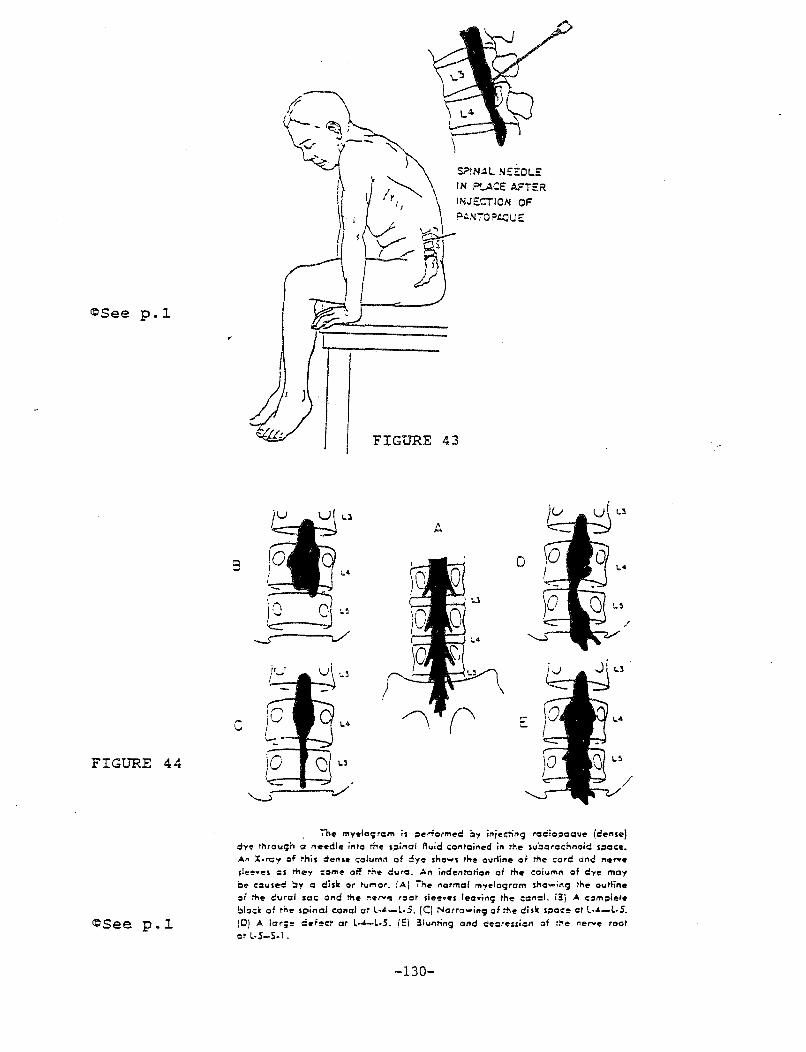
~Seep.~
FIGURE 44
~Seep.~
c
}
I ~~
r-C, ~::2) ~/"
S?lNAl N::!DLE: IN P'_ACE AFTER
INJECTION OF
P.:...'fiO PAOU E
FIGURE 43
D
The myeloc;rcm is ~erformed i:ly ir~je=::ir~c; roc:!io;:.oque (cense) dye rnrouc;n 12 ,,.,.,die in~o me s~inol Auid .:oMoir~ed in tile suborcc:!-onoic spac:e. A.n x.~., of this der~se column of :lye s!-oows '"" ourline of me c:ord one ,.,,...e slee•eS CIS they :orne off tne duro. ,a., j,deMI'OilO" of me c;ofum, of dye may C<' cause<: :,y = disl< or h.tmor. (A.) Tne ,ormol fflyeloc;rom snc-i"9 Ike oul'line of me durol sec: onci the ,.,.,,.."!! roor slee•es lec•i"9 tne c:c,cl. i3j A. c:::~m>:olete
bl<:lc:K o~ ,;,., s~:~inoi c:onci or L·-'-L·S. (C) Norrowir~g of tile disk s~oc:e ot L·-'-L·S. (D) A lar;" ciefe<:t or L-4-L-5. (El 31unring ond de:::~reuio, of :he ,.,,..., root or L·S-S-1.
-130-
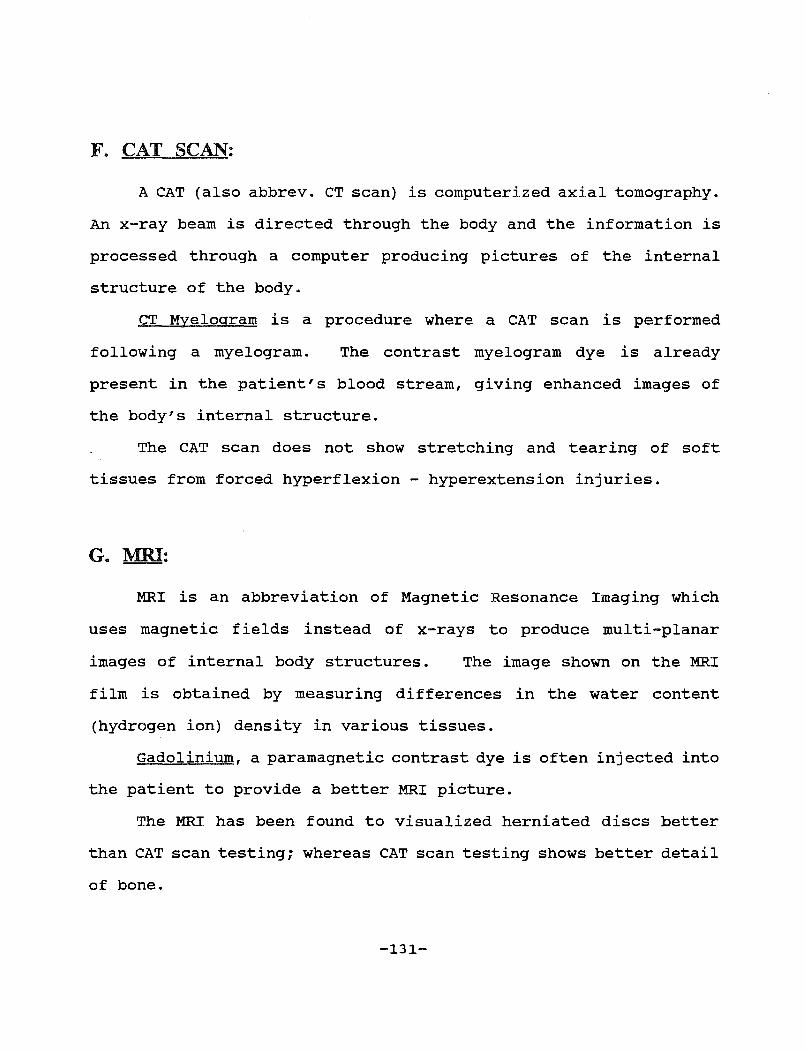
F. CAT SCAN:
A CAT (also abbrev. CT scan} is computerized axial tomography.
An x-ray beam is directed through the body and the information is
processed through a computer producing pictures of the internal
structure of the body.
CT Myelogram is a procedure where a CAT scan is performed
following a myelogram. The contrast myelogram dye is already
present in the patient's blood stream, giving enhanced images of
the body's internal structure.
The CAT scan does not show stretching and tearing of soft
tissues from forced hyperflexion - hyperextension injuries.
G. :MRI:
MRI is an abbreviation of Magnetic Resonance Imaging which
uses magnetic fields instead of x-rays to produce multi-planar
images of internal body structures. The image shown on the MRI
film is obtained by measuring differences in the water content
(hydrogen ion} density in various tissues.
Gadolinium, a paramagnetic contrast dye is often injected into
the patient to provide a better MRI picture.
The MRI has been found to visualized herniated discs better
than CAT scan testing; whereas CAT scan testing shows better detail
of bone.
-131-
An MRI should not be performed on patients who wear pacemakers
or who have metal plates in their body since this would adversely
effect the magnetic field.
Like the CAT scan, the MRI does not show stretching and
tearing of soft tissues from forced hyperflexion - hyperextension
injuries.
-132-
PART FIVE
DRUGS USED TO TREAT NECK AND BACK IN.RJRIES
INTRODUCTION
To treat neck and back injuries, physicians often prescribe
pain killers (analgesics); anti-inflammatory drugs to decrease
inflammation; muscle relaxants to decrease muscle spasm; anti-
anxiety drugs to treat anxiety; anti-depressants to treat pain and
depression resulting from the injury; and sleeping pills
(hypnotics) to help clients awakened by pain to fall asleep.
I. CLASSIFICATION OF DRUGS
A. BASED UPON ACTION
Drugs are classified based upon what they do -- their action
in the body. Some major classifications are: Analgesics,
Antibiotics, Antidiabetic Agents, Anti-Inflammatories, Anti-
Depressants, Muscle Relaxants, to name a few.
B. BASED UPON GROUP FORMULA
Drugs are further classified based upon their group chemical
formula. A group chemical formula is the basic chemical nucleus
shared by all drugs within the group which is
responsible for drug activity. Although all drugs within the group
-133-
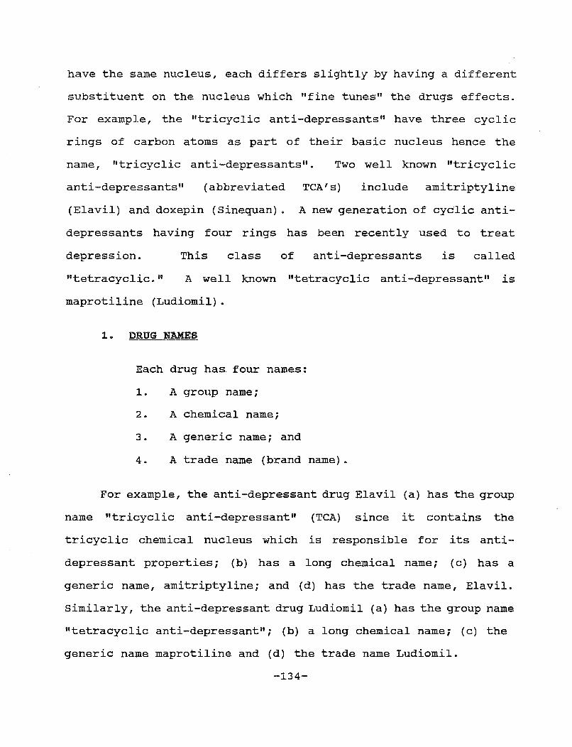
have the same nucleus, each differs slightly by having a different
substituent on the nucleus which "fine tunes" the drugs effects.
For example, the "tricyclic anti-depressants" have three cyclic
rings of carbon atoms as part of their basic nucleus hence the
name, "tricyclic anti-depressants". Two well known "tricyclic
anti-depressants" (abbreviated TCA's) include amitriptyline
(Elavil) and doxepin {Sinequan). A new generation of cyclic anti-
depressants having four rings has been recently used to treat
This class of anti-depressants is called depression.
"tetracyclic." A well known "tetracyclic anti-depressant" is
maprotiline (Ludiomil) •
1. DRUG NAMES
Each drug has four names:
1. A group name;
2. A chemical name;
3 0 A generic name; and
4. A trade name (brand name).
For example, the anti-depressant drug Elavil (a) has the group
name "tricyclic anti-depressant" {TCA) since it contains the
tricyclic chemical nucleus which is responsible for its anti-
depressant properties; (b) has a long chemical name; {c) has a
generic name, amitriptyline; and (d) has the trade name, Elavil.
Similarly, the anti-depressant drug Ludiomil (a) has the group name
"tetracyclic anti-depressant"; (b) a long chemical name; (c) the
generic name maprotiline and (d) the trade name Ludiomil.
-134-
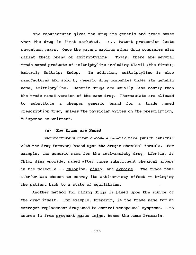
The manufacturer gives the drug its generic and trade names
when the drug is first marketed. U. s. Patent protection lasts
seventeen years. once the patent expires other drug companies also
market their brand of amitriptyline. Today, there are several
trade named products of amitriptyline including Elavil (the first) ;
Amitril; Emitrip; Endep. In addition, amitriptyline is also
manufactured and sold by generic drug companies under its generic
name, Amitriptyline. Generic drugs are usually less costly than
the trade named version of the same drug. Pharmacists are allowed
to substitute a cheaper generic brand for a trade named
prescription drug, unless the physician writes on the prescription,
"Dispense as written".
(a) How Drugs are Named
Manufacturers often choose a generic name (which "sticks"
with the drug forever) based upon the drug's chemical formula. For
example, the generic name for the anti-anxiety drug, Librium, is
Chlor diaz epoxide, named after three substituent chemical groups
in the molecule -- chlorine, diazo, and expoide. The trade name
Librium was chosen to convey its anti-anxiety effect -- bringing
the patient back to a state of equilibrium.
Another method for naming drugs is based upon the source of
the drug itself. For example, Premarin, is the trade name for an
estrogen replacement drug used to control menopausal symptoms. Its
source is from ~egnant mares urine, hence the name Premarin.
-135-

Lastly, some drugs are named as the manufacturer's "in house
joke". For example, the trade name anti-depressant, Trilafon, is
shorthand for "try laughing"!
II. ANALGESICS
A. INTRODUCTION
The term analgesic refers to a drug that relieves pain and is
derived from the word "analgia" meaning absence of pain (from the
Greek "an" meaning negative and "algas" meaning pain). Some
analgesics use "algas" (pain) as part of its trade name, i.e.
"Synalgos-DC" the trade name for the narcotic pain killer
Dihydrocodeine.
Analgesics are classified as either narcotic or non-narcotic.
B. NARCOTIC ANALGESICS
1. SISTER MORPHINE, BROTHER CODEINE AND A LITTLE VINEGAR -ENTER THE MORPHINE NARCOTICS
Opium is the oldest drug in history. Opium is the air dried
juice from the opium poppy plant. The name Opium is derived from
the Greek word opion - meaning "juice". The opium "juice" is
analogous to "chemical soup" and contains the drugs Morphine and
Codeine, in addition to two dozen other alkaloids. (Alkaloid is a
generic term referring to alkaline organic chemicals derived from
plants) .
-136-

Codeine is methylmorphine - the chemical brother of Morphine
(almost a molecular twin). Morphine is a stronger pain killer than
Morphine, one of the
strongest pain killers, is also a powerful sedative. Indeed, the
name Morphine is derived from the Greek word for sleep.
Over seventy-five years ago organic chemists tried to
synthesize a derivative of Morphine hoping to retain its pain
killing properties, with less addictive qualities. They added
acidic acid (common vinegar) to Morphine and came up with
diacetylmorphine, which they expected to become a "heroic drug" in
the treatment of pain, and named it Heroin. Cutting's Handbook of
Pharmacology, 7th Ed. (1984), page 625, describes the properties of
Heroin as, "More potent than Morphine and would be highly
·satisfactory if it were not also addicting; not permitted in the
United States."
Codeine is found in prescription cough medicines and is also
classified as an anti-tussive. (From the Latin "tussis", meaning
cough).
(a) The Morphine Group
Today, Morphine and Codeine are synthesized in the
laboratory rather than extracting the chemicals from the opium
poppy. Morphine is prescribed in the form of its sulphate, salt.
Codeine is widely prescribed in the prescription drug Tylenol with
Codeine. The prescription product is sold as Tylenol #1; Tylenol
#2; Tylenol #3;
-137-
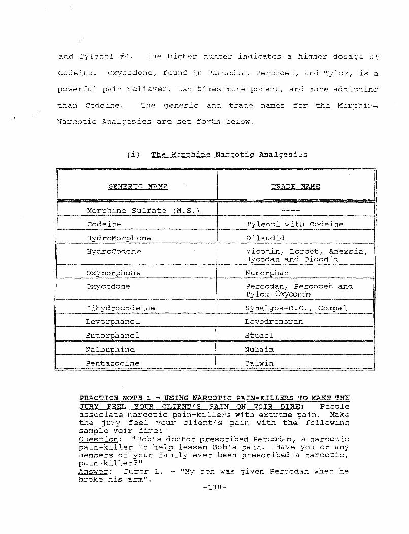
and Tylenol #4. The higher number indicates a higher dosage of
Codeine. Oxycodone, found in Percodan, Percocet, and Tylox, is a
powerful pain reliever, ten times more potent, and more addicting
than Codeine. The generic and trade names for the Morphine
Narcotic Analgesics are set forth below.
(i) The Morphine Narcotic Analgesics
II GENERIC NAME TRADE NAME - II
;: I I
/
Morphine Sulfate (M.S. ) ----Codeine Tylenol with Codeine
HydroMorphone Dilaudid
HydroCodone Vicodin, Lorcet, Anexsia, Hycodan and Dicodid
oxymorphone Numorphan
oxycodone Percodan, Percocet and I Tylox, Oxycontin
Dihydrocodeine Synalgos-D. c. , Compal
Levorphanol Levodromoran
Butorphanol Studol
Nalbuphine Nub aim
Pentazocine Tal win
PRACTICE NOTE 1 - USING NARCOTIC PAIN-KILLERS TO MAKE THE JURY FEEL YOUR CLIENT'S PAIN ON VOIR DIRE: People associate narcotic pain-killers with extreme pain. Make the jury feel your client's pain with the following sample voir dire: Question: "Bob's doctor prescribed Percodan, a narcotic pain-killer to help lessen Bob's pain. Have you or any members of your family ever been prescribed a narcotic, pain-killer?" Answer: Juror l. - "My son was given Percodan when he broke his arm" .
-138-
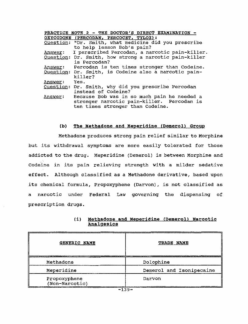
PRACTICE NOTE 2 - THE DOCTOR'S DIRECT EXAMINATION -OXYCODONE (PERCODAN, PERCOCET, TYLOX): Question: "Dr. Smith, what medicine did you prescribe
to help lesson Bob's pain? Answer: I perscribed Percodan, a narcotic pain-killer. Question: Dr. Smith, how strong a narcotic pain-killer
is Percodan? Answer: Percodan is ten times stronger than Codeine. Question: Dr. Smith, is Codeine also a narcotic pain-
killer? Answer: Question:
Answer:
(b)
Yes. Dr. Smith, why did you prescribe Percodan instead of Codeine? Because Bob was in so much pain he needed a stronger narcotic pain-killer. Percodan is ten times stronger than Codeine.
The Methadone and Meperidine (Demerol) Group
Methadone produces strong pain relief similar to Morphine
but its withdrawal symptoms are more easily tolerated for those
addicted to the drug. Meperidine (Demerol) is between Morphine and
Codeine in its pain relieving strength with a milder sedative
effect. Although classified as a Methadone derivative, based upon
its chemical formula, Propoxyphene (Darvon), is not classified as
a narcotic under Federal Law governing the dispensing of
prescription drugs.
I
(i) Methadone and Meperidine (Demerol) Narcotic Analgesics
GENERIC NAME
I TRADE NAME
Methadone Dolophine
Meperidine Demerol and Isonipecaine
Propoxyphene Darvon (Non-Narcotic)
-139-
I
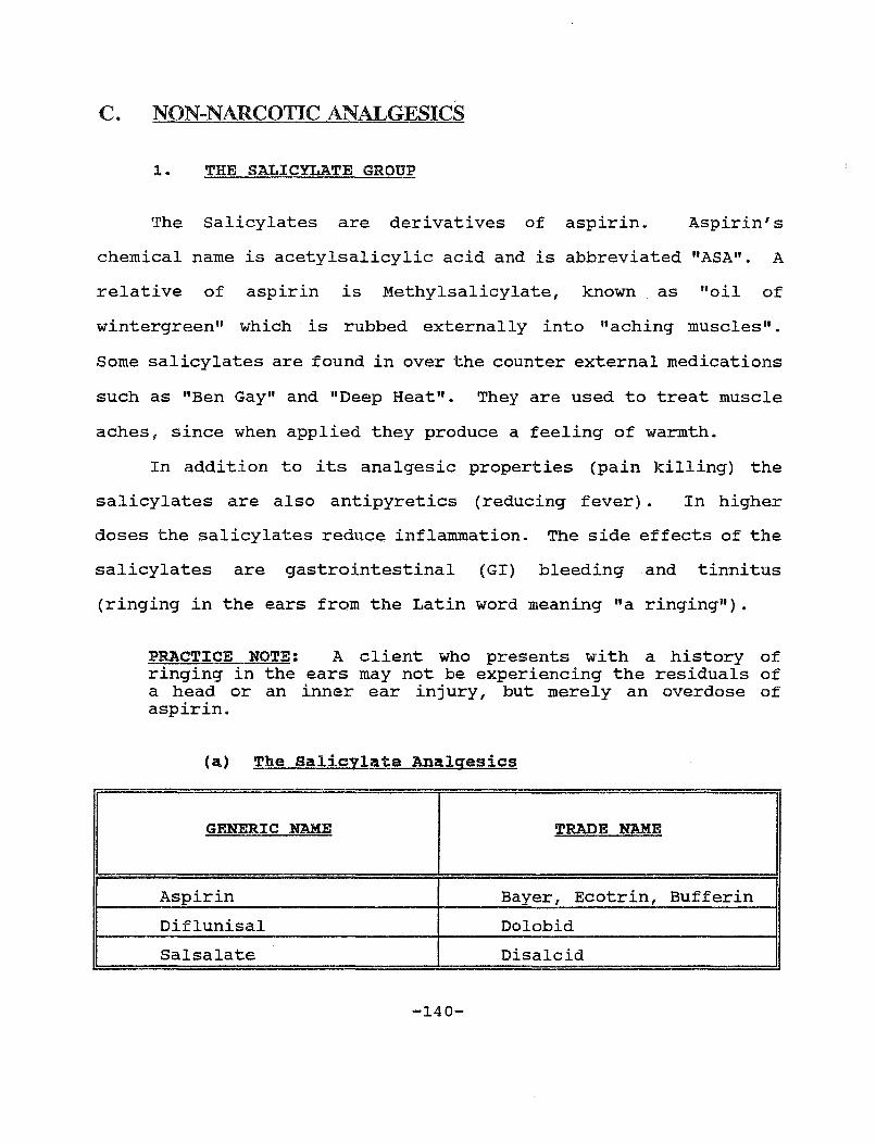
C. NON-NARCOTIC ANALGESICS
1. THE SALICYLATE GROUP
The Salicylates are derivatives of aspirin. Aspirin's
chemical name is acetylsalicylic acid and is abbreviated "ASA". A
relative of aspirin is Methylsalicylate, known as "oil of
wintergreen" which is rubbed externally into "aching muscles".
Some salicylates are found in over the counter external medications
such as "Ben Gay" and "Deep Heat". They are used to treat muscle
aches, since when applied they produce a feeling of warmth.
In addition to its analgesic properties (pain killing) the
salicylates are also antipyretics (reducing fever) . In higher
doses the salicylates reduce inflammation. The side effects of the
salicylates are gastrointestinal (GI) bleeding and tinnitus
I
(ringing in the ears from the Latin word meaning "a ringing").
PRACTICE NOTE: A client who presents with a history of ringing in the ears may not be experiencing the residuals of a head or an inner ear injury, but merely an overdose of aspirin.
(a) The Salicylate Analgesics
GENERIC NAME
I TRADE NAME
I Aspirin Bayer, Ecotrin, Buffer in
Diflunisal Dolobid
Salsa late Disalcid
-140-
2. ACETAMINOPHEN (TYLENOL)
Acetaminophen relieves pain and reduces fever, but does not
relieve inflammation like aspirin. Acetaminophen does not have the
gastrointestinal side effects of aspirin but may be toxic to the
liver (where is it metabolized) with chronic use. Frequent alcohol
intake (such as a few glasses of wine with dinner) while taking
acetaminophen presents an increased risk to the patient for
developing liver toxicity.
3. NSAIDs
Non-Steroidal Anti-inflammatory Drugs are abbreviated NSAIDs.
These drugs such as Motrin and Naprosyn, in addition to relieving
inflammation, also relieve pain. A new NSAID, ketorolac (Toradol)
recently has been used to treat pain. Because of its
gastrointestinal irritating side effects, Toradol's use is limited
to 5-14 days duration. NSAIDs are discussed in detail below.
4. ANTI-DEPRESSANTS
Anti-depressants such as amitriptyline (Elavil), fluoxetine
(Prozac), sertraline (Zoloft) and Venlafaxine (Effexor) have
recently been used to treat chronic pain.
-141-
III. ANTI-INFLAMJ\fATORY DRUGS
A. STEROIDAL ANTI-INFLAl\IMATORY DRUGS
1. DISCOVERY OF THE STEROIDS
Medicine exploded in the 1940's with the discovery of the
steroids leading to several Nobel Prizes and the development of new
wonder drugs to treat arthritis. The name steroid was derived from
an old compound called sterol. In the 1940's when organic chemists
first developed the formula for steroids they noticed that it was
similar to the formula for an old compound named sterol. The
suffix "oid" (from the Latin meaning "similar to") was added to
sterol giving the name steroids. Steroids have a characteristic 4-
r ing chemical nucleus; the same nucleus found in sterol, in
cholesterol, and in the sex hormones.
2. THE HORMONES OF THE ADRENAL CORTEX
Our body produces three types of steroid hormones in the
adrenal gland located just above the kidney. The outer layer of
the adrenal gland, called the cortex, produces these hormones.
These steroids are referred to as the adrenal cortical hormones
(from the adrenal cortex). In addition, physiologically active
steroids are called "corticosteroids" (referring again to the
adrenal cortex).
3. THE THREE CLASSES OF STEROIDS
Steroids are divided into three classes based upon their
effect on the body:
-142-
(i) Female (Estrogen) and Male (Androgen) Sex Hormones;
(ii) The Mineralcorticoids;
(iii) The Glucocorticoids.
All three classes of steroid hormones are produced by the
adrenal cortex. (1) Estrogen and Testosterone, the sex hormones;
(2) Aldosterone, a mineralcorticoid; and (3) Cortisone and
Hydrocortisone, glucocorticoids.
The Mineralcorticoids regulate the body's electrolytes by
controlling sodium retention and the excretion of potassium
important body minerals -- hence the name Mineralcorticoids.
The Glucocorticoids increase the body's blood glucose -- hence
the name Glucocorticoids. In addition, Glucocorticoids decrease
inflammation. synthesis of more potent derivatives of the body's
own glucocorticoids (Cortisone and Hydrocortisone), lead to the
development of today's potent "steroidal anti-inflammatory drugs"
such as dexamethasone (Decadron), and triamcinolone (Aristocort).
synthesis of derivatives of the body's sex hormones, Estrogen
and Testosterone, have resulted in female "birth control pills" and
"Anabolic Steroids", the latter promote nitrogen retention and
weight gain. (The term "anabolic" is derived from the word
"anabolism"-- retention of nitrogen).
PRACTICE NOTE: Anabolic Steroids, misused by athletes, have given the generic term "steroid" a "bad name". As a result, many lay people associate "steroid" with drug misuse. If the plaintiff were prescribed an anti-inflammatory steroid, the jury should be informed by the treating doctor, that this class of steroids (glucocorticoids) is vastly different from the anabolic steroids misused by athletes.
-143-
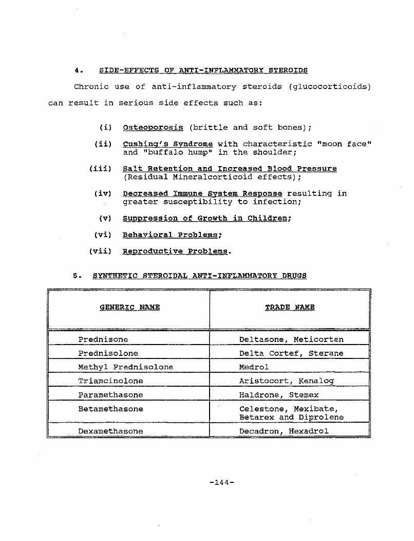
4. SIDE-EFFECTS OF ANTI-INFLAMMATORY STEROIDS
Chronic use of anti-inflammatory steroids (glucocorticoids)
can result in serious side effects such as:
I
(i) osteoporosis (brittle and soft bones);
(ii) cushing's Syndrome with characteristic "moon face" and "buffalo hump" in the shoulder;
(iii) Salt Retention and Increased Blood Pressure (Residual Mineralcorticoid effects);
(iv) Decreased Immune system Response resulting in greater susceptibility to infection;
(v) suppression of Growth in Children;
(vi) Behavioral Problems;
(vii) Reproductive Problems.
5. SYNTHETIC STEROIDAL ANTI-INFLAMMATORY DRUGS
GENERIC NAME
I TRADE NAME
Prednisone Deltasone, Meticorten
Prednisolone Delta Cortef, Sterane
Methyl Prednisolone Medrol
Triamcinolone Aristocort, Kenalog
Paramethasone Haldrone, Stem ex
Betamethasone Celestone, Mexibate, Betarex and Diprolene
Dexamethasone Decadron, Hexadrol
-144-
I
B. NSAIDs
Non-Steroidal Anti-Inflammatory Drugs are abbreviated NSAIDs.
Included within this classification are all drugs which reduce
inflammation which are not steroids. The NSAIDs have side-effects,
but they are not as serious as the steroidal anti-inflammatory
drugs.
The NSAIDs have two physiological effects on the body --
reducing inflammation and relieving pain. Some NSAIDs have greater
effectiveness at reducing inflammation, while others have greater
effectiveness at reducing pain, such as ketorolac (Toradol).
All NSAIDs produce gastrointestinal side-effects such as
nausea, vomiting and ulcers. Approximately one-quarter of all
people who take NSAIDs develop gastrointestinal side-effects. The
NSAIDs with the highest toxicity are Indocin 1 Tolectin 1 Meclomen 1
and Orudis.
PRACTICE NOTE Increased Specials Clients prescribed NSAIDs for long term chronic pain should have blood tests taken year 1 i.e. 1 a chem. profile, to monitor kidney and liver damage caused by long NSAIDs.
-145-
Blood Tests: use to treat 1-2 times per for possible term use of
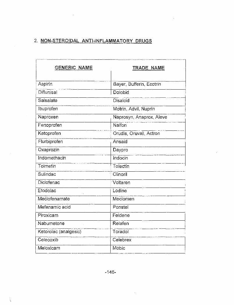
2. NON-STEROIDAL ANTI-INFLAMMATORY DRUGS
~ENERIC NAME TRADE NAME
Aspirin Bayer, Bufferin, Ecotrin
Diflunisal Dolo bid
Salsalate Disalcid
Ibuprofen Motrin, Advil, Nuprin
Naproxen Naprosyn, Anaprox, Aleve
Fenoprofen Nalfon
Ketoprofen Orudis, Oruvail, Actron
Flurbiprofen An said
Oxaprozin Daypro
Indomethacin lndocin
Tolmetin To lectin
Sulindac Clinoril
Diclofenac ·Voltaren
Etodolac Lodine
Meclofenamate Meclomen
Mefenamic acid Ponstel
Piroxicam Feldene
Nabumetone Relafen
Ketorolac (analgesic) Toradol
Celecoxib Celeb rex
Meloxicam Mabie
-146-
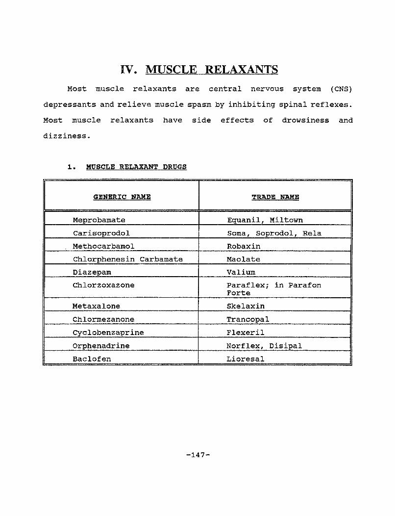
IV. MUSCLE RELAXANTS Most muscle relaxants are central nervous system (CNS)
depressants and relieve muscle spasm by inhibiting spinal reflexes.
Most muscle relaxants have side effects of drowsiness and
dizziness.
1. MUSCLE RELAXANT DRUGS
GENERIC NAME TRADE NAME
Meprobamate Equanil, Mil town
Carisoprodol Soma, Soprodol, Rela
Methocarbamol Rob ax in
Chlorphenesin Carbamate Mao late
Diazepam Valium
Chlorzoxazone Paraflex; in Parafon Forte
Metaxalone Skelaxin
Chlormezanone Trancopal
Cyclobenzaprine Flexeril
orphenadrine Norflex, Disipal
Baclofen Lioresal
-147-
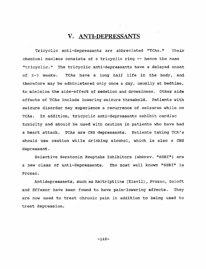
V.. ANTI-DEPRESSANTS
Tricyclic anti-depressants are abbreviated "TCAs." Their
chemical nucleus consists of a tricyclic ring -- hence the name
"tricyclic." The tricyclic anti-depressants have a delayed onset
of 2-3 weeks. TCAs have a long half life in the body, and
therefore may be administered only once a day, usually at bedtime,
to minimize the side-effect of sedation and drowsiness. Other side
effects of TCAs include lowering seizure threshold. Patients with
seizure disorder may experience a recurrence of seizures while on
TCAs. In addition, tricyclic anti-depressants exhibit cardiac
toxicity and should be used with caution in patients who have had
a heart attack. TCAs are CNS depressants. Patients taking TCA's
should use caution while drinking alcohol, which is also a CNS
depressant.
Selective Seratonin Reuptake Inhibitors (abbrev. "SSRI") are
a new class of anti-depressants.
Prozac.
The most well known "SSRI" is
Antidepressants, such as Amitriptline (Elavil), Prozac, Zoloft
and Effexor have been found to have pain-lowering effects. They
are now used to treat chronic pain in addition to being used to
treat depression.
-148-

The most commonly prescribed anti-depressants are set forth below:
1. ANTI-DEPRESSANT DRUGS
GENERIC NAME TRADE NAME
Amitriptyline. (TCA) Elavil, Amitril, Emitrip, Endep Amoxapine (TCA) Asendin Clomipramine (TCA) Anafranil Desimiprimine (TCA) Norpramin, Pertofrane Doxepin (TCA) Sinequan, Adapin Imipramine (TCA) Tofranil, Pesamine, Dramine, Janimine Nortriptyline (TCA) Aventyl, Pamelor, Allegron Protriptyline (TCA) Vivactil Trazodone (TCA Related) Desyrel, Molipaxia Trimipramine (TCA) Surmontil Citalopram (SSRI) Celexa Escitalopram (SSRI) Lexapro Fluoxetine (SSRI) Prozac Fluvoxamine (SSRI) Luvox Paroxetine (SSRI) Paxil, Seroxat Sertraline (SSRI) Zoloft Duloxetine (SNRI) Cymba Ita Nefazodone (SNRI) Serzone Reboxetine (SNRI) Edronax Venlafaxine (SNRI) Effexor Mirtazapine (NaSSA) Remeron, Zispin Bupropion (AK) Wellbutrin Maprotiline (NRI) Ludiomil
TCA = Tricyclic Antidepressant SSRI = Selective Serotonin Reuptake Inhibitor SNRI =Serotonin and Noradrenaline Reuptake Inhibitor NaSSA = Noradrenergic and Specific Serotonergic Antidepressant AK = Aminoketone NRI =Noradrenaline Reuptake Inhibitor
-149-
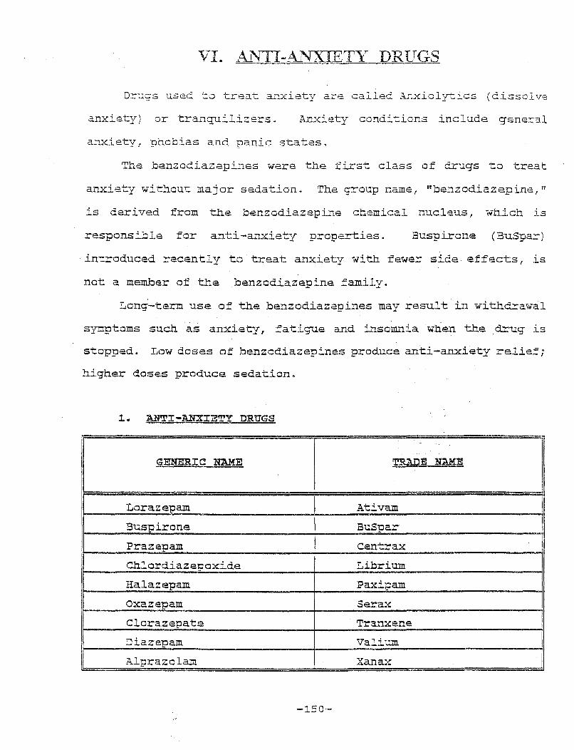
VI. AL'ITI-Al'fXIETr-.- DRlTGS
Drugs used to treat anxiety are called Anxiolytics (dissolve
anxiety) or tranquilizers. Anxiety conditions include gener."al
anxiety: phobias and panic states,
The benzodiazepines were the first class of drugs to treat
anxiety without major sedation. The group namer rrbenzodiazepine, 11
is derived from the benzodiazepine chemical nucleus, which is
responsible for anti-anxiety properties. Buspirone (BuSpar)
introduced recently to treat anxiety with fewer side effects, is
not a member af the benzadiazepine family.
Long-term use of the benzodiazepines may result in withdrawal
symptoms such as anxiety, fatigue and insomnia when the drug is
stopped. Low doses of benzodiazepines produce anti-anxiety relief;
higher doses produce sedation.
~. ANTI-ANXIETY DRUGS
I GENERIC NAME
I TRADE NAME
I Larazepam Ativam
Buspirane BuS par
Prazepam centrax
Chlordiazepoxide Librium
Halazepam Paxipa:m
oxazepam Serax
Clarazepate Tranxene
Diazepam Valium
Alprazala:m X an ax
-150-
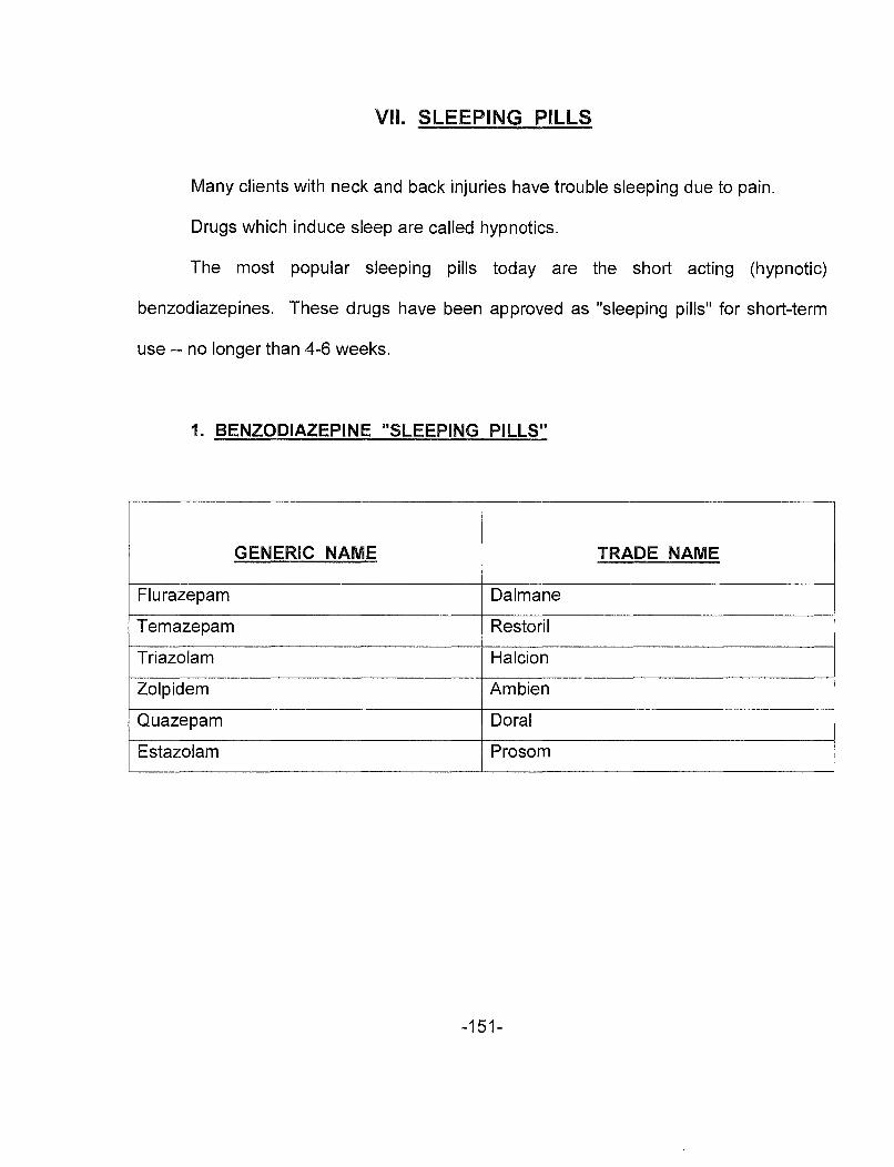
VII. SLEEPING PILLS
Many clients with neck and back injuries have trouble sleeping due to pain.
Drugs which induce sleep are called hypnotics.
The most popular sleeping pills today are the short acting (hypnotic)
benzodiazepines. These drugs have been approved as "sleeping pills" for short-term
use -- no longer than 4-6 weeks.
1. BENZODIAZEPINE "SLEEPING PILLS"
GENERIC NAME TRADE NAME
Flurazepam Dalmane
Temazepam Restoril
Triazolam Halcion
Zolpidem Am bien
Quazepam Dora I
Estazolam Prosom
-151-
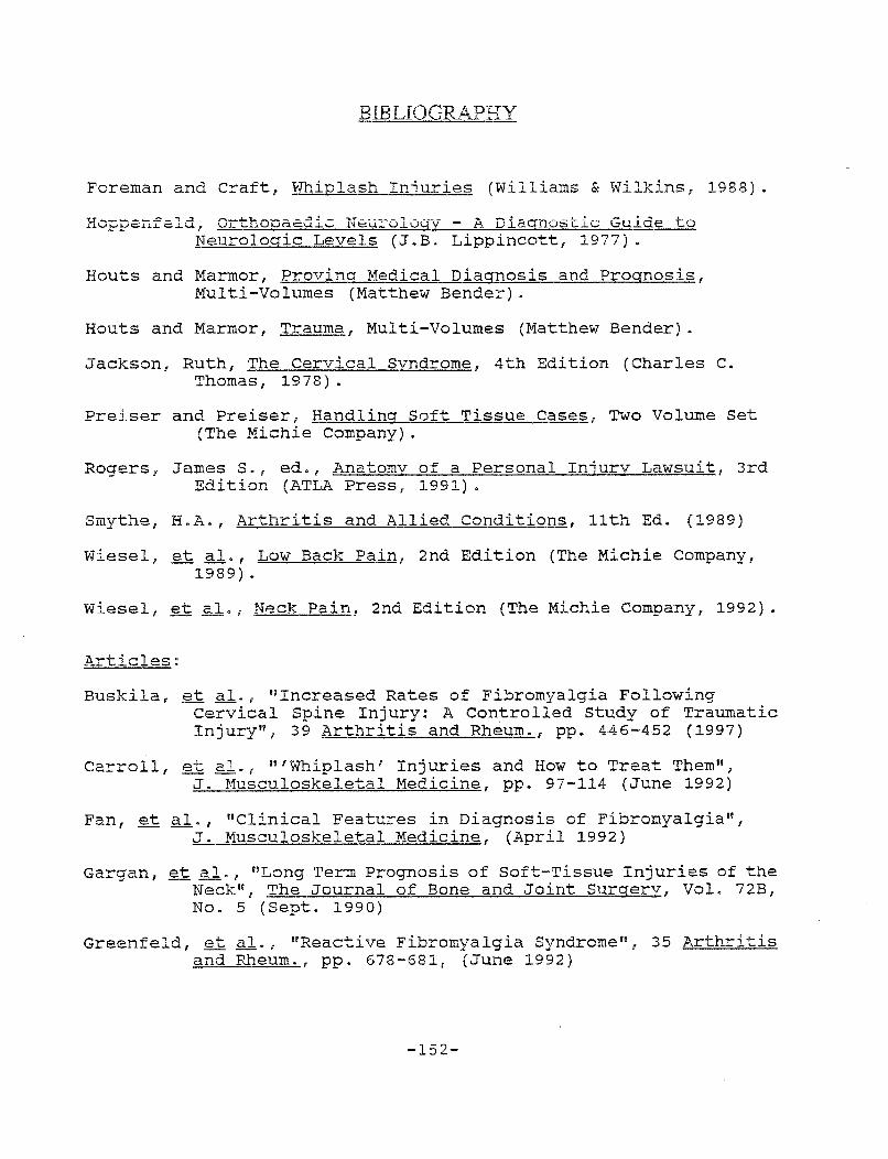
BIBLIOGR.APHY
Foreman and Craft, Whiolash Inluries (Williams & Wilkins, 1988).
Hcppenfeld, orthopaedic Neurolog-y - A Diagnostic ut.uae to Neurologic Levels (J.B. Lippincott, 1977).
Houts and Marmor, Provina Medical Diaqnosis and Prognosis, Multi-Volumes (Matthew Bender).
Houts and Marmor, Trauma, Multi-Volumes (Matthew Bender).
Jackson, Ruth 1 The Cervical svndrome, 4th Edition (Charles c. Thomas, 1978).
Preiser and Preiser, Handling Soft Tissue Cases, Two Volume Set (The Michie Company) .
Rogers, James s., ed., Anatomy of a Personal Injury Lawsuit, 3rd Edition (ATLA Press, 1991).
Smythe, H.A., Arthritis and Allied Conditions, 11th Ed. (1989)
Wiesel, et al., Low Back Pain, 2nd Edition (The Michie Company, 1989).
Wiesel, et al., Neck Pain, 2nd Edition (The Michie Company 1 1992).
Articles:
Buskila, et al., "Increased Rates of Fibromyalgia Following Cervical Spine Injury: A Controlled Study of Traumatic Injury", 39 Arthritis and Rheum., pp. 446-452 (1997)
carroll, et al. 1 "'Whiplash 1 Injuries and How to Treat Them", J. Musculoskeletal Medicine, pp. 97-114 (June 1992)
Fan, et al., 11 Clinical Features in Diagnosis of Fibromyalgia", J. Musculoskeletal Medicine, (April 1992)
Gargan, et al., "Long Term Prognosis of Soft-Tissue Injuries of the Neck", The Journal of Bone and Joint Surgerv, Vol. 72B, No. 5 (Sept. 1990)
Greenfeld, et al. , 11 Reacti ve Fibromyalgia Syndrome", 35 Arthritis and Rheum., pp. 678-681, (June 1992)
-152-
Recommended