Practical Course Receptor Biochemistry 2019
Preliminary scheduling for the practical course Receptorbiochemistry. Due to the work
with living cells changes might become necessary if cell growth is not as expected.
Explicit time schedules will be arranged individually.
If you have problems/questions please contact Dr.Karin Mörl [email protected]
Responsible Person: Dr. Karin Mörl
Supervision: Dr. Karin Mörl
Victoria Behr
Kristin Löbner
Sarina Rudolf
Corinna Schüß
Isabelle Ziffert
Einführung für Bock 1 und 2: 3.4.2019, 10:30, Seminarraum 4.Stock, Brüderstr.34
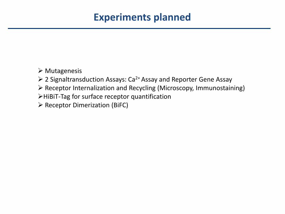
Experiments planned
Mutagenesis 2 Signaltransduction Assays: Ca2+ Assay and Reporter Gene Assay Receptor Internalization and Recycling (Microscopy, Immunostaining)HiBiT-Tag for surface receptor quantification Receptor Dimerization (BiFC)
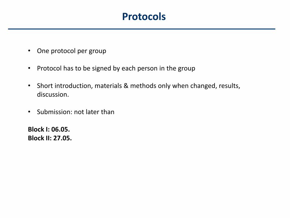
Protocols
• One protocol per group
• Protocol has to be signed by each person in the group
• Short introduction, materials & methods only when changed, results, discussion.
• Submission: not later than
Block I: 06.05.Block II: 27.05.

You have to follow basic principles and guidelines for working safely in LaboratoriesIf you work with hazardous materials you have to know the corresponding „Risiko- und Sicherheitssätze“, risk and safetysentences which can be found here: http://gestis.itrust.de/nxt/gateway.dll/gestis_de/000000.xml?f=templates&fn=default.htm&vid=gestisdeu:sdbdeuEven in small amounts, you have to label hazardous materialPoisonous substances have to be shut awayYou have to wear a lab coat. You have to wear gloves and protective glasses when needed. Do not touch labware, equipment , door handles etc. with contaminated glovesHandle hazardous material in a fume hoodKeep working benches clean and tidyKeep hazardous waste as small as possible and discard appropriatelyDon‘t eat and drink in the labsExperimental work is only allowed when supervisor is presentBe informed on directions for use of lab equipment, ask before you use itIf you realize a defect, immediately tell the supervisorDon‘t use gas and air supplying devices in improper wayChemical burns and burns: Rinse with cold water immediately for at least 10 minandIn case of emergency ifor supervisor, dispatcher number on each telephoneFire-extinguisher are to be used all-purposeSmall injuries have to be documented by supervisors for insurance reasonsPregnancy should be communicated as early as possibleOnly students taking part in the course are allowed to enter the labHandle sharps carefully (scalpels and cannula) and discard properly in special containersDon‘t use open fireBe informed on position of fire-extinguisher, emergency showers, electricity emergency stop buttons, alarm signals andescape routes
Instructions for safety working
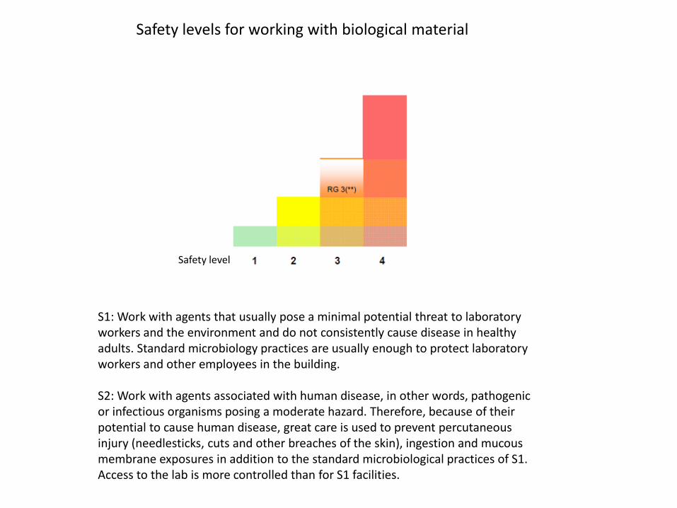
S1: Work with agents that usually pose a minimal potential threat to laboratory workers and the environment and do not consistently cause disease in healthy adults. Standard microbiology practices are usually enough to protect laboratory workers and other employees in the building.
S2: Work with agents associated with human disease, in other words, pathogenic or infectious organisms posing a moderate hazard. Therefore, because of their potential to cause human disease, great care is used to prevent percutaneous injury (needlesticks, cuts and other breaches of the skin), ingestion and mucous membrane exposures in addition to the standard microbiological practices of S1. Access to the lab is more controlled than for S1 facilities.
Safety level
Safety levels for working with biological material
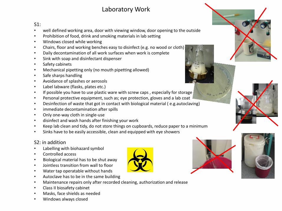
S1: • well defined working area, door with viewing window, door opening to the outside• Prohibition of food, drink and smoking materials in lab setting• Windows closed while working• Chairs, floor and working benches easy to disinfect (e.g. no wood or cloth)• Daily decontamination of all work surfaces when work is complete• Sink with soap and disinfectant dispenser• Safety cabinets• Mechanical pipetting only (no mouth pipetting allowed)• Safe sharps handling• Avoidance of splashes or aerosols• Label labware (flasks, plates etc.)• If possible you have to use plastic ware with screw caps , especially for storage• Personal protective equipment, such as; eye protection, gloves and a lab coat• Desinfection of waste that got in contact with biological material ( e.g.autoclaving)• immediate decontamination after spills• Only one-way cloth in single-use• disinfect and wash hands after finishing your work• Keep lab clean and tidy, do not store things on cupboards, reduce paper to a minimum• Sinks have to be easily accessible, clean and equipped with eye showers
S2: in addition• Labelling with biohazard symbol• Controlled access• Biological material has to be shut away• Jointless transition from wall to floor• Water tap operatable without hands• Autoclave has to be in the same building• Maintenance repairs only after recorded cleaning, authorization and release• Class II biosafety cabinet• Masks, face shields as needed• Windows always closed
Laboratory Work
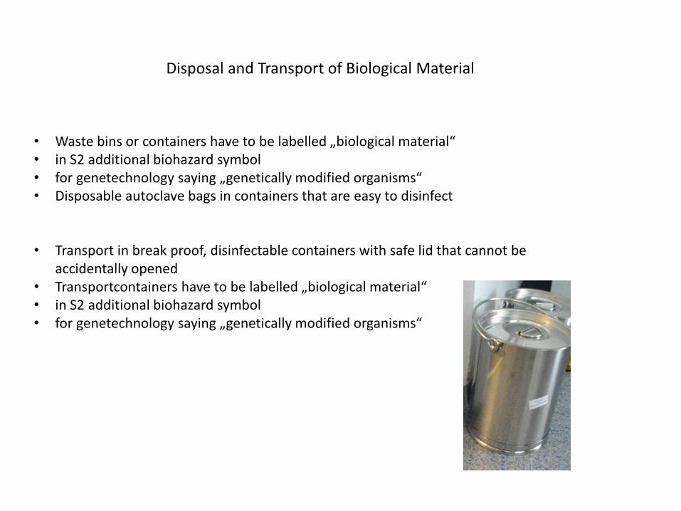
Disposal and Transport of Biological Material
• Waste bins or containers have to be labelled „biological material“• in S2 additional biohazard symbol• for genetechnology saying „genetically modified organisms“ • Disposable autoclave bags in containers that are easy to disinfect
• Transport in break proof, disinfectable containers with safe lid that cannot beaccidentally opened
• Transportcontainers have to be labelled „biological material“• in S2 additional biohazard symbol• for genetechnology saying „genetically modified organisms“
Please no cell phone when experiments are performed or discussed!
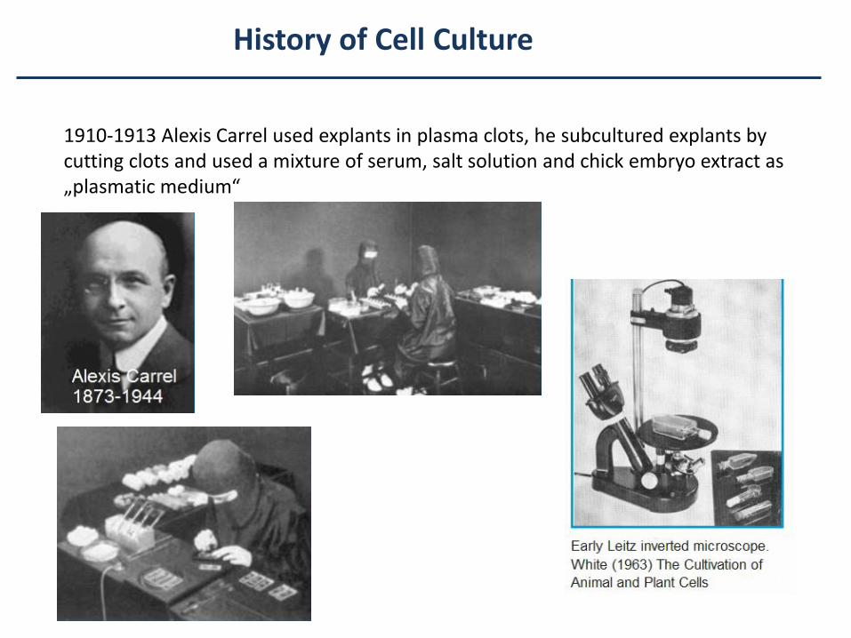
History of Cell Culture
1910-1913 Alexis Carrel used explants in plasma clots, he subcultured explants by cutting clots and used a mixture of serum, salt solution and chick embryo extract as „plasmatic medium“
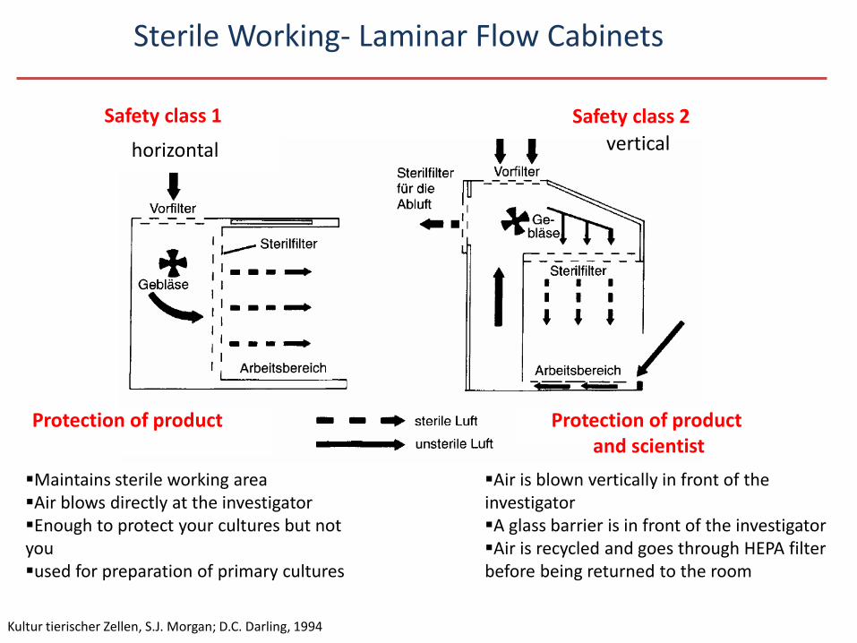
Safety class 1 Safety class 2
Protection of product
Kultur tierischer Zellen, S.J. Morgan; D.C. Darling, 1994
Protection of product and scientist
vertical
Sterile Working- Laminar Flow Cabinets
horizontal
Maintains sterile working areaAir blows directly at the investigatorEnough to protect your cultures but not youused for preparation of primary cultures
Air is blown vertically in front of the investigatorA glass barrier is in front of the investigatorAir is recycled and goes through HEPA filter before being returned to the room
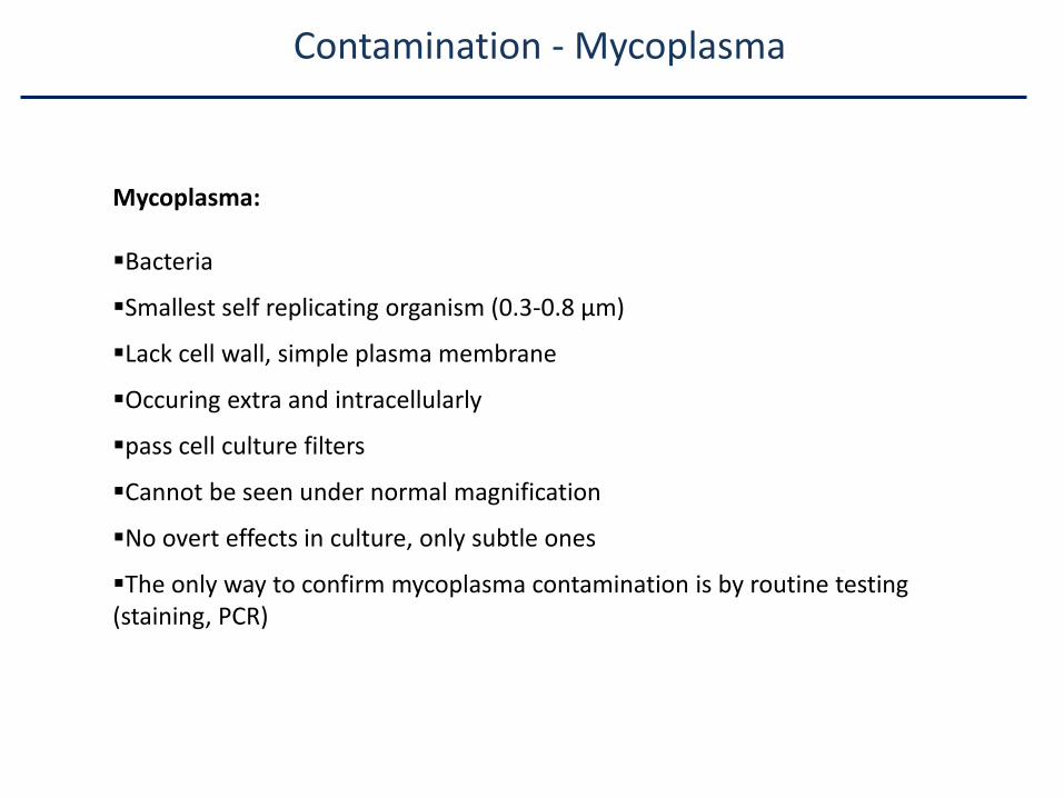
Contamination - Mycoplasma
Mycoplasma:
Bacteria
Smallest self replicating organism (0.3-0.8 µm)
Lack cell wall, simple plasma membrane
Occuring extra and intracellularly
pass cell culture filters
Cannot be seen under normal magnification
No overt effects in culture, only subtle ones
The only way to confirm mycoplasma contamination is by routine testing (staining, PCR)
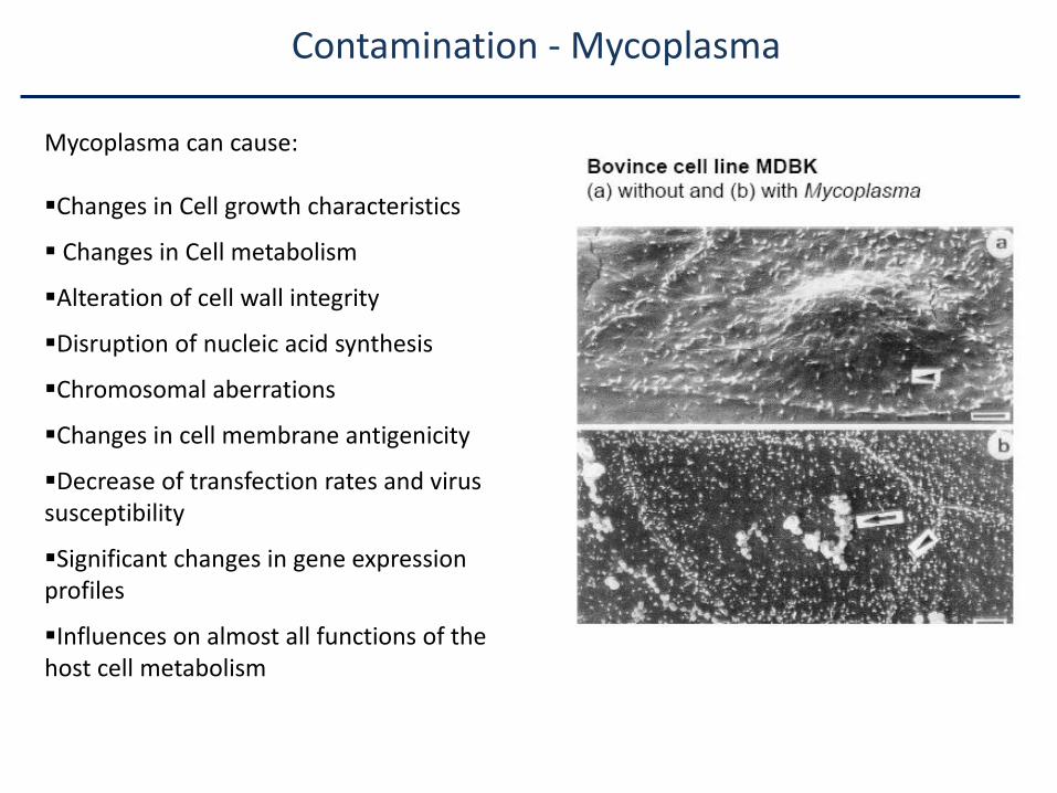
Contamination - Mycoplasma
Mycoplasma can cause:
Changes in Cell growth characteristics
Changes in Cell metabolism
Alteration of cell wall integrity
Disruption of nucleic acid synthesis
Chromosomal aberrations
Changes in cell membrane antigenicity
Decrease of transfection rates and virus susceptibility
Significant changes in gene expression profiles
Influences on almost all functions of the host cell metabolism
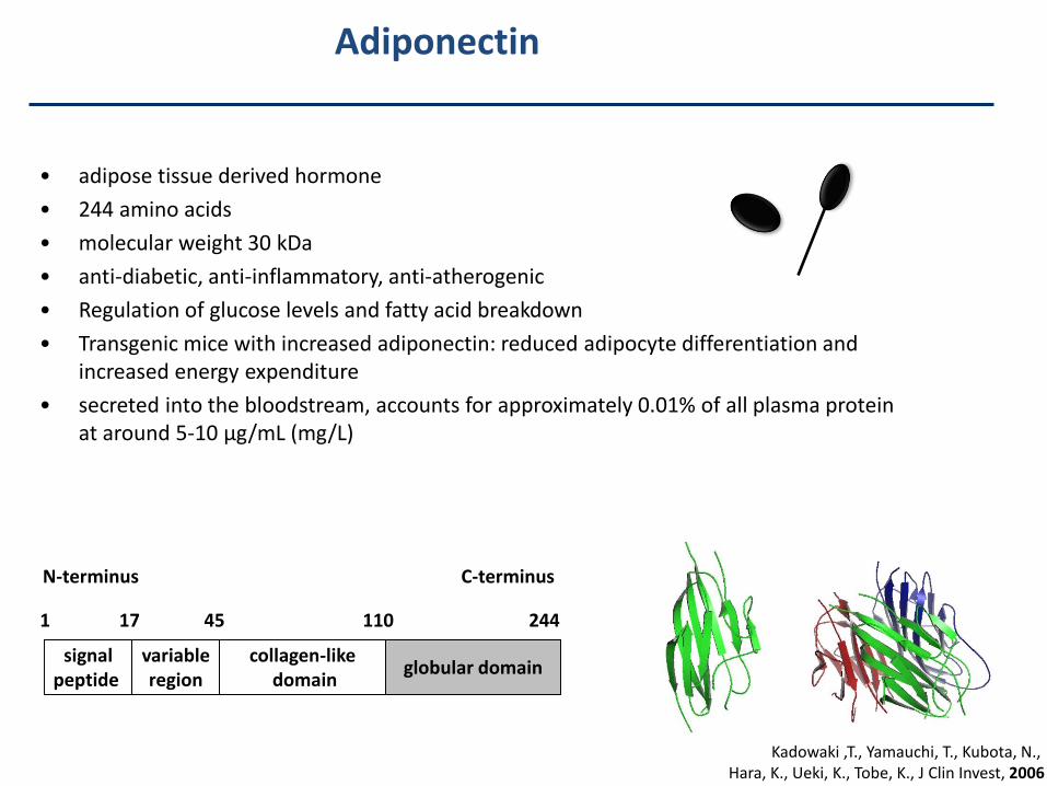
• adipose tissue derived hormone
• 244 amino acids
• molecular weight 30 kDa
• anti-diabetic, anti-inflammatory, anti-atherogenic
• Regulation of glucose levels and fatty acid breakdown
• Transgenic mice with increased adiponectin: reduced adipocyte differentiation and increased energy expenditure
• secreted into the bloodstream, accounts for approximately 0.01% of all plasma protein at around 5-10 μg/mL (mg/L)
N-terminus C-terminus
signalpeptide
variableregion
collagen-likedomain
globular domain
1 17 45 110 244
Kadowaki ,T., Yamauchi, T., Kubota, N., Hara, K., Ueki, K., Tobe, K., J Clin Invest, 2006
Adiponectin
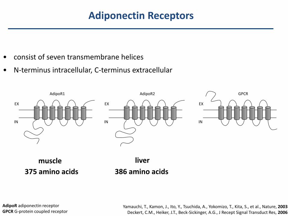
Adiponectin Receptors
• consist of seven transmembrane helices
• N-terminus intracellular, C-terminus extracellular
muscle liver
375 amino acids 386 amino acids
Yamauchi, T., Kamon, J., Ito, Y., Tsuchida, A., Yokomizo, T., Kita, S., et al., Nature, 2003Deckert, C.M., Heiker, J.T., Beck-Sickinger, A.G., J Recept Signal Transduct Res, 2006
AdipoR2
EX
IN
GPCR
EX
IN
AdipoR1
EX
IN
AdipoR adiponectin receptorGPCR G-protein coupled receptor
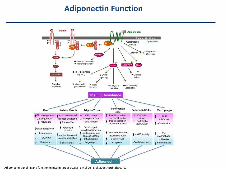
Adiponectin Function
Adiponectin signaling and function in insulin target tissues, J Mol Cell Biol. 2016 Apr;8(2):101-9.
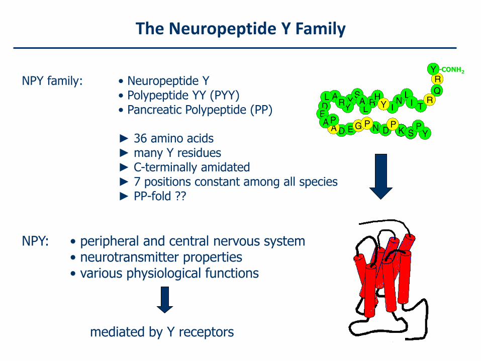
The Neuropeptide Y Family
NPY family: • Neuropeptide Y • Polypeptide YY (PYY)• Pancreatic Polypeptide (PP)
► 36 amino acids► many Y residues► C-terminally amidated► 7 positions constant among all species► PP-fold ??
NPY: • peripheral and central nervous system• neurotransmitter properties• various physiological functions
--CONH2
mediated by Y receptors
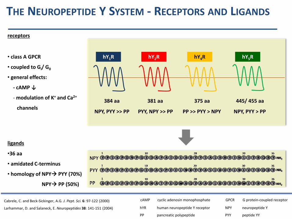
NPY, PYY >> PP PYY, NPY >> PP PP >> PYY > NPY NPY, PYY > PP
THE NEUROPEPTIDE Y SYSTEM - RECEPTORS AND LIGANDS
Cabrele, C. and Beck-Sickinger, A.G. J. Pept. Sci. 6: 97-122 (2000)
Larhammar, D. and Salaneck, E. Neuropeptides 38: 141-151 (2004) hYR human neuropeptide Y receptor NPY neuropeptide Y
PP pancreatic polypeptide PYY peptide YY
hY1R hY2R hY4R hY5R
384 aa 381 aa 375 aa 445/ 455 aa
• class A GPCR
• coupled to Gi/ G0
• general effects:
- cAMP ↓
- modulation of K+ and Ca2+
channels
receptors
ligands
•36 aa
• amidated C-terminus
• homology of NPY PYY (70%)
NPY PP (50%)
cAMP cyclic adenosin monophosphate GPCR G protein-coupled receptor
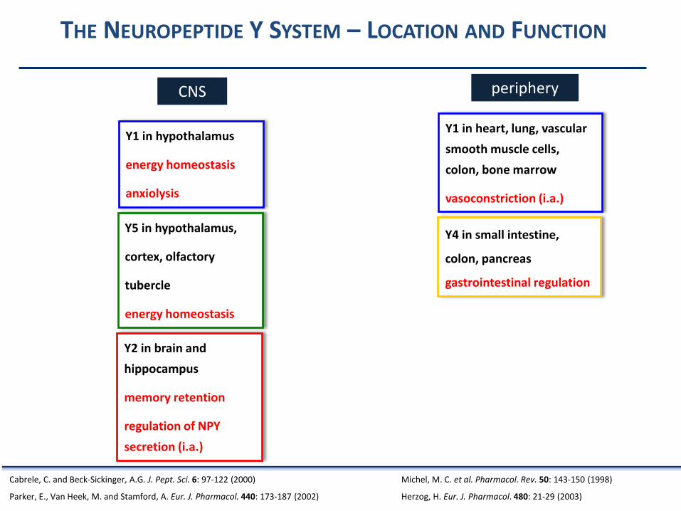
THE NEUROPEPTIDE Y SYSTEM – LOCATION AND FUNCTION
Y1 in hypothalamus
energy homeostasis
anxiolysis
Y4 in small intestine,
colon, pancreas
gastrointestinal regulation
Y2 in brain and
hippocampus
memory retention
regulation of NPY
secretion (i.a.)
Y5 in hypothalamus,
cortex, olfactory
tubercle
energy homeostasis
CNS periphery
Y1 in heart, lung, vascular
smooth muscle cells,
colon, bone marrow
vasoconstriction (i.a.)
Cabrele, C. and Beck-Sickinger, A.G. J. Pept. Sci. 6: 97-122 (2000)
Parker, E., Van Heek, M. and Stamford, A. Eur. J. Pharmacol. 440: 173-187 (2002)
Michel, M. C. et al. Pharmacol. Rev. 50: 143-150 (1998)
Herzog, H. Eur. J. Pharmacol. 480: 21-29 (2003)
THERAPEUTIC POTENTIAL OF THE HUMAN NEUROPEPTIDE Y RECEPTORS
ObesityEpilepsyBreast carcinomasEndocrine tumorsNeuroblastomasIschemic diseases
NPY System - Selective Ligands to Study Structure andFunction
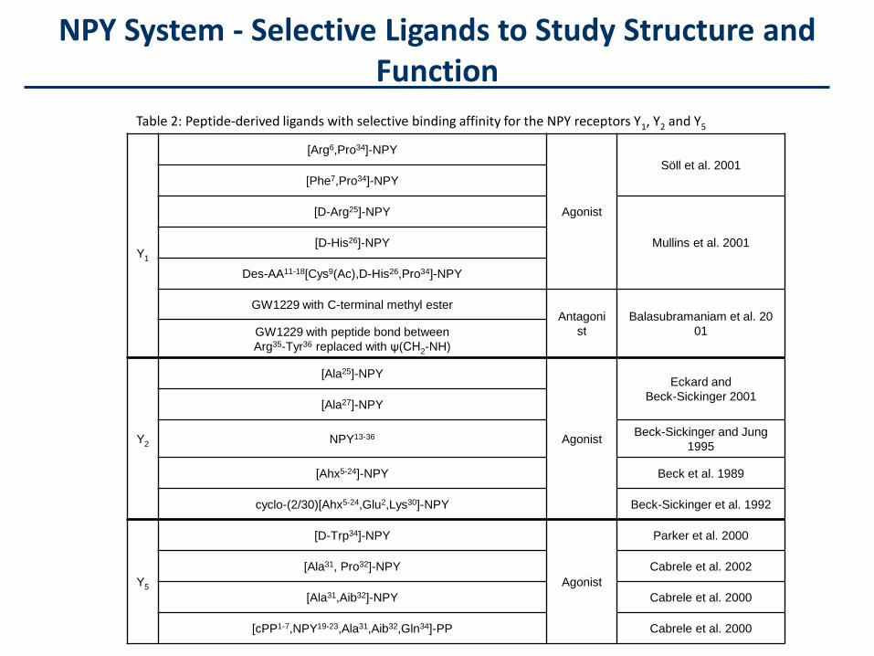
Table 2: Peptide-derived ligands with selective binding affinity for the NPY receptors Y1, Y2 and Y5
Y1
[Arg6,Pro34]-NPY
Agonist
Söll et al. 2001
[Phe7,Pro34]-NPY
[D-Arg25]-NPY
Mullins et al. 2001[D-His26]-NPY
Des-AA11-18[Cys9(Ac),D-His26,Pro34]-NPY
GW1229 with C-terminal methyl esterAntagoni
st
Balasubramaniam et al. 20
01GW1229 with peptide bond between
Arg35-Tyr36 replaced with ψ(CH2-NH)
Y2
[Ala25]-NPY
Agonist
Eckard and
Beck-Sickinger 2001[Ala27]-NPY
NPY13-36 Beck-Sickinger and Jung
1995
[Ahx5-24]-NPY Beck et al. 1989
cyclo-(2/30)[Ahx5-24,Glu2,Lys30]-NPY Beck-Sickinger et al. 1992
Y5
[D-Trp34]-NPY
Agonist
Parker et al. 2000
[Ala31, Pro32]-NPY Cabrele et al. 2002
[Ala31,Aib32]-NPY Cabrele et al. 2000
[cPP1-7,NPY19-23,Ala31,Aib32,Gln34]-PP Cabrele et al. 2000
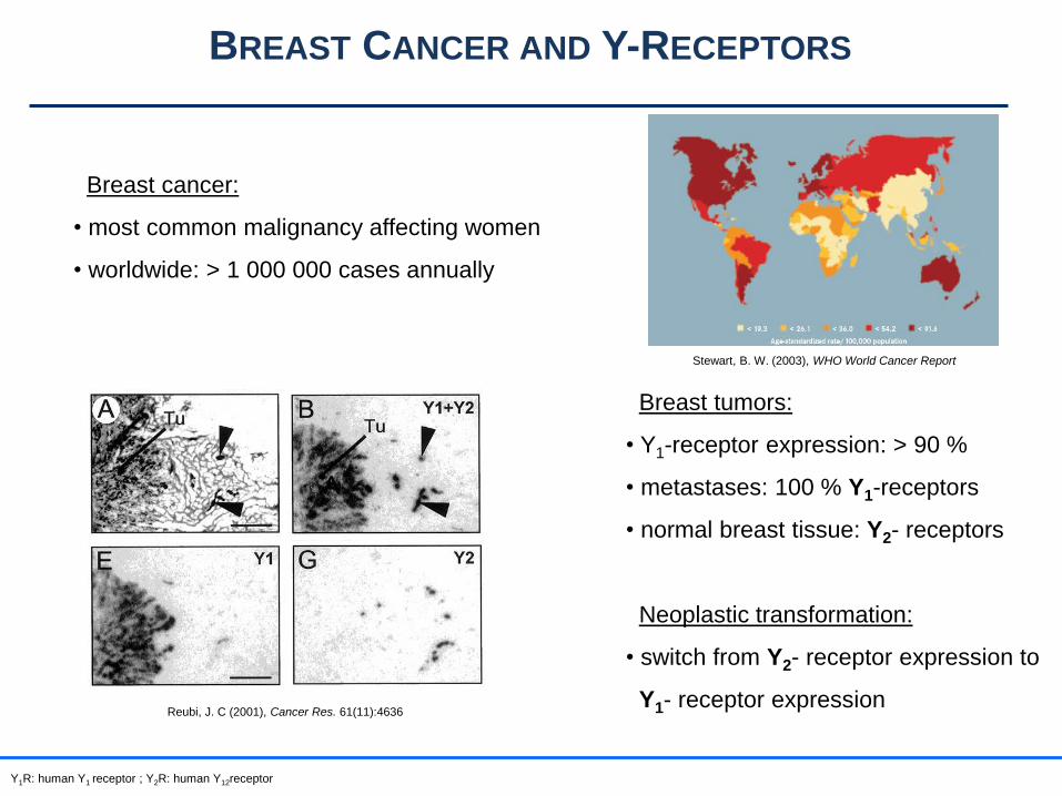
BREAST CANCER AND Y-RECEPTORS
Breast cancer:
• most common malignancy affecting women
• worldwide: > 1 000 000 cases annually
Stewart, B. W. (2003), WHO World Cancer Report
Breast tumors:
• Y1-receptor expression: > 90 %
• metastases: 100 % Y1-receptors
• normal breast tissue: Y2- receptors
Neoplastic transformation:
• switch from Y2- receptor expression to
Y1- receptor expressionReubi, J. C (2001), Cancer Res. 61(11):4636
Y1R: human Y1 receptor ; Y2R: human Y12receptor
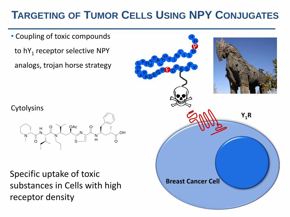
• Coupling of toxic compounds
to hY1 receptor selective NPY
analogs, trojan horse strategy
Breast Cancer Cell
Y1R
N
OAc
N
S
NH
O
OH
O
HN
N
O
O
Cytolysins
TARGETING OF TUMOR CELLS USING NPY CONJUGATES
Specific uptake of toxicsubstances in Cells with high receptor density
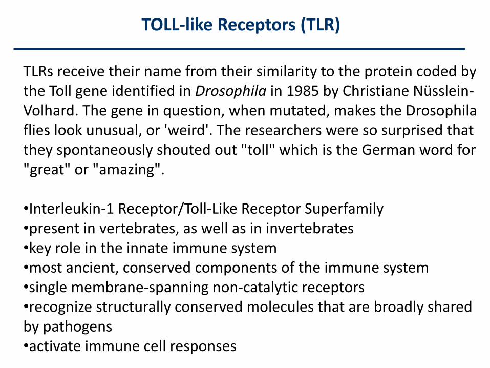
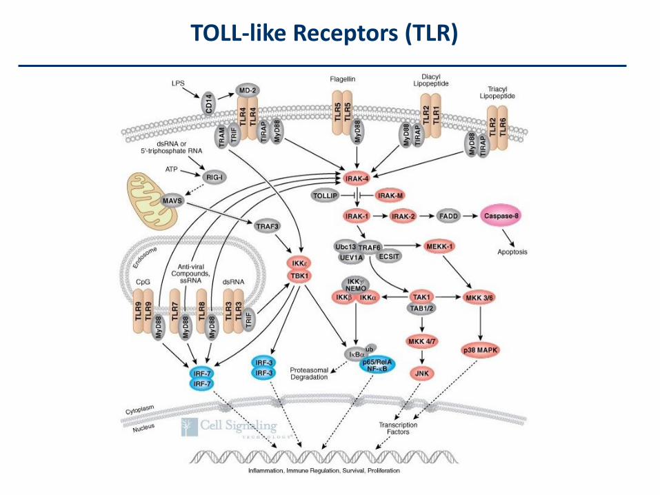
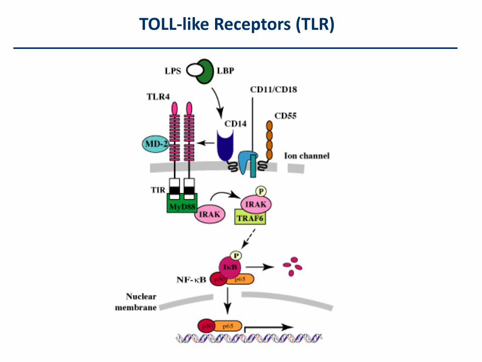
TOLL-like Receptors (TLR)
TLRs receive their name from their similarity to the protein coded bythe Toll gene identified in Drosophila in 1985 by Christiane Nüsslein-Volhard. The gene in question, when mutated, makes the Drosophilaflies look unusual, or 'weird'. The researchers were so surprised thatthey spontaneously shouted out "toll" which is the German word for"great" or "amazing".
•Interleukin-1 Receptor/Toll-Like Receptor Superfamily•present in vertebrates, as well as in invertebrates•key role in the innate immune system•most ancient, conserved components of the immune system•single membrane-spanning non-catalytic receptors•recognize structurally conserved molecules that are broadly sharedby pathogens•activate immune cell responses
TOLL-like Receptors (TLR)
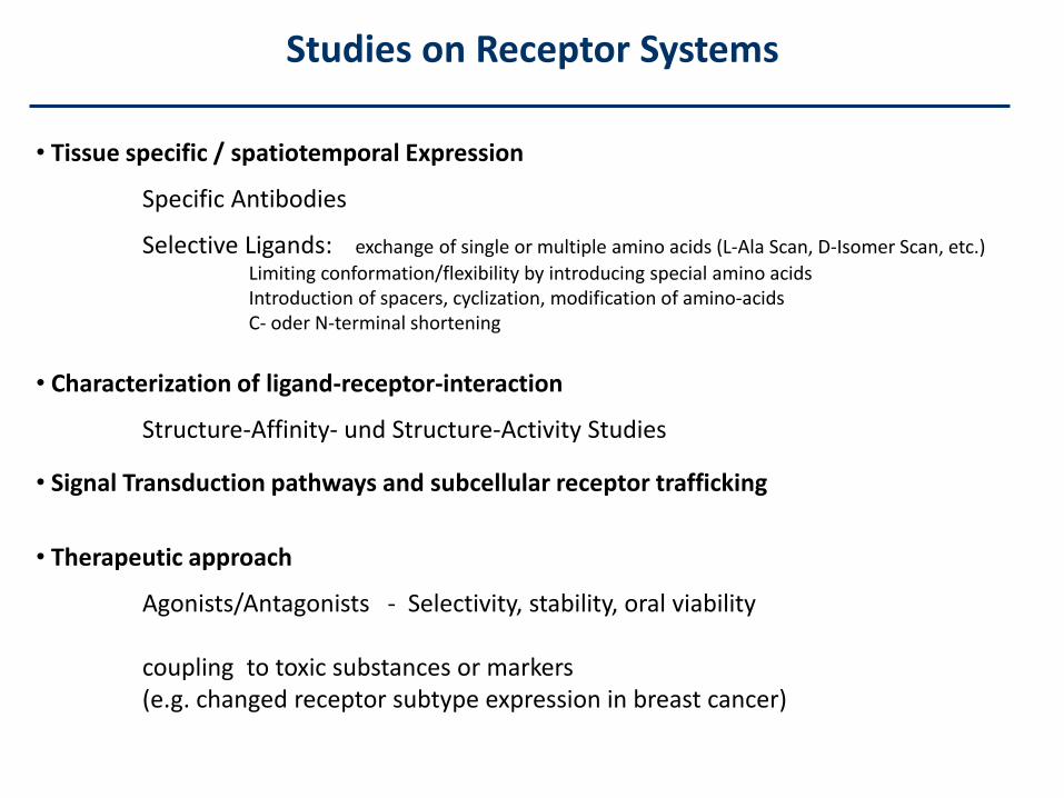
• Tissue specific / spatiotemporal Expression
Specific Antibodies
Selective Ligands: exchange of single or multiple amino acids (L-Ala Scan, D-Isomer Scan, etc.)
Limiting conformation/flexibility by introducing special amino acidsIntroduction of spacers, cyclization, modification of amino-acidsC- oder N-terminal shortening
• Characterization of ligand-receptor-interaction
Structure-Affinity- und Structure-Activity Studies
• Signal Transduction pathways and subcellular receptor trafficking
• Therapeutic approach
Agonists/Antagonists - Selectivity, stability, oral viability
coupling to toxic substances or markers(e.g. changed receptor subtype expression in breast cancer)
Studies on Receptor Systems
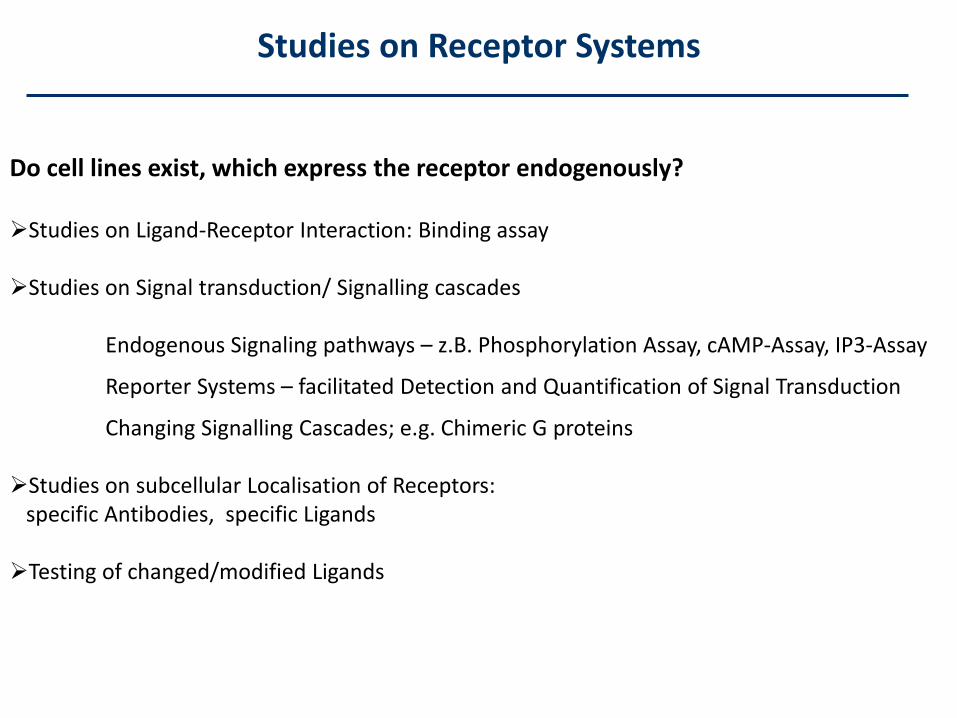
Do cell lines exist, which express the receptor endogenously?
Studies on Ligand-Receptor Interaction: Binding assay
Studies on Signal transduction/ Signalling cascades
Endogenous Signaling pathways – z.B. Phosphorylation Assay, cAMP-Assay, IP3-Assay
Reporter Systems – facilitated Detection and Quantification of Signal Transduction
Changing Signalling Cascades; e.g. Chimeric G proteins
Studies on subcellular Localisation of Receptors:specific Antibodies, specific Ligands
Testing of changed/modified Ligands
Studies on Receptor Systems
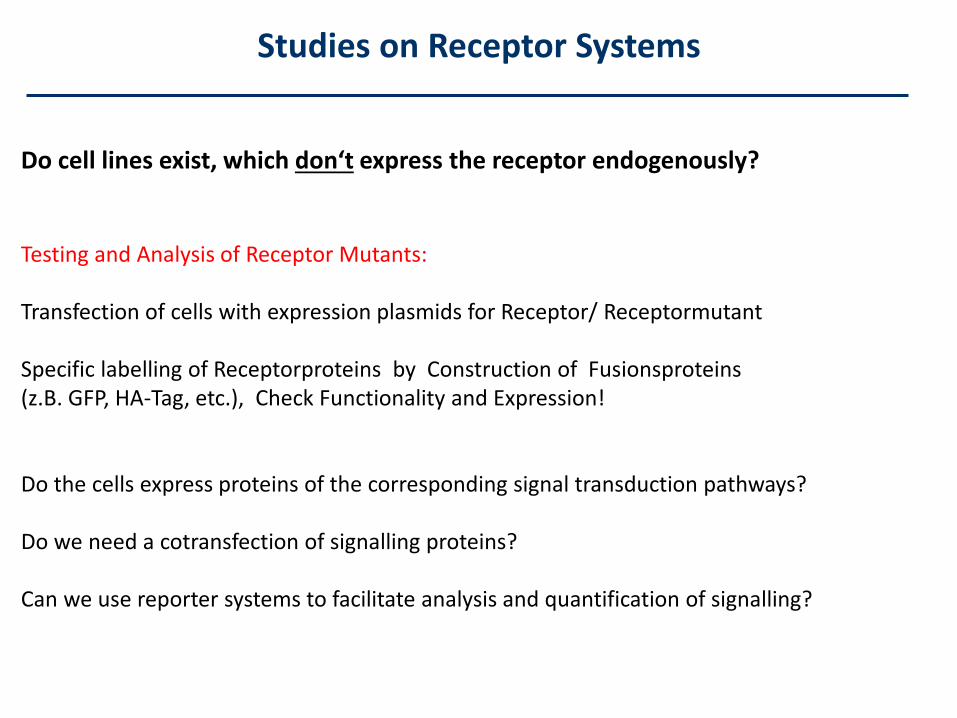
Do cell lines exist, which don‘t express the receptor endogenously?
Testing and Analysis of Receptor Mutants:
Transfection of cells with expression plasmids for Receptor/ Receptormutant
Specific labelling of Receptorproteins by Construction of Fusionsproteins (z.B. GFP, HA-Tag, etc.), Check Functionality and Expression!
Do the cells express proteins of the corresponding signal transduction pathways?
Do we need a cotransfection of signalling proteins?
Can we use reporter systems to facilitate analysis and quantification of signalling?
Studies on Receptor Systems
Transfection of Cells
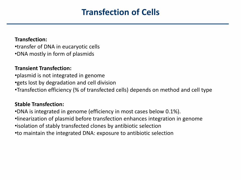
Transfection:•transfer of DNA in eucaryotic cells•DNA mostly in form of plasmids
Transient Transfection: •plasmid is not integrated in genome•gets lost by degradation and cell division•Transfection efficiency (% of transfected cells) depends on method and cell type
Stable Transfection: •DNA is integrated in genome (efficiency in most cases below 0.1%). •linearization of plasmid before transfection enhances integration in genome•isolation of stably transfected clones by antibiotic selection•to maintain the integrated DNA: exposure to antibiotic selection
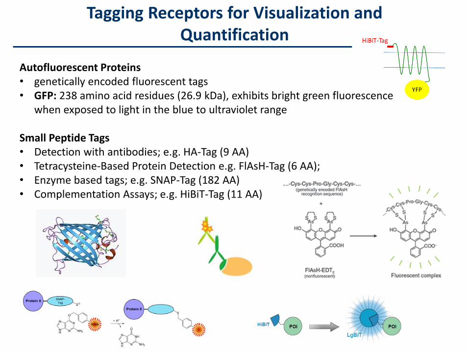
Tagging Receptors for Visualization andQuantification
Autofluorescent Proteins• genetically encoded fluorescent tags• GFP: 238 amino acid residues (26.9 kDa), exhibits bright green fluorescence
when exposed to light in the blue to ultraviolet range
Small Peptide Tags• Detection with antibodies; e.g. HA-Tag (9 AA)• Tetracysteine-Based Protein Detection e.g. FlAsH-Tag (6 AA);• Enzyme based tags; e.g. SNAP-Tag (182 AA)• Complementation Assays; e.g. HiBiT-Tag (11 AA)
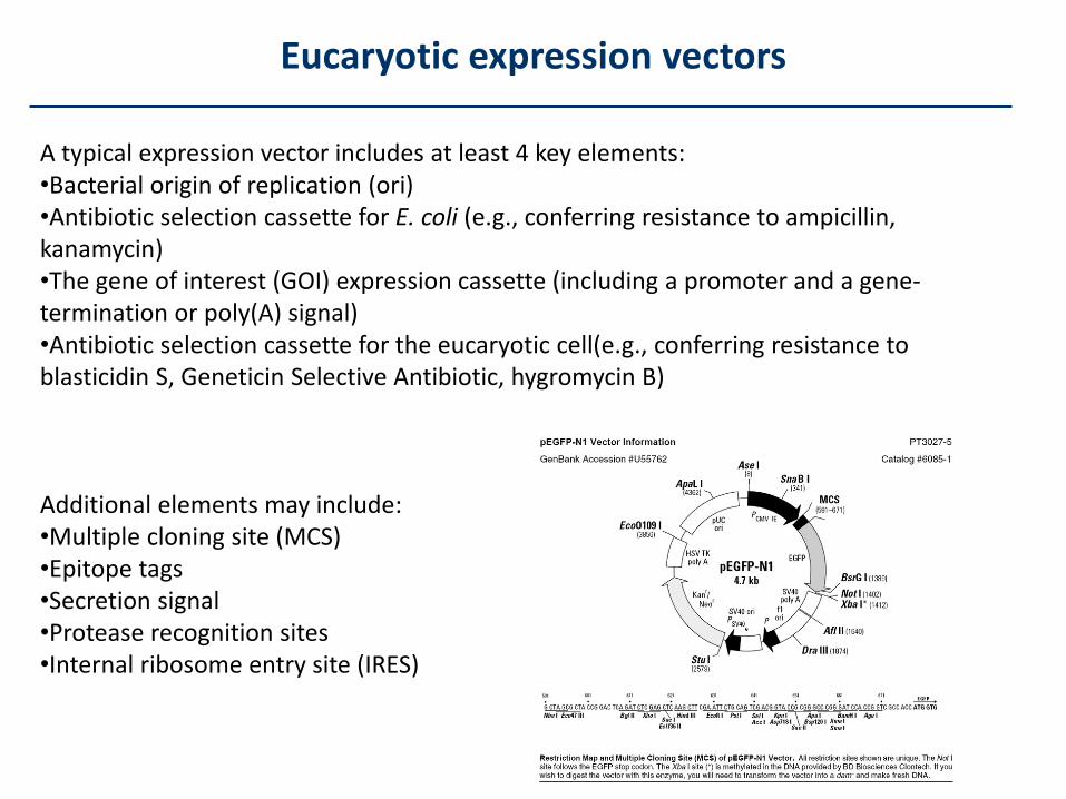
A typical expression vector includes at least 4 key elements:•Bacterial origin of replication (ori)•Antibiotic selection cassette for E. coli (e.g., conferring resistance to ampicillin, kanamycin)•The gene of interest (GOI) expression cassette (including a promoter and a gene-termination or poly(A) signal)•Antibiotic selection cassette for the eucaryotic cell(e.g., conferring resistance toblasticidin S, Geneticin Selective Antibiotic, hygromycin B)
Additional elements may include:•Multiple cloning site (MCS) •Epitope tags•Secretion signal•Protease recognition sites•Internal ribosome entry site (IRES)
Eucaryotic expression vectors
Experiment 1 – Receptor Internalization
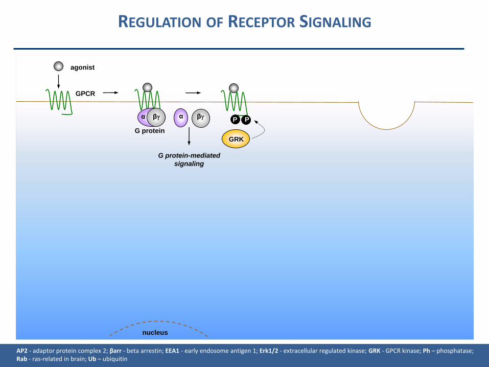
REGULATION OF RECEPTOR SIGNALING
nucleus
α β α β
G protein-mediated
signaling
GRK
P P
G protein
agonist
GPCR
AP2 - adaptor protein complex 2; βarr - beta arrestin; EEA1 - early endosome antigen 1; Erk1/2 - extracellular regulated kinase; GRK - GPCR kinase; Ph – phosphatase; Rab - ras-related in brain; Ub – ubiquitin
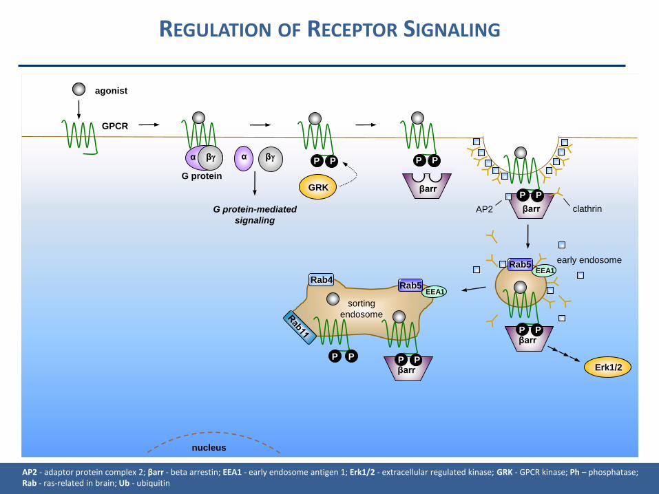
nucleus
sorting
endosome
Rab5EEA1
Rab5EEA1
α β α β
G protein-mediated
signaling
GRK
P P
βarr
βarr
P P
βarrP PP P
P P
G protein
agonist
GPCR
βarrP P
AP2 clathrin
early endosome
Erk1/2
Rab4
AP2 - adaptor protein complex 2; βarr - beta arrestin; EEA1 - early endosome antigen 1; Erk1/2 - extracellular regulated kinase; GRK - GPCR kinase; Ph – phosphatase; Rab - ras-related in brain; Ub - ubiquitin
REGULATION OF RECEPTOR SIGNALING
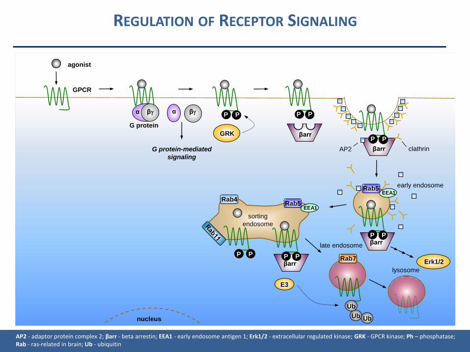
nucleus
lysosome
sorting
endosome
Rab5EEA1
Rab5EEA1
Rab7
α β α β
G protein-mediated
signaling
GRK
P P
βarr
βarr
P P
βarrP P
E3
Ub
Ub
Ub
P P
P P
G protein
agonist
GPCR
βarrP P
late endosome
AP2 clathrin
early endosome
Erk1/2
Rab4
AP2 - adaptor protein complex 2; βarr - beta arrestin; EEA1 - early endosome antigen 1; Erk1/2 - extracellular regulated kinase; GRK - GPCR kinase; Ph – phosphatase; Rab - ras-related in brain; Ub - ubiquitin
REGULATION OF RECEPTOR SIGNALING
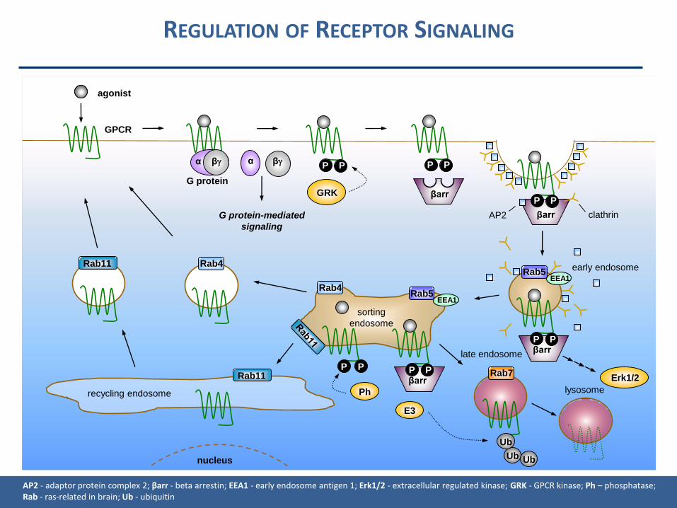
nucleus
lysosome
sorting
endosome
recycling endosome
Rab5EEA1
Rab5EEA1
Rab4
Rab7Rab11
Rab11
α β α β
G protein-mediated
signaling
GRK
P P
βarr
βarr
P P
βarrP P
E3
Ub
Ub
Ub
P P
P P
G protein
agonist
GPCR
Ph
βarrP P
late endosome
AP2 clathrin
early endosome
Erk1/2
Rab4
AP2 - adaptor protein complex 2; βarr - beta arrestin; EEA1 - early endosome antigen 1; Erk1/2 - extracellular regulated kinase; GRK - GPCR kinase; Ph – phosphatase; Rab - ras-related in brain; Ub - ubiquitin
REGULATION OF RECEPTOR SIGNALING

Impact of internalization on signaling
Desensitization of receptors Resensitization of receptors Degradation of receptors Different signalling pathways
Regulation of receptor density in the cellmembrane
Impact on therapeutical applications(e.g. biased ligands, cancer therapy)
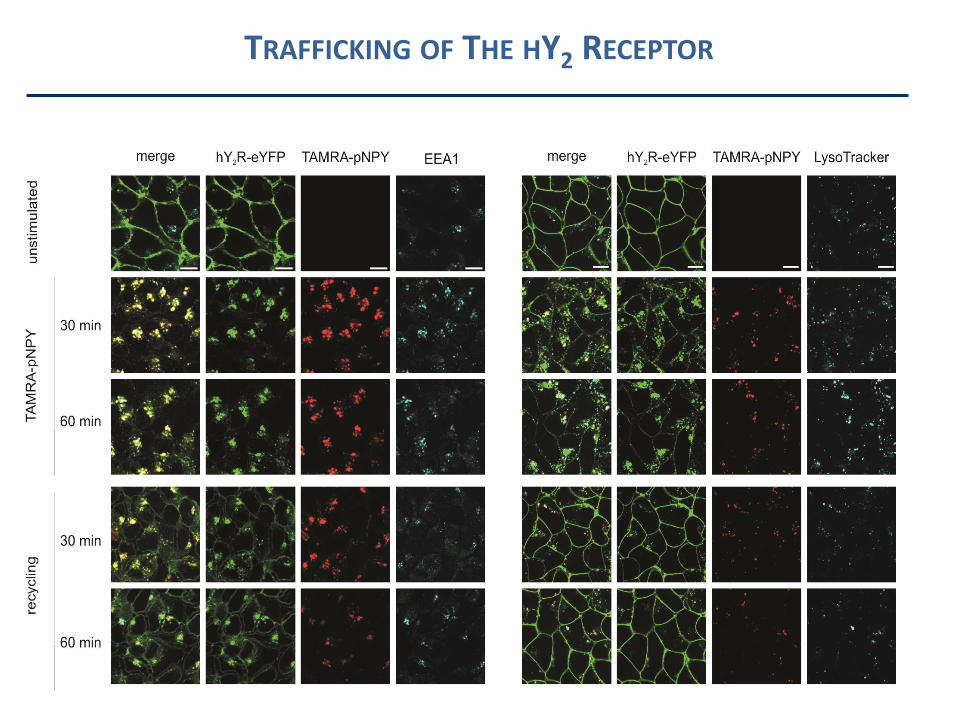
TRAFFICKING OF THE HY2 RECEPTOR
New synthesis or reycycling?
Cycloheximide
• fungicide produced by the bacterium Streptomyces griseus
• blocking eukaryotic translational elongation by interfering with the
translocation step in protein synthesis (movement of two tRNA molecules
and mRNA in relation to the ribosome)
Brefeldin A
• inhibits vesicle formation and transport between the endoplasmic
reticulum and the Golgi apparatus
TRAFFICKING OF THE HY2 RECEPTOR
Experiment 2 – HiBiT Tag for surface receptorquantification

HIBIT PROTEIN TAGGING SYSTEM – CLONING VECTOR
IL-6 Signal sequence
HiBiT Tag
YFP
HiBiT-Tag
HiBiT-hY1R-YFPHiBiT-hY2R-YFP
hY1R-YFPhY2R-YFP
To test: Control:
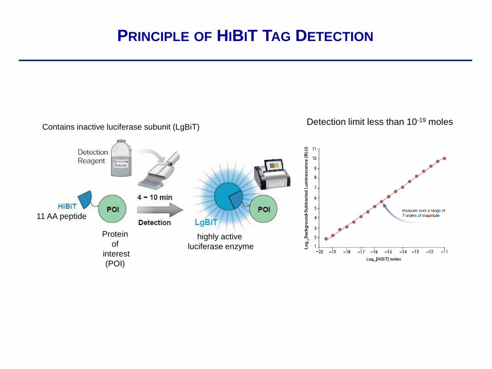
PRINCIPLE OF HIBIT TAG DETECTION
11 AA peptide
Protein
of
interest
(POI)
Contains inactive luciferase subunit (LgBiT)
highly active
luciferase enzyme
Detection limit less than 10-19 moles
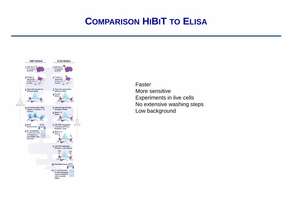
COMPARISON HIBIT TO ELISA
Faster
More sensitive
Experiments in live cells
No extensive washing steps
Low background
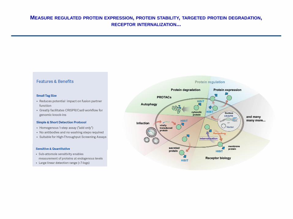
MEASURE REGULATED PROTEIN EXPRESSION, PROTEIN STABILITY, TARGETED PROTEIN DEGRADATION,
RECEPTOR INTERNALIZATION...
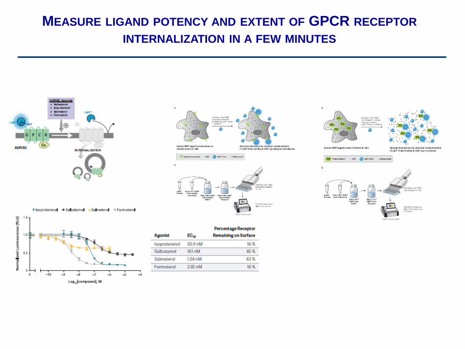
MEASURE LIGAND POTENCY AND EXTENT OF GPCR RECEPTOR
INTERNALIZATION IN A FEW MINUTES
Experiment 3 - Dimerization
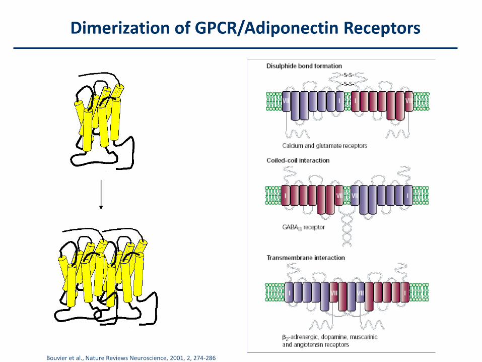
Dimerization of GPCR/Adiponectin Receptors
Bouvier et al., Nature Reviews Neuroscience, 2001, 2, 274-286
Bimolecular fluorescence complementation (BiFC)
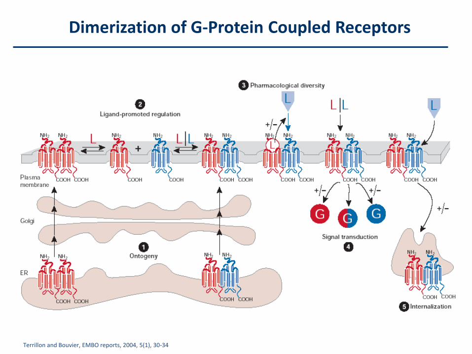
Dimerization of G-Protein Coupled Receptors
Terrillon and Bouvier, EMBO reports, 2004, 5(1), 30-34
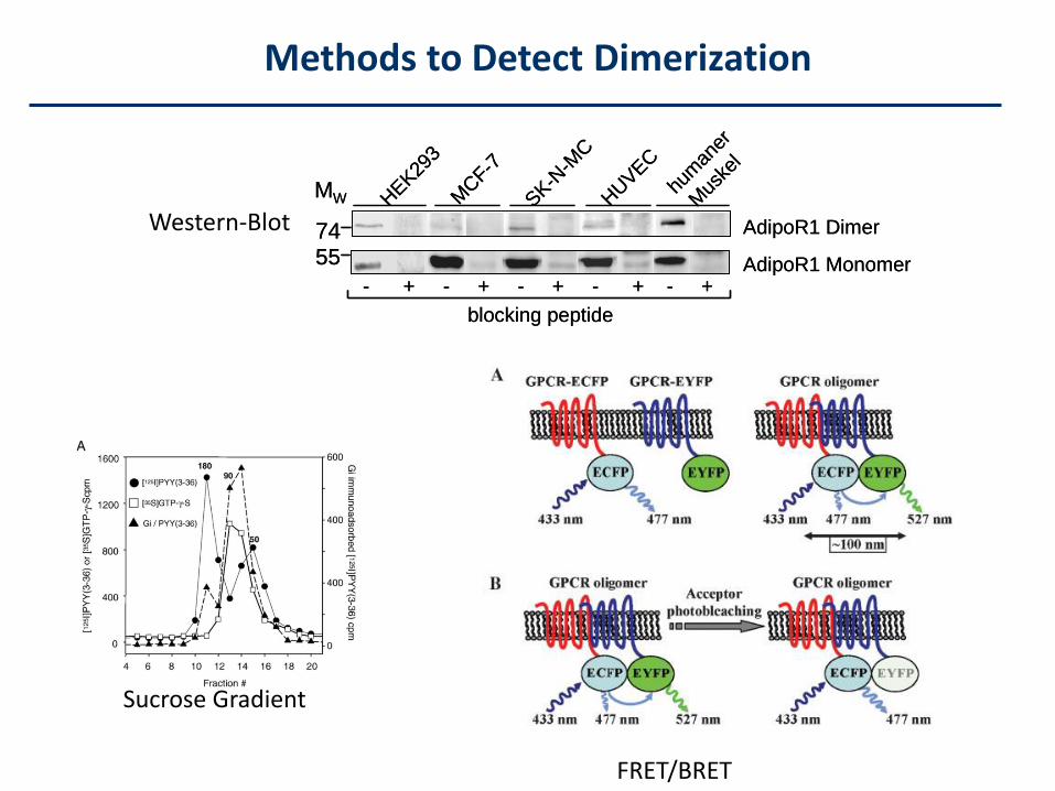
Methods to Detect Dimerization
Western-BlotM
CF-7
SK-N
-MC
HUVEC
hum
aner
Mus
kel
AdipoR1 Monomer
AdipoR1 Dimer
55
MW
- + - + - + - + - +
blocking peptide
74
HEK
293
MCF-7
SK-N
-MC
HUVEC
hum
aner
Mus
kel
AdipoR1 Monomer
AdipoR1 Dimer
55
MW
- + - + - + - + - +
blocking peptide
74
HEK
293
FRET/BRET
Sucrose Gradient
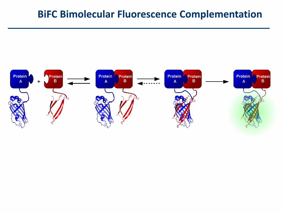
BiFC Bimolecular Fluorescence Complementation
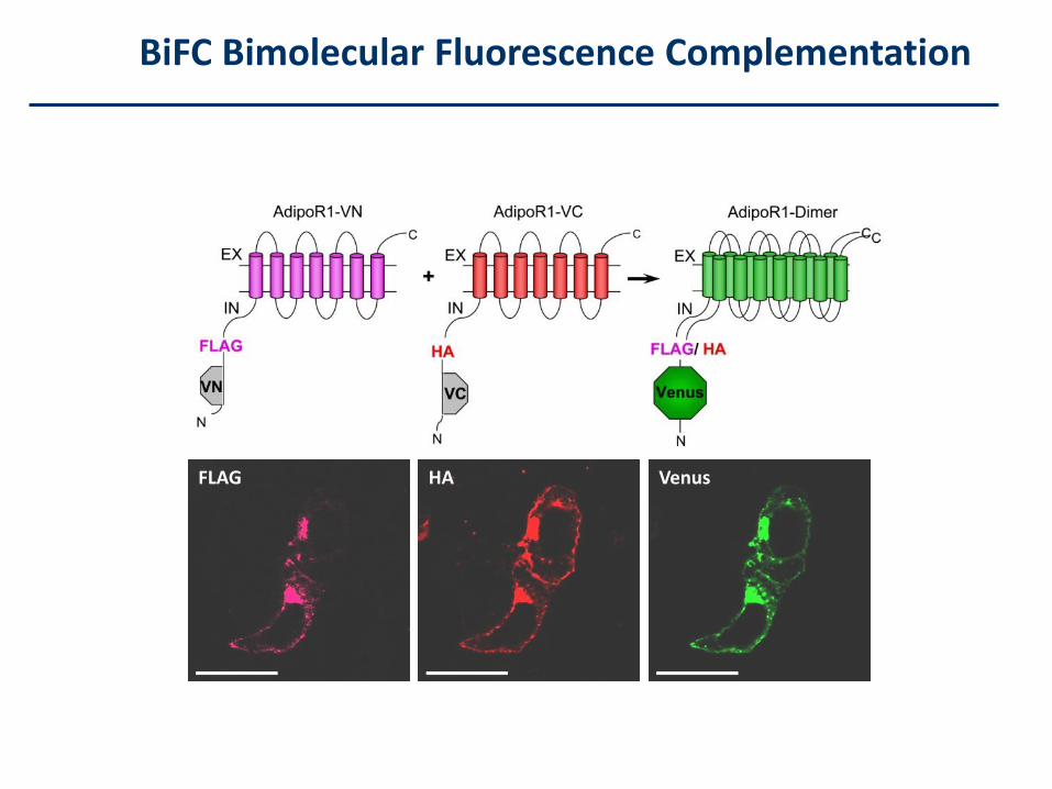
FLAG HA Venus
BiFC Bimolecular Fluorescence Complementation
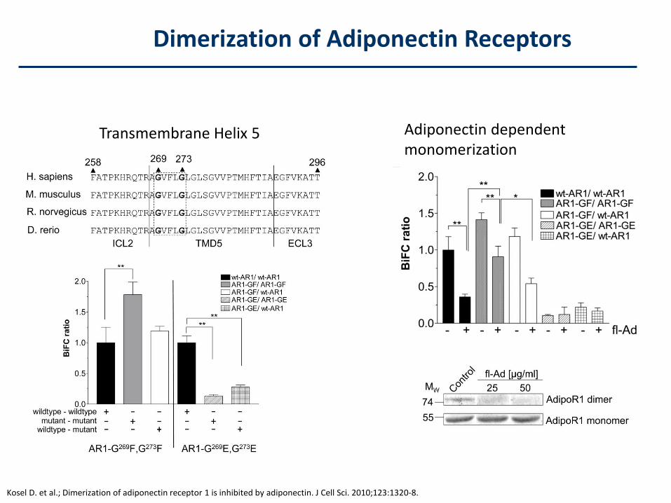
Transmembrane Helix 5 Adiponectin dependent monomerization
Dimerization of Adiponectin Receptors
Kosel D. et al.; Dimerization of adiponectin receptor 1 is inhibited by adiponectin. J Cell Sci. 2010;123:1320-8.
Experiment 4 – Ca2+ Assay
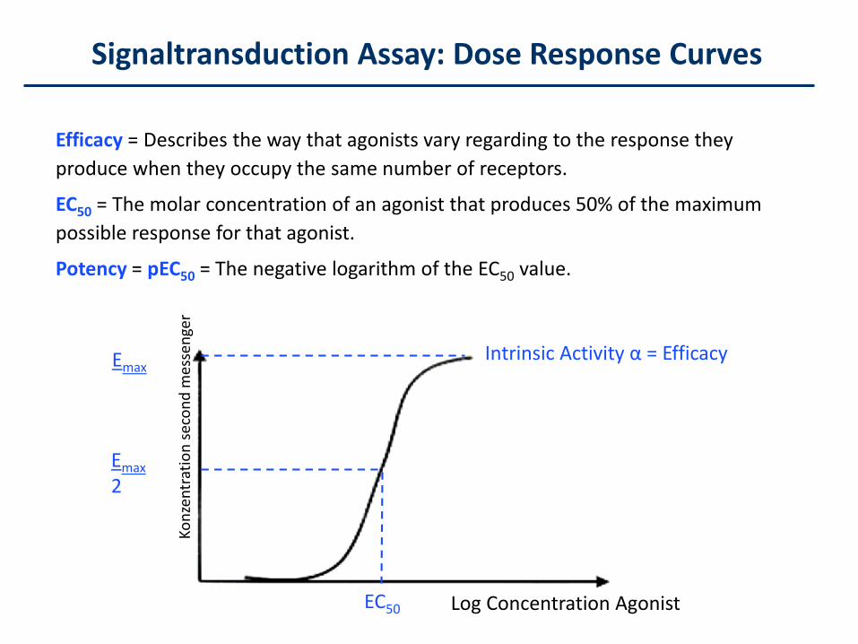
Efficacy = Describes the way that agonists vary regarding to the response they
produce when they occupy the same number of receptors.
EC50 = The molar concentration of an agonist that produces 50% of the maximum
possible response for that agonist.
Potency = pEC50 = The negative logarithm of the EC50 value.
Emax
2
Emax
EC50
Intrinsic Activity α = Efficacy
Signaltransduction Assay: Dose Response Curves
Log Concentration Agonist
Ko
nze
ntr
atio
n s
eco
nd
mes
sen
ger
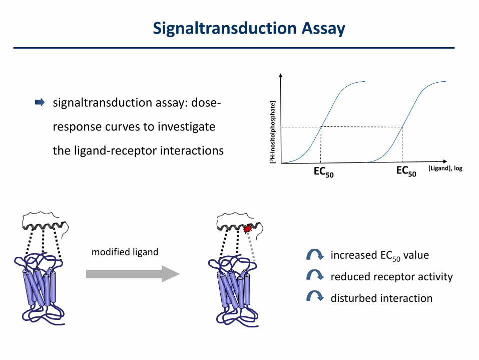
signaltransduction assay: dose-
response curves to investigate
the ligand-receptor interactions
EC50
[3H
-In
osi
tolp
ho
sph
ate
]
[Ligand], logEC50
modified ligand increased EC50 value
reduced receptor activity
disturbed interaction
Signaltransduction Assay
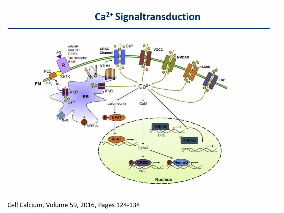
Cell Calcium, Volume 59, 2016, Pages 124-134
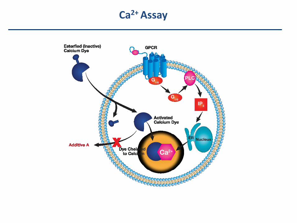
Ca2+ Signaltransduction
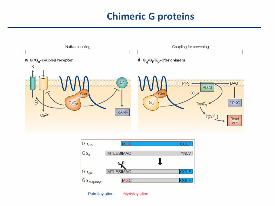
Chimeric G proteins
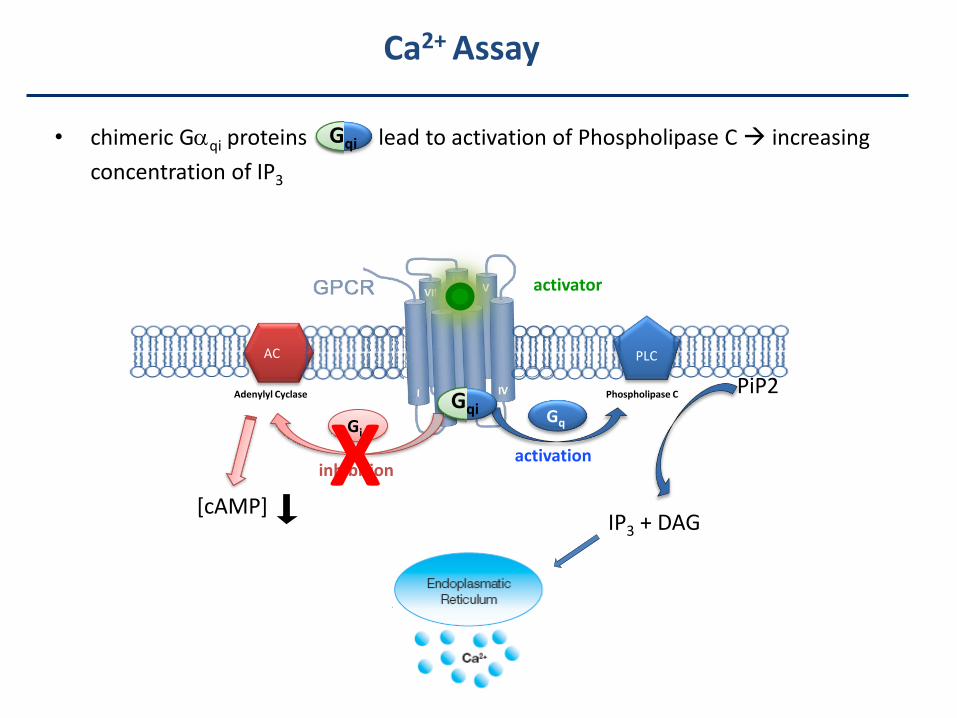
• chimeric Gqi proteins lead to activation of Phospholipase C increasing
concentration of IP3
activator
inhibition
Gi
[cAMP]
Adenylyl Cyclase
activation
Gq
IP3 + DAG
Gqi
PLC
PiP2
AC
Phospholipase C
Gqi
Ca2+ Assay
X
Ca2+ Assay

Experiment 5 – Reportergene assay
TOLL-like Receptors (TLR)
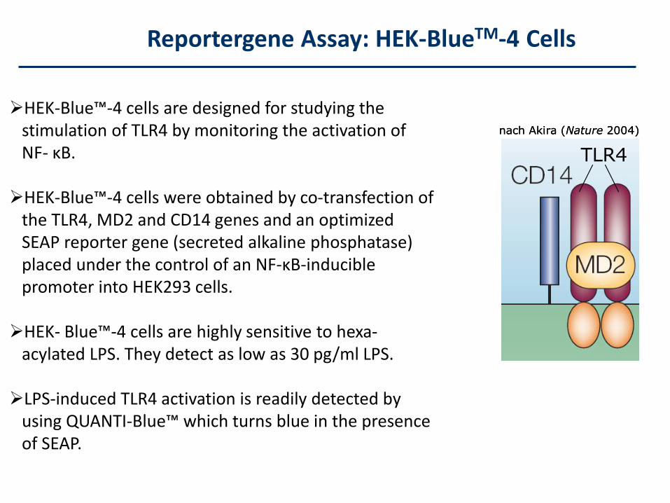
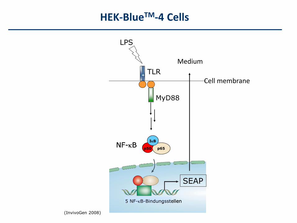
Reportergene Assay: HEK-BlueTM-4 Cells
HEK-Blue™-4 cells are designed for studying thestimulation of TLR4 by monitoring the activation ofNF- κB.
HEK-Blue™-4 cells were obtained by co-transfection of the TLR4, MD2 and CD14 genes and an optimizedSEAP reporter gene (secreted alkaline phosphatase)placed under the control of an NF-κB-inducible promoter into HEK293 cells.
HEK- Blue™-4 cells are highly sensitive to hexa-acylated LPS. They detect as low as 30 pg/ml LPS.
LPS-induced TLR4 activation is readily detected by using QUANTI-Blue™ which turns blue in the presenceof SEAP.
nach Akira (Nature 2004)
TLR4
nach Akira (Nature 2004)
TLR4
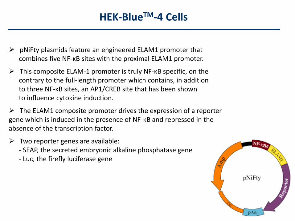
HEK-BlueTM-4 Cells
pNiFty plasmids feature an engineered ELAM1 promoter thatcombines five NF-κB sites with the proximal ELAM1 promoter.
This composite ELAM-1 promoter is truly NF-κB specific, on the contrary to the full-length promoter which contains, in additionto three NF-κB sites, an AP1/CREB site that has been shown to influence cytokine induction.
The ELAM1 composite promoter drives the expression of a reporter gene which is induced in the presence of NF-κB and repressed in the absence of the transcription factor.
Two reporter genes are available:- SEAP, the secreted embryonic alkaline phosphatase gene - Luc, the firefly luciferase gene
(InvivoGen 2008)
LPS
TLR
MyD88
p50 p65
IB
NF-Bp50 p65
IB
NF-B
SEAP
5 NF-B-Bindungsstellen5 NF-B-Bindungsstellen
HEK-BlueTM-4 Cells
Cell membrane
Medium
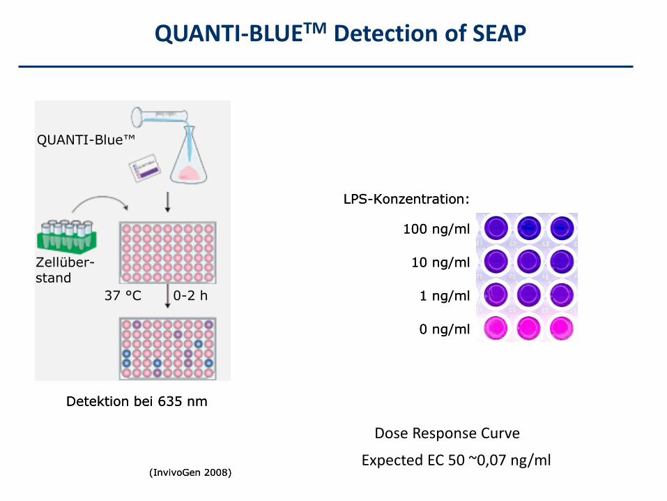
QUANTI-BLUETM Detection of SEAP
(InvivoGen 2008)
QUANTI-Blue™
37 °C 0-2 h
Detektion bei 635 nm
Zellüber-stand
(InvivoGen 2008)
QUANTI-Blue™
37 °C 0-2 h37 °C 0-2 h
Detektion bei 635 nm
Zellüber-stand
LPS-Konzentration:
100 ng/ml
10 ng/ml
1 ng/ml
0 ng/ml
LPS-Konzentration:
100 ng/ml
10 ng/ml
1 ng/ml
0 ng/ml
Dose Response Curve
Expected EC 50 ~0,07 ng/ml
(InvivoGen 2008)
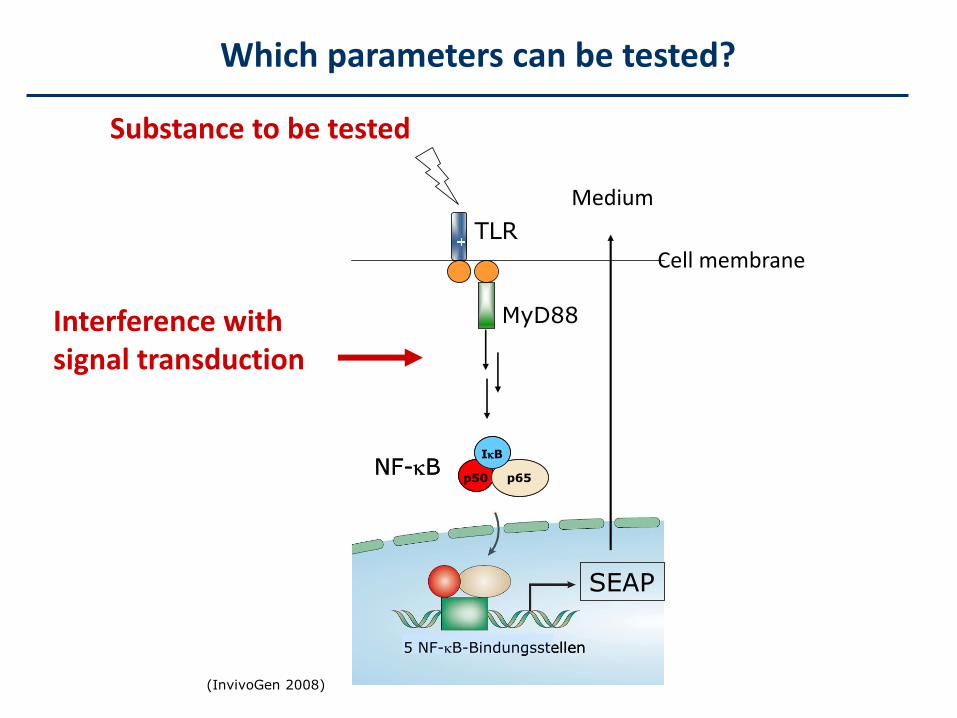
LPS
TLR
MyD88
p50 p65
IB
NF-Bp50 p65
IB
NF-B
SEAP
5 NF-B-Bindungsstellen5 NF-B-Bindungsstellen
Which parameters can be tested?
Cell membrane
Medium
Substance to be tested
Interference with signal transduction
Experiment 6 - Mutagenesis
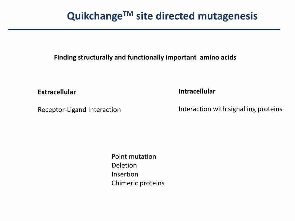
QuikchangeTM site directed mutagenesis
Finding structurally and functionally important amino acids
Extracellular
Receptor-Ligand Interaction
Intracellular
Interaction with signalling proteins
Point mutationDeletionInsertionChimeric proteins
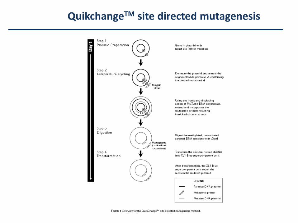
QuikchangeTM site directed mutagenesis
Recommended