Sam Beddar, Professor The University of Texas MD Anderson Cancer Center &
The University of Texas Graduate School of Biomedical Sciences
Plastic Scintillation Detectors: Present Status and Their Application for Quality Assurance
and In Vivo Dosimetry
Luc Beaulieu, Professor Department of Physics, Université Laval
Department of Radiation Oncology, CHU de Québec
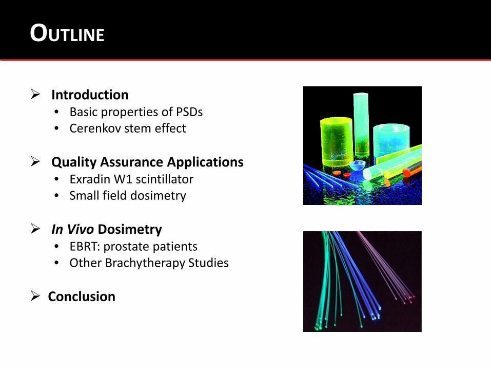
Introduction • Basic properties of PSDs • Cerenkov stem effect
Quality Assurance Applications
• Exradin W1 scintillator • Small field dosimetry
In Vivo Dosimetry
• EBRT: prostate patients • Other Brachytherapy Studies
Conclusion
OUTLINE
• The University of Texas MD Anderson Cancer Center & l’Université Laval have one license agreement with Standard Imaging.
• SB & LB had an NIH/NCI SBIR Phase I grant (1R43 CA153824-01) with Standard Imaging.
• LB & SB had Sponsored Research Agreement with Standard Imaging.
• SB had phase I, II, III Sponsored Research Agreements with Radiadyne, LLC.
• Scintillating Fiber Dosimetry Arrays. US Patent: 8,183,534, Date Issued:
May 22, 2012. • Real-time in vivo Dosimetry Using Scintillation Detectors. US Patent:
61/143,294 filed on 01/08/2009, pending.
DISCLOSURES
LECTURES SERIES AT AAPM
• AAPM 2008 --- introducing PSDs, basics & properties “Scintillation Dosimetry: Review, New Innovations and Applications”
• AAPM 2010 --- further studies & validation of PSDs “Scintillation Dosimetry: From Plastics to Liquids and from Photons/Electrons to Protons”
• AAPM 2013 --- application of commercial PSDs “PSDs: Present Status and their Applications for Quality Assurance and In Vivo Dosimetry”

Introduction &
Present Status
Introduction Quality Assurance Small Field Dosimetry In Vivo Dosimetry Conclusion
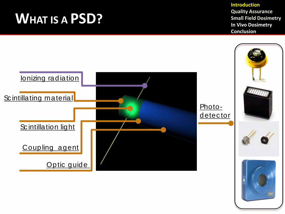
WHAT IS A PSD?
Scintillating material
Ionizing radiation
Scintillation light
Optic guide
Coupling agent
Photo-detector
Introduction Quality Assurance Small Field Dosimetry In Vivo Dosimetry Conclusion
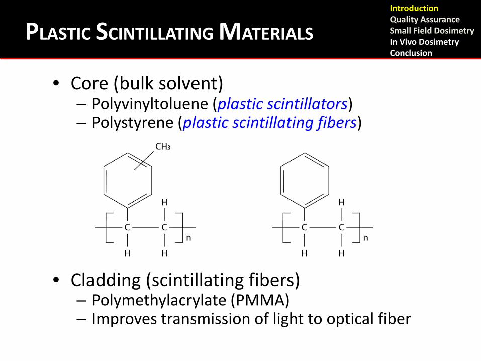
• Core (bulk solvent) – Polyvinyltoluene (plastic scintillators) – Polystyrene (plastic scintillating fibers)
• Cladding (scintillating fibers) – Polymethylacrylate (PMMA) – Improves transmission of light to optical fiber
Introduction Quality Assurance Small Field Dosimetry In Vivo Dosimetry Conclusion
PLASTIC SCINTILLATING MATERIALS
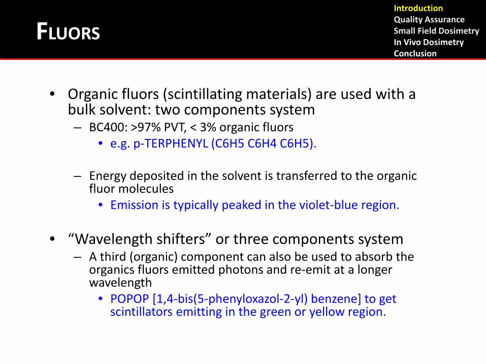
• Organic fluors (scintillating materials) are used with a bulk solvent: two components system – BC400: >97% PVT, < 3% organic fluors
• e.g. p-TERPHENYL (C6H5 C6H4 C6H5).
– Energy deposited in the solvent is transferred to the organic fluor molecules
• Emission is typically peaked in the violet-blue region.
• “Wavelength shifters” or three components system – A third (organic) component can also be used to absorb the
organics fluors emitted photons and re-emit at a longer wavelength
• POPOP [1,4-bis(5-phenyloxazol-2-yl) benzene] to get scintillators emitting in the green or yellow region.
FLUORS Introduction Quality Assurance Small Field Dosimetry In Vivo Dosimetry Conclusion
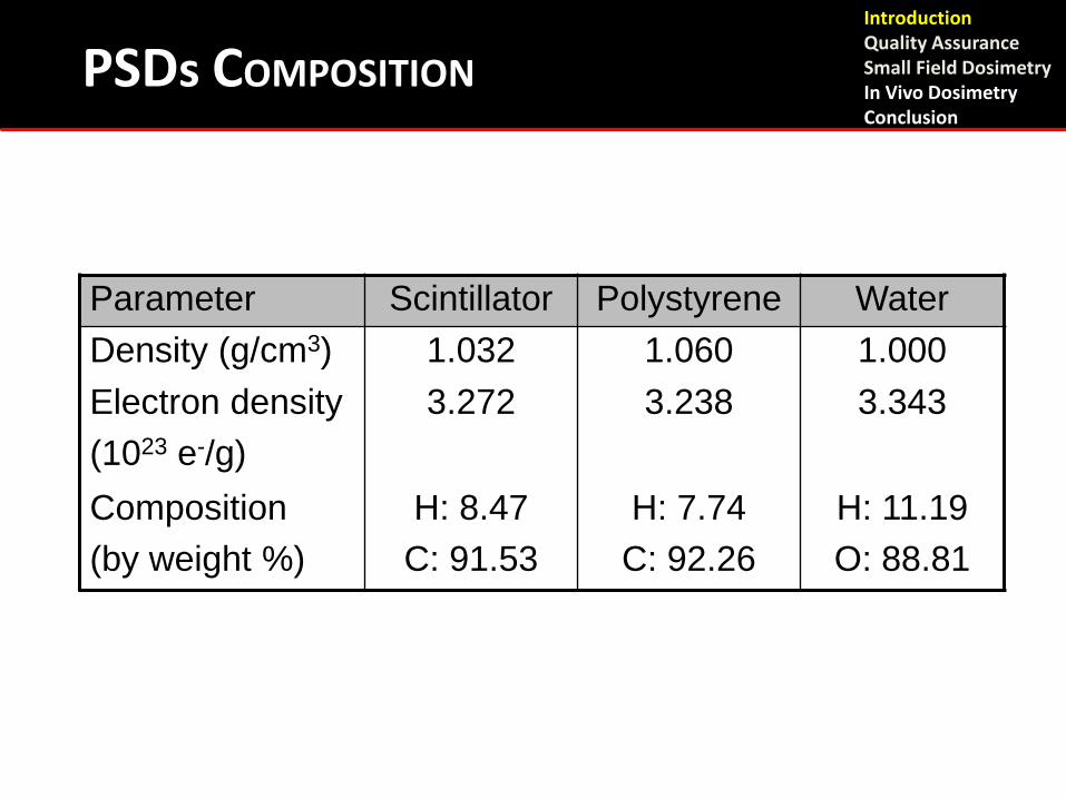
PSDs COMPOSITION
Parameter Scintillator Polystyrene Water Density (g/cm3) 1.032 1.060 1.000 Electron density (1023 e-/g)
3.272 3.238 3.343
Composition (by weight %)
H: 8.47 C: 91.53
H: 7.74 C: 92.26
H: 11.19 O: 88.81
Introduction Quality Assurance Small Field Dosimetry In Vivo Dosimetry Conclusion
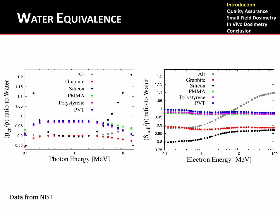
WATER EQUIVALENCE
Data from NIST
Introduction Quality Assurance Small Field Dosimetry In Vivo Dosimetry Conclusion
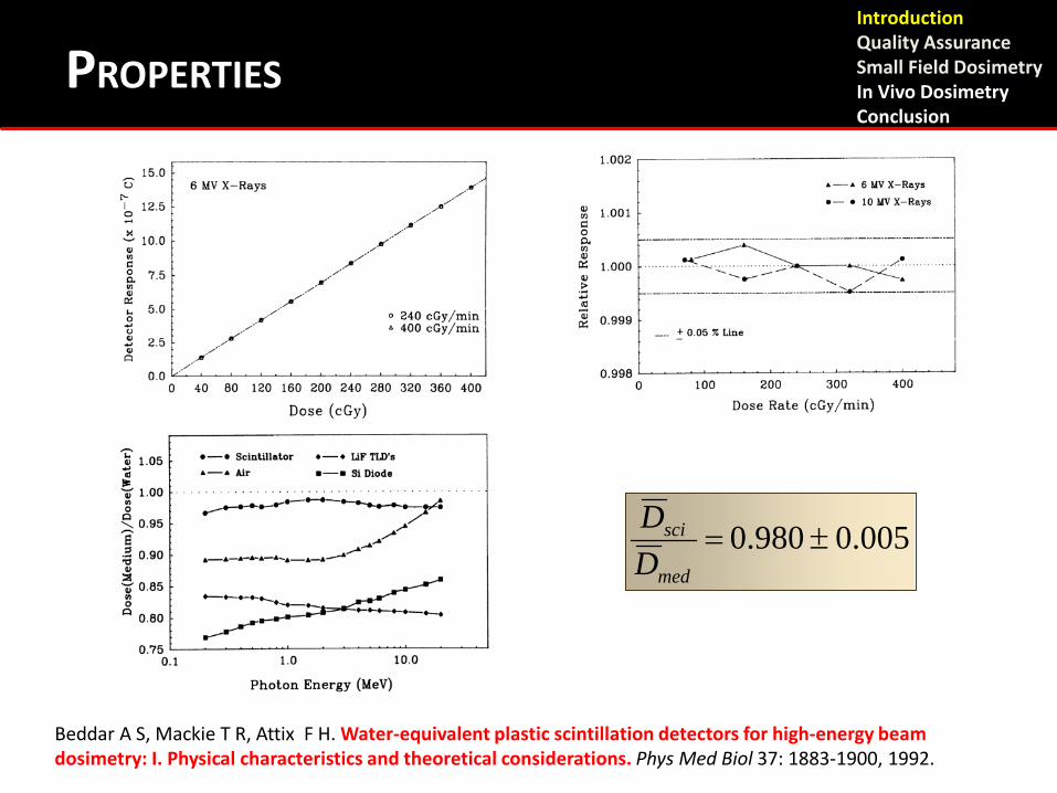
PROPERTIES
Beddar A S, Mackie T R, Attix F H. Water-equivalent plastic scintillation detectors for high-energy beam dosimetry: I. Physical characteristics and theoretical considerations. Phys Med Biol 37: 1883-1900, 1992.
0.005 0.980 ±=med
sci
DD
Introduction Quality Assurance Small Field Dosimetry In Vivo Dosimetry Conclusion
• A decrease from the optimal scintillation efficiency, or quenching, can occur under various conditions
– For organic scintillators, possible thermal quenching
– Radiation damage can decrease the efficiency (Ionizations lead to temporary and/or permanent molecular
damage) • Increased absorption due to defects (plastics turn yellow) • Need > kGy accumulated doses (104 to 105 Gy)
– High LET: proton and ion beams
• Overlapping excitation sites and molecule damages
QUENCHING EFFECT Introduction Quality Assurance Small Field Dosimetry In Vivo Dosimetry Conclusion
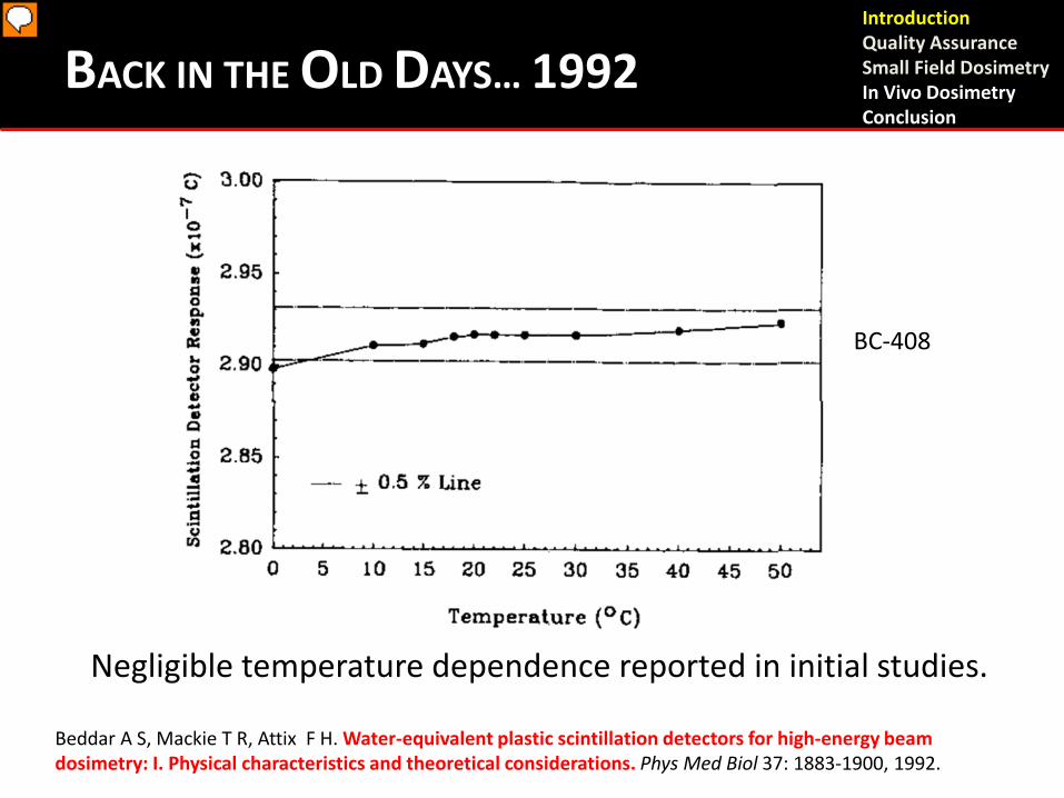
BACK IN THE OLD DAYS… 1992
Negligible temperature dependence reported in initial studies.
BC-408
Introduction Quality Assurance Small Field Dosimetry In Vivo Dosimetry Conclusion
Beddar A S, Mackie T R, Attix F H. Water-equivalent plastic scintillation detectors for high-energy beam dosimetry: I. Physical characteristics and theoretical considerations. Phys Med Biol 37: 1883-1900, 1992.
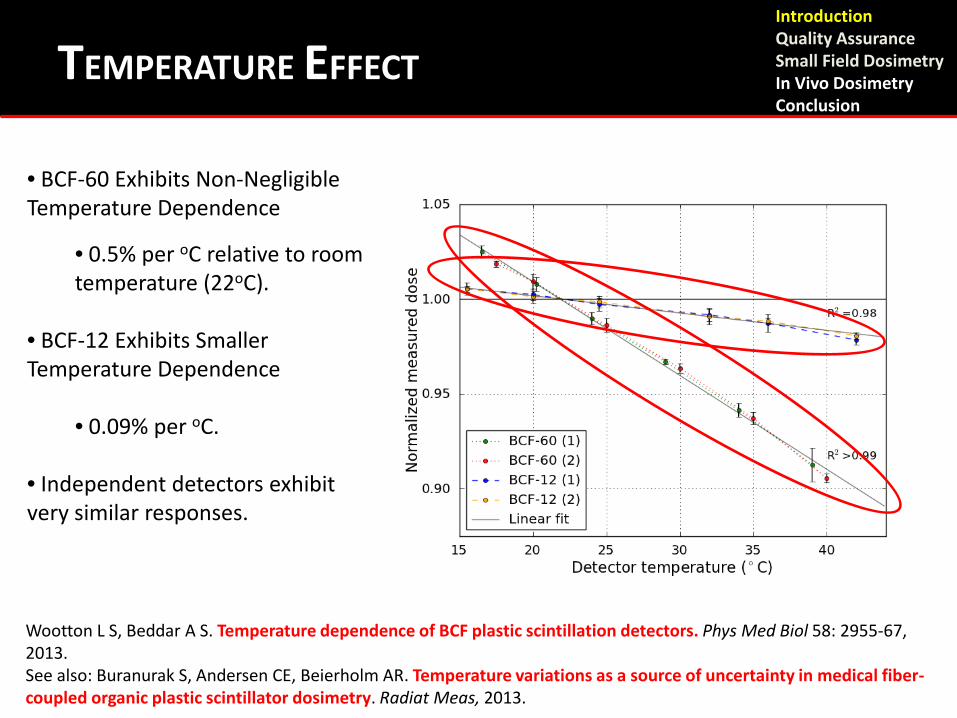
TEMPERATURE EFFECT
• BCF-60 Exhibits Non-Negligible Temperature Dependence
• 0.5% per oC relative to room temperature (22oC).
• BCF-12 Exhibits Smaller Temperature Dependence
• 0.09% per oC.
• Independent detectors exhibit very similar responses.
Wootton L S, Beddar A S. Temperature dependence of BCF plastic scintillation detectors. Phys Med Biol 58: 2955-67, 2013. See also: Buranurak S, Andersen CE, Beierholm AR. Temperature variations as a source of uncertainty in medical fiber-coupled organic plastic scintillator dosimetry. Radiat Meas, 2013.
Introduction Quality Assurance Small Field Dosimetry In Vivo Dosimetry Conclusion

• JB Birks, The Theory and Practice of Scintillation Counting,
Pergamon Press Book, MacMillan, New York, 1964. [Chapters 3 and 6]
• GF Knoll, Radiation Detection and Measurement, 3rd Edition, John Wiley and Sons, 2000. [Chapter 8]
• WR Leo, Techniques for Nuclear and Particle Physics Experiments, 2nd edition, Springer-Verlag, 1992. [Chapter 7]
• FH Attix, Introduction to Radiological Physics and Radiation Dosimetry, John Wiley and Sons, 1986. [Chapter 15]
SCINTILLATION PROCESS : REFERENCES Introduction Quality Assurance Small Field Dosimetry In Vivo Dosimetry Conclusion

The Čerenkov Challenge
Introduction Quality Assurance Small Field Dosimetry In Vivo Dosimetry Conclusion
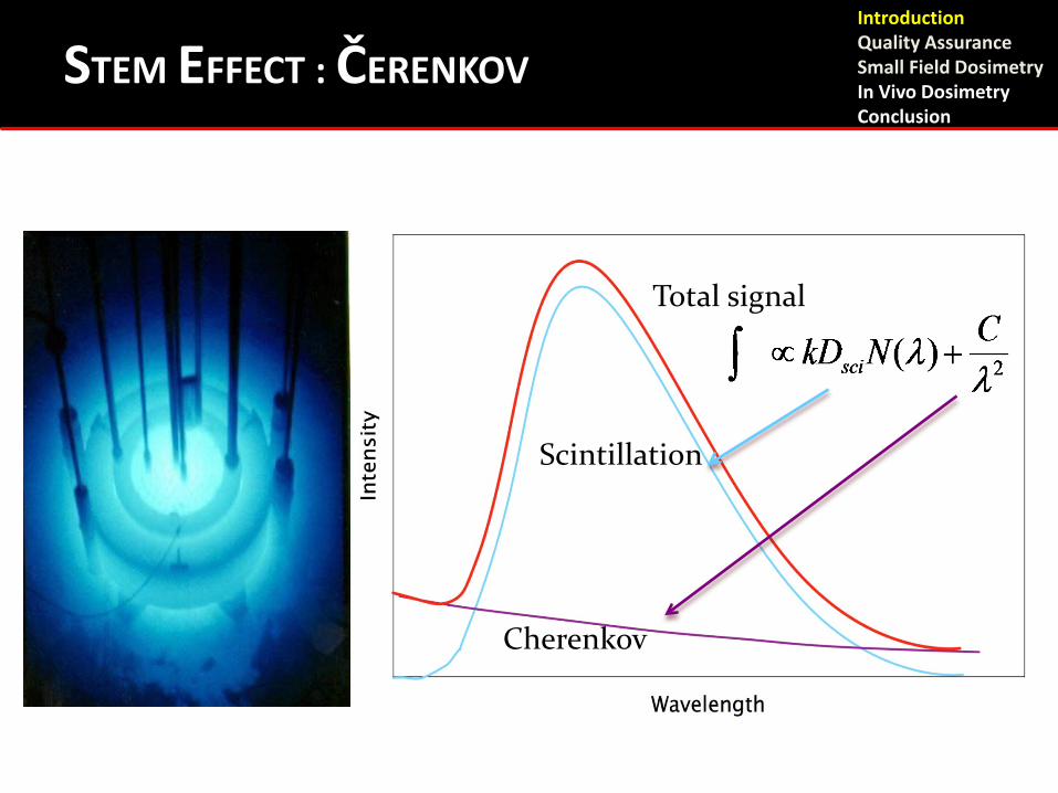
Total signal
Scintillation
Cherenkov
STEM EFFECT : ČERENKOV Introduction Quality Assurance Small Field Dosimetry In Vivo Dosimetry Conclusion
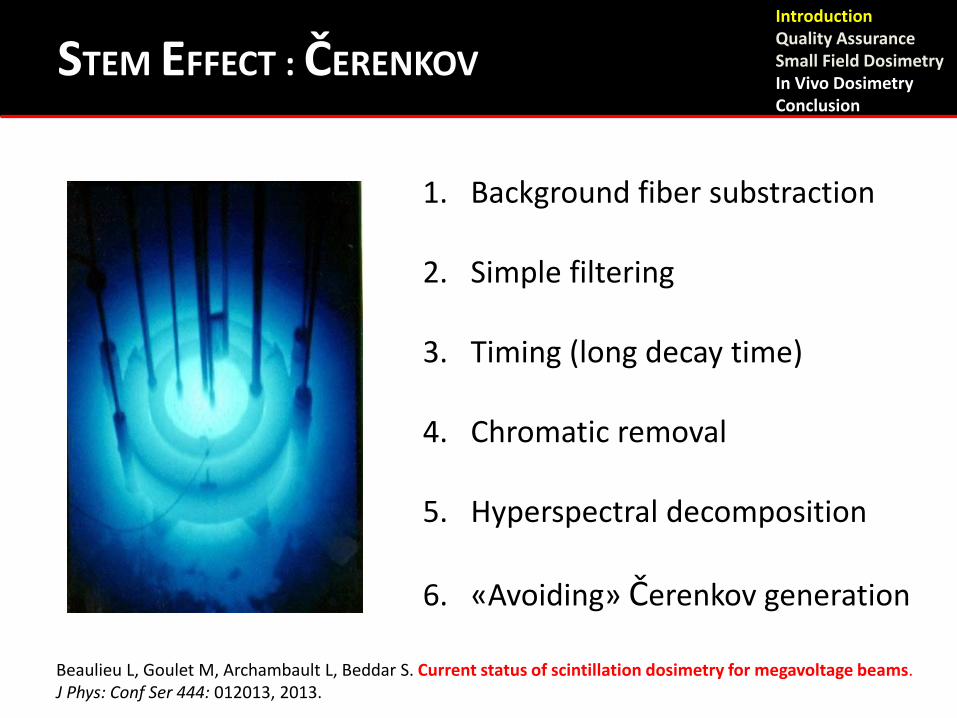
1. Background fiber substraction
2. Simple filtering
3. Timing (long decay time)
4. Chromatic removal
5. Hyperspectral decomposition
6. «Avoiding» Čerenkov generation
Beaulieu L, Goulet M, Archambault L, Beddar S. Current status of scintillation dosimetry for megavoltage beams. J Phys: Conf Ser 444: 012013, 2013.
Introduction Quality Assurance Small Field Dosimetry In Vivo Dosimetry Conclusion
STEM EFFECT : ČERENKOV
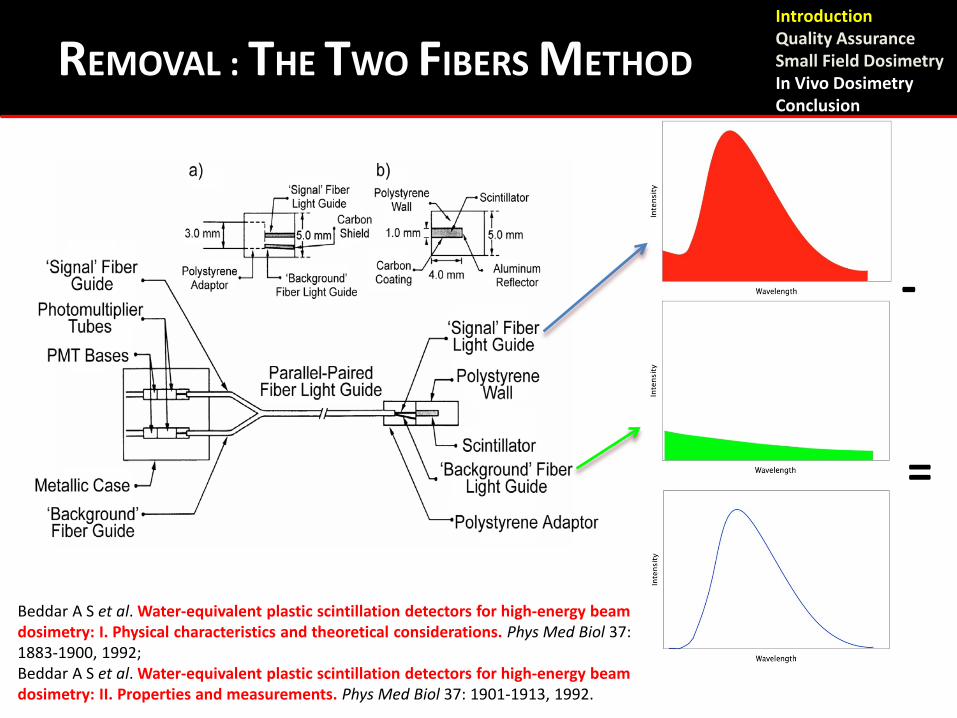
-
=
REMOVAL : THE TWO FIBERS METHOD Introduction Quality Assurance Small Field Dosimetry In Vivo Dosimetry Conclusion
Beddar A S et al. Water-equivalent plastic scintillation detectors for high-energy beam dosimetry: I. Physical characteristics and theoretical considerations. Phys Med Biol 37: 1883-1900, 1992; Beddar A S et al. Water-equivalent plastic scintillation detectors for high-energy beam dosimetry: II. Properties and measurements. Phys Med Biol 37: 1901-1913, 1992.
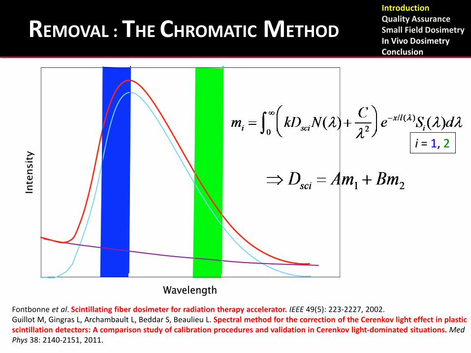
i = 1, 2
Fontbonne et al. Scintillating fiber dosimeter for radiation therapy accelerator. IEEE 49(5): 223-2227, 2002. Guillot M, Gingras L, Archambault L, Beddar S, Beaulieu L. Spectral method for the correction of the Cerenkov light effect in plastic scintillation detectors: A comparison study of calibration procedures and validation in Cerenkov light-dominated situations. Med Phys 38: 2140-2151, 2011.
Introduction Quality Assurance Small Field Dosimetry In Vivo Dosimetry Conclusion
REMOVAL : THE CHROMATIC METHOD
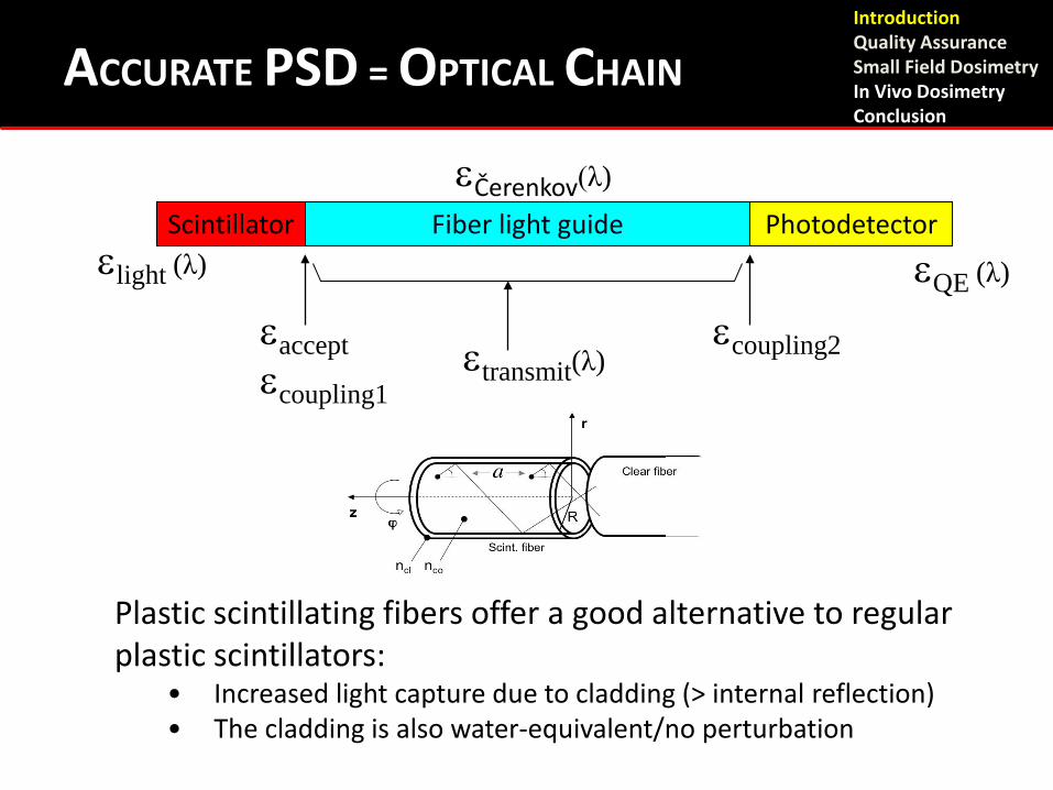
Scintillator Fiber light guide Photodetector
εaccept εcoupling1
εtransmit(λ)
εcoupling2
εlight (λ)
εQE (λ)
εČerenkov(λ)
ACCURATE PSD = OPTICAL CHAIN
Plastic scintillating fibers offer a good alternative to regular plastic scintillators:
• Increased light capture due to cladding (> internal reflection) • The cladding is also water-equivalent/no perturbation
Introduction Quality Assurance Small Field Dosimetry In Vivo Dosimetry Conclusion
Linear response to dose
Dose rate independence
Energy independence
Particle type independence for photons and electrons
Insensitive to RF fields
Real-time readout
Spatial resolution
ADVANTAGES OF PLASTIC SCINTILLATORS Introduction Quality Assurance Small Field Dosimetry In Vivo Dosimetry Conclusion

Quality Assurance
Introduction Quality Assurance Small Field Dosimetry In Vivo Dosimetry Conclusion
No time to go over all PSD-based devices proposed in the literature. A recent review can be found here:
Beaulieu L, Goulet M, Archambault L, Beddar S. Current status of scintillation dosimetry for megavoltage beams. J Phys : Conf Ser 444, 012013, 2013.
QA REFERENCES Introduction Quality Assurance Small Field Dosimetry In Vivo Dosimetry Conclusion
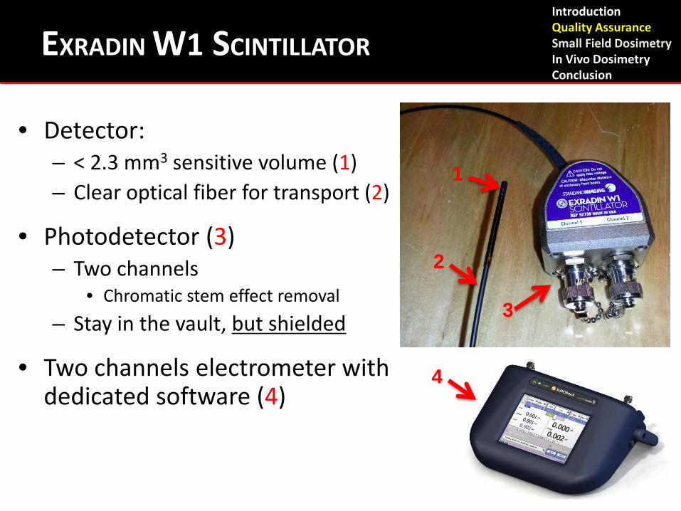
EXRADIN W1 SCINTILLATOR
• Detector: – < 2.3 mm3 sensitive volume (1) – Clear optical fiber for transport (2)
• Photodetector (3) – Two channels
• Chromatic stem effect removal – Stay in the vault, but shielded
• Two channels electrometer with dedicated software (4)
1
2
3
4
Introduction Quality Assurance Small Field Dosimetry In Vivo Dosimetry Conclusion
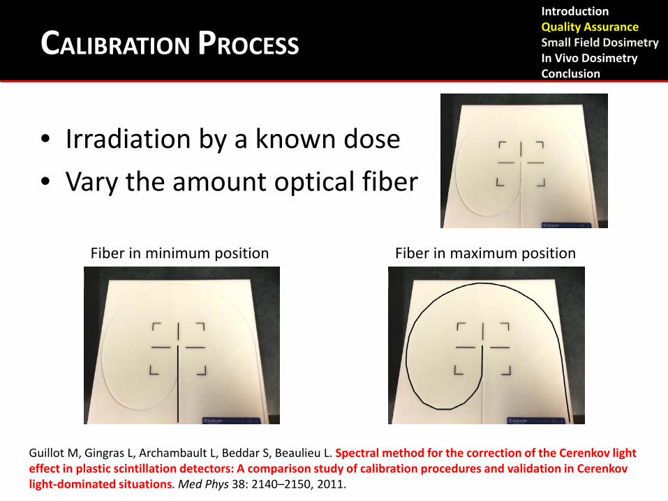
CALIBRATION PROCESS
• Irradiation by a known dose • Vary the amount optical fiber
The calibration phantom
Fiber in minimum position Fiber in maximum position
Guillot M, Gingras L, Archambault L, Beddar S, Beaulieu L. Spectral method for the correction of the Cerenkov light effect in plastic scintillation detectors: A comparison study of calibration procedures and validation in Cerenkov light-dominated situations. Med Phys 38: 2140–2150, 2011.
Introduction Quality Assurance Small Field Dosimetry In Vivo Dosimetry Conclusion
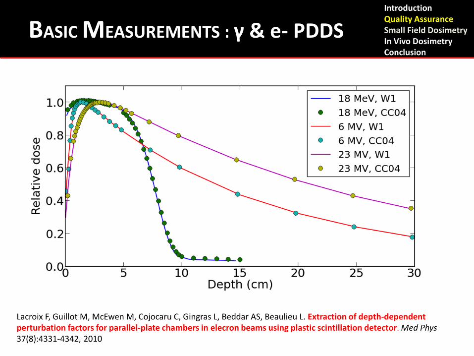
BASIC MEASUREMENTS : γ & e- PDDS
Lacroix F, Guillot M, McEwen M, Cojocaru C, Gingras L, Beddar AS, Beaulieu L. Extraction of depth-dependent perturbation factors for parallel-plate chambers in elecron beams using plastic scintillation detector. Med Phys 37(8):4331-4342, 2010
Introduction Quality Assurance Small Field Dosimetry In Vivo Dosimetry Conclusion
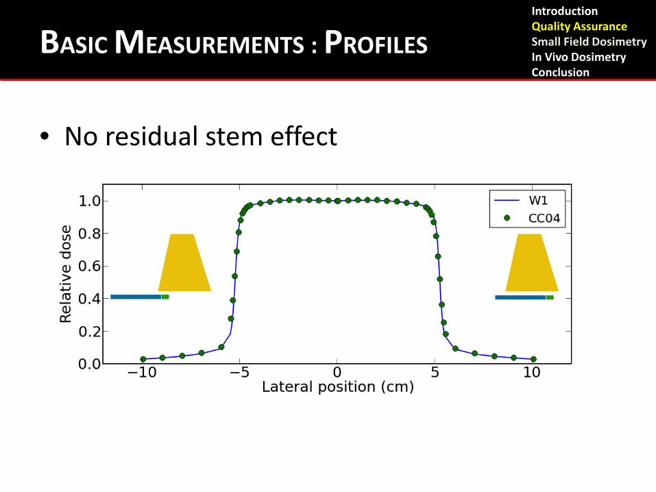
BASIC MEASUREMENTS : PROFILES
• No residual stem effect
Introduction Quality Assurance Small Field Dosimetry In Vivo Dosimetry Conclusion
Small Field Dosimetry
Introduction Quality Assurance Small Field Dosimetry In Vivo Dosimetry Conclusion
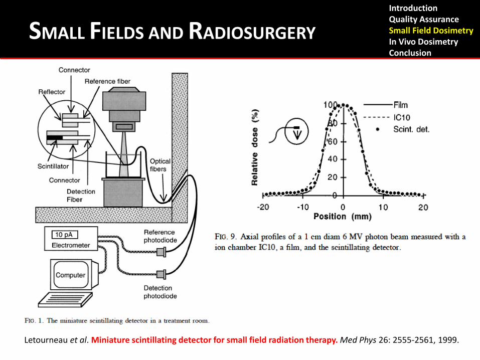
SMALL FIELDS AND RADIOSURGERY
Letourneau et al. Miniature scintillating detector for small field radiation therapy. Med Phys 26: 2555-2561, 1999.
Introduction Quality Assurance Small Field Dosimetry In Vivo Dosimetry Conclusion
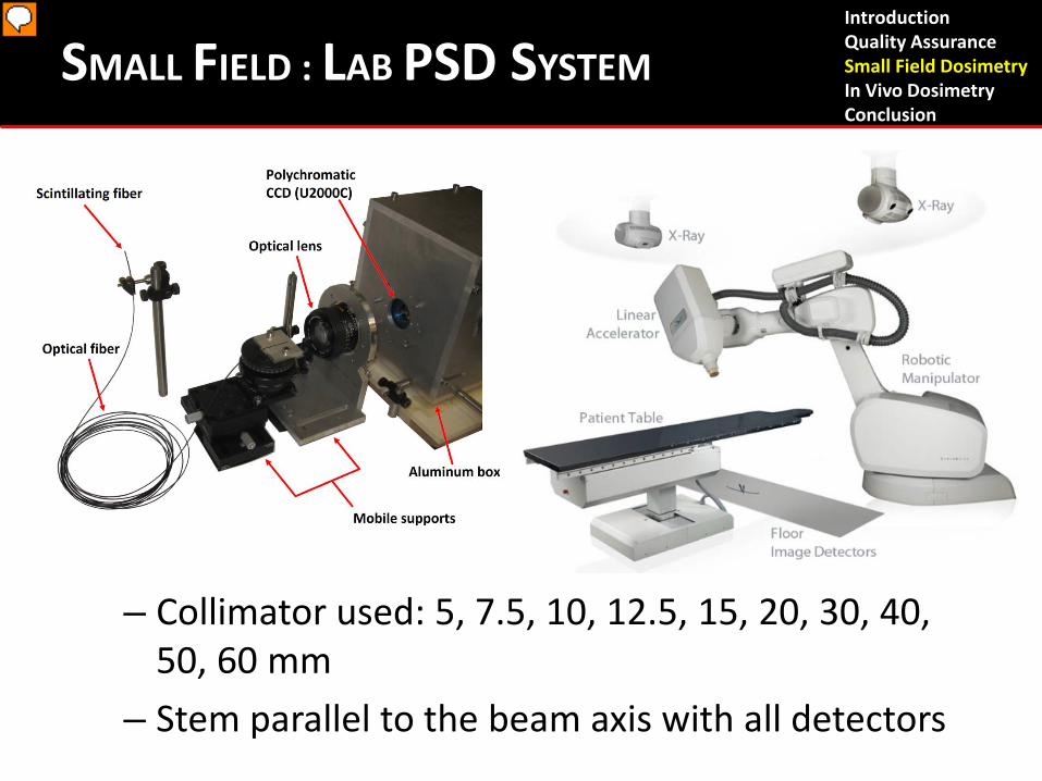
SMALL FIELD : LAB PSD SYSTEM
– Collimator used: 5, 7.5, 10, 12.5, 15, 20, 30, 40, 50, 60 mm
– Stem parallel to the beam axis with all detectors
Introduction Quality Assurance Small Field Dosimetry In Vivo Dosimetry Conclusion
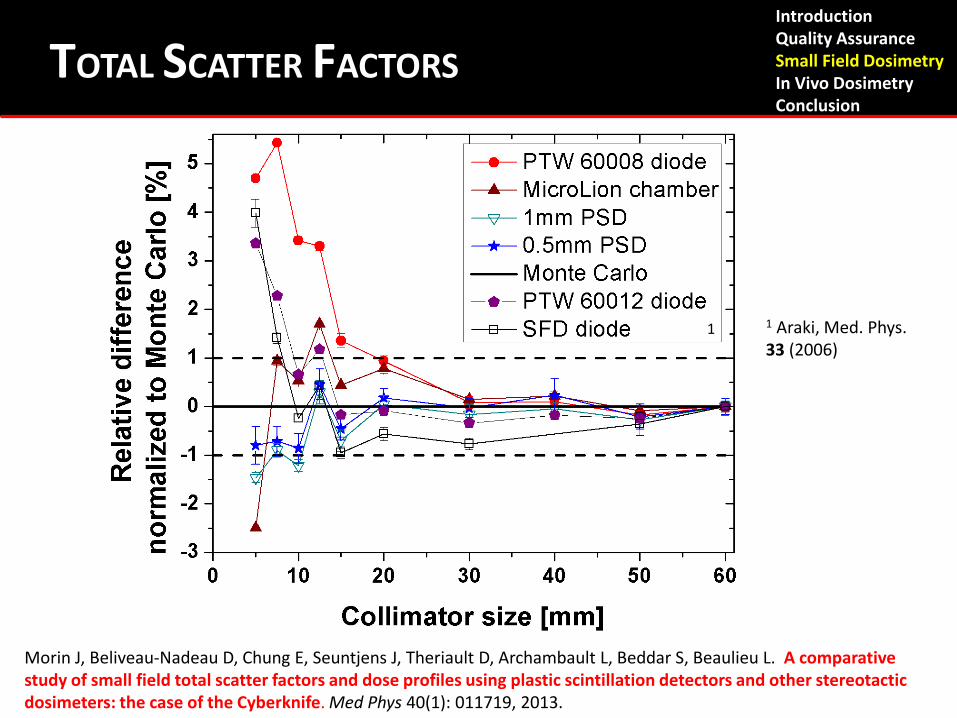
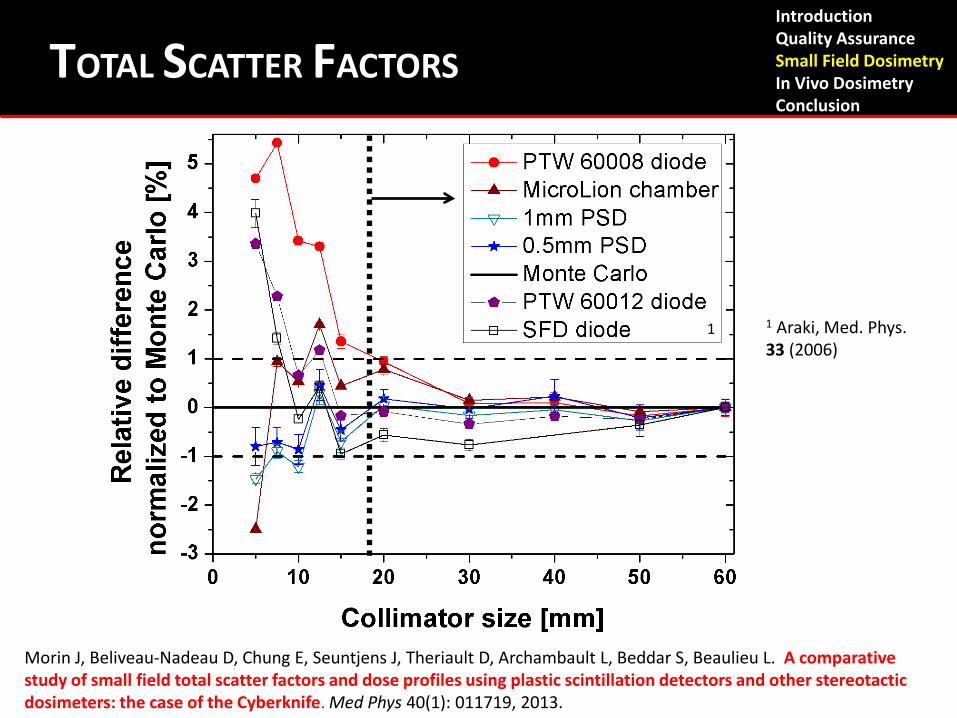
Total scatter factors
1 Araki, Med. Phys. 33 (2006)
1
Morin J, Beliveau-Nadeau D, Chung E, Seuntjens J, Theriault D, Archambault L, Beddar S, Beaulieu L. A comparative study of small field total scatter factors and dose profiles using plastic scintillation detectors and other stereotactic dosimeters: the case of the Cyberknife. Med Phys 40(1): 011719, 2013.
TOTAL SCATTER FACTORS Introduction Quality Assurance Small Field Dosimetry In Vivo Dosimetry Conclusion
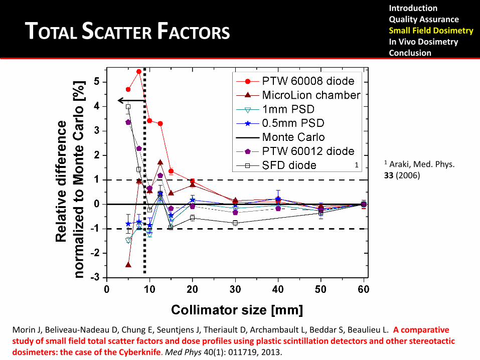
Total scatter factors
1 Araki, Med. Phys. 33 (2006)
1
Introduction Quality Assurance Small Field Dosimetry In Vivo Dosimetry Conclusion
TOTAL SCATTER FACTORS
Morin J, Beliveau-Nadeau D, Chung E, Seuntjens J, Theriault D, Archambault L, Beddar S, Beaulieu L. A comparative study of small field total scatter factors and dose profiles using plastic scintillation detectors and other stereotactic dosimeters: the case of the Cyberknife. Med Phys 40(1): 011719, 2013.
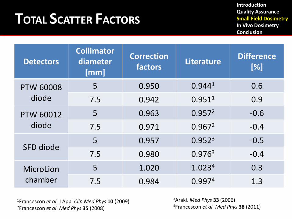
Detectors Collimator diameter
[mm]
Correction factors Literature Difference
[%]
PTW 60008 diode
5 0.950 0.9441 0.6 7.5 0.942 0.9511 0.9
PTW 60012 diode
5 0.963 0.9572 -0.6 7.5 0.971 0.9672 -0.4
SFD diode 5 0.957 0.9523 -0.5
7.5 0.980 0.9763 -0.4
MicroLion chamber
5 1.020 1.0234 0.3 7.5 0.984 0.9974 1.3
3Araki. Med Phys 33 (2006) 4Francescon et al. Med Phys 38 (2011)
1Francescon et al. J Appl Clin Med Phys 10 (2009) 2Francescon et al. Med Phys 35 (2008)
Introduction Quality Assurance Small Field Dosimetry In Vivo Dosimetry Conclusion
TOTAL SCATTER FACTORS
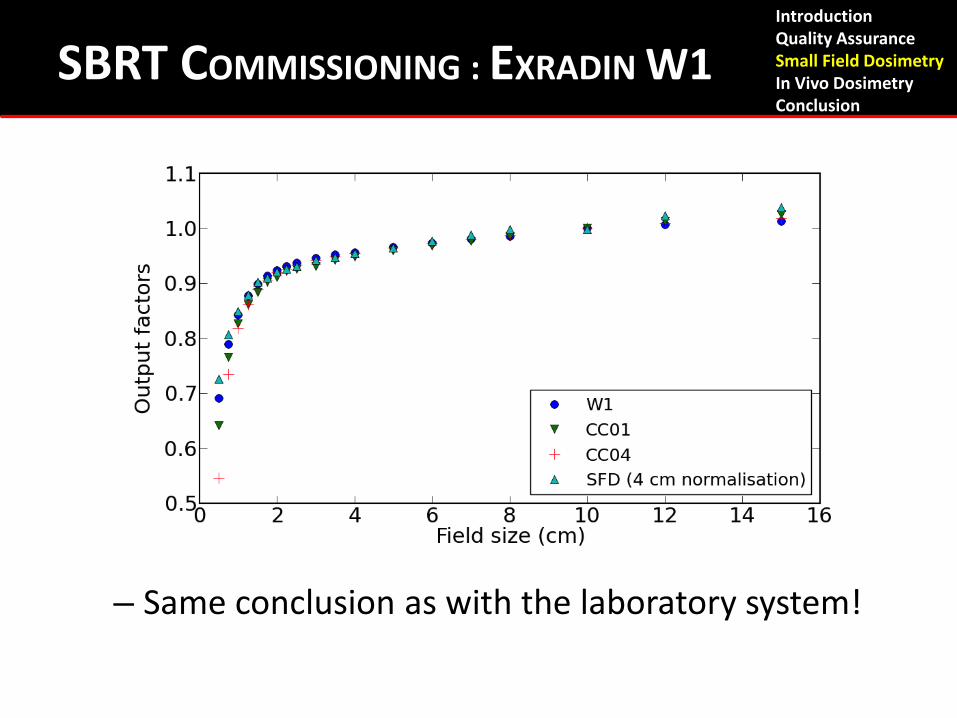
SBRT COMMISSIONING : EXRADIN W1
– Same conclusion as with the laboratory system!
Introduction Quality Assurance Small Field Dosimetry In Vivo Dosimetry Conclusion
DOSE RATE INDEPENDENCE
• High dose rate delivery: > 2000 MU/min ? • How do Ion Chambers fit in?
– Pion is affected by dose rate
• Comparison to the Exradin W1 PSD
Introduction Quality Assurance Small Field Dosimetry In Vivo Dosimetry Conclusion
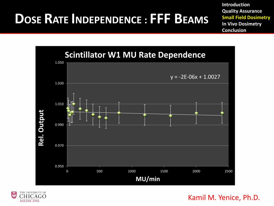
Kamil M. Yenice, Ph.D.
y = -2E-06x + 1.0027
0.950
0.970
0.990
1.010
1.030
1.050
0 500 1000 1500 2000 2500
Rel.
Out
put
MU/min
Scintillator W1 MU Rate Dependence
Introduction Quality Assurance Small Field Dosimetry In Vivo Dosimetry Conclusion
DOSE RATE INDEPENDENCE : FFF BEAMS
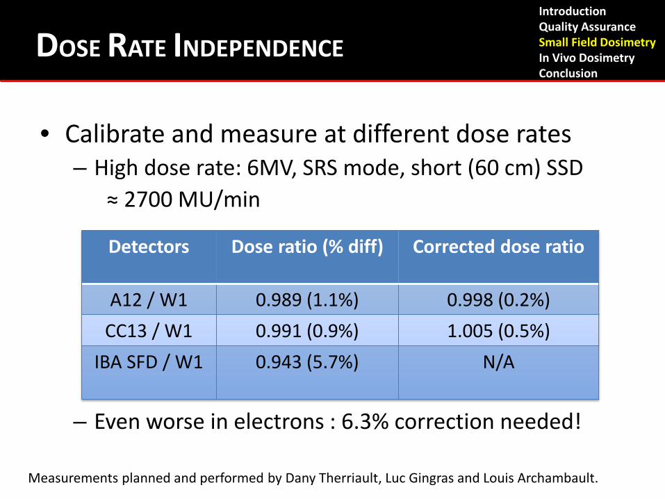
• Calibrate and measure at different dose rates – High dose rate: 6MV, SRS mode, short (60 cm) SSD ≈ 2700 MU/min
– Even worse in electrons : 6.3% correction needed!
Detectors Dose ratio (% diff) Corrected dose ratio
A12 / W1 0.989 (1.1%) 0.998 (0.2%) CC13 / W1 0.991 (0.9%) 1.005 (0.5%)
IBA SFD / W1 0.943 (5.7%) N/A
Measurements planned and performed by Dany Therriault, Luc Gingras and Louis Archambault.
Introduction Quality Assurance Small Field Dosimetry In Vivo Dosimetry Conclusion
DOSE RATE INDEPENDENCE
• Ion chambers affected by changes in Pion – Fully corrected by measuring Pion at a given dose
rate – W1 PSD is independent of dose rate at least up to
2700MU/min (max. tested!)
Introduction Quality Assurance Small Field Dosimetry In Vivo Dosimetry Conclusion
DOSE RATE INDEPENDENCE
Measurements planned and performed by Dany Therriault, Luc Gingras and Louis Archambault.
In Vivo Dosimetry
Introduction Quality Assurance Small Field Dosimetry In Vivo Dosimetry Conclusion
• Advantages of internal in-vivo dosimetry: – Point of measurement can be placed directly
adjacent to organ at risk or within treatment volume.
• Direct verification of treatment. • Detect adverse events or treatment variances,
potentially stop treatment and re-assess. • Clinical validation to monitor patient treatment
delivery is underway.
IN-VIVO DOSIMETRY Introduction Quality Assurance Small Field Dosimetry In Vivo Dosimetry Conclusion
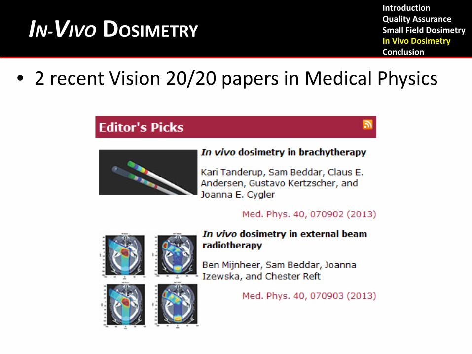
• 2 recent Vision 20/20 papers in Medical Physics
Introduction Quality Assurance Small Field Dosimetry In Vivo Dosimetry Conclusion
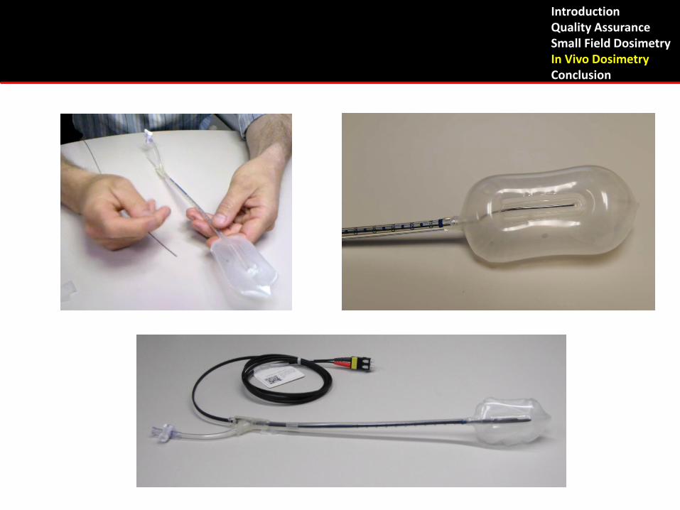
IN-VIVO DOSIMETRY
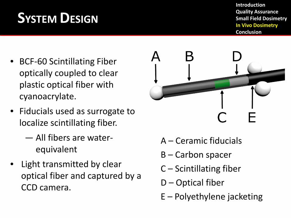
A – Ceramic fiducials B – Carbon spacer C – Scintillating fiber D – Optical fiber E – Polyethylene jacketing
SYSTEM DESIGN
• BCF-60 Scintillating Fiber optically coupled to clear plastic optical fiber with cyanoacrylate.
• Fiducials used as surrogate to localize scintillating fiber.
— All fibers are water-equivalent
• Light transmitted by clear optical fiber and captured by a CCD camera.
Introduction Quality Assurance Small Field Dosimetry In Vivo Dosimetry Conclusion
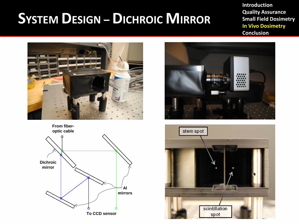
CURRENT EXPERIMENTAL SETUP
To CCD sensor
From fiber-optic cable
Dichroic mirror
Al mirrors
Introduction Quality Assurance Small Field Dosimetry In Vivo Dosimetry Conclusion
SYSTEM DESIGN – DICHROIC MIRROR
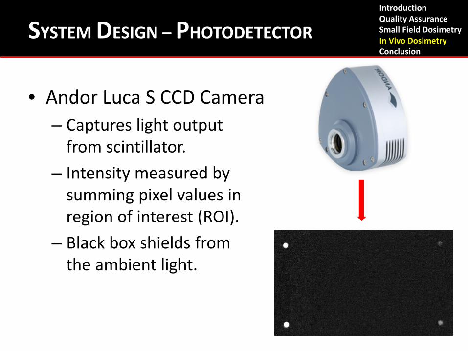
• Andor Luca S CCD Camera – Captures light output
from scintillator. – Intensity measured by
summing pixel values in region of interest (ROI).
– Black box shields from the ambient light.
Introduction Quality Assurance Small Field Dosimetry In Vivo Dosimetry Conclusion
SYSTEM DESIGN – PHOTODETECTOR
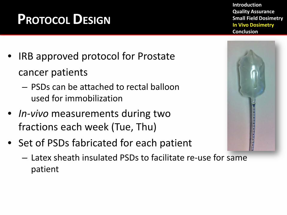
• IRB approved protocol for Prostate cancer patients – PSDs can be attached to rectal balloon
used for immobilization
• In-vivo measurements during two fractions each week (Tue, Thu)
• Set of PSDs fabricated for each patient – Latex sheath insulated PSDs to facilitate re-use for same
patient
PROTOCOL DESIGN Introduction Quality Assurance Small Field Dosimetry In Vivo Dosimetry Conclusion
• Daily CT for in-vivo fractions – Necessary to localize detectors
• Simple validation of PSDs performed after each treatment
• 200 cGy delivered in simple, static, fixed geometry • Deviations > 2% are considered indicative of loss of proper
function • Non-functioning detectors re-calibrated or discarded and re-
fabrication of new detectors
• 5 Patients enrolled (142 total measurements). – Only 5 thrown out due to problems with software (2)
or detectors’ malfunction (3).
Introduction Quality Assurance Small Field Dosimetry In Vivo Dosimetry Conclusion
PROTOCOL DESIGN
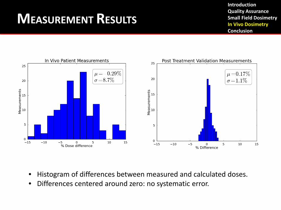
MEASUREMENT RESULTS
• Histogram of differences between measured and calculated doses. • Differences centered around zero: no systematic error.
Introduction Quality Assurance Small Field Dosimetry In Vivo Dosimetry Conclusion
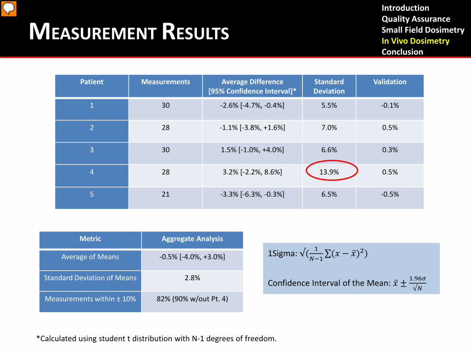
Patient Measurements Average Difference [95% Confidence Interval]*
Standard Deviation
Validation
1 30 -2.6% [-4.7%, -0.4%] 5.5% -0.1%
2 28 -1.1% [-3.8%, +1.6%] 7.0% 0.5%
3 30 1.5% [-1.0%, +4.0%] 6.6% 0.3%
4 28 3.2% [-2.2%, 8.6%] 13.9% 0.5%
5 21 -3.3% [-6.3%, -0.3%] 6.5% -0.5%
Metric Aggregate Analysis
Average of Means -0.5% [-4.0%, +3.0%]
Standard Deviation of Means 2.8%
Measurements within ± 10% 82% (90% w/out Pt. 4)
*Calculated using student t distribution with N-1 degrees of freedom.
Introduction Quality Assurance Small Field Dosimetry In Vivo Dosimetry Conclusion
MEASUREMENT RESULTS
1Sigma: √( 1𝑁−1
∑(𝑥 − �̅�)2)
Confidence Interval of the Mean: �̅� ± 1.96𝜎
√𝑁

• Histogram of differences between measured and calculated doses. • Differences centered around zero: no systematic error.
Introduction Quality Assurance Small Field Dosimetry In Vivo Dosimetry Conclusion
MEASUREMENT RESULTS
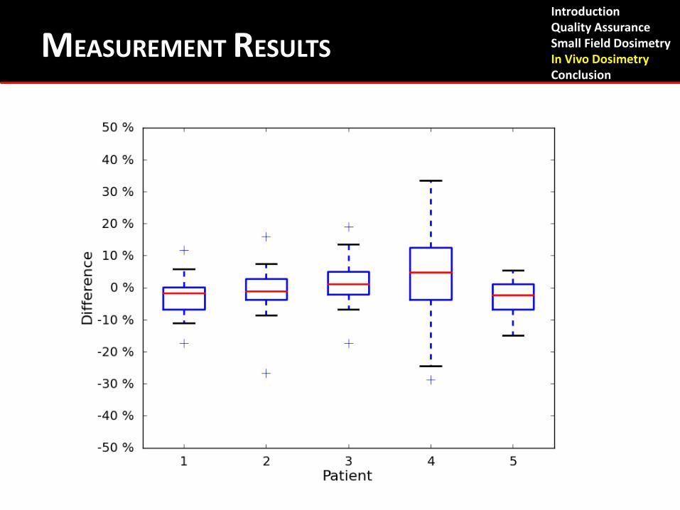
Introduction Quality Assurance Small Field Dosimetry In Vivo Dosimetry Conclusion
MEASUREMENT RESULTS
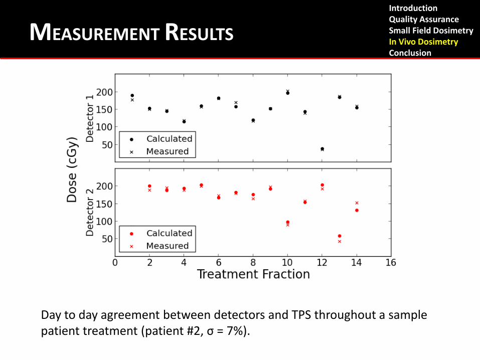
Day to day agreement between detectors and TPS throughout a sample patient treatment (patient #2, σ = 7%).
Introduction Quality Assurance Small Field Dosimetry In Vivo Dosimetry Conclusion
MEASUREMENT RESULTS
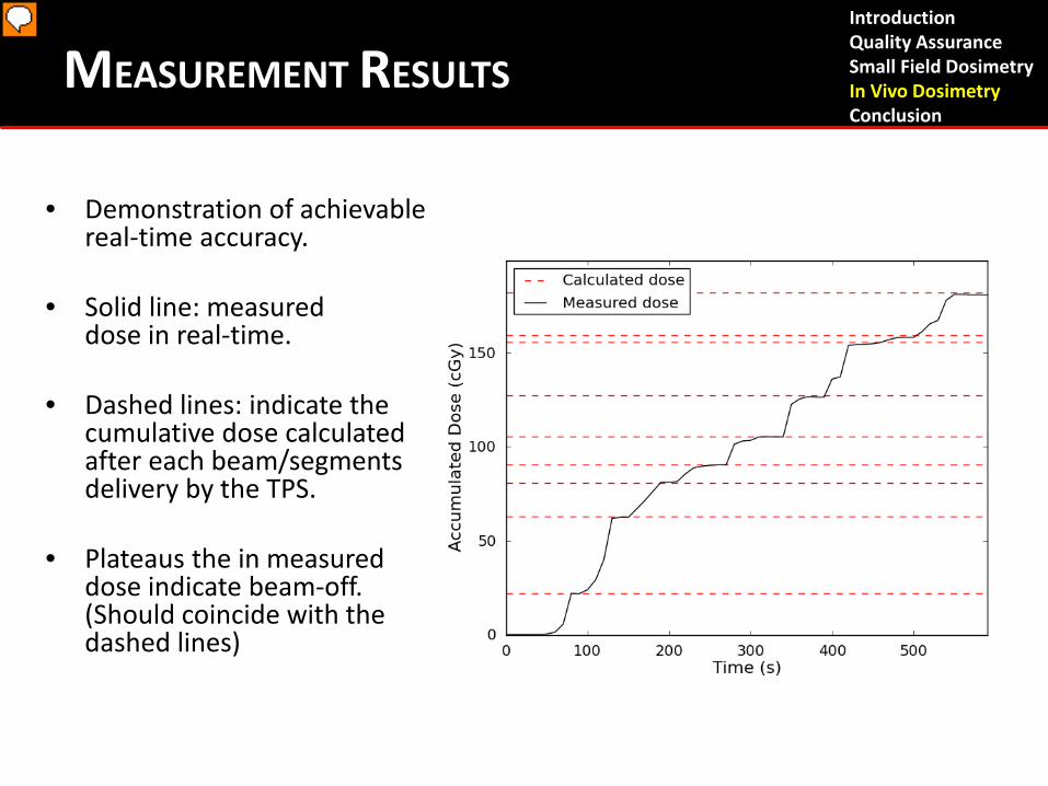
• Demonstration of achievable real-time accuracy.
• Solid line: measured dose in real-time.
• Dashed lines: indicate the cumulative dose calculated after each beam/segments delivery by the TPS.
• Plateaus the in measured dose indicate beam-off. (Should coincide with the dashed lines)
Introduction Quality Assurance Small Field Dosimetry In Vivo Dosimetry Conclusion
MEASUREMENT RESULTS
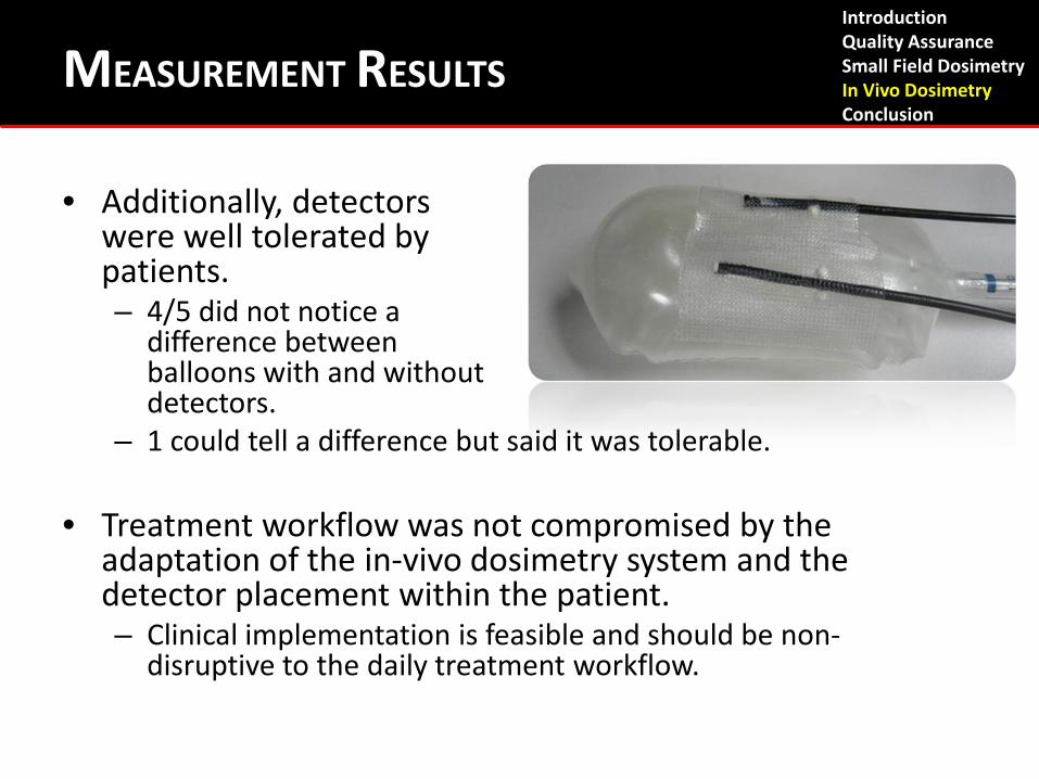
• Additionally, detectors were well tolerated by patients. – 4/5 did not notice a
difference between balloons with and without detectors.
– 1 could tell a difference but said it was tolerable.
• Treatment workflow was not compromised by the adaptation of the in-vivo dosimetry system and the detector placement within the patient. – Clinical implementation is feasible and should be non-
disruptive to the daily treatment workflow.
Introduction Quality Assurance Small Field Dosimetry In Vivo Dosimetry Conclusion
MEASUREMENT RESULTS
DISCUSSION
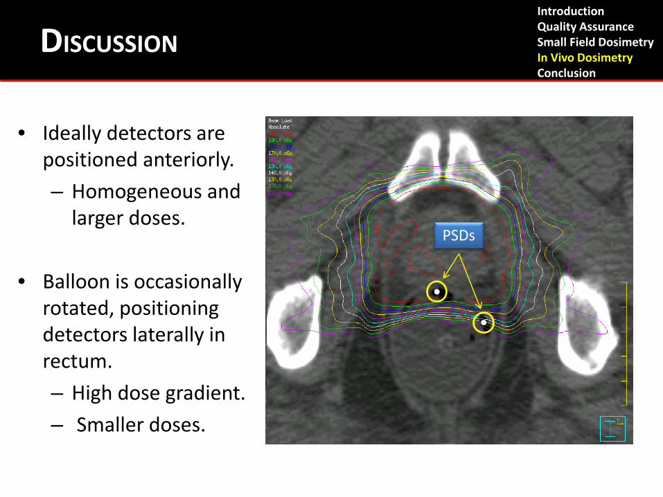
PSDs
• Ideally detectors are positioned anteriorly. – Homogeneous and
larger doses.
• Balloon is occasionally rotated, positioning detectors laterally in rectum. – High dose gradient. – Smaller doses.
Introduction Quality Assurance Small Field Dosimetry In Vivo Dosimetry Conclusion
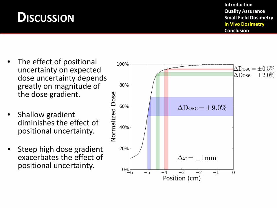
• The effect of positional uncertainty on expected dose uncertainty depends greatly on magnitude of the dose gradient.
• Shallow gradient
diminishes the effect of positional uncertainty.
• Steep high dose gradient exacerbates the effect of positional uncertainty.
Introduction Quality Assurance Small Field Dosimetry In Vivo Dosimetry Conclusion
DISCUSSION
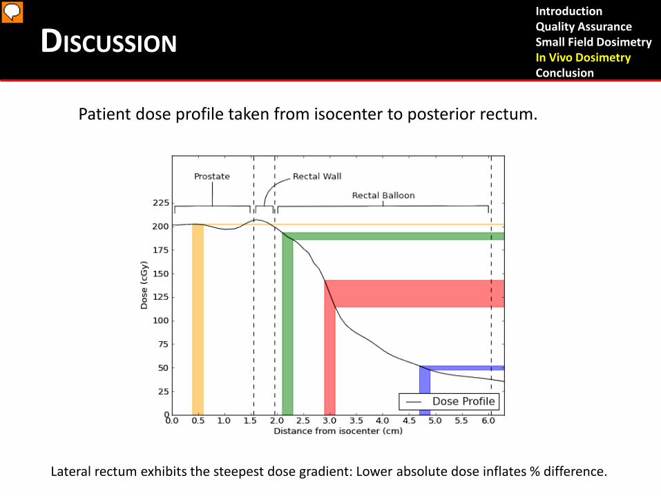
Patient dose profile taken from isocenter to posterior rectum.
Introduction Quality Assurance Small Field Dosimetry In Vivo Dosimetry Conclusion
DISCUSSION
Lateral rectum exhibits the steepest dose gradient: Lower absolute dose inflates % difference.
• Measurements with calculated doses > 170 cGy (corresponding to an anteriorly positioned detector) exhibit -1.4% ± 4.7%* average agreement.
• Measurements with calculated doses < 170 cGy (corresponding to laterally/posteriorly positioned detectors) exhibit 0.7% ± 11.1%* average agreement.
• Anterior dose measurements are more consistent.
*Mean and standard deviation of 65 and 72 measurements respectively, considered in aggregate regardless of patient of origin.
Introduction Quality Assurance Small Field Dosimetry In Vivo Dosimetry Conclusion
DISCUSSION
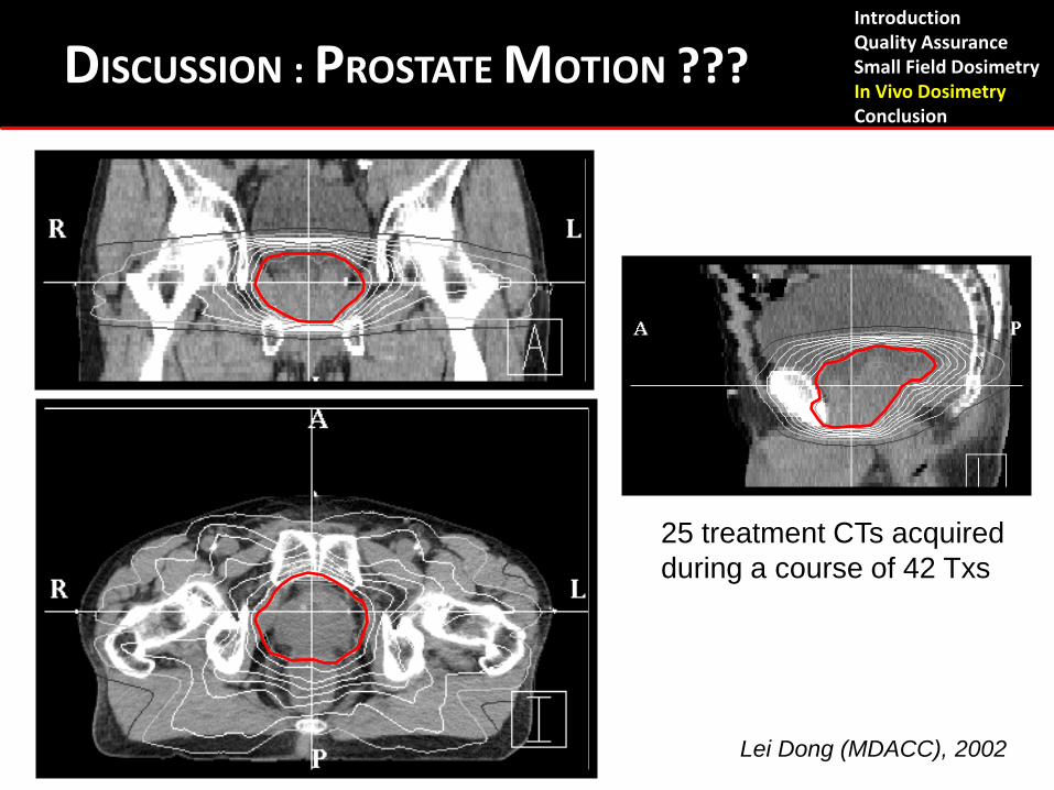
25 treatment CTs acquired during a course of 42 Txs
Lei Dong (MDACC), 2002
Introduction Quality Assurance Small Field Dosimetry In Vivo Dosimetry Conclusion
DISCUSSION : PROSTATE MOTION ???
OTHER PSD PROTOTYPES Introduction Quality Assurance Small Field Dosimetry In Vivo Dosimetry Conclusion
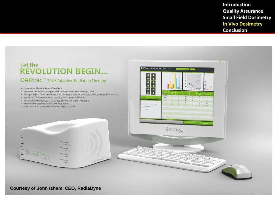
OARTRAC SYSTEM
Courtesy of John Isham, CEO, RadiaDyne
Introduction Quality Assurance Small Field Dosimetry In Vivo Dosimetry Conclusion
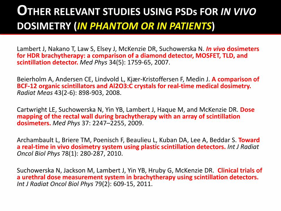
Lambert J, Nakano T, Law S, Elsey J, McKenzie DR, Suchowerska N. In vivo dosimeters for HDR brachytherapy: a comparison of a diamond detector, MOSFET, TLD, and scintillation detector. Med Phys 34(5): 1759-65, 2007. Beierholm A, Andersen CE, Lindvold L, Kjær-Kristoffersen F, Medin J. A comparison of BCF-12 organic scintillators and Al2O3:C crystals for real-time medical dosimetry. Radiat Meas 43(2-6): 898-903, 2008. Cartwright LE, Suchowerska N, Yin YB, Lambert J, Haque M, and McKenzie DR. Dose mapping of the rectal wall during brachytherapy with an array of scintillation dosimeters. Med Phys 37: 2247–2255, 2009. Archambault L, Briere TM, Poenisch F, Beaulieu L, Kuban DA, Lee A, Beddar S. Toward a real-time in vivo dosimetry system using plastic scintillation detectors. Int J Radiat Oncol Biol Phys 78(1): 280-287, 2010. Suchowerska N, Jackson M, Lambert J, Yin YB, Hruby G, McKenzie DR. Clinical trials of a urethral dose measurement system in brachytherapy using scintillation detectors. Int J Radiat Oncol Biol Phys 79(2): 609-15, 2011.
OTHER RELEVANT STUDIES USING PSDs FOR IN VIVO DOSIMETRY (IN PHANTOM OR IN PATIENTS)
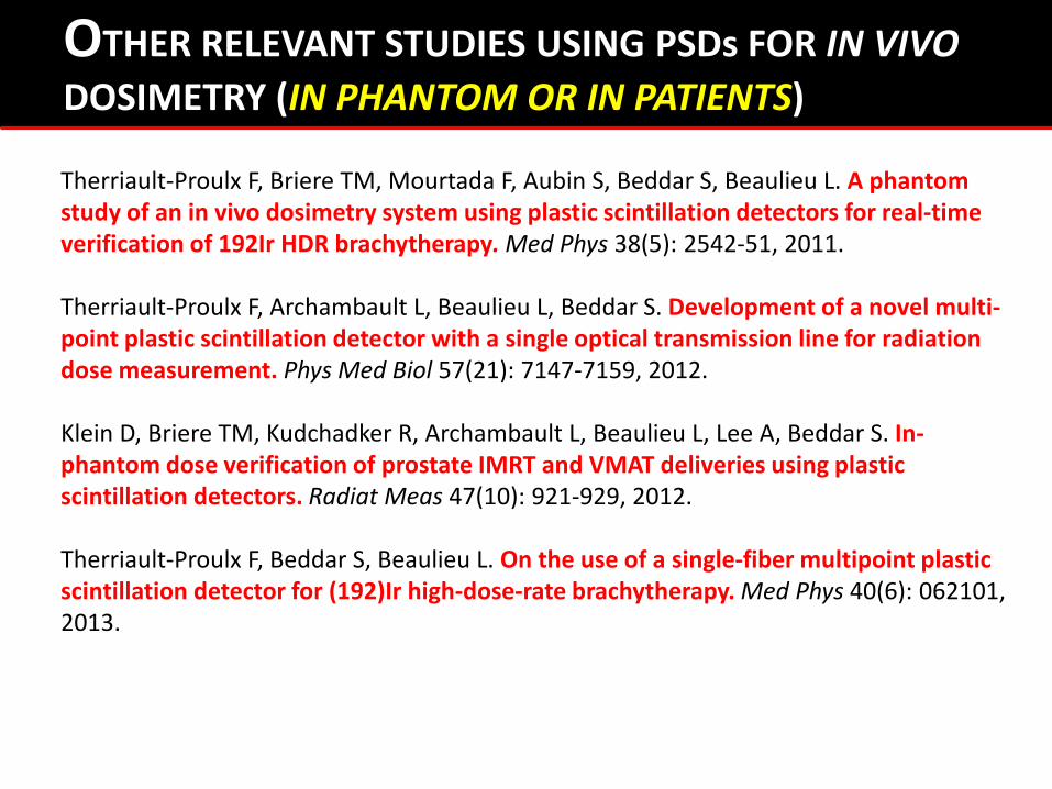
Therriault-Proulx F, Briere TM, Mourtada F, Aubin S, Beddar S, Beaulieu L. A phantom study of an in vivo dosimetry system using plastic scintillation detectors for real-time verification of 192Ir HDR brachytherapy. Med Phys 38(5): 2542-51, 2011. Therriault-Proulx F, Archambault L, Beaulieu L, Beddar S. Development of a novel multi-point plastic scintillation detector with a single optical transmission line for radiation dose measurement. Phys Med Biol 57(21): 7147-7159, 2012. Klein D, Briere TM, Kudchadker R, Archambault L, Beaulieu L, Lee A, Beddar S. In-phantom dose verification of prostate IMRT and VMAT deliveries using plastic scintillation detectors. Radiat Meas 47(10): 921-929, 2012. Therriault-Proulx F, Beddar S, Beaulieu L. On the use of a single-fiber multipoint plastic scintillation detector for (192)Ir high-dose-rate brachytherapy. Med Phys 40(6): 062101, 2013.
OTHER RELEVANT STUDIES USING PSDs FOR IN VIVO DOSIMETRY (IN PHANTOM OR IN PATIENTS)

ACKNOWLEDGEMENTS
F.H. Attix S. Aubin S.F. de Boer J. Boivin A. Bourgoin T.M. Briere P. Després J.C. Gagnon M.T. Gillin L. Gingras M. Goulet M. Guillemette
M. Guillot A. Ikhlef S. Klawikowski D. Klein R. Kudchadker F. Lacroix S. Law A. Lee F. Lessard T.R. Mackie R. Mohan J. Morin F. Mourtada
M. Plamondon F. Poenisch D. Robertson N. Sahoo C.H. Sibata J.V. Siebers N. Suchowerska R.C. Tailor F. Therriault-Proulx M. Villeneuve L. Wang L. Wootton C. Zeringue
… and all others who have contributed to the field of scintillation dosimetry.
ACKNOWLEDGEMENTS
Co-authors
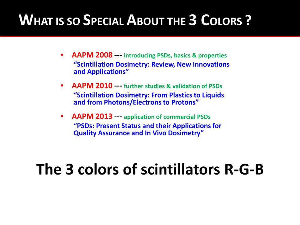
WHAT IS SO SPECIAL ABOUT THE 3 COLORS ?
• AAPM 2008 --- introducing PSDs, basics & properties “Scintillation Dosimetry: Review, New Innovations and Applications”
• AAPM 2010 --- further studies & validation of PSDs “Scintillation Dosimetry: From Plastics to Liquids and from Photons/Electrons to Protons”
• AAPM 2013 --- application of commercial PSDs “PSDs: Present Status and their Applications for Quality Assurance and In Vivo Dosimetry”
The 3 colors of scintillators R-G-B

Recommended