Phillip D Jones, DVM, MS
Diplomate American College of Veterinary Surgeons
NAVICULAR SYNDROME AND HEEL PAIN IN THE PERFORMANCE HORSE
OUTLINE
• Causes for caudal heel pain
• Diagnostics and therapies
• Shoeing recommendations
CAUDAL HEEL PAIN
• Navicular syndrome• Degenerative process that changes the bone-initiation
and progression of the disease is a result of excessive and prolonged forces of compression on the bone
• Important factors- signalment, conformation, and use
NAVICULAR SYNDROME
• Mild to moderate intermittent lameness
• Insidious onset
• Perceived as shoulder lameness
• Poor confirmation
• Bilateral lameness
• Lameness switches to contralateral limb after unilateral PD block
NAVICULAR SYNDROME
• Important PE findings -contracted heels -atrophied frog-small foot compared with body size
NAVICULAR SYNDROME
• Medical therapy -shoeing changes-anti-inflammatory medications -rest
• Surgical therapy- PD neurectomy (criteria! & complications!)
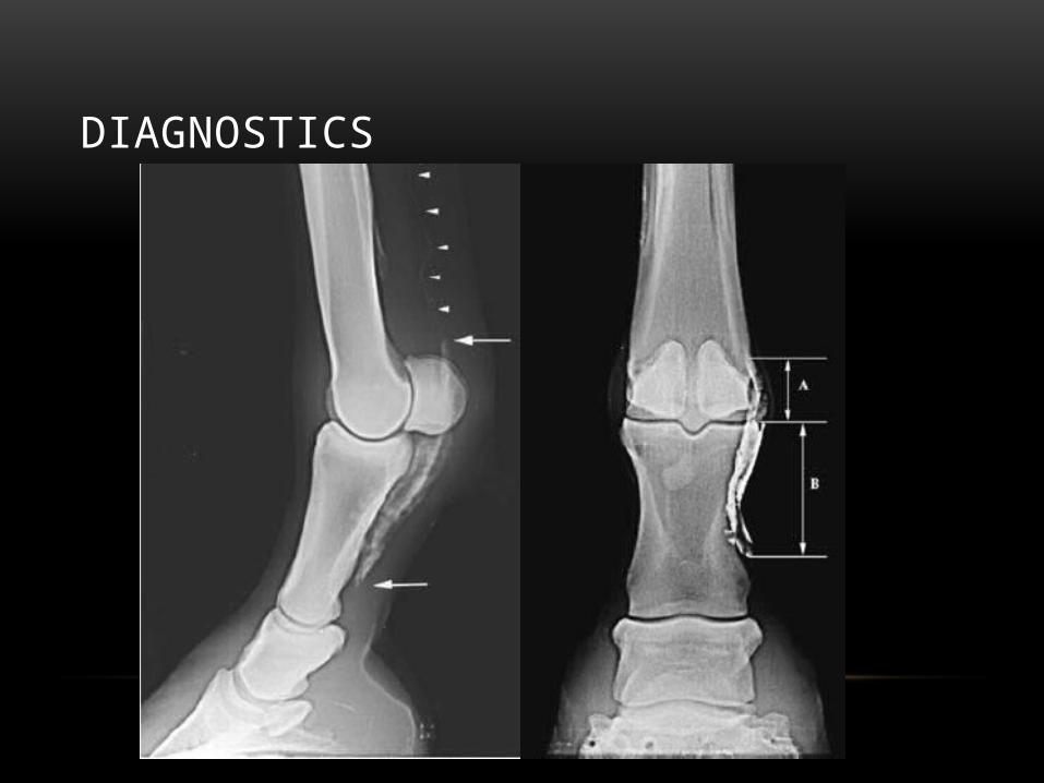
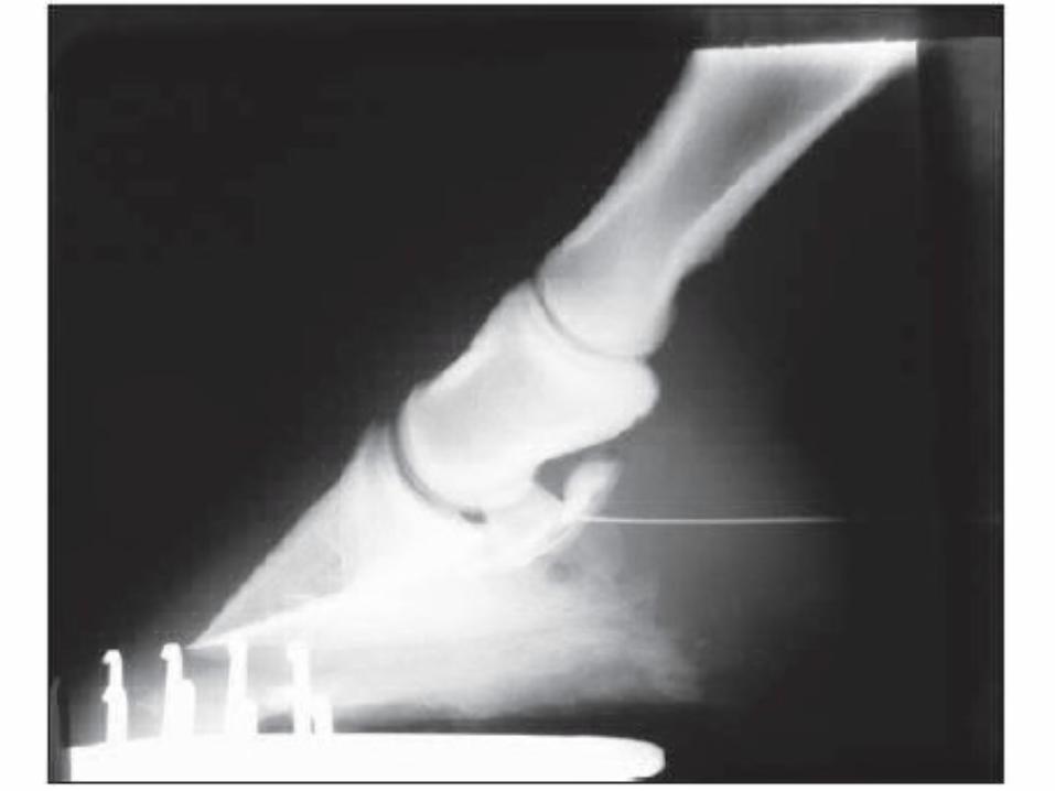
DIAGNOSTICS
• Localization
• Palmar Digital Nerve block-Mepivacaine: 1 -1.5ml /nerve
DIAGNOSTICS

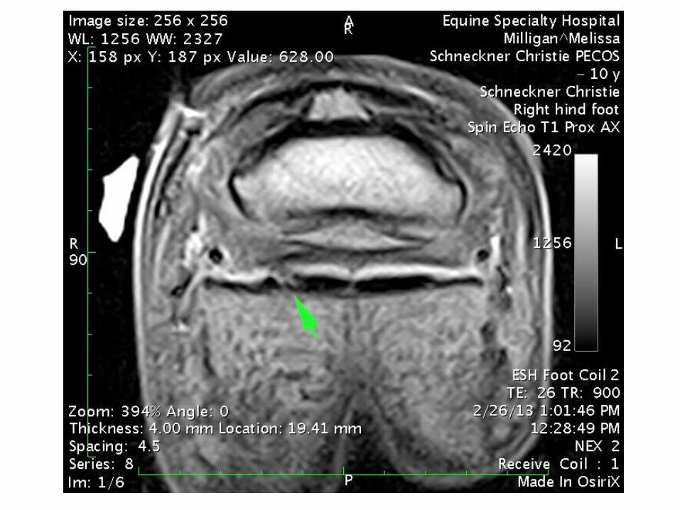
NAVICULAR SYNDROME
• Changes within the navicular bone -edema -vascular stasis -enlargement of the nutrient foraminae -cyst-like medullary areas -subchondral bone changes -changes in the flexor surface -fragmentation of the distal border
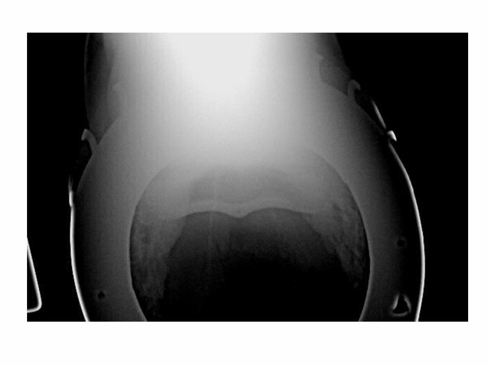
ADD NORMAL NB
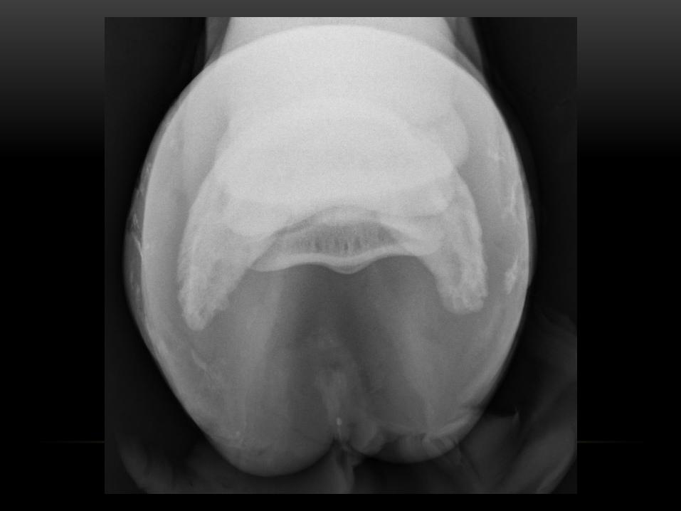
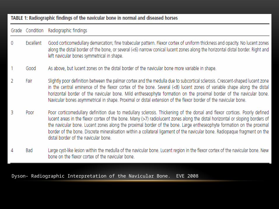
Dyson- Radiographic Interpretation of the Navicular Bone. EVE 2008
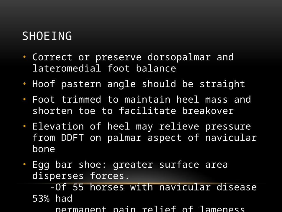
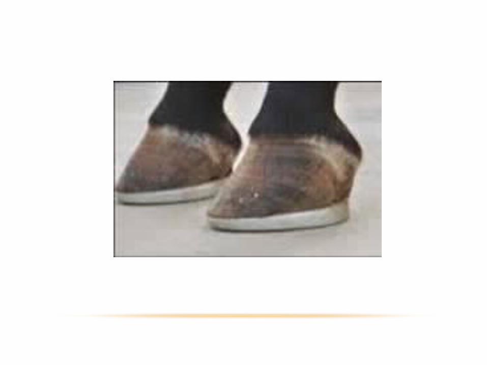
SHOEING
• Correct or preserve dorsopalmar and lateromedial foot balance
• Hoof pastern angle should be straight
• Foot trimmed to maintain heel mass and shorten toe to facilitate breakover
• Elevation of heel may relieve pressure from DDFT on palmar aspect of navicular bone
• Egg bar shoe: greater surface area disperses forces. -Of 55 horses with navicular disease 53% had permanent pain relief of lameness after egg bar shoes in 12-40 month follow up.
SHOEING
• Key points:
• Correct and maintain dorsopalmar and lateromedial balance
• Ease breakover
• Maintain heel mass
• Protect palmar aspect of the hoof from concussion
MEDICAL THERAPIES
• Intra-articular injection
• Intra-bursal injection
• Tilduronic acid (Tildren)
• NSAID’s
• Isoxuprine-2.2% oral bioavailability
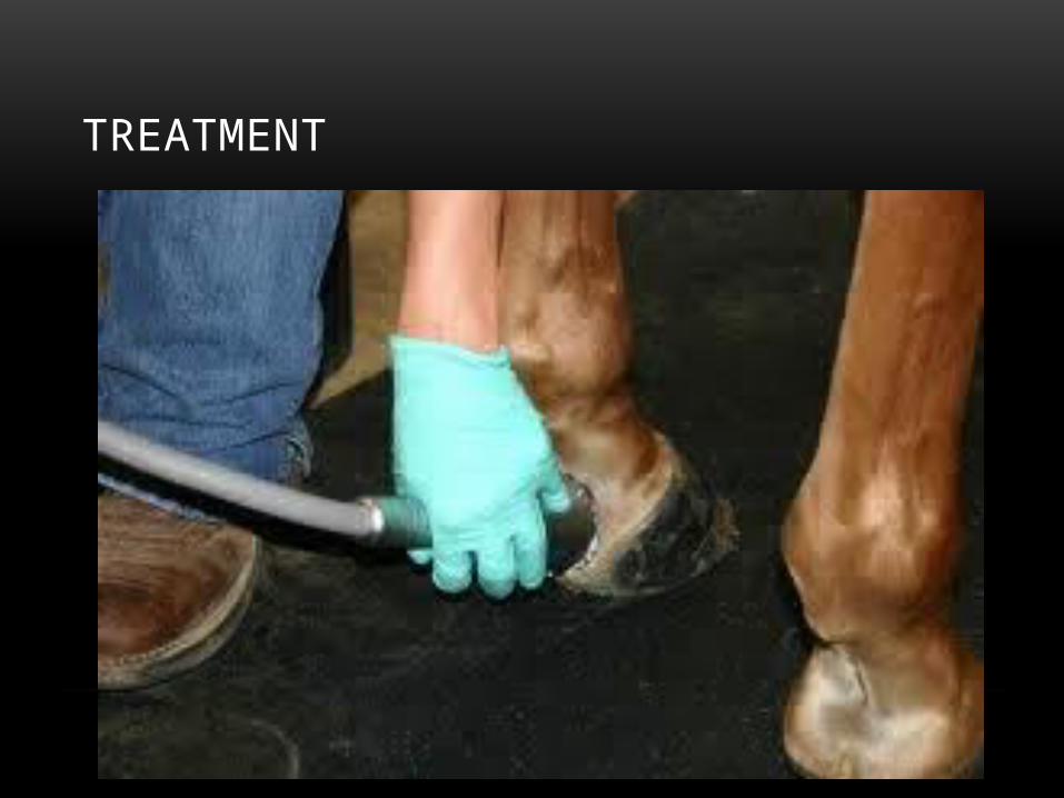
TREATMENT
CAUDAL HEEL PAIN
• Desmititis of the collateral ligaments.
• Tendonitis of the DDFT at 3 possible locations: -insertion -palmar to the navicular bone -proximal to the navicular bone
• Desmitis of the impar ligament.
• Desmitis of the distal annular ligament.
• Synovitis in the distal interphaleangeal joint.
• Synovitis in the navicular bursa.
• Cystic lesion in the second phalanx
TREATMENT
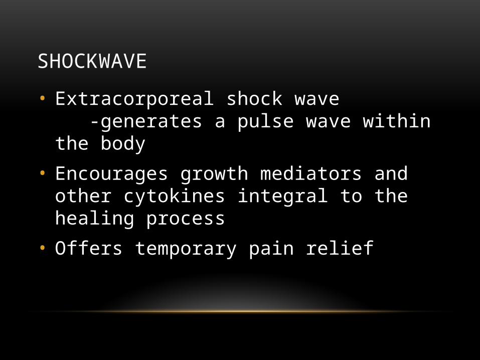
SHOCKWAVE
• Extracorporeal shock wave -generates a pulse wave within the body
• Encourages growth mediators and other cytokines integral to the healing process
• Offers temporary pain relief
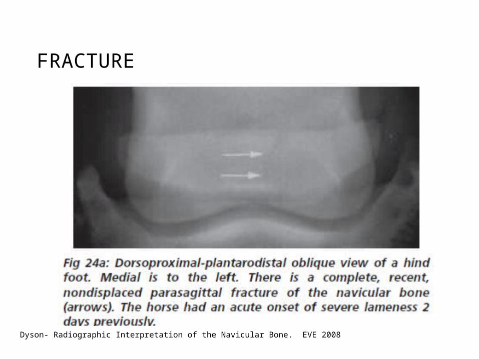
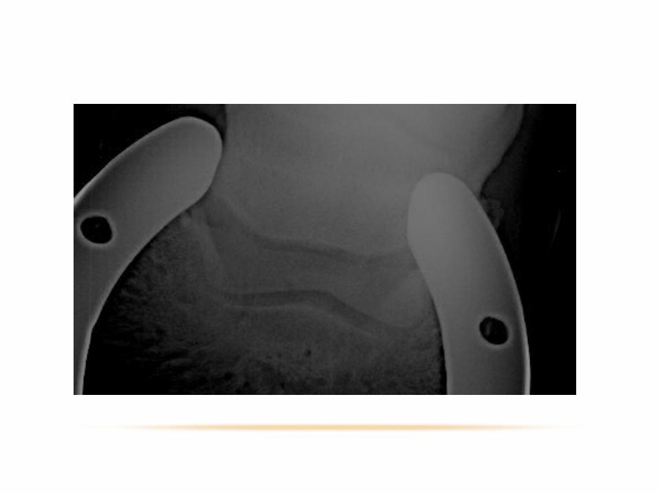
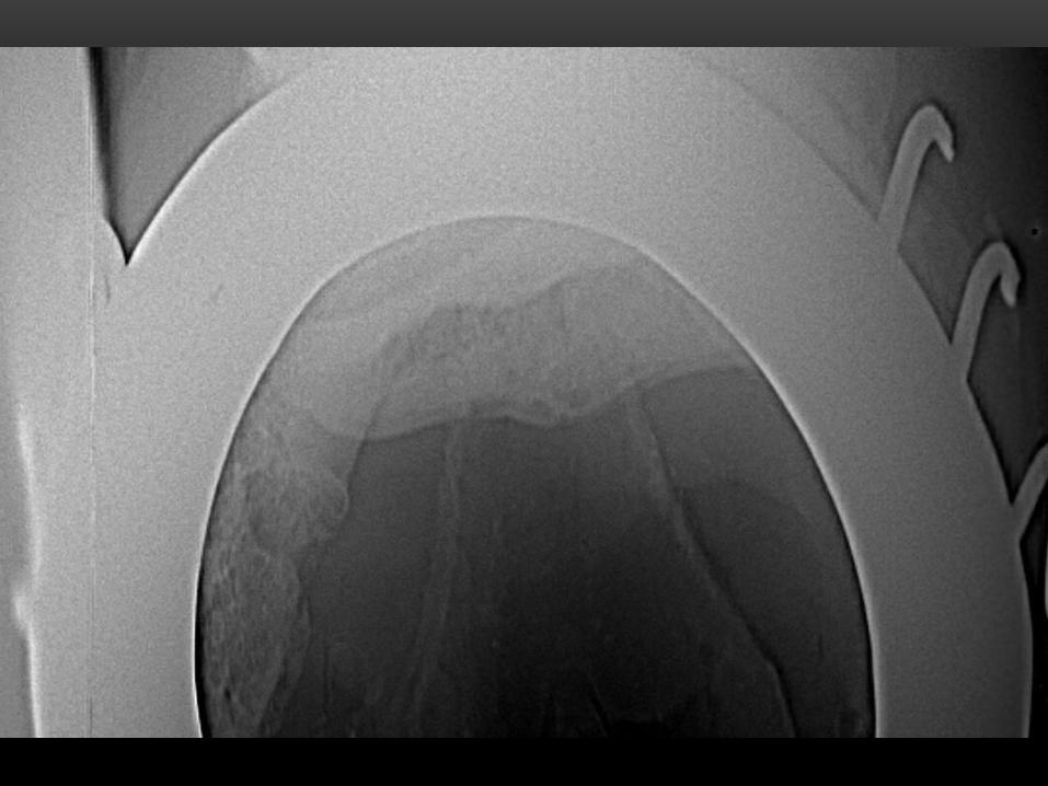
FRACTURE
Dyson- Radiographic Interpretation of the Navicular Bone. EVE 2008
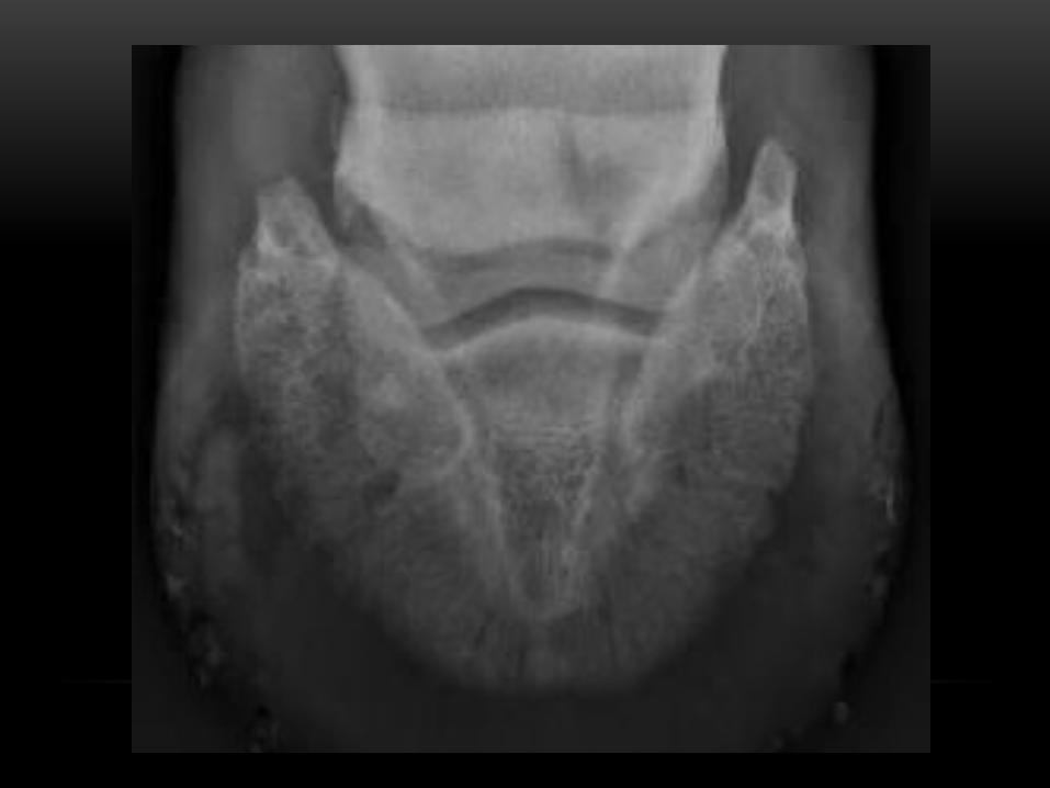
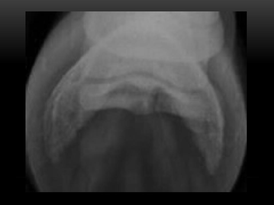
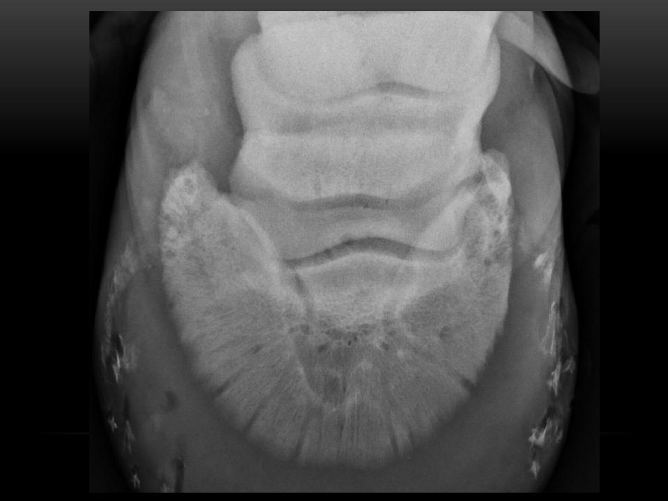
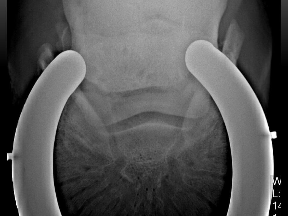
BIPARTITE NAVICULAR BONE
• Develops as 2 separate centers of ossification that never unite
• Unilateral or bilateral
• Broad well defined lucent line between the 2 pieces
• Horse may be clinically normal, or have episodic lameness in full athletic function
• No history of acute lameness as in fracture
BIPARTITE
Dyson- Radiographic Interpretation of the Navicular Bone. EVE 2008

?
Recommended