Thromboembolism and haemorrhage in pregnancy and delivery
Petr Krepelka
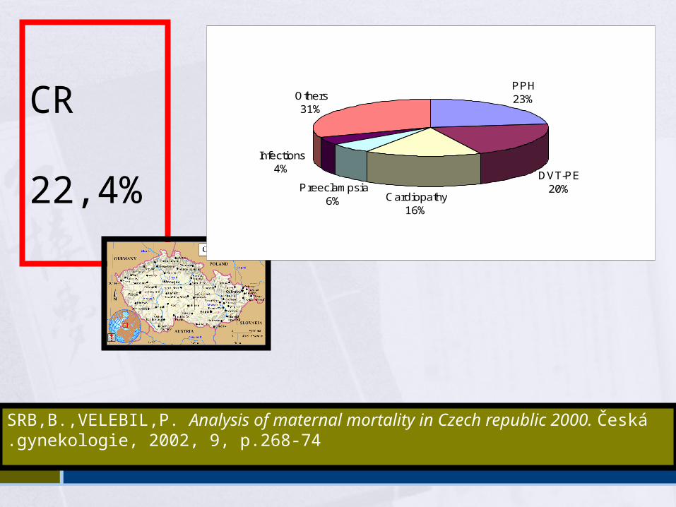
CR 22,4%
SRB,B.,VELEBIL,P. Analysis of maternal mortality in Czech republic 2000. Česká gynekologie, 2002, 9, p.268-74.
PPH23%
DVT-PE20%
Cardiopathy16%
Others31%
Infections4%
Preeclampsia6%
Deep venous thrombosis and pulmonary embolism in pregnancy
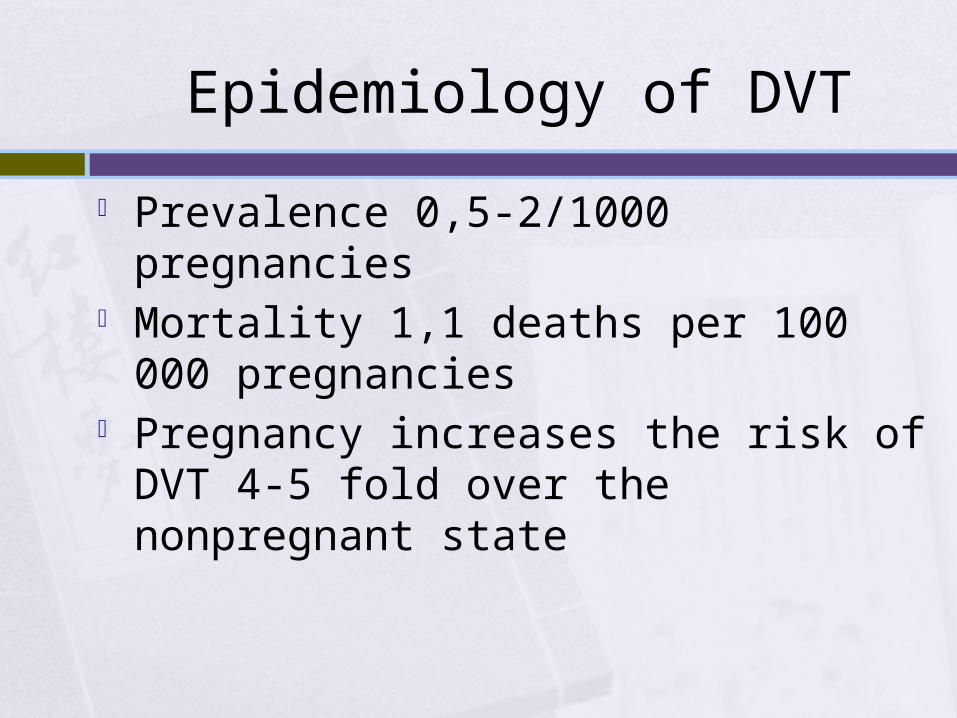
Epidemiology of DVT
Prevalence 0,5-2/1000 pregnancies Mortality 1,1 deaths per 100 000
pregnancies Pregnancy increases the risk of DVT 4-5 fold
over the nonpregnant state
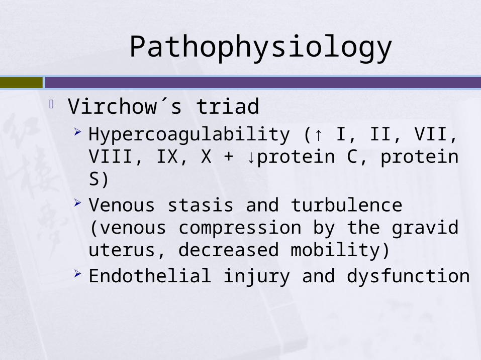
Pathophysiology
Virchow´s triad Hypercoagulability (↑ I, II, VII, VIII, IX, X +
↓protein C, protein S) Venous stasis and turbulence (venous
compression by the gravid uterus, decreased mobility)
Endothelial injury and dysfunction
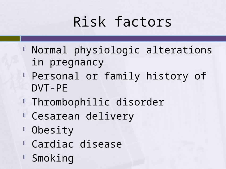
Risk factors
Normal physiologic alterations in pregnancy Personal or family history of DVT-PE Thrombophilic disorder Cesarean delivery Obesity Cardiac disease Smoking
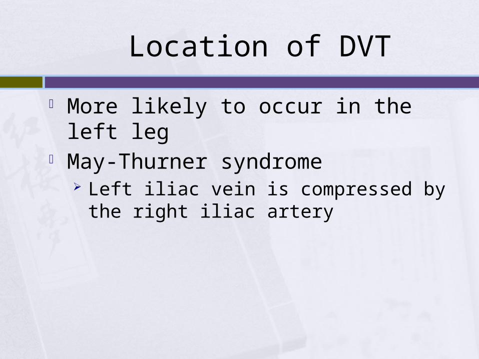
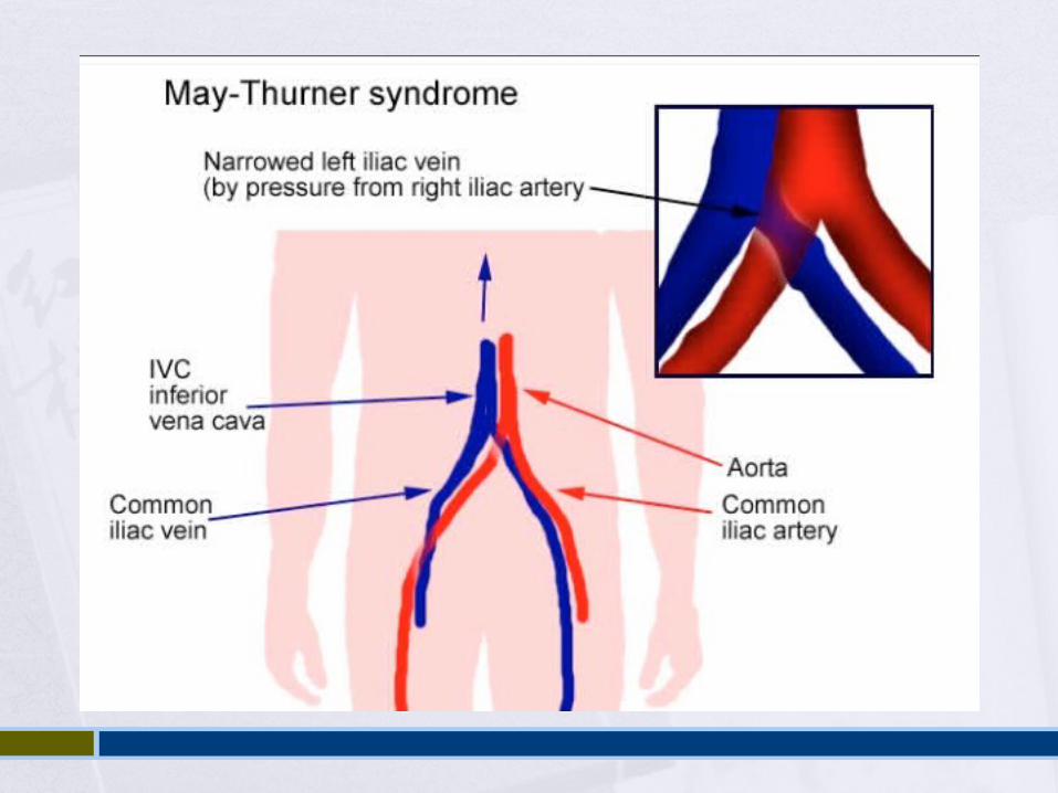
Location of DVT
More likely to occur in the left leg May-Thurner syndrome
Left iliac vein is compressed by the right iliac artery
Sequelae of DVT
Pulmonary hypertension Post-thrombotic syndrome (pain, cramps,
heaviness, paresthesia, edema, skin induration, hyperpigmentation, venous ectasia, redness)
Venous insufficiency
History and physical examination DVT
Signs and symptoms are nonspecific 2 most common symptoms
Pain Swelling of the lower extremity (mid-calf
circumference difference of ≥2 cm)
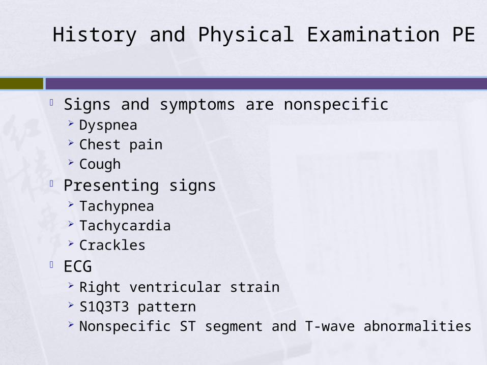
History and Physical Examination PE
Signs and symptoms are nonspecific Dyspnea Chest pain Cough
Presenting signs Tachypnea Tachycardia Crackles
ECG Right ventricular strain S1Q3T3 pattern Nonspecific ST segment and T-wave abnormalities
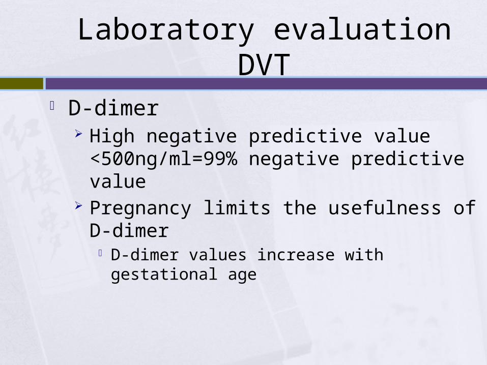
Laboratory evaluation DVT
D-dimer High negative predictive value <500ng/ml=99%
negative predictive value Pregnancy limits the usefulness of D-dimer
D-dimer values increase with gestational age
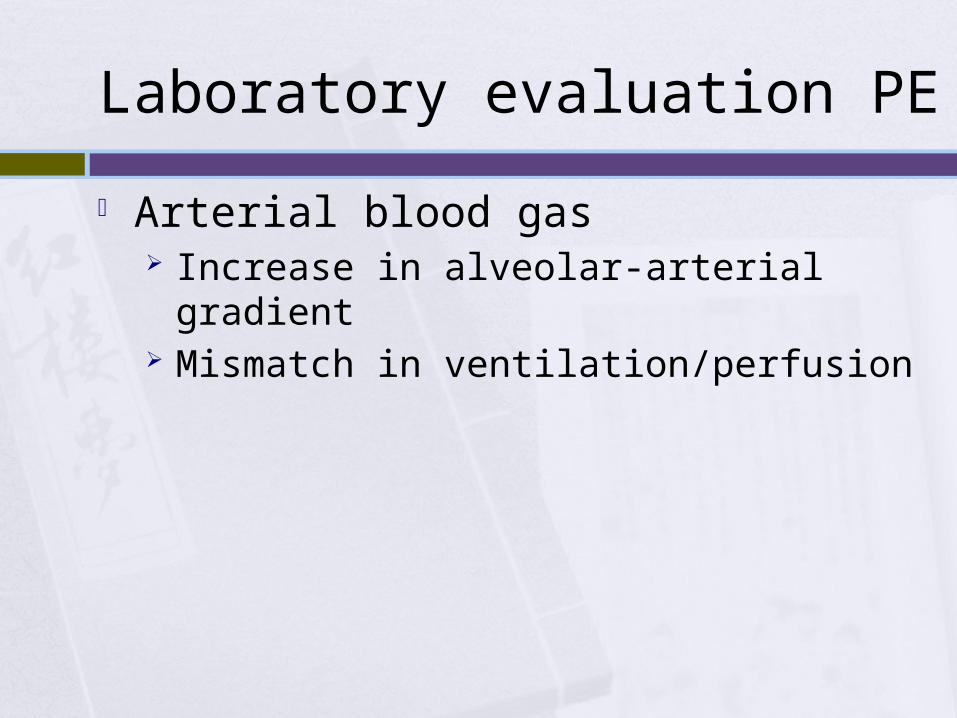
Laboratory evaluation PE
Arterial blood gas Increase in alveolar-arterial gradient Mismatch in ventilation/perfusion
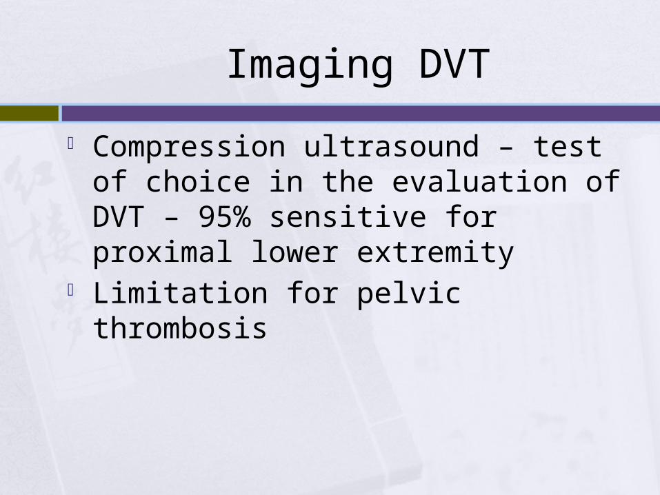
Imaging DVT
Compression ultrasound – test of choice in the evaluation of DVT – 95% sensitive for proximal lower extremity
Limitation for pelvic thrombosis
Imaging PE
Spiral CT pulmonary angiography (CT-PA) Normal chest radiograph
Ventilation-perfusion (V/Q) scan Abnormal chest radiograph or knonw
pulmonary disease
Therapy Indirect thrombin inhibitors
unfractionated heparin low molecular weight heparins synthetic heparin pentasaccharides orally administered Factor Xa inhibitors (eg, rivaroxaban)
Direct thrombin inhibitors Argatroban Lepirudin Bivalirudin
Vitamin K antagonist Warfarin
Heparin (both unfractionated and low molecular weight) is the preferred drugs for management of VTE in pregnancy
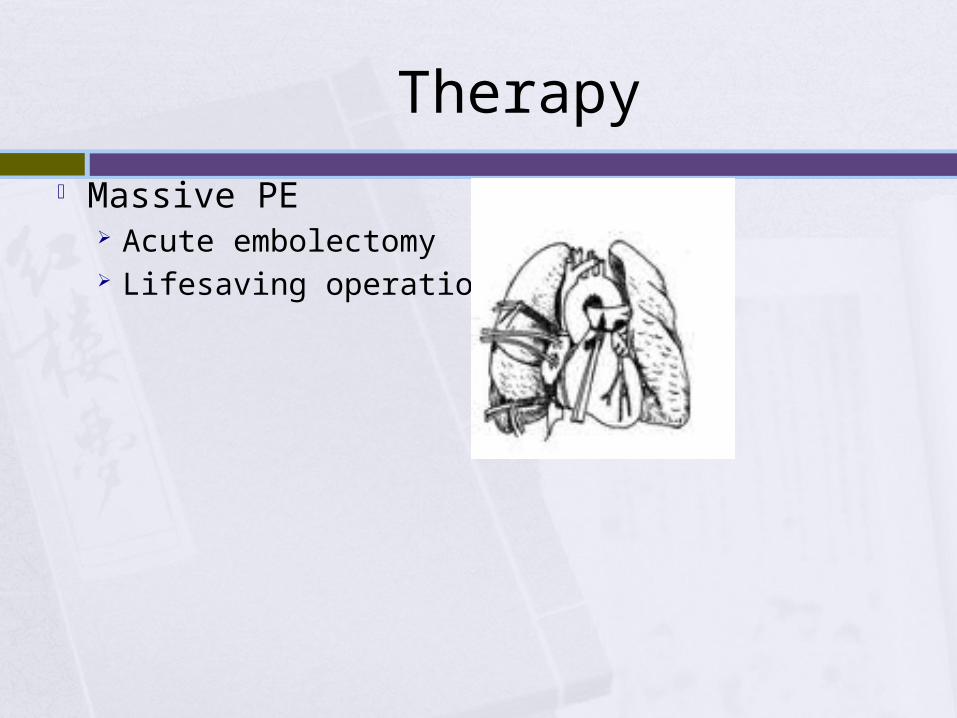
Therapy Massive PE
Acute embolectomy Lifesaving operation
Bleeding in pregnancy
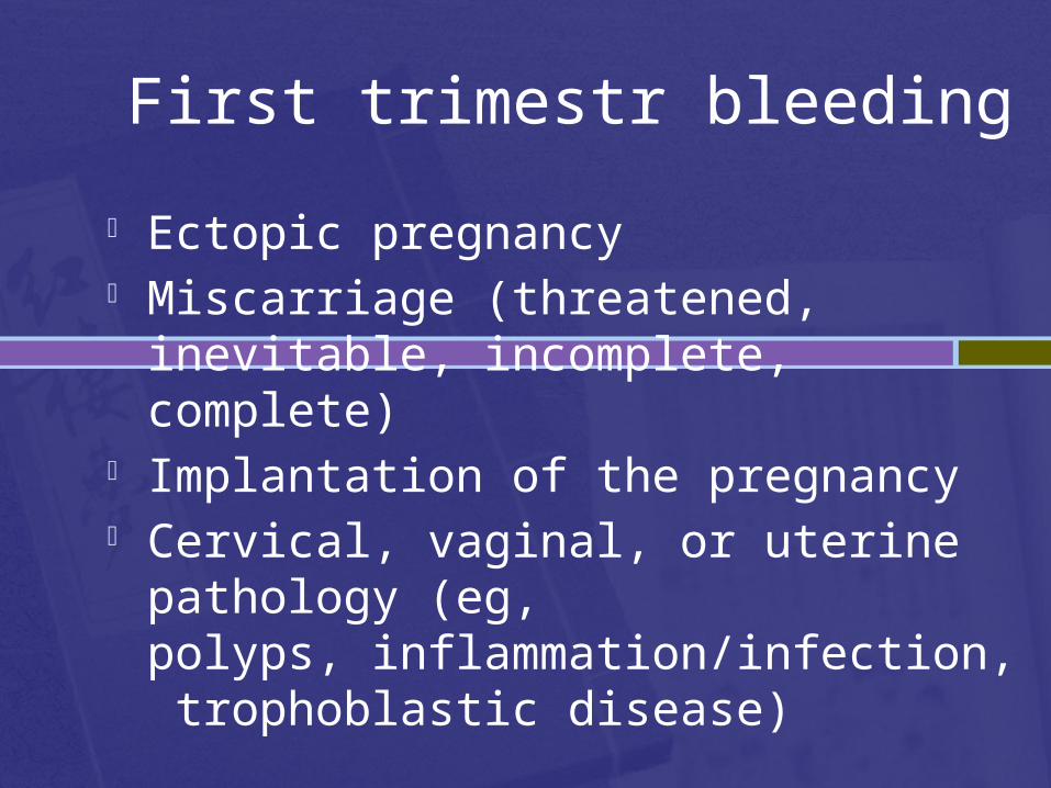
First trimestr bleeding
Ectopic pregnancy Miscarriage (threatened, inevitable,
incomplete, complete) Implantation of the pregnancy Cervical, vaginal, or uterine pathology (eg,
polyps, inflammation/infection, trophoblastic disease)
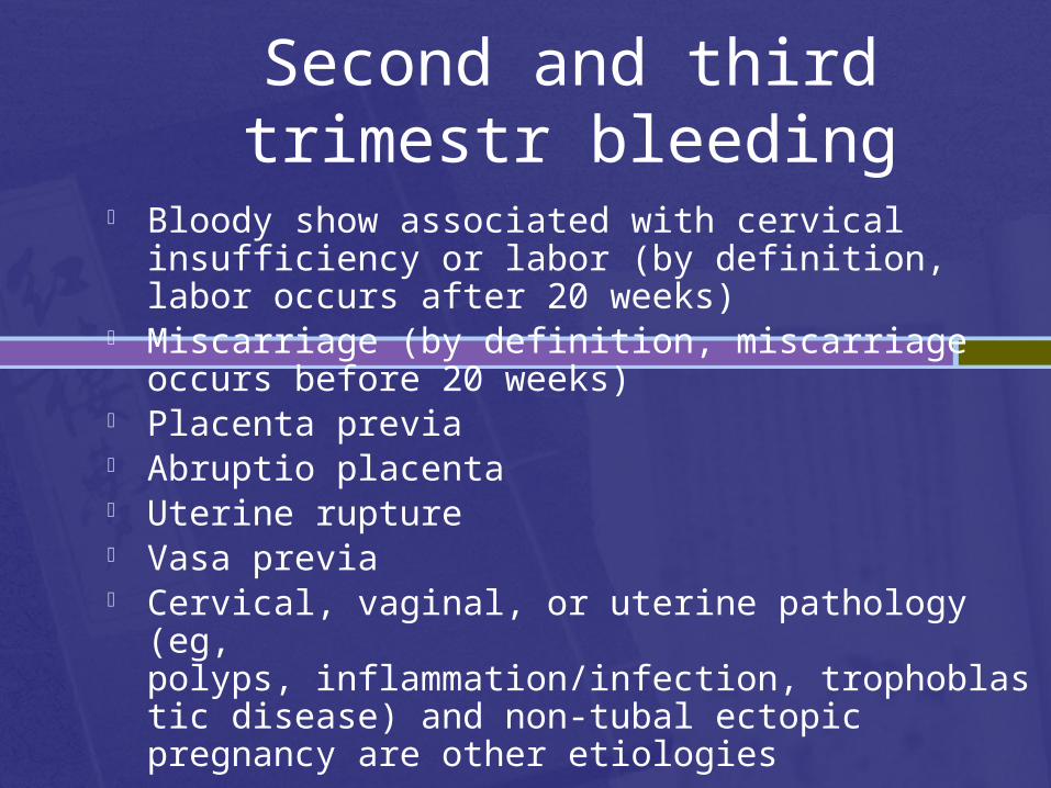
Second and third trimestr bleeding
Bloody show associated with cervical insufficiency or labor (by definition, labor occurs after 20 weeks)
Miscarriage (by definition, miscarriage occurs before 20 weeks)
Placenta previa Abruptio placenta Uterine rupture Vasa previa Cervical, vaginal, or uterine pathology (eg,
polyps, inflammation/infection, trophoblastic disease) and non-tubal ectopic pregnancy are other etiologies
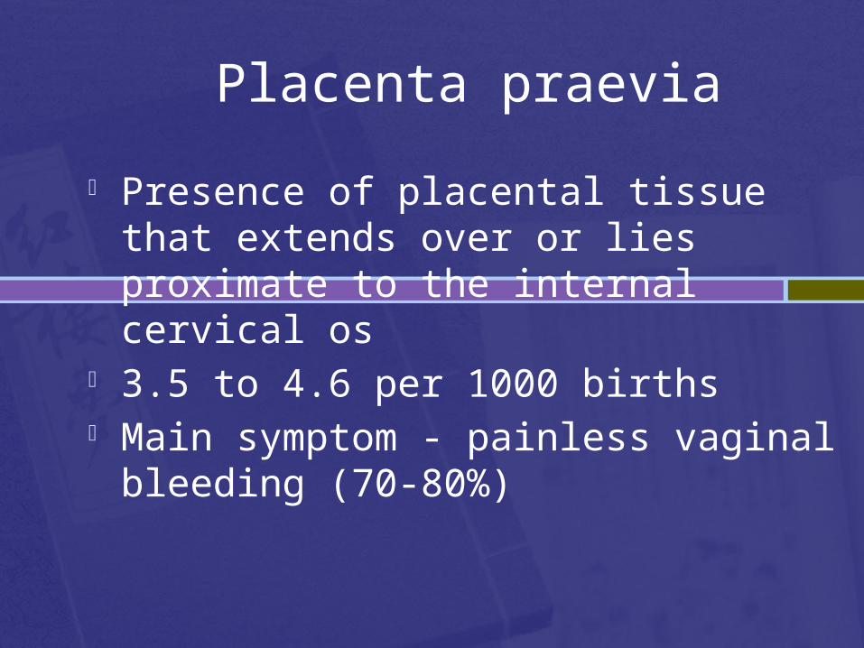
Placenta praevia
Presence of placental tissue that extends over or lies proximate to the internal cervical os
3.5 to 4.6 per 1000 births Main symptom - painless vaginal bleeding
(70-80%)
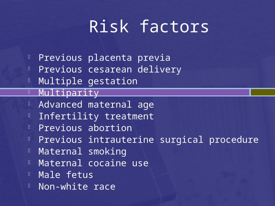
Risk factors
Previous placenta previa Previous cesarean delivery Multiple gestation Multiparity Advanced maternal age Infertility treatment Previous abortion Previous intrauterine surgical procedure Maternal smoking Maternal cocaine use Male fetus Non-white race
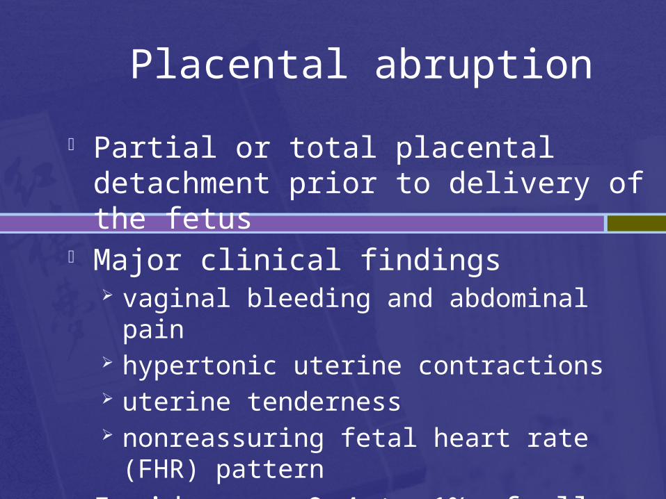
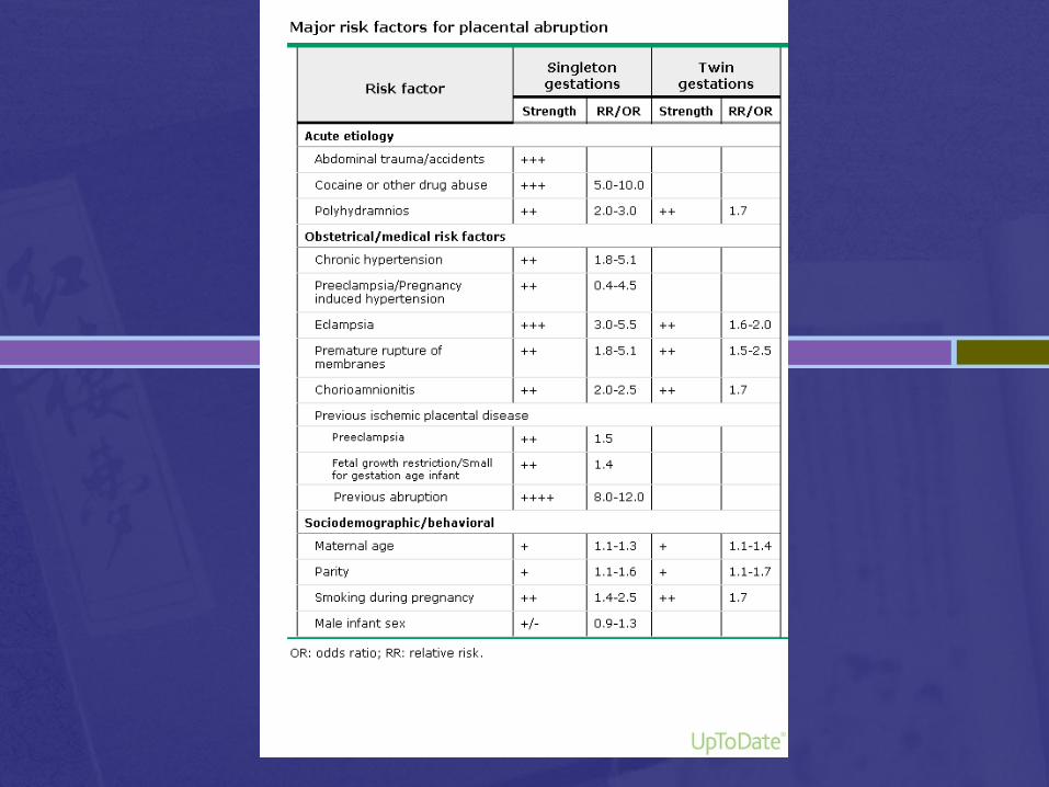
Placental abruption
Partial or total placental detachment prior to delivery of the fetus
Major clinical findings vaginal bleeding and abdominal pain hypertonic uterine contractions uterine tenderness nonreassuring fetal heart rate (FHR) pattern
Incidence - 0.4 to 1% of all pregnancies
Primary postpartum haemorrhage
Epidemiology
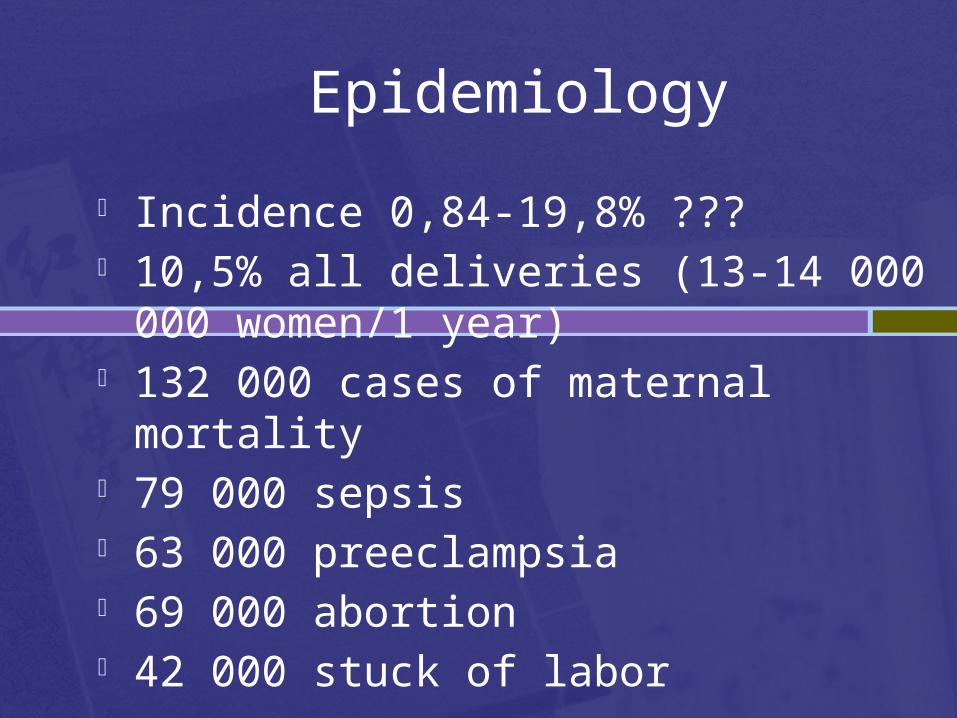
Incidence 0,84-19,8% ??? 10,5% all deliveries (13-14 000 000
women/1 year) 132 000 cases of maternal mortality 79 000 sepsis 63 000 preeclampsia 69 000 abortion 42 000 stuck of labor
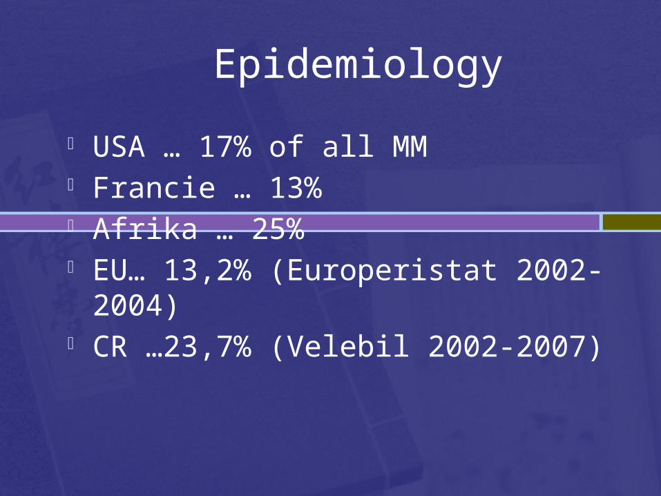
USA … 17% of all MM Francie … 13% Afrika … 25% EU… 13,2% (Europeristat 2002-2004) CR …23,7% (Velebil 2002-2007)
Epidemiology
Definition of PPH
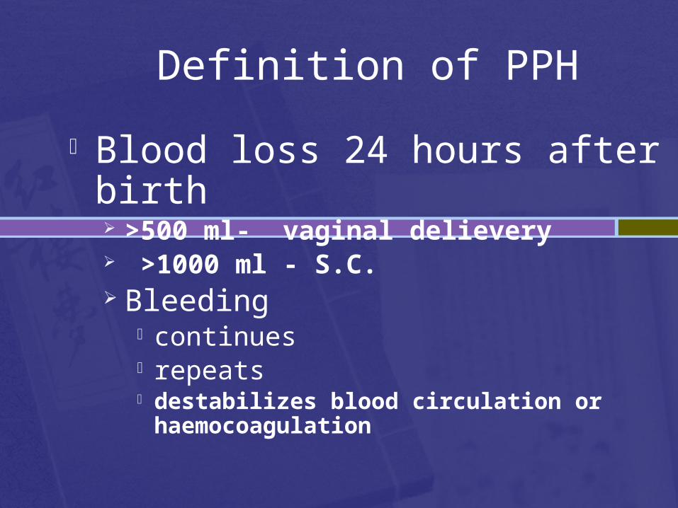
Blood loss 24 hours after birth >500 ml- vaginal delievery >1000 ml - S.C. Bleeding
continues repeats destabilizes blood circulation or
haemocoagulation
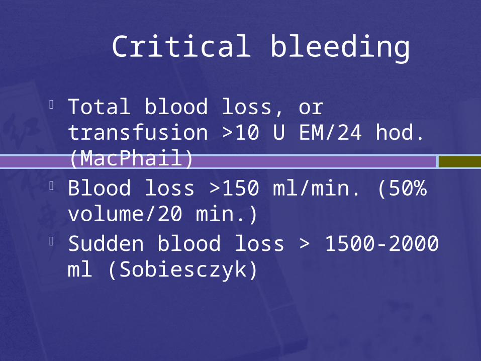
Critical bleeding
Total blood loss, or transfusion >10 U EM/24 hod. (MacPhail)
Blood loss >150 ml/min. (50% volume/20 min.)
Sudden blood loss > 1500-2000 ml (Sobiesczyk)
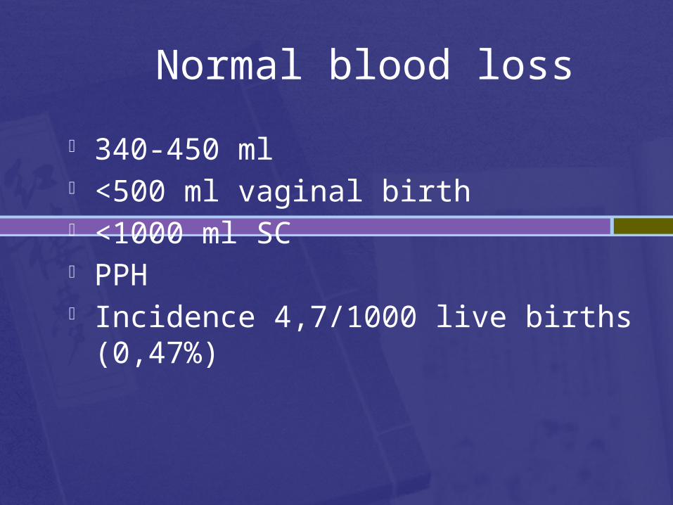
Normal blood loss
340-450 ml <500 ml vaginal birth <1000 ml SC PPH Incidence 4,7/1000 live births (0,47%)
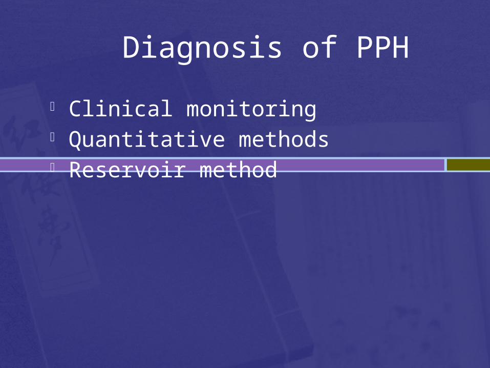
Clinical monitoring Quantitative methods Reservoir method
Diagnosis of PPH

1 150
2 300
3 450
4 600
5 750
6 900
7 1050
8 1200
9 1350
10 1500
11 1650
12 1800
13 1950
14 2100
15 2250
16 2400
17 2550
18 2700
19 2850
20 3000
15%
20%
25%
30%
35%
40%
1
2
3
4
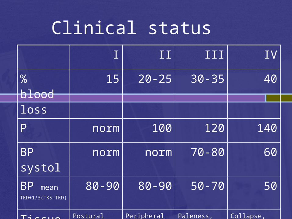
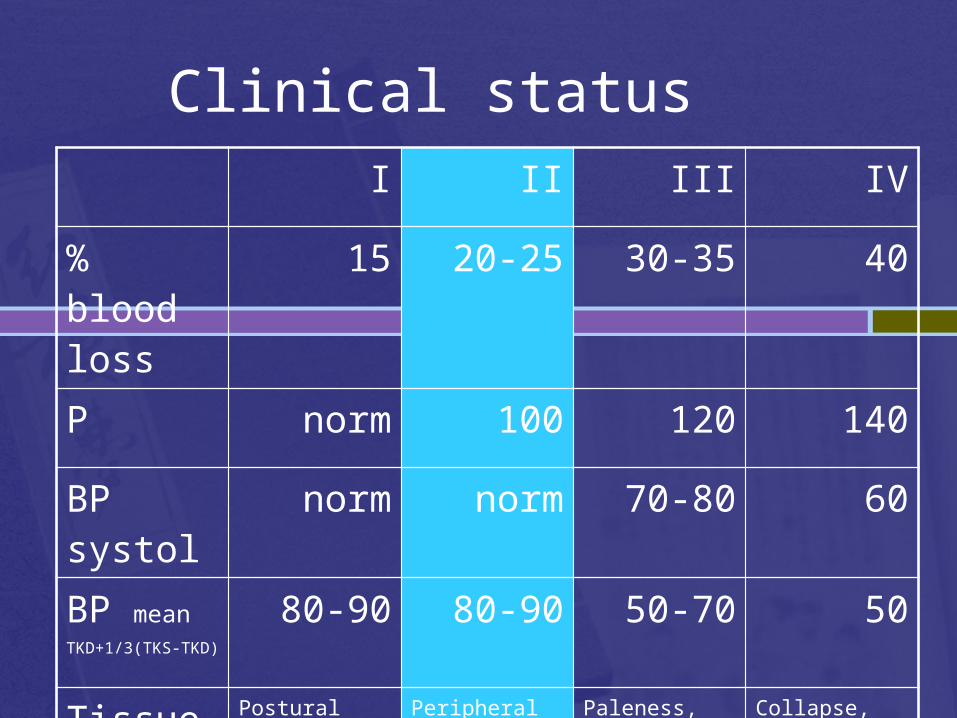
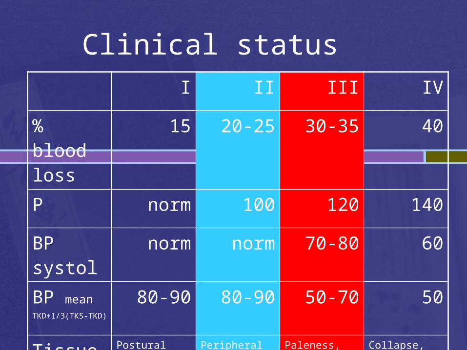
Clinical statusI II III IV
% blood loss
15 20-25 30-35 40
P norm 100 120 140
BP systol norm norm 70-80 60
BP meanTKD+1/3(TKS-TKD)
80-90 80-90 50-70 50
Tissue perfusion
Postural hypotension
Peripheral vasoconstriction
Paleness, restlessness, oliguria
Collapse, anuria, gasping breathing
Clinical statusI II III IV
% blood loss
15 20-25 30-35 40
P norm 100 120 140
BP systol norm norm 70-80 60
BP meanTKD+1/3(TKS-TKD)
80-90 80-90 50-70 50
Tissue perfusion
Postural hypotension
Peripheral vasoconstriction
Paleness, restlessness, oliguria
Collapse, anuria, gasping breathing
Clinical statusI II III IV
% blood loss
15 20-25 30-35 40
P norm 100 120 140
BP systol norm norm 70-80 60
BP meanTKD+1/3(TKS-TKD)
80-90 80-90 50-70 50
Tissue perfusion
Postural hypotension
Peripheral vasoconstriction
Paleness, restlessness, oliguria
Collapse, anuria, gasping breathing
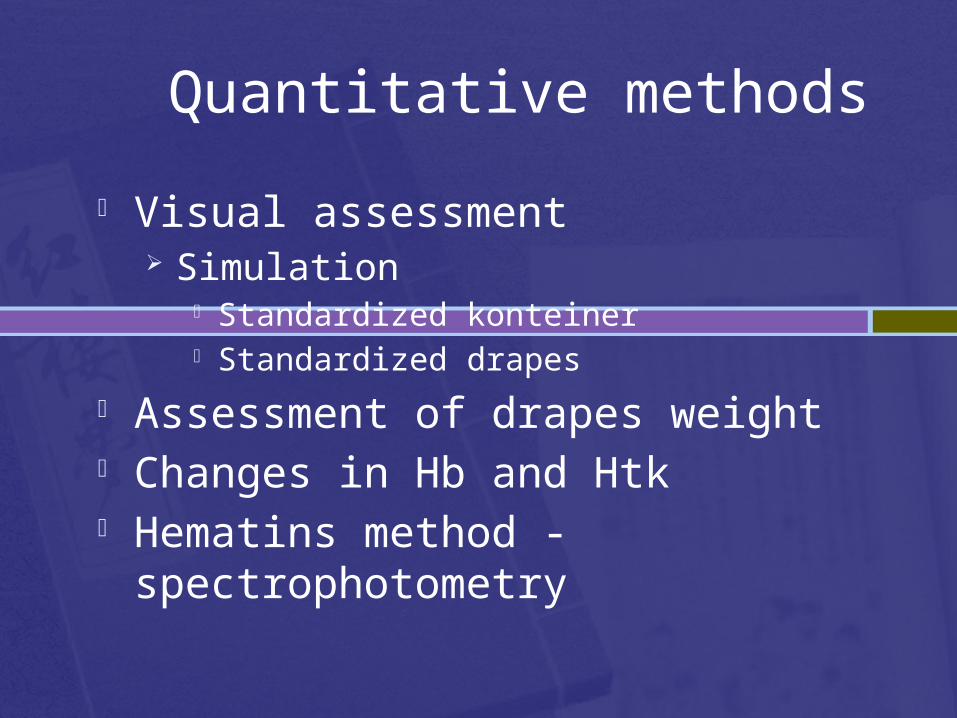
Quantitative methods
Visual assessment Simulation
Standardized konteiner Standardized drapes
Assessment of drapes weight Changes in Hb and Htk Hematins method - spectrophotometry
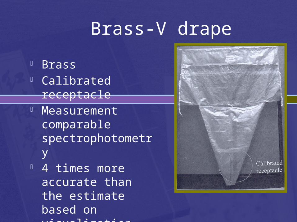
Brass-V drape
Brass Calibrated receptacle Measurement
comparable spectrophotometry
4 times more accurate than the estimate based on visualization
PPH - etiology
The causes of postpartum hemorrhage can be thought of as the four TsToneTissue Trauma Thrombin
Uterine atony
Multiple gestation High parity Prolonged labor Chorioamnionitis Augmented labor Tocolytic agents
Tissue – retained uterine contents
Products of conception Blood clots
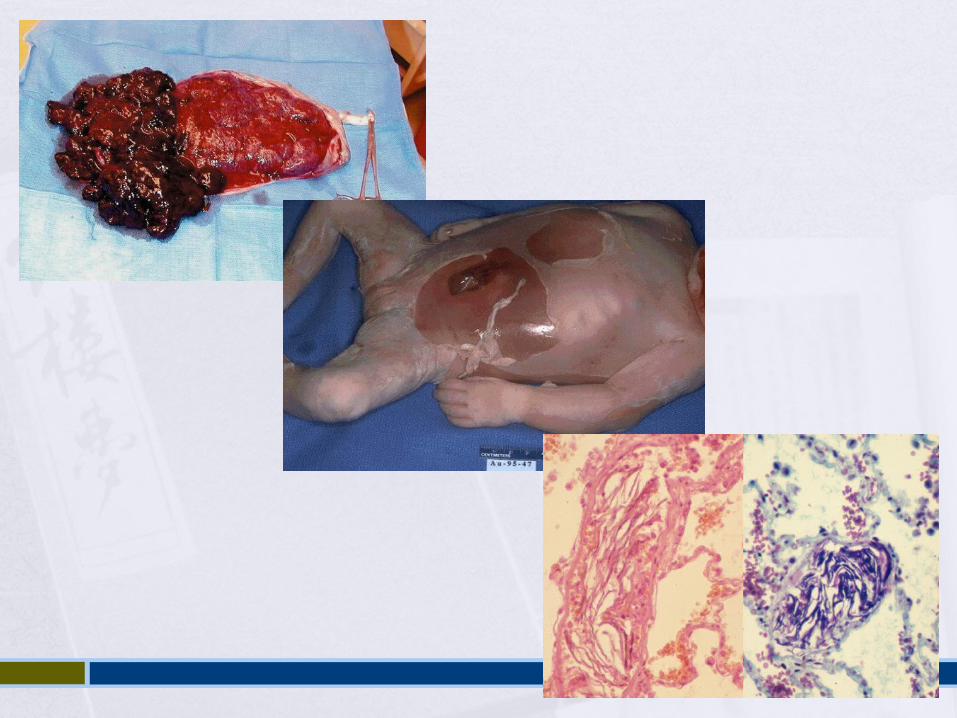
Tissue – placental abnormalities
Congenital – bicorporate uterus Location – placenta praevia Attachment – placenta accreta Acquired structural – previous surgery
Trauma
Planned – SC, episiotomy Unplaned - vaginal/cervical tear, surgical
trauma
Thrombin – coagulation disorders
Congenital - Von Willebrand's disease Acquired – DIC, dilutional coagulopathy,
heparin
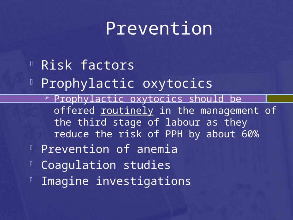
Prevention
Risk factors Prophylactic oxytocics
Prophylactic oxytocics should be offered routinely in the management of the third stage of labour as they reduce the risk of PPH by about 60%
Prevention of anemia Coagulation studies Imagine investigations
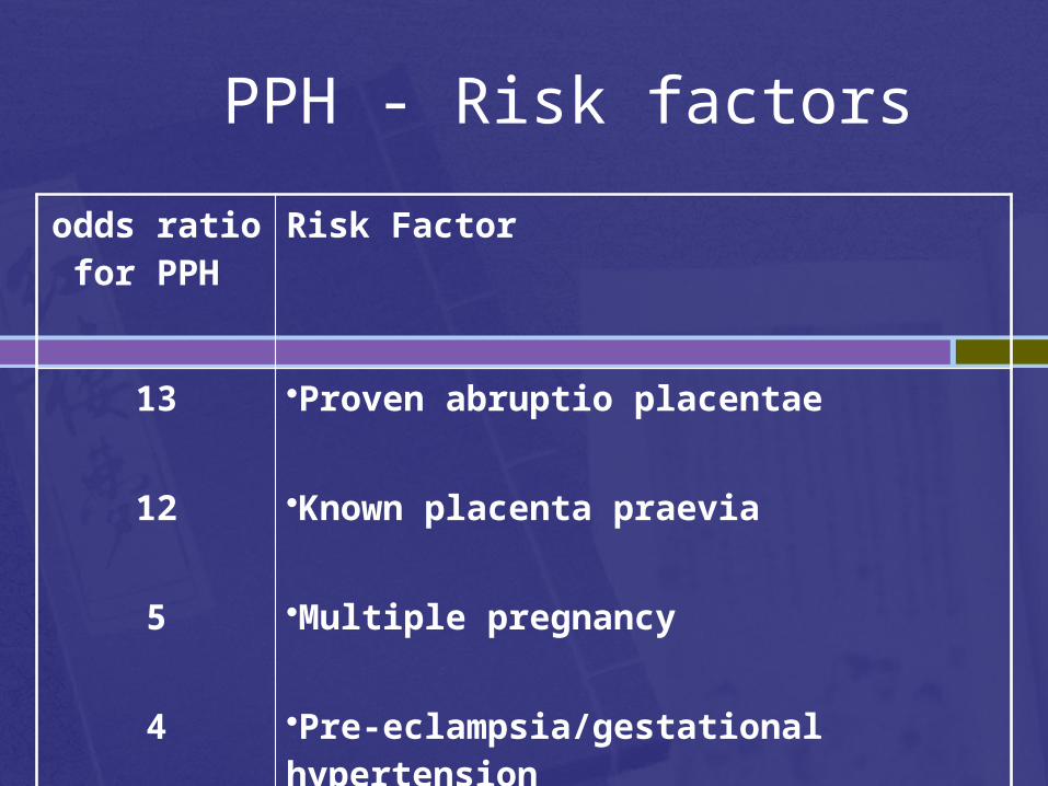
PPH - Risk factors
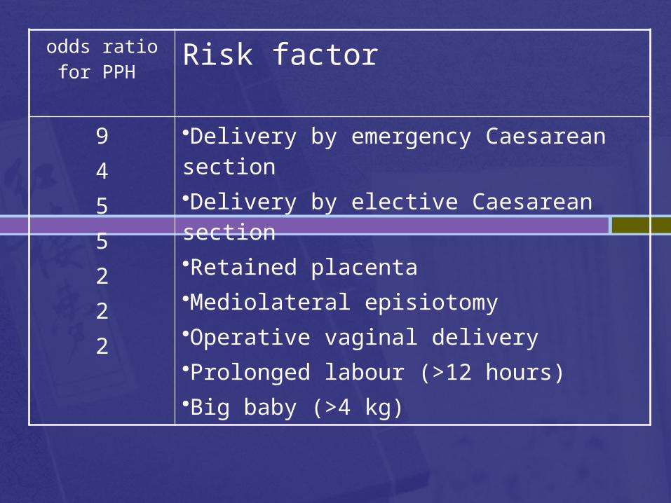
odds ratio for PPH
Risk Factor
13
12
5
4
•Proven abruptio placentae
•Known placenta praevia
•Multiple pregnancy
•Pre-eclampsia/gestational hypertension
odds ratio for PPH Risk factor
9455222
•Delivery by emergency Caesarean section •Delivery by elective Caesarean section •Retained placenta •Mediolateral episiotomy •Operative vaginal delivery •Prolonged labour (>12 hours) •Big baby (>4 kg)
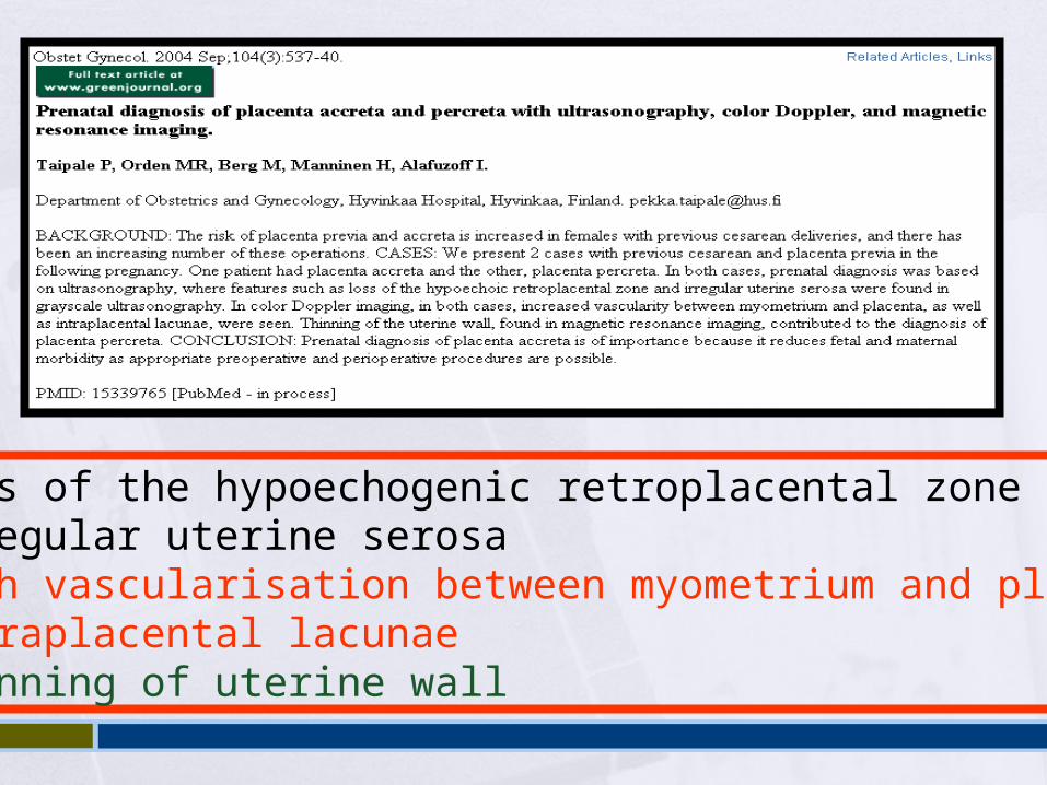
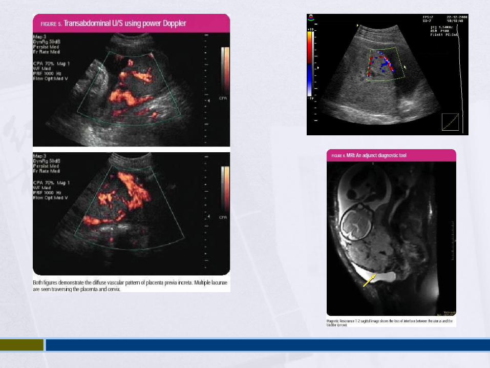
• loss of the hypoechogenic retroplacental zone• irregular uterine serosa• high vascularisation between myometrium and placenta• intraplacental lacunae• thinning of uterine wall
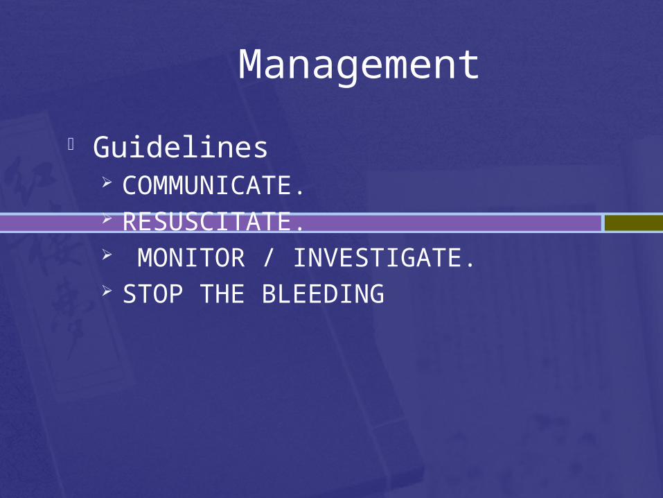
Management
Guidelines COMMUNICATE. RESUSCITATE. MONITOR / INVESTIGATE. STOP THE BLEEDING
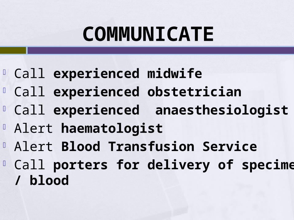
COMMUNICATE
Call experienced midwife Call experienced obstetrician Call experienced anaesthesiologist Alert haematologist Alert Blood Transfusion Service Call porters for delivery of specimens / blood
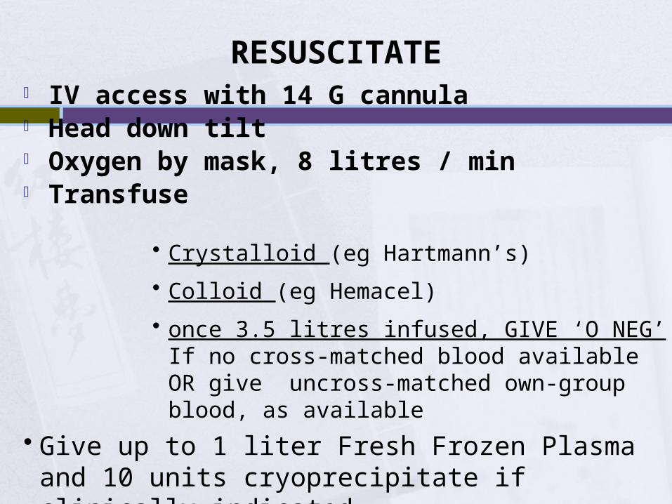
RESUSCITATE IV access with 14 G cannula Head down tilt Oxygen by mask, 8 litres / min Transfuse
• Crystalloid (eg Hartmann’s)
• Colloid (eg Hemacel)
• once 3.5 litres infused, GIVE ‘O NEG’ If no cross-matched blood available OR give uncross-matched own-group blood, as available
• Give up to 1 liter Fresh Frozen Plasma and 10 units cryoprecipitate if clinically indicated
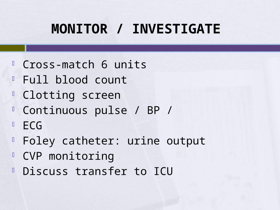
MONITOR / INVESTIGATE
Cross-match 6 units Full blood count Clotting screen Continuous pulse / BP / ECG Foley catheter: urine output CVP monitoring Discuss transfer to ICU
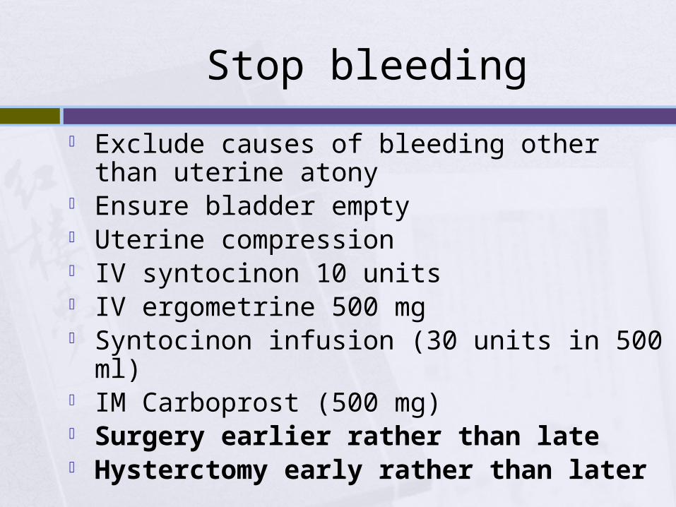
Stop bleeding
Exclude causes of bleeding other than uterine atony
Ensure bladder empty Uterine compression IV syntocinon 10 units IV ergometrine 500 mg Syntocinon infusion (30 units in 500 ml) IM Carboprost (500 mg) Surgery earlier rather than late Hysterctomy early rather than later
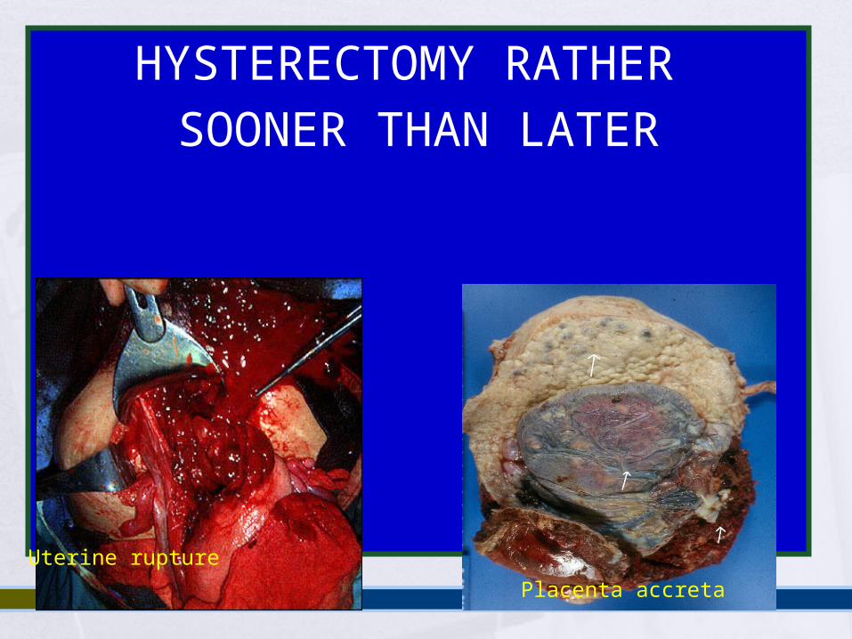
HYSTERECTOMY RATHER SOONER THAN LATER
Uterine rupture
Placenta accreta
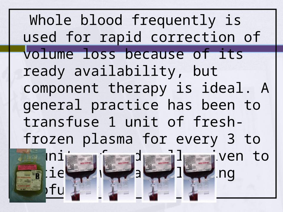
Whole blood frequently is used for rapid correction of volume loss because of its ready availability, but component therapy is ideal. A general practice has been to transfuse 1 unit of fresh-frozen plasma for every 3 to 4 units of red cells given to patients who are bleeding profusely
Genital tract laceration
Genital trauma always must be eliminated first if the uterus is firm
Uterine atony
Explore the uterine cavity. Inspect vagina and cervix for lacerations. If the cavity is empty, massage and give
methylergometrine 0.2 mg, the dose can be repeated every 2 to 4 hours.
Rectal 800mcg. Misoprostol is beneficial (unfortunately is not accesible)
Bimanual compression
Retained placenta
Retained placental fragments are a leading cause of early and delayed postpartum hemorrhage
Treatment is manual removal On rare occasions, a retained placenta is an
undiagnosed placenta accreta, and massive bleeding may occur during attempted manual removal
Placenta accreta
Placenta accreta is defined as an abnormal implantation of the placenta in the uterine wall, of which there are three types:
1. accreta vera, in which the placenta adheres to the myometrium without invasion into the muscle.
2. increta, in which it invades into the myometrium.
3. percreta, in which it invades the full thickness of the uterine wall and possibly other pelvic structures, most frequently the bladder
Placenta accreta
In a patient with a previous cesarean section and a placenta previa Previous one has 14% risk of placenta accreta Previous two has 24% risk of placenta accreta Previous three has 44% risk of placenta accreta
Uterine rupture
Rupture of the uterus is described as complete or incomplete and should be differentiated from dehiscence of a cesarean section scar
Complete rupture describes a full-thickness defect of the uterine wall and serosa resulting in direct communication between the uterine cavity and the peritoneal cavity
Incomplete rupture describes a defect of the uterine wall that is contained by the visceral peritoneum or broad ligament in patients with prior cesarean section
Dehiscence describes partial separation of the scar with minimal bleeding, with the peritoneum and fetal membranes remaining intact
Management
The identification or suspicion of uterine rupture must be followed by an immediate and simultaneous response from the obstetric team
Surgery should not be delayed owing to hypovolemic shock because it may not be easily reversible until the hemorrhage is controlled
Upon entering the abdomen, aortic compression can be applied to decrease bleeding
Oxytocin should be administered to effect uterine contraction to assist in vessel constriction and to decrease bleeding
Hemostasis can then be achieved by ligation of the hypogastric artery, uterine artery, or ovarian arteries
Management At this point, a decision must be made to perform hysterectomy or to
repair the rupture site. In most cases, hysterectomy should be performed
In selected cases, repair of the rupture can be attempted. When rupture occurs in the body of the uterus
Bladder rupture must be ruled out by clearly mobilizing and inspecting the bladder to ensure that it is intact. This avoids injury on repair of the defect as well
A lower segment lateral rupture can cause transection of the uterine vessels. The vessels can retract toward the pelvic side wall, and the site of bleeding must be isolated before placing clamps to avoid injury to the ureter and iliac vessels.
Typically, longitudinal tears, especially those in a lateral position, should be treated by hysterectomy, whereas low transverse tears may be repaired
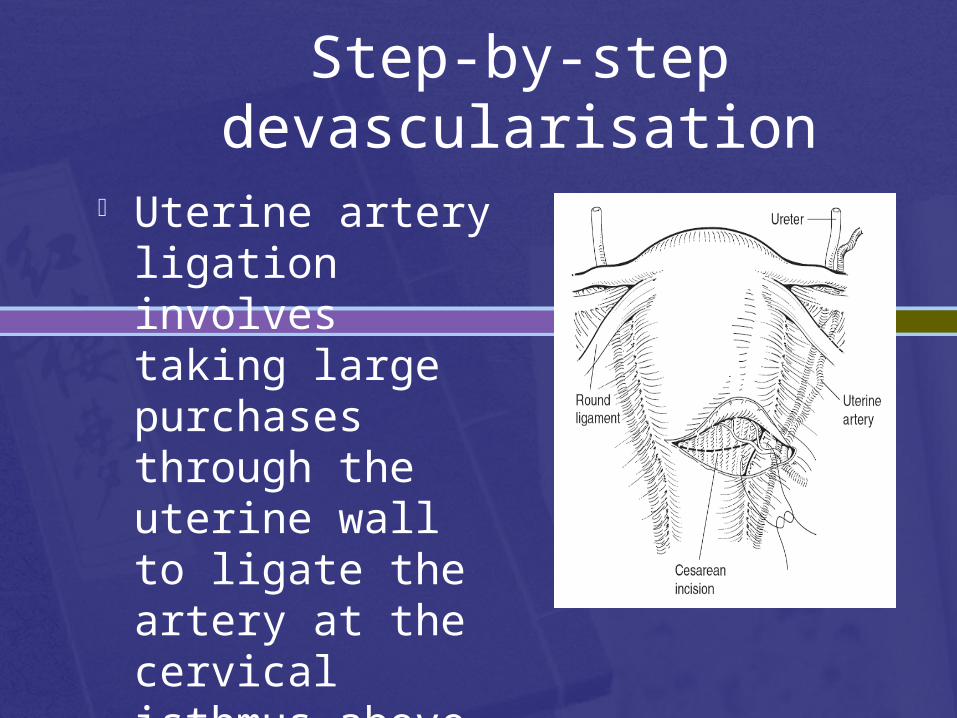
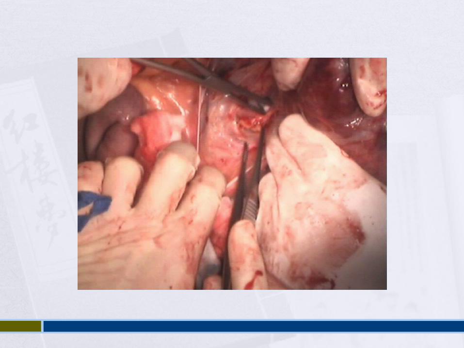
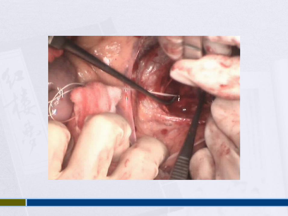
Step-by-step devascularisation
Uterine artery ligation involves taking large purchases through the uterine wall to ligate the artery at the cervical isthmus above the bladder flap
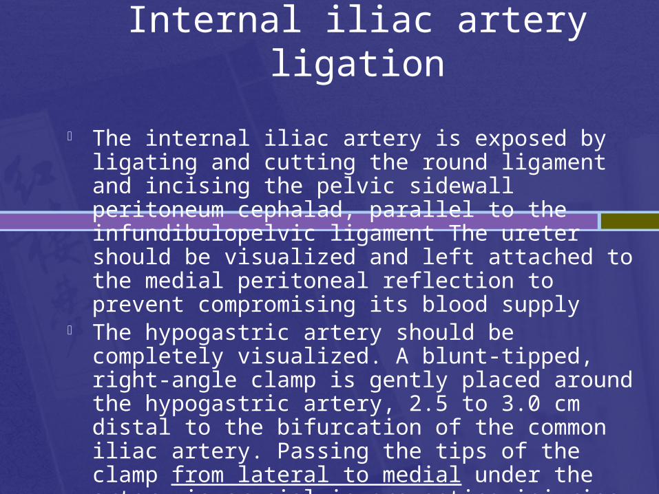
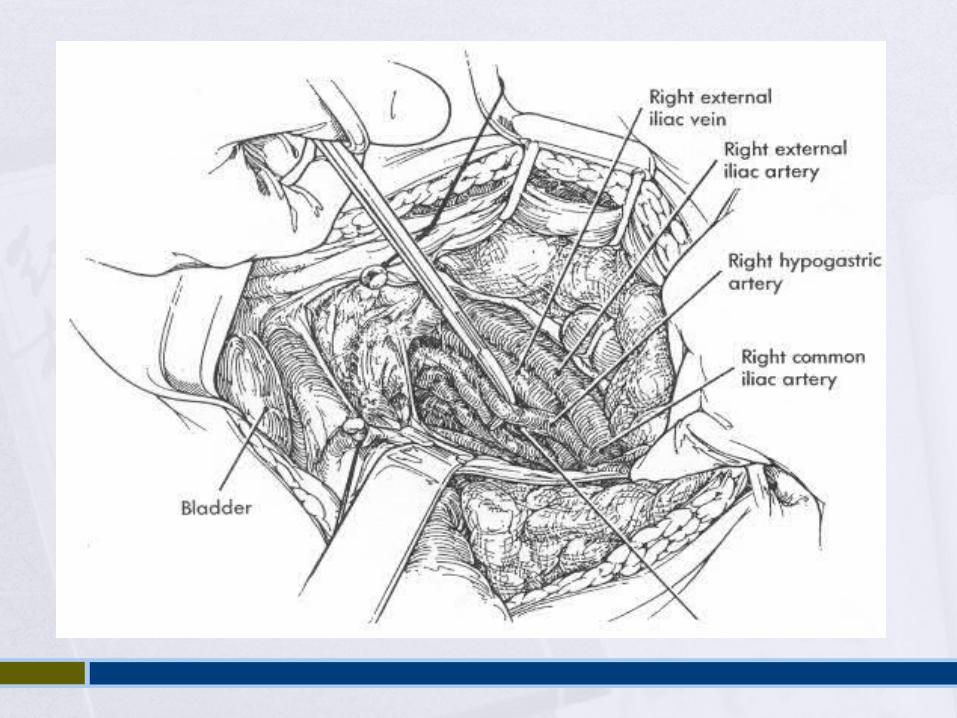
Internal iliac artery ligation
The internal iliac artery is exposed by ligating and cutting the round ligament and incising the pelvic sidewall peritoneum cephalad, parallel to the infundibulopelvic ligament The ureter should be visualized and left attached to the medial peritoneal reflection to prevent compromising its blood supply
The hypogastric artery should be completely visualized. A blunt-tipped, right-angle clamp is gently placed around the hypogastric artery, 2.5 to 3.0 cm distal to the bifurcation of the common iliac artery. Passing the tips of the clamp from lateral to medial under the artery is crucial in preventing injuries to the underlying hypogastric vein
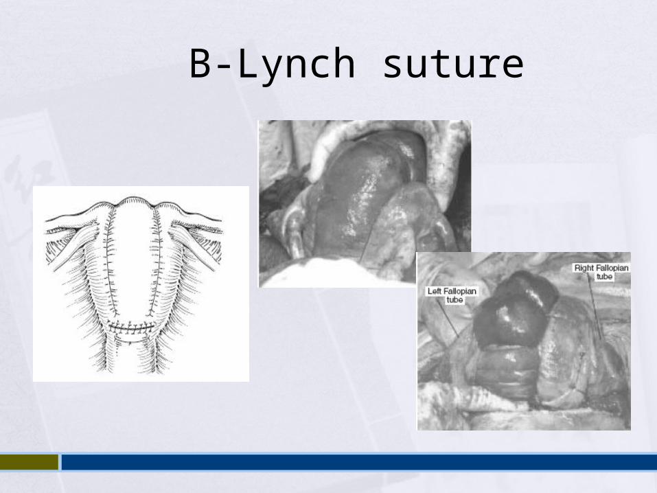
B-Lynch suture
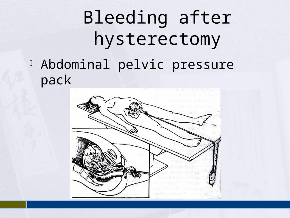
Bleeding after hysterectomy
Abdominal pelvic pressure pack
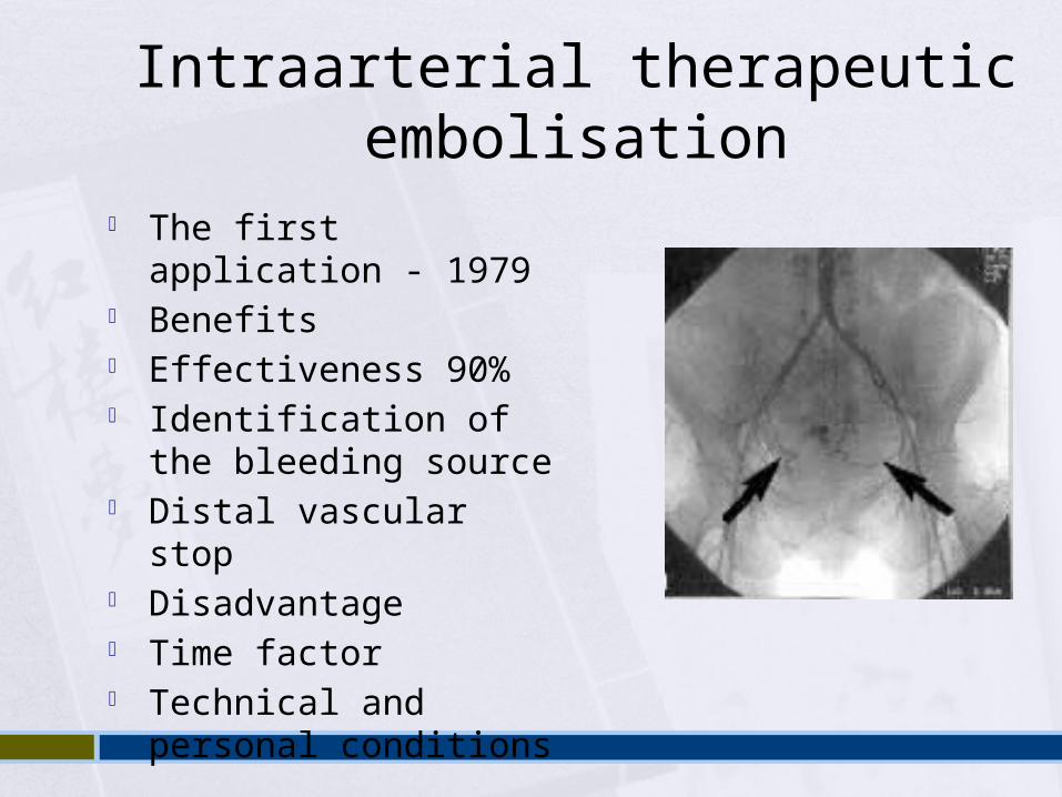
Intraarterial therapeutic embolisation
The first application - 1979
Benefits Effectiveness 90% Identification of the
bleeding source Distal vascular stop Disadvantage Time factor Technical and personal
conditions

Thank you for your attention …[email protected]
Recommended


![Course: Law of the European Union [07] Free Movement of Individuals Filip Křepelka (krepelka@law.muni.cz) Masarykova univerzitakrepelka@law.muni.cz](https://img.pdfslide.us/doc/110x75/56649e7e5503460f94b8233e/course-law-of-the-european-union-07-free-movement-of-individuals-filip-krepelka.jpg)