1/15/2012
1
Patella Instability
Dr. Tudor H. Hughes M.D., FRCRDepartment of RadiologyUniversity of California School of MedicineSan Diego, California
• Presentations:
• Subluxation or Dislocation
Patella Instability
• Maltracking with Anterior knee pain
Lateral Patellar Dislocation
• Anteroposterior radiograph of the knee showing a laterally dislocated patella. The patella usually spontaneously reduces and this appearance is rare.
• The patella is reduced, but note the osteochondralfragment adjacent to the medial patella and the small concave defect at the medial patellar margin.
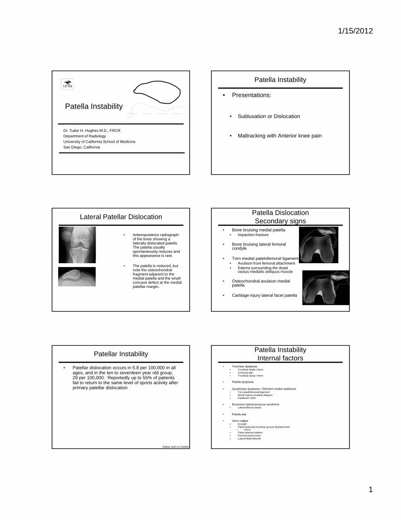
Patella DislocationSecondary signs
• Bone bruising medial patella• Impaction fracture
• Bone bruising lateral femoral condyle
• Torn medial patelofemoral ligamento ed a pate o e o a ga e t• Avulsion from femoral attachment• Edema surrounding the distal
vastus medialis obliquus muscle
• Osteochondral avulsion medial patella
• Cartilage injury lateral facet patella
Patellar Instability
• Patellar dislocation occurs in 5.8 per 100,000 in all ages, and in the ten to seventeen year old group, 29 per 100,000. Reportedly up to 55% of patients fail to return to the same level of sports activity after primary patellar dislocation
Dejour and Le Coultre
Patella InstabilityInternal factors
• Trochlear dysplasia• Trochlear depth <4mm• Crossing sign• Trochlear bump >3mm
• Patella dysplasia
• Quadriceps dysplasia / Deficient medial stabilizers• Torn patellofemoral ligament• Weak Vastus medialis obliquus• Patella tilt >20%
• Excessive lateral pressure syndrome• Lateral fibrous bands
• Patella alta
• Genu valgus• Q angle• Tibial tuberosity trochlear groove displacement
• >20mm• Tibial external rotation• Femoral anteversion• Lateral tibial tubercle
1/15/2012
2
Patella Instability Patellar tracking
• First 20 degrees of flexion• Tibia derotates (internally)• Q angle decreases• Patella enters trochlea laterally
• 20 - 30 degrees of flexion• Patella lifts away from trochlea• Patella most prominent
• Above 30 degrees of flexion• Patella settles into trochlea
Patellar tracking
• 30 - 90 degrees of flexion• Stability rarely a problem• Instability seen with anatomic derangement
• 90 - 135 degrees of flexion90 135 degrees of flexion• Patella shift laterally• Patella rotates (pronates)
• Full flexion• Further lateral shift
History• Skeletal findings prove that the knee joint has been in
existence for over 320 million years
• The Eryops, the ancestors of the reptiles, birds and mammals, seems to be the first creature in the animal kingdom with a bicondylar knee joint.
Th t ll f l j i t h l b t d l• The patellofemoral joint, however, only began to develop some 65 million years ago.
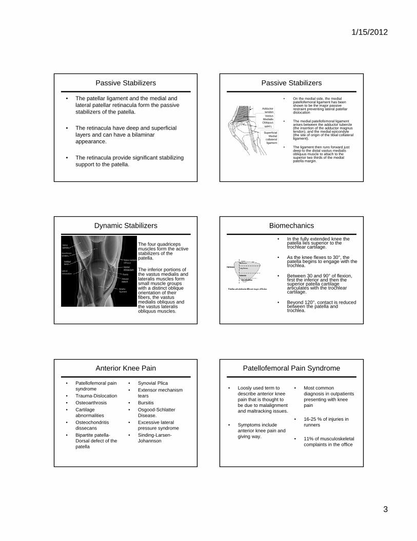
Patella Facets
• The posterior surface of the patella articulates with the trochlear groove along the anterior surface of the femoral condyles to form the patellofemoral joint.
• The posterior patella has a medial and lateral facet. A variable, usually small, odd facet lies along the medial border of the patella.
Patellar facets
Lateral
Medial
Odd
1/15/2012
3
Passive Stabilizers
• The patellar ligament and the medial and lateral patellar retinacula form the passive stabilizers of the patella.
• The retinacula have deep and superficial• The retinacula have deep and superficial layers and can have a bilaminar appearance.
• The retinacula provide significant stabilizing support to the patella.
Passive Stabilizers
• On the medial side, the medial patellofemoral ligament has been shown to be the major passive restraint preventing lateral patellar dislocation
• The medial patellofemoral ligament arises between the adductor tubercle (the insertion of the adductor magnus
Adductor tendonVastus
MedialisObliquus
MPFL (the insertion of the adductor magnus tendon), and the medial epicondyle (the site of origin of the tibial collateral ligament).
• The ligament then runs forward just deep to the distal vastus medialis obliquus muscle to attach to the superior two thirds of the medial patella margin.
Superficial Medial
collateral ligament
Dynamic Stabilizers
• The four quadriceps muscles form the active stabilizers of the patella.
• The inferior portions ofThe inferior portions of the vastus medialis and lateralis muscles form small muscle groups with a distinct oblique orientation of their fibers, the vastus medialis obliquus and the vastus lateralis obliquus muscles.
Biomechanics
• In the fully extended knee the patella lies superior to the trochlear cartilage.
• As the knee flexes to 30°, the patella begins to engage with the trochlea.
• Between 30 and 90° of flexion, first the inferior and then the superior patella cartilage articulates with the trochlear cartilage.
• Beyond 120°, contact is reduced between the patella and trochlea.
Anterior Knee Pain
• Patellofemoral pain syndrome
• Trauma-Dislocation• Osteoarthrosis• Cartilage
• Synovial Plica• Extensor mechanism
tears• Bursitis• Osgood-Schlatter
abnormalities• Osteochondritis
dissecans• Bipartite patella-
Dorsal defect of the patella
Disease. • Excessive lateral
pressure syndrome• Sinding-Larsen-
Johannson
Patellofemoral Pain Syndrome
• Loosly used term to describe anterior knee pain that is thought to be due to malalignmentand maltracking issues.
• Most common diagnosis in outpatients presenting with knee pain
g
• Symptoms include anterior knee pain and giving way.
• 16-25 % of injuries in runners
• 11% of musculoskeletal complaints in the office
1/15/2012
4
Definitions
• Patellofemoral alignment refers to the static relationship between the patella and the trochlea at a given degree of knee flexion.
• Patellofemoral tracking refers to the dynamic patellofemoral alignment during knee motion.
Limitations of Radiology
• Measures of alignment will vary depending on the degree of knee flexion.
• Imaging studies of the patellofemoral joint for tracking should focus on the first 30-45 degrees of flexion In early flexion is when anatomical factorsflexion. In early flexion is when anatomical factors such as patella alta, trochlear dysplasia and abnormalities of the soft tissue restraints of the patella have the most pronounced effect in producing abnormal tracking.
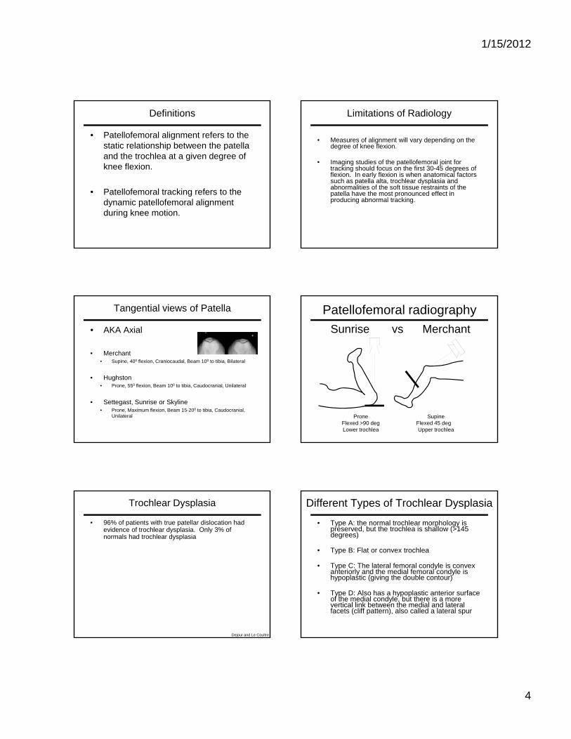
Tangential views of Patella
• AKA Axial
• Merchant• Supine, 400 flexion, Craniocaudal, Beam 100 to tibia, Bilateral
• Hughston• Prone, 550 flexion, Beam 100 to tibia, Caudocranial, Unilateral
• Settegast, Sunrise or Skyline• Prone, Maximum flexion, Beam 15-200 to tibia, Caudocranial,
Unilateral
Patellofemoral radiographySunrise vs Merchant
ProneFlexed >90 degLower trochlea
SupineFlexed 45 degUpper trochlea
Trochlear Dysplasia
• 96% of patients with true patellar dislocation had evidence of trochlear dysplasia. Only 3% of normals had trochlear dysplasia
Dejour and Le Coultre
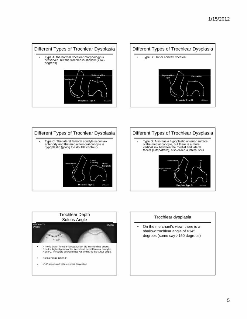
Different Types of Trochlear Dysplasia
• Type A: the normal trochlear morphology is preserved, but the trochlea is shallow (>145 degrees)
• Type B: Flat or convex trochlea
T C Th l t l f l d l i• Type C: The lateral femoral condyle is convex anteriorly and the medial femoral condyle is hypoplastic (giving the double contour)
• Type D: Also has a hypoplastic anterior surface of the medial condyle, but there is a more vertical link between the medial and lateral facets (cliff pattern), also called a lateral spur
1/15/2012
5
Different Types of Trochlear Dysplasia• Type A: the normal trochlear morphology is
preserved, but the trochlea is shallow (>145 degrees)
Different Types of Trochlear Dysplasia• Type B: Flat or convex trochlea
Different Types of Trochlear Dysplasia• Type C: The lateral femoral condyle is convex
anteriorly and the medial femoral condyle is hypoplastic (giving the double contour)
Different Types of Trochlear Dysplasia• Type D: Also has a hypoplastic anterior surface
of the medial condyle, but there is a more vertical link between the medial and lateral facets (cliff pattern), also called a lateral spur
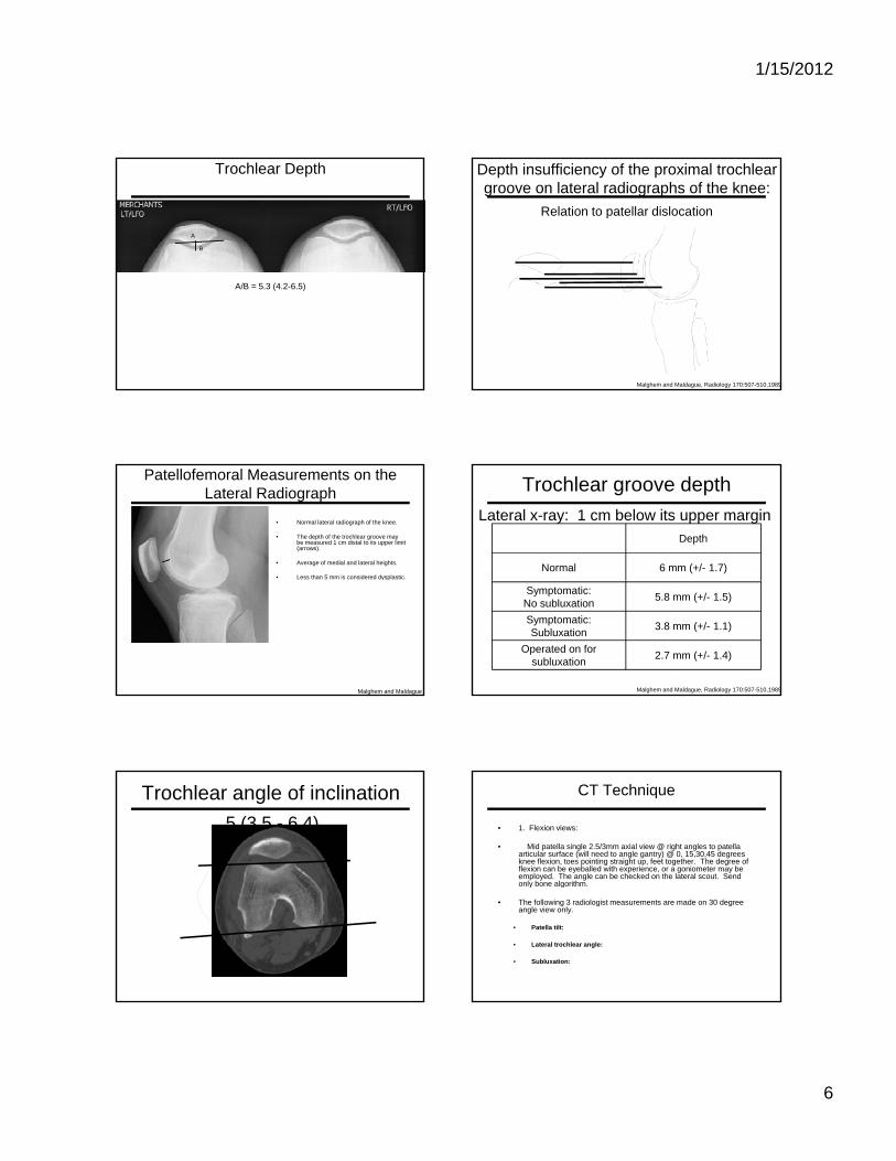
Trochlear DepthSulcus Angle
BA C
1400
• A line is drawn from the lowest point of the intercondylar sulcus, B, to the highest points of the lateral and medial femoral condyles, A and C. The angle between lines AB and BC is the sulcus angle.
• Normal range 136+/–6°
• >145 associated with recurrent dislocation
Trochlear dysplasia
• On the merchant’s view, there is a shallow trochlear angle of >145 degrees (some say >150 degrees)
1/15/2012
6
Trochlear Depth
A
B
A/B = 5.3 (4.2-6.5)
Depth insufficiency of the proximal trochlear groove on lateral radiographs of the knee:
Relation to patellar dislocation
Malghem and Maldague, Radiology 170:507-510,1989
Patellofemoral Measurements on the Lateral Radiograph
• Normal lateral radiograph of the knee.
• The depth of the trochlear groove may be measured 1 cm distal to its upper limit (arrows).
• Average of medial and lateral heights.
• Less than 5 mm is considered dysplastic. y p
Malghem and Maldague
Trochlear groove depthLateral x-ray: 1 cm below its upper margin
Depth
Normal 6 mm (+/- 1.7)
Malghem and Maldague, Radiology 170:507-510,1989
Symptomatic:No subluxation 5.8 mm (+/- 1.5)
Symptomatic:Subluxation 3.8 mm (+/- 1.1)
Operated on for subluxation 2.7 mm (+/- 1.4)
Trochlear angle of inclination5 (3.5 - 6.4)
CT Technique
• 1. Flexion views:
• Mid patella single 2.5/3mm axial view @ right angles to patella articular surface (will need to angle gantry) @ 0, 15,30,45 degrees knee flexion, toes pointing straight up, feet together. The degree of flexion can be eyeballed with experience, or a goniometer may be employed. The angle can be checked on the lateral scout. Send only bone algorithm.o y bo e a go t
• The following 3 radiologist measurements are made on 30 degree angle view only.
• Patella tilt:
• Lateral trochlear angle:
• Subluxation:
1/15/2012
7
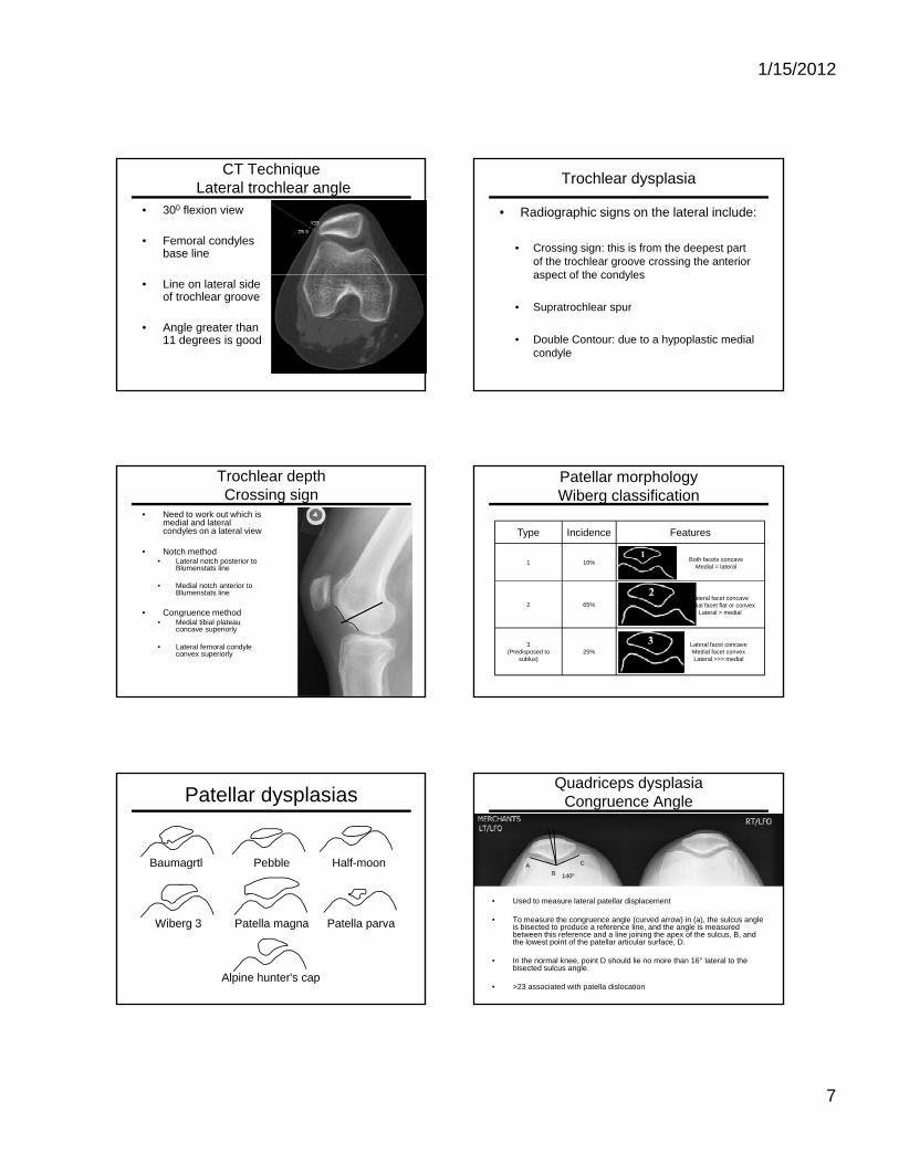
CT TechniqueLateral trochlear angle
• 300 flexion view
• Femoral condyles base line
• Line on lateral side of trochlear groove
• Angle greater than 11 degrees is good
Trochlear dysplasia
• Radiographic signs on the lateral include:
• Crossing sign: this is from the deepest part of the trochlear groove crossing the anterior aspect of the condylesaspect of the condyles
• Supratrochlear spur
• Double Contour: due to a hypoplastic medial condyle
Trochlear depthCrossing sign
• Need to work out which is medial and lateral condyles on a lateral view
• Notch method• Lateral notch posterior to
Blumenstats line
• Medial notch anterior to Blumenstats line
• Congruence method• Medial tibial plateau
concave superiorly
• Lateral femoral condyle convex superiorly
Patellar morphologyWiberg classification
Type Incidence Features
1 10% Both facets concaveMedial = lateral
2 65%Lateral facet concave
Medial facet flat or convexLateral > medial
3 (Predisposed to
sublux)25%
Lateral facet concaveMedial facet convexLateral >>> medial
Patellar dysplasias
Baumagrtl Pebble Half-moon
Wiberg 3
Alpine hunter's cap
Patella magna Patella parva
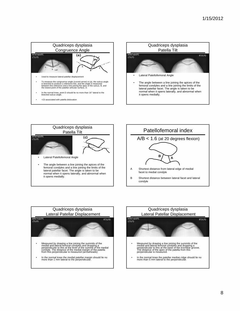
Quadriceps dysplasiaCongruence Angle
BA C
1400
• Used to measure lateral patellar displacement
• To measure the congruence angle (curved arrow) in (a), the sulcus angle is bisected to produce a reference line, and the angle is measured between this reference and a line joining the apex of the sulcus, B, and the lowest point of the patellar articular surface, D.
• In the normal knee, point D should lie no more than 16° lateral to the bisected sulcus angle.
• >23 associated with patella dislocation
1/15/2012
8
Quadriceps dysplasiaCongruence Angle
BA C
1400
• Used to measure lateral patellar displacement
• To measure the congruence angle (curved arrow) in (a), the sulcus angle is bisected to produce a reference line, and the angle is measured between this reference and a line joining the apex of the sulcus, B, and the lowest point of the patellar articular surface, D.
• In the normal knee, point D should lie no more than 16° lateral to the bisected sulcus angle.
• >23 associated with patella dislocation
Quadriceps dysplasiaPatella Tilt
• Lateral Patellofemoral Angle
• The angle between a line joining the apices of the femoral condyles and a line joining the limits of the lateral patellar facet. The angle is taken to be normal when it opens laterally, and abnormal when it opens medially.
Quadriceps dysplasiaPatella Tilt
• Lateral Patellofemoral Angle
• The angle between a line joining the apices of the femoral condyles and a line joining the limits of the lateral patellar facet. The angle is taken to be normal when it opens laterally, and abnormal when it opens medially.
Patellofemoral indexA/B < 1.6 (at 20 degrees flexion)
B AB
A Shortest distance from lateral edge of medial facet to medial condyle
B Shortest distance between lateral facet and lateral condyle
Quadriceps dysplasiaLateral Patellar Displacement
• Measured by drawing a line joining the summits of the medial and lateral femoral condyles and dropping a perpendicular to this at the level of the summit of the medial condyle. The distance of the medial margin of the patella from this perpendicular is measured (arrowheads).
• In the normal knee the medial patellar margin should lie no more than 1 mm lateral to the perpendicular.
Quadriceps dysplasiaLateral Patellar Displacement
• Measured by drawing a line joining the summits of the medial and lateral femoral condyles and dropping a perpendicular to this at the base of the trochlear groove. The distance of the apex of the patella from this perpendicular is measured.
• In the normal knee the patellar median ridge should lie no more than 5 mm lateral to the perpendicular.
1/15/2012
9
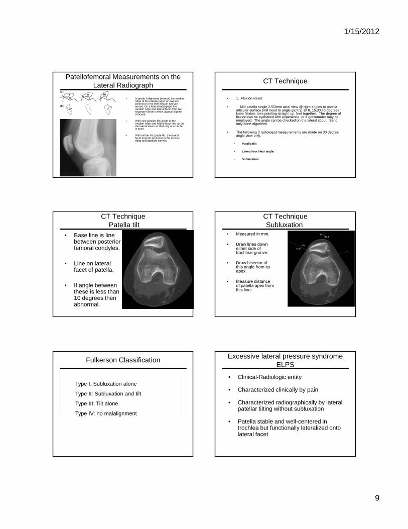
Patellofemoral Measurements on the Lateral Radiograph
• In grade I alignment (normal) the median ridge of the patella (open arrow) lies posterior to the lateral facet (curved arrow). On a lateral radiograph the median ridge and lateral facet form two separate borders which appear slightly concave.
• With mild patellar tilt (grade II) the median ridge and lateral facet line up on g pthe lateral views so that only one border is seen.
• With further tilt (grade III), the lateral facet projects posterior to the median ridge and appears convex.
CT Technique
• 1. Flexion views:
• Mid patella single 2.5/3mm axial view @ right angles to patella articular surface (will need to angle gantry) @ 0, 15,30,45 degrees knee flexion, toes pointing straight up, feet together. The degree of flexion can be eyeballed with experience, or a goniometer may be employed. The angle can be checked on the lateral scout. Send only bone algorithm.only bone algorithm.
• The following 3 radiologist measurements are made on 30 degree angle view only.
• Patella tilt:
• Lateral trochlear angle:
• Subluxation:
CT TechniquePatella tilt
• Base line is line between posterior femoral condyles.
• Line on lateralLine on lateral facet of patella.
• If angle between these is less than 10 degrees then abnormal.
CT TechniqueSubluxation
• Measured in mm.
• Draw lines down either side of trochlear groove.
• Draw bisector of this angle from its apex.
• Measure distance of patella apex from this line.
Type I: Subluxation alone
Type II: Subluxation and tilt
Type III: Tilt alone
Fulkerson Classification
Type III: Tilt alone
Type IV: no malalignment
Excessive lateral pressure syndromeELPS
• Clinical-Radiologic entity
• Characterized clinically by pain
• Characterized radiographically by lateralCharacterized radiographically by lateral patellar tilting without subluxation
• Patella stable and well-centered in trochlea but functionally lateralized onto lateral facet
1/15/2012
10
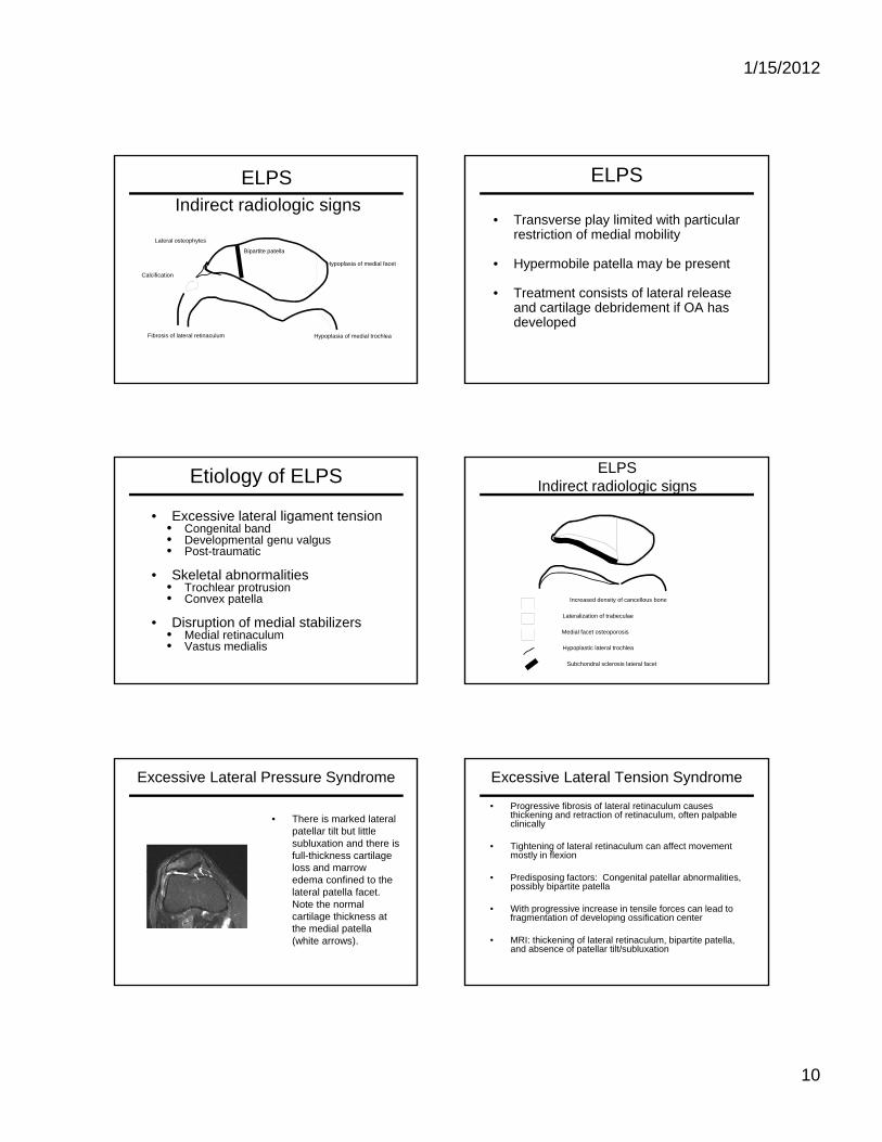
ELPSIndirect radiologic signs
Hypoplasia of medial facet
Bipartite patella
Lateral osteophytes
Hypoplasia of medial trochleaFibrosis of lateral retinaculum
Calcification
ELPS
• Transverse play limited with particular restriction of medial mobility
• Hypermobile patella may be presentyp p y p
• Treatment consists of lateral release and cartilage debridement if OA has developed
Etiology of ELPS
• Excessive lateral ligament tension• Congenital band• Developmental genu valgus• Post-traumatic
Sk l t l b liti• Skeletal abnormalities• Trochlear protrusion• Convex patella
• Disruption of medial stabilizers• Medial retinaculum• Vastus medialis
ELPSIndirect radiologic signs
Increased density of cancellous bone
Lateralization of trabeculae
Medial facet osteoporosis
Hypoplastic lateral trochlea
Subchondral sclerosis lateral facet
Excessive Lateral Pressure Syndrome
• There is marked lateral patellar tilt but little subluxation and there is full-thickness cartilage loss and marrow edema confined to the lateral patella facet. Note the normal cartilage thickness at the medial patella (white arrows).
Excessive Lateral Tension Syndrome
• Progressive fibrosis of lateral retinaculum causes thickening and retraction of retinaculum, often palpable clinically
• Tightening of lateral retinaculum can affect movement mostly in flexion
• Predisposing factors: Congenital patellar abnormalities, possibly bipartite patella
• With progressive increase in tensile forces can lead to fragmentation of developing ossification center
• MRI: thickening of lateral retinaculum, bipartite patella, and absence of patellar tilt/subluxation
1/15/2012
11
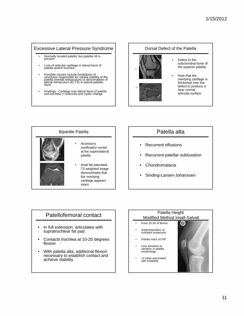
Excessive Lateral Pressure Syndrome• Normally located patella, but patellar tilt is
present
• Loss of articular cartilage in lateral facet of patella and/or trochlea
• Possible causes include breakdown of• Possible causes include breakdown of structures responsible for medial stability of the patella (medial retinaculum) or abnormalities of lateral retinaculum (ELTS) or lateral patellar facet
• Findings: Cartilage loss lateral facet of patella and trochlea +/-sclerosis and cystic change
Dorsal Defect of the Patella
• Defect in the subchondral bone of the superior patella.
N t th t th
T1
• Note that the overlying cartilage is thickened over the defect to produce a near normal articular surface
T2
Bipartite Patella
• Accessory ossification center at the superolateral patella.
• Axial fat-saturated T2-weighted image demonstrates that the overlying cartilage appears intact.
Patella alta
• Recurrent effusions
• Recurrent patellar subluxation
• Chondromalacia
• Sinding-Larsen-Johanssen
Patellofemoral contact
• In full extension, articulates with supratrochlear fat pad
• Contacts trochlea at 10-20 degrees gflexion
• With patella alta, additional flexion necessary to establish contact and achieve stability
Patella HeightModified Method Insall-Salvati
• Knee 20-30 of flexion
• Superimposition of condyles posteriorly
• Patella index AT/AP P
• Less sensitive to variation in patella morphology
• >2 (alta) associated with instability
A
T
1/15/2012
12
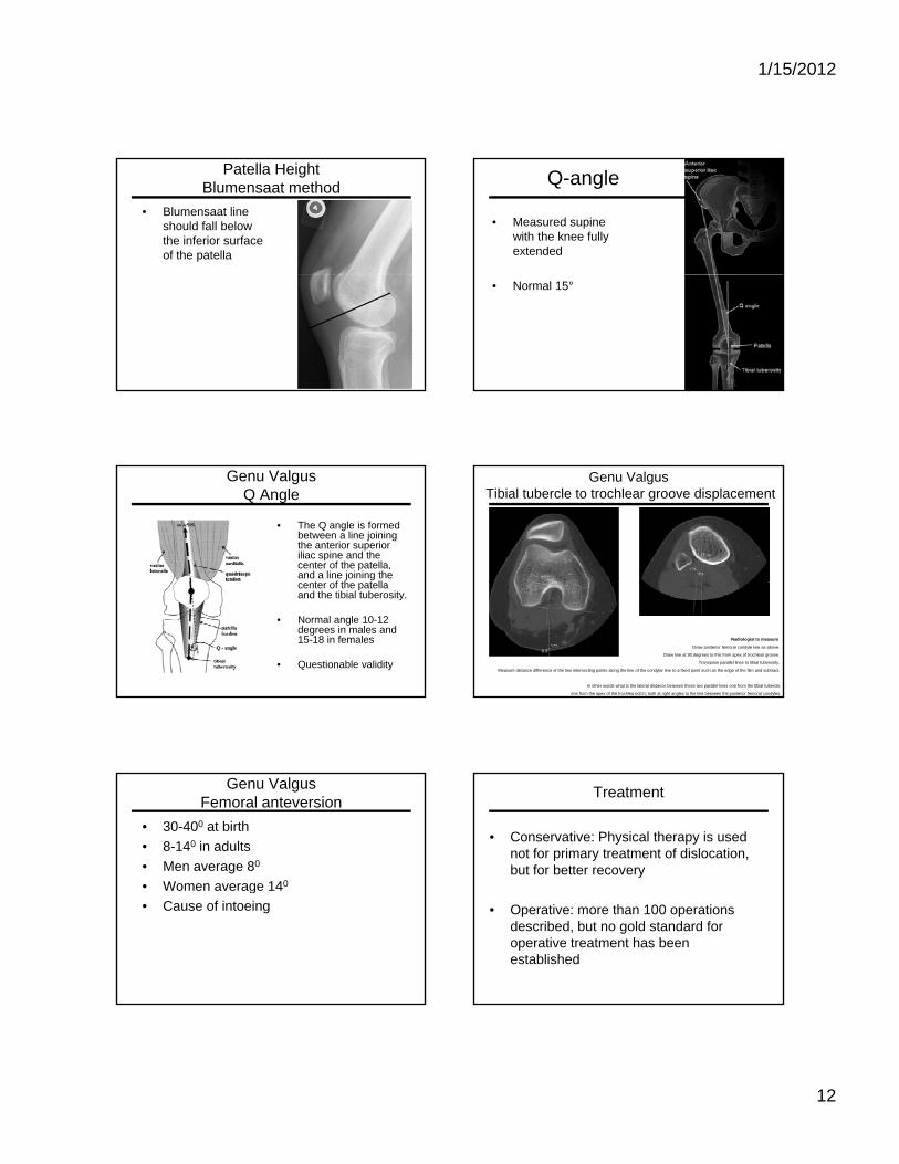
Patella HeightBlumensaat method
• Blumensaat line should fall below the inferior surface of the patella
Q-angle
• Measured supine with the knee fully extended
• Normal 15°
Genu ValgusQ Angle
• The Q angle is formed between a line joining the anterior superior iliac spine and the center of the patella, and a line joining the
t f th t llcenter of the patella and the tibial tuberosity.
• Normal angle 10-12 degrees in males and 15-18 in females
• Questionable validity
Genu Valgus Tibial tubercle to trochlear groove displacement
Radiologist to measure
Draw posterior femoral condyle line as above.
Draw line at 90 degrees to this from apex of trochlear groove.
Transpose parallel lines to tibial tuberosity.
Measure distance difference of the two intersecting points along the line of the condylar line to a fixed point such as the edge of the film and subtract.
In other words what is the lateral distance between these two parallel lines one from the tibial tubercle,
one from the apex of the trochlea notch, both at right angles to the line between the posterior femoral condyles.
• 30-400 at birth• 8-140 in adults• Men average 80
• Women average 140
Genu ValgusFemoral anteversion
Women average 14• Cause of intoeing
Treatment
• Conservative: Physical therapy is used not for primary treatment of dislocation, but for better recovery
• Operative: more than 100 operations described, but no gold standard for operative treatment has been established
1/15/2012
13
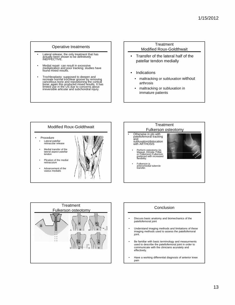
Operative treatments
• Lateral release: the only treatment that has actually been shown to be definitively INEFFECTIVE.
• Medial repair: can result in excessive medialization and poor tracking; studies have found mixed results.
• Trochleoplasty: supposed to deepen and recreate normal trochlear groove by removing cancellous bone and repositioning the cortical bone; again this produced mixed results. It has limited use in the US due to concerns about irreversible articular and subchondral injury.
TreatmentModified Roux-Goldthwait
• Transfer of the lateral half of the patellar tendon medially
Indications• Indications• maltracking or subluxation without
arthrosis• maltracking or subluxation in
immature patients
Modified Roux-Goldthwait
• Procedure• Lateral patellar
retinacular release
• Medial transfer of the lateral aspect patellarlateral aspect patellar tendon
• Plication of the medial retinaculum
• Advancement of the vastus medialis
TreatmentFulkerson osteotomy
• Otherwise in pts with patellofemoral tracking and subluxation/dislocation with ARTHOSIS
• Perform osteotomy (ie. Maquet Elmslie-TrillatMaquet, Elmslie Trillat, or Fulkerson) Fulkerson preferred with increased flexibility.
• Fulkerson is anteromedial tubercle transfer.
TreatmentFulkerson osteotomy Conclusion
• Discuss basic anatomy and biomechanics of the patellofemoral joint
• Understand imaging methods and limitations of these imaging methods used to assess the patellofemoral g g pjoint.
• Be familiar with basic terminology and measurments used to describe the patellofemoral joint in order to communicate with the clinicians acurately and effectively.
• Have a working differential diagnosis of anterior knee pain
Recommended