2013, 1 (3), 273-290
273
Particle Size and Agglomeration Affect the Toxicity Levels of Silver
Nanoparticle Types in Aquatic Environment
Mohammad Reza Kalbassi1*
, Seyed Ali Johari2, Mahdi Soltani
3 and Il Je Yu
4
1 Professor, Faculty of Marine Sciences, Tarbiat Modares University, Noor, Iran 2 Assistant Professor, Faculty of Natural Resources, University of Kurdistan, Sanandaj, Iran 3 Professor, Faculty of Veterinary Medicine, Tehran University, Iran 4 Professor, Toxicological Research Center, Hoseo University, Sechul-ri, Baebang-myun, Asan , Sonth Korea
Received: 25 August 2011 / Accepted: 10 June 2013 / Published Online: 24 November 2013
ABSTRACT In order to understand the importance of particle size and agglomeration for nano-
eco-toxicological studies in aquatic environments, the acute toxicity of two different types
(suspended powder and colloidal) of silver nanoparticles (AgNPs) were studied in alevin and
juvenile rainbow trout. Fish were exposed to each type of AgNPs at nominal concentrations of
0.032, 0.1, 0.32, 1, 3.2, 10, 32, and 100 mgl-1
. Lethal concentrations (LC) were calculated using a
Probit analysis. Some physical and chemical characteristics of silver nanoparticles were
determined. In the case of colloidal form, particles were well dispersed in the water column and
retained their size; but in the case of suspended powder, particles were agglomerated to large
clumps and precipitated on the bottom. In alevins, the calculated 96 h LC50 values were 0.25 and
28.25 mgl-1
for colloidal and suspended powder AgNPs respectively. In the case of juveniles, the
96h LC50 of colloidal form was 2.16 mgl-1
, but suspended powder did not caused mortality in fish
even after 21 days. The results showed that both in alevin and juvenile stages, colloidal form is
much toxic than suspended powder; this shows increase of nanoparticles size due to
agglomeration, will reduce the toxicity. Silver nanoparticles are toxic materials and their release
into the water environment should be avoided.
Key words: Aquatic Nanotoxicology, Agglomeration, Rainbow trout, Silver Nanoparticle, Size-
Dependent Toxicity
1 INTRODUCTION
Manufactured nanomaterials are materials with
diameters ranging from 1 to 100nm, and
nanotechnology is one of the rapid growing parts
of the new technology. Although the applications
of nanoparticles are increasing broadly in many
fields, concerns about their environmental and
health impacts remain unresolved. The use of
nanomaterials is also likely to result in their
release into aquatic environments and may pose
risks to the aquatic ecosystems. The aquatic
ecotoxicology of engineered nanomaterials,
aquatic nanoecotoxicology, is a relatively new
and evolving field.
Silver nanoparticles, have been, and
continue to be, recognized worldwide as either
a cure or as a preventive for bacterial, fungal,
and viral diseases (Murr, 2008). Nano silver is
*Corresponding author: Faculty of Marine Sciences, Tarbiat Modares University, Noor, Iran, Tel: +98 122 625 3101,
+98 911 220 4336, E-mail: [email protected]
[ D
OR
: 20.
1001
.1.2
3222
700.
2013
.1.3
.5.9
]
[ D
ownl
oade
d fr
om e
cope
rsia
.mod
ares
.ac.
ir o
n 20
22-0
3-24
]
1 / 18

M.R. Kalbassi et al. _________________________________________ ECOPERSIA (2013) Vol. 1(3)
274
about 24 percentages of commonly used
nanomaterials in consumer products (Woodrow
Wilson Database, 2011). Nanoscale silver is
used in a range of products including water
treatment, textiles, washing machines,
dyes/paints and varnishes, polymers, medical
applications, sinks and sanitary ceramics as well
as various consumer applications such as
disinfectants, cosmetics, cleaning agents, baby
bottles, etc (Senjen, 2009). The extensive
application of nanoscale silver might eventually
lead to the release of these particles into the
environment (Benn and Westerhoff, 2008). The
European market for silver-containing biocidal
products is planned to reach between 110 and
230 metric tons of silver annually by 2010 and a
significant portion of this will be nanosilver
(Blaser, et al., 2008). Also Blaser et al, (2008)
assessed 68% increase of the silver load in waste
water due to silver-containing biocidal products
from 2010 to 2015. Recent studies by
researchers has been focused on the toxicity of
silver nanomaterials in aquatic environments,
especially in the case of fishes (Asharani et al.,
2008; Lee et al., 2007; Yeo and Kang, 2008;
Bar-Ilan et al., 2009; Chae et al., 2009; Choi et
al., 2009; Griffitt et al., 2009; Wu et al., 2009;
Bilberg et al., 2010; Powers et al., 2010).
Although most of those have been focused on
zebra fish (Asharani et al., 2008; Yeo and Kang,
2008; Bar-Ilan et al., 2009; Choi et al., 2009;
Griffitt et al., 2009; Powers et al., 2010) and
there is only one in vivo study in regard to
chronic toxicity of nanosilver in rainbow trout
(Scown et al., 2010).
It is recognized that, when the size of a
particle decreases to the nanoscale, the physical
properties of the particle will change; this means
nano-sized particles, have optical, electrical and
magnetic properties that differ substantially from
larger particles of the same compounds
(Dowling et al., 2004). Furthermore size of
particles affects the toxicity on the cells and
organisms (Nowack and Bucheli, 2007; Carlson
et al., 2008; Inoue et al., 2009). The smaller
size of nanoparticles might allow it to enter an
organism more easily than its conventional
counterpart which may lead to changing the
toxicological properties of particle.
One of the most commonly applied animal
tests in regulatory ecotoxicology to this day is
the fish acute lethality test (Schirmer, et al.,
2008). In this study, the acute toxicity of
colloidal silver nanoparticles (smaller size in
aquatic environment) was compared with
suspended silver nanoparticles (larger size due to
agglomeration in aquatic environment) on alevin
(sac fry) and juvenile rainbow trout. Alevin stage
is ecotoxicologically important because at this
stage of life cycle, the fish are still receiving
nutrition only from the yolk sac with no
alimentary relation to the environment (fish have
endogenous feeding). Therefore results of this
stage, will shown only external impacts of
chemicals on fishes. Also early-life stages in fish
are known to be the most sensitive to
environmental perturbation (Weis and Weis,
1989). While juveniles have exogenous feeding,
and are related to the environment via gills, skin,
and alimentary canal (Handy et al., 2008, B).
The main objective of this study was to
determine the toxicity of two different types of
silver nanoparticles (colloidal and powdered
forms) with different sizes and degrees of
agglomeration in rainbow trout.
2 MATERIALS AND METHODS
2.1 Characterization of silver nanoparticles
The colloidal AgNPs at pH 2.4, type L
(commercial name: Nanocid) were purchased
from Nano Nasb Pars Co. Ltd., Tehran, Iran. The
colloid product was synthesized using a novel
process involving the photo-assisted reduction of
Ag+ to metallic nanoparticles, registered under
United States Patent Application No:
20090013825 (Rahman Nia, 2009). Briefly, 4.5g
of LABS (Linear alkyl benzene sulfonate) was
[ D
OR
: 20.
1001
.1.2
3222
700.
2013
.1.3
.5.9
]
[ D
ownl
oade
d fr
om e
cope
rsia
.mod
ares
.ac.
ir o
n 20
22-0
3-24
]
2 / 18

Effects of Particle Size and Agglomeration on Toxicity levels of Ag-NPs ________ ECOPERSIA (2013) Vol. 1(3)
275
dissolved in 95 ml of distilled water and then
added to a solution containing 0.32 g of silver
nitrate. After mixing thoroughly, 0.2g of a
hydrazine solution (0.03 M) was added, resulting
in the formation of a yellowish silver colloidal
solution. According to information provided by
the manufacturer, the product was a water-based
colloidal suspension containing 4000mgl-1
spherical silver nanoparticles (average size
16.6nm).
The powdered type AgNPs and dispersant
reagent (bacterial polysaccharide) were
purchased from Xuzhou Hongwu Nanometer
Material Co. Ltd., Jiangsu, China. The water
solubility of this type of AgNPs was very low
and particles were settling on the bottom of
vessel; so it was necessary to disperse particles
in water column via dispersant reagent. A stock
suspension of 500 mgl-1
dispersed particles was
prepared according the manufacturer
recommendations. Briefly, 100mg suspend-
ing reagent was added to 1L of deionized water,
stirring on magnet stirrer, and 500 mg of
powdered silver nanoparticles was added to with
continues stirring for 24 hours. The pH of final
mixture was determined as 7.7.
The hydrodynamic sizes and also surface
charge (zeta potential) of the colloidal and
suspended powder silver nanoparticles were
measured in four replicate runs, each run 6
measurements, using zetasizer (Malvern
Instruments Inc, UK, Model: 3000HSa).
To determine the concentrations of silver in
the stocks of colloidal and suspended powder
AgNPs, equal volumes of each stock and 69%
HNO3 were mixed resulting in dissolution of the
silver particles. The concentrations of silver in
each digested solution were then measured using
inductively coupled plasma-atomic emission
spectroscopy (ICP-AES, Model: 3410 ARL,
Switzerland).
TEM analyses of the dry powder, suspended
powder and colloidal type AgNPs were
performed using an H-7100FA transmission
electron microscope (Hitachi, Japan) with an
acceleration voltage of 125kV. For each type the
diameters of 700 randomly selected particles
were measured at a magnification of 100,000
using Axio Vision digital image processing
software (Release 4.8.2.0, Carl Zeiss Micro
Imaging GmbH, Germany). EDX analyses of the
dry powder, suspended powder and colloidal
type AgNPs were performed using an EX200
Energy-dispersive x-ray analyzer (Horiba,
Japan).
To determine the crystalline phase of dry
powdered AgNPs, X-ray diffraction (XRD) was
performed with a Philips X'Pert-MPD X-ray
Diffraction System (Netherland) (Tube: Cu kα,
λ: 1.54056 A°, Step Size: 0.02 °/s, Voltage:
40kV, Current: 40mA). Also X-ray fluorescence
(XRF) chemical analysis of dry powdered
AgNPs was performed using a Philips PW2404
X-ray Fluorescence Spectrometer (Netherland).
2.2 Rainbow Trout
Alevin rainbow trout (n=480) from the same
brood stock were randomly selected 2 days after
hatching, and exposed in 1L cylindrical glass
beakers containing the desired concentration of
the test chemical at 10±0.5 °C with a semi-
statistic exposure regime and aerated using 2 cm
air stones. The beakers were covered with a
special dark plastic due to the light sensitivity of
the embryos.
Juvenile rainbow trout with a weighing
15.47±0.83 g (mean ± S.E.) obtained from
Marzan Ghezel Trout Farm, (Mazandaran,
Iran.) and were maintained in the aquatic
organisms laboratory in 1000 L tanks supplied
by a semi-static system with dechlorinated tap
water under a 12/12 hour light/dark cycle and
were fed pelleted feed (Chineh, Iran), at 1% of
their body weight at 10-14°C. After one week
of adaptation, the juveniles (n=480) were
transferred to 90L cylindrical tanks (10
[ D
OR
: 20.
1001
.1.2
3222
700.
2013
.1.3
.5.9
]
[ D
ownl
oade
d fr
om e
cope
rsia
.mod
ares
.ac.
ir o
n 20
22-0
3-24
]
3 / 18

M.R. Kalbassi et al. _________________________________________ ECOPERSIA (2013) Vol. 1(3)
276
fish/tank) in triplicates and allowed to adapt
for 24 h prior to the start of the experiments.
Each tank was continuously aerated using a
5cm spherical air stone. To minimize risk of
the Ag particle absorption to food or fecal
material, and also keeping a constant water
quality, feeding of fish were stopped 48h prior to
the experiments.
All the animals were treated humanely as
regards the alleviation of suffering, and all the
laboratory procedures involving the animals
were reviewed and approved by an Animal Care
and Use Committee in accordance with the
Animal Welfare Act and Interagency Research
Animal Committee guidelines (Nickum et al.,
2004).
The tap water was dechlorinated by adding
1mgl-1
sodium thiosulfate followed by
vigorous aeration for at least 48 hours in
1000L reservoir tanks. The dechlorinated tap
water was then used as the water source of
experiments, and some of its chemical
characteristics, including the ammonium,
sulfide, magnesium, total hardness, potassium,
calcium hardness, and chloride were measured
using a Palintest photometer (Model: 8000,
UK), while the sodium was measured using a
Philips atomic absorption spectrophotometer
(Model: PU9400X). The means of the
chemical characteristics for the dechlorinated
tap water are shown in Table 1. Also, the pH
and dissolved oxygen recorded daily and were
8.02±0.14 and 8±0.21mg l-1
, respectively. The
means of water temperature in the alevin
beakers and juvenile tanks were 10±0.5°C and
12±2°C, respectively.
2.3 Exposing to silver nanoparticles
Logarithmic series of each type of AgNPs
were used according to the OECD guideline
for chemicals testing (OECD, 2000). The
selected concentrations were 100, 32, 10, 3.2,
1, 0.32, 0.1, and 0.032 mg l-1
for both colloidal
and suspended powder AgNPs. 10 healthy
alevin and/or ten juvenile rainbow trout were
transferred directly to each prepared
concentration in triplicate (30fish/treatment).
Control groups (without chemicals) were also
included for each treatment. In addition to
control groups, three dispersant controls
including 20 mg l-1
bacterial polysaccharide
were also used to make sure this dispersant is
not lethal for fish; this amount is equal to the
concentration of dispersant which was added
with maximum concentration of suspended
powder AgNPs. Fish were exposed to the
materials for continuously 4 days in a semi-
static exposure regime (100% water change
after 48 hour with re-dosing after change). The
aeration and water flow in the tanks dispersed
each dose around the tank in less than one
minute and helped to maintenance of
suspension during the exposure. To determine
the actual silver concentrations in the exposure
tanks, water samples were collected from the
middle of the water column one hour after
dosing the tank. The samples were then placed
in brown glass vessels, acidified with HNO3 to
reduce the pH to less than 2, and kept at 4ºC.
Prior to taking measurements, the water
samples were digested with 69% HNO3 and
the concentrations of silver measured using a
Philips model PU9400X atomic absorption
spectrophotometer. The means of the actual
silver concentrations in suspended powder and
colloidal AgNPs treatments are shown in
Table 2.
In each treatment and during 4 day, dead fish
were removed every 24 hours and considered as
mortality rate. The LC10, LC50, and LC90
values (with 95% confidence limits) were
determined using entering the number of dead
fish in each concentration/time to the EPA Probit
analysis program (version 1.5). In all cases the
standard deviations (SD) were calculated using
Microsoft Office Excel.
[ D
OR
: 20.
1001
.1.2
3222
700.
2013
.1.3
.5.9
]
[ D
ownl
oade
d fr
om e
cope
rsia
.mod
ares
.ac.
ir o
n 20
22-0
3-24
]
4 / 18

Effects of Particle Size and Agglomeration on Toxicity levels of Ag-NPs ________ ECOPERSIA (2013) Vol. 1(3)
277
Table 1 Chemical characteristics of dechlorinated tap water used for toxicity tests in all experiments
Variable NH4+
S2-
Mg2+
Cl- Na
+ K
+
Calcium
Hardness
Total
Hardness
mg l-1 0.1±0.01
Not
detectable 39±1.15 2.4±0.2 13.8±0.11 3.9±0.1 26±3.78 150±3.60
Table 2 Comparison of nominal silver concentrations versus actual concentrations. Actual concentrations were
measured one hour after dosing of fish tanks with silver nanoparticles (ND = not detectable)
Nominal AgNP
Conc. (mg l-1
)
Actual Ag Conc. in exposure tanks of
colloidal AgNPs (mg l-1
, mean± SD)
Actual Ag Conc. in exposure tanks of
suspended powder AgNPs (mg l-1
, mean± SD)
100 108.1±2.26 10.5±1.76
32 37.3±1.67 1.9±0.45
10 13.3±0.42 1±0.14
3.2 3.7±0.14 0.3±0.07
1 1.3±0.1 0.1±0.03
0.32 0.37±0.07 0.02±0.02
0.1 0.1±0.03 ND
0.032 0.03±0.02 ND
3 RESULTS
3.1 Particle characterization
Base on the results of zetasizer instrument
(Figure 1), hydrodynamic size distribution of
AgNPs in the suspended powder stock
solution, ranged from about 100 to over 300
nm; with a mean average size (ZAve) of 0.2
µm (196.1 nm). In the colloidal solution,
hydrodynamic size distribution of AgNPs
ranged from 3.9 to 163.5 nm and the zeta
average (mean particles size) was 54.8 nm;
also in this case, totally tree classes of
particles were distinguishable: 10-50nm
(33.6%), 50-100nm (20.5%), and 100-165nm
(45.9%). Also according to zetasizer
instrument information, zeta potential of
colloidal and suspended powder AgNPs had an
average of -53.33±7.86 mV and +1.03±0.13
mV respectively. A zeta potential range from
±40 to ±60 mV is a sign of good stability for
colloids (ASTM, 1985).
The colloidal AgNPs observed by TEM were
spherical in shape (Figure 2. A), with a
maximum diameter of 129 nm: 65.14% of the
particles had diameters between 1 and 13 nm
(Figure 3. A), just 2.28% of the particles had
diameters more than 100nm, and the CMD
(count median diameter) for the particles was
6.47nm (Figure 4. A). Also the geometric mean
diameter (GMD) and geometric standard
deviation (GSD) of colloidal silver nanoparticles
were 12.65nm and 1.46 respectively.
The dry powdered AgNPs observed by TEM
were spherical in shape (Figure 2. B), with a
maximum diameter of 161 nm: 85.97% of the
particles had diameters between 1 and 45nm
(Figure 3. B), just 1.34% of the particles had
diameters more than 100nm, and the CMD for
the particles was 17.97nm (Figure 4. B). Also
the GMD and GSD of dry powdered silver
nanoparticles were 14.39nm and 1.31
respectively.
[ D
OR
: 20.
1001
.1.2
3222
700.
2013
.1.3
.5.9
]
[ D
ownl
oade
d fr
om e
cope
rsia
.mod
ares
.ac.
ir o
n 20
22-0
3-24
]
5 / 18

M.R. Kalbassi et al. _________________________________________ ECOPERSIA (2013) Vol. 1(3)
278
Size distribution(s)
5 10 50 100 500 1000Diameter (nm)
20
40
60
80
% in
cla
ss
Size distribution(s)
5 10 50 100 500 1000Diameter (nm)
10
20
% in
cla
ss
Figure 1 Size distribution in stock solutions of suspended powder (left) and colloidal (right) AgNPs,
Determined by dynamic light scattering method (Zetasizer)
B:
Figure 2 TEM micrographs of colloidal (above) and dry powdered (below) silver nanoparticles.
Scale bars are 200 and 100 nm in left and right images respectively
[ D
OR
: 20.
1001
.1.2
3222
700.
2013
.1.3
.5.9
]
[ D
ownl
oade
d fr
om e
cope
rsia
.mod
ares
.ac.
ir o
n 20
22-0
3-24
]
6 / 18

Effects of Particle Size and Agglomeration on Toxicity levels of Ag-NPs ________ ECOPERSIA (2013) Vol. 1(3)
279
Figure 3 Size distribution of particles in colloidal (A) and dry powdered (B) AgNPs based on number
frequency determined from transmission electron microscope data
Figure 4 Size distribution of particles in colloidal (A) and dry powdered (B) AgNPs based on cumulative
frequency determined from transmission electron microscope data. (CMD:
Cumulative median diameter)
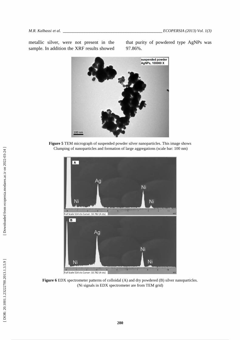
In the case of suspended powder AgNPs,
TEM images showed that in an aqueous
environment the nanoparticles clumping and
form large aggregates, many of which are larger
than 100nm (Figure 5). This result is consistent
with the results of hydrodynamic size
distribution which was obtained from the
dynamic light scattering method (zetasizer).
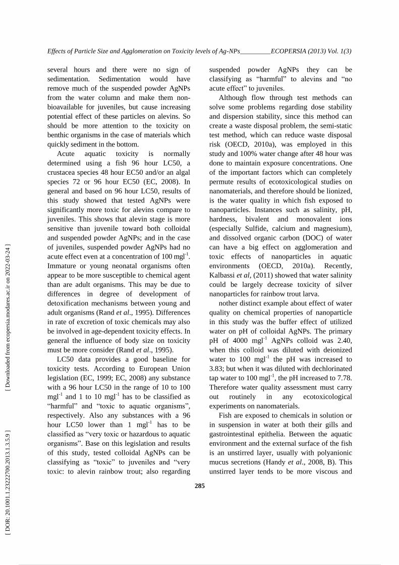
As seen in figures 6, EDX analyses were
confirmed that only elemental silver was
presented in both colloidal and dry powdered
AgNPs. According to the ICP-AES results,
the concentrations of Ag ions in the acid
digested stocks of colloidal and suspended
powder AgNPs were 3980 and 447.2 mgl-1
,
respectively. The XRD pattern of powdered
type AgNPs is shown in Figure 7; as can be
seen, all diffraction peaks correspond to the
characteristic face centered cubic (FCC)
silver lines. Also results of the XRD pattern
confirms the crystallinity of powdered type
AgNPs and presence of elemental
crystalline silver, also other phases, except
Particle Diameter (nm) Particle Diameter (nm)
Particle Diameter (nm) Particle Diameter (nm)
[ D
OR
: 20.
1001
.1.2
3222
700.
2013
.1.3
.5.9
]
[ D
ownl
oade
d fr
om e
cope
rsia
.mod
ares
.ac.
ir o
n 20
22-0
3-24
]
7 / 18
M.R. Kalbassi et al. _________________________________________ ECOPERSIA (2013) Vol. 1(3)
280
metallic silver, were not present in the
sample. In addition the XRF results showed
that purity of powdered type AgNPs was
97.86%.
Figure 5 TEM micrograph of suspended powder silver nanoparticles. This image shows
Clumping of nanoparticles and formation of large aggregations (scale bar: 100 nm)
Figure 6 EDX spectrometer patterns of colloidal (A) and dry powdered (B) silver nanoparticles.
(Ni signals in EDX spectrometer are from TEM grid)
100 nm
Full Scale 514 cts Cursor: 10.782 (4 cts)
Full Scale 514 cts Cursor: 10.782 (4 cts)
[ D
OR
: 20.
1001
.1.2
3222
700.
2013
.1.3
.5.9
]
[ D
ownl
oade
d fr
om e
cope
rsia
.mod
ares
.ac.
ir o
n 20
22-0
3-24
]
8 / 18

Effects of Particle Size and Agglomeration on Toxicity levels of Ag-NPs ________ ECOPERSIA (2013) Vol. 1(3)
281
3.2 Determination of lethal concentrations
(LC)
The mortality of controls during the
experiment was one alevin in one of the
control groups. No mortality was observed in
dispersant controls. Results of LC
determinations are shown in Table 3. The
lowest concentrations of colloidal and
suspended powder AgNPs which were caused
100% mortality in alevins after 96 hour were 1
and 100 mgl-1
, respectively. In alevins,
calculated median lethal concentration (LC50)
values for the colloidal AgNPs were 2.75,
0.44, 0.35 and 0.25 mgl-1
, after 24, 48, 72 and
96 hour, respectively. While these values for
the suspended powder AgNPs were 186.42,
69.37, 36.93, and 28.25 mgl-1
, respectively
(Table 3). According to these results it is clear
that compared to the suspended powder AgNPs,
much less concentrations of the colloidal
AgNPs are toxic to alevins.
The Mortality of controls and dispersant
controls of juveniles was zero during the
experiments. The lowest concentration of
colloidal AgNPs which were caused 100%
mortality in juveniles was 3.2 mgl-1
after 96
hour. Also as shown in Table 4, in juveniles the
calculated LC50 values of colloidal AgNPs
were 3.76, 3.13, 2.39 and 2.16 mgl-1
at 24, 48,
72 and 96 hour, respectively. Survey on
survival rates of juveniles, which exposed to
suspended powder AgNPs, showed that even in
maximum concentration (100 mgl-1
) and after
21 day exposure, no mortality was observed. In
toxicity test of the suspended powder AgNPs on
juveniles, rapid aggregation was occurred so
that large clumps were observable in the water
column. Also rapid sedimentation of these
clumps was observed in exposure tanks and
because juveniles were swim in water column,
most of these particles were away from fish
contacts.
Figure 7 X-Ray diffraction pattern of powdered AgNPs
[ D
OR
: 20.
1001
.1.2
3222
700.
2013
.1.3
.5.9
]
[ D
ownl
oade
d fr
om e
cope
rsia
.mod
ares
.ac.
ir o
n 20
22-0
3-24
]
9 / 18

M.R. Kalbassi et al. _________________________________________ ECOPERSIA (2013) Vol. 1(3)
282
Table 3 Lethal-concentration values with lower and upper 95% confidence limits (CL) of colloidal and
suspended powder silver nanoparticles (AgNPs) for rainbow trout alevin during 96h
Toxicity Time (h)/AgNP type Colloidal Suspended powder
Average LC10
(mg l-1
)
24 0.90 (0.06-1.80) 18.60 (7.41-26.70)
48 0.16 (0.10-0.22) 11.93 (5.63-20.12)
72 0.12 (0.07-0.16) 8.95 (4.42-17.01)
96 0.08 (0.04-0.11) 7.10 (3.94-11.68)
Average LC50
(mg l-1
)
24 2.75 (1.20-7.16) 186.42 (93.42-448.91)
48 0.44 (0.34-0.56) 69.37 (27.94-174.30)
72 0.35 (0.27-0.45) 36.93 (14.62-83.39)
96 0.25 (0.18-0.32) 28.25 (7.97-35.46))
Average LC90
(mg l-1
)
24 4.67 (2.46-10.76) 1868.42 (490.83-5815.19)
48 1.21 (0.90-1.88) 403.18 (327.82-1734.55)
72 1.02 (0.75-1.610) 152.25 (39.53-561.43)
96 0.75 (0.55-1.23) 112.32 (30.74-217.91)
Table 4 Lethal-concentration values with lower and upper 95% confidence limits (CL) of colloidal and
suspended powder silver nanoparticles (AgNPs) for rainbow trout juvenile during 96h
Toxicity Time (h)/AgNP type Colloidal Suspended powder
Average LC10
(mg l-1
)
24 2.63 (2.15-2.93) >100
48 2.56 (2.15-2.78) >100
72 1.74 (0.89-2.10) >100
96 1.60 (0.51-1.99) >100
Average LC50
(mg l-1
)
24 3.76 (3.44-4.24) >100
48 3.13 (2.91-3.33) >100
72 2.39 (1.87-2.66) >100
96 2.16 (1.34-2.43) >100
Average LC90
(mg l-1
)
24 5.39 (4.67-7.22) >100
48 3.84 (3.57-4.41) >100
72 3.28 (2.93-4.49) >100
96 2.91 (2.60-4.06) >100
3.3 Observable effects of silver particles on
fish
In the case of alevins, agglomeration of the
colloidal AgNPs was happened in contact
with mucoproteins of the fish. These
agglomerated particles were trapped under
the gill operculum and inside the mouth of
alevins (Figure 8). Also about suspended
[ D
OR
: 20.
1001
.1.2
3222
700.
2013
.1.3
.5.9
]
[ D
ownl
oade
d fr
om e
cope
rsia
.mod
ares
.ac.
ir o
n 20
22-0
3-24
]
10 / 18

Effects of Particle Size and Agglomeration on Toxicity levels of Ag-NPs ________ ECOPERSIA (2013) Vol. 1(3)
283
powder AgNPs rapid sedimentation was
occurred in experimental beakers; and since
alevins don’t have active swimming and stay
in the bottom of the beakers, the sediments of
AgNPs were in close contact with the fish. In
the case of juveniles similar to alevins, the
colloidal AgNPs were agglomerated in
contact with mucus of the gill and at higher
concentrations (>1 mgl-1
) the mucus secretion
increased and lengthy filamentous mixtures
of mucus-nanosilver were connected to the
gill (Figure 9).
Figure 8 Agglomerated colloidal (left) and suspended powder (right) AgNPs in contact with fish mucus
Figure 9 Secreted fish mucus rapidly agglomerates colloidal AgNPs on the surface of the gills; example fish
from 10 mg l-1
(left) and 3.2 mg l-1
(right) concentrations after 24 h.
4 DISCUSSION
Particle size is a critical parameter in the
assessment of environmental, health and safety
aspects of nanoscale materials (OECD,
2010b). Nanoparticles (NPs) often exhibit
special physical and chemical properties and
activities due to their small size and
homogeneous composition, structure or
surface characteristics, which are not present at
the larger scale. The size of the nanoparticle
implies that it has a large surface area to come
in contact with the cells and hence, it will have
[ D
OR
: 20.
1001
.1.2
3222
700.
2013
.1.3
.5.9
]
[ D
ownl
oade
d fr
om e
cope
rsia
.mod
ares
.ac.
ir o
n 20
22-0
3-24
]
11 / 18

M.R. Kalbassi et al. _________________________________________ ECOPERSIA (2013) Vol. 1(3)
284
a higher percentage of interaction than bigger
particles. In particular NPs possess a much
higher specific surface area than their larger
counterparts of the same material, and the
proportion of atoms on the surface versus the
interior of the particle is also much larger for
NPs (Handy et al., 2008, A).
The intrinsic properties of nanomaterials,
such as enhanced reactivity and unique surface
structures, can result in higher dissolution
rates, reduction and oxidation reactions, or
increased generation of reactive oxygen
species (ROS), all of which can in turn affect
toxicity in a size-dependent manner (Auffan et
al., 2009).
Although there are almost no
ecotoxicological data that have systematically
investigated particle-size effects, such studies
are important as they inform the need for
additional hazard assessments for materials in
nano form.
In the present study we investigated the
toxicity of two different types of silver
nanoparticles on alevin and juvenile of rainbow
trout. In the first type, the colloidal AgNPs,
nano particles were dispersed well in aqueous
environment and the agglomeration was very
low (results from TEM microscopes); while in
the second type, which was prepared by
suspending powdered AgNPs in the water, the
nanoparticles were agglomerated and formed
larger size clumps. When comparing the
colloidal and suspended powder AgNPs, for
alevin and juvenile rainbow trout, the colloidal
form (smaller size, well dispersed) were
comparatively more toxic than suspended
powder form (bigger, agglomerated).
Kashiwada (2006) showed a particle-size effect
on the accumulation of fluorescent NPs in the
Japanese medaka, with the smaller particles
accumulating more quickly. Also Palaniappan
and Pramod (2010) reported that LC50 values
of nano TiO2 and micro TiO2 were 30 and
100ppm, respectively for zebrafish; which show
the positive effect of reduction of particle size
on increasing of toxicity. On the other hand
Bar-Ilan et al, (2009) investigated the size-
dependent toxicity of nanosilver on zebrafish
embryonic development using 3, 10, 50, and
100 nm silver nanoparticles. In their study
LC50 values (93.31μM for 3nm particles to
137.26 μM for 100nm particles) indicated that
toxicity is loosely size-dependent, although
only at certain concentrations and time points;
although mortality was similar across sizes, but
the smaller size groups (3 and 10nm) of
nanosilver produced a higher incidence of sub-
lethal effects than the larger sizes (50 and
100nm).
According to the results of actual
concentration measurements of silver in the
samples were taken from middle of water
column (Table 2), it is clear that the nominal
concentrations of silver in the samples from
colloidal AgNPs is approximately equal to its
actual concentrations with an excellent
correlation (R2=0.99); but in the case of the
samples from suspended powder AgNPs, actual
concentrations of silver were approximately 10
to 16 time less than nominal concentrations. So
in the case of suspended powder AgNPs, it
seems that aggregation on the one hand causing
sedimentation of particles, makes large amount
of silver go out of the water column; and on the
other hand making larger clumps with smaller
surface area which release less amount of silver
ions to the water. Griffitt et al, (2009) infer that
metallic nanoparticles added to water tend to
aggregate simultaneously to form larger
particles, dissolve to release soluble metal ions,
and sediment out of the water column. In the
present study dispersion stability of suspended
powder AgNPs was very low and despite
application of suspending reagent and strong
aeration of tanks, sedimentation was observable;
moreover aggregations of particles accelerate the
precipitation of AgNPs. While colloidal AgNPs
were remained in a stable suspension for up to
[ D
OR
: 20.
1001
.1.2
3222
700.
2013
.1.3
.5.9
]
[ D
ownl
oade
d fr
om e
cope
rsia
.mod
ares
.ac.
ir o
n 20
22-0
3-24
]
12 / 18
Effects of Particle Size and Agglomeration on Toxicity levels of Ag-NPs _________ ECOPERSIA (2013) Vol. 1(3)
285
several hours and there were no sign of
sedimentation. Sedimentation would have
remove much of the suspended powder AgNPs
from the water column and make them non-
bioavailable for juveniles, but cause increasing
potential effect of these particles on alevins. So
should be more attention to the toxicity on
benthic organisms in the case of materials which
quickly sediment in the bottom.
Acute aquatic toxicity is normally
determined using a fish 96 hour LC50, a
crustacea species 48 hour EC50 and/or an algal
species 72 or 96 hour EC50 (EC, 2008). In
general and based on 96 hour LC50, results of
this study showed that tested AgNPs were
significantly more toxic for alevins compare to
juveniles. This shows that alevin stage is more
sensitive than juvenile toward both colloidal
and suspended powder AgNPs; and in the case
of juveniles, suspended powder AgNPs had no
acute effect even at a concentration of 100 mgl-1.
Immature or young neonatal organisms often
appear to be more susceptible to chemical agent
than are adult organisms. This may be due to
differences in degree of development of
detoxification mechanisms between young and
adult organisms (Rand et al., 1995). Differences
in rate of excretion of toxic chemicals may also
be involved in age-dependent toxicity effects. In
general the influence of body size on toxicity
must be more consider (Rand et al., 1995).
LC50 data provides a good baseline for
toxicity tests. According to European Union
legislation (EC, 1999; EC, 2008) any substance
with a 96 hour LC50 in the range of 10 to 100
mgl-1
and 1 to 10 mgl-1
has to be classified as
“harmful” and “toxic to aquatic organisms”,
respectively. Also any substances with a 96
hour LC50 lower than 1 mgl-1 has to be
classified as “very toxic or hazardous to aquatic
organisms”. Base on this legislation and results
of this study, tested colloidal AgNPs can be
classifying as “toxic” to juveniles and “very
toxic: to alevin rainbow trout; also regarding
suspended powder AgNPs they can be
classifying as “harmful” to alevins and “no
acute effect” to juveniles.
Although flow through test methods can
solve some problems regarding dose stability
and dispersion stability, since this method can
create a waste disposal problem, the semi-static
test method, which can reduce waste disposal
risk (OECD, 2010a), was employed in this
study and 100% water change after 48 hour was
done to maintain exposure concentrations. One
of the important factors which can completely
permute results of ecotoxicological studies on
nanomaterials, and therefore should be lionized,
is the water quality in which fish exposed to
nanoparticles. Instances such as salinity, pH,
hardness, bivalent and monovalent ions
(especially Sulfide, calcium and magnesium),
and dissolved organic carbon (DOC) of water
can have a big effect on agglomeration and
toxic effects of nanoparticles in aquatic
environments (OECD, 2010a). Recently,
Kalbassi et al, (2011) showed that water salinity
could be largely decrease toxicity of silver
nanoparticles for rainbow trout larva.
nother distinct example about effect of water
quality on chemical properties of nanoparticle
in this study was the buffer effect of utilized
water on pH of colloidal AgNPs. The primary
pH of 4000 mgl-1
AgNPs colloid was 2.40,
when this colloid was diluted with deionized
water to 100 mgl-1
the pH was increased to
3.83; but when it was diluted with dechlorinated
tap water to 100 mgl-1
, the pH increased to 7.78.
Therefore water quality assessment must carry
out routinely in any ecotoxicological
experiments on nanomaterials.
Fish are exposed to chemicals in solution or
in suspension in water at both their gills and
gastrointestinal epithelia. Between the aquatic
environment and the external surface of the fish
is an unstirred layer, usually with polyanionic
mucus secretions (Handy et al., 2008, B). This
unstirred layer tends to be more viscous and
[ D
OR
: 20.
1001
.1.2
3222
700.
2013
.1.3
.5.9
]
[ D
ownl
oade
d fr
om e
cope
rsia
.mod
ares
.ac.
ir o
n 20
22-0
3-24
]
13 / 18

M.R. Kalbassi et al. _________________________________________ ECOPERSIA (2013) Vol. 1(3)
286
move more slowly than bulk water, thereby
holding nanoparticles at the external surface of
the organism (Handy et al., 2008, B). The
various ligands present on the cell surface also
are predominantly anionic. Nanoparticles
should generally diffuse across the mucous
layer more slowly than single molecules such as
electrolytes and metal ions, and cationic
nanoparticles might bind to strands of
mucoproteins hindering their uptake (Handy
and Shaw, 2007; Handy et al., 2008, B). Cell
surfaces also might present ligands for
nanosilver (e.g., gill epithelium is
predominantly anionic) (Handy and Eddy,
2004). The role of mucous secretion from fish’s
body is very sensible in this study. The mucus
layer on the gills and body surface can connect
particles to each other and may be an effective
barrier to entry of the nanoparticles into the
body cells. Agglomeration can abrogate the
properties associated with nano-sized particles
by reducing its effective surface area (Greulich
et al., 2009).
In conclusion, results of this short-term
toxicity test can demonstrate some differences
between well dispersed and agglomerated silver
nanoparticles; although both acute and chronic
ecotoxicity testing should be undertaken in
order to build mathematical relationships
between acute and chronic toxicity for
categories of nanomaterials (OECD, 2010b).
Totally it is recommended that the release of
untreated silver particle waste into the
environment be controlled.
5 ACKNOWLEDGEMENT
We gratefully acknowledge the support of the
Tarbiat Modares University of I. R. Iran, who
funded this research through the PhD Thesis
project. Also this research was partially
supported by the Green Nanotechnology
program through the National Research
Foundation of Korea funded by the Korean
Ministry of Education, Science and
Technology. We thank Dr. Ji Hyun Lee for
technical assistance in the analysis of TEM
images.
6 REFERENCES
ASTM, Zeta Potential of Colloids in Water and
Waste Water. ASTM Standard D 4187-
82, Am. Soc. Testing Mats. 1985.
Asharani, P.V., Wu, Y.L., Gong, Z. and
Valiyaveetteil, S. Toxicity of silver
nanoparticles in Zebrafish models.
Nanotechnology, 2008; 19(25): 255102.
Auffan, M., Rose, J., Wiesner, MR. and Bottero
JY. Chemical stability of metallic
nanoparticles: A parameter controlling
their potential cellular toxicity in vitro.
Environ. Pollut., 2009; 157: 1127-1133.
Bar-Ilan, O., Albrecht, R.M., Fako, V.E. and
Furgeson, D.Y. Toxicity assessments of
multisized gold and silver nanoparticles
in Zebrafish embryos. Small, 2009;
5(16): 1897-910.
Benn, T.M., Westerhoff, P., 2008. Nanoparticle
silver released into water from
commercially available sock fabrics.
Environ. Sci. Technol., 42(11): 4133-4139.
Bilberg, K., Malte, H., Wang, T. and Baatrup,
E. Silver nanoparticles and silver nitrate
cause respiratory stress in Eurasian perch
(Perca fluviatilis). Aquat. Toxicol., 2010;
96: 159-165.
Blaser, S.A., Scheringer, M., Macleod, M. and
Hungerbühler, K. Estimation of
cumulative aquatic exposure and risk due
to silver: Contribution of nano-
functionalized plastics and textiles. Sci.
Total Environ., 2008; 390: 396-409.
Carlson, C., Schrand, A. M., Braydich-Stolle,
L.K., Hess, K.L., Jones, R.L., Schlager,
J.J. and Hussain, S.M. Unique cellular
interaction of silver nanoparticles: size-
[ D
OR
: 20.
1001
.1.2
3222
700.
2013
.1.3
.5.9
]
[ D
ownl
oade
d fr
om e
cope
rsia
.mod
ares
.ac.
ir o
n 20
22-0
3-24
]
14 / 18

Effects of Particle Size and Agglomeration on Toxicity levels of Ag-NPs _________ ECOPERSIA (2013) Vol. 1(3)
287
dependent generation of reactive oxygen
species. J. Phys. Chem., B, 2008;
112(43): 13608-13619.
Chae, Y.J., Pham, C.H., Lee, J., Bae, E., Yi, J.
and Gu, M. B. Evaluation of the toxic
impact of silver nanoparticles on
Japanese medaka (Oryzias latipes).
Aquat. Toxicol., 2009; 94: 320-327.
Choi, J. E., Kim, S., Ahn, J.H., Youn, P., Kang,
J.S., Park, K., Yi, J. and Ryu, D.
Induction of oxidative stress and
apoptosis by silver nanoparticles in the
liver of adult Zebrafish. Aquat. Toxicol.,
2009; 100(2): 151-159.
Dowling, A., Clift, R., Grobert, N., Hutton, D.,
Oliver, R., O’Neill, O., Pethica, J., Inoue,
K.I., Takano, H., Yanagisawa, R., Koike,
E. and Shimada, A. Size effects of latex
nanomaterials on lung inflammation in
mice. Toxicol. Appl. Pharm., 2009;
234(1): 68-76.
EC, Annex VI of Directive 1999/45/EC to
consolidated version of directive
67/548/EEC. General classification and
labeling requirements for dangerous
substances and preparations. 1999.
EC, Regulation (EC) No 1272/2008 of the
European Parliament and of the Council
of 16 December 2008 on classification,
labeling and packaging of substances and
mixtures, Official J. Eur. Union,
31.12.2008.
Greulich, C., Kittler, S., Epple, M., Muhr, G.,
Koller, M., Studies on the
biocompatibility and interaction of silver
nanoparticles with human mesenchymal
stem cells (hMSCs). Langenbeck's
Archives of Surgery, 2009; 394: 495-502.
Griffitt, R.J., Hyndman, K., Denslow, N.D., and
Barber, D.S. Comparison of molecular
and histological changes in zebrafish gills
exposed to metallic nanoparticles.
Toxicol. Sci. 2009; 107(2): 404-415.
Handy, R.D. and Eddy, F.B. Transport of
solutes across biological membranes in
eukaryotes: An environmental
perspective, In: Physicochemical Kinetics
and Transport at Biointerfaces 2004;
(337-356). John Wiley, New York.
Handy, R.D., Henry, T.B., Scown, T.M.,
Johnston, B.D. and Tyler, C.R. (B).
Manufactured nanoparticles: their uptake
and effects on fish: a mechanistic analysis.
Ecotoxicology, 2008; 17: 396-409.
Handy, R.D., Kammer, F.v.d., Lead, J.R.,
Hassellöv, M., Owen, R. and Crane, M. (A).
The ecotoxicology and chemistry of
manufactured nanoparticles. Ecotoxicology,
2008; 17, 287-314.
Handy, R.D. and Shaw, B.J., Ecotoxicity of
nanomaterials to fish: Challenges for
ecotoxicity testing. Integr. Environ.
Assess. Manage., 2007; 3: 458-460.
Kalbassi, M.R., Salari Joo, H. and Johari, S.A.
Toxicity of Silver nanoparticles in
aquatic ecosystems: salinity as the main
cause of reducing toxicity. Iran. J.
Toxicol., 2011; 5(12 and 13): 436-443.
Kashiwada, S. Distribution of nanoparticles in
the See-through medaka (Oryzias
latipes). Environ. Health Perspect., 2006;
114: 1697-1702.
Lee, K.J., Nallathamby, P.D., Browning, L.M.,
Osgood, C.J. and Hancy, X.H. In Vivo
imaging of transport and biocompatibility
of single silver nanoparticles in early
development of Zebrafish embryos. J.
Am. Chem. Soc., (ACSNANO), 2007;
1(2): 133-143.
Murr, L.E., Nanoparticulate materials in
antiquity: The good, the bad and the ugly.
Mater. Charact., 2009; 60(4): 261-270.
[ D
OR
: 20.
1001
.1.2
3222
700.
2013
.1.3
.5.9
]
[ D
ownl
oade
d fr
om e
cope
rsia
.mod
ares
.ac.
ir o
n 20
22-0
3-24
]
15 / 18

M.R. Kalbassi et al. _________________________________________ ECOPERSIA (2013) Vol. 1(3)
288
Nickum, J.G., Bart Jr., H.L., Bowser, P.R.,
Greer, I.E., Hubbs, C., Jenkins, J.A.,
MacMillan, J.R., Rachlin, J.W., Rose,
J.D., Sorensen, P.W. and Tomasso, J.R.
Guidelines for the use of fishes in
research. Am. Fish. Soc., Bethesda,
Maryland, 2004; 54P.
Nowack, B. and Bucheli, T.D. Occurrence,
behavior and effects of nanoparticles in
the environment. Environ. Pollut., 2007;
150: 5-22.
OECD, Guidelines for the Testing of
Chemicals. Section 2: Effects on Biotic
Systems Test No. 215: Fish, Juvenile
Growth Test. Organ. Econ. Coop. Dev.,
Paris, France. 2000, 25 p.
OECD, Environment, Health and Safety
Publications, Series on the Safety of
Manufactured Nanomaterials, No. 24:
Preliminary guidance notes on sample
preparation and dosimetry for the safety
testing of manufactured nanomaterials,
ENV/JM/MONO (2010)25, Organ. Econ.
Coop. Dev., Paris, France. 2010a, 67 p.
OECD, Environment, Health and Safety
Publications, Series on the Safety of
Manufactured Nanomaterials, No. 25:
Guidance manual for the testing of
manufactured nanomaterials, OECD
Sponsorship program, ENV/JM/MONO
(2009)20/REV, Organ. Econ. Coop. Dev.,
Paris, France. 2010b, 92 p.
Palaniappan, PL. and Pramod, KS. FTIR study
of the effect of nTiO2 on the biochemical
constituents of gill tissues of Zebrafish
(Danio rerio). Food. Chem. Toxicol.,
2010; 48: 2337-2343.
Pidgeon, N., Porritt, J., Ryan, J., Seaton, A.,
Tendler, S., Welland, M. and Whatmore,
R. Nanoscience and nanotechnologies:
opportunities and uncertainties; The Royal
Society, The Royal Academy of
Engineering: 29/07/2004. 2004.
Powers, C.M., Yen, J., Linney, E.A., Seidler,
F.J. and Slotkin, T.A. Silver exposure in
developing Zebrafish (Danio rerio):
Persistent effects on larval behavior and
survival. Neurotoxicol. Teratol., 2010;
32: 391-397.
Rahman Nia, J. Preparation of colloidal
nanosilver. US Patent application docket
20090013825, 15 January 2009.
Rand, G.M., Wells, P.G. and McCarty, L.S.
Introduction to aquatic toxicology, in:
Rand, G. M. (Editor) Fundamentals of
aquatic ecotoxicology: effects,
environment fate, and risk assessment
(Second edition), Taylor and Francis,
Washington. 1995, 1125 p.
Salari Joo H., MR Kalbassi, IJ Yu, JH Lee, SA
Johari. 2013. Bioaccumulation of silver
nanoparticles in Rainbow trout
(Oncorhynchus mykiss): Influence of
concentration and salinity. Aquat.
Toxicol., 7: 398-406.
Schirmer, K., Tanneberger, K., I. Kramer, N.,
Volker, D., Scholz, S., Hafner, C., E.J.
Lee, L., C. Bols, N., L.M. and Hermens,
J. Developing a list of reference
chemicals for testing alternatives to
whole fish toxicity tests. Aquat. Toxicol.,
2008; 90: 128-137.
Senjen, R. Can nanotechnologies assist in
solving 21st century environmental
challenges? A critical review of
opportunities and risks. The European
Environmental Bureau (EEB).
Nanomaterials, Health and Environ. Conc.
2009; 2: 17P.
Scown, T.M., Santos, E.M., Johnston, B.D.,
Gaiser, B., Baalousha, M., Mitov, S.,
Lead, J.R., Stone, V., Fernandes, T.F.,
[ D
OR
: 20.
1001
.1.2
3222
700.
2013
.1.3
.5.9
]
[ D
ownl
oade
d fr
om e
cope
rsia
.mod
ares
.ac.
ir o
n 20
22-0
3-24
]
16 / 18

Effects of Particle Size and Agglomeration on Toxicity levels of Ag-NPs _________ ECOPERSIA (2013) Vol. 1(3)
289
Jepson, M., van Aerle, R. and Tyler, C.R.
Effects of aqueous exposure to silver
nanoparticles of different sizes in
Rainbow trout. Toxicol. Sci., 2010;
115(2): 521-534.
Weis, J.S. and Weis, P. Effects of
environmental pollutants on early fish
development. Rev. Aquat. Sci., 1989; 1:
45-73.
Woodrow Wilson Database, Nanotechnology
consumer product inventory. http://www.
nanotechproject.org/inventories/consumer/
analysis_draft/. 2011.
Wu, Y., Zhoua, Q., Li, H., Liua, W., Wanga, T.
and Jianga, G. Effects of silver
nanoparticles on the development and
histopathology biomarkers of Japanese
medaka (Oryzias latipes) using the
partial-life test. Aquat. Toxicol., 2009;
100(2): 160-167.
Yeo, M. and Kang, M. Effects of nanometer
sized silver materials on biological
toxicity during Zebrafish embryogenesis.
Bull. Korean Chem. Soc., 2008; 29(6):
1179-1184.
[ D
OR
: 20.
1001
.1.2
3222
700.
2013
.1.3
.5.9
]
[ D
ownl
oade
d fr
om e
cope
rsia
.mod
ares
.ac.
ir o
n 20
22-0
3-24
]
17 / 18

M.R. Kalbassi et al. _________________________________________ ECOPERSIA (2013) Vol. 1(3)
290
های آبیدر محیطنقره ررات نانوفرمهای مختلف سمیت بر تأثیر انباشتگی و انذازه ررات
4 ایل ج ی 3، هذی سلطبی2، سیذػلی جشی1هحوذسضب کلجبسی
داطگب تشثیت هذسس، س، ایشاىداطکذ ػلم دسیبیی، ،استبد -1
یشاى، اسذج، کشدستبى، داطگب هبثغ طجیؼی، داطکذ استبدیبس -2
ایشاى تشاى، داهپضضکی، داطگب تشاى، داطکذ ،استبد -3
داطگب سئ، آسبى، کش جثیضبسی، سنهشکض تحقیقبت ،استبد -4
1392آرس 3/ تبسیخ چبح: 1392 خشداد 20 / تبسیخ پزیشش: 1390 ضشیس 3تبسیخ دسیبفت:
بی آثی، ضبسی دس هحیطسن صیست ب دس هطبلؼبت برسات اجبضتگی آى اذاصث هظس ثشسسی تأثیش چکیذه
داس هبیبى جاى کلئیذ( دس لاسبی کیس صسد رسات قش )ضبهل پدس هؼلق سویت حبد د ع هتفبت ب
10، 2/3، 1، 32/0، 1/0 ،032/0 بی اسویب دس هؼشض غلظتکوبى هسد ثشسسی قشاس گشفت. هبیآلای سگیي قضل
بی کطذ ثب استفبد اص تجضی تحلیل پشثیت هحبسج رسات قش قشاس گشفتذ. غلظت س لیتش بدگشم هیلی 100
گشدیذ. دس هسد ب قش کلئیذی، رسات یض ثشسسی رسات قش هسد استفبد گشدیذذ. هطخصبت فیضیکی ضیویبیی ب
قش پدسی هؼلق، رسات دچبس ب یض حفظ ضذ ثد؛ اهب دس هسد بدذ اثؼبد آىخثی دس ستى آة پشاکذ ضذ ثث
( دس LC50)ب دس کف آة گشدیذ. غلظت کطذ هیبی طیی آىت بیتب اذاصفضایص اجبضتگی ضذذ ک هجش ث ا
گشم یلیه 25/22 25/0 تشتیتدس هؼلق ثقش کلئیذی پ داس، ثشای بدس هسد لاسبی کیس صسد سبػت، 96طی
گشم هیلی 16/2قش کلئیذی سبػت ثشای ب 96س لیتش هحبسج گشدیذ. دس هسد هبیبى جاى، غلظت کطذ هیبی د
ب سص دس هؼشض قشاسگیشی هبیبى جاى، ثبػث هشگ آى 21قش پدسی هؼلق، حتی طی دست آهذ، اهب بلیتش ثدس
قش کلئیذی سجت ث آلا، بداس هبیبى جاى قضلشدیذ. تبیج ایي هطبلؼ طبى داد ک دس لاسبی کیس صسدگ
رسات دس تیج چیي پذیذ اجبضتگی هجت افضایص اثؼبد باست، نثد تش هحلل پدس هؼلق ضذ ثسیبس سوی
ب ث ذی هادی ثسیبس سوی ثشای هبیبى هحسة گشدیذ اص سبیص آىرسات قش کلیی ضذ. بکبص سویت آب
اجتبة ود. هحیط صیست آثی ثبیذ کبهلا
ضبسی ب سن ،رسات قش ب آلای سگیي کوبى،قضل سویت اثست ث اذاص، ،خطشات صیست هحیطی کلمات کلیذی:
آثضیبى
[ D
OR
: 20.
1001
.1.2
3222
700.
2013
.1.3
.5.9
]
[ D
ownl
oade
d fr
om e
cope
rsia
.mod
ares
.ac.
ir o
n 20
22-0
3-24
]
Powered by TCPDF (www.tcpdf.org)
18 / 18
Recommended