Paediatrics in ED 2: Part A
Fevers
Dr Steve Costa
Emergency Medicine Training Hub
Ballarat & Grampians Region
23rd April 2015
Warm paediatric pearls . . .
Let’s be honest, at 4am, every hot child looks
miserable and sleepy.
Tessa Davies LITFL 2013
“If you can’t be caring – fake it, [just] make
sure everyone is engaged in the magic show”
Colin Parker SMACC 2013
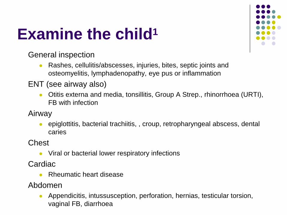
Examine the child1
General inspection
Rashes, cellulitis/abscesses, injuries, bites, septic joints and
osteomyelitis, lymphadenopathy, eye pus or inflammation
ENT (see airway also)
Otitis externa and media, tonsillitis, Group A Strep., rhinorrhoea (URTI),
FB with infection
Airway
epiglottitis, bacterial trachiitis, , croup, retropharyngeal abscess, dental
caries
Chest
Viral or bacterial lower respiratory infections
Cardiac
Rheumatic heart disease
Abdomen
Appendicitis, intussusception, perforation, hernias, testicular torsion,
vaginal FB, diarrhoea

Non-infectious fever
Medication
Immunization reactions
Central nervous system dysfunction
Malignancy (eg, leukemia)
Chronic inflammatory conditions
inflammatory bowel disease
juvenile idiopathic arthritis
Enviromental
Teething (<38.5°C)
Don’t miss lists
meningitis, herpes simplex encephalitis
UTI
pneumonia
septic arthritis
Kawasaki disease (fever 5/7 days)
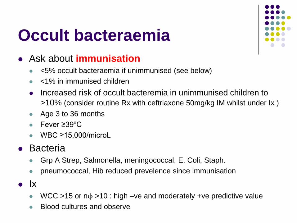
Occult bacteraemia
Ask about immunisation <5% occult bacteraemia if unimmunised (see below)
<1% in immunised children
Increased risk of occult bacteremia in unimmunised children to
>10% (consider routine Rx with ceftriaxone 50mg/kg IM whilst under Ix )
Age 3 to 36 months
Fever ≥39ºC
WBC ≥15,000/microL
Bacteria Grp A Strep, Salmonella, meningococcal, E. Coli, Staph.
pneumococcal, Hib reduced prevelence since immunisation
Ix WCC >15 or nϕ >10 : high –ve and moderately +ve predictive value
Blood cultures and observe

Management – under 3 months
bloods (FBC, CRP, B/C), urine, CXR, stool
culture if indicated.
Perform a lumbar puncture if: less than 1
month; 1-3 months and unwell; or 1-3 months
with WCC<5×10⁹/L or >15×10/L.
Give IV antibiotics for the same criteria as the
LP (i.e. lumbar puncture = IV antibiotics).

Management – 3 months or
older
Investigate fever with no source if they have
any red features
FBC, CRP, B/C and urine. Consider LP, CXR,
UEC and gas if indicated.
Prepare to investigate fever with no source if
there are any amber features – paediatrician
may halt these investigations.
FWT urine for all children with fever (over
37.5) and no source, even if they are green.

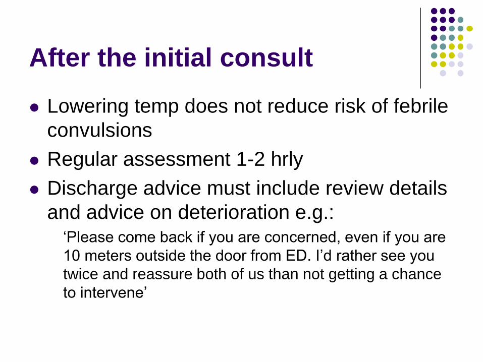
After the initial consult
Lowering temp does not reduce risk of febrile
convulsions
Regular assessment 1-2 hrly
Discharge advice must include review details
and advice on deterioration e.g.: ‘Please come back if you are concerned, even if you are
10 meters outside the door from ED. I’d rather see you
twice and reassure both of us than not getting a chance
to intervene’
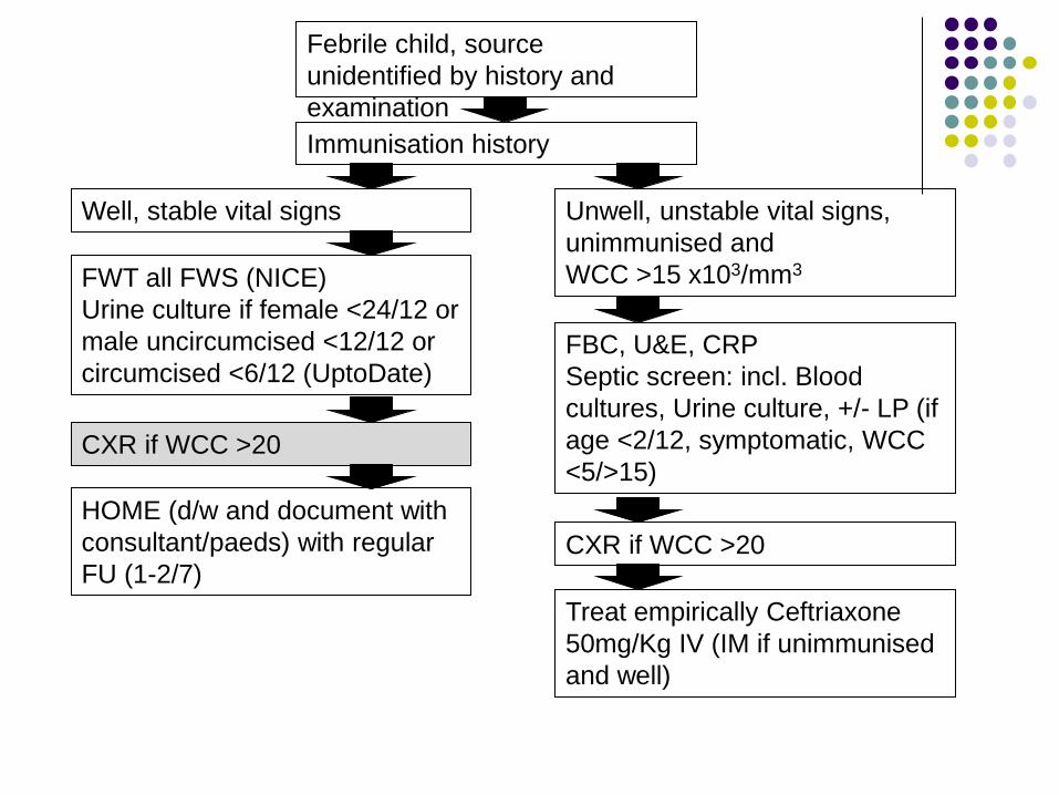
Febrile child, source
unidentified by history and
examination
Well, stable vital signs
Treat empirically Ceftriaxone
50mg/Kg IV (IM if unimmunised
and well)
FBC, U&E, CRP
Septic screen: incl. Blood
cultures, Urine culture, +/- LP (if
age <2/12, symptomatic, WCC
<5/>15)
Unwell, unstable vital signs,
unimmunised and
WCC >15 x103/mm3
CXR if WCC >20
Immunisation history
FWT all FWS (NICE)
Urine culture if female <24/12 or
male uncircumcised <12/12 or
circumcised <6/12 (UptoDate)
HOME (d/w and document with
consultant/paeds) with regular
FU (1-2/7)
CXR if WCC >20
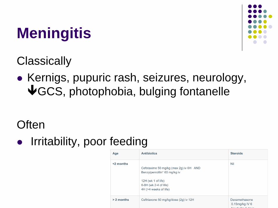
Meningitis
Classically
Kernigs, pupuric rash, seizures, neurology,
GCS, photophobia, bulging fontanelle
Often
Irritability, poor feeding

Treatment for Meningitis
(not including resuscitation)
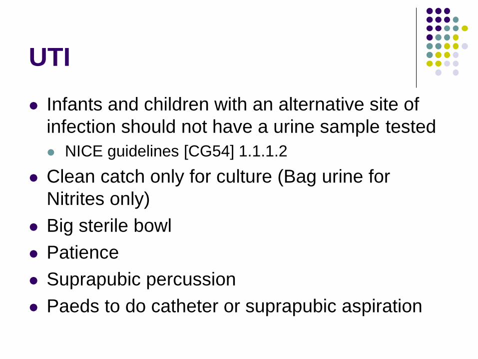
UTI
Infants and children with an alternative site of
infection should not have a urine sample tested
NICE guidelines [CG54] 1.1.1.2
Clean catch only for culture (Bag urine for
Nitrites only)
Big sterile bowl
Patience
Suprapubic percussion
Paeds to do catheter or suprapubic aspiration

UTIs
High temp (>38°C) and bacteriuria or temp 37-38°C and
loin pain and bacteriuria have pyelonephritis
<3/12 old – refer paeds and Rx with low resistance Abx
(ceph or Augmentin)
Imaging (with paeds r/v) if <6/12 old or recurrent
Treatment commenced on balance of risk, unwell child,
speed of test results
FWT/microscopy Leucocytes +ve only should be treated
as UTI if symptomatic only

Pneumonia
CXR
persistent chest signs between clinicians
Investigation for unresolved fever
20-40% of <5yo with high fever and WCC
>20-25 x103/mm3 have radiographic
pneumonia EVEN IF EXAMINATION NORMAL

Kawasaki
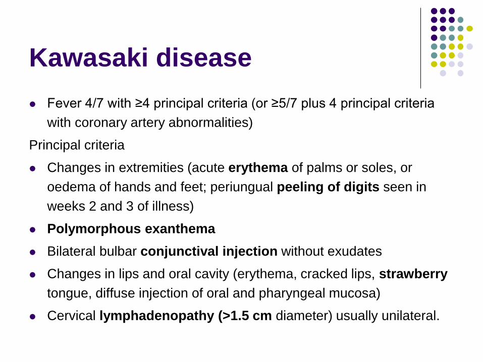
Kawasaki disease
Fever 4/7 with ≥4 principal criteria (or ≥5/7 plus 4 principal criteria
with coronary artery abnormalities)
Principal criteria
Changes in extremities (acute erythema of palms or soles, or
oedema of hands and feet; periungual peeling of digits seen in
weeks 2 and 3 of illness)
Polymorphous exanthema
Bilateral bulbar conjunctival injection without exudates
Changes in lips and oral cavity (erythema, cracked lips, strawberry
tongue, diffuse injection of oral and pharyngeal mucosa)
Cervical lymphadenopathy (>1.5 cm diameter) usually unilateral.

Febrile convulsions
A convulsion associated with an elevated temperature
greater than 38°C.
A child younger than six years of age.
No evidence of central nervous system infection or
inflammation (i.e. not meningitis or encephalitis)
No evidence of acute systemic metabolic abnormality
that may produce convulsions
No history of previous afebrile seizures
Generalised rather than focal
Short (< 15 min) rather than prolonged
Single rather than multiple
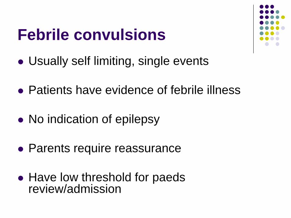
Febrile convulsions
Usually self limiting, single events
Patients have evidence of febrile illness
No indication of epilepsy
Parents require reassurance
Have low threshold for paeds review/admission

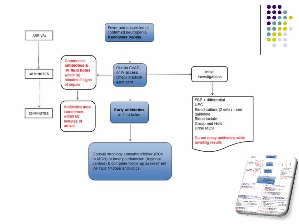
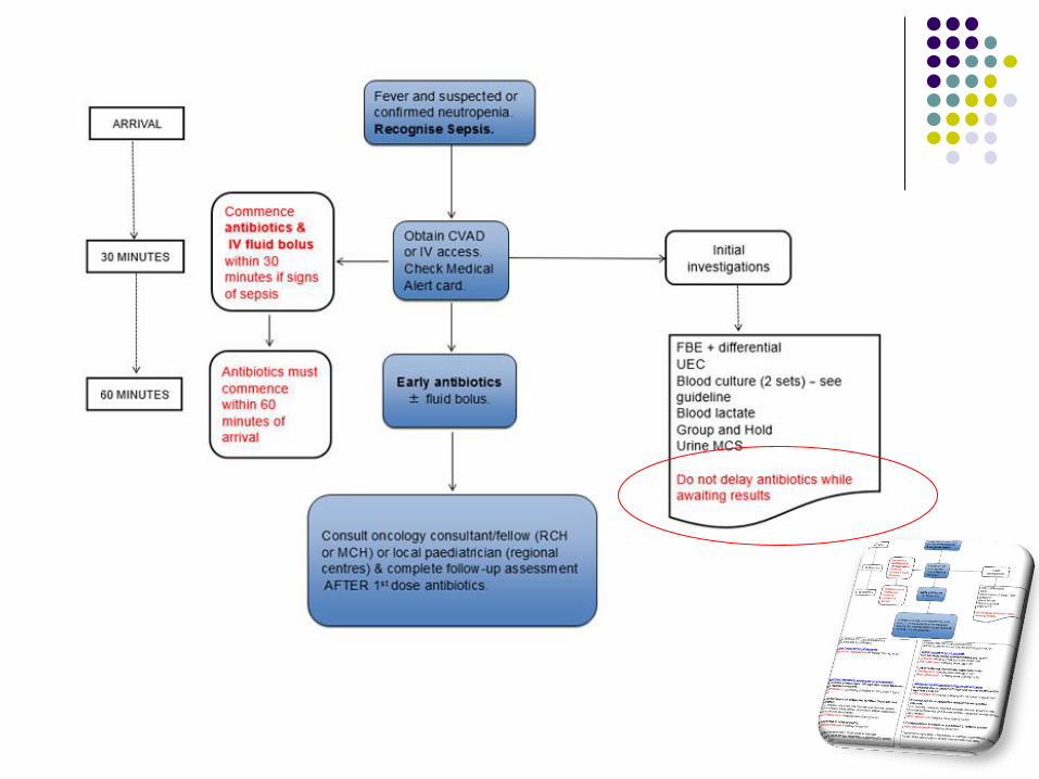
Febrile Neutropaenia (FN)
Bacteraemia is diagnosed in up to one-third
of children with FN
High risk in leukaemia and lymphoma, organ
transplant, relapse therapy*
Antibiotics <30min if septic and <60min if not
Then d/w oncologist
NB:
Peripheral BCs are no longer routinely recommended in oncology patients
Always label BC bottle with site from which blood has been taken including
specific lumen.
aerobic bottle should be inoculated preferentially *See RCH guidelines for specifics
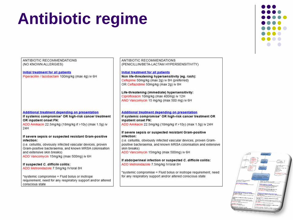
Antibiotic regime

Questions?

Paediatrics in ED 2: part B Lots of stuff that isn’t fevers or
advanced life support (although there is
other bits in later lectures too that are
important as well and tie in nicely actually)
Dr Steve Costa
Emergency Medicine Training Hub
Ballarat & Grampians Region
1st April 2015
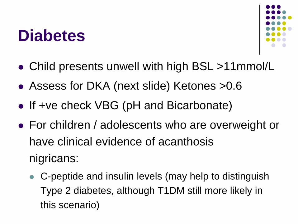
Diabetes
Child presents unwell with high BSL >11mmol/L
Assess for DKA (next slide) Ketones >0.6
If +ve check VBG (pH and Bicarbonate)
For children / adolescents who are overweight or
have clinical evidence of acanthosis
nigricans:
C-peptide and insulin levels (may help to distinguish
Type 2 diabetes, although T1DM still more likely in
this scenario)
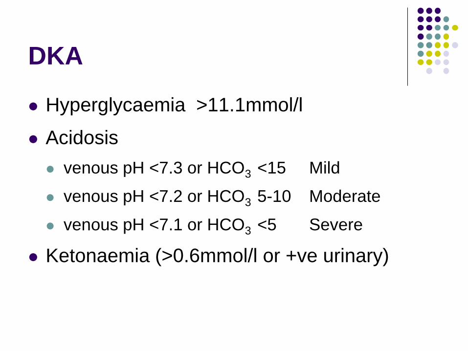
DKA
Hyperglycaemia >11.1mmol/l
Acidosis
venous pH <7.3 or HCO3 <15 Mild
venous pH <7.2 or HCO3 5-10 Moderate
venous pH <7.1 or HCO3 <5 Severe
Ketonaemia (>0.6mmol/l or +ve urinary)

Treatment
Fluids (must precede insulin by 1 hour) Add 5-10% glucose once BSL<15mmol/L
If not shocked (CRT<2secs) or clinically dehydrated, bolus fluid may be
avoided
Insulin 0.1U/Kg/hr
Electrolyte replacement Corrected Na = measured Na +0.4[glucose in mmol/l) – 5.5
K+ depletion 3 and 6 mmol kg−1
Anion gap = (Na+K) – (Cl+HCO3) Normal<18mmol/l
Increased in DKA as Ketones (weak acid) consumes HCO3
Confounding Cl- acidosis and lactic acidosis

Treatment
Fluids Add 5-10% glucose once BSL<15mmol/L
If not shocked (CRT<2secs) or clinically dehydrated, bolus fluid may be
avoided
Insulin 0.1U/Kg/hr
Electrolyte replacement Corrected Na = measured Na + 0.4[glucose in mmol/l) – 5.5
K+ depletion 3 and 6 mmol kg−1
Anion gap = (Na+K) – (Cl+HCO3) Normal<18mmol/l
Increased in DKA as Ketones (weak acid) consumes HCO3
Confounding Cl- acidosis and lactic acidosis
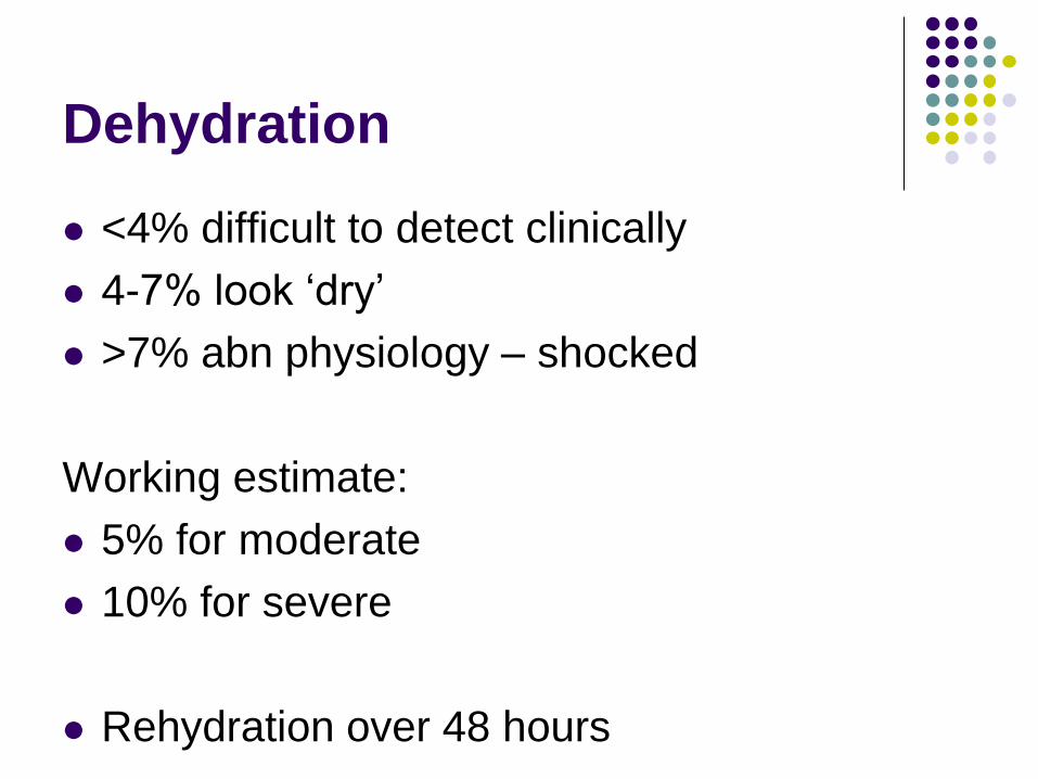
Dehydration
<4% difficult to detect clinically
4-7% look ‘dry’
>7% abn physiology – shocked
Working estimate:
5% for moderate
10% for severe
Rehydration over 48 hours
Presentation
New undiagnosed case
Illness
Missed doses or insulin
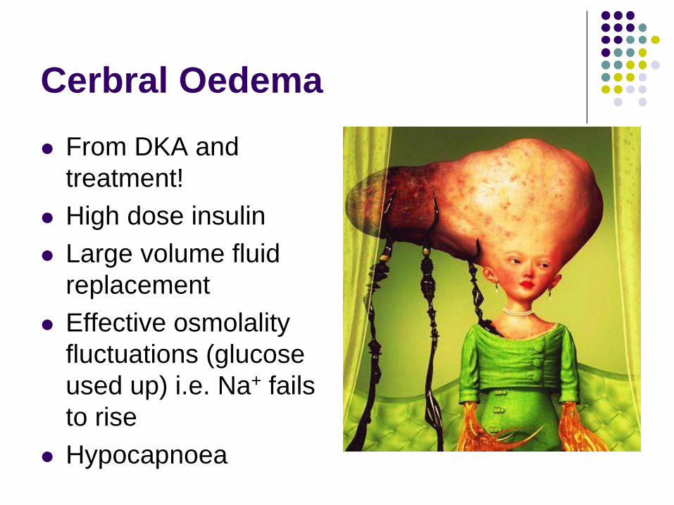
Cerbral Oedema
From DKA and
treatment!
High dose insulin
Large volume fluid
replacement
Effective osmolality
fluctuations (glucose
used up) i.e. Na+ fails
to rise
Hypocapnoea
Cerebral Oedema
younger age;
newly diagnosed diabetes;
longer duration of symptoms;
raised serum urea;
initial pH <7.1;
extreme hypocapnia (P CO 2 <2 kPa) at presentation;
hypocapnia in association with mechanical ventilation;
>40 ml kg−1 total fluid given in first 4 h;
bicarbonate therapy;
an attenuated increase, or a decrease in corrected plasma sodium
during treatment;
bolus insulin therapy.

Venous blood sample (IV access)
FBE
Blood glucose, urea, electrolytes (sodium, potassium,
calcium, magnesium, phosphate)
Blood ketones (bedside test)
Venous blood gas (including bicarbonate)
Investigations for precipitating cause/ septic work up
For all newly diagnosed patients:
Insulin antibodies, GAD antibodies, coeliac screen (total IgA,
anti-gliadin Ab, tissue transglutaminase Ab) and thyroid function
tests (TSH and FT4

DKA
Resuscitation
ABC if required
Hypertonic saline 3% 3-5ml/kg if COe suspected
NGT
Adequate IV access
ECG
IDUC
Fluids
Intravascular volume support and dehydration
replenishment
Acidosis confounds perfusion
Smaller volume boluses 10ml/kg N saline x2
Rehydration over 48hours

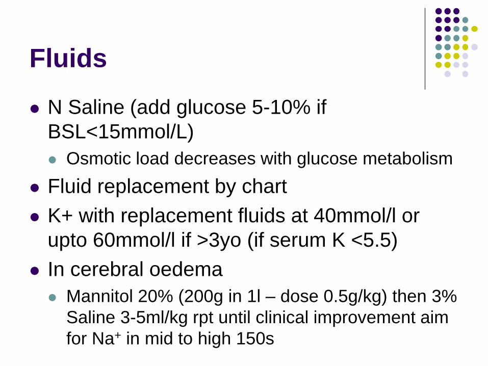
Fluids
N Saline (add glucose 5-10% if
BSL<15mmol/L)
Osmotic load decreases with glucose metabolism
Fluid replacement by chart
K+ with replacement fluids at 40mmol/l or
upto 60mmol/l if >3yo (if serum K <5.5)
3% Saline 3-5ml/kg rpt until clinical
improvement aim for Na+ in mid to high 150s
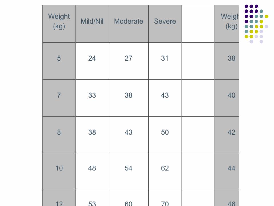

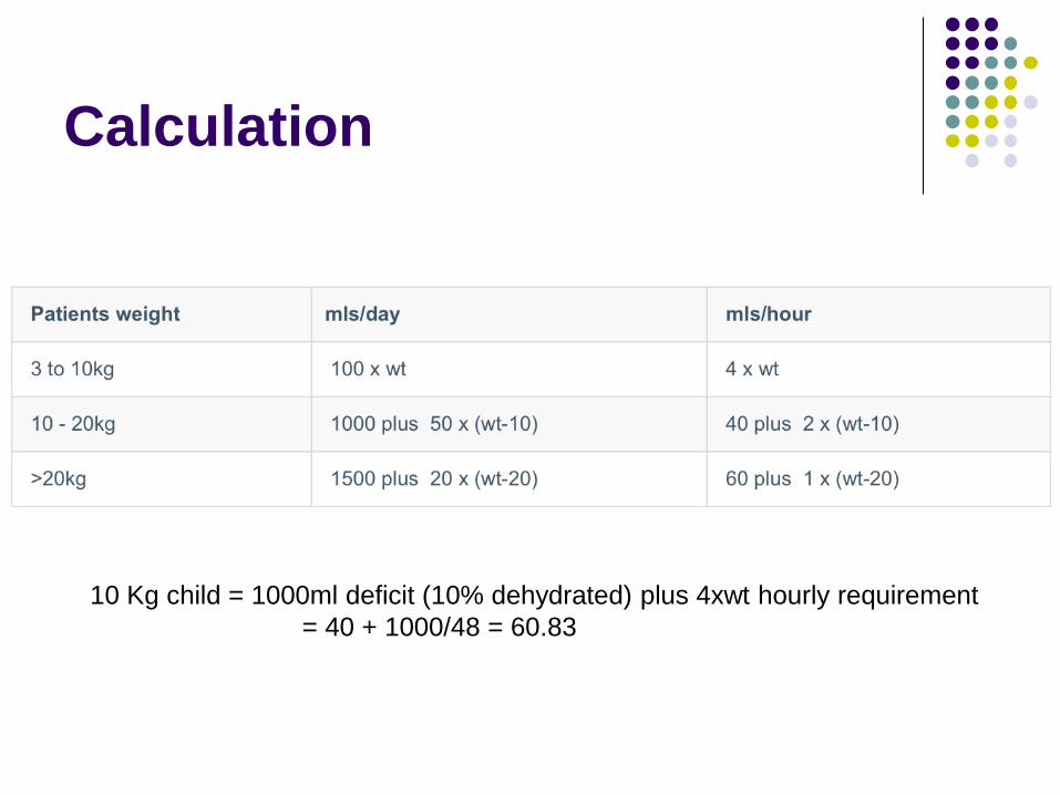
Calculation
10 Kg child = 1000ml deficit (10% dehydrated) plus 4xwt hourly requirement
= 40 + 1000/48 = 60.83
Fluids
N Saline (add glucose 5-10% if
BSL<15mmol/L)
Osmotic load decreases with glucose metabolism
Fluid replacement by chart
K+ with replacement fluids at 40mmol/l or
upto 60mmol/l if >3yo (if serum K <5.5)
In cerebral oedema
Mannitol 20% (200g in 1l – dose 0.5g/kg) then 3%
Saline 3-5ml/kg rpt until clinical improvement aim
for Na+ in mid to high 150s

Insulin
Insulin rate depends on history:
If new presentation or DM with BSL <15
0.1 Unit/Kg/hr if BSL >15mmol/L
If known DM and BSL >15
0.05 Unit/Kg/hr if BSL <15mmol/L
Give as infusion of 50 Units insulin with
49.5ml N saline

Questions

Pain relief and wounds

Pain relief
Analgesia is a central pillar of emergency medicine
Rapid appropriate analgesia
Paracetamol
Ibuprofen
Intranasal fentanyl
Sucrose
Painless procedures
AnGel cream
Ketamine sedation
Adjuncts to pain relief
Non-pharmacological techniques

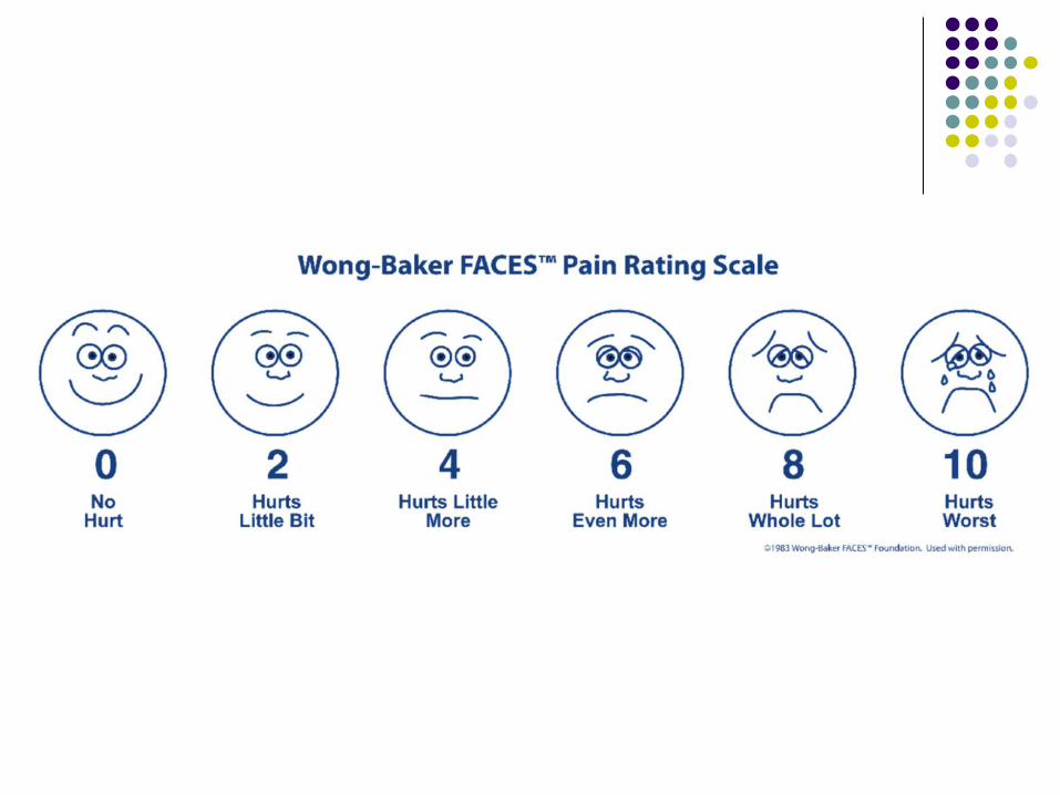
Pain assessment
Use their language (sore, ouch, hurt)
Be developmentally appropriate
Consider using dolls/toys as a medium
Consider other issues
Non-verbal children are very vulnerable to
having their pain under estimated
Children do not often have the skills to
understand or express themselves as adults
Non-pharmacological
interventions
Minimise fear and distress
Make pain more tolerable
To give the child a sense of control over the
situation and their behaviour
To teach and enhance coping strategies for
the child
To instruct parents in techniques to assist
their child

Techniques
Information: explain, explain, explain...
Parents’ presence
Choices and control
Laughter and fun
Deep breathing
Heat / Cold
Distraction Box: Tactile and audio-visual
distraction, games, DVDs, magic etc
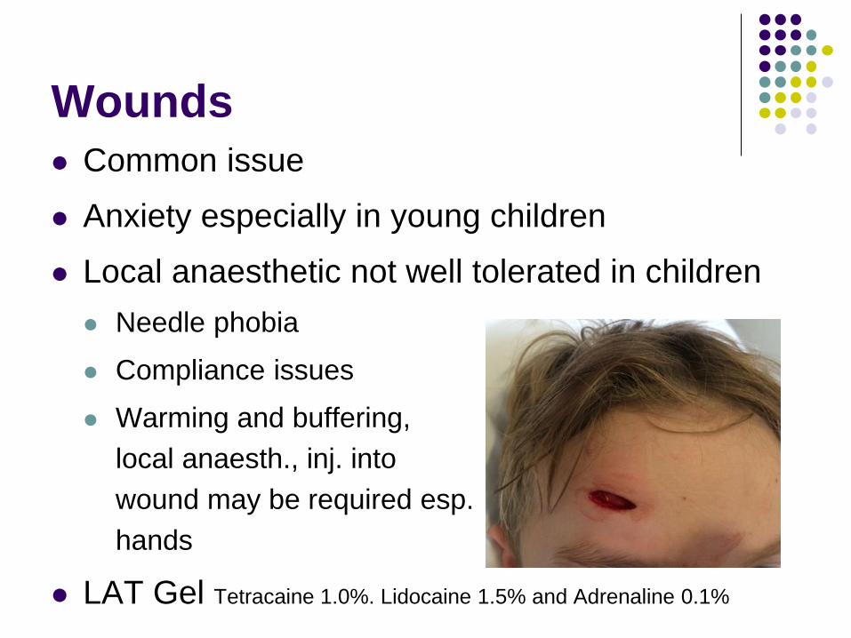
Wounds
Common issue
Anxiety especially in young children
Local anaesthetic not well tolerated in children
Needle phobia
Compliance issues
Warming and buffering,
local anaesth., inj. into
wound may be required esp.
hands
LAT Gel Tetracaine 1.0%. Lidocaine 1.5% and Adrenaline 0.1%
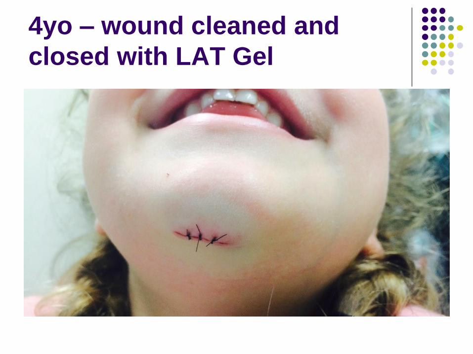
4yo – wound cleaned and
closed with LAT Gel
LAT Gel
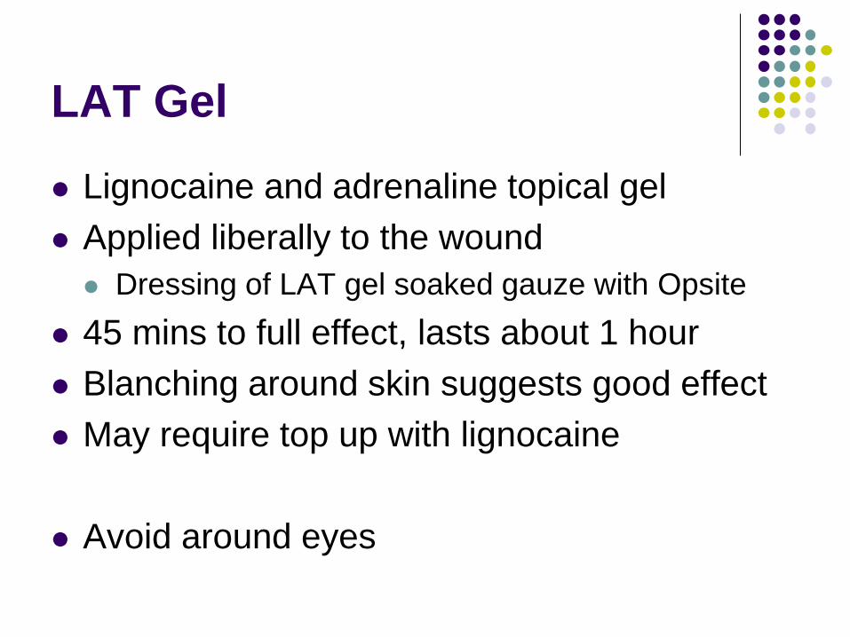
Lignocaine and adrenaline topical gel
Applied liberally to the wound
Dressing of LAT gel soaked gauze with Opsite
45 mins to full effect, lasts about 1 hour
Blanching around skin suggests good effect
May require top up with lignocaine
Avoid around eyes
Ketamine
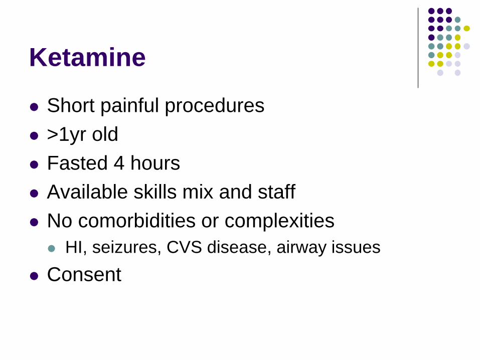
Short painful procedures
>1yr old
Fasted 4 hours
Available skills mix and staff
No comorbidities or complexities
HI, seizures, CVS disease, airway issues
Consent
Ketamine sedation
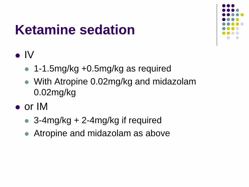
IV
1-1.5mg/kg +0.5mg/kg as required
With Atropine 0.02mg/kg and midazolam
0.02mg/kg
or IM
3-4mg/kg + 2-4mg/kg if required
Atropine and midazolam as above
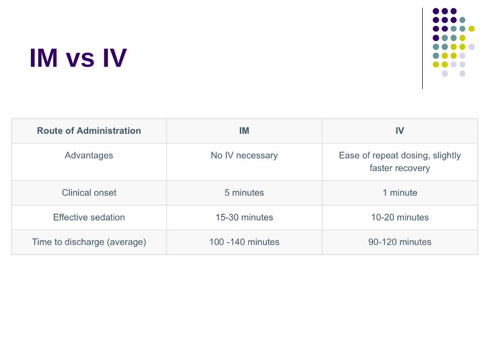
IM vs IV

Preparation
Airway trolley in room
Airways skills doctor able to recognise and
intervene is airway an issue
O2 Sats and cardiac monitoring
Nurse to monitor patient and someone to do
the procedure
Ketamine sedation
potent sedative, amnestic, analgesic and
anaesthetic agent
Dissociative catalepsy with nystagmus
Airway reflexes maintained
Emergence phenomena and agitation
Hypersalivation
Laryngospasm
Emesis
Apnoea
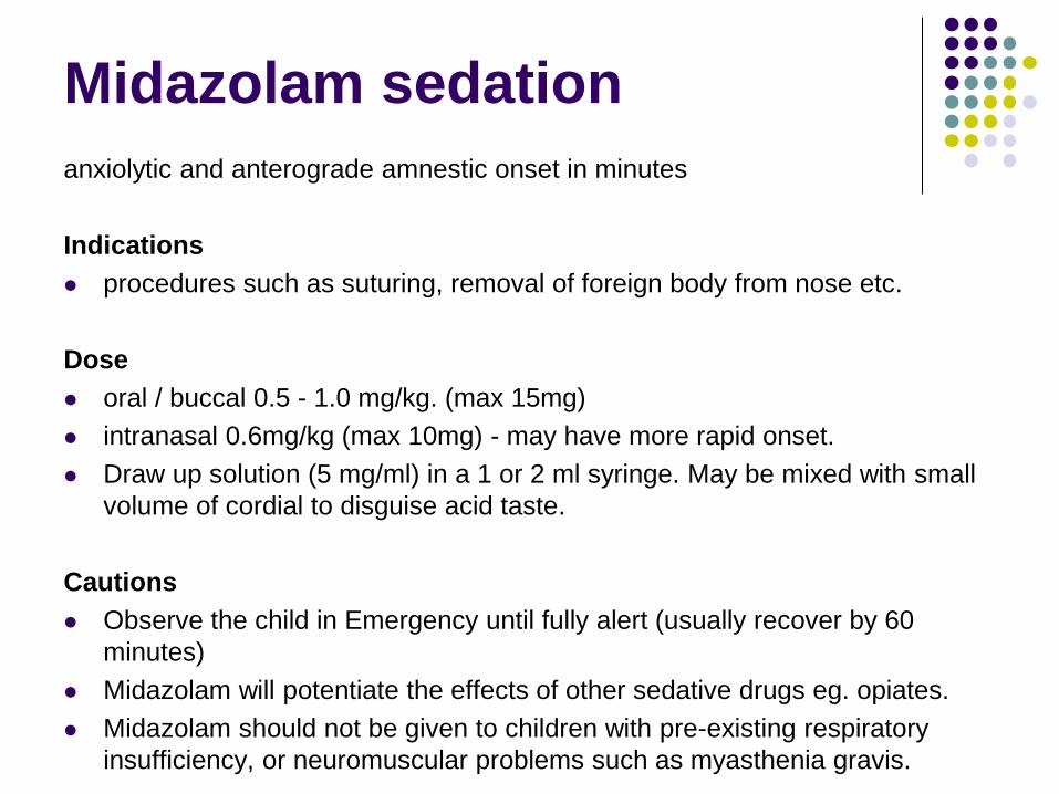
Midazolam sedation
anxiolytic and anterograde amnestic onset in minutes
Indications
procedures such as suturing, removal of foreign body from nose etc.
Dose
oral / buccal 0.5 - 1.0 mg/kg. (max 15mg)
intranasal 0.6mg/kg (max 10mg) - may have more rapid onset.
Draw up solution (5 mg/ml) in a 1 or 2 ml syringe. May be mixed with small
volume of cordial to disguise acid taste.
Cautions
Observe the child in Emergency until fully alert (usually recover by 60
minutes)
Midazolam will potentiate the effects of other sedative drugs eg. opiates.
Midazolam should not be given to children with pre-existing respiratory
insufficiency, or neuromuscular problems such as myasthenia gravis.

Case
7/12yo child, only child young parents
Brought into ED at 0300 for third visit in 2/7
‘Inconsolable’ according to parents (currently
asleep
Obs completely normal in ED but ‘seemed
hot earlier’
How many causes can you suggest for the
‘inconsolable child’
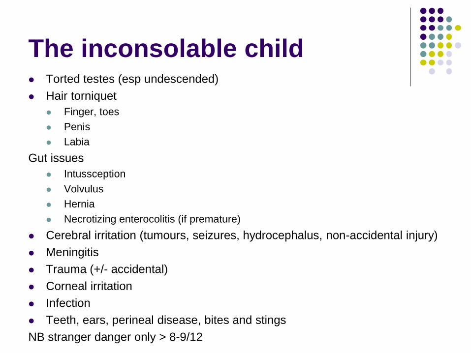
The inconsolable child Torted testes (esp undescended)
Hair torniquet
Finger, toes
Penis
Labia
Gut issues
Intussception
Volvulus
Hernia
Necrotizing enterocolitis (if premature)
Cerebral irritation (tumours, seizures, hydrocephalus, non-accidental injury)
Meningitis
Trauma (+/- accidental)
Corneal irritation
Infection
Teeth, ears, perineal disease, bites and stings
NB stranger danger only > 8-9/12
Asthma Bronchiolitis and
croup
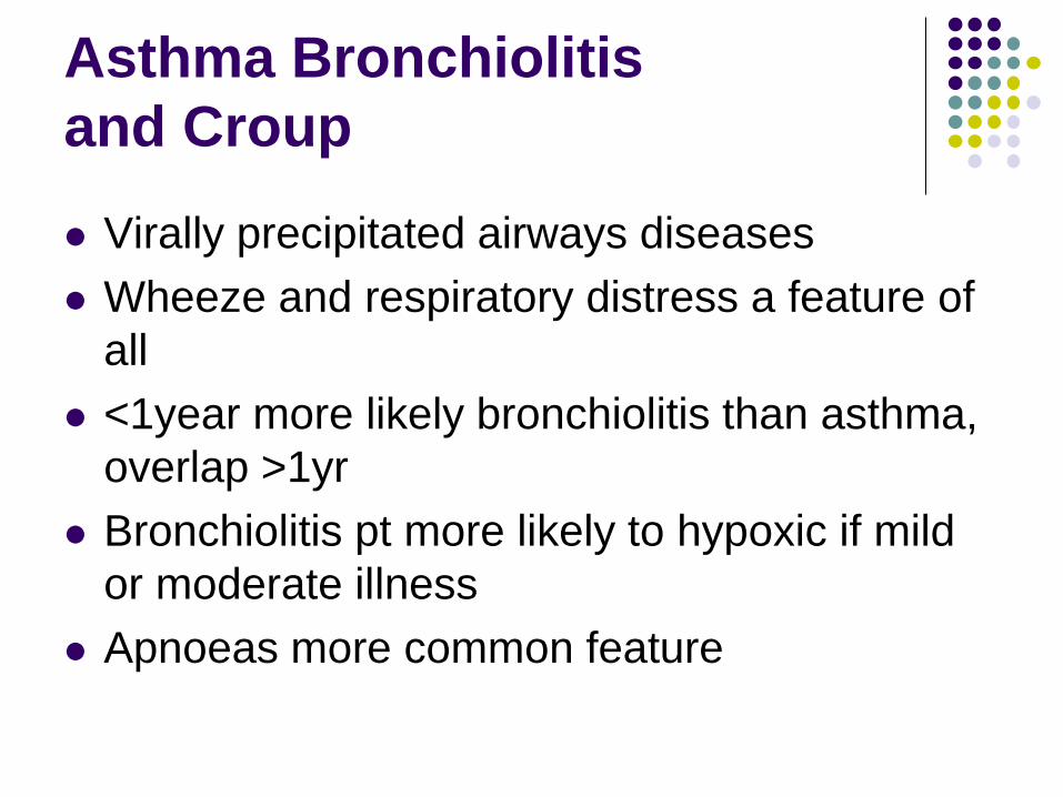
Asthma Bronchiolitis
and Croup
Virally precipitated airways diseases
Wheeze and respiratory distress a feature of
all
<1year more likely bronchiolitis than asthma,
overlap >1yr
Bronchiolitis pt more likely to hypoxic if mild
or moderate illness
Apnoeas more common feature
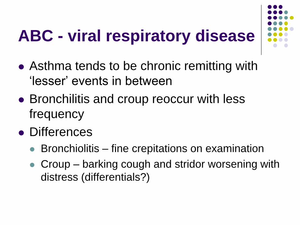
ABC - viral respiratory disease
Asthma tends to be chronic remitting with
‘lesser’ events in between
Bronchilitis and croup reoccur with less
frequency
Differences
Bronchiolitis – fine crepitations on examination
Croup – barking cough and stridor worsening with
distress (differentials?)

Treatment
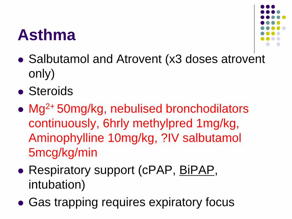
Asthma
Salbutamol and Atrovent (x3 doses atrovent
only)
Steroids
Mg2+ 50mg/kg, nebulised bronchodilators
continuously, 6hrly methylpred 1mg/kg,
Aminophylline 10mg/kg, ?IV salbutamol
5mcg/kg/min
Respiratory support (cPAP, BiPAP,
intubation)
Gas trapping requires expiratory focus

Asthma Severity Signs
Mild
Normal mental state
Subtle or no increased work of breathing accessory
muscle use/recession.
Able to talk normally
Moderate
Normal mental state
Some increased work of breathing accessory muscle
use/recession
Tachycardia
Some limitation of ability to talk
Severe
Agitated/distressed
Moderate-marked increased work of breathing
accessory muscle use/recession.
Tachycardia
Marked limitation of ability to talk
Note: wheeze is a poor predictor of severity.
Critical
Confused/drowsy
Maximal work of breathing accessory muscle
use/recession
Exhaustion
Marked tachycardia
Unable to talk
SILENT CHEST, wheeze may be absent if there is
poor air entry.

Bronchiolitis
Trickiling snot running into lungs
Treatment largely supportive
EM:RAP advocating upper airway (nasal)
saline and mucous aspiration
O2 (sats>92%), fluids (restricted 80%),
cPAP, ventilation
May be a subsection of patients that respond
to asthma protocol – difficult to identify
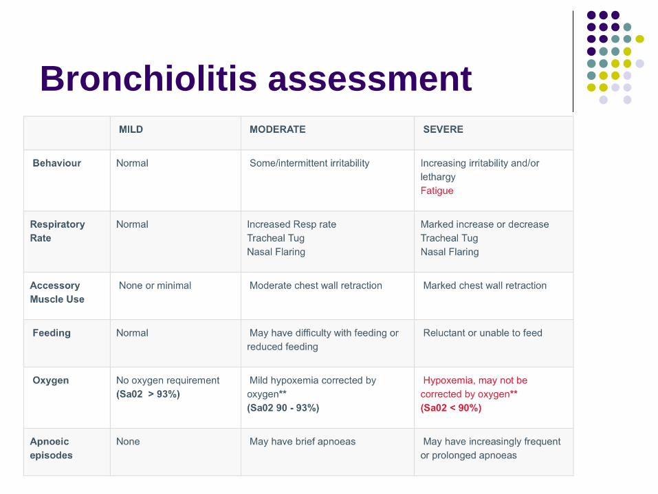
Bronchiolitis assessment

Croup
Upper airway obstruction, stridor makes you . . .
!!
Uncommon <6/12
Risk of severe croup if subglottic stenosis,
downs, prev. severe croup.
Prednisolone 1mg/kg
Neb Adrenaline 1mg (1ml of 1:1000 to 5ml with
saline) rpt if req.
IV dexamethasone 0.6mg/kg to 12mg
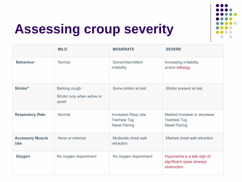
Assessing croup severity

Any Questions?

Thankyou


Other stuff
Fevers
Rashes
oncology
IV access
Abuse
Confronting and distressing
Risk of being wrong
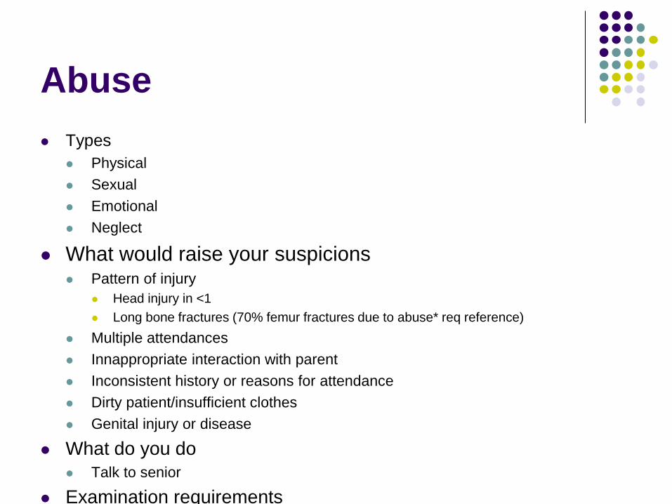
Abuse
Types
Physical
Sexual
Emotional
Neglect
What would raise your suspicions Pattern of injury
Head injury in <1
Long bone fractures (70% femur fractures due to abuse* req reference)
Multiple attendances
Innappropriate interaction with parent
Inconsistent history or reasons for attendance
Dirty patient/insufficient clothes
Genital injury or disease
What do you do
Talk to senior
Examination requirements
Need to treat emergencies
Risk of contamination or elimination of potential forensic evidence
Appropriate skills required for forensic examination
CHILDREN IN NEED OF PROTECTION Children, Youth and Families Act 2005 - SECT 162PART 4.1 CHILDREN IN
NEED OF PROTECTION
Recommended