Pacemaker and ICD infections
Recent insightsBJ Rijnders, MD, PhD
Internal Medicine
Section Infectious Dis.
Erasmus MC
Rotterdam
BVIKM 2007
Clinical presentation and diagnosis
Epidemiology
Etiology and origin of microorganismsOverall incidence
Treatment
- Antibiotic therapy
- Removal of PM/ICD ?
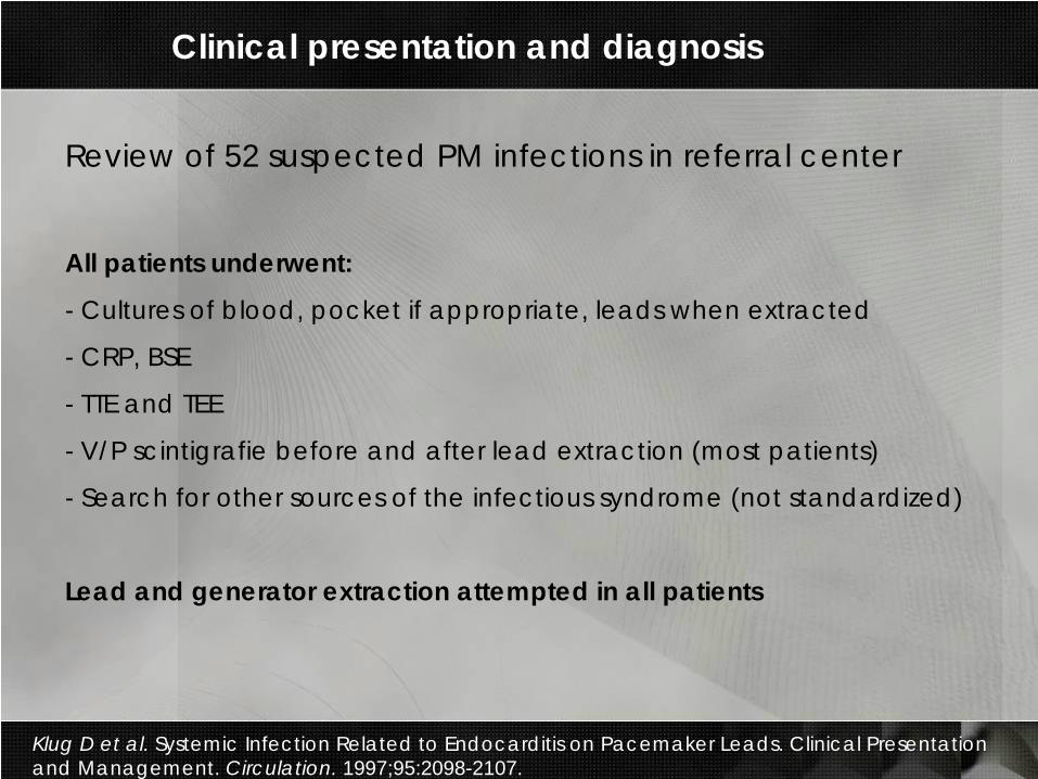
Clinical presentation and diagnosis
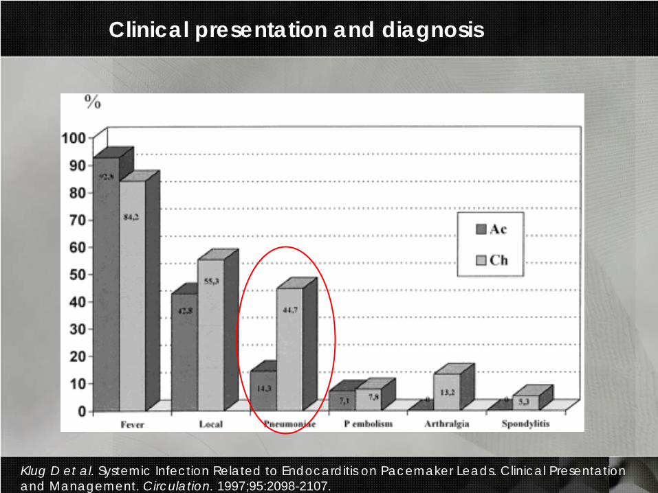
Klug D et al. Systemic Infection Related to Endocarditis on Pacemaker Leads. Clinical Presentation and Management. Circulation. 1997;95:2098-2107.
Review of 52 suspected PM infections in referral center
All patients underwent:
- Cultures of blood, pocket if appropriate, leads when extracted
- CRP, BSE
- TTE and TEE
- V/P scintigrafie before and after lead extraction (most patients)
- Search for other sources of the infectious syndrome (not standardized)
Lead and generator extraction attempted in all patients
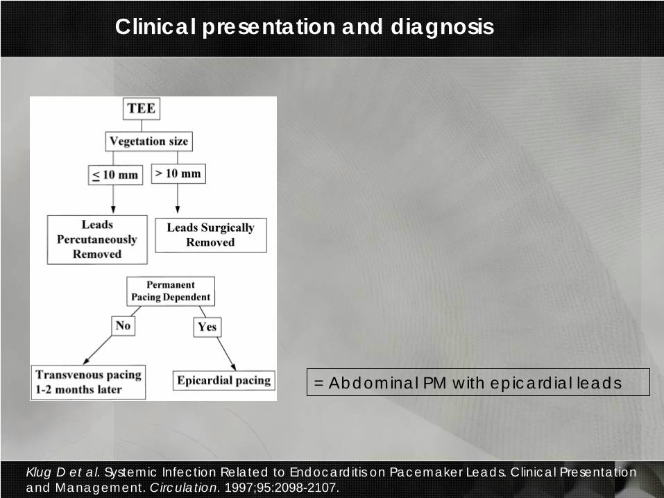
Clinical presentation and diagnosis
Klug D et al. Systemic Infection Related to Endocarditis on Pacemaker Leads. Clinical Presentation and Management. Circulation. 1997;95:2098-2107.
= Abdominal PM with epicardial leads
Clinical presentation and diagnosis
Klug D et al. Systemic Infection Related to Endocarditis on Pacemaker Leads. Clinical Presentation and Management. Circulation. 1997;95:2098-2107.
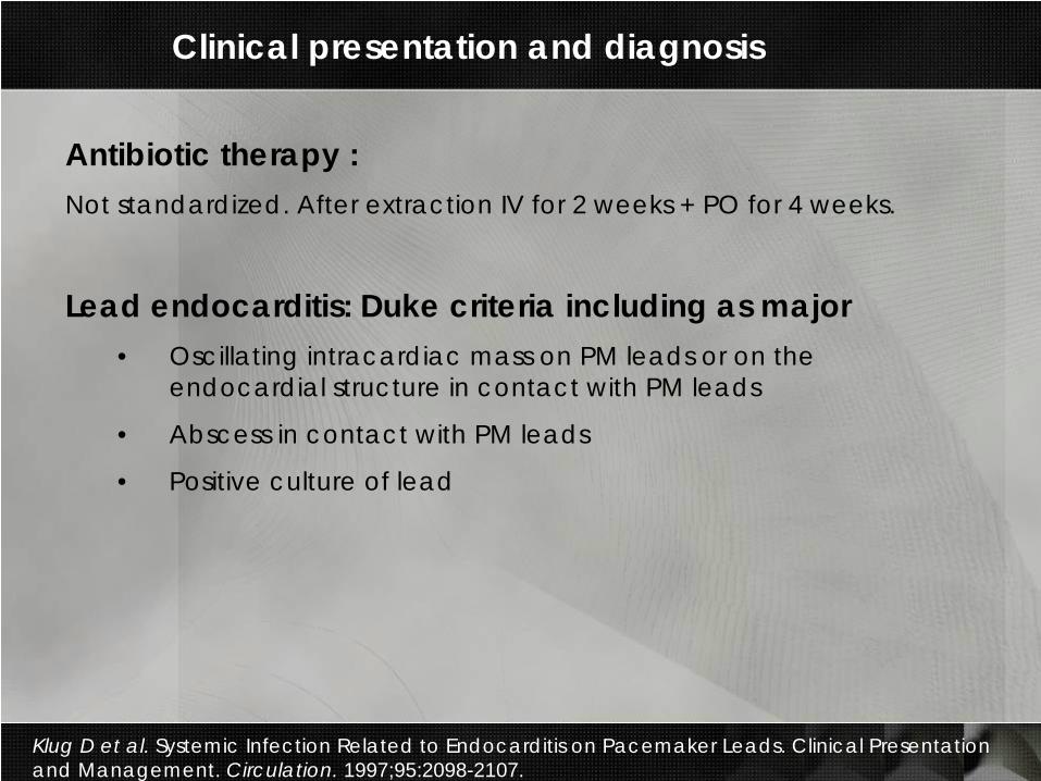
Antibiotic therapy : Not standardized. After extraction IV for 2 weeks + PO for 4 weeks.
Lead endocarditis: Duke criteria including as major• Oscillating intracardiac mass on PM leads or on the
endocardial structure in contact with PM leads
• Abscess in contact with PM leads
• Positive culture of lead
Clinical presentation and diagnosis
Klug D et al. Systemic Infection Related to Endocarditis on Pacemaker Leads. Clinical Presentation and Management. Circulation. 1997;95:2098-2107.
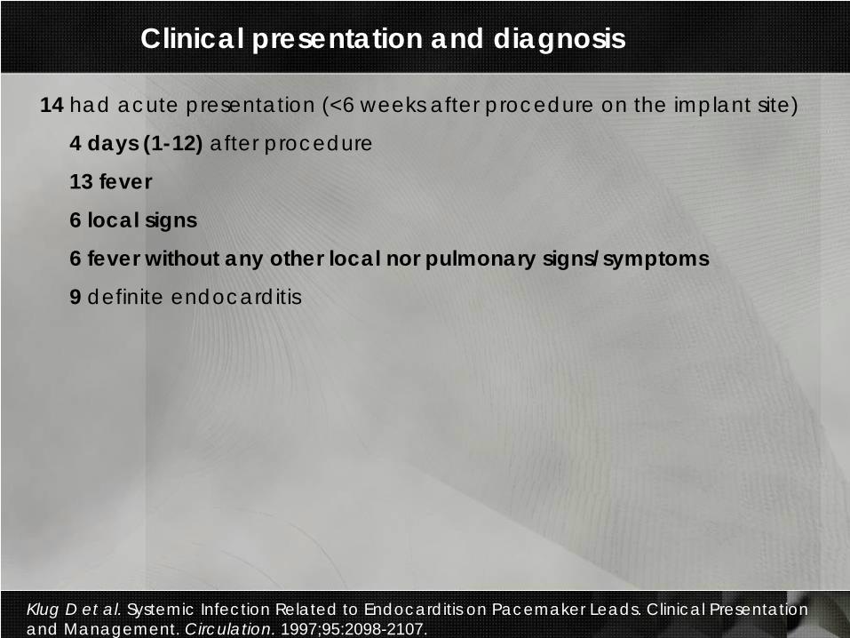
14 had acute presentation (<6 weeks after procedure on the implant site)
4 days (1-12) after procedure
13 fever
6 local signs
6 fever without any other local nor pulmonary signs/symptoms
9 definite endocarditis
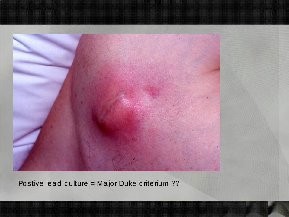
Positive lead culture = Major Duke criterium ??
Clinical presentation and diagnosis
Klug D et al. Systemic Infection Related to Endocarditis on Pacemaker Leads. Clinical Presentation and Management. Circulation. 1997;95:2098-2107.
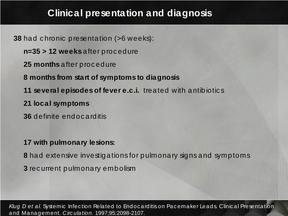
38 had chronic presentation (>6 weeks):
n=35 > 12 weeks after procedure
25 months after procedure
8 months from start of symptoms to diagnosis
11 several episodes of fever e.c.i. treated with antibiotics
21 local symptoms
36 definite endocarditis
17 with pulmonary lesions:
8 had extensive investigations for pulmonary signs and symptoms
3 recurrent pulmonary embolism
Clinical presentation and diagnosis
Klug D et al. Systemic Infection Related to Endocarditis on Pacemaker Leads. Clinical Presentation and Management. Circulation. 1997;95:2098-2107.
Clinical presentation and diagnosis
Klug D et al. Systemic Infection Related to Endocarditis on Pacemaker Leads. Clinical Presentation and Management. Circulation. 1997;95:2098-2107.
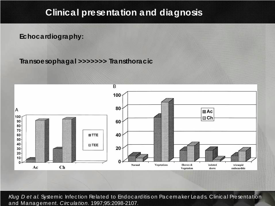
Echocardiography:
Transoesophagal >>>>>>> Transthoracic
Clinical presentation and diagnosis
Klug D et al. Systemic Infection Related to Endocarditis on Pacemaker Leads. Clinical Presentation and Management. Circulation. 1997;95:2098-2107.
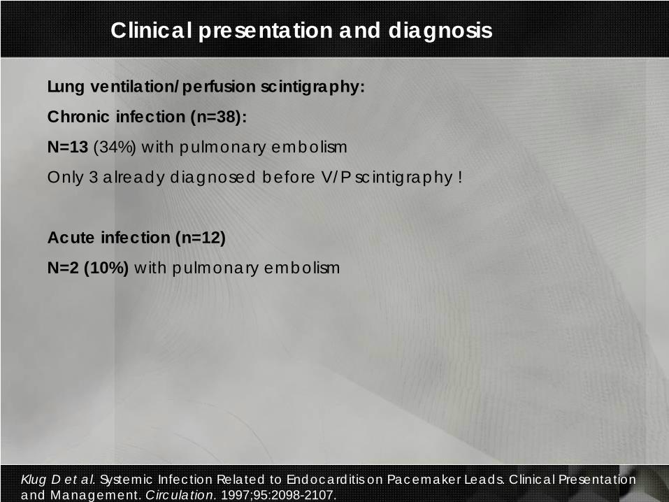
Lung ventilation/perfusion scintigraphy:
Chronic infection (n=38):
N=13 (34%) with pulmonary embolism
Only 3 already diagnosed before V/P scintigraphy !
Acute infection (n=12)
N=2 (10%) with pulmonary embolism
Clinical presentation and diagnosis
Klug D et al. Systemic Infection Related to Endocarditis on Pacemaker Leads. Clinical Presentation and Management. Circulation. 1997;95:2098-2107.
Treatment and outcome
Klug D et al. Systemic Infection Related to Endocarditis on Pacemaker Leads. Clinical Presentation and Management. Circulation. 1997;95:2098-2107.
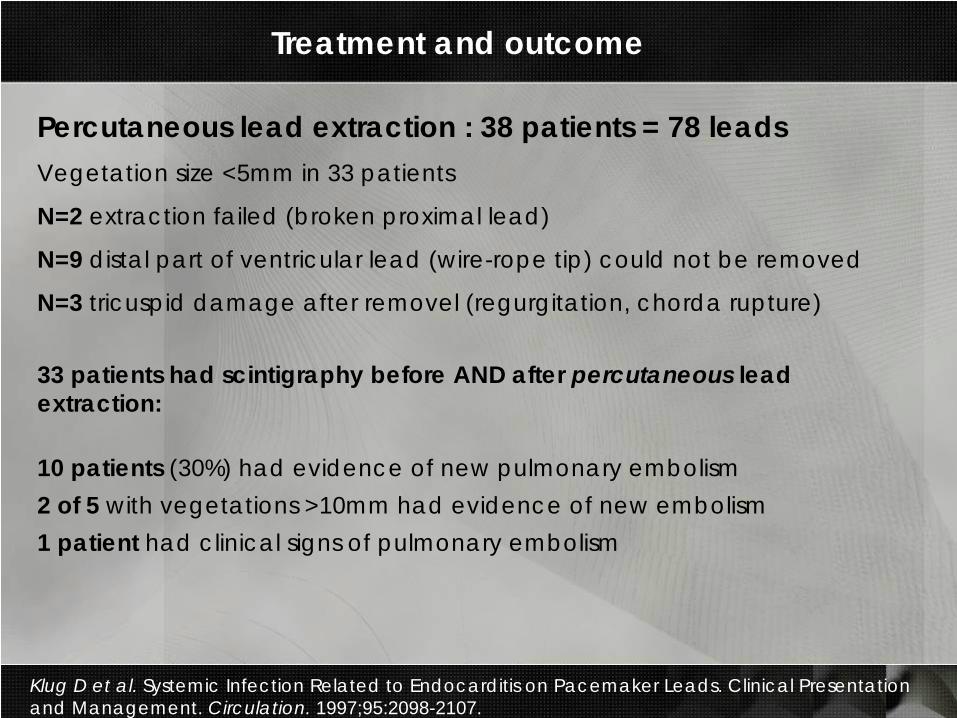
Percutaneous lead extraction : 38 patients = 78 leadsVegetation size <5mm in 33 patients
N=2 extraction failed (broken proximal lead)
N=9 distal part of ventricular lead (wire-rope tip) could not be removed
N=3 tricuspid damage after removel (regurgitation, chorda rupture)
33 patients had scintigraphy before AND after percutaneous lead extraction:
10 patients (30%) had evidence of new pulmonary embolism2 of 5 with vegetations >10mm had evidence of new embolism1 patient had clinical signs of pulmonary embolism
Treatment and outcome
Klug D et al. Systemic Infection Related to Endocarditis on Pacemaker Leads. Clinical Presentation and Management. Circulation. 1997;95:2098-2107.
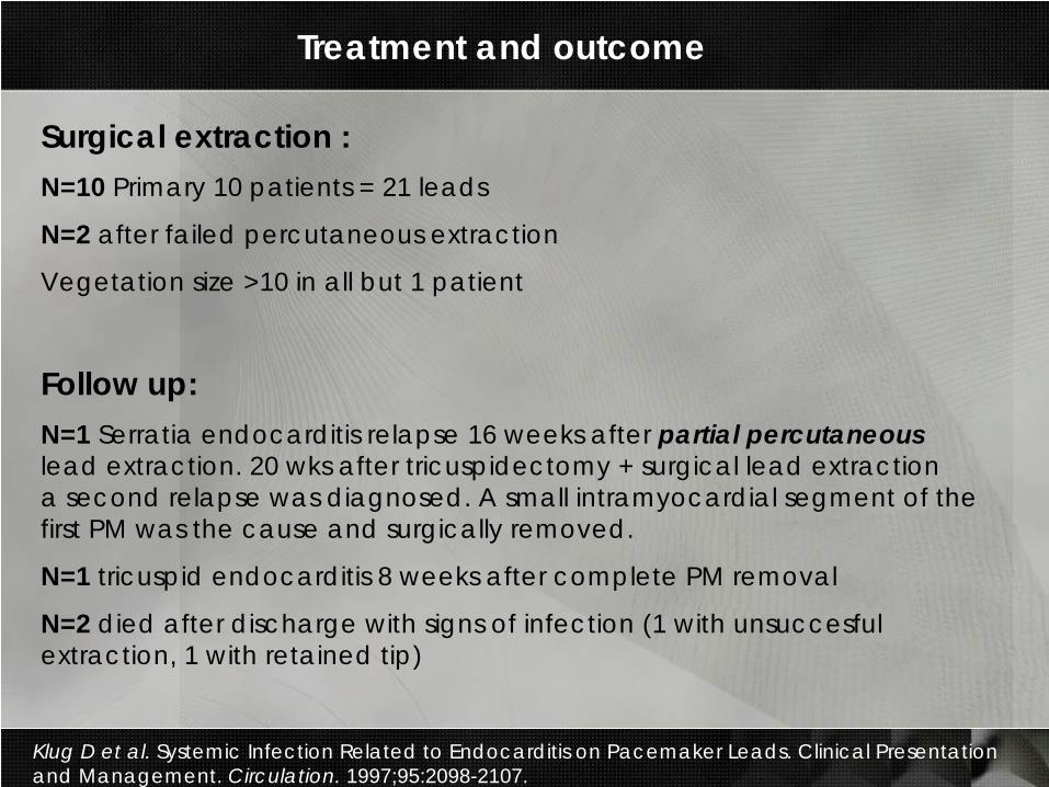
Surgical extraction : N=10 Primary 10 patients = 21 leads
N=2 after failed percutaneous extraction
Vegetation size >10 in all but 1 patient
Follow up:N=1 Serratia endocarditis relapse 16 weeks after partial percutaneous lead extraction. 20 wks after tricuspidectomy + surgical lead extraction a second relapse was diagnosed. A small intramyocardial segment of the first PM was the cause and surgically removed.
N=1 tricuspid endocarditis 8 weeks after complete PM removal
N=2 died after discharge with signs of infection (1 with unsuccesful extraction, 1 with retained tip)
Clinical presentation and diagnosis
Klug D et al. Systemic Infection Related to Endocarditis on Pacemaker Leads. Clinical Presentation and Management. Circulation. 1997;95:2098-2107.
“ The diagnosis of systemic infection related to PM-lead infection
must be systematically considered in the presence of chronic fever,
recurrent bronchitis, or pulmonary infection or in case of recurrent or
persistent evidence of infection at the implant site “
“ No correlation between vegetation size and pulmonary migration
was observed during percutaneous lead extraction. Perhaps we
should expand the indications for percutaneous removal to include
patients with larger vegetation sizes “
Clinical presentation and diagnosis
Epidemiology
Overall incidenceEtiology and origin of microorganisms
Treatment
- Antibiotic therapy
- Removal of PM/ICD ?
Epidemiology
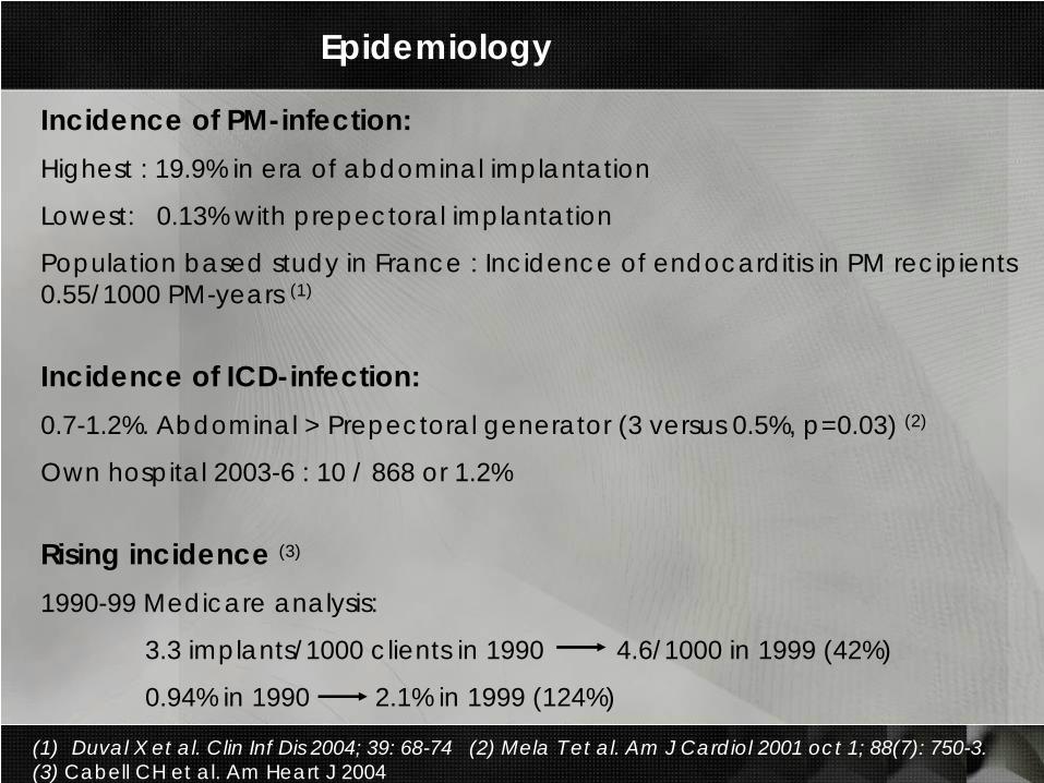
(1) Duval X et al. Clin Inf Dis 2004; 39: 68-74 (2) Mela T et al. Am J Cardiol 2001 oct 1; 88(7): 750-3.(3) Cabell CH et al. Am Heart J 2004
Incidence of PM-infection:Highest : 19.9% in era of abdominal implantation
Lowest: 0.13% with prepectoral implantation
Population based study in France : Incidence of endocarditis in PM recipients 0.55/1000 PM-years (1)
Incidence of ICD-infection:0.7-1.2%. Abdominal > Prepectoral generator (3 versus 0.5%, p=0.03) (2)
Own hospital 2003-6 : 10 / 868 or 1.2%
Rising incidence (3)
1990-99 Medicare analysis:
3.3 implants/1000 clients in 1990 4.6/1000 in 1999 (42%)
0.94% in 1990 2.1% in 1999 (124%)
Epidemiology: Sources of infection
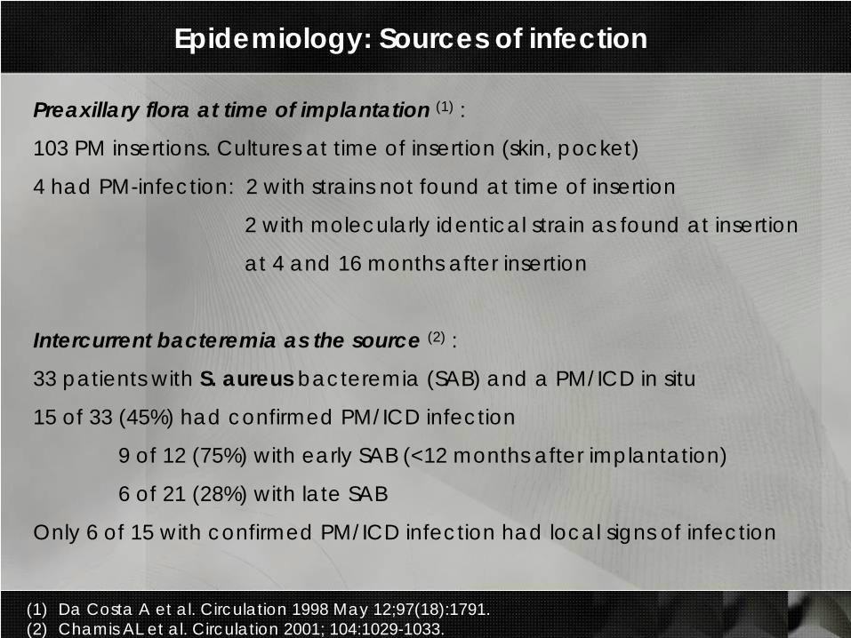
(1) Da Costa A et al. Circulation 1998 May 12;97(18):1791.(2) Chamis AL et al. Circulation 2001; 104:1029-1033.
Preaxillary flora at time of implantation (1) :
103 PM insertions. Cultures at time of insertion (skin, pocket)
4 had PM-infection: 2 with strains not found at time of insertion
2 with molecularly identical strain as found at insertion
at 4 and 16 months after insertion
Intercurrent bacteremia as the source (2) :
33 patients with S. aureus bacteremia (SAB) and a PM/ICD in situ
15 of 33 (45%) had confirmed PM/ICD infection
9 of 12 (75%) with early SAB (<12 months after implantation)
6 of 21 (28%) with late SAB
Only 6 of 15 with confirmed PM/ICD infection had local signs of infection
Epidemiology: Sources of infection
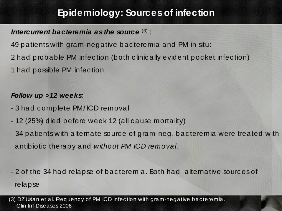
(3) DZ Uslan et al. Frequency of PM ICD infection with gram-negative bacteremia. Clin Inf Diseases 2006
Intercurrent bacteremia as the source (3) :
49 patients with gram-negative bacteremia and PM in situ:
2 had probable PM infection (both clinically evident pocket infection)
1 had possible PM infection
Follow up >12 weeks:
- 3 had complete PM/ICD removal
- 12 (25%) died before week 12 (all cause mortality)
- 34 patients with alternate source of gram-neg. bacteremia were treated with
antibiotic therapy and without PM ICD removal.
- 2 of the 34 had relapse of bacteremia. Both had alternative sources of
relapse
Clinical presentation and diagnosis
Epidemiology
Etiology and origin of microorganismsOverall incidence
Treatment
- Antibiotic therapy
- Removal of PM/ICD ?
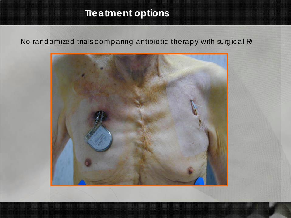
Treatment options
No randomized trials comparing antibiotic therapy with surgical R/
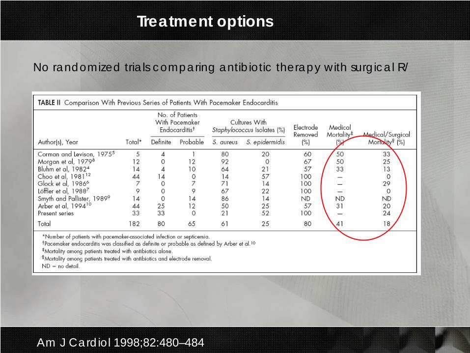
Treatment options
No randomized trials comparing antibiotic therapy with surgical R/
Am J Cardiol 1998;82:480–484
Treatment options
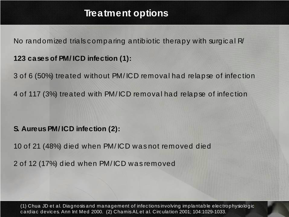
No randomized trials comparing antibiotic therapy with surgical R/
123 cases of PM/ICD infection (1):
3 of 6 (50%) treated without PM/ICD removal had relapse of infection
4 of 117 (3%) treated with PM/ICD removal had relapse of infection
S. Aureus PM/ICD infection (2):
10 of 21 (48%) died when PM/ICD was not removed died
2 of 12 (17%) died when PM/ICD was removed
(1) Chua JD et al. Diagnosis and management of infections involving implantable electrophysiologic cardiac devices. Ann Int Med 2000. (2) Chamis AL et al. Circulation 2001; 104:1029-1033.
Treatment options
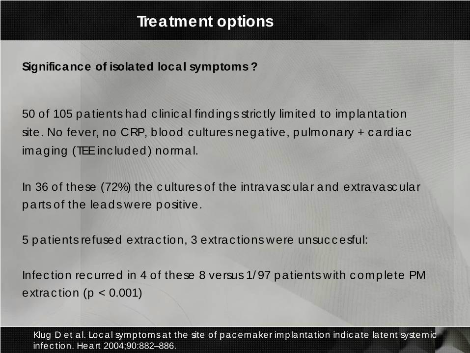
Significance of isolated local symptoms ?
50 of 105 patients had clinical findings strictly limited to implantation site. No fever, no CRP, blood cultures negative, pulmonary + cardiac imaging (TEE included) normal.
In 36 of these (72%) the cultures of the intravascular and extravascularparts of the leads were positive.
5 patients refused extraction, 3 extractions were unsuccesful:
Infection recurred in 4 of these 8 versus 1/97 patients with complete PM extraction (p < 0.001)
Klug D et al. Local symptoms at the site of pacemaker implantation indicate latent systemic infection. Heart 2004;90:882–886.
Treatment options
Percutaneous extraction and large vegetations:
Robbins MJ et al. Influence of vegetation size on clinical outcome of right-sided infective endocarditis. Am J Med 1986;80:165–71.
This study reported that a vegetation size of 10 mm was a bad prognostic factor (=surgery was needed for ongoing infection)
(1) 38 percutaneous removals. 9 patients had vegetations >10mm.
5 of 9 had evidence of PE. All 5 survived with AB and anticoagulant R/1 developed small lung abces (S. aureus)
(2) 9 percutaneous removals with vegetations >10mm. Non had clinically apparent PE.
(1) Meier-Ewert HK et al. Am Heart J 2003;146: 339–44.
{
(2) Victor F et al. Heart 1999;81:82-7.
Treatment
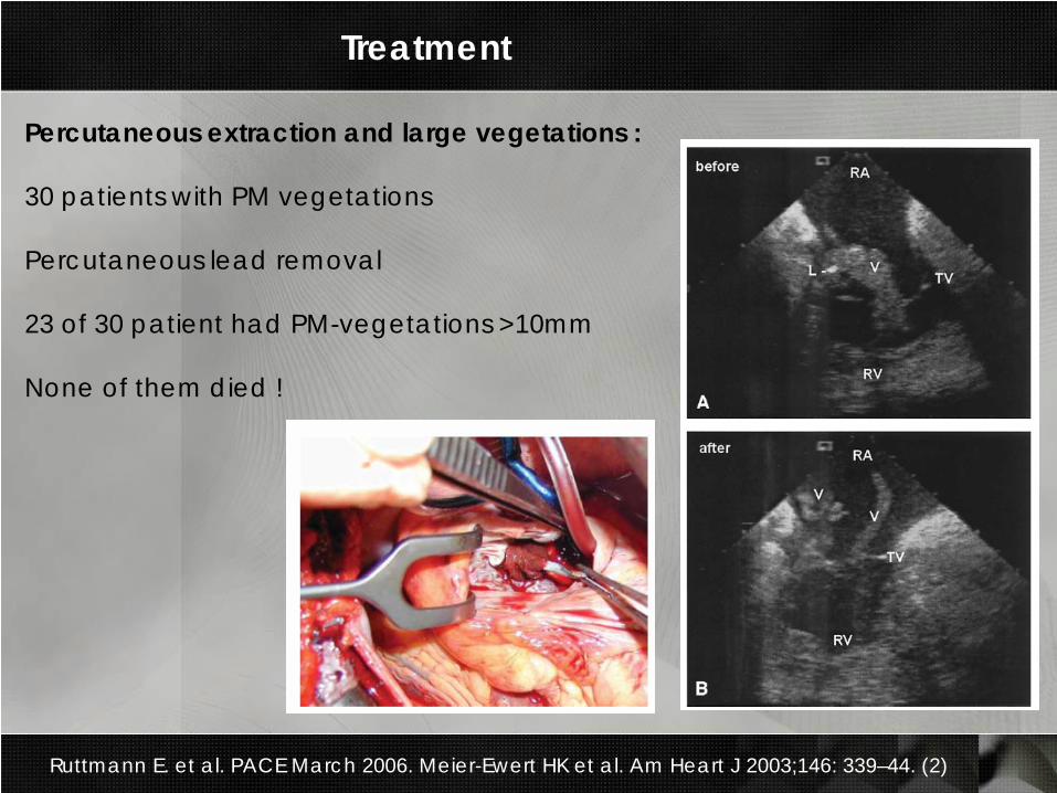
Percutaneous extraction and large vegetations :
30 patients with PM vegetations
Percutaneous lead removal
23 of 30 patient had PM-vegetations >10mm
None of them died !
Ruttmann E. et al. PACE March 2006. Meier-Ewert HK et al. Am Heart J 2003;146: 339–44. (2)
Treatment
Antibiotic therapy : Recent hot trials !
Vancomycine versus vanco + genta versus vanco + genta + rifa for
the treatment PM-lead endocarditis treated without lead extraction.
Double blind, double dummy controlled trial. Cinderella PM et al.
New Engl J Med 2007, in press.
Daptomycine or vanco + genta + rifa for the treatment of PM-lead
endocarditis treated without lead extraction. A double blind double
dummy controlled non-inferiority trial. Riding Hood LR et al. JAMA
2007, in press.
Treatment
Antibiotic therapy : Recent trials.
Vancomycine versus vanco + genta versus vanco + genta + rifa for
the treatment PM-lead endocarditis treated without lead extraction.
Double blind, double dummy controlled trial. Cinderella PM et al.
New Engl J Med 2007, in press.
Daptomycine or vanco + genta + rifa for the treatment of PM-lead
endocarditis treated without lead extraction. A double blind double
dummy controlled non-inferiority trial. Riding Hood LR et al. JAMA
2007, in press.
Treatment: Antibiotic therapy
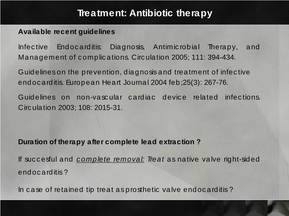
Available recent guidelines
Infective Endocarditis: Diagnosis, Antimicrobial Therapy, and Management of complications. Circulation 2005; 111: 394-434.
Guidelines on the prevention, diagnosis and treatment of infective endocarditis. European Heart Journal 2004 feb;25(3): 267-76.
Guidelines on non-vascular cardiac device related infections. Circulation 2003; 108: 2015-31.
Duration of therapy after complete lead extraction ?
If succesful and complete removal: Treat as native valve right-sided
endocarditis ?
In case of retained tip treat as prosthetic valve endocarditis ?
Time for questions and coffee !
Recommended