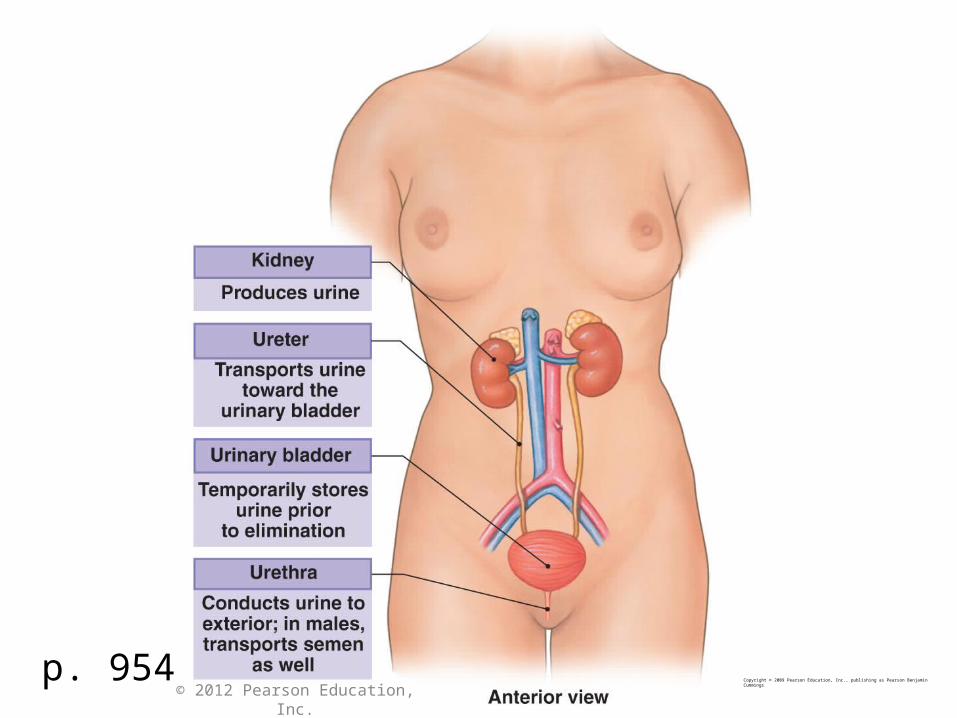
p. 954Copyright © 2009 Pearson Education, Inc., publishing as Pearson Benjamin Cummings
© 2012 Pearson Education, Inc.
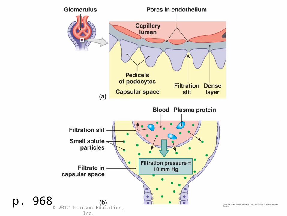
p. 968Copyright © 2009 Pearson Education, Inc., publishing as Pearson Benjamin Cummings
© 2012 Pearson Education, Inc.
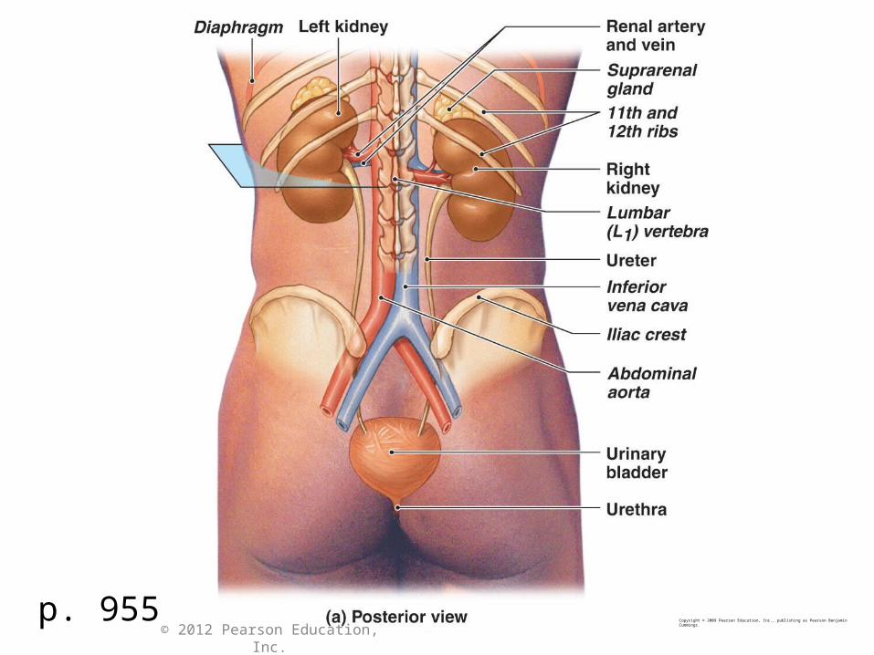
p. 955Copyright © 2009 Pearson Education, Inc., publishing as Pearson Benjamin Cummings
© 2012 Pearson Education, Inc.
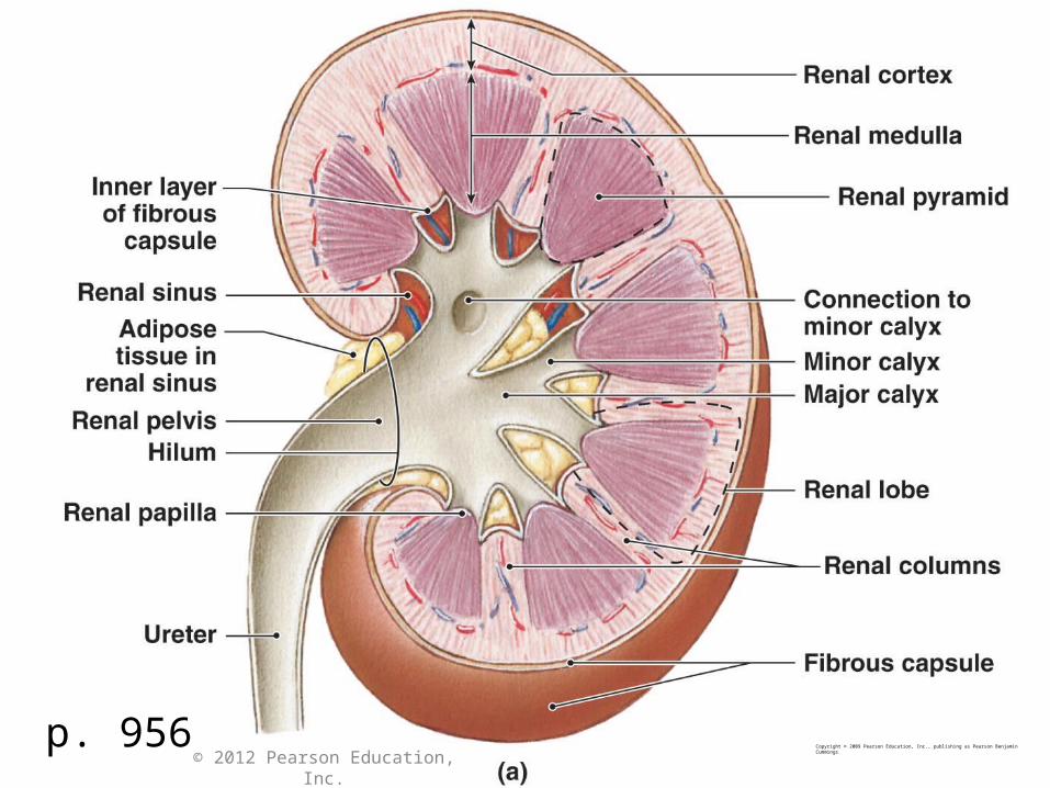
p. 956Copyright © 2009 Pearson Education, Inc., publishing as Pearson Benjamin Cummings
© 2012 Pearson Education, Inc.
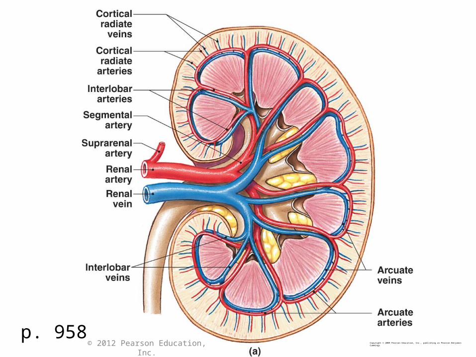
p. 958Copyright © 2009 Pearson Education, Inc., publishing as Pearson Benjamin Cummings© 2012 Pearson Education, Inc.
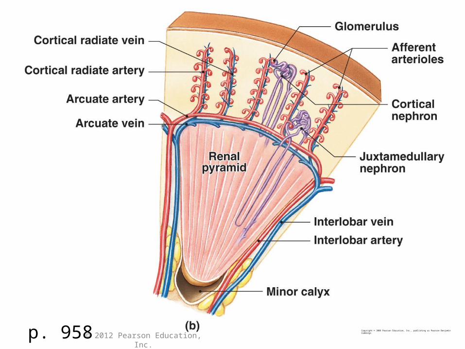
Copyright © 2009 Pearson Education, Inc., publishing as Pearson Benjamin Cummingsp. 958 © 2012 Pearson Education, Inc.
© 2012 Pearson Education, Inc.
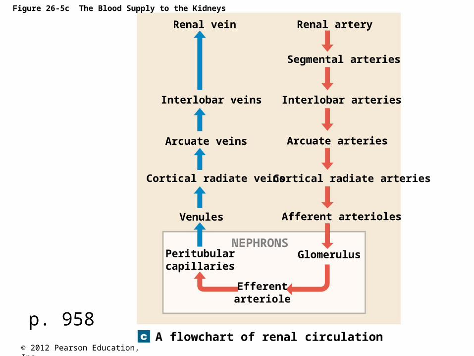
Figure 26-5c The Blood Supply to the Kidneys
A flowchart of renal circulation
Renal vein Renal artery
Segmental arteries
Interlobar arteries
Arcuate arteries
Cortical radiate arteries
Afferent arterioles
Glomerulus
Efferentarteriole
Peritubularcapillaries
Interlobar veins
Arcuate veins
Cortical radiate veins
Venules
NEPHRONS
p. 958
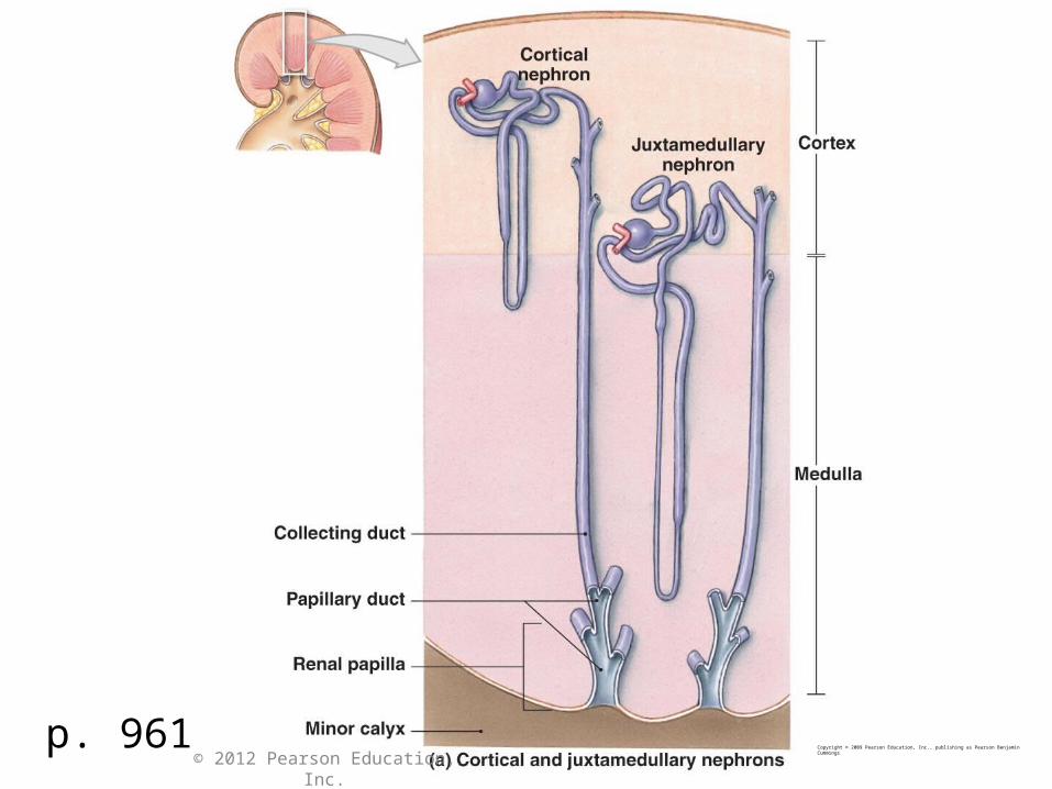
p. 961Copyright © 2009 Pearson Education, Inc., publishing as Pearson Benjamin Cummings
© 2012 Pearson Education, Inc.
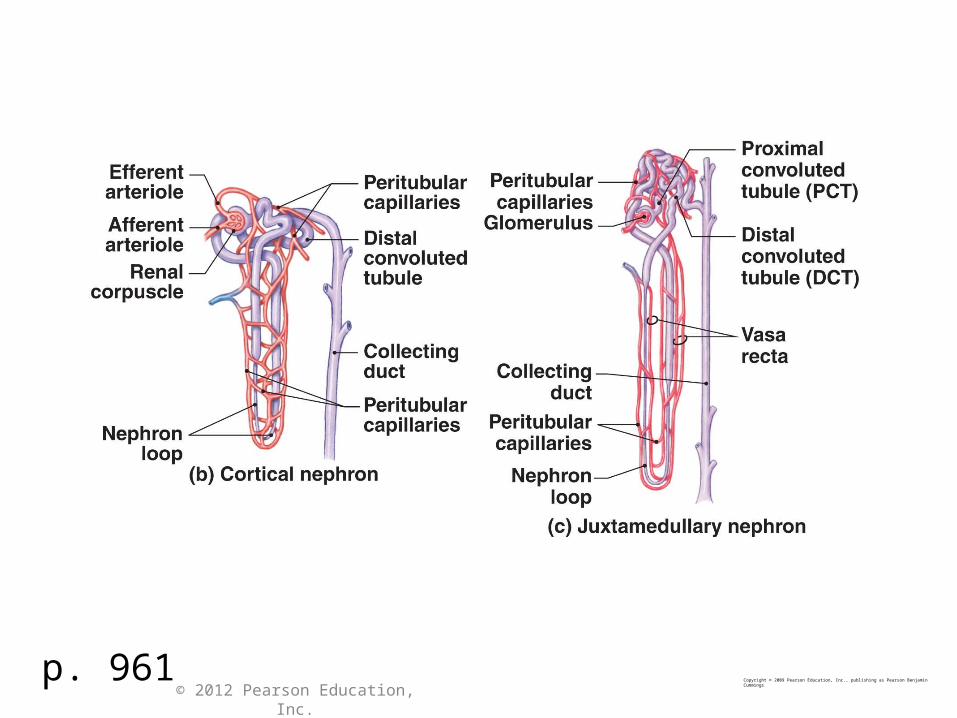
p. 961Copyright © 2009 Pearson Education, Inc., publishing as Pearson Benjamin Cummings
© 2012 Pearson Education, Inc.
p. 959Copyright © 2009 Pearson Education, Inc., publishing as Pearson Benjamin Cummings
© 2012 Pearson Education, Inc.
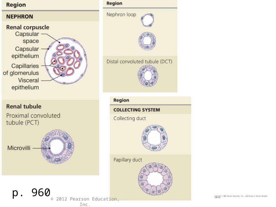
p. 960Copyright © 2009 Pearson Education, Inc., publishing as Pearson Benjamin Cummings
© 2012 Pearson Education, Inc.

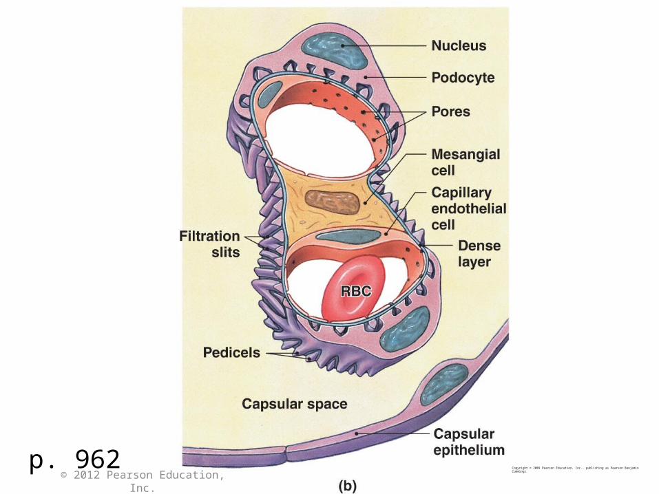
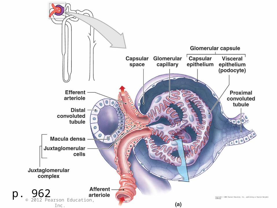
p. 962Copyright © 2009 Pearson Education, Inc., publishing as Pearson Benjamin Cummings
© 2012 Pearson Education, Inc.
p. 962Copyright © 2009 Pearson Education, Inc., publishing as Pearson Benjamin Cummings
© 2012 Pearson Education, Inc.
© 2012 Pearson Education, Inc.
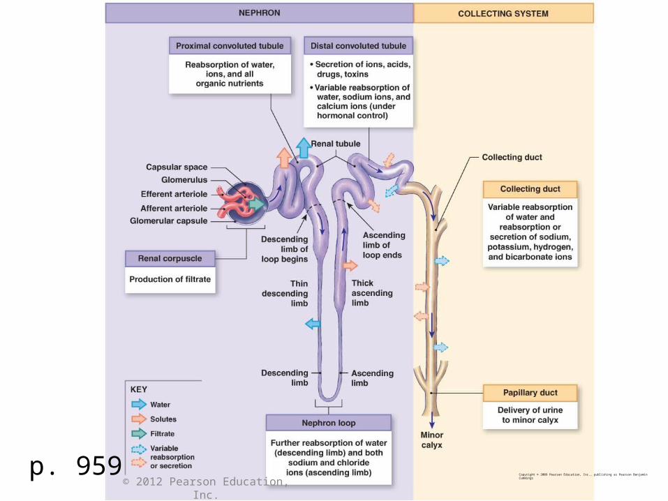
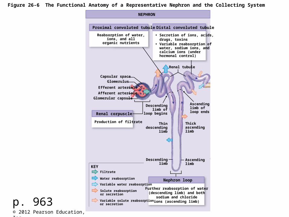
Figure 26-6 The Functional Anatomy of a Representative Nephron and the Collecting System
NEPHRON
Distal convoluted tubuleProximal convoluted tubule
Renal corpuscle
Nephron loop
KEY
• Secretion of ions, acids, drugs, toxins• Variable reabsorption of water, sodium ions, and calcium ions (under hormonal control)
Reabsorption of water,ions, and all
organic nutrients
Renal tubule
Capsular spaceGlomerulus
Efferent arteriole
Afferent arterioleGlomerular capsule
Production of filtrate
Descendinglimb of
loop begins
Thindescending
limb
Ascendinglimb ofloop ends
Thickascendinglimb
Descendinglimb
Ascendinglimb
Further reabsorption of water(descending limb) and both
sodium and chlorideions (ascending limb)
Filtrate
Water reabsorption
Variable water reabsorption
Solute reabsorptionor secretion
Variable solute reabsorptionor secretionp. 963
© 2012 Pearson Education, Inc.
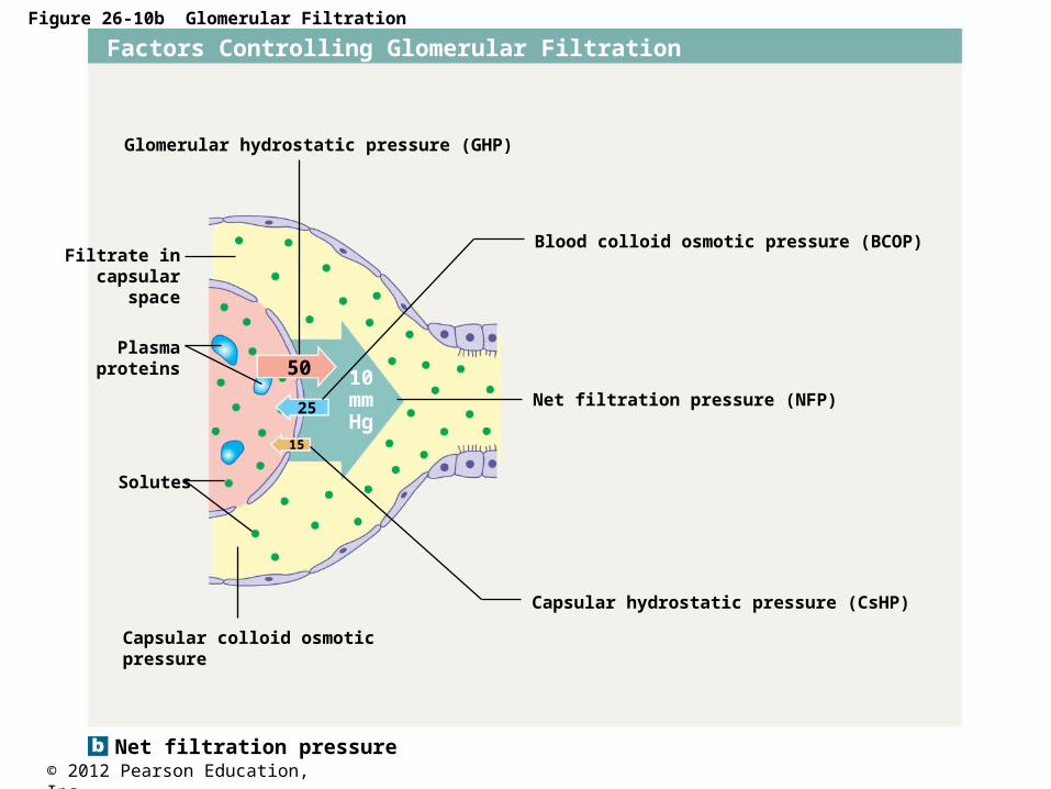
Figure 26-10b Glomerular Filtration
Net filtration pressure
Factors Controlling Glomerular Filtration
Filtrate incapsular
space
Plasmaproteins
Solutes
50
25
15
10mmHg
Capsular colloid osmoticpressure
Capsular hydrostatic pressure (CsHP)
Net filtration pressure (NFP)
Blood colloid osmotic pressure (BCOP)
Glomerular hydrostatic pressure (GHP)

© 2012 Pearson Education, Inc.
Figure 26-12 Transport Activities at the PCT
KEY
Leak channel
Countertransport
Exchange pump
Cotransport
Diffusion
Reabsorption
Secretion
Peritubularcapillary
Peritubularfluid
Osmoticwaterflow
Glucoseand otherorganicsolutes
Cells ofproximalconvolutedtubule
Tubular fluid
Lumen containingtubular fluid
Cuboidalepithelial cells
p. 973
© 2012 Pearson Education, Inc.
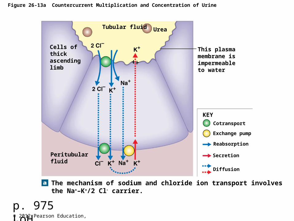
Figure 26-13a Countercurrent Multiplication and Concentration of Urine
The mechanism of sodium and chloride ion transport involvesthe Na–K/2 Cl carrier.
KEY
Cotransport
Exchange pump
Reabsorption
Secretion
Diffusion
This plasmamembrane isimpermeableto water
UreaTubular fluid
Peritubularfluid
Cells of thickascendinglimb
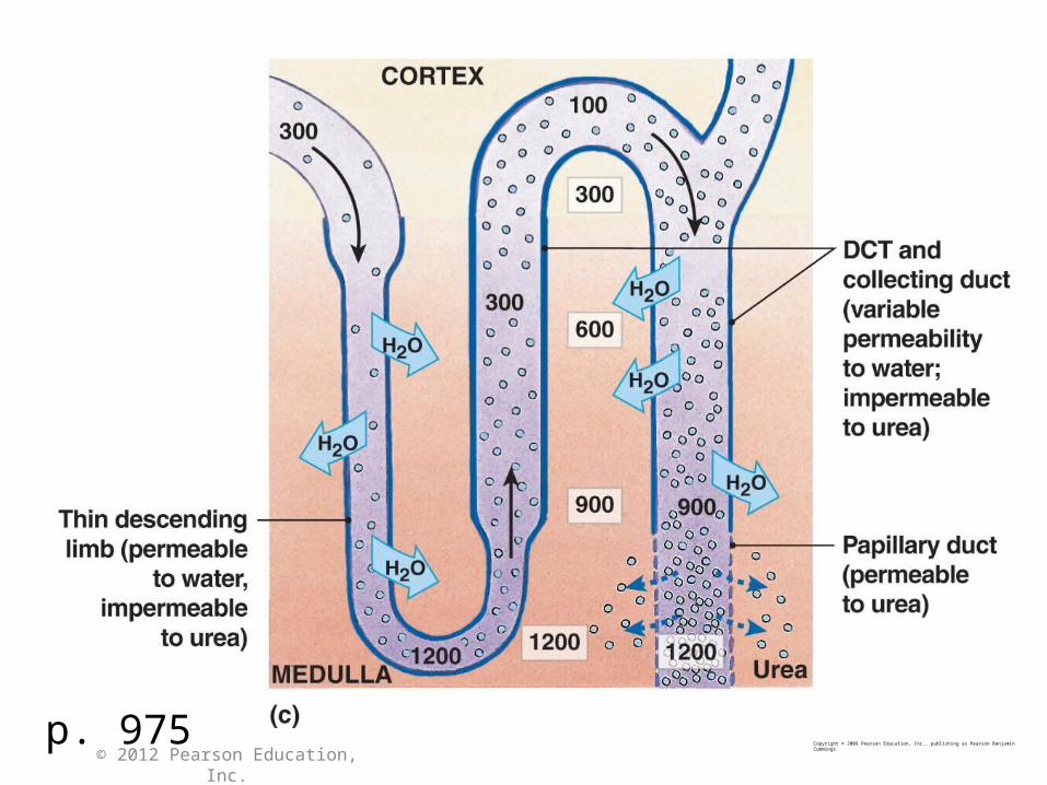
p. 975 LoH
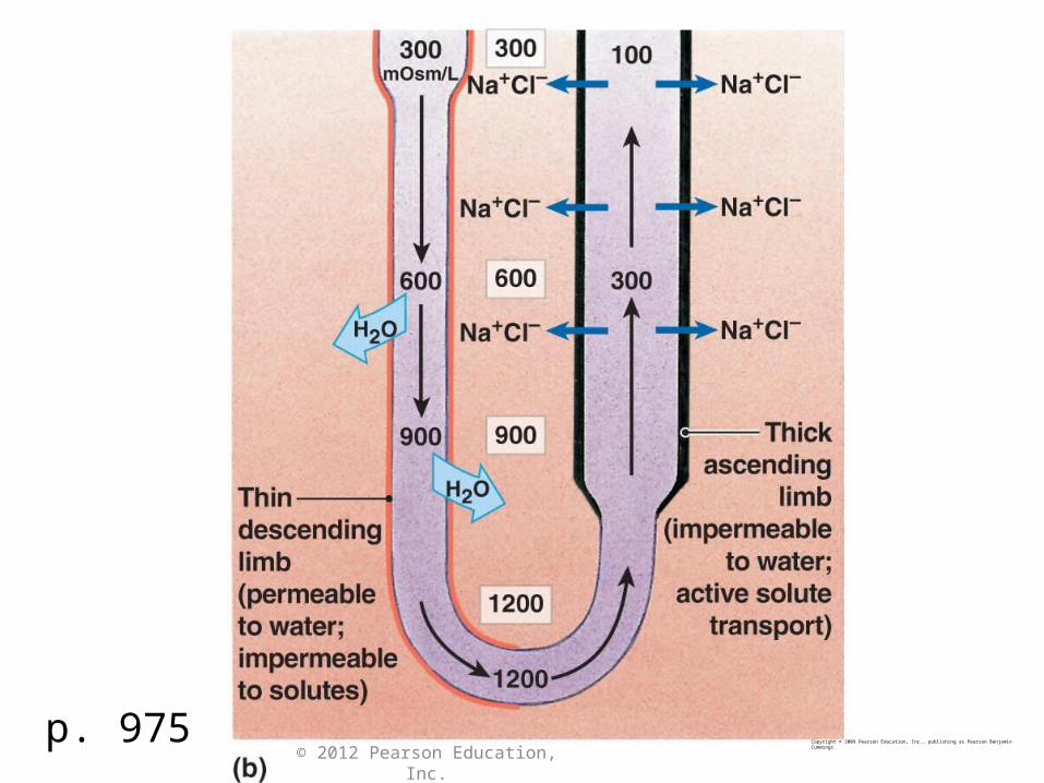
p. 975Copyright © 2009 Pearson Education, Inc., publishing as Pearson Benjamin Cummings
© 2012 Pearson Education, Inc.
© 2012 Pearson Education, Inc.
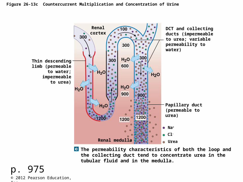
Figure 26-13c Countercurrent Multiplication and Concentration of Urine
The permeability characteristics of both the loop and the collecting duct tend to concentrate urea in the tubular fluid and in the medulla.
Renalcortex
DCT and collecting ducts (impermeable to urea; variablepermeability towater)
Thin descendinglimb (permeable
to water;impermeable
to urea)
Papillary duct(permeable tourea)
Renal medulla Urea
Na
Cl
p. 975
p. 962Copyright © 2009 Pearson Education, Inc., publishing as Pearson Benjamin Cummings
© 2012 Pearson Education, Inc.
© 2012 Pearson Education, Inc.
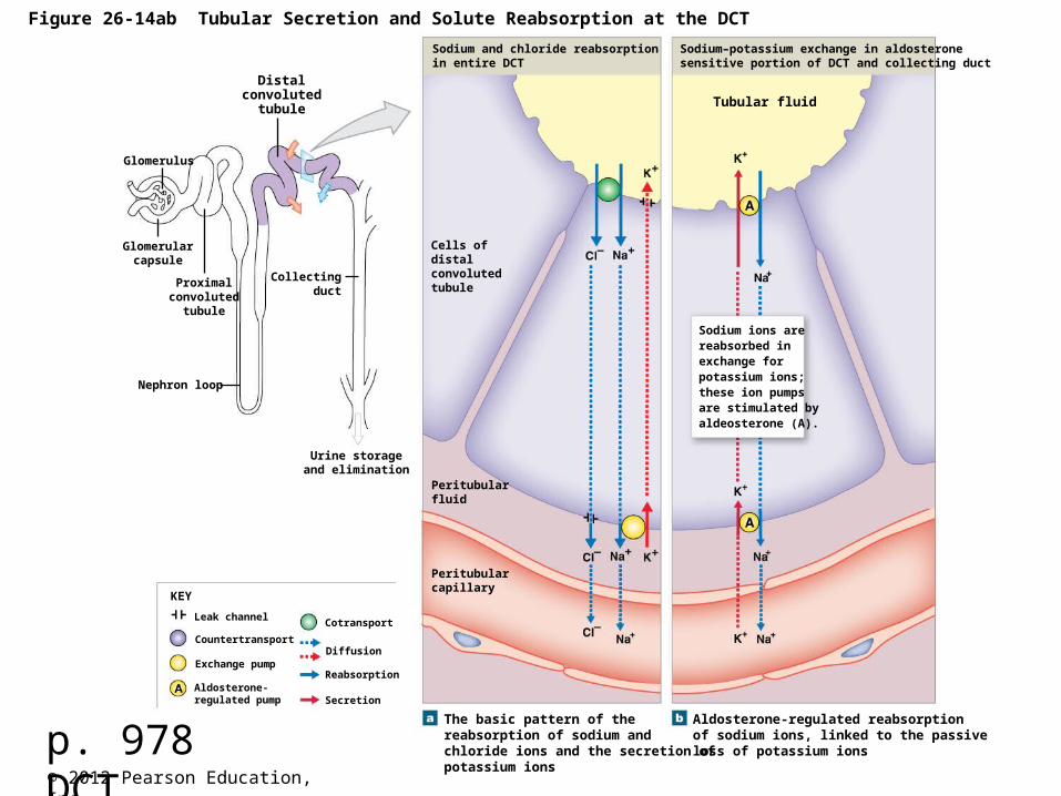
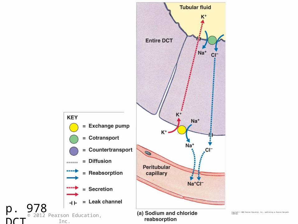
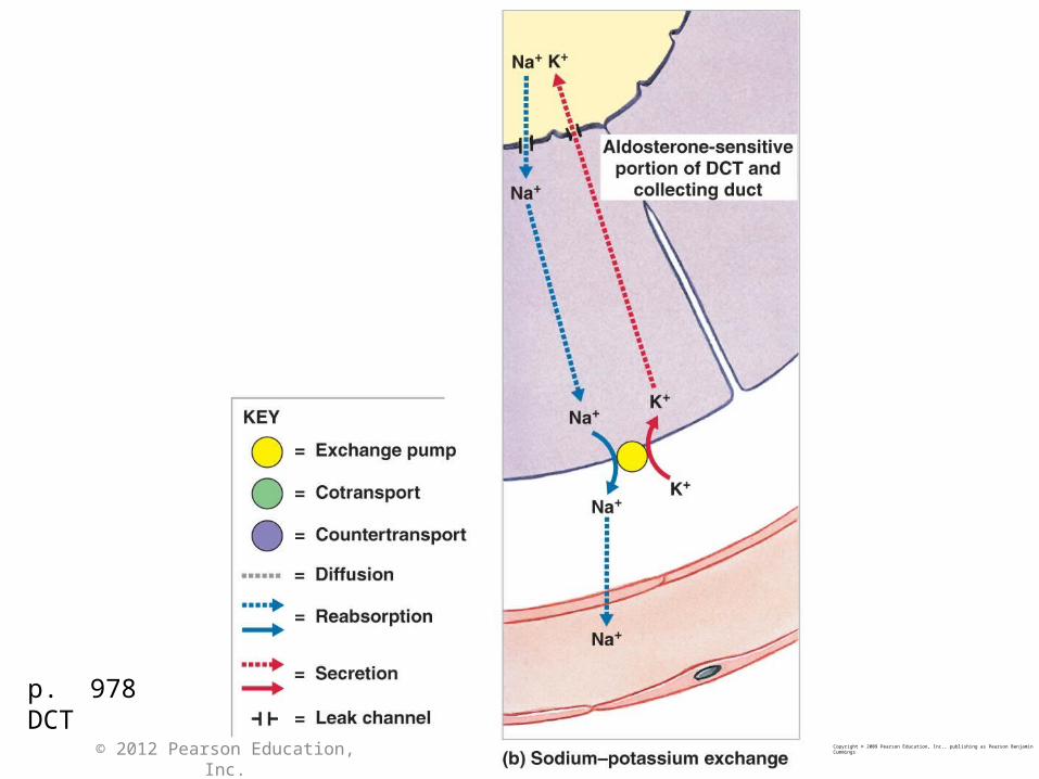
Figure 26-14ab Tubular Secretion and Solute Reabsorption at the DCT
The basic pattern of thereabsorption of sodium andchloride ions and the secretion ofpotassium ions
Aldosterone-regulated reabsorptionof sodium ions, linked to the passiveloss of potassium ions
Peritubularcapillary
Peritubularfluid
Cells of distalconvolutedtubule
Sodium ions arereabsorbed inexchange forpotassium ions;these ion pumpsare stimulated byaldeosterone (A).
Tubular fluid
Sodium–potassium exchange in aldosteronesensitive portion of DCT and collecting duct
Sodium and chloride reabsorptionin entire DCT
Distalconvoluted
tubule
Glomerulus
Glomerularcapsule
Proximalconvoluted
tubule
Urine storageand elimination
Collectingduct
Nephron loop
KEY
Secretion
Reabsorption
Diffusion
Cotransport
Aldosterone-regulated pump
Exchange pump
Countertransport
Leak channel
p. 978 DCT
Copyright © 2009 Pearson Education, Inc., publishing as Pearson Benjamin Cummings
© 2012 Pearson Education, Inc.
p. 978 DCT
p. 978 DCT
Copyright © 2009 Pearson Education, Inc., publishing as Pearson Benjamin Cummings
© 2012 Pearson Education, Inc.
© 2012 Pearson Education, Inc.
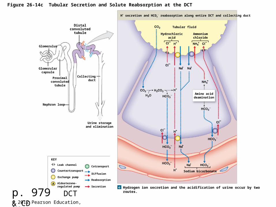
Figure 26-14c Tubular Secretion and Solute Reabsorption at the DCT
Distalconvoluted
tubule
Glomerulus
Glomerularcapsule
Proximalconvoluted
tubule
Urine storageand elimination
Collectingduct
Nephron loop
KEY
Secretion
Reabsorption
Diffusion
Cotransport
Aldosterone-regulated pump
Exchange pump
Countertransport
Leak channel
Sodium bicarbonate
Hydrogen ion secretion and the acidification of urine occur by tworoutes.
Amino aciddeamination
Hydrochloricacid
Ammoniumchloride
Tubular fluid
H+ secretion and HCO3- reabsorption along entire DCT and collecting duct
p. 979 DCT & CD
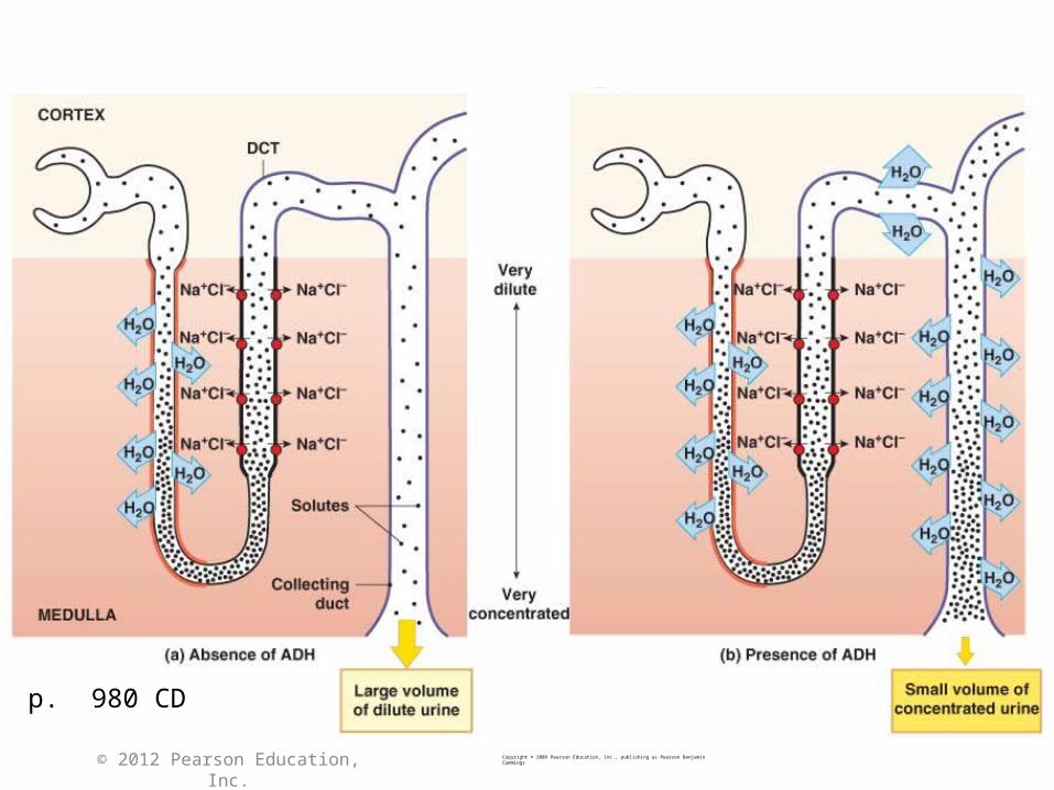
p. 980 CD
Copyright © 2009 Pearson Education, Inc., publishing as Pearson Benjamin Cummings
© 2012 Pearson Education, Inc.
p. 975Copyright © 2009 Pearson Education, Inc., publishing as Pearson Benjamin Cummings
© 2012 Pearson Education, Inc.
© 2012 Pearson Education, Inc.
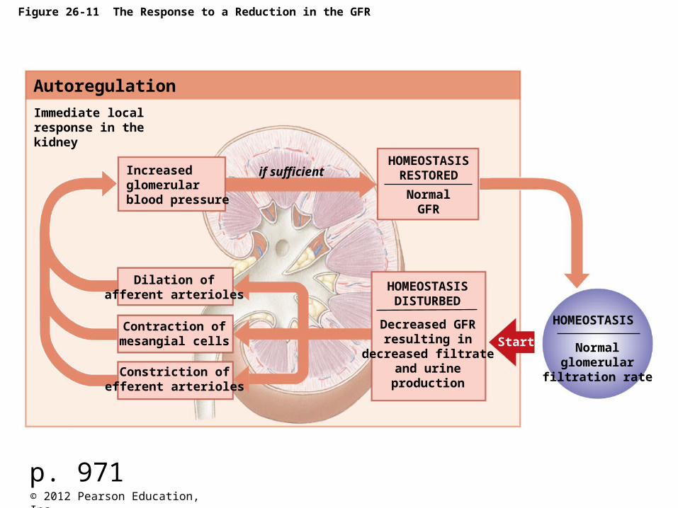
Figure 26-11 The Response to a Reduction in the GFR
Autoregulation
Normalglomerular
filtration rate
HOMEOSTASIS
HOMEOSTASISRESTORED
HOMEOSTASISDISTURBED
Immediate localresponse in thekidney
Increasedglomerularblood pressure
if sufficient
Dilation ofafferent arterioles
Contraction ofmesangial cells
Constriction ofefferent arterioles
NormalGFR
Decreased GFRresulting in
decreased filtrateand urine
production
Start
p. 971
© 2012 Pearson Education, Inc.
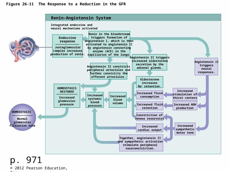
Figure 26-11 The Response to a Reduction in the GFR
Renin–Angiotensin System
Endocrineresponse
Integrated endocrine andneural mechanisms activated
Juxtaglomerularcomplex increasesproduction of renin.
Angiotensin II constrictsperipheral arterioles and
further constricts theefferent arterioles.
Renin in the bloodstreamtriggers formation of
angiotensin I, which is thenactivated to angiotensin IIby angiotensin converting
enzyme (ACE) in thecapillaries of the lungs.
Angiotensin II triggersincreased aldosterone
secretion by theadrenal glands.
Aldosteroneincreases
Na retention.HOMEOSTASIS
RESTORED
Increasedglomerularpressure
Increasedsystemic
bloodpressure
Increasedblood
volume
Increased fluidconsumption
Increased fluidretention
Constriction ofvenous reservoirs
Increasedcardiac output
Increasedstimulation ofthirst centers
Increased ADHproduction
Angiotensin IItriggersneural
responses.
Increasedsympatheticmotor tone
Together, angiotensin IIand sympathetic activation
stimulate peripheralvasoconstriction.
Normalglomerular
filtration rate
HOMEOSTASIS
p. 971
Recommended