OMM for the Family
Physician
OMED 2014
Seattle Washington
Kevin D. Treffer, D.O., FACOFP
Associate Professor
Interim Chair, Department of OMM
KCUMB-COM
Objectives
At the end of the session the attendee will
be able to:
1. Apply a thorough history and physical for
an upper extremity musculoskeletal
complaint
2. Perform the appropriate tests in the
examination of the patient and know how
to interpret them
3. Integrate structural evaluation into the
examination
4. Integrate OMM into the management
plan.
Case # 1
Upper Extremity
Case #1
A 34 year old auto worker presents with insidious
onset of right shoulder pain. He notes the pain is
increased when he has his arm above his head and
the extremity gets heavy and cool at times. He
admits to tingling into the extremity but denies
numbness. He has only noticed loss of strength
due to the pain in the noted position. No new
traumas to the shoulder have occurred however,
he uses an impact wrench all day on the assembly
line.. He rates the pain as a 7/10 at its worst.
Case #1
Examination reveals a well developed adult male
with normal affect and good eye contact during
the encounter. Vital signs are stable. Adson’s test
is weakly positive on the right and Wright’s
abduction test is positive on the right. Abduction
at the right shoulder is limited to 150 degrees.
Negative impingement sign, Tinnel’s, Phalen’s,
Yeargason’s, Speed’s, arm drop, and empty can
tests are noted. Deep tendon reflexes are +2/4
bilaterally and strengths are 5/5 in the upper and
lower extremities. Sensory is intact
Case #1
There are TART findings at the upper thoracic
and cervical spine (T1-5RRSL and C3-5 FRRSR).
Tender points are noted for the supraspinatus,
infraspinatus, scalene, subscapularis, long head of
the biceps, levator scapula, and serratus posterior
muscles. Marked tension is noted in the scalene
and pectoral minor muscles. Right first rib is
superior. Right glenohumeral joint is
anterior/inferior.
Case #1
Impression
Thoracic Outlet Syndrome
Arthralgia
Somatic Dysfunction
Cervical, Thoracic, Rib, Upper
Extremity
What is Thoracic Outlet
Syndrome?
• Pain
• Paresthesias
• Weakness,
coolness,
heaviness in upper
extremity
• Symptoms
aggravated by:
• elevation of arms
• exaggerated
movement of the
head and neck
What is Thoracic Outlet
Syndrome?
• Three sites of compression
• Scalene muscles
• Infraclavicular space
• Pectoralis minor muscle
What is Thoracic Outlet
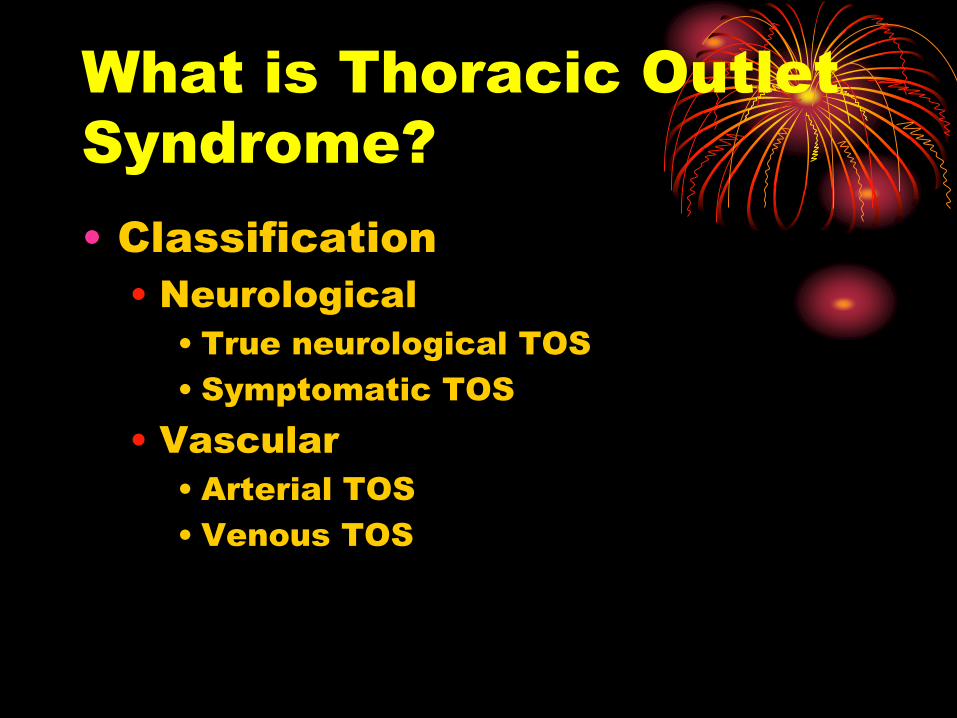
Syndrome?
• Classification
• Neurological
• True neurological TOS
• Symptomatic TOS
• Vascular
• Arterial TOS
• Venous TOS

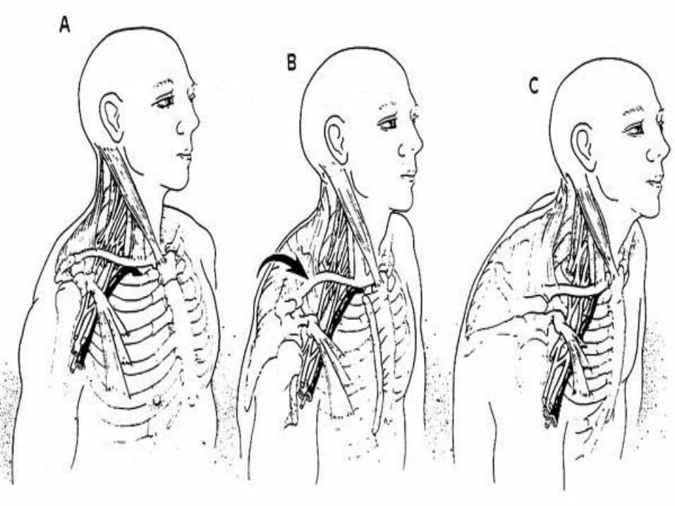
ETIOLOGIES
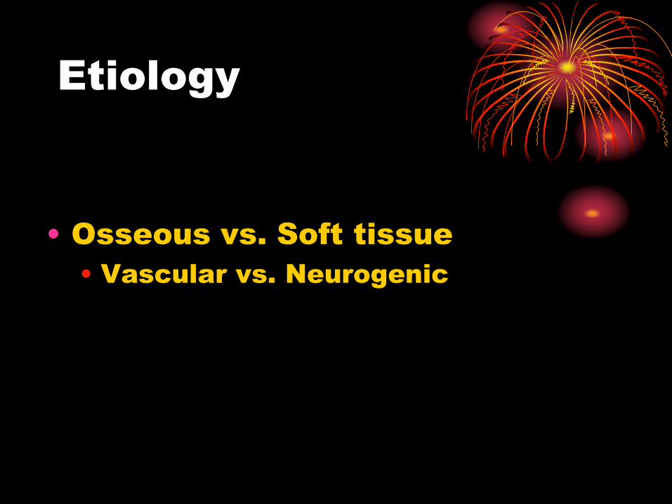
Etiology
• Osseous vs. Soft tissue
• Vascular vs. Neurogenic

Etiology: Soft Tissue
• Scalene and/or pectoral muscle
restriction
• associated postural and structural
change
• C-spine
hyperflexion/hyperextension
• Whiplash
• Apical tumor of Lung
• Pancoast’s tumor
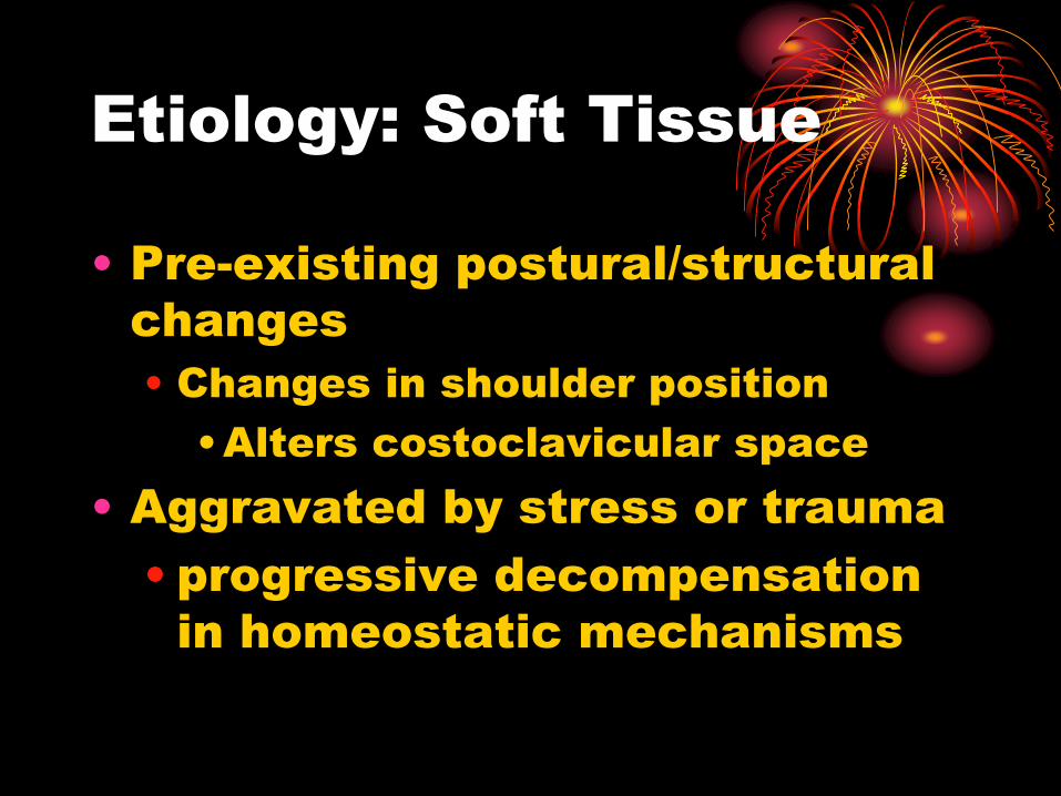
Etiology: Soft Tissue
• Pre-existing postural/structural
changes
• Changes in shoulder position
•Alters costoclavicular space
• Aggravated by stress or trauma
• progressive decompensation
in homeostatic mechanisms
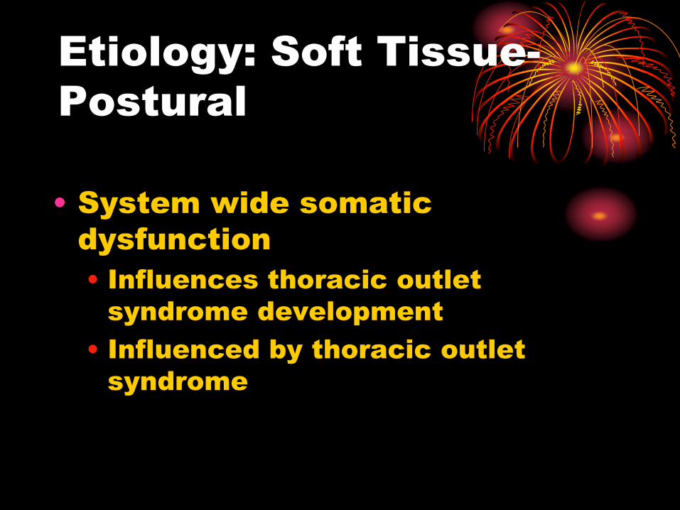
Etiology: Soft Tissue-
Postural
• System wide somatic
dysfunction
• Influences thoracic outlet
syndrome development
• Influenced by thoracic outlet
syndrome
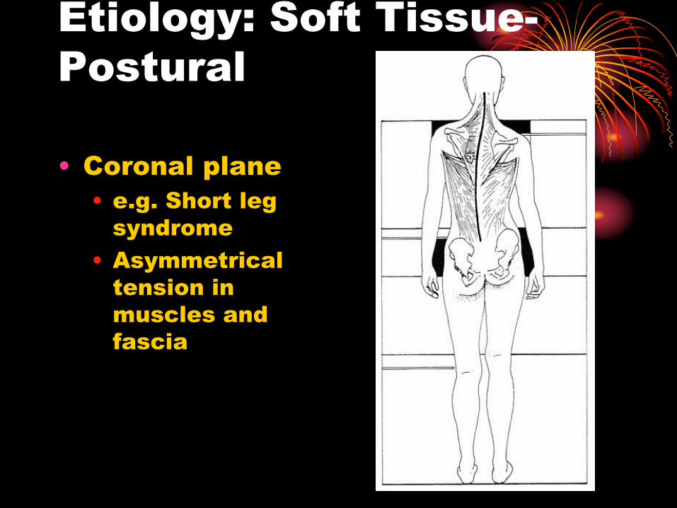
Etiology: Soft Tissue-
Postural
• Coronal plane
• e.g. Short leg
syndrome
• Asymmetrical
tension in
muscles and
fascia
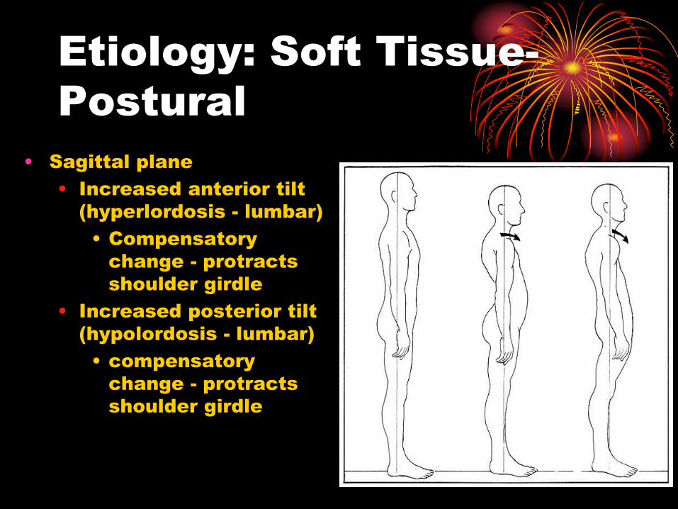
Etiology: Soft Tissue-
Postural
• Sagittal plane
• Increased anterior tilt
(hyperlordosis - lumbar)
• Compensatory
change - protracts
shoulder girdle
• Increased posterior tilt
(hypolordosis - lumbar)
• compensatory
change - protracts
shoulder girdle
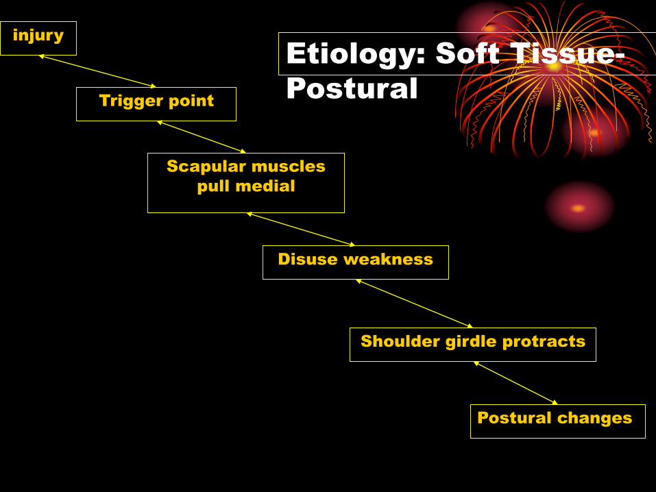
injury
Trigger point
Scapular muscles
pull medial
Disuse weakness
Shoulder girdle protracts
Etiology: Soft Tissue-
Postural
Postural changes
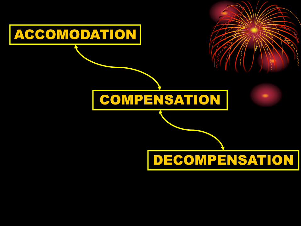
ACCOMODATION
COMPENSATION
DECOMPENSATION
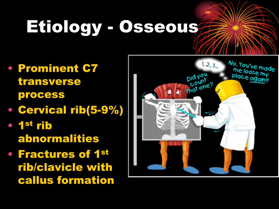
Etiology - Osseous
• Prominent C7
transverse
process
• Cervical rib(5-9%)
• 1st
rib
abnormalities
• Fractures of 1st
rib/clavicle with
callus formation
DIAGNOSIS
OF
THORACIC OUTLET
SYNDROME
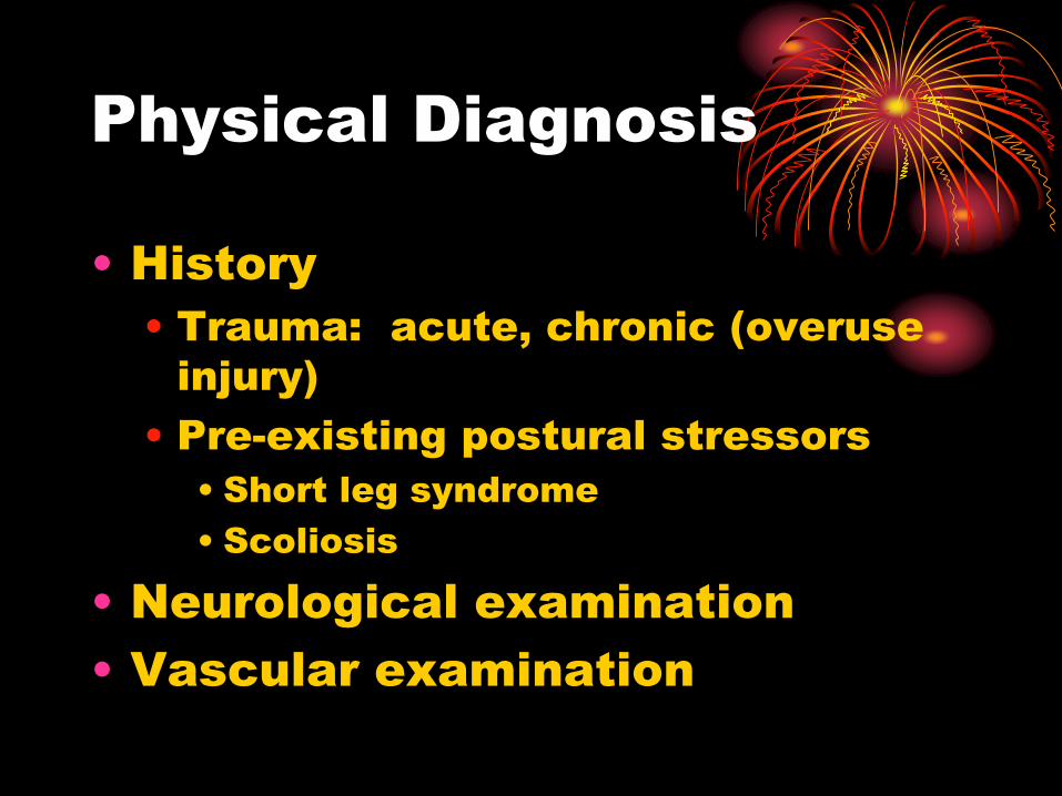
Physical Diagnosis
• History
• Trauma: acute, chronic (overuse
injury)
• Pre-existing postural stressors
• Short leg syndrome
• Scoliosis
• Neurological examination
• Vascular examination
Physical Diagnosis
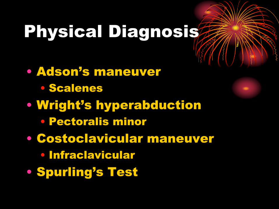
• Adson’s maneuver
• Scalenes
• Wright’s hyperabduction
• Pectoralis minor
• Costoclavicular maneuver
• Infraclavicular
• Spurling’s Test
Physical Diagnosis
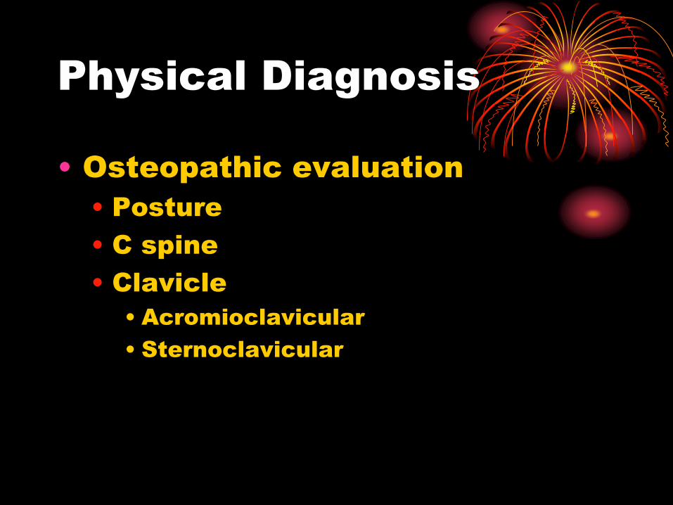
• Osteopathic evaluation
• Posture
• C spine
• Clavicle
• Acromioclavicular
• Sternoclavicular
Physical Diagnosis
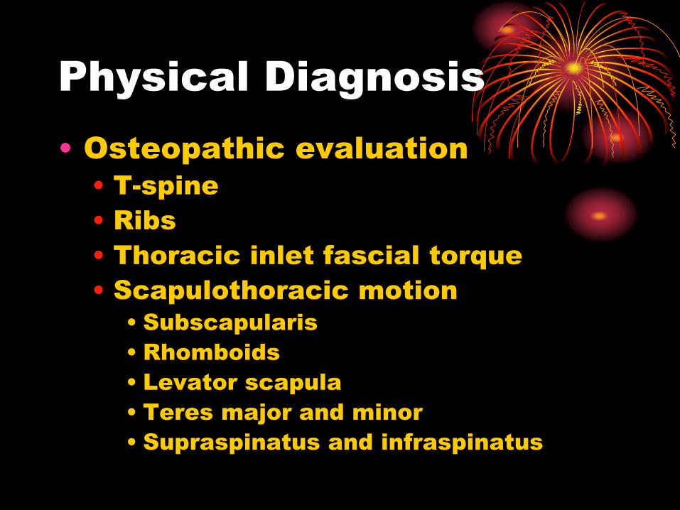
• Osteopathic evaluation
• T-spine
• Ribs
• Thoracic inlet fascial torque
• Scapulothoracic motion
• Subscapularis
• Rhomboids
• Levator scapula
• Teres major and minor
• Supraspinatus and infraspinatus
Testing: Anatomic
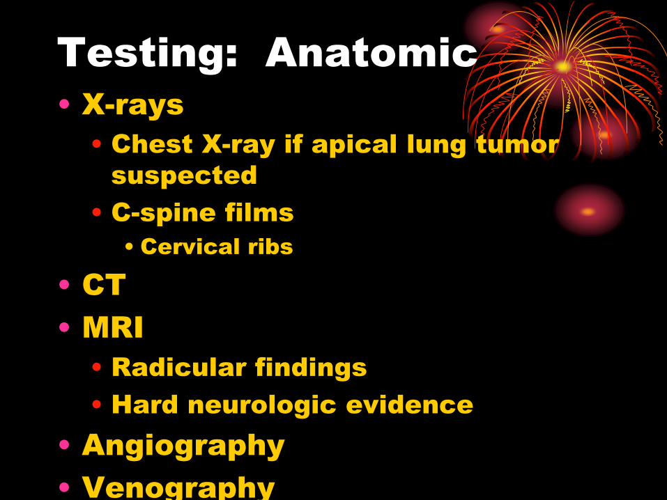
• X-rays
• Chest X-ray if apical lung tumor
suspected
• C-spine films
• Cervical ribs
• CT
• MRI
• Radicular findings
• Hard neurologic evidence
• Angiography
• Venography
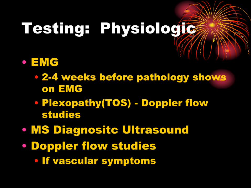
Testing: Physiologic
• EMG
• 2-4 weeks before pathology shows
on EMG
• Plexopathy(TOS) - Doppler flow
studies
• MS Diagnositc Ultrasound
• Doppler flow studies
• If vascular symptoms
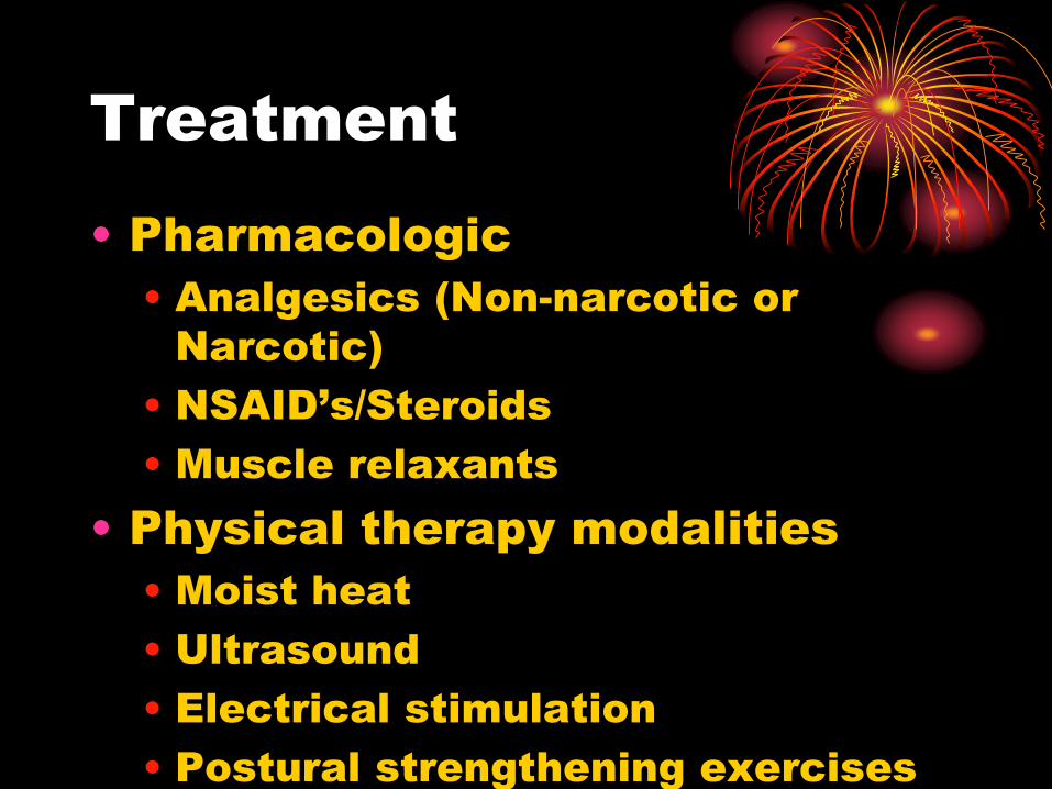
Treatment
• Pharmacologic
• Analgesics (Non-narcotic or
Narcotic)
• NSAID’s/Steroids
• Muscle relaxants
• Physical therapy modalities
• Moist heat
• Ultrasound
• Electrical stimulation
• Postural strengthening exercises
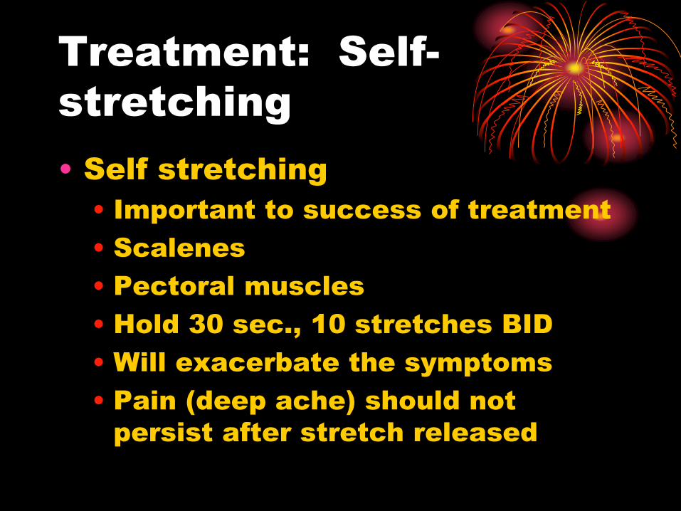
Treatment: Self-
stretching
• Self stretching
• Important to success of treatment
• Scalenes
• Pectoral muscles
• Hold 30 sec., 10 stretches BID
• Will exacerbate the symptoms
• Pain (deep ache) should not
persist after stretch released
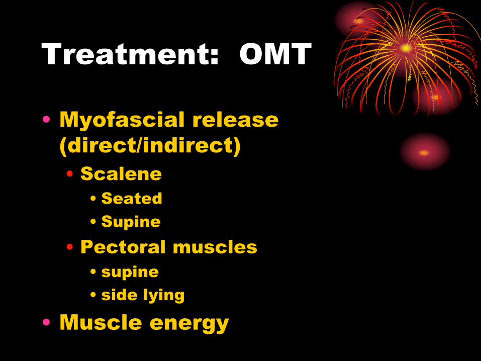
Treatment: OMT
• Myofascial release
(direct/indirect)
• Scalene
• Seated
• Supine
• Pectoral muscles
• supine
• side lying
• Muscle energy

Treatment: OMT
• High velocity low amplitude
• Spray and stretch
• Ethyl chloride
• Counterstrain

Treatment: Postural
• Muscle strengthening
• Light weights/high repetition
• Resistance exercises (elastic
bands)
• Avoid shoulder abduction >45
degrees
• reactivates trigger point in
parascapular muscles
Treatment: Surgical
• Failure of conservative therapy
• Options
• Scalenectomy
• First rib removal
Case #1 Questions
• What kind of neuropathy is TOS?
• Entrapment or Plexopathy
• What is the difference on physical
exam between this and a
radiculopathy?
• Radiculopathy has muscular
weakness, decreased DTR,
paresthesias in dermatome pattern,
EMG may be positive
• Entrapment will have negative EMG
normal neural examination
Case #1 Questions
• What diagnostic procedures
need to be done in this patient
and why?
• C-spine films, EMG, CT/MRI, US,
Doppler
• How can OMM be used in the
management of this patient
• Decide on modalities to be used
safely
Hands-On Practice at
OMM
Counterstrain Upper Extremity
Case #1
Treatment:
We will be reviewing the named tender points
today. You will treat a few significant points and
associated somatic dysfunctions then address the
home stretches by teaching the patient how to
perform them and what to expect. Included in the
treatment is patient homework for them to do.
Stretches to the scalenes, and pectoralis minor
would be appropriate for the patient.

Counterstrain Treatment
Treatment:
If using counterstrain you must decide which
tender points are most tender and start there. You
can still treat other areas but realize that by treating
these tender points you are treating somatic
dysfunction and relieving the pain you should
expect improvement in range of motion, tissue
texture changes and asymmetry.
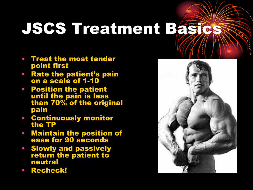
JSCS Treatment Basics
• Treat the most tender
point first
• Rate the patient’s pain
on a scale of 1-10
• Position the patient
until the pain is less
than 70% of the original
pain
• Continuously monitor
the TP
• Maintain the position of
ease for 90 seconds
• Slowly and passively
return the patient to
neutral
• Recheck!
Diagnostic and Treatment
Approach to the Upper
Extremities
• Always check the joint above and below
the area of chief complaint!!!
• Patients often present with wrist pain,
decreased motion or grip strength and the
problem could be originating in the elbow,
radius or ulna
• Treat proximal before distal as a general
rule
• i.e. examine and treat (if necessary) the
shoulder before the elbow, or wrist
before the hand.
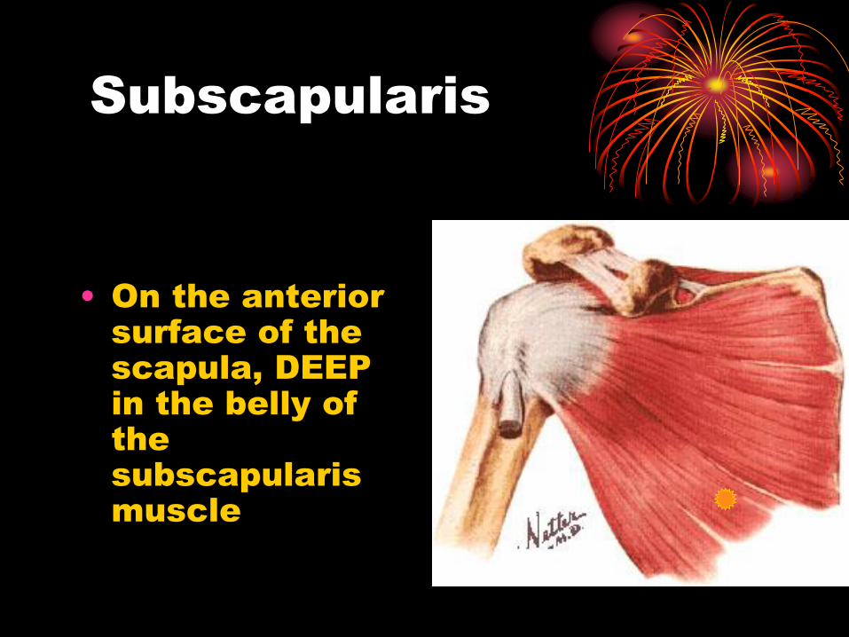
Subscapularis
• On the anterior
surface of the
scapula, DEEP
in the belly of
the
subscapularis
muscle
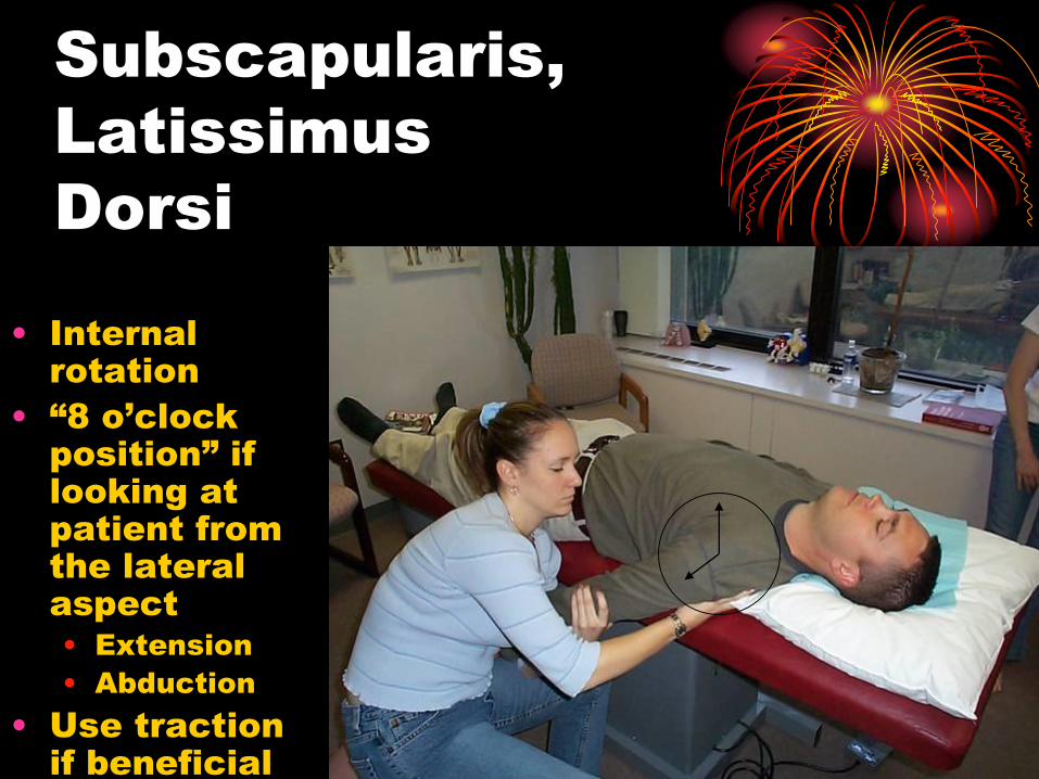
Subscapularis,
Latissimus
Dorsi
• Internal
rotation
• “8 o’clock
position” if
looking at
patient from
the lateral
aspect
• Extension
• Abduction
• Use traction
if beneficial
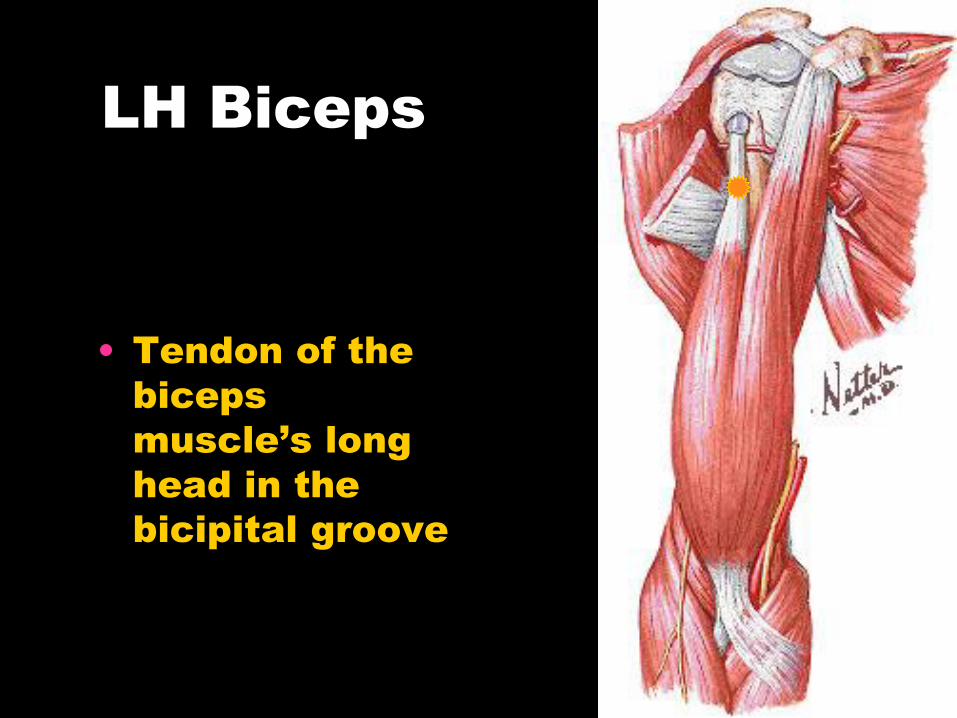
LH Biceps
• Tendon of the
biceps
muscle’s long
head in the
bicipital groove
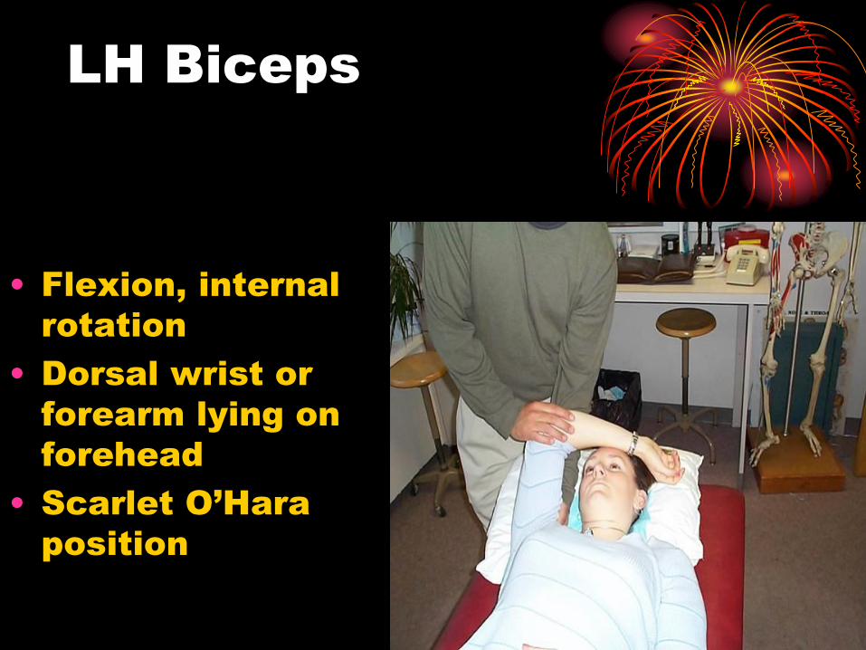
LH Biceps
• Flexion, internal
rotation
• Dorsal wrist or
forearm lying on
forehead
• Scarlet O’Hara
position
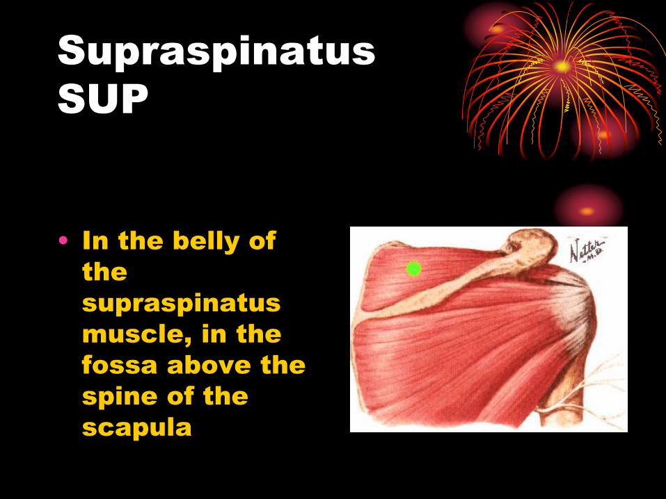
Supraspinatus
SUP
• In the belly of
the
supraspinatus
muscle, in the
fossa above the
spine of the
scapula
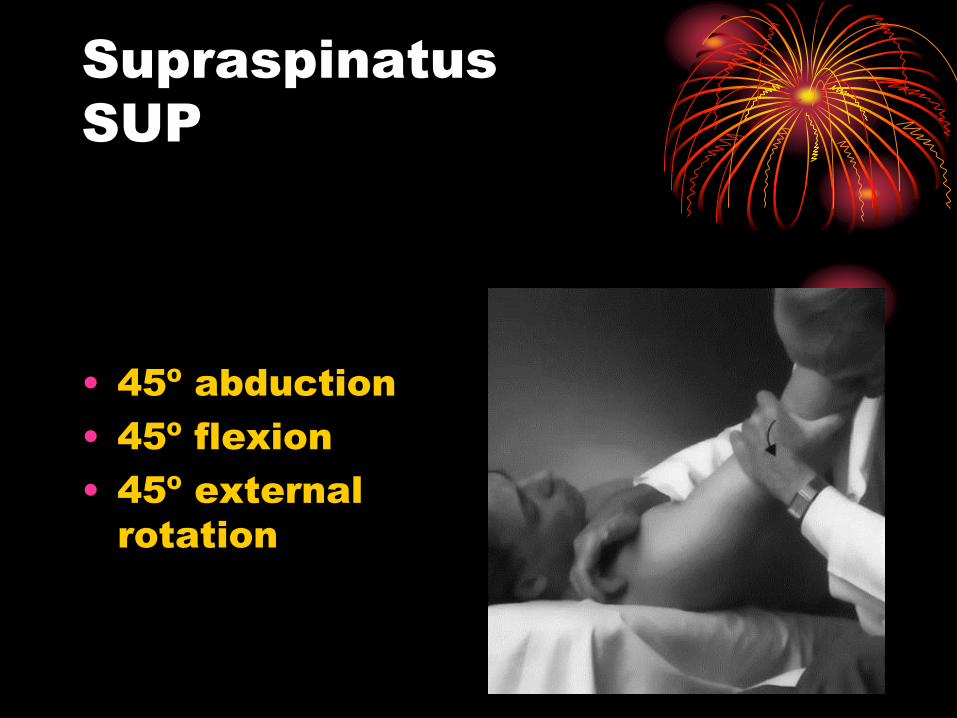
Supraspinatus
SUP
• 45º abduction
• 45º flexion
• 45º external
rotation
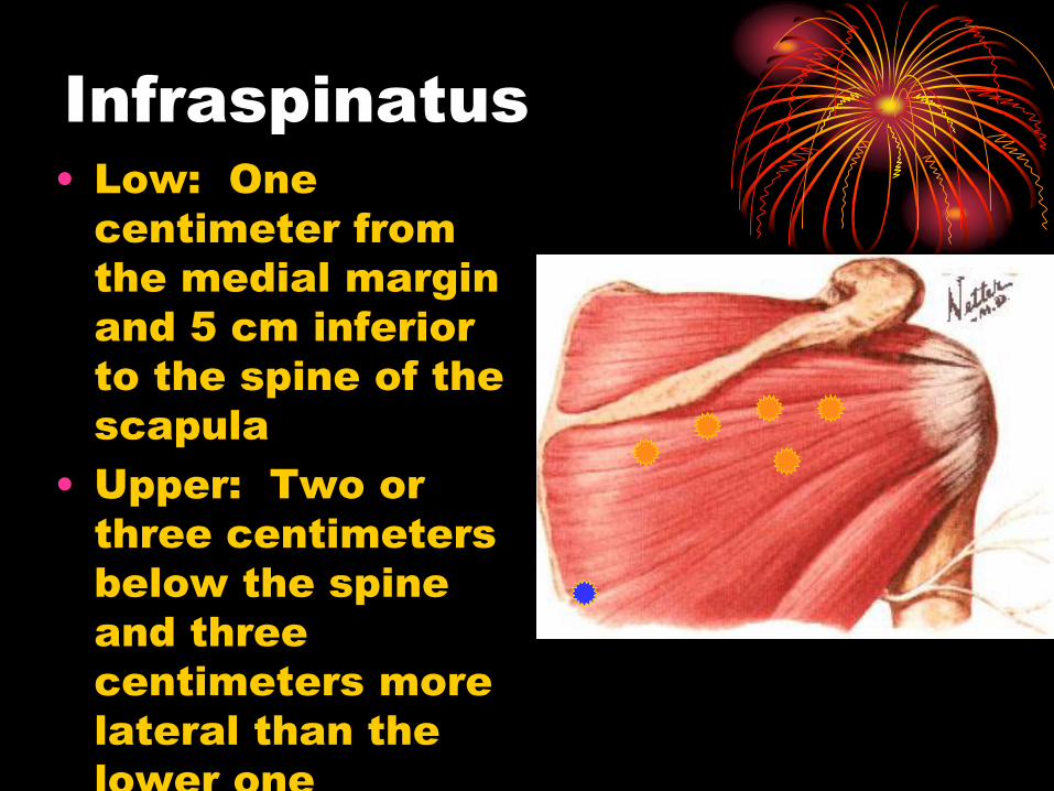
Infraspinatus
• Low: One
centimeter from
the medial margin
and 5 cm inferior
to the spine of the
scapula
• Upper: Two or
three centimeters
below the spine
and three
centimeters more
lateral than the
lower one
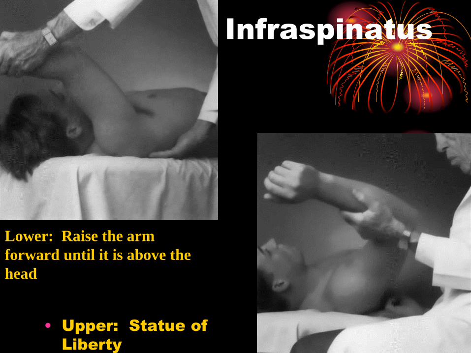
Infraspinatus
• Upper: Statue of
Liberty
Lower: Raise the arm
forward until it is above the
head
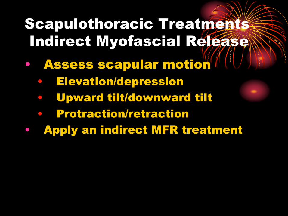
Scapulothoracic Treatments
Indirect Myofascial Release
• Assess scapular motion
• Elevation/depression
• Upward tilt/downward tilt
• Protraction/retraction
• Apply an indirect MFR treatment

Myofascial Release to the
Scalene Muscles
• Patient is in supine position
• Doc at head of table
• With one hand side bend the head
and neck away from the side to be
stretched
• With the other hand provide caudad
compression on shoulder girdle for
opposing stretch
• Other hand may also provide a
perpendicular force to treat the muscles
Myofascial Release to the
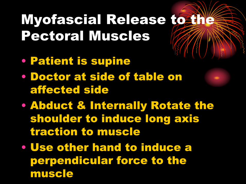
Pectoral Muscles
• Patient is supine
• Doctor at side of table on
affected side
• Abduct & Internally Rotate the
shoulder to induce long axis
traction to muscle
• Use other hand to induce a
perpendicular force to the
muscle
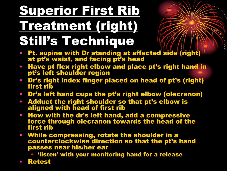
Superior First Rib
Treatment (right)
Still’s Technique
• Pt. supine with Dr standing at affected side (right)
at pt’s waist, and facing pt’s head
• Have pt flex right elbow and place pt’s right hand in
pt’s left shoulder region
• Dr’s right index finger placed on head of pt’s (right)
first rib
• Dr’s left hand cups the pt’s right elbow (olecranon)
• Adduct the right shoulder so that pt’s elbow is
aligned with head of first rib
• Now with the dr’s left hand, add a compressive
force through olecranon towards the head of the
first rib
• While compressing, rotate the shoulder in a
counterclockwise direction so that the pt’s hand
passes near his/her ear
• ‘listen’ with your monitoring hand for a release
• Retest
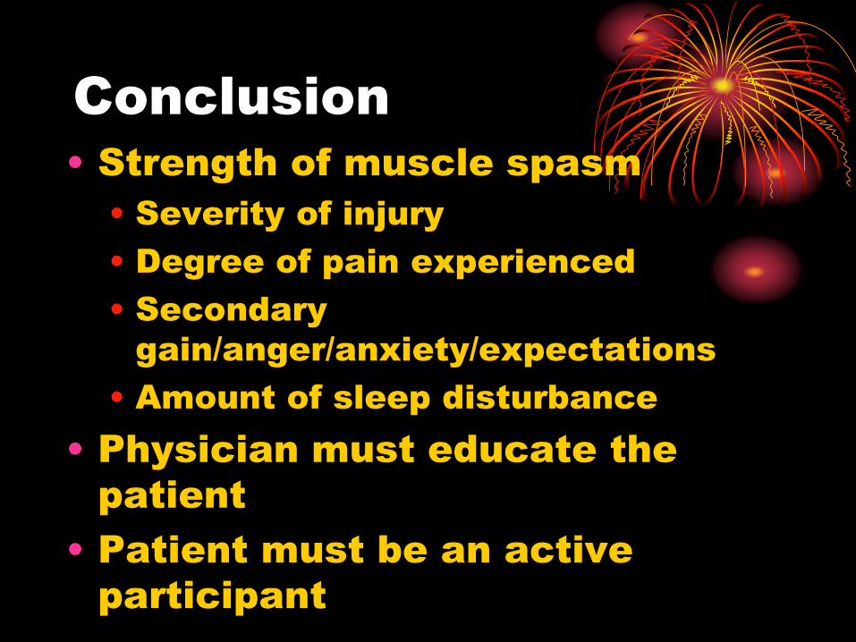
Conclusion
• Strength of muscle spasm
• Severity of injury
• Degree of pain experienced
• Secondary
gain/anger/anxiety/expectations
• Amount of sleep disturbance
• Physician must educate the
patient
• Patient must be an active
participant
Recommended