Eur. J. Biochem. 160, 305-310 (1986) 0 FEBS 1986
Nucleotide sequence of a cDNA clone encoding Scylliorhinus caniculus protamine 22 Frederique BERLOT-PICARD, Guilane VODJDANI and Janine DOLY Departement de Biologie Moleculaire des Interferons, Institut de Recherches sur le Cancer, Villejuif
(Received May 14/July 7,1986) - EJB 86 0482
A cDNA library was constructed from a protamine-enriched fraction of dogfish (Scylliorhinus caniculus) mRNA. The nucleotide sequence of a 440-bp insert was determined, and its produced protein sequence confirmed its identification as a cysteine-rich protamine 2 2 [Martinage, A., Gusse, M., Belaiche, D., Sautiere, P. and Chevaillier, P. (1985) Biochim. Biophys. Acta 831, 172- 1781. The frequency of utilization of the different triplets coding for arginine, which represents 30-70% of the total amino acid residues for trout, mouse and dogfish protamines, is discussed. An alternative repetitive sequence of CGC-AGG was found in the N terminus of the protein. Analysis of the 3' flanking region after the mRNA-terminating TAA codon identified an inverted repeat sequence and an ACCA sequence, which may be possible vestiges of a histone-like termination signal.
Spermatogenesis in many animal species is characterized by dramatic morphological and biological changes in nuclear organization. In spermatozoa, histones, which are highly conserved basic eucaryotic proteins organizing DNA in nucleosome units, are replaced by the protamines, a family of small and arginine-rich proteins (for a review see [l, 21). These changes in chromatin produce a DNA-protamine complex that is extremely compact, devoid of nucleosomal structure and inactive in RNA synthesis [3]. The DNA-protamine complex seems to protect the genetic information during storage of spermatozoa in testes until fecundation [4, 51. A recent study of Hecht and his colleagues [6] shows that pro- tamines seem to play a prominent part in certain types of sterility in the mouse.
The most extensive investigations have been accomplished by Dixon et al. [7 - 91 on the expression and evolution of the protamine gene family of the trout. These basic proteins were also found in mammals and birds [lo- 121.
The dogfish is an interesting model, as spermatogenesis is characterized by the synchronous development of germinal cells within follicles and as it was shown that two basic nuclear proteins (S1 and S2) occur transiently during nuclear elonga- tion [13, 141. Four protamines, known as Z1, Z2,23 and S4, have been described in the dogfish [15]. Among these only 23 is a typical arginine-rich protamine while the other three are characterized by high arginine and cysteine contents. The latter feature differentiates them in their structure from bird and other fish protamines [12, 16, 171. The protamines from placental mammals also contain cysteine residues (8- 18%), which are involved in the disulfide cross-linkage with DNA in sperm chromatin.
Correspondence to J. Doly, Biologie Moleculaire des Interferons,
Abbreviations. bp, base pairs in DNA; SDS, sodium dodecyl IRSC, 7, rue Guy Mocquet, F-94802 Villejuif Cedex, France
sulfate.
We describe here a cloned and fully sequenced cDNA of the 22 dogfish protamine, the only one containing a conserved methionine residue at its N terminus [18]. Analysis of the nucleotide sequence and in particular the frequency of the codons used for the essential amino acids is presented and suggests no evident divergence of these proteins in different species.
Special attention is devoted to the 3' non-coding region described to be involved in the cell-cycle regulation of histone gene expression. An inverted repeat sequence between the TAA stop codon and the ACCA sequence is demonstrated for the 22 dogfish protamine. A parallel between the histone palindromes processing signals and 22 structure is discussed.
MATERIALS AND METHODS
Dogfish (Scylliorhinus caniculus) testes were collected from freshly killed animals at the French Station de Biologie Mar- ine de Concarneau. After excision, gonads were frozen immediately in liquid nitrogen and stored at -70°C.
Enzymes and chemical products
Proteinase K was purchased from Merck and RNase A from Worthington. Oligo(dT)-cellulose (T3) and 12 - 18 (dT) primer were purchased from Collaborative Research. Rabbit reticulocyte lysate, [3H]arginine 48 Ci/mmol and [35Ss]- methionine 1220 Ci/mmol, cloning and sequencing MI 3 kit, all the radioactive nucleotides and their derivatives were purchased from Amersham International. DNA polymerase I from E. coli, Klenow fragment and Kornberg polymerase, nuclease S1 and tRNA calf liver were purchased from Boehringer Mannheim. Reverse transcriptase from avian myeloblastosis virus was purchased from BRL. Terminal deoxyribonucleotidyl transferase was from PL Biochemicals and restriction endonucleases were from Biolabs or
306
Appligine, T4 DNA ligase was purchased from Appligkne (Illkirch-Graffenstaden, France).
Bacterial strains and vectors
E. coli strains HB 101 [19], JM 101 [20] were used as host strains for pBR322, M13mp8 and M13mp9 vectors respective- ly.
Preparation of mRNA
mRNA was extracted from frozen dogfish testes by the modified procedures of Feramisco et al. [21] and Chirgwin et al. [22]. Testes were homogenized in a Waring blender for 3 min in 4 M guanidium isothiocyanate made in sodium ace- tate 50 mM, pH 5.1 instead of Tris/HCI pH 7. An equal volume of phenol saturated with 0.1 M sodium acetate pH 5.1 and 0.1% hydroxyquinoleine, preheated to 60°C, was added and the mixture was vigourously shaken for 15 min at 60"C, then rapidly frozen in acetone at -70°C. The homogenate was centrifuged at 12000 x g for 20 min and the recovered aqueous phase was re-extracted with phenol/ chlorofonn/isoamyl alcohol (25: 24: 1) at 60°C as already de- scribed, twice with chloroform/isoamyl alcohol at room tem- perature and then precipitated with two volumes of ethanol at - 70 "C for 2 h or at - 20 "C overnight. After centrifugation the pellets were dissolved in 0.1 M Tris/HCl pH 7.4, 50 mM NaCI, 10 mM EDTA and 0.2% sodium dodecyl sulfate and treated for 2 h with 200 pg/ml proteinase K [23]. The mixture was then extracted as before: twice at 60°C, twice at room temperature and then precipitated with ethanol.
Poly(A)-rich mRNA, after heating at 65 "C, were purified by two successive cycles of chromatography on a column of oligo(dT)-cellulose. Fractionation was carried out by sedi- mentation on a 10 - 30% sucrose gradient in 0.1 M Tris/HCl pH 7.6, 0.3 M KC1, 10 mM EDTA at 288000 x g for 24 h at 4 "C.
Translation assay and protein analysis
After each step the poly(A)-rich mRNA preparation was analyzed by in vitro translation in rabbit reticulocyte lysate containing 30 pCi [3H]arginine (globin mRNA was used as control). Incubation was carried out for 60 min at 30 "C. The incorporation was determined by trichloroacetic acid pre- cipitation according to Pelham and Jackson [24]. The synthe- sized products were analyzed by polyacrylamide gel electro- phoresis: each lysate was treated for 30min at 37°C with RNase A (0.2pg/ml) and 40pg for the four dogfish pro- tamines were added as a carrier before trichloroacetic acid precipitation at 4°C. After centrifugation the pellets were extracted overnight with 0.25 M HC1. Acid-soluble proteins were precipitated with 25% trichloroacetic acid the pre- cipitates were washed successively with acidified acetone and acetone, dried and redissolved in 0.9 M acetic acid, 0.5 M 2- mercaptoethanol and 8 M urea. The samples were run on 6.25 M urea/l7% polydcrylamide gel according to the proce- dure described by Panyim and Chalkley [25].
The synthesized proteins were identified either by autoradiography on Fuji X-Ray film or by counting radioac- tivity of the gel slices corresponding to the position of the protamine markers (as indicated by the Coomassie blue coloration) in a mixture of Lipoluma/lumasolve/water (10: 1 :0.2) (Kontron).
cDNA preparation and cloning
Double-stranded cDNA were synthesized as described [26] from the 6 - 8 S fraction of a 10 - 30% sucrose gradient tailed with dC residues [27] and annealed [28] to a PstI-digested pBR322 plasmid tailed with dG residues. E. coli HBlOl strain [19] was transformed according to Mandel and Higa [29].
Differential hybridization colony screening
Tetracyclin-resistant and ampicillin-sensitive colonies were selected and 1340 clones were picked, grown in microtiter plates, then replicated on nitrocellulose filters (Schleicher & Schull) and screened by differential colony hybridization. Double-stranded cDNA probes were synthesized using poly(A)-rich mRNA from the 4- 6 S fraction containing only the mRNA for S4 protamine and from the 9 - 12 S fraction (this fraction translates Z1, 22, 23) of the sucrose gradient and nick-translated 1301 with [a-32P]dCTP. Prehybridization (1 h) and hybridization (16 h) were carried out at 42°C in a solution containing 5 x NaCl/Cit (1 x NaCl/Cit is 0.15 M NaC1,0.015 M sodium citrate pH 7), 1 x Denhardt's solution (0.02% each of bovine serum albumin, Ficoll 400 and polyvinylpyrrolidone), 50% formamide (Fluka) 25 mM Na2HP04 and 10% dextran sulfate [31].
Hybrid-selected translation Positive clones were selected and hybridization with
mRNA was performed as described by Ricciardi et al. [32] and modified according to Iatrou et al. [33]. 10 pg DNA from a single clone were spotted in each well of a BRL dot-blot apparatus and the nitrocellulose was then cut into small rect- angles. Filters from 12 clones were hybridized in the same tube with 40 pg poly(A)-rich mRNA for 4 h at 60°C in 50% formamide, 0.1 M Pipes pH 6.4,0.2% SDS, 0.6 M NaCl and 100 pg/ml poly(rA). After washing the filters twice with 4, 3, 2, 1 and 0.2 x SET (1 x SET is 0.15 M NaCl, 0.03 M Tris/HCl pH 8, 2mM EDTA) containing 0.2% SDS and once with 0.2 x SET, each filter was separated, eluted twice in boiling distilled water for 1 min. The eluted mRNAs were rapidly frozen in liquid nitrogen and ethanol-precipitated with tRNA as carrier and translated in rabbit reticulocyte lysate.
DNA sequence determination
Nucleotide sequences were determined by the dideoxy method of Sanger et al. [34] after subcloning suitable fragments in M13-based cloning vectors [35] and labelling with [35S]dCTP.
General techniques
Plasmid DNA was prepared as reported by Clewell and Helinski [36] or by Marko et al. [37] for large-scale preparation and according to the procedure of Birnboim and Doly [38] for rapid and small-scale analytic preparation. DNA fragments were recovered from agarose gel electrophoresis as described by Girwitz et al. [39].
RESULTS AND DISCUSSION mRNA preparation and construction of the cDNA library
130 mg total RNA were obtained from 100 g testes and the recovered poly(A)-rich mRNA fraction comprised 1.2%
307
0 . 5 4 s 6 s 8 5 9 s 12s 16s
- S e d i m e n t a t i o n
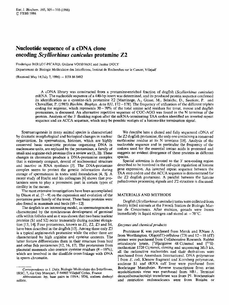
Fig. 1. Sucrose-gradient analysis of the poly(A)-rich mRNA protamine isolated from dog testes. Five gradient fractions were collected and assayed by in vitro translation
of the total RNA. After a 10--30% sucrose gradient an ex- tremely complex profile (Fig. 1) was obtained. Five fractions were separated and the analytical gel electrophoresis (6% polyacrylamide/S M urea) of the poly(A)-rich mRNA and non-polydenylated RNA showed that this material was not damaged (data not shown). These fractions, namely 4-6 S, 6-8 S, 8-9 S , 9- 12 S and 12- 16 S , were translated in rabbit reticulocyte lysate with [3H]arginine. The 4- 6 S frac- tion contains only the mRNA for S4.
The Z1,22,Z3 were found in the 9- 12 S fraction. More- over, the 6 - 8 S fraction translated all four protamines at a high level. On the basis of these results and the low level of contaminating proteins the 6 - 8 S fraction was chosen for the construction of the library (see Materials and Methods).
Screening of the cDNA library Transformants were screened by differential colony hy-
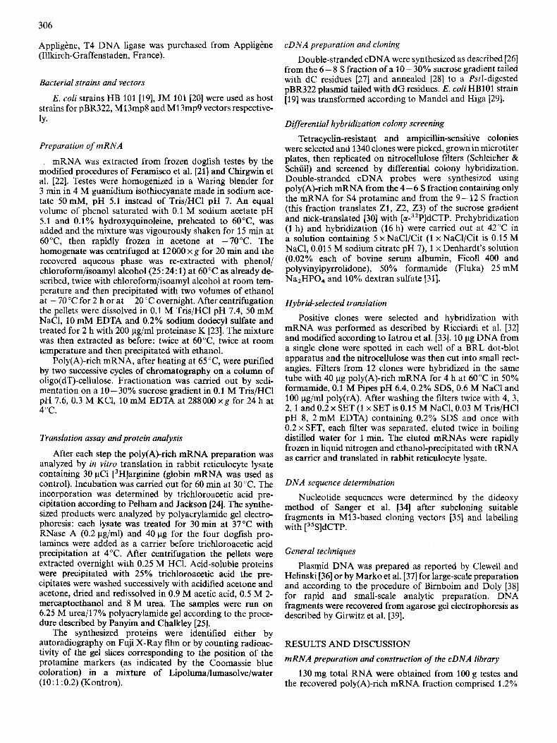
bridization: 144 clones hybridized very strongly with the two cDNA probes, 31 clones hybridized strongly with the 4 - 6 S probe and 45 clones hybridized with 9 - 12 S probe. Plasmid DNA from clones hybridizing strongly with the 9 - 12 S probe were prepared and hybridized to poly(A)-rich mRNA. The eluted mRNA was tested by in vitro translation using [3H]arginine. As in the four existing dogfish protamines, only one (22) contains a methionine residue at the N terminus, this feature was used to find whether a given clone incorporated [3sS]methionine, and therefore encodes the 22 protamine. After electrophoresis the parts of the gel corresponding to the protamine markers were cut out and counted for tritiated radioactivity. Incorporation of 3H was found in the region where Z1 and 22 protamines migrate. Another translation experiment was carried out using [3 'Slmethionine and [3H]arginine in order to determine whether the eluted mRNA corresponds to Z1 or to 22 protein. After one week of ex- posure the autoradiogram (Fig. 2) showed a strong incorpora- tion of radioactivity corresponding to [35S]methionine (specific activity of [35S]methionine is approximately 30 times greater than that of [3H]arginine in this translation exper- iment) at the level of the 22 protamine. At the migration level of the 23 protamine a low 3H band is visualized; it could correspond to a cross-hybridization of 22 mRNA and 23 mRNA on the DNA of the clone which has been later defined by sequencing as a protamine clone. We thus obtained two clones (10C4 and 10D1) whose cDNA corresponds to the 22 protamine gene.
Identification and nucleotide sequence of 10C4 and lODl protamine clones
Plasmid DNA from the two clones was extracted and digested with PstI endonuclease to estimate the size of the
Fig. 2. Autoradiogram of a polyacrylamide gel of polypeptides synthe- sized in the rabbit reticulocyte Iysate system in the presence of [ 3 H ] - arginine and [35S]methionine. Lane 1, endogenous translation; lane 2, total poly(A)-rich mRNA (2 pg) translation product; lane 3, trans- lation product of the mRNA eluted from the hybridization-transla- tion experiment of clone 1 OD1
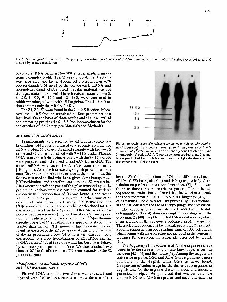
insert. We found that clones 10C4 and lODl contained a cDNA of 370 base pairs (bp) and 440 bp respectively. A re- striction map of each insert was determined (Fig. 3) and was found to show the same restriction pattern. The nucleotide sequence determination confirmed that the two clones encode for the same protein, 10D1 cDNA has a longer poly(A) tail of 70 residues. The PstI-Hue111 fragments (Fig. 3) were cloned at the PstI-SmaI sites of the M13 mp9 phage and sequenced.
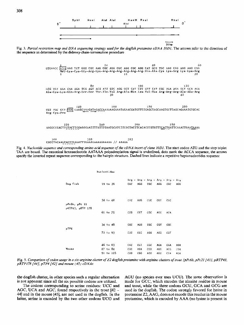
The amino acid sequence deduced from the nucleotide determination (Fig. 4) shows a complete homology with the protamine 22 [40] except for the last C-terminal residue, which is an arginine in the previously published protein sequence. The nucleotide sequence of the dogfish protamine 22 presents a coding region with an open reading frame of 138 nucleotides, which begins with an ATG sequence included in the consensus sequence for eucaryotic initiation site described by Kozak
The frequency of the codon used for the arginine residue seems to be the same as for the other known species such as the trout [42-441 and the mouse [45]. Among the six possible codons for arginine, CGC and AGA/G are significantly more abundant in the dogfish while CGA is never found. Comparison of codon usage for the cluster of six arginines in dogfish and for the arginine cluster in trout and mouse is presented in Fig. 5. We point out that whereas only two codons (CGC and AGG) are present and occur alternately in
~411.
308
S p h l R s a l A l u l A l u l H a e l l l R s a l R s a l G ' A l u l 3'
- . - 20 bp
Fig. 3. Partial restriction map and DNA sequencing strategy used for the dogfish protarnine cDNA IODI. The arrows refer to the direction of the sequence as determined by the dideoxy-chain-termination procedure
20 40 6 0
i.let-Lys-Cys-Gly-Arg-Lys-Arg-Arg-Arg-Arg-Arg-Arg-His-Ala-Cys-Lys-Arg-Lys-Lys-Arg- 1 10 20
GTGAACC IhTG( AAG T G T GGG CGC AAG CGC AGG CGC AGG CGC AGG C A T GCG TGC AAG CGG AAG AAG CGG
80 100 1 2 0 GCG T G T AAA CAA AGA TCG AGT ACG A T T GTC AGG GCT CAT C T T G T T CAT CGC AGA AGA GCT GCA AGA Ala-Cys-Lys-Gln-Arg-Ser-Ser-Thr-Ile-Val-Arg-Ala-His-Leu-Val-His-Arg-Arg-Arg-Ala-Ala-Arg-
30 4 0
140 160 180 200 CGT TGC C C T GAAGC~A~GATA,GACCAAAAAAGAAATAGAACGATGTTTTGAGCTAGCAAGTGCTTAGCAGAAATGTGCAC A r g - C y s - P r o
220 240 260 280 A A G G C C G A C T T G T P _ C T ~ G ~ A G G G A A T T T T A T T T G A A T G C A T C T T C G C T A C T T C A C A C C C G T G T T T ~ A T T C A A A T T G A A T A A A A
300 320 393 CGCCTACAAgTP_CzxEAGATTTGAAAGAAAAAAAAAA / / AAAAA
Fig. 4. Nucleotide sequence and corresponding amino acid sequence of the cDNA insert of clone IODI. The start codon ATG and the stop triplet TAA are boxed. The canonical hexanucleotide AATAAA polyadenylation signal is underlined, dots mark the ACCA sequence, the arrows specify the inverted repeat sequence corresponding to the hairpin structure. Dashed lines indicate a repetitive heptanucleotides sequence
Nucleot ides
Dog f i s h
34 t o 48 C G C pPc6b. pPc 21
pRTP43, pRTP 178
61 t o 7 5 CGG
34 t o 4 8 CGC
pTP4 72 to 93 CGC
Mouse 46 t o 63 CGC
67 to 84 CGC
91 t o 108 C G G
AGG CGC
C G T CGC
AGG CGC
CGC A G G
CGT CGC
A G A C G G
CGG AGG
C G T
A G G
CGT
A G G
AGA
A G G
A G G
C G C
A G A
CGC
C G T
CGA AGA
A G G C G A
C G A AGA
Fig. 5. Comparison of codon usage in a six-arginine cluster of 2 2 dogfish protarnine with arginine clusters of trout (pPc6b, pPcZI(43J. pRTP43, pRTPl78 [44] , pTP4 [42] and mouse (45) cDNAs
the dogfish cluster, in other species such a regular alternation is not apparent since all the six possible codons are utilized.
The codons corresponding to serine residues: UCC and AGC, UCA and AGC, found respectively in the trout [42- 441 and in the mouse [45], are not used in the dogfish. In the latter, serine is encoded by the two other codons UCG and
AGU (no species ever uses UCU). The same observation is made for GCC, which encodes the alanine residue in mouse and trout, while the three codons GCU, GCA and GCG are used in the dogfish. The codon strongly favored for lysine in protamine 22, AAG, does not encode this residue in the mouse protarnine, which is encoded by AAA (no lysine is present in
309
the known protamine of the trout). It is of interest to note that in all protamine cDNAs previously described, TAG is the stop codon, while, in the dogfish it is TAA.
Analysis of the 3’ non-coding region
Comparison with the 3‘ end of the other published prot- amine cDNA sequences shows no pronounced homology (20%). The nucleotide sequence of our 22 protamine (Fig. 4) present a complete 3’ non-coding region of 254 bp (nucleo- tides 139 - 393) with the canonical signal AATAAA necessary for polyadenylation and/or cleavage of the transcript [46, 471. The CACTG sequence was found 10 bp upstream of the AATAAA, which was recognized by Benoist et al. [48] and Berget [49] to be adjacent to the site of poly(A) addition in several sequenced DNAs and to be a recognition element related to cleavage site selection. This sequence was located upstream or downstream the polyadenylation signal [49].
A similar element, CAATG, is also found 14 bp upstream the AATAAA in the mouse (cDNA published by Kleene et al. [45] and CACTG, 27 bp upstream in the trout [42-441. Moreover, 13 bp downstream of the stop codon in the 22 dogfish cDNA we found a ACCA sequence. This sequence is highly conserved within the 3’ non-coding region of almost all the histone DNAs investigated so far [50, 511. It has been suggested to be a regulatory signal active in termination of transcription or maturation of a longer precursor (for a review see [52]). The ACCA sequence in histone is immediately pre- ceded by a 16-bp SGGCzCTTTTCAGiGCC3’ conserved DNA sequence, w h i c w a hyphenated i&erted repeat sequence and which forms a stable hairpin loop structure. Another striking feature of histone gene structure, is the conserved CAAGAAAGA sequence located in the spacer DNA, a few nucleotides farther downstream from the ACCA sequence [51,53]. These two characteristic sequences: inverted DNA repeat and spacer DNA are necessary for an efficient processing and for a correct generation of the so-called ‘true 3’ end’ of histone mRNAs [54]. Surprisingly we found in our 22 protamine sequence, immediately upstream of the ACCA signal, an inverted repeat sequence 5:CTATGATAG,3’ (Fig. 4) and five nucleotides downstream the ACCA a S’GAAATAGAA3’ sequence, which may be correlated to the conserved S‘CAAGAAAGA3‘ signal. In most of the hstone genes where the hairpin and the ACCA.. .CAAGAAAGA sequences are found, the putative polyadenylation signal AATAAA was almost invariably missing. This observation is in agreement with the fact that the histone mRNAs are in most cases non polyadenylated except an H1 mRNA of Xenopus laevis, which has the usual conserved terminal se- quence (ACCA ...) and yet is polyadenylated [55], and six other already described histone genes from different species as mentioned by Nordstrom et al. [56]. The 22 dogfish protamine structure, determined in this paper, suggests that the ACCA signal became inactive in the termination of transcription and that AATAAA is the only signal used, so that all the messengers are polyadenylated. Consequently we can suggest that the inverted repeat sequence and the spacer DNA surrounding the ACCA core have evolved from an original histone-like ancestor. In such a case our S’CTATGATAG?’ sequence has probably diverged by a series bf deletions and point mutations, leading to a weaker hairpin loop structure and to a non-functional spacer DNA signal.
We are grateful to Pr P. Chevaillier and to Dr M. Gusse for kindly providing the Scylliorhinus caniculus protamine markers. We thank
S. Pellegrino for typing the manuscript. The present investigations was supported by grants from the Centre National de la Recherche Scientfique (ER: 272; ATP-CNRS 06931) and from the Association pour la Recherche sur le Cancer (1042).
REFERENCES 1. Bloch, D. P. (1969) Genet. Suppl. 61.93 - 111. 2. Bloch, D. P. (1976) in Handbook ofgenetics (King, R. C., ed.)
3. Kierszenbaum, A. L. & Tres, L. L. (1975) J. Cell Biol. 65,258-
4. Bedford, J. M. & Calvin, H. I. (1974) J. Exp. Zool. 188, 137-
5. Kopecny, V. & Pavlok, A. (1975) J. Exp. Zool. 191,85-96. 6. Hecht, N. B., Bower, P. A., Kleene, K. C. & Distel, R. J. (1985)
Differentiation 29, 189- 193. 7. Davies, P. L., Dixon, G. H., Ferrier, L. N., Gedamu, L. & Latrou,
K. (1976) Prog. Nucleic Acids Res. Mol. Biol. 19, 135-155. 8. Aiken, J. M., McKenzie, D. I., Zhao, Z., States, J. C. & Dixon,
G. H. (1983) Nucleic Acidr Res. 11,4907-4922. 9. Aiken, J. M., Miller, F. D., Hagen, F., McKenzie, D. I., Krawetz,
S . A., van de Sande, J. H., Rattner, J. B. & Dixon, G. H. (1985) Biochemistry 24, 6268 - 6276.
10. Coelingh, J. P., Monfoort, C. H., Rozijin, T. H., Gevers-Leuven, J. A., Shiphof, R., Steyn-Parve, E. P., Braunitzer, G., Shrank, B. & Ruhfus, A. (1972) Biochim. Biophys. Acta 285, 1 - 14.
11. Gaastra, W., Lukkes-Hofstra, J. & Kolk, A. H. J. (1978) Biochem. Genet. 16, 525-529.
12. Nakano, M., Tobita, T. & Ando, T. (1976) Int. J . Peptide Protein Res. 8, 565 - 578.
13. Mellinger, J. (1965) Zellforschung 67, 653-673. 14. Chauviere, M., Laine, B., Sautiere, P. & Chevaillier, P. (1983)
15. Gusse, M. & Chevaiilier, P. (1978) Cytobiologie 16,421 -443. 16. Ando, T. & Suzuki, K. (1966) Biochim. Biophys. Acta 121,427-
17. Ando, T. & Watanabe, S. (1969) Int. J . Protein Res. I , 221 -224. 18. Gusse, M., Sautiere, P., Chauvikre, M. & Chevaillier, P. (1983)
19. Boyer, H. W. & Roulland-Dussoix, D. (1969) J. Mol. Biol. 41,
20. Messing, J., Crea, R. & Seeburg, P. H. (1981) Nucleic Acids Res.
21. Feramisco, J. R., Smart, J. E., Burridge, K., Helfman, D. M. & Thomas, G. P. (1982) J . Biol. Chem. 257, 13 024- 11 031.
22. Chirgwin, J. M., Przybyla, A. E., MacDonald. R. J. & Rutter, W. J. (1979) Biochemistry 18, 5294-5299.
23. Maniatis, T., Fritsch, E. F. & Sambrook, J. (1982) Molecular cloning: A laboratory manual, Cold Springer Harbor Labora- tory, Cold Spring Harbor, New York.
24. Pelham, H. R. B. & Jackson, R. J. (1976) Eur. J . Biochem. 67.
25. Panyim, S. & Chalkley, S. (1969) Arch. Biochem. Biophys. 130,
26. Wickens, M. P., Buell, G. N. & Schimke, R. T. (1978) J . B i d .
27. Roychoudhury, R., Jay, E. & Wu, R. (1976) Nucleic Acids Rex
28. Villa-Komaroff, L., Efstratiadis, A., Broome, S., Lomedico, P., Tizard, R., Naber, S. P., Chick, W. L. & Gilbert, W. (1978) Proc. Nut1 Acad. Sci. USA 75,3727 - 3731.
vol. 5, pp. 139- 167, Plenum Press, New York.
270.
156.
FEBS Lett. 152,231-235.
429.
Biochim. Biophys. Acta 748,93-98.
459 -472.
9, 309 - 321.
247-256.
337- 346.
Chem. 253,2483 - 2495.
3,101-116.
29. Mandel, M. & Higa, A. (1970) J. Mol. Biol. 53, 159- 162. 30. Rigby, P. W. J., Dieckmann, M., Rhodes, C. & Berg, P. (1977)
31. Wahl, G. M., Stern, M. & Stark, G. R. (1979) Proc. Nut1 Acad.
32. Ricciardi, R. P., Miller, J. S. & Roberts, B. E. (1979) Proc. Natl
33. Iatrou, K., Tsitilou, S. G., Goldsmith, M. R. & Kafatos, F. C.
J . Mol. Biol. 113,237-251.
Sci. USA 76,3683 -3687.
Acad. Sci. USA 76,4927 - 4931.
(1980) Cell 20,659 - 669.
310
34. Sanger, F., Nicklen, S. & Coulson, A. R. (1977) Proc. Natl Acad.
35. Messing, J. & Vieira, J. (1982) Gene 19, 269-276. 36. Clewell, D. B. & Helinski, D. R. (1969) Proc. Nut1 Acad. Sci.
37. Marko, M. A., Chipperfield, R. & Birnboim, H. C. (1982) Anal. Biochem. 12,382 - 387.
38. Birnboim, H. C., Doly, J. (1979) Nucleic Acids Res. 7, 1513- 1523.
39. Girvitz, S. C., Bacchetti, S., Rainbow, A. J. & Graham, F. L. (1 980) Anal. Biochem. 106,492 -496.
40. Martinage, A., Gusse, M., Belaiche, D., Sautiere, P., Chevaillier, P. (1985) Biochim. Biophys. Acta 831, 172-178.
41. Kozak, M. (1984) Nucleic Acids Res. 12, 857 - 872. 42. Gedamu, L., Wosnick, M. A., Connor, W., Watson, D. C. &
Dixon, G. H. (1981) Nucleic Aciak Res. 9, 1463- 1482. 43. Sakai, M., Fuji-Kuriyama, Y., Saito, T. & Muramatsu, M. (1981)
J . Biochem. (Tokyo) 89, 1863 - 1868. 44. Jenkins, J. R. (1979) Nature (Lond.) 279,809-811. 45. Kleene, K. C., Distel, R. J. & Hecht, N. B. (1985) Biochemistry
Sci. USA 74, 5463 - 5467.
USA 62, 1159-1166.
24,719-722.
46. Fitzgerald, M. & Shenk, T. (1981) Cell 24, 251 -260. 47. Montell, C., Fisher, E. F., Caruthers, M. H. & Berk, A. J. (1983)
48. Benoist, C., O’Hare, K., Breathnach, R. & Chambon, P. (1980)
49. Berget, S. M. (1984) Nature (Lond.) 309, 179-182. 50. Busslinger, M., Portmann, R. & Birnstiel, M. L. (1979) Nucleic
51. Hentschel, C. C. & Birnstiel, M. L. (1981) Cell25, 301 -313. 52. Birnstiel, M. L., Busslinger, M. & Strub, K. (1985) Cell 41, 349-
53. Georgiev, 0. & Birnstiel, M. L. (1985) EMBO J. 4,481 -489. 54. Birchmeier, C., Grosschedl, R. & Birnstiel, M. L. (1982) Cell28,
55. Zernik, M., Heintz, N., Boime, I. & Roeder, R. G. (1980) Cell
56. Nordstrom, J. L., Hall, S. L. & Kessler, M. M. (1985) Proc. Nut1
Nature (Lond.) 305,600-605.
Nucleic Acids Res. 8 , 127 - 142.
Acids Res. 6 , 2997 - 3008.
359.
739 - 745.
22, 807-815.
Acad. Sci. USA 82,1094- 1098.
Recommended