1558-1748 (c) 2018 IEEE. Personal use is permitted, but republication/redistribution requires IEEE permission. See http://www.ieee.org/publications_standards/publications/rights/index.html for more information.
This article has been accepted for publication in a future issue of this journal, but has not been fully edited. Content may change prior to final publication. Citation information: DOI 10.1109/JSEN.2018.2887107, IEEE SensorsJournal
> REPLACE THIS LINE WITH YOUR PAPER IDENTIFICATION NUMBER (DOUBLE-CLICK HERE TO EDIT) <
1
Abstract—The increased prevalence of chronic disease in aging
population entails health risks and imposes significant economic
and social burden. It is essential to provide comfortable, cost-
effective, easy-to-use unobtrusive and wearable systems for
personal well-being and healthcare. Novel flexible material-based
non-invasive and wearable sensors offer an efficient and cost-
effective solution which enables the continuous and real-time
monitoring of important physiological signs of the human-beings,
the assessment of personal health conditions and provides
feedback from remote and home monitoring. In this paper, novel
flexible material-based wearable sensors, devised into body sensor
networks to capture and monitor vital bio-signals including,
Electroencephalography (EEG), Electrocardiography (ECG) and
respiratory, are proposed. Silver nanowires (Ag NWs) and
polydimethylsiloxane (PDMS) composite material, carbon foam
and graphene-based fiber are used to sense the EEG, ECG and
respiratory respectively. With different flexible materials, the
smart hat and smart jacket are designed to affix the sensors which
enable long-term health monitoring of vital signals seamlessly.
Meanwhile, the corresponding acquisition circuits are developed
and mounted with the proposed electrodes on the garments. More
importantly, a comprehensive protocol is designed to validate the
performance of the proposed system while some standard sensors
and commercial devices are used for comparison. The evaluation
results demonstrate the proposed system presents a comparable
performance with the existing system. In summary, the proposed
sensing system offers an unobtrusive, detachable, expandable,
user-friendly and comfortable solution for physiological signal
monitoring. It can be expected to use for the remote healthcare
monitoring and provide personalized information of health, fitness,
and diseases.
Index Terms—Novel flexible materials, Vital Bio-signal
measurements, Body sensor networks
This work was supported by National Key R&D Program of China (Grant
No.2017YFE0112000), China Postdoctoral Science Foundation Grant (Grant
No.2018T110346 and No.2018M632019) and Shanghai Municipal Science and Technology Major Project (Grant No.2017SHZDZX01).
Chen Chen, Zeyu Wang, and Wei Li are with the Center for Intelligent
Medical Electronics, School of Information Science and Technology, Fudan University, Shanghai, China (E-mail: chenchen_fd@ fudan.edu.cn;
[email protected]; [email protected]).
Hongyu Chen and Linkai Tao are with the Center for Intelligent Medical Electronics, School of Information Science and Technology, Fudan University,
Shanghai, China, and the Department of Industrial Design, Eindhoven
University of Technology, Netherlands. (E-mail: [email protected]; [email protected]).
I. INTRODUCTION
HE aging population combined with the increased
prevalence of chronic disease are becoming significant
socioeconomic burdens for present-day society. Besides this,
the accompanying costs are increased rapidly in terms of the
prescription drugs, medical instruments, hospital care and so on
[1], [2]. Thus, seeking for the innovative solutions and new
technologies to improve the quality of healthcare services and
to provide the early detection/intervention at an affordable price
is extremely important. The emerging technology Body Sensor
Networks (BSNs) provides long-term monitoring of
physiological signals without interrupting the human’s normal
activities. It combines the intelligent, flexible, light in weight
novel sensors with the human body and offers a comprehensive
solution to providing a detailed information regarding the
personalized health and fitness, tracking the personal wellbeing,
detecting the health risk, facilitating timely intervention to acute
events such as stroke, epilepsy and heart attack and so on [3],
[4]. Physiological signs such as EEG, ECG, and respiration play
a vital role in healthcare monitoring. Specifically, EEG can
provide high temporal resolutions and reflect the dynamics of
brain activities for neurobiological disorders diagnosis; ECG
and respiration signals can furthermore provide a
comprehensive cardiopulmonary, respiration and physical
actives information for daily actives analysis and disease
analysis like arrhythmias, ischemia, sleep apnea, etc. As an
indispensable part of the remote healthcare monitoring, EEG,
ECG, and respiration sensing systems attracted the attention of
many researchers and entrepreneurs in recent years.
The sensing systems/devices have evolved from bench-top,
Wei Yuan is with Printable Electronics Research Centre, Suzhou Institute of
Nanotech and Nano-Bionics, Chinese Academy of Sciences, Suzhou, China.
(E-mail: [email protected]). Yuting Zhao, Gaoshan Huang, and Yongfeng Mei are with the Department
of Material Science, Fudan University, Shanghai, China. (E-mail:
[email protected]; [email protected]; [email protected]). Zherui Cao and Ranran Wang are with Shanghai Institute of Ceramics,
Chinese Academy of Sciences, Shanghai, China. (E-mail:
[email protected]; [email protected]). Wei Chen is with the Center for Intelligent Medical Electronics, School of
Information Science and Technology, Fudan University, and Shanghai Key
Laboratory of Medical Imaging Computing and Computer Assisted Intervention, Shanghai China. (Phone: +86-021-65643781; E-mail:
Novel Flexible Material-based Unobtrusive and
Wearable Body Sensor Networks for Vital Sign
Monitoring
Chen Chen, Zeyu Wang, Wei Li, Hongyu Chen, Wei Yuan, Linkai Tao, Yuting Zhao, Gaoshan
Huang, Yongfeng Mei, Zherui Cao, Ranran Wang, Wei Chen, Senior Member, IEEE
T
1558-1748 (c) 2018 IEEE. Personal use is permitted, but republication/redistribution requires IEEE permission. See http://www.ieee.org/publications_standards/publications/rights/index.html for more information.
This article has been accepted for publication in a future issue of this journal, but has not been fully edited. Content may change prior to final publication. Citation information: DOI 10.1109/JSEN.2018.2887107, IEEE SensorsJournal
> REPLACE THIS LINE WITH YOUR PAPER IDENTIFICATION NUMBER (DOUBLE-CLICK HERE TO EDIT) <
2
and portable devices, to the recent garment and small gadgets
based wearable devices. Wearable devices have grown rapidly
in popularity recently. Until now, several various commercial
wearable devices are available on the market like
BioStampRC® by mc10, USA [5], Wearable biosensor by
Philips [6], HUAWEI WATCH by Huawei, China [7], Apple
Watch by Apple, USA [8], Hexoskin Smart Shirts by Hexoskin
Wearable Body Metrics, Canada [9], etc. Most of existing
commercial products are presented or manufactured in the form
of watch or adhesive sticker. It may integrate ECG electrodes,
accelerometer and gyroscope to monitor the heart rate and
motion like the number of steps, climbing, and running. But the
concerns and doubts about the achievable signal quality
obtained by these devices remain controversial. And other bio-
signals like EEG and respiration rate were scarcely considered
in existing wearable devices.
Apart from commercial wearable devices, a variety of
wearable sensors/systems based on novel materials have been
reported in the literature. As for the EEG signal measurement,
Ng et al. [10] proposed a vacuum casting method to fabricate
the micro-spike EEG electrode and characterized the sensing
performance in terms of the impedance level and stability. Lin
et al. [11] designed a novel dry foam-based EEG electrode,
which was fabricated by electrically conductive polymer foam
covered with a conductive fabric. Recently, Fiedler et al. [12]
designed a polymer-based multipin dry EEG electrode, which
used a multi-phase chemical coating method to coat
polyurethane electrode with the silver. These proposed novel
dry EEG electrodes have overcome the limitations of
conventional wet electrodes such as skin preparation, and
conductive gel requirements. However, the micro-spike
electrode and polymer-based multipin electrode may cause the
discomfort due to their rigid pin design and polymer foam
seems more suitable for collecting the EEG signals originating
from the area without hair by simply affix the designed
electrodes pad to the skin with adhesive tapes.
Regarding other physiologic signals monitoring, like ECG,
several studies have also reported their developments. Pandian
et al. [13] proposed a customized ECG sensor using silicon
rubber with pure silver fillings, which was fabricated in the
form of a belt to acquire ECG signal. Yamamoto et al. [14]
presented a gel-less sticky ECG electrode that mixed multiwall
carbon nanotubes (CNT) with the adhesive ethoxylated
polyethyleneimine-polydimethylsiloxane (PEIE-PDMS). Pani
et al. [15] proposed a novel textile electrode based on woven
fabrics treated with PEDOT:PSS. Castrillón et al. [16] made a
comparative study on textile electrodes involving PEDOT:PSS
treated fabrics based on cotton, cotton–polyester, lycra and
polyester in terms of the contact impedance, electrode
polarization, noise, and long‑term performance. The novel ECG
sensors and systems are developed to overcome the main
limitations of the widely used silver/silver chloride (Ag/AgCl)
disposable ECG electrodes. However, there also have some
drawbacks, like, the textile electrodes may result in a high
contact impedance due to the materials and belt-based ECG
may cause discomfort and not suitable for long-term monitoring.
For measuring respiration signal, a textile capacitive
respiration sensor (TCRS) [17] and a fiber-optic deformation
sensor [18] were explored. The TCRS is fabricated with
conductive textile and polyester, and the respiration is derived
from the distance changes between two textile plates in the
TCRS, which measures the force from the abdominal diameter
changes caused by the respiratory movement. In [18], the fiber-
optic deformation sensor measures bending deformation
directly. Two fiber-optic sensors are bonded to an elastic belt
and a differential system for respiratory measurement is
established. Mostly, the prototypes of respiration monitoring
systems are designed as belt form. These proposed systems can
avoid the limitations of traditional pressure transducer or
thermocouple-based respiratory systems which need to be fitted
in or near the nostrils.
In general, both existing commercial devices and novel
wearable sensing systems proposed in the literature have shown
the dramatical improvement of the healthcare monitoring
systems. Despite its significant contributions in remote
healthcare monitoring systems, several issues still remain
challenging. 1) For long-term healthcare monitoring, the
wearable sensors should satisfy the ergonomic requirements,
e.g. they should be flexible and comfortable to wear; easy to
set-up and remove. 2) Regarding the bio-compatibility, the
wearable device should be hypo-allergenic and nontoxic to the
human body. 3) The wearable devices should ensure the
measurement accuracy while considering the cost and
efficiency, e.g. they should be easy to fabricate at low cost and
high efficiency. 4) The bio-signal sensing systems are
susceptible to the motion artifacts, thus flexible electrodes
which have smaller skin-electrode impedance and better
tolerance on motion artifacts are required to overcome the
limited sensitivity and reliability of the existing systems. A
robust healthcare system should be able to address these issues
and to enable continuous and uninterrupted monitoring. Thus, a
novel flexible material-based unobtrusive and wearable vital
bio-signal sensing system is proposed in this paper.
In this paper, a novel flexible Ag NWs/PDMS composite
material is proposed to measure the EEG signal. It is an ideal
material for sensing EEG signal due to its low impedance, high
safety, high conductivity, and good resistance stability. In the
existing work, two fabrication methods were widely used to
produce this kind of silicon rubber-based materials. One is
coating conductive layer on the silicon substrate by magnetron
sputtering or multi-phase chemical coating process. This
fabrication process has been used in some EEG electrodes
design [12], [19]. However, due to its inherent stiff character,
these metal coatings may cause cracks easily under bending or
twisting states, which makes these EEG sensors are not suitable
for long-term wearing. Another method is combining silicon
rubber and conductive additives to generate a conductive
composite [20], [21]. The conductive additives are usually
metal particles or flakes, these structures are difficult to form
conductive networks in composite, therefore, these composite
electrodes usually have high impedance and poor resistance
stability. In this paper, we use one-dimensional Ag NWs as
conductive additives rather than the structures of particles or
flakes [22], [23]. The Ag NWs/PDMS composite has high
1558-1748 (c) 2018 IEEE. Personal use is permitted, but republication/redistribution requires IEEE permission. See http://www.ieee.org/publications_standards/publications/rights/index.html for more information.
This article has been accepted for publication in a future issue of this journal, but has not been fully edited. Content may change prior to final publication. Citation information: DOI 10.1109/JSEN.2018.2887107, IEEE SensorsJournal
> REPLACE THIS LINE WITH YOUR PAPER IDENTIFICATION NUMBER (DOUBLE-CLICK HERE TO EDIT) <
3
conductivity and good resistance stability due to the forming of
wonderful Ag NWs conductive networks in silicon rubber. This
conductive composite electrode is more suitable for long-term
EEG measurement. For sensing the ECG signal, a novel
material proposed in [24] is used. This material has widely used
in electromagnetic shielding application [25] and sensing
pressure [26]. Due to its good conductivity and excellent
elasticity characteristics, it was first attempted to acquire the
ECG signal in this paper. As for sensing respiratory signal, the
graphene-based fiber material is applied. The material applies
the structure of the double covered yarn of the fiber substrate
endows the fiber an incorporation of ultrahigh sensitivity to
tensile strain deformation and wide maximal sensing range,
which shows superior strain-sensing performance compared
with other stretchable electronics [27]. Besides, conventional
flexible sensors [28], [29] that based on metal, which restricted
by their rigidity and may not fit properly with human bodies.
As a two-dimensional conductive nanomaterial, graphene
exhibits high conductivity and excellent combination with the
substrate. The fiber is light and flexible enough to conform to
arbitrary curved surfaces, indicating its conformability in the
robust and flexible wearable electronics. With the flexible and
stretchable Ag NWs/PDMS composite material, carbon foam,
and graphene-based fiber material, different sensor prototypes
for measuring EEG, ECG, and respiration signal are designed
by mounting the EEG sensors on the hat and embedded the
ECG and respiration sensors in the garment.
The rest of this paper is organized as follows: Section II gives
the overall requirements and design of the vital bio-signal
sensing system. Section III presents a brief description of the
novel flexible materials and electrodes prototypes proposed in
our system. Followed by a details explanation of the proposed
prototype of the sensing system in Section IV. Evaluations of
the proposed sensing system are presented in Section V. At last,
the brief discussion and conclusion are shown in Section VI and
VII, respectively.
II. VITAL BIO-SIGNAL SENSING SYSTEM
The main objective of the proposed sensing system is to
enable continuous monitoring of physiological signals, which
involves the sensing and monitoring of EEG, ECG, and
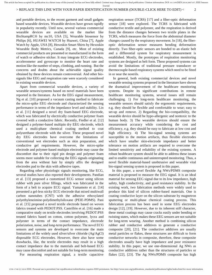
respiratory. Fig. 1 presents a conceptual architecture of the
proposed BSN for the remote healthcare monitoring, which
mainly involves the sensing system, local process unit for
monitoring and displaying the signals, cloud computing for the
further data analysis and medical intervention. Thus, we
proposed the following requirements based on the need-finding.
⚫ Continuously and long-term monitoring the EEG, ECG
and respiration signals simultaneously: all the components
are unobtrusive, detachable and user-friendly.
⚫ All the components are comfortable to wear: all the
sensors are flexible, small in dimensions, light in weight
and hypo-allergenic and nontoxic to the human body.
⚫ A reasonable layout of the sensors to avoid the signal
interruption between different sensors.
⚫ All the sensors conform well with the ergonomic
requirements which provide a good conductivity between
the skin and the electrodes.
⚫ A comfortable base (for example, clothes, hat) for
embedding or mounting the sensors to avoid the motion
artifacts.
⚫ The acquisition modules can be expandable to add more
monitoring functions if needed.
Fig 1. Conceptual architecture of the proposed BSN for remote healthcare
monitoring
To fulfill the requirements of the sensing system mentioned
above, novel flexible materials are explored to obtain the
physiological signals unobtrusively. Different sensors
prototypes are presented due to the flexible material properties
and ergonomic requirements. Meanwhile, to provide a flexible
solution for embedding and mounting the proposed electrodes,
two modules namely the smart hat and smart jacket which
includes the different acquisition systems for monitoring EEG,
ECG, and EMG are designed. These two modules are
detachable, expandable, user-friendly for measuring the EEG,
ECG, and respiration simultaneously. Moreover, the detailed
evaluations of the proposed sensing system are also provided to
verify the signal quality recorded by the proposed system in
comparison to different commercial devices.
III. NOVEL FLEXIBLE MATERIALS AND SENSING METHODS
In this section, novel materials for sensing the EEG, ECG,
and respiration signal were briefly introduced, more details
about these materials can be found in our recent publications
[30], [31], and [32] respectively. Based on these materials, the
corresponding sensing methods are also proposed.
A. Novel Flexible Materials
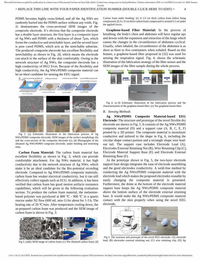
Ag NWs/PDMS Composite Material: In this paper, a
conductive and stretchable Ag NWs/PDMS composite material
is applied for sensing the EEG signal. Fig. 2a illustrates the
fabrication process, firstly, the prepared Ag NWs ink is screen
printed on a glass substrate, and then the printed Ag NWs
pattern is dried in a vacuum oven to form a uniform and
conductive film of Ag NWs network. Next, fresh PDMS liquid
is cast on top of the printed Ag NWs film, followed by curing
at 80 °C for 4 h. When peeled off the substrate, the Ag NWs
film is bonded to the cured PDMS and form the Ag NWs/PDMS
composite electrode. The Ag NWs film is actually buried just
below the PDMS surface, as shown in the scanning electron
microscopy (SEM) image of Fig. 2b. Before curing at high
temperature, the liquid PDMS penetrates into the pores of the
three-dimensional (3D) Ag NWs network. After curing, the
EEG acquisition
--Ag NWs/PDMS-based electrodes--Smart hat
--Neurobiological disorders
diagnosis, stroke, epilepsy.
ECG acquisition--Smart jacket
--Carbon foam
based electrodes--Heart diseasediagnosis
Respiration
--Smart jacket--Graphene-based fiber sensor
--Sleep apnea
Local process unit
--Signal display/processing/storage
--Phone, pad, PC
MedicalIntervention
1558-1748 (c) 2018 IEEE. Personal use is permitted, but republication/redistribution requires IEEE permission. See http://www.ieee.org/publications_standards/publications/rights/index.html for more information.
This article has been accepted for publication in a future issue of this journal, but has not been fully edited. Content may change prior to final publication. Citation information: DOI 10.1109/JSEN.2018.2887107, IEEE SensorsJournal
> REPLACE THIS LINE WITH YOUR PAPER IDENTIFICATION NUMBER (DOUBLE-CLICK HERE TO EDIT) <
4
PDMS becomes highly cross-linked, and all the Ag NWs are
randomly buried into the PDMS surface without any voids. Fig.
2c demonstrates the cross-sectional SEM images of the
composite electrode. It’s obvious that the composite electrode
has a double layer structure, the first layer is a composite layer
of Ag NWs and PDMS with a thickness of about 7μm, which
forms the conductive and stretchable layer, and the second layer
is pure cured PDMS, which acts as the stretchable substrate.
The produced composite electrode has excellent flexibility and
stretchability as shown in Fig. 2d, which means the electrode
can attach to the surface of the skin comfortably. Owing to the
network structure of Ag NWs, the composite electrode has a
high conductivity of 6912 S/cm. Because of the flexibility and
high conductivity, the Ag NWs/PDMS composite material can
be an ideal candidate for sensing the EEG signal.
Fig 2. (a) Schematic illustration of the fabrication process of Ag
NWs/PDMS composite electrode; SEM images of the surface morphology (b)
and the cross-section of the composite electrode (c); (d) Photographs of the
obtained Ag NWs/PDMS composite electrode, under bending and stretching
state.
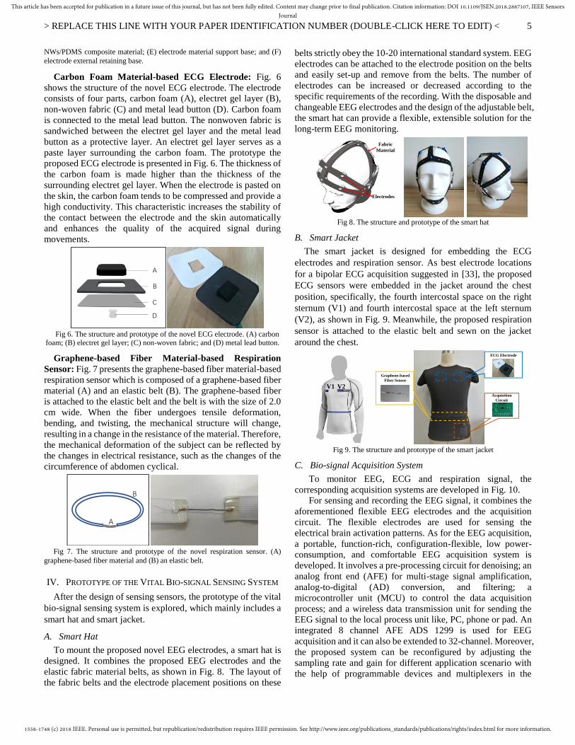
Carbon Foam Material: The carbon foam material has
excellent flexibility as shown in Fig. 3, which can provide
comfortable attachment. For Ag NWs material, it has high
conductivity due to the network structure of Ag NWs, which
make it be an ideal candidate for the Bio-potential recording
electrode. Compared to Ag NWs/PDMS composite materials,
carbon foam has weaker electrical conductivity, but it can still
effectively collect signals such as ECG. In addition, it has been
verified that carbon foam has good motion artifacts resistance
capabilities, which will be given in the following evaluation
section. To produce the carbon foam material, the melamine-
based polymer was pyrolyzed at 800 oC - 900 oC in a quartz
reactor under N2 flow (600 mL min-1) for about for 1-3 h. The
heating rate of 20 oC/min. After temperature cooling down, the
as-prepared carbon foam was produced and the SEM image of
carbon foam is shown in Fig. 3.
Fig 3. (a)(b) SEM image of carbon foam; (c) The obtained carbon foam; (d)
Carbon foam under bending; (e) A 1.0 cm thick carbon foam before being
compressed; (f) A 1.0 cm thick carbon foam compressed to around 0.3 cm under
the applied forces.
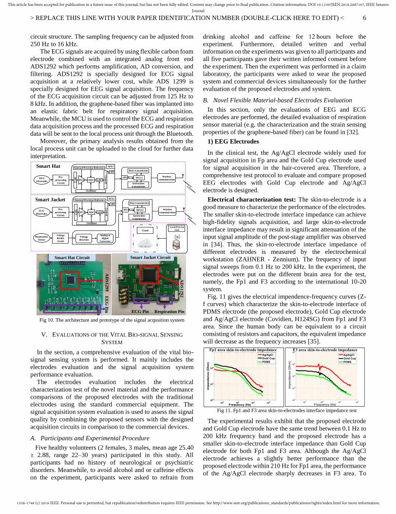
Graphene-based Fiber Material: In the process of
breathing, the body's chest and abdomen will have regular ups
and downs with the expansion and retraction of the lungs which
causes the changes in the circumference of abdomen cyclical.
Usually, when inhaled, the circumference of the abdomen is as
short as three to five centimeters when exhaled. Based on this
feature, a graphene-based fiber proposed in [32] was used for
sensing the respiration signal. Fig. 4. shows the schematic
illustration of the fabrication strategy of the fiber sensor and the
SEM images of the fiber sample during the whole process.
Fig 4. (a–d) Schematic illustration of the fabrication process and the
characterization of the graphene-based fiber; (e) The graphene-based fiber.
B. Sensing Methods
Ag NWs/PDMS Composite Material-based EEG
Electrode: The structure and prototype of the novel flexible dry
electrode are shown in Fig. 5. It consists of the Ag NWs/PDMS
composite material (D) and a support case (A, B, C, E, F)
printed by a 3D printer. The composite material is monolayer
conductive and tailored to the shape in Fig. 6, including the
circular shape contact portion and a rectangle shape wire lead-
out tail. The support case includes Electrode Lead (A),
Electrodes External Retaining Nut (B), Wire Retaining Clip (C),
Electrode Material Support Base (E) and Electrode External
Retaining Base (F).
As the prototype shown in Fig. 5, the two-layer electrode
support base design integrates the ease of electrode assembling
and the good electrodes conductivity. A weld-free method by
conducting the Ag NWs/PDMS composite material with the
electrode lead which makes the proposed electrodes reusable by
easily changing the composite material is presented.
Furthermore, the dome at the bottom of the electrode material
support base keeps the Ag NWs/PDMS composite material
above the bottom surface of the electrode external retaining
base. It would make the Ag NWs/PDMS composite material
contact with the skin properly when using the novel EEG
electrode.
Fig 5. The structure and prototype of the novel EEG electrode. (A) electrode
lead; (B) electrodes external retaining nut; (C) wire retaining clip; (D) Ag
a b c d
e f
e
1558-1748 (c) 2018 IEEE. Personal use is permitted, but republication/redistribution requires IEEE permission. See http://www.ieee.org/publications_standards/publications/rights/index.html for more information.
This article has been accepted for publication in a future issue of this journal, but has not been fully edited. Content may change prior to final publication. Citation information: DOI 10.1109/JSEN.2018.2887107, IEEE SensorsJournal
> REPLACE THIS LINE WITH YOUR PAPER IDENTIFICATION NUMBER (DOUBLE-CLICK HERE TO EDIT) <
5
NWs/PDMS composite material; (E) electrode material support base; and (F)
electrode external retaining base.
Carbon Foam Material-based ECG Electrode: Fig. 6
shows the structure of the novel ECG electrode. The electrode
consists of four parts, carbon foam (A), electret gel layer (B),
non-woven fabric (C) and metal lead button (D). Carbon foam
is connected to the metal lead button. The nonwoven fabric is
sandwiched between the electret gel layer and the metal lead
button as a protective layer. An electret gel layer serves as a
paste layer surrounding the carbon foam. The prototype the
proposed ECG electrode is presented in Fig. 6. The thickness of
the carbon foam is made higher than the thickness of the
surrounding electret gel layer. When the electrode is pasted on
the skin, the carbon foam tends to be compressed and provide a
high conductivity. This characteristic increases the stability of
the contact between the electrode and the skin automatically
and enhances the quality of the acquired signal during
movements.
Fig 6. The structure and prototype of the novel ECG electrode. (A) carbon
foam; (B) electret gel layer; (C) non-woven fabric; and (D) metal lead button.
Graphene-based Fiber Material-based Respiration
Sensor: Fig. 7 presents the graphene-based fiber material-based
respiration sensor which is composed of a graphene-based fiber
material (A) and an elastic belt (B). The graphene-based fiber
is attached to the elastic belt and the belt is with the size of 2.0
cm wide. When the fiber undergoes tensile deformation,
bending, and twisting, the mechanical structure will change,
resulting in a change in the resistance of the material. Therefore,
the mechanical deformation of the subject can be reflected by
the changes in electrical resistance, such as the changes of the
circumference of abdomen cyclical.
Fig 7. The structure and prototype of the novel respiration sensor. (A)
graphene-based fiber material and (B) an elastic belt.
IV. PROTOTYPE OF THE VITAL BIO-SIGNAL SENSING SYSTEM
After the design of sensing sensors, the prototype of the vital
bio-signal sensing system is explored, which mainly includes a
smart hat and smart jacket.
A. Smart Hat
To mount the proposed novel EEG electrodes, a smart hat is
designed. It combines the proposed EEG electrodes and the
elastic fabric material belts, as shown in Fig. 8. The layout of
the fabric belts and the electrode placement positions on these
belts strictly obey the 10-20 international standard system. EEG
electrodes can be attached to the electrode position on the belts
and easily set-up and remove from the belts. The number of
electrodes can be increased or decreased according to the
specific requirements of the recording. With the disposable and
changeable EEG electrodes and the design of the adjustable belt,
the smart hat can provide a flexible, extensible solution for the
long-term EEG monitoring.
Fig 8. The structure and prototype of the smart hat
B. Smart Jacket
The smart jacket is designed for embedding the ECG
electrodes and respiration sensor. As best electrode locations
for a bipolar ECG acquisition suggested in [33], the proposed
ECG sensors were embedded in the jacket around the chest
position, specifically, the fourth intercostal space on the right
sternum (V1) and fourth intercostal space at the left sternum
(V2), as shown in Fig. 9. Meanwhile, the proposed respiration
sensor is attached to the elastic belt and sewn on the jacket
around the chest.
Fig 9. The structure and prototype of the smart jacket
C. Bio-signal Acquisition System
To monitor EEG, ECG and respiration signal, the
corresponding acquisition systems are developed in Fig. 10.
For sensing and recording the EEG signal, it combines the
aforementioned flexible EEG electrodes and the acquisition
circuit. The flexible electrodes are used for sensing the
electrical brain activation patterns. As for the EEG acquisition,
a portable, function-rich, configuration-flexible, low power-
consumption, and comfortable EEG acquisition system is
developed. It involves a pre-processing circuit for denoising; an
analog front end (AFE) for multi-stage signal amplification,
analog-to-digital (AD) conversion, and filtering; a
microcontroller unit (MCU) to control the data acquisition
process; and a wireless data transmission unit for sending the
EEG signal to the local process unit like, PC, phone or pad. An
integrated 8 channel AFE ADS 1299 is used for EEG
acquisition and it can also be extended to 32-channel. Moreover,
the proposed system can be reconfigured by adjusting the
sampling rate and gain for different application scenario with
the help of programmable devices and multiplexers in the
A
B
C
D
A
B
Fabric
Material
Electrodes
V1 V2
A
B
C
D
e
ECG Electrode
Graphene-based
Fiber Sensor
Acquisition
Circuit
1558-1748 (c) 2018 IEEE. Personal use is permitted, but republication/redistribution requires IEEE permission. See http://www.ieee.org/publications_standards/publications/rights/index.html for more information.
This article has been accepted for publication in a future issue of this journal, but has not been fully edited. Content may change prior to final publication. Citation information: DOI 10.1109/JSEN.2018.2887107, IEEE SensorsJournal
> REPLACE THIS LINE WITH YOUR PAPER IDENTIFICATION NUMBER (DOUBLE-CLICK HERE TO EDIT) <
6
circuit structure. The sampling frequency can be adjusted from
250 Hz to 16 kHz.
The ECG signals are acquired by using flexible carbon foam
electrode combined with an integrated analog front end
ADS1292 which performs amplification, AD conversion, and
filtering. ADS1292 is specially designed for ECG signal
acquisition at a relatively lower cost, while ADS 1299 is
specially designed for EEG signal acquisition. The frequency
of the ECG acquisition circuit can be adjusted from 125 Hz to
8 kHz. In addition, the graphene-based fiber was implanted into
an elastic fabric belt for respiratory signal acquisition.
Meanwhile, the MCU is used to control the ECG and respiration
data acquisition process and the processed ECG and respiration
data will be sent to the local process unit through the Bluetooth.
Moreover, the primary analysis results obtained from the
local process unit can be uploaded to the cloud for further data
interpretation.
MU
X ADC LPFPGA
Co
ntr
ol
SPI
Oscillator
Internal Precision ReferenceAFE1
Pre-
processing
Circuit
Data transmission
Instruction
transmission
MCU1SPI
Data transmission
Instruction
transmission
MCU2SPI
Voltage
divider
circuit
Voltage
detection
Analog to
digital
Converter
Wireless
communication
Wireless
communication
Local Process
UnitCloud
MU
X ADC LPFPGA
Co
ntr
ol
SPI
Oscillator
Internal Precision ReferenceAFE2
Pre-
processing
Circuit
Smart Hat
Smart Jacket
Respiration
Sensor
ECG
Electrodes
EEG
Electrodes
Fig 10. The architecture and prototype of the signal acqusition system
V. EVALUATIONS OF THE VITAL BIO-SIGNAL SENSING
SYSTEM
In the section, a comprehensive evaluation of the vital bio-
signal sensing system is performed. It mainly includes the
electrodes evaluation and the signal acquisition system
performance evaluation.
The electrodes evaluation includes the electrical
characterization test of the novel material and the performance
comparisons of the proposed electrodes with the traditional
electrodes using the standard commercial equipment. The
signal acquisition system evaluation is used to assess the signal
quality by combining the proposed sensors with the designed
acquisition circuits in comparison to the commercial devices.
A. Participants and Experimental Procedure
Five healthy volunteers (2 females, 3 males, mean age 25.40
± 2.88, range 22–30 years) participated in this study. All
participants had no history of neurological or psychiatric
disorders. Meanwhile, to avoid alcohol and or caffeine effects
on the experiment, participants were asked to refrain from
drinking alcohol and caffeine for 12 hours before the
experiment. Furthermore, detailed written and verbal
information on the experiments was given to all participants and
all five participants gave their written informed consent before
the experiment. Then the experiment was performed in a claim
laboratory, the participants were asked to wear the proposed
system and commercial devices simultaneously for the further
evaluation of the proposed electrodes and system.
B. Novel Flexible Material-based Electrodes Evaluation
In this section, only the evaluations of EEG and ECG
electrodes are performed, the detailed evaluation of respiration
sensor material (e.g. the characterization and the strain sensing
properties of the graphene-based fiber) can be found in [32].
1) EEG Electrodes
In the clinical test, the Ag/AgCl electrode widely used for
signal acquisition in Fp area and the Gold Cup electrode used
for signal acquisition in the hair-covered area. Therefore, a
comprehensive test protocol to evaluate and compare proposed
EEG electrodes with Gold Cup electrode and Ag/AgCl
electrode is designed.
Electrical characterization test: The skin-to-electrode is a
good measure to characterize the performance of the electrodes.
The smaller skin-to-electrode interface impedance can achieve
high-fidelity signals acquisition, and large skin-to-electrode
interface impedance may result in significant attenuation of the
input signal amplitude of the post-stage amplifier was observed
in [34]. Thus, the skin-to-electrode interface impedance of
different electrodes is measured by the electrochemical
workstation (ZAHNER - Zennium). The frequency of input
signal sweeps from 0.1 Hz to 200 kHz. In the experiment, the
electrodes were put on the different brain area for the test,
namely, the Fp1 and F3 according to the international 10-20
system.
Fig. 11 gives the electrical impendence-frequency curves (Z-
f curves) which characterize the skin-to-electrode interface of
PDMS electrode (the proposed electrode), Gold Cup electrode
and Ag/AgCl electrode (Covidien, H124SG) from Fp1 and F3
area. Since the human body can be equivalent to a circuit
consisting of resistors and capacitors, the equivalent impedance
will decrease as the frequency increases [35].
Fig 11. Fp1 and F3 area skin-to-electrodes interface impedance test
The experimental results exhibit that the proposed electrode
and Gold Cup electrode have the same trend between 0.1 Hz to
200 kHz frequency band and the proposed electrode has a
smaller skin-to-electrode interface impedance than Gold Cup
electrode for both Fp1 and F3 area. Although the Ag/AgCl
electrode achieves a slightly better performance than the
proposed electrode within 210 Hz for Fp1 area, the performance
of the Ag/AgCl electrode sharply decreases in F3 area. To
AD
S1
29
2
ECG Pin Respiration Pin
Smart Jacket Circuit
AD
S1
29
9E
EG
Pin
Smart Hat Circuit
1558-1748 (c) 2018 IEEE. Personal use is permitted, but republication/redistribution requires IEEE permission. See http://www.ieee.org/publications_standards/publications/rights/index.html for more information.
This article has been accepted for publication in a future issue of this journal, but has not been fully edited. Content may change prior to final publication. Citation information: DOI 10.1109/JSEN.2018.2887107, IEEE SensorsJournal
> REPLACE THIS LINE WITH YOUR PAPER IDENTIFICATION NUMBER (DOUBLE-CLICK HERE TO EDIT) <
7
acquire the EEG signal from the hairy and non-hairy area, it is
well known that the largest obstacles are the influence of the
hair and different thickness of stratum corneum, which act as
barriers to ionic current and thus significantly increases the
impedance. Meanwhile, the electrical nature of the interface
between the biopotential electrodes and scalp also plays a vital
role in impedance. Even the skin preparation will be performed
before applying the Ag/AgCl electrodes, some hair will still
adhere to the surface of the conductive gels which will
significantly influence the conductivity between the electrodes
and scalp. Hence, in practice, the Ag/AgCl electrode is only
used in the frontal brain area without the influence of hair
instead of other areas of the brain. In comparison to Ag/AgCl
electrodes, the conductive medium of the proposed PDMS
electrodes is Ag NWs conductive networks rather than the ion
medium of the conductive gels, which ensures the conductivity
between the electrodes and the scalp. Moreover, with the
flexibility of the proposed electrodes, it would also potentially
ensure the good conductive and thus decrease the impedance.
Electrode signal quality test: After evaluating the EEG
electrodes characterization, the signal quality obtained by these
electrodes were also investigated. To ensure the same signal
conditioning environment for comparing the signal sensing
ability, the electrodes are connected to the same device,
Compumedics Grael Polysomnography (Grael PSG).
Fig. 12 presents the EEG signals and the corresponding
spectrum obtained from Fp1 and F3 area using the proposed
flexible EEG electrode, Ag/AgCl electrode, and Gold Cup
electrode. In Fp1 area, eye blink signals are clearly observed
and the signals collected by the three electrodes have high
consistency. Due to the different sensing ability of the
electrodes, there are some differences in signal amplitude, but
the frequency components are the same. While, in F3 area, the
Ag/AgCl electrode fails to obtain the EEG signal because of the
hair. However, the proposed flexible EEG electrode and the
Gold Cup electrode can still achieve favorable signal quality.
Fig 12. EEG signal and spectrum of Fp1 and F3 area
Electrodes signal quality evaluation results have proved that
the proposed electrodes are suitable for EEG signal acquisition
and can achieve comparable performance with the wet
electrodes like the Gold Cup and Ag/AgCl electrodes.
2) ECG Electrodes
For evaluating the performance and applicability of the
proposed carbon foam-based ECG electrode, a comparison of
the proposed ECG electrode and Ag/AgCl electrode is
presented. In the clinical test, the wet electrode Ag/AgCl
electrode is widely used. Thus, for ECG electrodes evaluation,
only Ag/AgCl electrode is involved and tested for the
comparison.
Electrical characterization test: Using ZAHNER-Zennium
with the same experiment condition aforementioned for the
EEG test, the skin-to-electrode interface impedance of the
proposed ECG electrode and the Ag/AgCl electrode are
measured. Fig. 13 demonstrates that the impedance
corresponding to the proposed Carbon Foam (CF) electrode is
slightly larger than that of the Ag/AgCl electrode. The
impedance of the Ag/AgCl electrode is nearly 566 kOhm in
near dc range, while for the carbon foam-based electrode is
about 675 kOhm. This is mainly because before using the
Ag/AgCl electrode, the skin preparation is performed by
removing the epidermal stratum corneum and applying
conductive gels. This preparation would potentially facilitate
the transduction of the ionic currents into electric current and
provide a low impedance path for signal transduction, therefore,
lower the skin-electrode impedance. While before applying
carbon foam electrode, there is no requirement for any skin
preparation.
Fig 13. Skin-electrode impedance of ECG electrodes
Electrode signal quality test: To evaluate the signal quality
obtained by the proposed ECG electrodes, we connect the
proposed ECG electrode and Ag/AgCl electrode to a
commercial device Shimmer-3. Fig. 14 shows that the proposed
ECG electrode can acquire comparable signal quality with
respect to Ag/AgCl electrode. The shapes of both signals have
high similarity and R peaks of both signals can be easily
detected. To measure the similarity between the ECG signal
obtained by the proposed ECG electrodes and the ECG signal
acquired by the Ag/AgCl electrode, the Pearson cross-
correlation coefficient is calculated. For the Pearson cross-
correlation coefficient, it lies between −1 to +1, where 1 is the
total positive correlation, 0 is no correlation, and −1 is the total
negative correlation. If the cross-correlation coefficient is close
to +1, then it indicates that there is a strong linear positive
correlation. In this paper, a high Pearson cross-correlation
coefficient between both signals is obtained, which can reach
0.9568. It indicates that is a strong linear positive correlation
between these two signals, which also indicates that there is a
high similarity in terms of morphological attributes between
these two signals.
11.79
+-
Scale
0 1 2 3 4 5 6 7 8 9 10 11 12
3
2
1
Time (s)
Ma
gn
itu
de
Fp1 EEG signal waveform
1-Ag/AgCl electrode 2-The proposed electrode 3-Gold Cup electrode
0 10 20 30 40 50 60
-160
-140
-120
-100
-80
-60
Frequency (Hz)
Po
we
r 1
0*l
og
10(
V2/H
z)
Fp1 EEG signal spectrum
1 Ag/AgCl electrode
2 The proposed electrode
3 Gold Cup electrode
193.1
+-
Scale
0 1 2 3 4 5 6 7 8 9 10 11 12 13 14 15 16 17 18
2
1
Time (s)
Ma
gn
itu
de
F3 EEG signal waveform
1-The proposed electrode 2-Gold Cup electrode
0 10 20 30 40 50 60
-50
-40
-30
-20
-10
0
10
20
30
40
Frequency (Hz)
Po
we
r 1
0*l
og
10(
V2/H
z)
F3 EEG signal spectrum
1 The proposed electrode
2 Gold Cup electrode
1558-1748 (c) 2018 IEEE. Personal use is permitted, but republication/redistribution requires IEEE permission. See http://www.ieee.org/publications_standards/publications/rights/index.html for more information.
This article has been accepted for publication in a future issue of this journal, but has not been fully edited. Content may change prior to final publication. Citation information: DOI 10.1109/JSEN.2018.2887107, IEEE SensorsJournal
> REPLACE THIS LINE WITH YOUR PAPER IDENTIFICATION NUMBER (DOUBLE-CLICK HERE TO EDIT) <
8
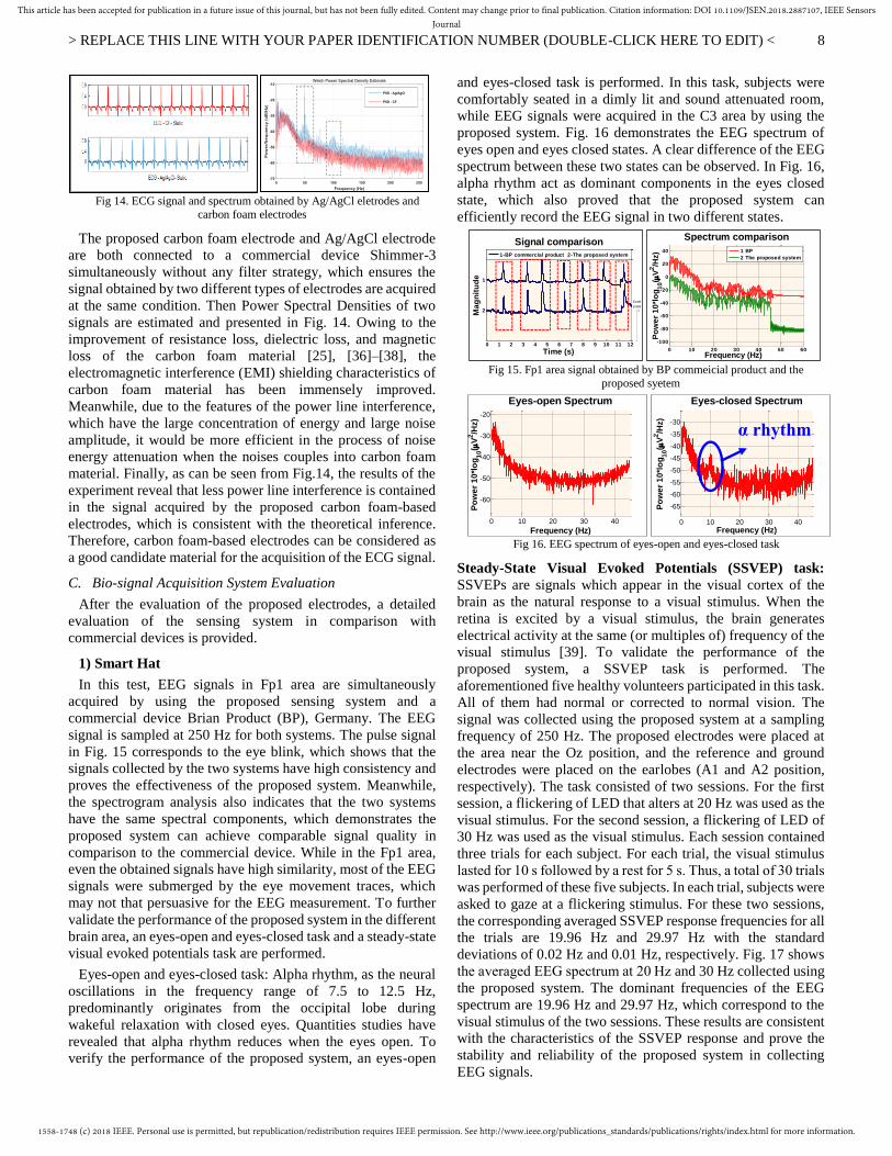
Fig 14. ECG signal and spectrum obtained by Ag/AgCl eletrodes and
carbon foam electrodes
The proposed carbon foam electrode and Ag/AgCl electrode
are both connected to a commercial device Shimmer-3
simultaneously without any filter strategy, which ensures the
signal obtained by two different types of electrodes are acquired
at the same condition. Then Power Spectral Densities of two
signals are estimated and presented in Fig. 14. Owing to the
improvement of resistance loss, dielectric loss, and magnetic
loss of the carbon foam material [25], [36]–[38], the
electromagnetic interference (EMI) shielding characteristics of
carbon foam material has been immensely improved.
Meanwhile, due to the features of the power line interference,
which have the large concentration of energy and large noise
amplitude, it would be more efficient in the process of noise
energy attenuation when the noises couples into carbon foam
material. Finally, as can be seen from Fig.14, the results of the
experiment reveal that less power line interference is contained
in the signal acquired by the proposed carbon foam-based
electrodes, which is consistent with the theoretical inference.
Therefore, carbon foam-based electrodes can be considered as
a good candidate material for the acquisition of the ECG signal.
C. Bio-signal Acquisition System Evaluation
After the evaluation of the proposed electrodes, a detailed
evaluation of the sensing system in comparison with
commercial devices is provided.
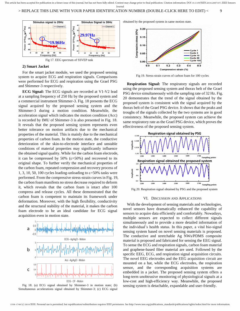
1) Smart Hat
In this test, EEG signals in Fp1 area are simultaneously
acquired by using the proposed sensing system and a
commercial device Brian Product (BP), Germany. The EEG
signal is sampled at 250 Hz for both systems. The pulse signal
in Fig. 15 corresponds to the eye blink, which shows that the
signals collected by the two systems have high consistency and
proves the effectiveness of the proposed system. Meanwhile,
the spectrogram analysis also indicates that the two systems
have the same spectral components, which demonstrates the
proposed system can achieve comparable signal quality in
comparison to the commercial device. While in the Fp1 area,
even the obtained signals have high similarity, most of the EEG
signals were submerged by the eye movement traces, which
may not that persuasive for the EEG measurement. To further
validate the performance of the proposed system in the different
brain area, an eyes-open and eyes-closed task and a steady-state
visual evoked potentials task are performed.
Eyes-open and eyes-closed task: Alpha rhythm, as the neural
oscillations in the frequency range of 7.5 to 12.5 Hz,
predominantly originates from the occipital lobe during
wakeful relaxation with closed eyes. Quantities studies have
revealed that alpha rhythm reduces when the eyes open. To
verify the performance of the proposed system, an eyes-open
and eyes-closed task is performed. In this task, subjects were
comfortably seated in a dimly lit and sound attenuated room,
while EEG signals were acquired in the C3 area by using the
proposed system. Fig. 16 demonstrates the EEG spectrum of
eyes open and eyes closed states. A clear difference of the EEG
spectrum between these two states can be observed. In Fig. 16,
alpha rhythm act as dominant components in the eyes closed
state, which also proved that the proposed system can
efficiently record the EEG signal in two different states.
Fig 15. Fp1 area signal obtained by BP commeicial product and the
proposed syetem
Fig 16. EEG spectrum of eyes-open and eyes-closed task
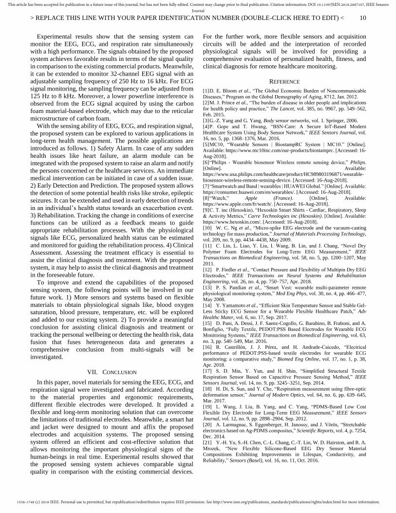
Steady-State Visual Evoked Potentials (SSVEP) task:
SSVEPs are signals which appear in the visual cortex of the
brain as the natural response to a visual stimulus. When the
retina is excited by a visual stimulus, the brain generates
electrical activity at the same (or multiples of) frequency of the
visual stimulus [39]. To validate the performance of the
proposed system, a SSVEP task is performed. The
aforementioned five healthy volunteers participated in this task.
All of them had normal or corrected to normal vision. The
signal was collected using the proposed system at a sampling
frequency of 250 Hz. The proposed electrodes were placed at
the area near the Oz position, and the reference and ground
electrodes were placed on the earlobes (A1 and A2 position,
respectively). The task consisted of two sessions. For the first
session, a flickering of LED that alters at 20 Hz was used as the
visual stimulus. For the second session, a flickering of LED of
30 Hz was used as the visual stimulus. Each session contained
three trials for each subject. For each trial, the visual stimulus
lasted for 10 s followed by a rest for 5 s. Thus, a total of 30 trials
was performed of these five subjects. In each trial, subjects were
asked to gaze at a flickering stimulus. For these two sessions,
the corresponding averaged SSVEP response frequencies for all
the trials are 19.96 Hz and 29.97 Hz with the standard
deviations of 0.02 Hz and 0.01 Hz, respectively. Fig. 17 shows
the averaged EEG spectrum at 20 Hz and 30 Hz collected using
the proposed system. The dominant frequencies of the EEG
spectrum are 19.96 Hz and 29.97 Hz, which correspond to the
visual stimulus of the two sessions. These results are consistent
with the characteristics of the SSVEP response and prove the
stability and reliability of the proposed system in collecting
EEG signals.
8.858
+-
Scale
0 1 2 3 4 5 6 7 8 9 10 11 12
2
1
Time (s)
Ma
gn
itu
de
Signal comparison
1-BP commercial product 2-The proposed system
0 10 20 30 40 50 60
-100
-80
-60
-40
-20
0
20
40
Frequency (Hz)
Po
we
r 1
0*l
og
10(
V2/H
z)
Spectrum comparison
1 BP
2 The proposed system
Frequency (Hz)
Po
we
r 1
0*l
og
10(
V2/H
z)
Eyes-open Spectrum
0 10 20 30 40
-60
-50
-40
-30
-20
0 10 20 30 40
-65
-60
-55
-50
-45
-40
-35
-30
Frequency (Hz)
Po
we
r 1
0*l
og
10(
V2/H
z)
Eyes-closed Spectrum
α rhythm
1558-1748 (c) 2018 IEEE. Personal use is permitted, but republication/redistribution requires IEEE permission. See http://www.ieee.org/publications_standards/publications/rights/index.html for more information.
This article has been accepted for publication in a future issue of this journal, but has not been fully edited. Content may change prior to final publication. Citation information: DOI 10.1109/JSEN.2018.2887107, IEEE SensorsJournal
> REPLACE THIS LINE WITH YOUR PAPER IDENTIFICATION NUMBER (DOUBLE-CLICK HERE TO EDIT) <
9
Fig 17. EEG spectrum of SSVEP task
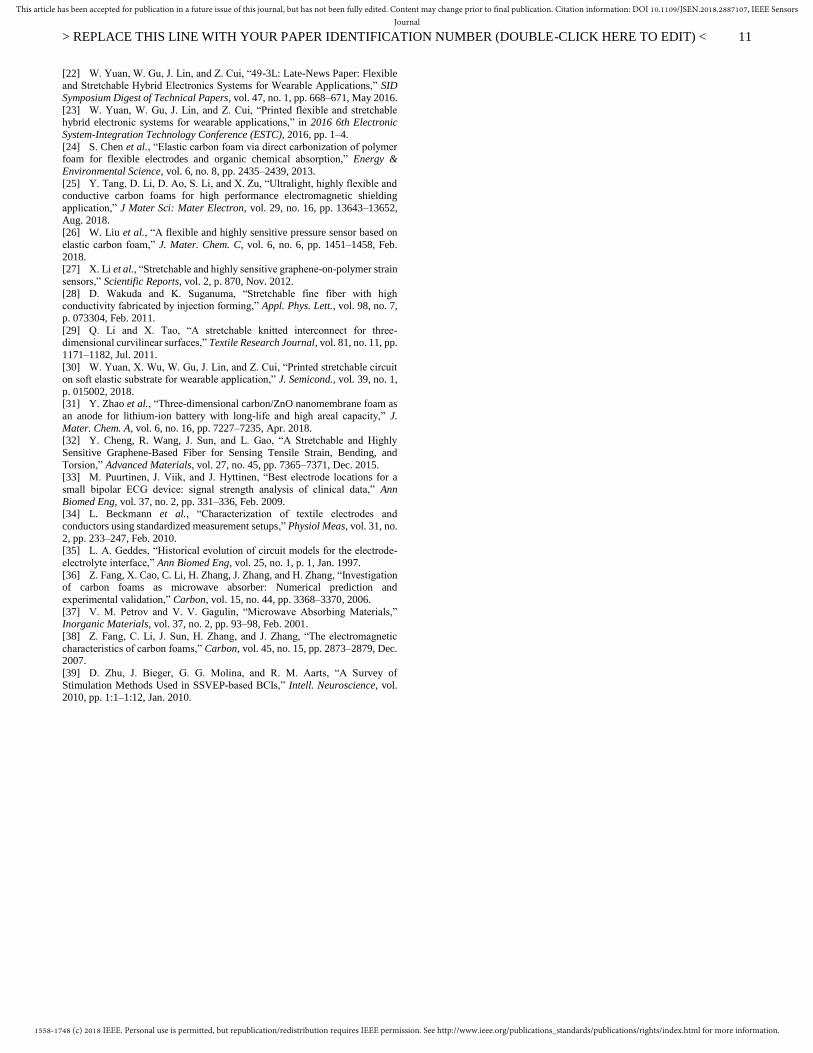
2) Smart Jacket
For the smart jacket module, we used the proposed sensing
system to acquire ECG and respiration signals. Comparisons
were performed for ECG and respiration using the Grael PSG
and Shimmer-3 respectively.
ECG Signal: The ECG signals are recorded at V1-V2 lead
at a sampling frequency of 250 Hz by the proposed system and
a commercial instrument Shimmer-3. Fig. 18 presents the ECG
signal acquired by the proposed sensing system and the
Shimmer-3 during a motion condition. Meanwhile, the
acceleration signal which indicates the motion condition (Acc)
is recorded by IMU of Shimmer-3 is also presented in Fig. 18.
It reveals that the proposed sensing system represents even
better tolerance on motion artifacts due to the mechanical
properties of the material. This is mainly due to the mechanical
properties of carbon foam. In the motion state, the conductive
deterioration of the skin-to-electrode interface and unstable
conditions of material properties may significantly influence
the obtained signal quality. While for the carbon foam electrode,
it can be compressed by 50% (=50%) and recovered to its
original shape. To further verify the mechanical properties of
the carbon foam, repeated compression and recovery process of
1, 3, 10, 50, 100 cycles loading-unloading to =50% tasks were
performed. From the compressive stress-strain curves in Fig. 19,
the carbon foam manifests no stress decrease required to deform
it, which reveals that the carbon foam is intact after 100
compress and release cycles. All these demonstrated that the
carbon foam is competent to maintain its framework under
deformation. Moreover, with the high flexibility, conductivity
and the structural stability of the material, it makes the carbon
foam electrode to be an ideal candidate for ECG signal
acquisition even in motion state.
Fig 18. (a) ECG signal obtained by Shimmer-3 in motion state; (b)
Simultaneous accelerations signal obtained by Shimmer-3; (c) ECG signal
obtained by the proposed system in same motion state.
Fig 19. Stress-strain curves of carbon foam for 100 cycles
Respiration Signal: The respiratory signals are recorded
using the proposed sensing system and thorax belt of the Grael
PSG device simultaneously with the sampling rate of 32 Hz. Fig.
20 demonstrates that the trend of the signal obtained by the
proposed system is consistent with the signal acquired by the
thorax belt of the Grael PSG device. It shows that the peaks and
troughs of the signals collected by the two systems are in good
consistency. Meanwhile, the proposed system can achieve the
same respiratory rate as the Grael PSG device, which proves the
effectiveness of the proposed sensing system.
Fig 20. Respiration signal obtained by PSG and the proposed system
VI. DISCUSSION AND APPLICATIONS
With the development of sensing materials and technologies,
novel sensors have dramatically enhanced the capability of
sensors to acquire data efficiently and comfortably. Nowadays,
multiple sensors are expected to collect different signals
simultaneously and to provide a more detailed information of
the individual’s health status. In this paper, a vital bio-signal
sensing system based on novel sensing materials is proposed.
The conductive and stretchable Ag NWs/PDMS composite
material is proposed and fabricated for sensing the EEG signal.
To sense the ECG and respiration signals, carbon foam material
and graphene-based fiber material are used. Followed by the
specific EEG, ECG, and respiration signal acquisition circuits.
The novel EEG electrodes and the EEG acquisition circuit are
mounted on a hat, while the ECG electrodes, the respiration
sensor, and the corresponding acquisition systems are
embedded in a jacket. The proposed sensing system offers a
long-term unobtrusive monitoring of physiological signals at a
low-cost and high-efficiency way. Meanwhile, the proposed
sensing system is detachable, expandable and user-friendly.
0 10 20 30 40 50
-60
-40
-20
0
20
Frequency (Hz)
Po
we
r 1
0*l
og
10(
V2/H
z)
Stimulus signal is 20Hz
X: 19.96
Y: -17.08
Channel 1
19.96Hz
0 10 20 30 40 50
-60
-40
-20
0
20
40
Frequency (Hz)
Po
we
r 1
0*l
og
10(
V2/H
z)
Stimulus Signal is 30Hz
X: 29.97
Y: -16.92
Channel 1
29.97Hz
a
b
c
Respiration signal obtained the proposed system
Am
pli
tud
eA
mp
litu
de
Sampling points
Sampling points
1200 1300 1400 1500 1600 1700
1200 1300 1400 1500 1600 1700
-1.16
-1.154
-1.148
-1.144
4.4
4.6
4.8
5.0
Respiration signal obtained by PSG
1558-1748 (c) 2018 IEEE. Personal use is permitted, but republication/redistribution requires IEEE permission. See http://www.ieee.org/publications_standards/publications/rights/index.html for more information.
This article has been accepted for publication in a future issue of this journal, but has not been fully edited. Content may change prior to final publication. Citation information: DOI 10.1109/JSEN.2018.2887107, IEEE SensorsJournal
> REPLACE THIS LINE WITH YOUR PAPER IDENTIFICATION NUMBER (DOUBLE-CLICK HERE TO EDIT) <
10
Experimental results show that the sensing system can
monitor the EEG, ECG, and respiration rate simultaneously
with a high performance. The signals obtained by the proposed
system achieves favorable results in terms of the signal quality
in comparison to the existing commercial products. Meanwhile,
it can be extended to monitor 32-channel EEG signal with an
adjustable sampling frequency of 250 Hz to 16 kHz. For ECG
signal monitoring, the sampling frequency can be adjusted from
125 Hz to 8 kHz. Moreover, a lower powerline interference is
observed from the ECG signal acquired by using the carbon
foam material-based electrode, which may due to the reticular
microstructure of carbon foam.
With the sensing ability of EEG, ECG, and respiration signal,
the proposed system can be explored to various applications in
long-term health management. The possible applications are
introduced as follows. 1) Safety Alarm. In case of any sudden
health issues like heart failure, an alarm module can be
integrated with the proposed system to raise an alarm and notify
the persons concerned or the healthcare services. An immediate
medical intervention can be initiated in case of a sudden issue.
2) Early Detection and Prediction. The proposed system allows
the detection of some potential health risks like stroke, epileptic
seizures. It can be extended and used in early detection of trends
in an individual’s health status towards an exacerbation event.
3) Rehabilitation. Tracking the change in conditions of exercise
functions can be utilized as a feedback means to guide
appropriate rehabilitation processes. With the physiological
signals like ECG, personalized health status can be estimated
and monitored for guiding the rehabilitation process. 4) Clinical
Assessment. Assessing the treatment efficacy is essential to
assist the clinical diagnosis and treatment. With the proposed
system, it may help to assist the clinical diagnosis and treatment
in the foreseeable future.
To improve and extend the capabilities of the proposed
sensing system, the following points will be involved in our
future work. 1) More sensors and systems based on flexible
materials to obtain physiological signals like, blood oxygen
saturation, blood pressure, temperature, etc. will be explored
and added to our existing system. 2) To provide a meaningful
conclusion for assisting clinical diagnosis and treatment or
tracking the personal wellbeing or detecting the health risk, data
fusion that fuses heterogeneous data and generates a
comprehensive conclusion from multi-signals will be
investigated.
VII. CONCLUSION
In this paper, novel materials for sensing the EEG, ECG, and
respiration signal were investigated and fabricated. According
to the material properties and ergonomic requirements,
different flexible electrodes were developed. It provided a
flexible and long-term monitoring solution that can overcome
the limitations of traditional electrodes. Meanwhile, a smart hat
and jacket were designed to mount and affix the proposed
electrodes and acquisition systems. The proposed sensing
system offered an efficient and cost-effective solution that
allows monitoring the important physiological signs of the
human-beings in real time. Experimental results showed that
the proposed sensing system achieves comparable signal
quality in comparison with the existing commercial devices.
For the further work, more flexible sensors and acquisition
circuits will be added and the interpretation of recorded
physiological signals will be involved for providing a
comprehensive evaluation of personalized health, fitness, and
clinical diagnosis for remote healthcare monitoring.
REFERENCE
[1] D. E. Bloom et al., “The Global Economic Burden of Noncommunicable
Diseases,” Program on the Global Demography of Aging, 8712, Jan. 2012. [2] M. J. Prince et al., “The burden of disease in older people and implications
for health policy and practice,” The Lancet, vol. 385, no. 9967, pp. 549–562,
Feb. 2015. [3] G.-Z. Yang and G. Yang, Body sensor networks, vol. 1. Springer, 2006.
[4] P. Gope and T. Hwang, “BSN-Care: A Secure IoT-Based Modern
Healthcare System Using Body Sensor Network,” IEEE Sensors Journal, vol. 16, no. 5, pp. 1368–1376, Mar. 2016.
[5] MC10, “Wearable Sensors | BiostampRC System | MC10.” [Online].
Available: https://www.mc10inc.com/our-products/biostamprc. [Accessed: 16-Aug-2018].
[6] “Philips - Wearable biosensor Wireless remote sensing device,” Philips.
[Online]. Available: https://www.usa.philips.com/healthcare/product/HC989803196871/wearable-
biosensor-wireless-remote-sensing-device. [Accessed: 16-Aug-2018].
[7] “Smartwatch and Band | wearables | HUAWEI Global.” [Online]. Available: https://consumer.huawei.com/en/wearables/. [Accessed: 16-Aug-2018].
[8] “Watch,” Apple (France). [Online]. Available: https://www.apple.com/fr/watch/. [Accessed: 16-Aug-2018].
[9] C. T. inc (Hexoskin), “Hexoskin Smart Shirts - Cardiac, Respiratory, Sleep
& Activity Metrics,” Carre Technologies inc (Hexoskin). [Online]. Available: https://www.hexoskin.com/. [Accessed: 16-Aug-2018].
[10] W. C. Ng et al., “Micro-spike EEG electrode and the vacuum-casting
technology for mass production,” Journal of Materials Processing Technology, vol. 209, no. 9, pp. 4434–4438, May 2009.
[11] C. Lin, L. Liao, Y. Liu, I. Wang, B. Lin, and J. Chang, “Novel Dry
Polymer Foam Electrodes for Long-Term EEG Measurement,” IEEE Transactions on Biomedical Engineering, vol. 58, no. 5, pp. 1200–1207, May
2011.
[12] P. Fiedler et al., “Contact Pressure and Flexibility of Multipin Dry EEG Electrodes,” IEEE Transactions on Neural Systems and Rehabilitation
Engineering, vol. 26, no. 4, pp. 750–757, Apr. 2018.
[13] P. S. Pandian et al., “Smart Vest: wearable multi-parameter remote physiological monitoring system,” Med Eng Phys, vol. 30, no. 4, pp. 466–477,
May 2008.
[14] Y. Yamamoto et al., “Efficient Skin Temperature Sensor and Stable Gel-Less Sticky ECG Sensor for a Wearable Flexible Healthcare Patch,” Adv
Healthc Mater, vol. 6, no. 17, Sep. 2017.
[15] D. Pani, A. Dessì, J. F. Saenz-Cogollo, G. Barabino, B. Fraboni, and A. Bonfiglio, “Fully Textile, PEDOT:PSS Based Electrodes for Wearable ECG
Monitoring Systems,” IEEE Transactions on Biomedical Engineering, vol. 63,
no. 3, pp. 540–549, Mar. 2016. [16] R. Castrillón, J. J. Pérez, and H. Andrade-Caicedo, “Electrical
performance of PEDOT:PSS-based textile electrodes for wearable ECG
monitoring: a comparative study,” Biomed Eng Online, vol. 17, no. 1, p. 38, Apr. 2018.
[17] S. D. Min, Y. Yun, and H. Shin, “Simplified Structural Textile
Respiration Sensor Based on Capacitive Pressure Sensing Method,” IEEE
Sensors Journal, vol. 14, no. 9, pp. 3245–3251, Sep. 2014.
[18] H. Di, S. Sun, and Y. Che, “Respiration measurement using fibre-optic
deformation sensor,” Journal of Modern Optics, vol. 64, no. 6, pp. 639–645, Mar. 2017.
[19] L. Wang, J. Liu, B. Yang, and C. Yang, “PDMS-Based Low Cost
Flexible Dry Electrode for Long-Term EEG Measurement,” IEEE Sensors Journal, vol. 12, no. 9, pp. 2898–2904, Sep. 2012.
[20] A. Larmagnac, S. Eggenberger, H. Janossy, and J. Vörös, “Stretchable
electronics based on Ag-PDMS composites,” Scientific Reports, vol. 4, p. 7254, Dec. 2014.
[21] Y.-H. Yu, S.-H. Chen, C.-L. Chang, C.-T. Lin, W. D. Hairston, and R. A.
Mrozek, “New Flexible Silicone-Based EEG Dry Sensor Material Compositions Exhibiting Improvements in Lifespan, Conductivity, and
Reliability,” Sensors (Basel), vol. 16, no. 11, Oct. 2016.
1558-1748 (c) 2018 IEEE. Personal use is permitted, but republication/redistribution requires IEEE permission. See http://www.ieee.org/publications_standards/publications/rights/index.html for more information.
This article has been accepted for publication in a future issue of this journal, but has not been fully edited. Content may change prior to final publication. Citation information: DOI 10.1109/JSEN.2018.2887107, IEEE SensorsJournal
> REPLACE THIS LINE WITH YOUR PAPER IDENTIFICATION NUMBER (DOUBLE-CLICK HERE TO EDIT) <
11
[22] W. Yuan, W. Gu, J. Lin, and Z. Cui, “49-3L: Late-News Paper: Flexible and Stretchable Hybrid Electronics Systems for Wearable Applications,” SID
Symposium Digest of Technical Papers, vol. 47, no. 1, pp. 668–671, May 2016.
[23] W. Yuan, W. Gu, J. Lin, and Z. Cui, “Printed flexible and stretchable hybrid electronic systems for wearable applications,” in 2016 6th Electronic
System-Integration Technology Conference (ESTC), 2016, pp. 1–4.
[24] S. Chen et al., “Elastic carbon foam via direct carbonization of polymer foam for flexible electrodes and organic chemical absorption,” Energy &
Environmental Science, vol. 6, no. 8, pp. 2435–2439, 2013.
[25] Y. Tang, D. Li, D. Ao, S. Li, and X. Zu, “Ultralight, highly flexible and conductive carbon foams for high performance electromagnetic shielding
application,” J Mater Sci: Mater Electron, vol. 29, no. 16, pp. 13643–13652,
Aug. 2018. [26] W. Liu et al., “A flexible and highly sensitive pressure sensor based on
elastic carbon foam,” J. Mater. Chem. C, vol. 6, no. 6, pp. 1451–1458, Feb.
2018. [27] X. Li et al., “Stretchable and highly sensitive graphene-on-polymer strain
sensors,” Scientific Reports, vol. 2, p. 870, Nov. 2012.
[28] D. Wakuda and K. Suganuma, “Stretchable fine fiber with high conductivity fabricated by injection forming,” Appl. Phys. Lett., vol. 98, no. 7,
p. 073304, Feb. 2011.
[29] Q. Li and X. Tao, “A stretchable knitted interconnect for three-dimensional curvilinear surfaces,” Textile Research Journal, vol. 81, no. 11, pp.
1171–1182, Jul. 2011.
[30] W. Yuan, X. Wu, W. Gu, J. Lin, and Z. Cui, “Printed stretchable circuit on soft elastic substrate for wearable application,” J. Semicond., vol. 39, no. 1,
p. 015002, 2018. [31] Y. Zhao et al., “Three-dimensional carbon/ZnO nanomembrane foam as
an anode for lithium-ion battery with long-life and high areal capacity,” J.
Mater. Chem. A, vol. 6, no. 16, pp. 7227–7235, Apr. 2018. [32] Y. Cheng, R. Wang, J. Sun, and L. Gao, “A Stretchable and Highly
Sensitive Graphene-Based Fiber for Sensing Tensile Strain, Bending, and
Torsion,” Advanced Materials, vol. 27, no. 45, pp. 7365–7371, Dec. 2015. [33] M. Puurtinen, J. Viik, and J. Hyttinen, “Best electrode locations for a
small bipolar ECG device: signal strength analysis of clinical data,” Ann
Biomed Eng, vol. 37, no. 2, pp. 331–336, Feb. 2009. [34] L. Beckmann et al., “Characterization of textile electrodes and
conductors using standardized measurement setups,” Physiol Meas, vol. 31, no.
2, pp. 233–247, Feb. 2010. [35] L. A. Geddes, “Historical evolution of circuit models for the electrode-
electrolyte interface,” Ann Biomed Eng, vol. 25, no. 1, p. 1, Jan. 1997.
[36] Z. Fang, X. Cao, C. Li, H. Zhang, J. Zhang, and H. Zhang, “Investigation of carbon foams as microwave absorber: Numerical prediction and
experimental validation,” Carbon, vol. 15, no. 44, pp. 3368–3370, 2006.
[37] V. M. Petrov and V. V. Gagulin, “Microwave Absorbing Materials,” Inorganic Materials, vol. 37, no. 2, pp. 93–98, Feb. 2001.
[38] Z. Fang, C. Li, J. Sun, H. Zhang, and J. Zhang, “The electromagnetic
characteristics of carbon foams,” Carbon, vol. 45, no. 15, pp. 2873–2879, Dec. 2007.
[39] D. Zhu, J. Bieger, G. G. Molina, and R. M. Aarts, “A Survey of
Stimulation Methods Used in SSVEP-based BCIs,” Intell. Neuroscience, vol. 2010, pp. 1:1–1:12, Jan. 2010.
Recommended