Bulgarian Chemical Communications, Volume 52, Issue 4 (pp. 488-492) 2020 DOI: 10.34049/bcc.52.4.5286
488
New neurotensin analogue with improving effect on some affective symptoms in
Parkinson’s disease model in rats
A. Popatanasov1*, S. Abarova2, L. P. Tancheva1, T. Pajpanova3
1Institute of Neurobiology, Bulgarian Academy of Sciences, Acad. G. Bonchev Str., Bl. 23, Sofia, Bulgaria 2 Department of Medical Physics and Biophysics, Medical University, 2 Zdrave Str., Sofia, Bulgaria
3Institute of Molecular Biology, Bulgarian Academy of Sciences, Acad. G. Bonchev Str., Bl. 21, Sofia, Bulgaria
Received: June 02, 2020; Accepted: June 21, 2020
Neurotensin (NT) is a small neuropeptide acting as neurotransmitter and neuromodulator in the nervous system.
However, natural neurotensin is rapidly degraded in the body, therefore artificial analogues are needed to prolong its
bioavailability and effects. Parkinson’s disease (PD) is a neurological disease with specific motor, cognitive and
affective disturbances and is associated with high oxidative stress and mitochondrial dysfunction and degeneration of
the dopaminergic system. The scope of the study was to evaluate some effects of a new neurotensin analogue (NT2)
upon the behavior of rats with a model of PD induced with striatal injection of the neurotoxin 6-hydroxydopamine (6-
OHDA). The PD model was produced via striatal 6-OHDA (12 µg in 2 µl saline) injection of male Wistar rats. NT2
treatment was with an effective daily dose of 5 mg/kg i.p. for 5 days. NT2 effects were evaluated via behavioral tests
for locomotor activity and anxiety. Student’s t-test was used at p<0.05. The PD model was verified by rotarod tests on
the 2nd and 3rd week after the operation and was compared to sham operated animals. There was a significant
performance decrease in the mood and affective disturbances. The affective disturbances as anxiety in NT2-treated
animals were reduced (both on the 2nd and 3rd week) compared to PD-controls.
Key words: neurotensin, affective disorder, anxiety, Parkinson disease.
INTRODUCTION
Parkinson’s disease (PD) is one of the most
common neurodegenerative diseases with specific
motor, cognitive and affective disturbances and is
associated with high oxidative stress and
mitochondrial dysfunction and degeneration of the
dopaminergic system. Among the non-motor
symptoms, some of the most common ones are
depression and anxiety [1]. They may seriously
impact the wellbeing and performance of the
patients and are an additional source of stress for
the care givers [2]. However, the current therapies
are mostly focused to combat the motor symptoms,
while the non-motor ones often remain neglected
[3].
One of the first discovered connections between
the receptors of the different neurotransmitters and
neuromodulators was the one between the DA-
receptors and neurotensin (NT) receptors. Such
close connection suggests that NT is associated
with PD [4]. Additionally, several years after these
findings the researchers gradually started to
discover that NT can be related to some affective
symptoms in the psychiatric disorders [5], which
further stimulated such research and the quest for
NT-like agents.
Neurotensin is a tridecapeptide acting as
neurotransmitter and neuromodulator in the nervous
system. It is secreted in both the central nervous
system and the gut.
NT exercises its biologic effect from the specific
interaction of the peptide with three different cell-
surface receptors referred to as NTS1, NTS2 and
NTS3/sortilin [6]. However, natural neurotensin is
rapidly degraded in the body, therefore artificial
analogues are needed to prolong the bioavailability
and effects. Therefore, the object of this study is a
promising long-lasting NT analogue with code NT2
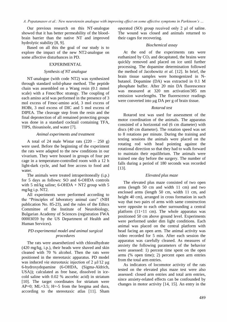
(Fig. 1) synthesized by Pajpanova et al. [7].
Fig. 1. The amino acid sequence of NT and NT2-
analogue.
* To whom all correspondence should be sent:
E-mail: [email protected] 2020 Bulgarian Academy of Sciences, Union of Chemists in Bulgaria
A. Popatanasov et al.: New neurotensin analogue with improving effect on some affective symptoms in Parkinson’s …
489
Our previous research on this NT-analogue
showed that it has better permeability of the blood-
brain barrier than the native NT and improved
hydrolytic stability [8, 9].
Based on all this the goal of our study is to
explore the impact of the new NT2-analogue on
some affective disturbances in PD.
EXPERIMENTAL
Synthesis of NT analogue
NT-analogue (with code NT2) was synthesized
through standard solid-phase method. The peptide
chain was assembled on a Wang resin (0.1 mmol
scale) with a Fmoc/Boc strategy. The coupling of
each amino acid was performed in the presence of 3
mol excess of Fmoc-amino acid, 3 mol excess of
HOBt, 3 mol excess of DIC and 5 mol excess of
DIPEA. The cleavage step from the resin and the
final deprotection of all remained protecting groups
was done in a standard cocktail containing TFA,
TIPS, thioanisole, and water [7].
Animal experiments and treatment
A total of 24 male Wistar rats (220 – 250 g)
were used. Before the beginning of the experiment
the rats were adapted to the new conditions in our
vivarium. They were housed in groups of four per
cage in a temperature-controlled room with a 12 h
light-dark cycle, and had free access to food and
water.
The animals were treated intraperitoneally (i.p.)
for 5 days as follows: SO and 6-OHDA controls
with 5 ml/kg saline; 6-OHDA + NT2 group with 5
mg/kg i.p. NT2.
All experiments were performed according to
the “Principles of laboratory animal care” (NIH
publication No. 85-23), and the rules of the Ethics
Committee of the Institute of Neurobiology,
Bulgarian Academy of Sciences (registration FWA
00003059 by the US Department of Health and
Human Services).
PD experimental model and animal surgical
procedures
The rats were anaesthetized with chloralhydrate
(420 mg/kg, i.p.), their heads were shaved and skin
cleaned with 70 % alcohol. Then the rats were
positioned in the stereotaxic apparatus. PD model
was induced via stereotaxic injection of 2 µl/12 µg
6-hydroxydopamine (6-OHDA, (Sigma-Aldrich,
USA)); calculated as free base, dissolved in ice-
cold saline with 0.02 % ascorbic acid) in striatum
[10]. The target coordinates for striatum were
AP=0; ML=3.5; H=-5 from the bregma and dura,
according to the stereotaxic atlas [11]. Sham
operated (SO) group received only 2 µl of saline.
The wound was closed and animals returned to
their cages for recovering.
Biochemical assay
At the end of the experiments rats were
euthanized by CO2 and decapitated, the brains were
quickly removed and placed on ice until further
processing. The dopamine determination followed
the method of Jacobowitz et al. [12]. In brief, the
brain tissue samples were homogenized in N-
butanol. Dopamine (DA) was extracted in 0.1 M
phosphate buffer. After 20 min DA fluorescence
was measured at 320 nm activation/385 nm
emission wavelengths. The fluorescence readings
were converted into µg DA per g of brain tissue.
Rotarod test
Rotarod test was used for assessment of the
motor coordination of the animals. The apparatus
consisted of a horizontal rod (6 cm diameter) with
discs (40 cm diameter). The rotation speed was set
to 8 rotations per minute. During the training and
testing sessions the animals were placed on the
rotating rod with head pointing against the
rotational direction so that they had to walk forward
to maintain their equilibrium. The animals were
trained one day before the surgery. The number of
falls during a period of 180 seconds was recorded
[13].
Elevated plus maze
The elevated plus maze consisted of two open
arms (length 50 cm and width 11 cm) and two
enclosed arms (length 50 cm, width 11 cm, and
height 40 cm), arranged in cross formation in such
way that two pairs of arms with same construction
were opposite to each other surrounding a central
platform (11×11 cm). The whole apparatus was
positioned 50 cm above ground level. Experiments
were performed under dim light conditions. Each
animal was placed on the central platform with
head facing an open arm. The animal activity was
video recorded for 5 min. After each session the
apparatus was carefully cleaned. As measures of
anxiety the following parameters of the behavior
were assessed: 1) percent time spent on the open
arms (% open time); 2) percent open arm entries
from the total arm entries.
As indicators of locomotor activity of the rats
tested on the elevated plus maze test were also
assessed: closed arm entries and total arm entries,
since anxiety-related effects can be confounded by
changes in motor activity [14, 15]. An entry in the
A. Popatanasov et al.: New neurotensin analogue with improving effect on some affective symptoms in Parkinson’s …
arm was considered when the whole body and four
paws were placed on the arm.
Open field test
In order to determine the nonspecific motor
effects which may have flustered the assessments in
the elevated plus maze test, assessment of the
locomotor activity was performed in the same
animals following the elevated plus maze test
procedures. The assessment of the locomotor
activity was done in the same room as the elevated
plus maze test.
Additionally, the number of entries from the
outer zone into the central zone of the arena was
measured which was used to estimate the anti-
thigmotactic ratio as an indicator of increased or
decreased anxiety of the tested animals. This ratio
is calculated by the number of entries into the
central arena of the open field to total distance
travelled, multiplied by 1000. Initially the rats were
placed in the center of the open field arena [16].
The open field apparatus comprised a cylinder
(diameter 1 m, height 40 cm) on contrasting
background. The arena was divided in two zones –
central zone and outer zone equidistantly
surrounding it. The testing sessions lasted for 20
min and were video recorded for analysis.
Statistical and data analysis
Results were expressed as means ± SEM.
Experimental data were analyzed by Student’s t-
test. Differences were considered significant at p <
0.05.
The video materials from open field and
elevated plus maze tests were processed with the
specialized neurobehavioral and video analysis
software Noldus EthoVision [17]; raw data of the
spatiotemporal analysis were imported into
Microsoft Excel 2007 (Microsoft Corp., USA) for
further processing. The total distance traveled,
velocities and entrance and time spent at important
areas of the apparatus were calculated with custom-
built algorithms.
RESULTS
Dopamine levels
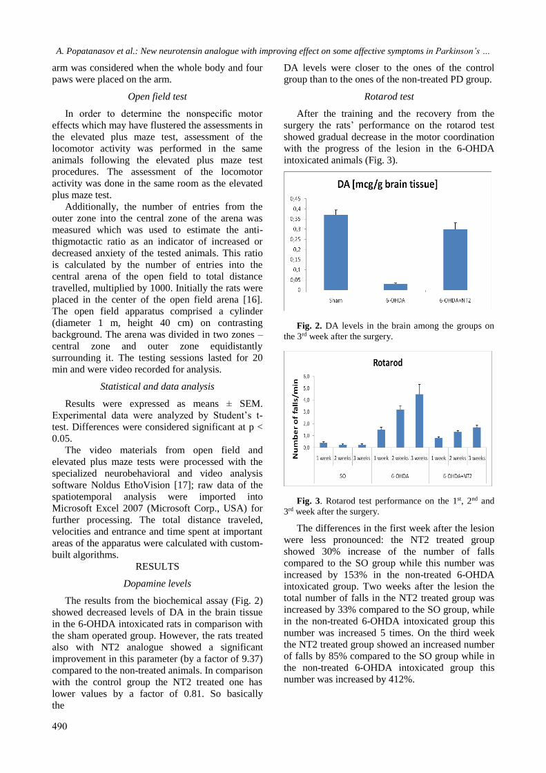
The results from the biochemical assay (Fig. 2)
showed decreased levels of DA in the brain tissue
in the 6-OHDA intoxicated rats in comparison with
the sham operated group. However, the rats treated
also with NT2 analogue showed a significant
improvement in this parameter (by a factor of 9.37)
compared to the non-treated animals. In comparison
with the control group the NT2 treated one has
lower values by a factor of 0.81. So basically
the
490
DA levels were closer to the ones of the control
group than to the ones of the non-treated PD group.
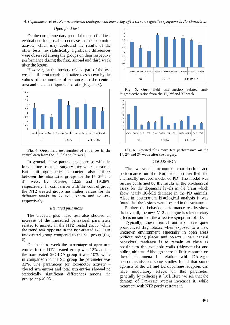
Rotarod test
After the training and the recovery from the
surgery the rats’ performance on the rotarod test
showed gradual decrease in the motor coordination
with the progress of the lesion in the 6-OHDA
intoxicated animals (Fig. 3).
Fig. 2. DA levels in the brain among the groups on
the 3rd week after the surgery.
Fig. 3. Rotarod test performance on the 1st, 2nd and
3rd week after the surgery.
The differences in the first week after the lesion
were less pronounced: the NT2 treated group
showed 30% increase of the number of falls
compared to the SO group while this number was
increased by 153% in the non-treated 6-OHDA
intoxicated group. Two weeks after the lesion the
total number of falls in the NT2 treated group was
increased by 33% compared to the SO group, while
in the non-treated 6-OHDA intoxicated group this
number was increased 5 times. On the third week
the NT2 treated group showed an increased number
of falls by 85% compared to the SO group while in
the non-treated 6-OHDA intoxicated group this
number was increased by 412%.
A. Popatanasov et al.: New neurotensin analogue with improving effect on some affective symptoms in Parkinson’s …
491
Open field test
On the complementary part of the open field test
evaluations for possible decrease in the locomotor
activity which may confound the results of the
other tests, no statistically significant differences
were observed among the groups on their respective
performance during the first, second and third week
after the lesion.
However, on the anxiety related part of the test
we see different trends and patterns as shown by the
values of the number of entrances in the central
area and the anti-thigmotactic ratio (Figs. 4, 5).
Fig. 4. Open field test number of entrances in the
central area from the 1st, 2nd and 3rd week.
In general, these parameters decrease with the
longer time from the surgery they were measured.
But anti-thigmotactic parameter also differs
between the intoxicated groups for the 1st, 2nd and
3rd week by 10.56%, 12.25 and 19.28%,
respectively. In comparison with the control group
the NT2 treated group has higher values for the
different weeks by 22.06%, 37.5% and 42.14%,
respectively.
Elevated plus maze
The elevated plus maze test also showed an
increase of the measured behavioral parameters
related to anxiety in the NT2 treated group, while
the trend was opposite in the non-treated 6-OHDA
intoxicated group compared to the SO group (Fig.
6).
On the third week the percentage of open arm
entries in the NT2 treated group was 12% and in
the non-treated 6-OHDA group it was 10%, while
in comparison to the SO group the parameter was
21%. The parameters for locomotor activity –
closed arm entries and total arm entries showed no
statistically significant differences among the
groups at p<0.05.
Fig. 5. Open field test anxiety related anti- thigmotactic ratios from the 1st, 2nd and 3rd week.
Fig. 6. Elevated plus maze test performance on the
1st, 2nd and 3rd week after the surgery.
DISCUSSION
The worsened locomotor coordination and
performance on the Rot-a-rod test verified the
chemically induced model of PD. The model was
further confirmed by the results of the biochemical
assay for the dopamine levels in the brain which
show nearly 10-fold decrease in the PD animals.
Also, in postmortem histological analysis it was
found that the lesions were located in the striatum.
Further, the behavior performance results show
that overall, the new NT2 analogue has beneficiary
effects on some of the affective symptoms of PD.
Typically, these fearful animals have quite
pronounced thigmotaxis when exposed to a new
unknown environment especially in open areas
without hiding places and objects. Their natural
behavioral tendency is to remain as close as
possible to the available walls (thigmotaxis) and
hiding objects. Although there is little research on
these phenomena in relation with DA-ergic
neurotransmission, some studies found that some
agonists of the D1 and D2 dopamine receptors can
have modulatory effects on this parameter,
generally by reducing it [18]. Here we see that the
damage of DA-ergic system increases it, while
treatment with NT2 partly restores it.
A. Popatanasov et al.: New neurotensin analogue with improving effect on some affective symptoms in Parkinson’s …
492
In general, the DA-ergic system is less
frequently accounted for as engaged in the anxiety
and the related disturbances but some studies
showed that through D1 and D2 dopamine
receptors in mesolimbic circuits the DA-ergic
system may also have a modulatory role in this
emotional behavior [19]. And here we observe a
similar pattern – destruction of a large part of the
dopaminergic system leads to affective disturbances
as shown in our tests.
The mesolimbic circuits of the dopaminergic
system also contain a relatively high level of co-
localized NT-receptors as some neuroanatomical
studies show [20]. In behavioral concordance with
this is the observed significant improvement in the
NT2 treated group performance in all the anxiety
tests, which can be related to stimulation of
compensatory mechanisms by the NT2 in this
pathway, such as the fact that NT elicits evoked DA
release in the striatum and prefrontal cortex [21]. A
clinic research by Ruiz et al. from 1992 [22]
showed that plasma levels of some neuropeptides
are significantly reduced in patients with depression
or anxiety disorder, which get restored after
recovery. With our study we go further by
confirming that there is a functional connection
between the NT associated part of the dopaminergic
system and some affective disturbances, since by
introducing the NT2 analogue into the plasma we
partially mitigate some of the affective symptoms
incurred by the massive selective neurotoxin
destruction of the dopaminergic system.
In our previous studies [9, 10] we showed the
beneficiary effects of NT2 on the behavior related
to the damaged DA-ergic circuits in PD in charge
for the motor and cognitive behavior. In this study
it was shown that NT2 impacts positively other
DA-ergic neural circuits related to the emotional
behavior.
CONCLUSION
The new NT-analogue is a promising agent for
the management of some of the non-motor
symptoms of PD as anxiety, and deserves further
exploration and development.
REFERENCES
1. R. F. Pfeiffer, Parkinsonism Relat. Disord., 22,
S119 (2016).
2. O. B. Leiknes, I. Tysnes, D. Aarsland, J. P. Larsen,
Acta Neurol. Scand., 122(6), 418 (2010).
3. A. Todorova, P. Jenner, K. R. Chaudhuri, Practical
Neurology, 14(5), 310 (2014).
4. F. St-Gelais, C. Jomphe, L. É. Trudeau, J.
Psychiatr. Neurosci., 31, 229 (2006).
5. R. Cáceda, B. Kinkead, C. B. Nemeroff, Peptides,
27(10), 2385 (2006).
6. J. P. Vincent, J. Mazella, P. Kitabgi, Trends
Pharmacol. Sci., 20, 302 (1999).
7. T. Dzimbova, S. Stoeva, L. Tancheva, A.
Georgieva, R. Kalfin, T. Pajpanova, in: Peptides
2014, (Proc. 33rd European Peptide Symp., Sofia,
2014), E. Naydenova, T. Pajpanova, D. Danalev
(eds.), European Peptide Society, Sofia, 2014, p.
260.
8. S. Michailova, T. Dzimbova, K. Kalíkova, E.
Tesařová, T. Pajpanova, Bulg. Chem. Commun.,
49(E), 113 (2017).
9. M., Lazarova, A., Popatanasov, R., Klissurov, S.,
Stoeva, T., Pajpanova, R., Kalfin, L. Tancheva. J.
Mol. Neurosci., 66(4), 552 (2018).
10. A. Popatanasov, S. Stoeva, M. Lazarova, L.
Traikov, T. Pajpanova, R. Kalfin, L. Tancheva.
Bulg. Chem. Commun., 49(E), 146 (2017).
11. G. Paxinos, C. Watson, The Rat Brain in Stereotaxic
Coordinates, Elsevier Acad. Press, San Diego, 2005.
12. D. M. Jacobowitz, J. S. Richardson, Pharmacol.
Biochem. Behav., 8(5), 515 (1978).
13. D. M. Pechlivanova, P. P. Markova, D. Popov, A.
G. Stoynev, Peptides, 39, 152 (2013).
14. R. D. Porsolt, G. Anton, N. Blavet, M. Jalfre, Eur. J.
Pharmacol., 47(4), 379 (1978).
15. G. R. Dawson, M. D. Tricklebank, Trends
Pharmacol. Sci., 16(2), 33 (1995).
16. A. Skórzewska, M. Lehner, A. Wisłowska-Stanek,
P. Krząścik, A. Ziemba, A. Płaźnik, Hormones and
Behavior, 65(1), 6 (2014).
17. P. Escudero, V. Hinz, A. de Polavieja, Nat.
Methods, 11(7),743 (2014).
18. P. Simon, R. Dupuis, J. Costentin, Behav. Brain
Res., 61(1), 59 (1994).
19. R. J. Rodgers, E. M. Nikulina, J. C. Cole,
Pharmacol. Biochem. Behav., 49(4), 985 (1994).
20. R. Quirion, C. C. Chiueh, H. D. Everist, A. Pert,
Brain Res., 327(1-2), 385 (1985).
21. E. Hetier, A. Boireau, P. Dubedat, J. C. Blanchard,
Naunyn Schmiedebergs Arch. Pharmacol., 337(1),
13 (1988).
22. J. P. Saiz, J. R. Carrasco, A. Hernanz, Archivos de
Neurobiologia, 55(1), 1 (1992).
Recommended