DR NILESH KATE
MBBS,MD
ASSOCIATE PROF
DEPT. OF PHYSIOLOGY
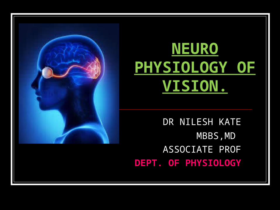
NEURO PHYSIOLOGY
OF VISION.
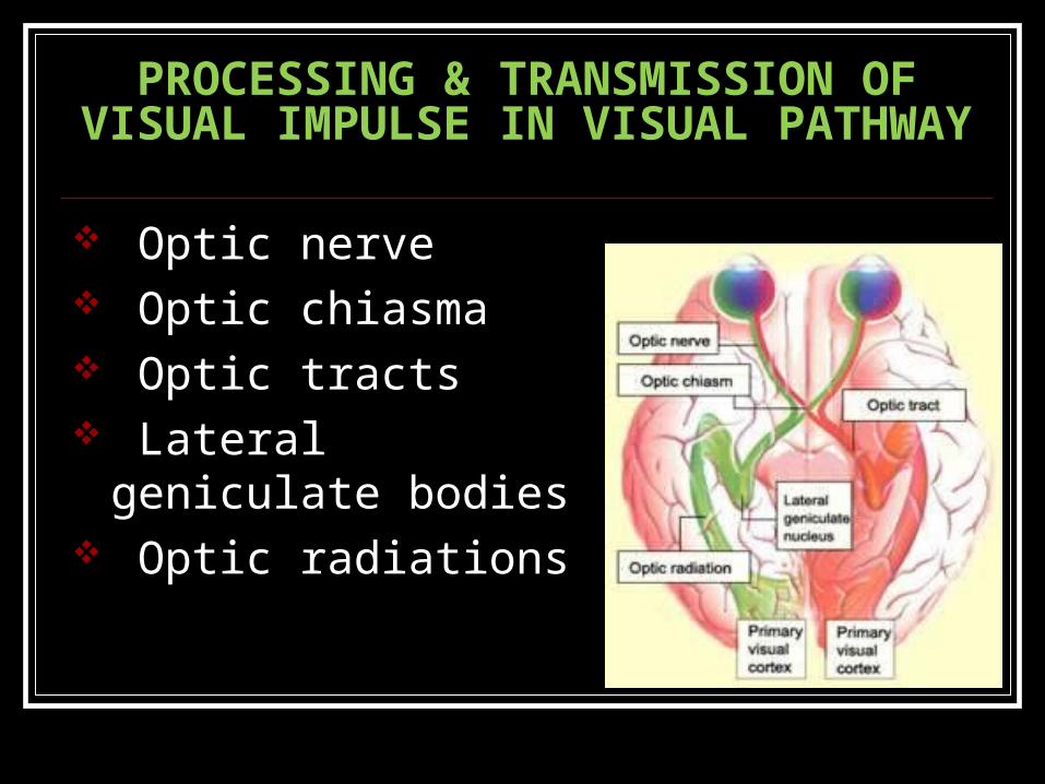
PROCESSING & TRANSMISSION OF VISUAL IMPULSE IN VISUAL PATHWAY
Optic nerve Optic chiasma Optic tracts Lateral geniculate bodies Optic radiations
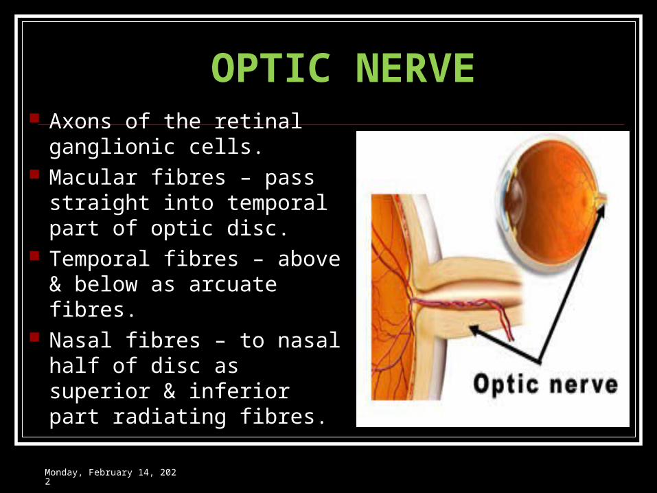
OPTIC NERVE Axons of the retinal
ganglionic cells. Macular fibres – pass
straight into temporal part of optic disc.
Temporal fibres – above & below as arcuate fibres.
Nasal fibres – to nasal half of disc as superior & inferior part radiating fibres.
Wednesday, May 3, 2023
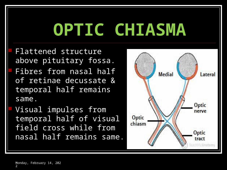
OPTIC CHIASMA Flattened structure above
pituitary fossa. Fibres from nasal half of
retinae decussate & temporal half remains same.
Visual impulses from temporal half of visual field cross while from nasal half remains same.
Wednesday, May 3, 2023
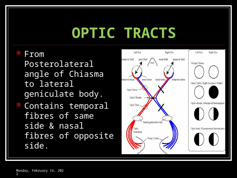
OPTIC TRACTS From Posterolateral
angle of Chiasma to lateral geniculate body.
Contains temporal fibres of same side & nasal fibres of opposite side.
Wednesday, May 3, 2023
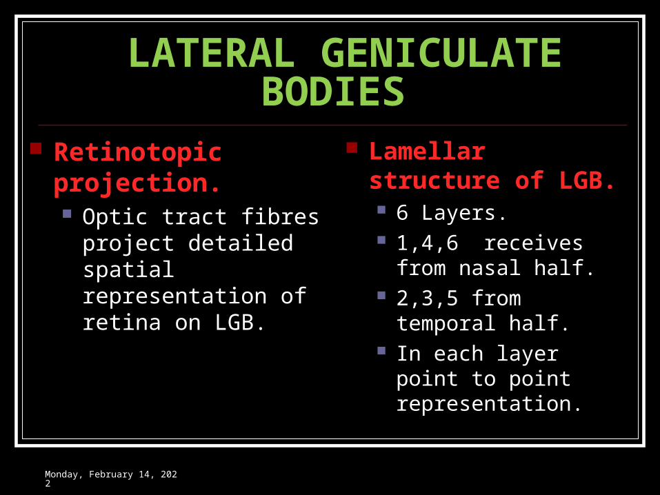
LATERAL GENICULATE BODIES
Retinotopic projection. Optic tract fibres project
detailed spatial representation of retina on LGB.
Lamellar structure of LGB. 6 Layers. 1,4,6 receives from
nasal half. 2,3,5 from temporal
half. In each layer point to
point representation.
Wednesday, May 3, 2023
LATERAL GENICULATE BODIES
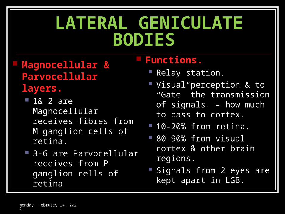
Magnocellular & Parvocellular layers. 1& 2 are Magnocellular
receives fibres from M ganglion cells of retina.
3-6 are Parvocellular receives from P ganglion cells of retina
Functions. Relay station. Visual perception & to
“Gate” the transmission of signals. – how much to pass to cortex.
10-20% from retina. 80-90% from visual cortex
& other brain regions. Signals from 2 eyes are
kept apart in LGB.
Wednesday, May 3, 2023
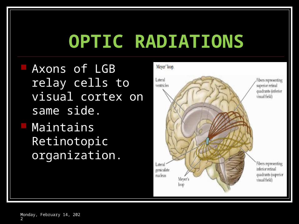
OPTIC RADIATIONS Axons of LGB relay
cells to visual cortex on same side.
Maintains Retinotopic organization.
Wednesday, May 3, 2023
PROCESSING & ANALYSIS OF VISUAL IMPULSE IN THE VISUAL CORTEX
Retinotopic organization Functional anatomy & organization of visual
cortex Visual areas (Classical nomenclature) Primary visual cortex Peristriate cortex Parastriate cortex
Modified nomenclature of visual areas Histological layers of primary visual cortex
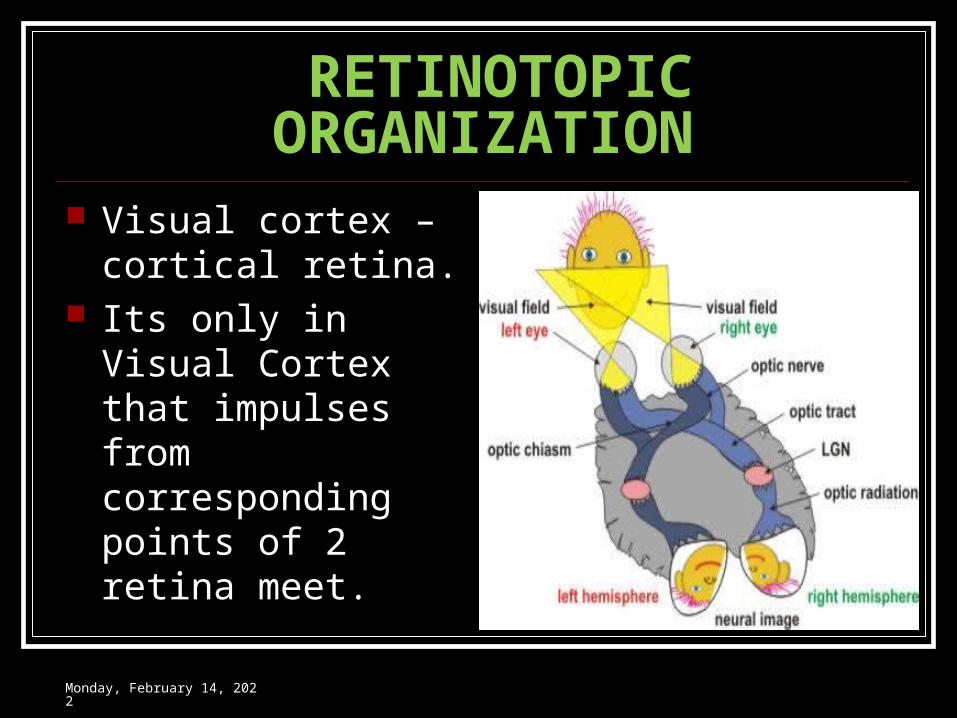
RETINOTOPIC ORGANIZATION Visual cortex – cortical
retina. Its only in Visual
Cortex that impulses from corresponding points of 2 retina meet.
Wednesday, May 3, 2023
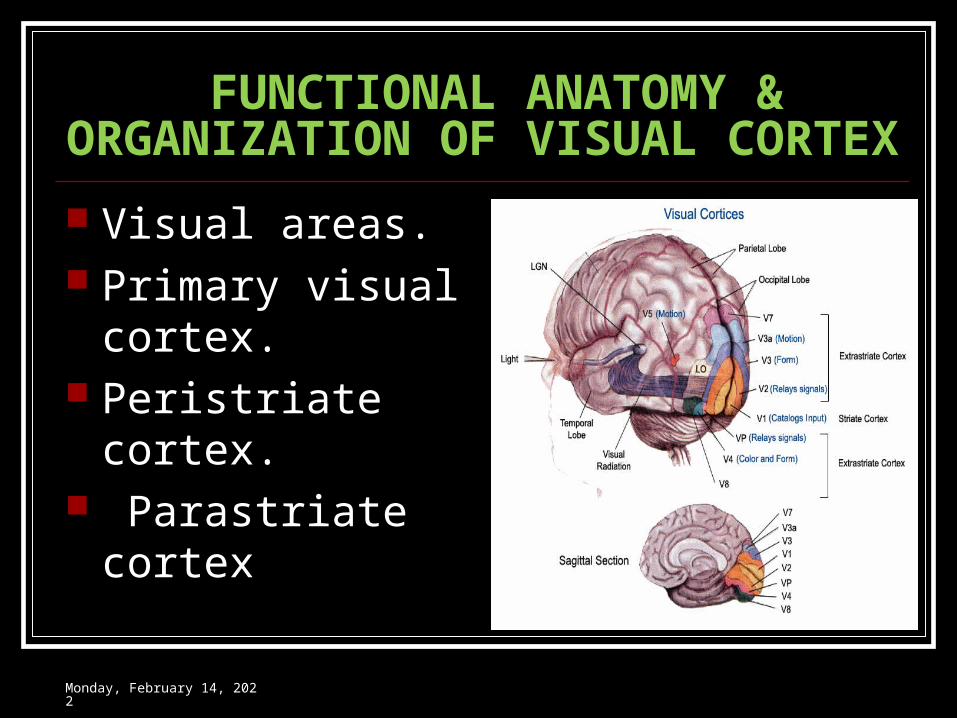
FUNCTIONAL ANATOMY & ORGANIZATION OF VISUAL CORTEX
Visual areas. Primary visual
cortex. Peristriate cortex. Parastriate cortex
Wednesday, May 3, 2023
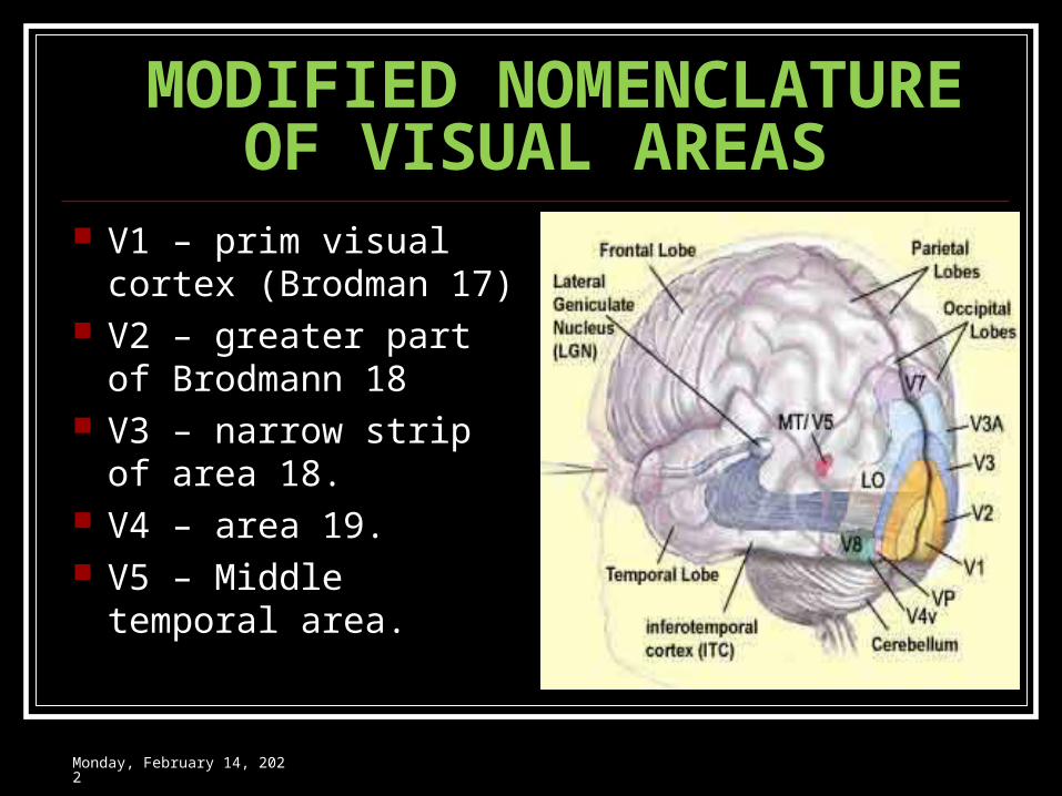
MODIFIED NOMENCLATURE OF VISUAL AREAS
V1 – prim visual cortex (Brodman 17)
V2 – greater part of Brodmann 18
V3 – narrow strip of area 18.
V4 – area 19. V5 – Middle temporal
area.
Wednesday, May 3, 2023
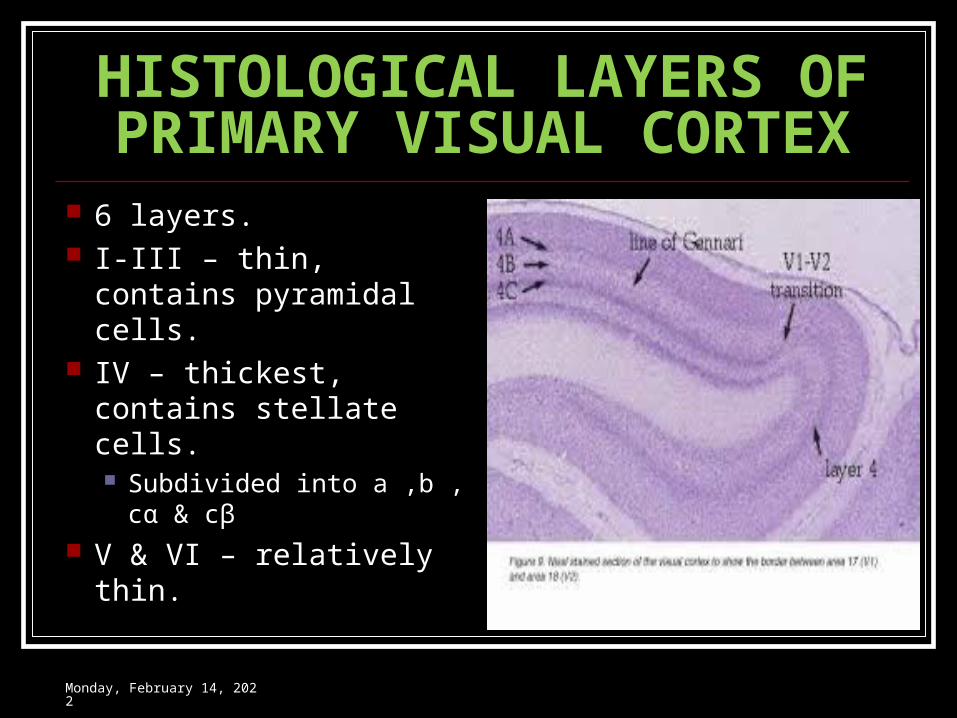
HISTOLOGICAL LAYERS OF PRIMARY VISUAL CORTEX
6 layers. I-III – thin, contains
pyramidal cells. IV – thickest, contains
stellate cells. Subdivided into a ,b , cα
& cβ V & VI – relatively thin.
Wednesday, May 3, 2023
PHYSIOLOGICAL CONSIDERATION OF VISUAL CORTEX
Concept of receptive field of striate cortex Columnar organization of the striate cortex
Orientation column Ocular dominance column The colour blobs
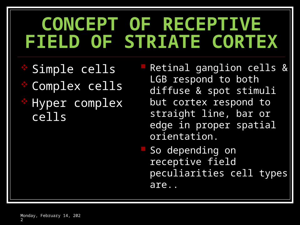
CONCEPT OF RECEPTIVE FIELD OF STRIATE CORTEX
Simple cells Complex cells Hyper complex cells
Retinal ganglion cells & LGB respond to both diffuse & spot stimuli but cortex respond to straight line, bar or edge in proper spatial orientation.
So depending on receptive field peculiarities cell types are..
Wednesday, May 3, 2023
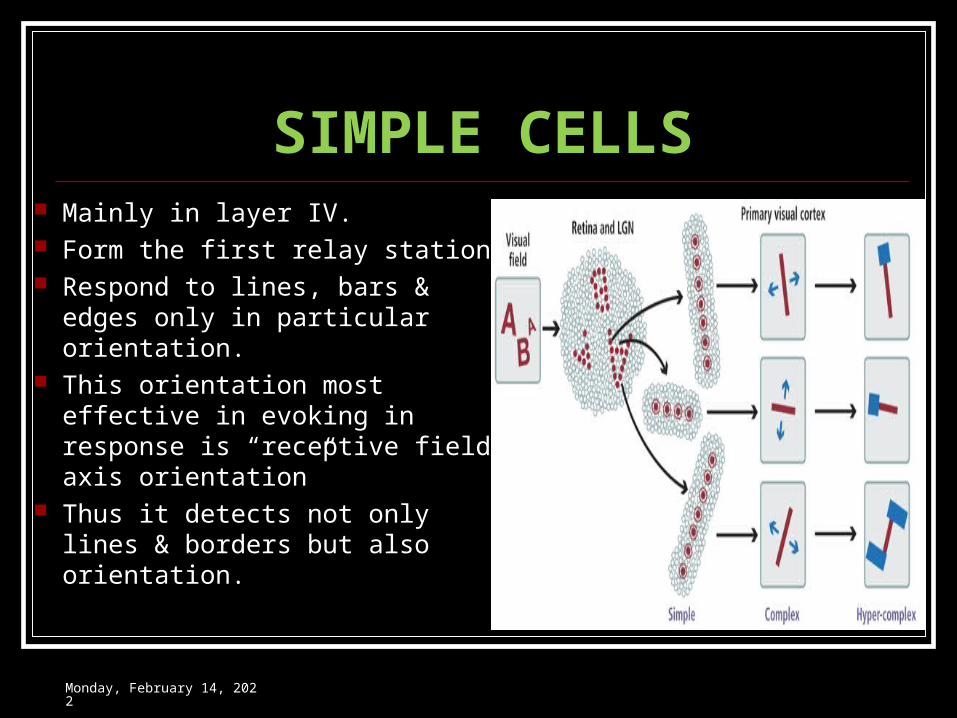
SIMPLE CELLS Mainly in layer IV. Form the first relay station. Respond to lines, bars & edges
only in particular orientation. This orientation most effective
in evoking in response is “receptive field axis orientation”
Thus it detects not only lines & borders but also orientation.
Wednesday, May 3, 2023
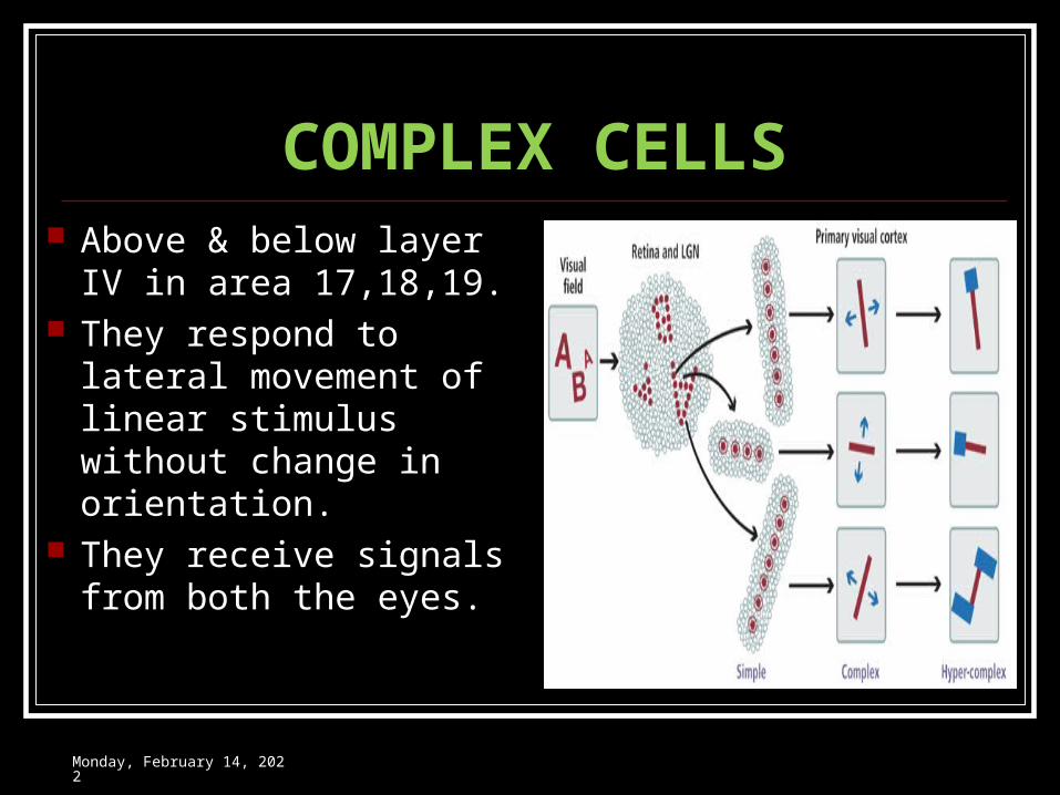
COMPLEX CELLS Above & below layer IV
in area 17,18,19. They respond to lateral
movement of linear stimulus without change in orientation.
They receive signals from both the eyes.
Wednesday, May 3, 2023
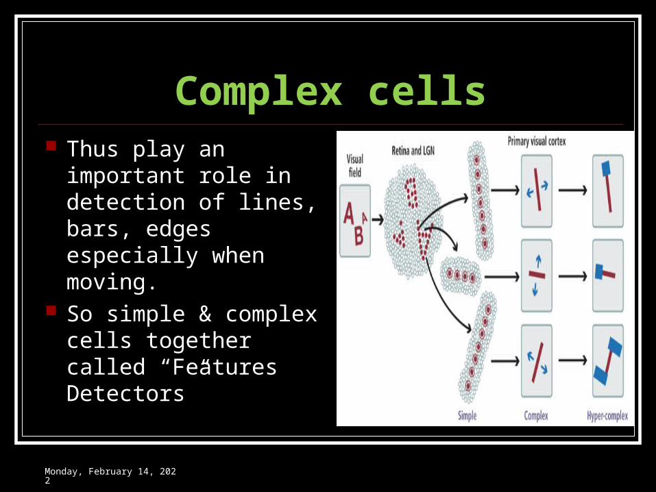
Complex cells Thus play an
important role in detection of lines, bars, edges especially when moving.
So simple & complex cells together called “Features Detectors”
Wednesday, May 3, 2023
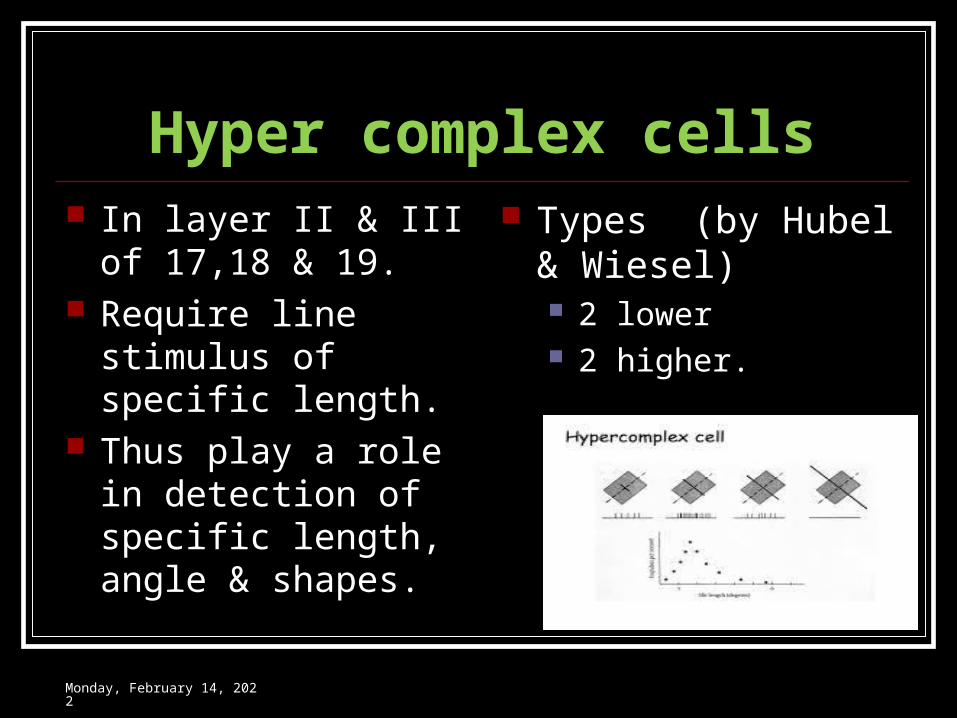
Hyper complex cells In layer II & III of
17,18 & 19. Require line stimulus
of specific length. Thus play a role in
detection of specific length, angle & shapes.
Types (by Hubel & Wiesel) 2 lower 2 higher.
Wednesday, May 3, 2023
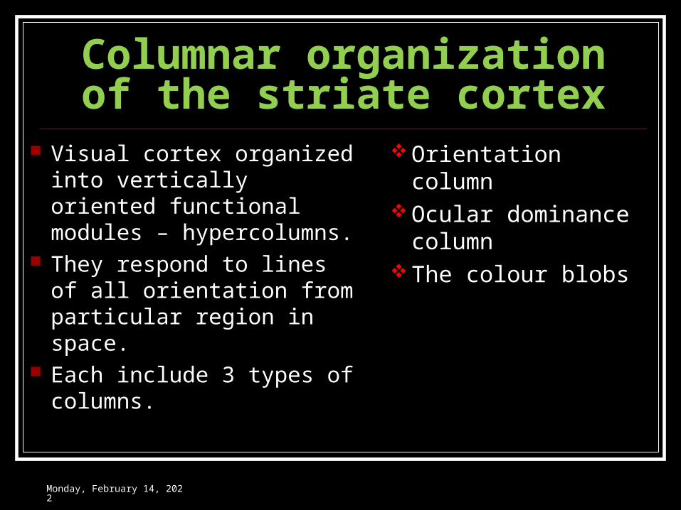
Columnar organization of the striate cortex
Visual cortex organized into vertically oriented functional modules – hypercolumns.
They respond to lines of all orientation from particular region in space.
Each include 3 types of columns.
Orientation column Ocular dominance
column The colour blobs
Wednesday, May 3, 2023
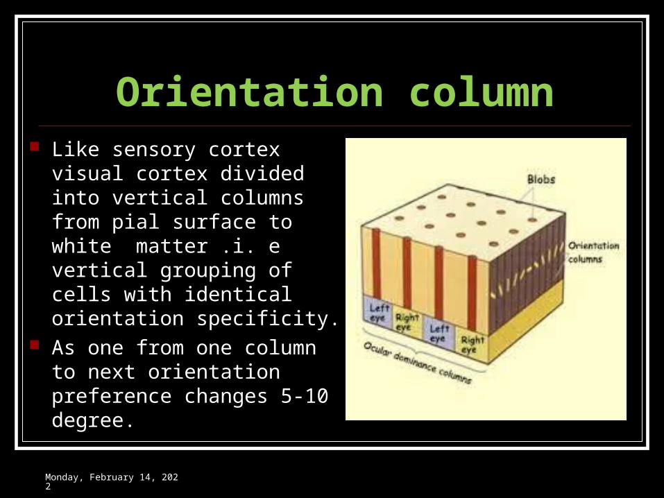
Orientation column Like sensory cortex visual
cortex divided into vertical columns from pial surface to white matter .i. e vertical grouping of cells with identical orientation specificity.
As one from one column to next orientation preference changes 5-10 degree.
Wednesday, May 3, 2023
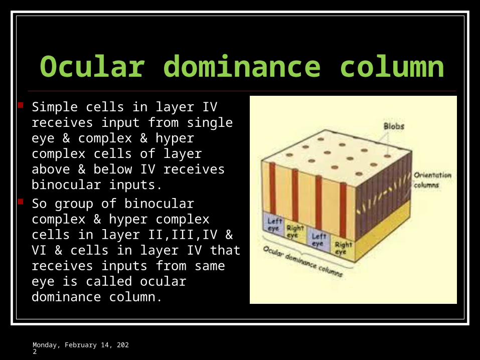
Ocular dominance column Simple cells in layer IV receives
input from single eye & complex & hyper complex cells of layer above & below IV receives binocular inputs.
So group of binocular complex & hyper complex cells in layer II,III,IV & VI & cells in layer IV that receives inputs from same eye is called ocular dominance column.
Wednesday, May 3, 2023
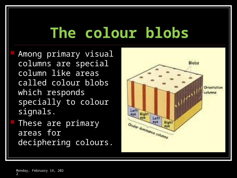
The colour blobs Among primary visual
columns are special column like areas called colour blobs which responds specially to colour signals.
These are primary areas for deciphering colours.
Wednesday, May 3, 2023
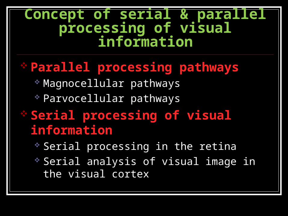
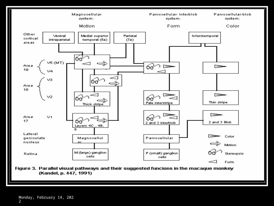
Concept of serial & parallel processing of visual information
Parallel processing pathways Magnocellular pathways Parvocellular pathways
Serial processing of visual information Serial processing in the retina Serial analysis of visual image in the visual cortex
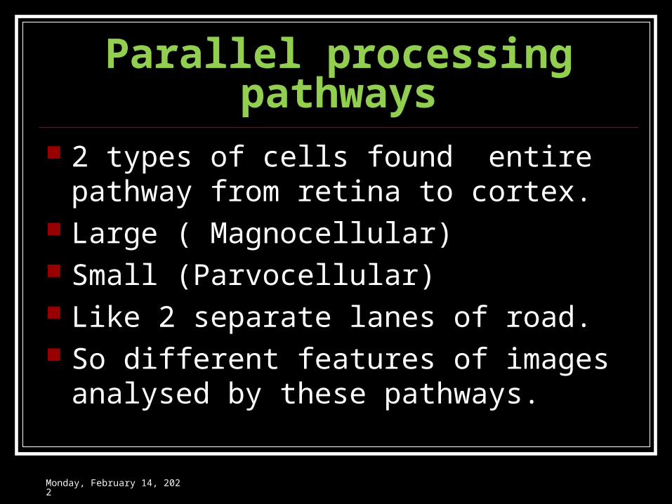
Parallel processing pathways 2 types of cells found entire pathway from
retina to cortex. Large ( Magnocellular) Small (Parvocellular) Like 2 separate lanes of road. So different features of images analysed by
these pathways.
Wednesday, May 3, 2023
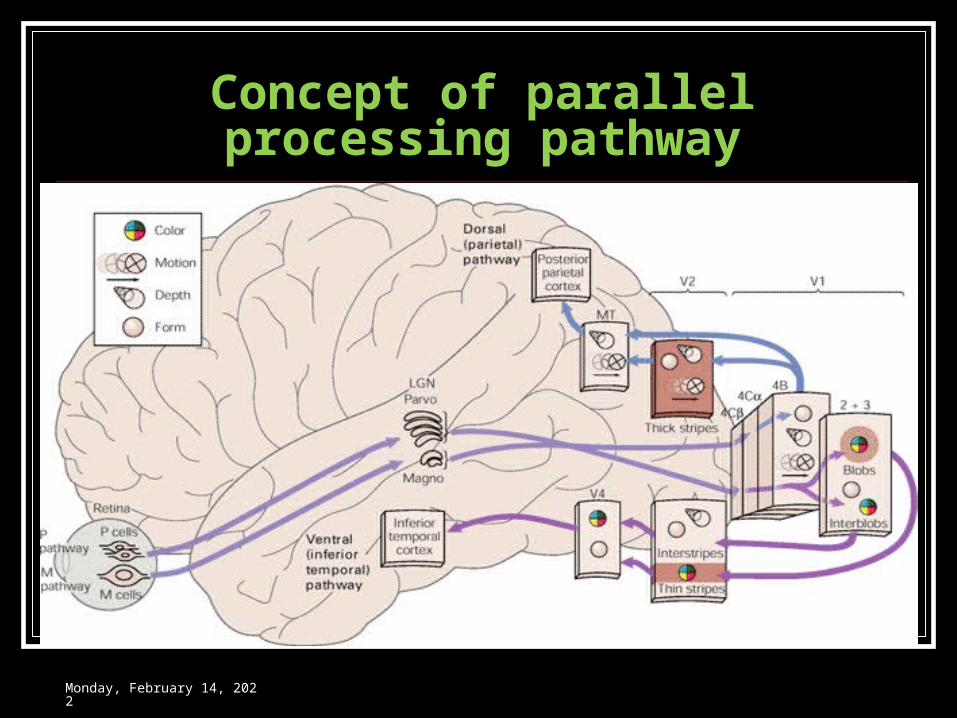
Concept of parallel processing pathway
Wednesday, May 3, 2023
Wednesday, May 3, 2023
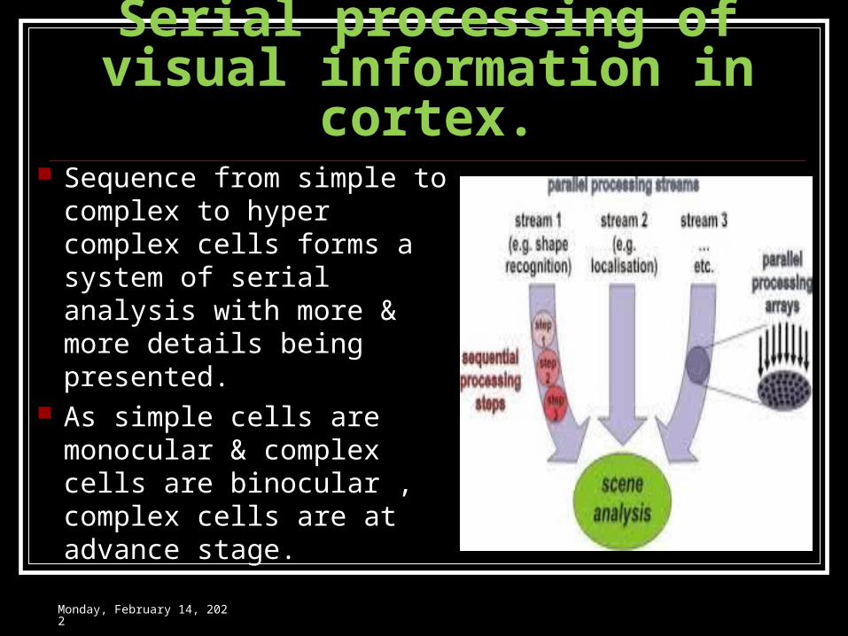
Serial processing of visual information in cortex.
Sequence from simple to complex to hyper complex cells forms a system of serial analysis with more & more details being presented.
As simple cells are monocular & complex cells are binocular , complex cells are at advance stage.
Wednesday, May 3, 2023
Wednesday, May 3, 2023
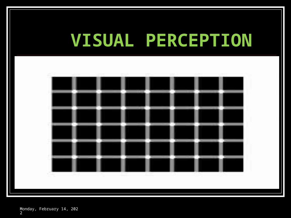
VISUAL PERCEPTION
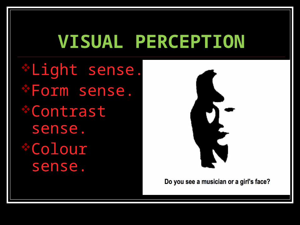
VISUAL PERCEPTIONLight sense.Form sense.Contrast sense.Colour sense.
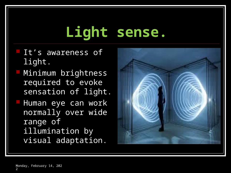
Light sense. It’s awareness of light. Minimum brightness
required to evoke sensation of light.
Human eye can work normally over wide range of illumination by visual adaptation.
Wednesday, May 3, 2023
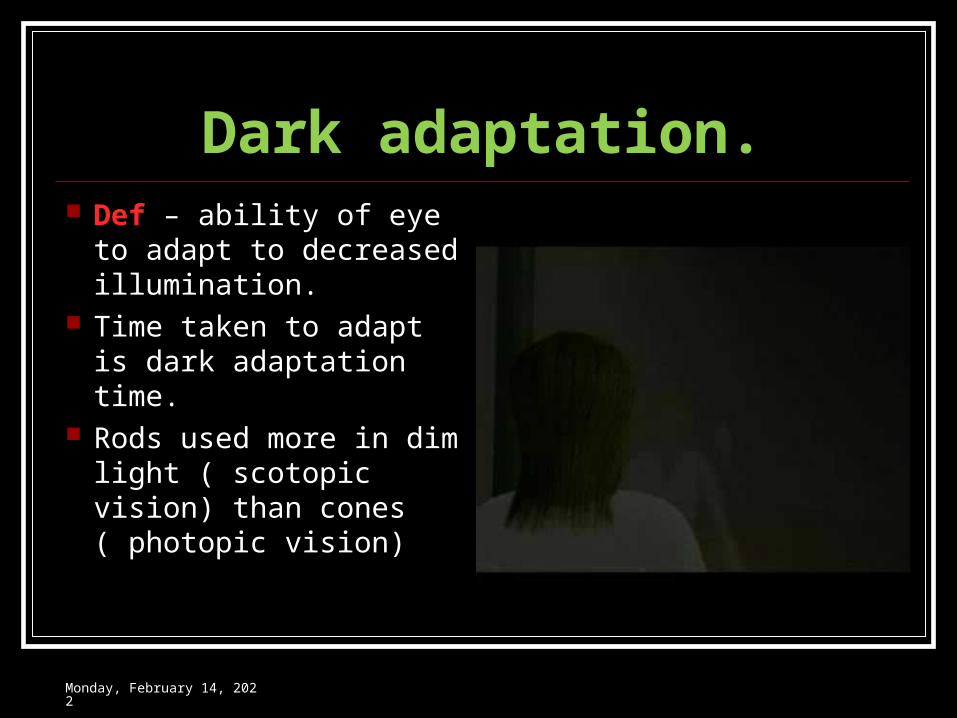
Dark adaptation. Def – ability of eye to
adapt to decreased illumination.
Time taken to adapt is dark adaptation time.
Rods used more in dim light ( scotopic vision) than cones ( photopic vision)
Wednesday, May 3, 2023
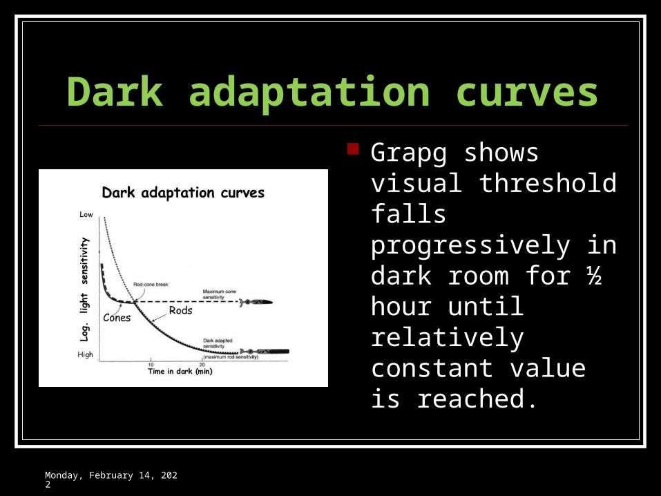
Dark adaptation curves Grapg shows visual
threshold falls progressively in dark room for ½ hour until relatively constant value is reached.
Wednesday, May 3, 2023
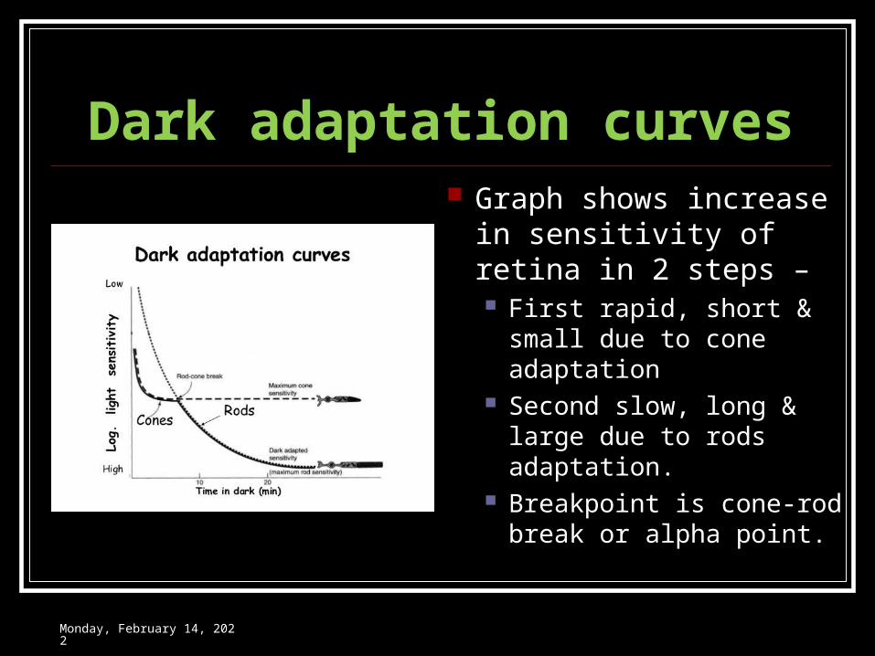
Dark adaptation curves Graph shows increase in
sensitivity of retina in 2 steps – First rapid, short & small
due to cone adaptation Second slow, long & large
due to rods adaptation. Breakpoint is cone-rod
break or alpha point.
Wednesday, May 3, 2023
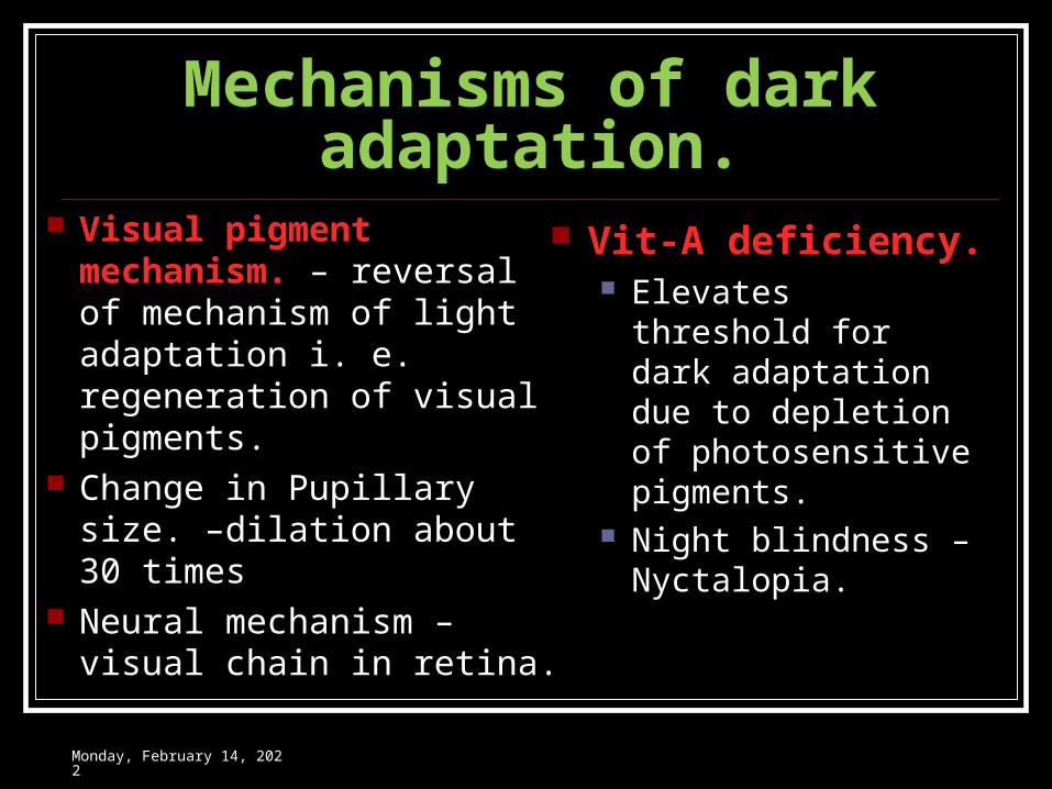
Mechanisms of dark adaptation.
Visual pigment mechanism. – reversal of mechanism of light adaptation i. e. regeneration of visual pigments.
Change in Pupillary size. –dilation about 30 times
Neural mechanism – visual chain in retina.
Vit-A deficiency. Elevates threshold for
dark adaptation due to depletion of photosensitive pigments.
Night blindness – Nyctalopia.
Wednesday, May 3, 2023
Light adaptation. Def – process by
which retina adapt itself to bright light.
Very quick, over in 5 min.
It’s merely disappearance of dark adaptation.
Wednesday, May 3, 2023
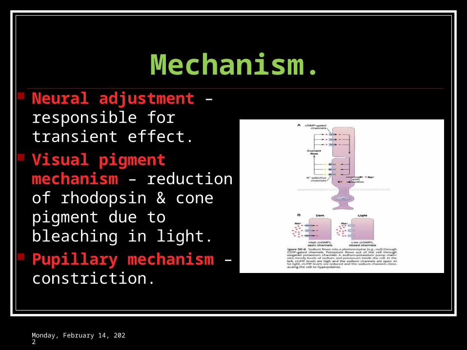
Mechanism. Neural adjustment –
responsible for transient effect.
Visual pigment mechanism – reduction of rhodopsin & cone pigment due to bleaching in light.
Pupillary mechanism – constriction.
Wednesday, May 3, 2023
The form sense Ability to discriminate
between shapes of objects.
Cones are imp. Snellen’s chart is
measure of form sense.
Wednesday, May 3, 2023
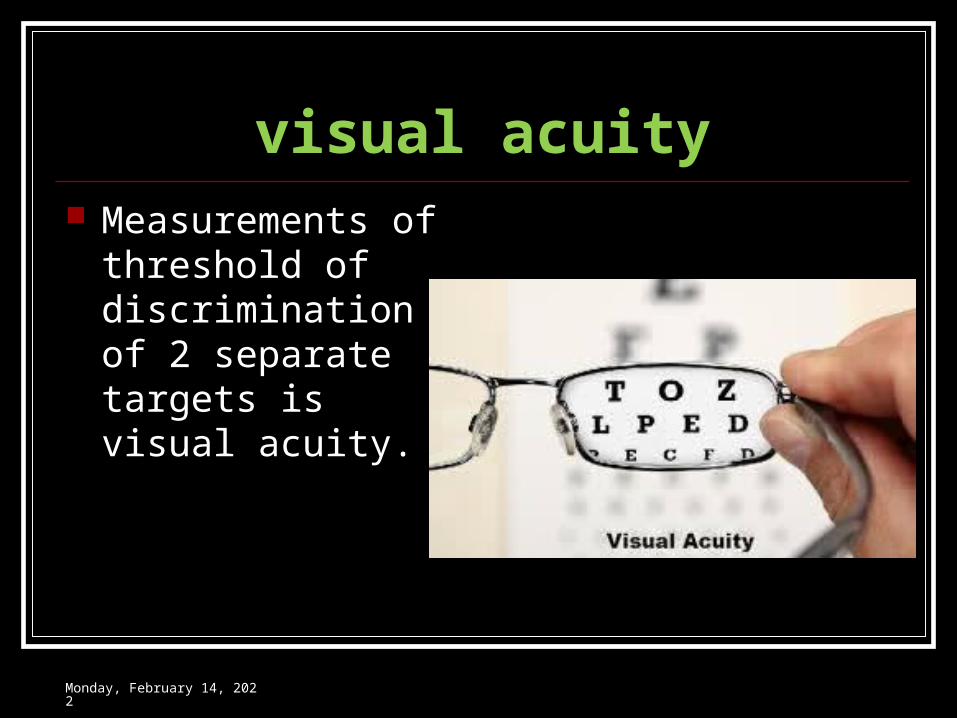
visual acuity Measurements of
threshold of discrimination of 2 separate targets is visual acuity.
Wednesday, May 3, 2023
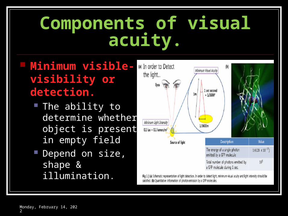
Components of visual acuity. Minimum visible-
visibility or detection. The ability to
determine whether object is present in empty field
Depend on size, shape & illumination.
Wednesday, May 3, 2023
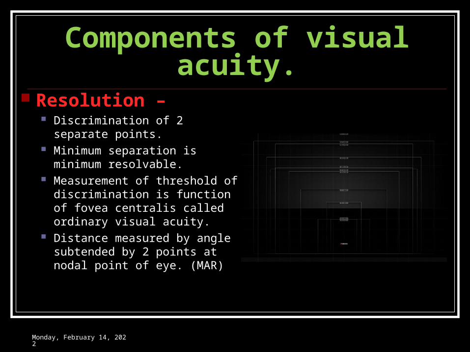
Components of visual acuity. Resolution –
Discrimination of 2 separate points.
Minimum separation is minimum resolvable.
Measurement of threshold of discrimination is function of fovea centralis called ordinary visual acuity.
Distance measured by angle subtended by 2 points at nodal point of eye. (MAR)
Wednesday, May 3, 2023
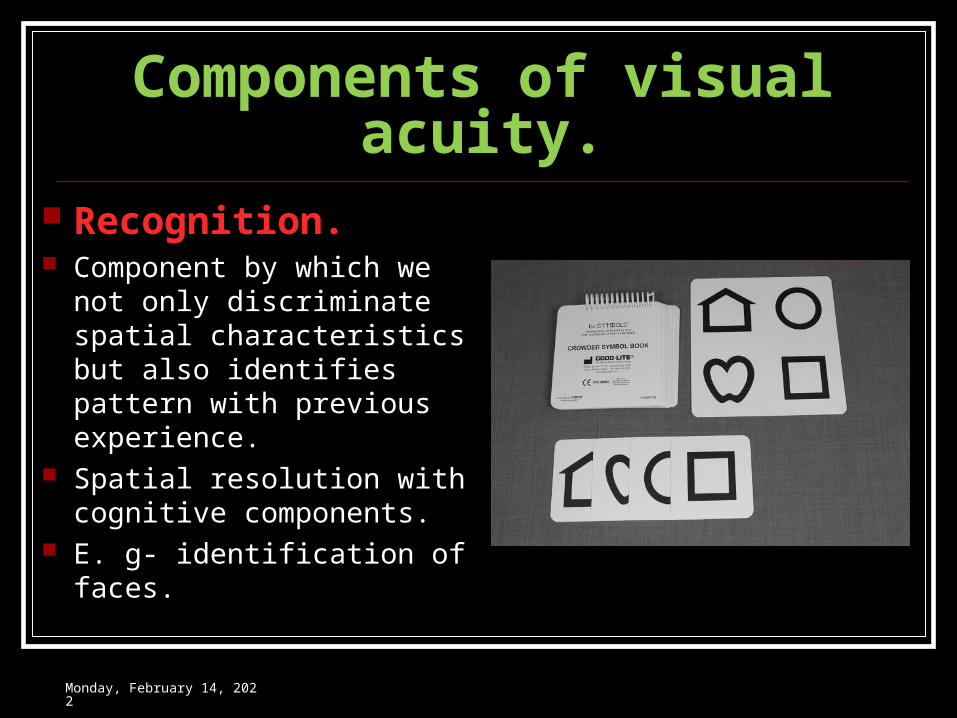
Components of visual acuity. Recognition. Component by which we not
only discriminate spatial characteristics but also identifies pattern with previous experience.
Spatial resolution with cognitive components.
E. g- identification of faces.
Wednesday, May 3, 2023
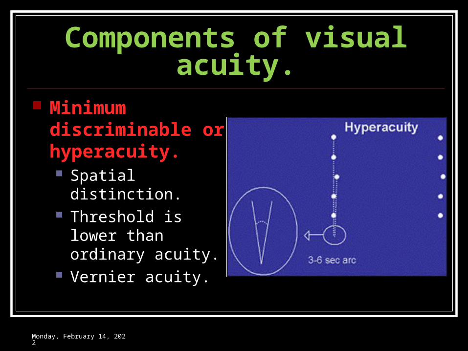
Components of visual acuity. Minimum
discriminable or hyperacuity. Spatial distinction. Threshold is lower
than ordinary acuity. Vernier acuity.
Wednesday, May 3, 2023
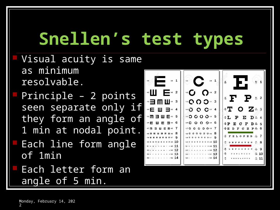
Snellen’s test types Visual acuity is same as
minimum resolvable. Principle – 2 points seen
separate only if they form an angle of 1 min at nodal point.
Each line form angle of 1min
Each letter form an angle of 5 min.
Wednesday, May 3, 2023
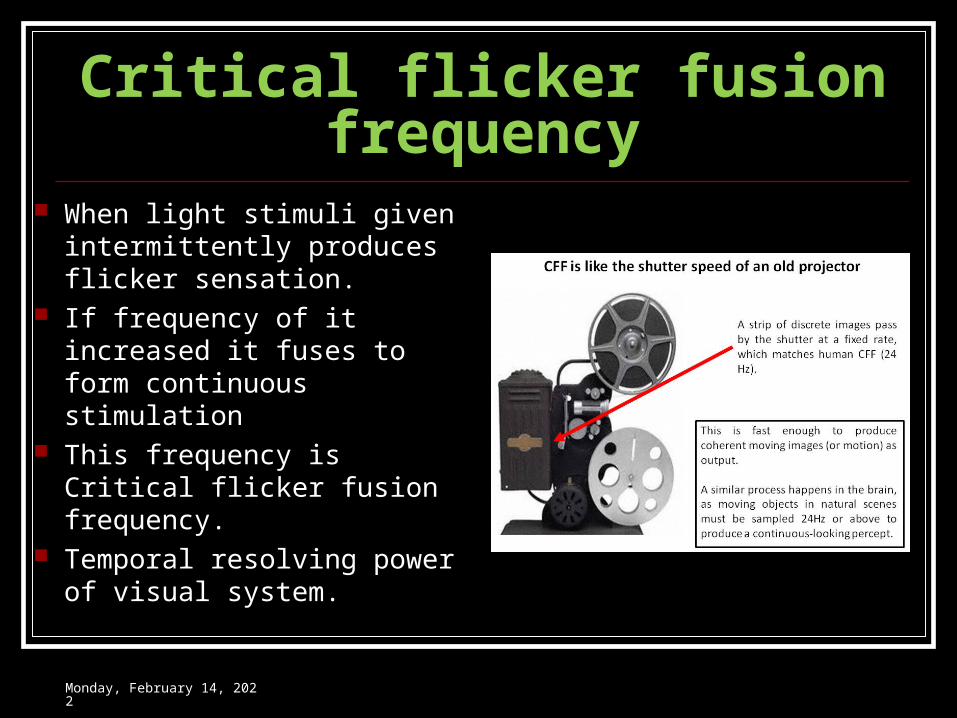
Critical flicker fusion frequency When light stimuli given
intermittently produces flicker sensation.
If frequency of it increased it fuses to form continuous stimulation
This frequency is Critical flicker fusion frequency.
Temporal resolving power of visual system.
Wednesday, May 3, 2023
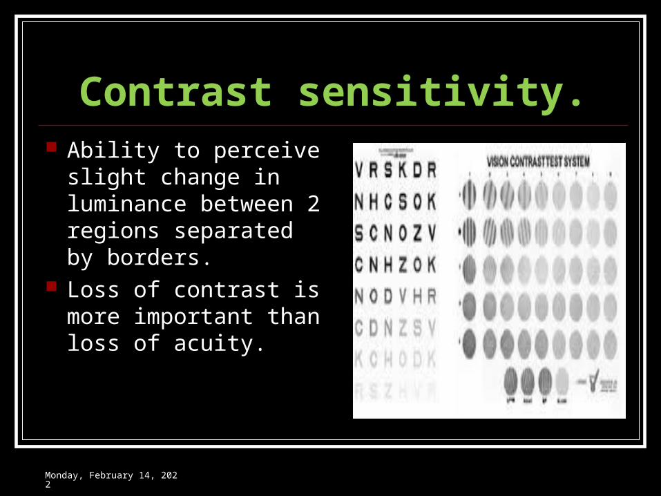
Contrast sensitivity. Ability to perceive
slight change in luminance between 2 regions separated by borders.
Loss of contrast is more important than loss of acuity.
Wednesday, May 3, 2023
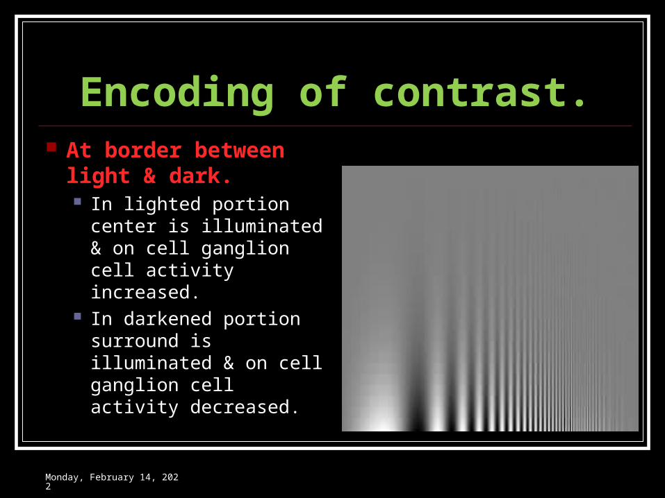
Encoding of contrast. At border between
light & dark. In lighted portion
center is illuminated & on cell ganglion cell activity increased.
In darkened portion surround is illuminated & on cell ganglion cell activity decreased.
Wednesday, May 3, 2023

THANK YOU
Recommended