Myasthenia Gravis
Shirley H. Wray, M.D., Ph.D.Professor of Neurology, Harvard Medical School
Director, Unit for Neurovisual DisordersMassachusetts General Hospital
HARVARD MEDICAL SCHOOLDEPARTMENT OF NEUROLOGY
MASSACHUSETTS GENERAL HOSPITAL
HARVARD MEDICAL SCHOOLDEPARTMENT OF NEUROLOGY
MASSACHUSETTS GENERAL HOSPITAL
HARVARD MEDICAL SCHOOLDEPARTMENT OF NEUROLOGY
MASSACHUSETTS GENERAL HOSPITAL
HistoryA detailed history often reveals evidence of early, unrecognized myasthenic features:
Intermittent diplopiaFrequent purchases of new glasses to correct blurry vision, difficulty focusing and/or early onset of convergence insufficnecy of a need for prism correctionUse of dark glasses to reduce diplopia or hide drooping eyelids
History continued.
Avoidance of certain foods that become difficult to chew and swallow
Cessation of activities that require prolonged use of muscle activity
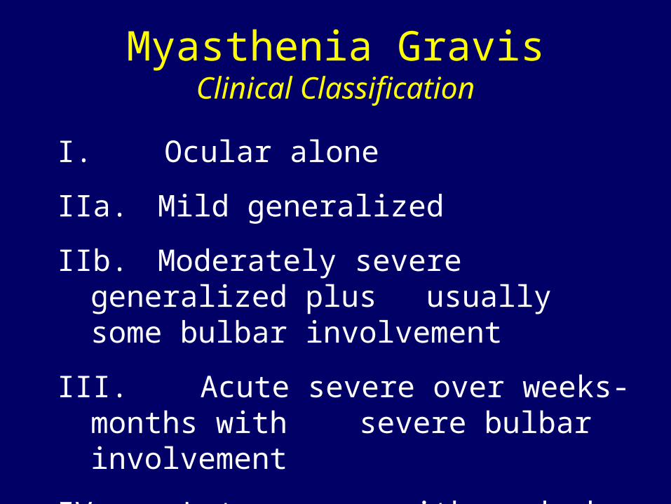
Myasthenia GravisClinical Classification
I. Ocular alone
IIa. Mild generalized
IIb. Moderately severe generalized plus usually some bulbar involvement
III. Acute severe over weeks-months with severe bulbar involvement
IV. Late severe with marked bulbar involvement
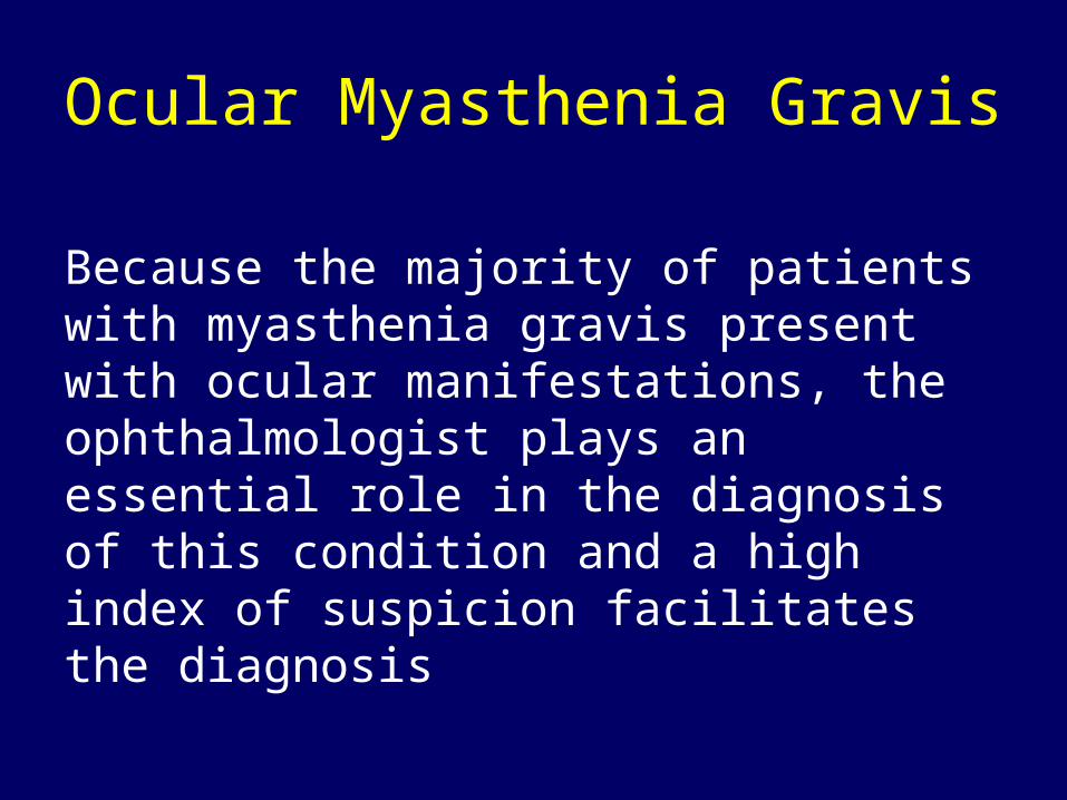
Ocular Myasthenia Gravis
Because the majority of patients with myasthenia gravis present with ocular manifestations, the ophthalmologist plays an essential role in the diagnosis of this condition and a high index of suspicion facilitates the diagnosis
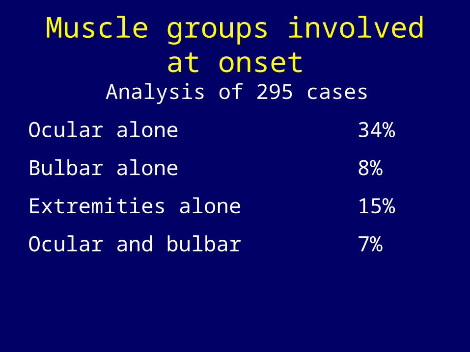
Muscle groups involved at onset
Analysis of 295 cases
Ocular alone 34%
Bulbar alone 8%
Extremities alone 15%
Ocular and bulbar 7%

Muscle group analysis continued.
Ocular and extremities 7%
Bulbar and extremities 6%
Ocular, bulbar and extremities 21%
Simpson et al, Acta Neurol. Scand. 1966 suppl. 23 pl
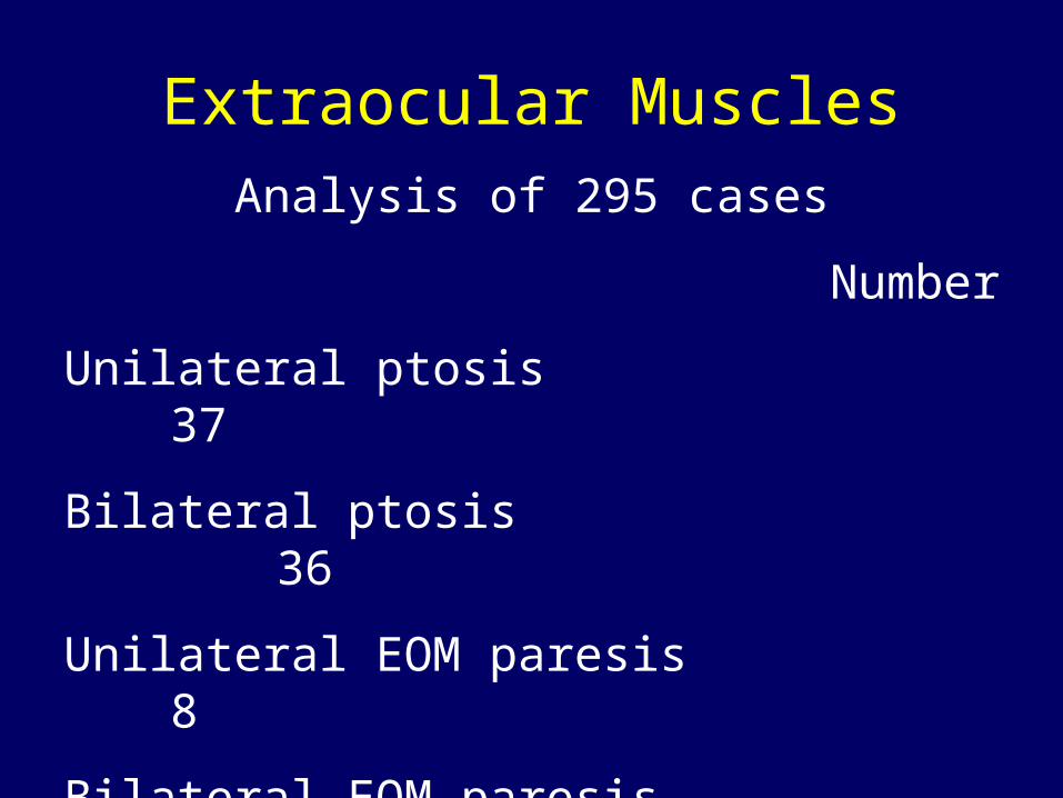
Extraocular MusclesAnalysis of 295 cases
Number
Unilateral ptosis 37
Bilateral ptosis 36
Unilateral EOM paresis 8
Bilateral EOM paresis 32
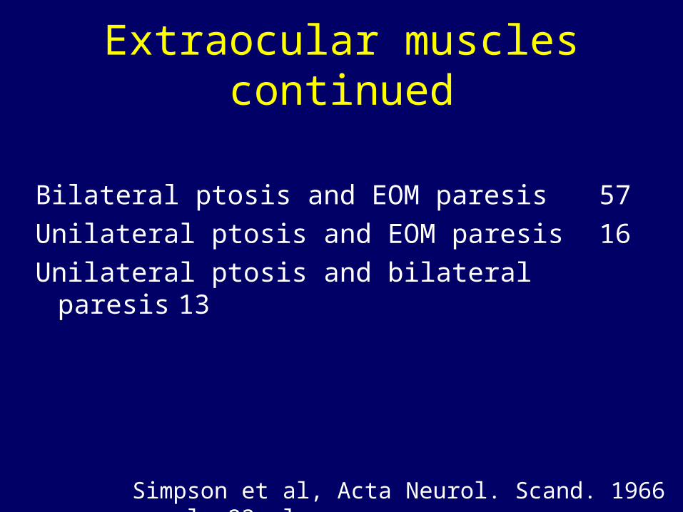
Extraocular muscles continued
Bilateral ptosis and EOM paresis 57
Unilateral ptosis and EOM paresis 16
Unilateral ptosis and bilateral paresis 13
Simpson et al, Acta Neurol. Scand. 1966 suppl. 23 pl
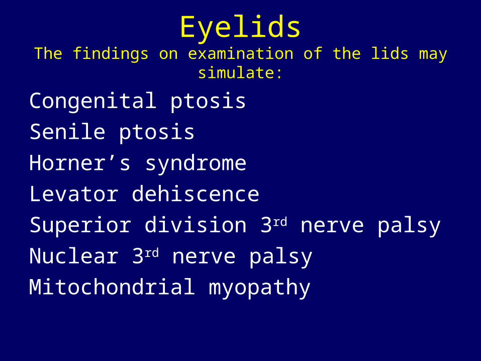
EyelidsThe findings on examination of the lids may simulate:
Congenital ptosis
Senile ptosis
Horner’s syndrome
Levator dehiscence
Superior division 3rd nerve palsy
Nuclear 3rd nerve palsy
Mitochondrial myopathy
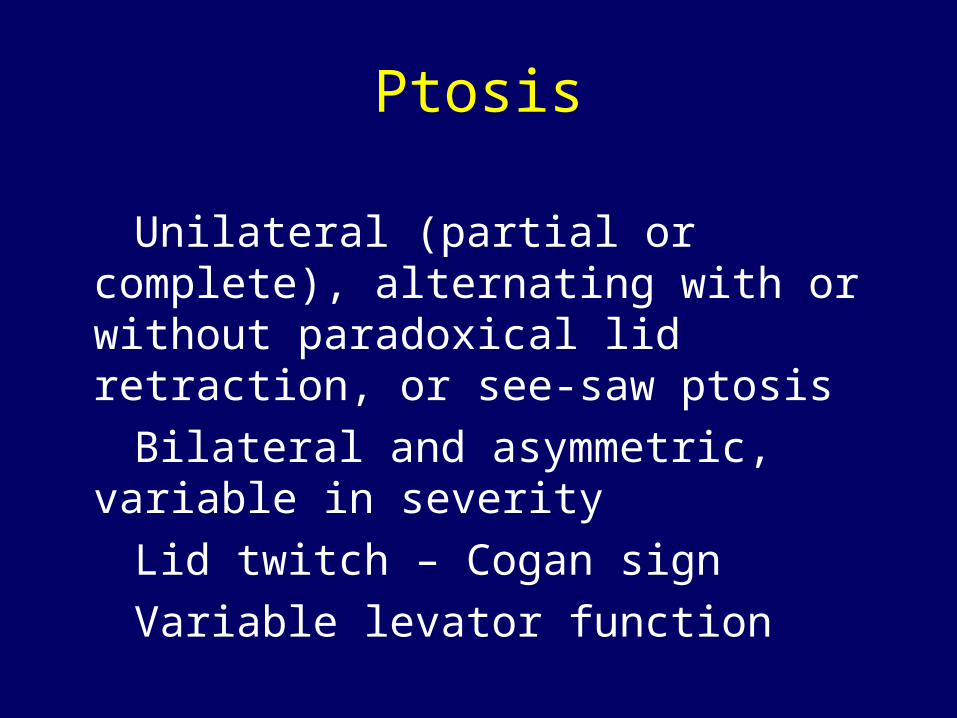
Ptosis
Unilateral (partial or complete), alternating with or without paradoxical lid retraction, or see-saw ptosis
Bilateral and asymmetric, variable in severity
Lid twitch – Cogan sign
Variable levator function
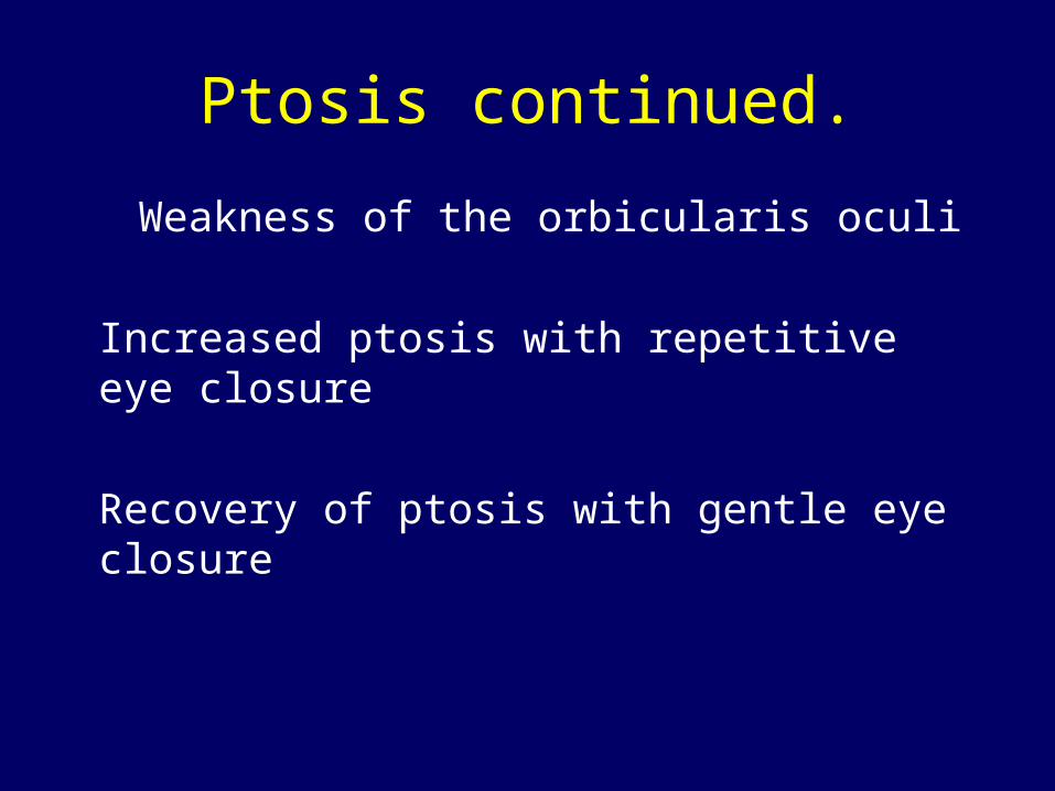
Ptosis continued.
Weakness of the orbicularis oculi
Increased ptosis with repetitive eye closure
Recovery of ptosis with gentle eye closure
To Document Ptosis
Measure the palpebral fissure before testing EOM, giving Tensilon, or using sympathomimetic drugs
Increase of ptosis on fatigue
Myasthenic lid twitch
Recovery of ptosis following gentle eye closure

To document ptosis continued.
Range of levator function
+ Weakness orbicularis oculi
Photograph and compare with family snapshots
Measure response to IV Tensilon

PtosisMyopathy Myasthenia
Symmetric Appearance Asymmetric
No Ptosis on fatigue Yes
No Lid twitch Yes
No Recovery w/ eye closure Yes
Constant Range lev. function Varies
Yes Weak orb. oculi Yes
Negative Tensilon test Positive
50% Family history Rare
Slowly Progressive Course Fluctuates
SaccadesExamination may show:
Intrasaccadic fatigue (slow in midflight)Decrescendo from saccade to saccade
Hypermetria of small saccades
Hypometria of large saccades
Quiver movements and “hyperfast” saccades
Fatigability of quick phases on OKN
Myasthenia Gravis
Myasthenia gravis is a disease of skeletal muscle acetylcholine receptors. The chemical transmitter, acetylcholine (ACh) is unable to bind to the receptors (AChR) on the postsynaptic membrane to transmit the nerve impulse to muscle fibers to produce a muscle contraction
Presentation (I)
MG occurs at any age, involves either sex and begins insidiously
Second and third decades commonest age of onset in women. Seventh and eighth decades in men
Patients complain of specific muscle weakness, not generalized fatigue
Presentation (II)
Ptosis or diplopia – initial symptoms in 65% of patients
Oropharyngeal muscle weakness – difficulty in swallowing and talking initial symptoms in 17% of patients
Limb weakness presenting symptom in only 10% of cases
Presentation (III)
Characteristically, severity of weakness fluctuates during the day, least in the morning, worsening as the day progresses, especially after prolonged use of affected muscles
In the era before corticosteroid treatment, approximately one-third of patients improved spontaneously, one third became worse and one third died

Presentation (IV)
Ocular myasthenia – if progressing to generalized MG usually does so within the first two years after onset
After 15 to 20 years, weakness becomes fixed. The Burnt-Out-Stage + muscle atrophy
Edrophonium Chloride Tensilon Test (10 Mg in 1 cc)
List all medications being taken
History of drug allergy and previous reaction to Tensilon
Perform the test in the ER with an ambu bag, atropine and adrenalin available in elderly patients and those with cardiac disease
Precautionary Steps:

Administration Procedure
The ideal dose of Tensilon cannot be predetermined
Give a 0.1 cc test dose and monitor pulse, blood pressure and clinical state
Follow with 0.3 cc aliquotes examining for a response in ptosis, EOM or Lancaster strabismus screen test after each one
Once improvement is seen -- STOP
The defect in neuromuscular transmission in Myasthenia Gravis is due to: The muscle end-plate membrane is
distorted
Acetylcholine receptors are lost from the tips of the folds, and antibodies attach to the postsynaptic membrane
Ach is released normally but absence of receptors prevents the transmitter binding to the muscle membrane
Acetylcholine Receptor Antibodies
75% of cases generalized MG have serum antibodies (Ab) that bind to huma AChR
54% with ocular MG have antibodies 10% MG cases with no binding antibodies have other antibodies
The AChR Ab tit varies widely among patients with similar degrees of weakness. The amount of Ab in the serum does not predict the severity of the disease in individual patients
Antibodies continued.
The Ab level may be low at onset on MG and gradually become elevated in late stage
Worthwhile to repeat test when initial values normal
The Presence of AChR Antibody is not diagnostic for MG, also present in:
Systemic lupus erythematosus
Inflammatory neuropathy
Amyotrophic lateral sclerosis
Rheumatoid arthritis in patients taking D-penicillamine
In cases of thymoma without MG

Association of MG with other diseases
Hyperthyroidism 6%
Rheumatoid arthritis, less than 2%
Systemic lupus erythematosus 2%
Diabetes mellitus 7%
Non thymus neoplasm 3%
Differential Diagnosis
Graves ophthalmopathy
Progressive External Ophthalmoplegia (PEO)
Oculopharyngeal Dystrophy
Myotonic Dystrophy
The Lambert-Eaton Myasthenic Syndrome (LEMS)
Guillian Barre Syndrome – Miller Fisher variant

Factors that Aggravate MG
Emotional stress
Systemic illness e.g. viral URI
Thyroid disease, hyper or hypo
Pregnancy
Menstrual Cycle
Increase in body temperature
Drugs
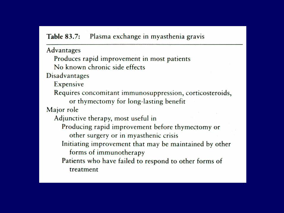
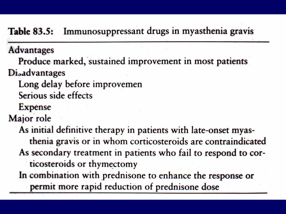
Treatment
Treatment decisions are based on the predicted response to a specific form of therapy
Treatment goals must be individualized according to the severity of the disease, the patient’s age and sex, and the degree of functional impairment
Treatment continued.
The response to any form of treatment is difficult to access because the severity of symptoms fluctuates. Spontaneous improvement, even remissions, occur without specific therapy, especially during the early stages of the disease

CHE Inhibitors (I)
Mestinon (Pyridostigmine bromide) first choice, dose 30-60 mg q 6-8 h/daily
Prostigmine (Neostigmine bromide) 7.5 – 15.0 mg q 6-8 h/daily
No fixed dosage schedle suits all aptients
CHE Inhibitors (II)
The need for ChE inhibitors varies from day to day and during the same day
Different muscles respond differently with any dose, certain muscles get stronger, others do not change and still others become weaker
The drug schedule should be titrated according to the pateints work load and muscle activity
ChE Inhibitors (III)
Many patients assume responsibility for their own drug dose
The goal is to keep the dose low enough to provide definite improvement 30 to 40 minutes later and allow theeffect to wear off before the next dose
Advise patients re: adverse effects of ChE inhibitors

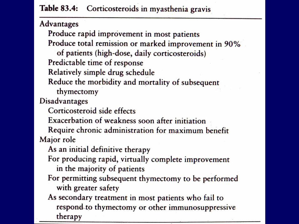
Prednisone
Marked improvement or complete relief of symptoms occurs in 75% of cases
Improvement in first 6 to 8 weeks, but strength may increase to total remission over months
Best responses in patients with recent onset MG, but chronic disease may also respond
Prednisone continued.
The severity of the disease does not predict the ultimate improvement
Patients with thymoma have an excellent response to prednisone before or after thymectomy

Dose
Prednisone 60 to 80 mg/day given until sustained improvement (usually 2 weeks) then alternate days beginning with 100-120 mg tapered over months to lowest dose necessary (usually less than 20 mg alternate days)


http://www.library.med.utah.edu/NOVEL
Recommended