1
11.1 Musculoskeletal Imaging
Musculoskeketal Sectional Anatomy & Clinical Imaging
Slide # 1
Carolyn Kaut Roth, RT (R)(MR)(CT)(M)(CV) FSMRTCEO Imaging Education Associates
www.imaginged.com [email protected]
11.1 Musculoskeletal Imaging
Part I• Planes of the Musculoskeletal system
S ti l A t f th t it
Outline
Slide # 2
• Sectional Anatomy of the upper extremity• Sectional Anatomy of the lower extremity
11.1 Musculoskeletal Imaging
Patient Preparation for MSK Imaging
• Patient Screening– Check for contraindications– Remove ALL metal– Change into gown
• Coil Selection– For routine– For vascular– For Arthrography
• Patient set-up & Positioning
Slide # 3
Patient set up & Positioning– Supine– Coil on joint– Hearing Protection– Patient Intercom
• Landmark– Per joint– Other
• Isocenter
11.1 Musculoskeletal ImagingTMJ TMJ
Anatomy Musculoskeletal System
Slide # 4
11.1 Musculoskeletal Imaging
Median Line
Mid-sagittal Plane
Parasagittal Planes
Axial
Mid FrontalCoronal Plane
TransversePlane
Imaging Planes - TMJ
Slide # 5
Sagittal
Coronal
MRI Images
Sagittal Axial Coronal
CT Images
11.1 Musculoskeletal Imaging
Localizer for sagittal oblique sections Localizer for coronal oblique sections
Overview musculoskeletal Anatomy - TMJ
Slide # 6
Sagittal Oblique TMJ Coronal Oblique TMJ
Temporal boneFossa(meniscus within)Mandibular
condyleMandible
2
11.1 Musculoskeletal Imaging
Shoulder radiograph
Corocoid vs coronoid… “There’s a sea (C) between two Nations”
Slide # 7CoroNoid CoroCoid CoroNoid
Sagittal reformatted CT Lateral radiography (Elbow)
Shoulder radiograph
TMJ 3D CT reformatted image
Shoulder CT
Sagittal MR Elbow
11.1 Musculoskeletal Imaging
Patient Preparation for TMJ MRI
• Patient Screening– Check for contraindications– Remove ALL metal– Change into gown
• Coil Selection– For routine TMJ
• Transmit/receive• Receive only
• Patient set up &
Slide # 8
• Patient set-up & Positioning– Supine - COIL ON TMJ– Pad under knees - elbows– Hearing Protection– Patient Intercom
• Landmark– CENTER EAM
• Isocenter
11.1 Musculoskeletal Imaging
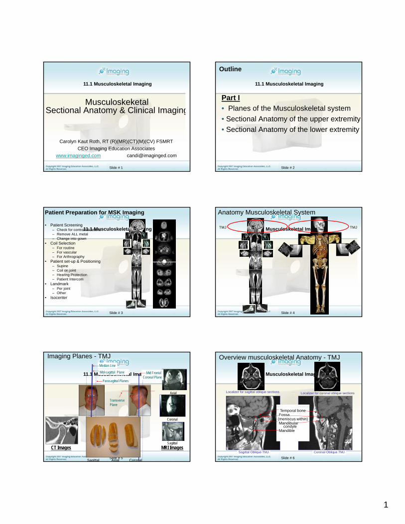
Basic “Vanilla” TMJ Protocol
• Localizer T1 – plane best3 plane loc
• Sagittal Oblique T2 & PD – CLOSED Generally TSE (aka FSE or RARE)
12 ish to fit anatomy3 or 4 mm / 1192 x 256TR = 4000 msTE = 30 ms 100 ms ETL= 8
• Coronal oblique T1closedGenerally TSE (aka FSE or RARE)
CO IL
Sensitivity profile of the RF coil
radius
diameter
Slide # 9
y ( )12 ish to fit anatomy3 or 4 mm / 1192 x 256TR =500-700msTE = min
• Sagittal oblique T1closed/openGenerally TSE (aka FSE or RARE)12 ish to fit anatomy3 or 4 mm / 1192 x 256TR =500-700msTE = min
11.1 Musculoskeletal Imaging
Sag T2
Left
Sagittal Oblique T2Generally TSE (aka FSE)12 ish to fit anatomy3 or 4 mm / 1192 x 256TR 4000
Slide # 10
Right
TR = 4000 msTE = 100 ms ETL= 8
11.1 Musculoskeletal Imaging
Sag T1
Left
Sagittal oblique T1…closedGenerally TSE (aka FSE)12 ish to fit anatomy3 or 4 mm / 1192 x 256
Slide # 11
Right
192 x 256TR =500-700msTE = min
11.1 Musculoskeletal Imaging
Sag T1 “open”
Left
Sagittal oblique T1…openGenerally TSE (aka FSE)12 ish to fit anatomy3 or 4 mm / 1192 x 256
Slide # 12
Right
192 x 256TR =500-700msTE = min
3
11.1 Musculoskeletal Imaging
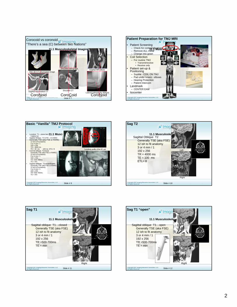
Open vs. Closed
Closed
Sagittal oblique T1closed/openGenerally TSE (aka FSE)12 ish to fit anatomy3 or 4 mm / 1192 x 256
Slide # 13
Open
192 x 256TR =500-700msTE = min
To evaluate meniscus
11.1 Musculoskeletal Imaging
Anatomy Musculoskeletal System
Shoulder
Shoulder
Slide # 14
11.1 Musculoskeletal Imaging
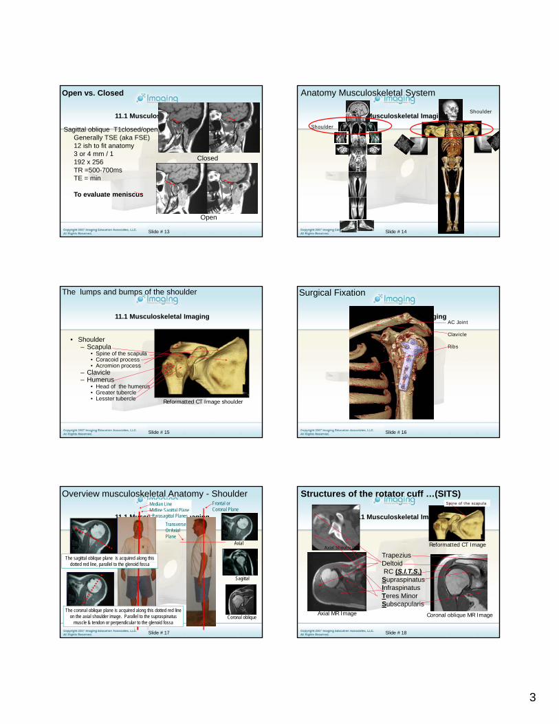
• Shoulder– Scapula
• Spine of the scapula• Coracoid process
The lumps and bumps of the shoulder
Slide # 15
p• Acromion process
– Clavicle– Humerus
• Head of the humerus• Greater tubercle• Lesster tubercle Reformatted CT Image shoulder
11.1 Musculoskeletal Imaging
Surgical Fixation
AC Joint
Clavicle
Ribs
Slide # 16
11.1 Musculoskeletal Imaging
Median LineMidline Sagittal Plane
Parasagittal Planes
Frontal orCoronal Plane
TransverseOr AxialPlane
Axial
The sagittal oblique plane is acquired along this f
Overview musculoskeletal Anatomy - Shoulder
Slide # 17
Sagittal
Coronal obliqueThe coronal oblique plane is acquired along this dotted red line
on the axial shoulder image. Parallel to the supraspinatus muscle & tendon or perpendicular to the glenoid fossa
dotted red line, parallel to the glenoid fossa
11.1 Musculoskeletal Imaging
Reformatted CT Image
TrapeziusDeltoid
Structures of the rotator cuff …(SITS)
Axial Shoulder CT
Spine of the scapula
Slide # 18
Axial MR Image Coronal oblique MR Image
DeltoidRC (S.I.T.S.)Supraspinatus Infraspinatus Teres Minor Subscapularis
4
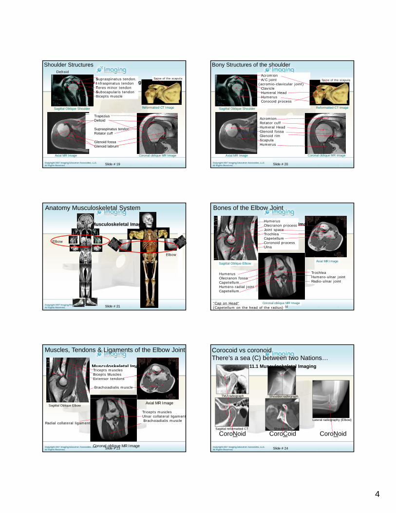
11.1 Musculoskeletal ImagingSupraspinatus tendon.Infraspinatus tendonTeres minor tendonSubscapularis tendonBicepts muscle
Reformatted CT Image
Shoulder Structures
Sagittal Oblique Shoulder
Deltoid
Spine of the scapula
Slide # 19
Axial MR Image
TrapeziusDeltoid
Supraspinatus tendonRotator cuff
Glenoid fossa Glenoid labrum
Coronal oblique MR Image
11.1 Musculoskeletal ImagingAcromionA/C joint
(acromio-clavicular joint)ClavicleHumeral HeadHumerusCorocoid process
Reformatted CT Image
Bony Structures of the shoulder
Sagittal Oblique Shoulder
Spine of the scapula
Slide # 20
Axial MR Image Coronal oblique MR Image
AcromionRotator cuffHumeral HeadGlenoid fossaGlenoid rimScapulaHumerus
11.1 Musculoskeletal Imaging
Anatomy Musculoskeletal System
Elbow
Elbow
Slide # 21
11.1 Musculoskeletal ImagingHumerusOlecranon processJoint spaceTrochleaCapetellumCoronoid processUlna
Bones of the Elbow Joint
Slide # 22
Axial MR Image
HumerusOlecranon fossaCapetellumHumero-radial jointCapetellum
TrochleaHumero-ulnar jointRadio-ulnar joint
“Cap on Head”(Capetellum on the head of the radius)
Coronal oblique MR Image
Sagittal Oblique Elbow
11.1 Musculoskeletal ImagingTricepts musclesBicepts MusclesExtensor tendons
Brachoiadialis muscle
Muscles, Tendons & Ligaments of the Elbow Joint
Slide # 23
Axial MR ImageSagittal Oblique Elbow
Coronal oblique MR Image
Tricepts musclesUlnar collateral ligamentBrachoiadialis muscle
Radial collateral ligament
11.1 Musculoskeletal Imaging
Corocoid vs coronoidThere’s a sea (C) between two Nations…
Shoulder radiographTMJ radiograph
Slide # 24
CoroNoid CoroCoid CoroNoidSagittal reformatted CT
Lateral radiography (Elbow)
Shoulder CT
5
11.1 Musculoskeletal Imaging
Anatomy Musculoskeletal System
WristWrist
Slide # 25
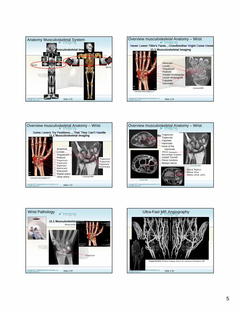
11.1 Musculoskeletal Imaging
NavicularLunateTriquetruim
Never Lower Tillie’s Pants…Grandmother Might Come Home
Overview musculoskeletal Anatomy – Wrist
Slide # 26
TriquetruimPisiformGreater MutlangularLesser MultangularCapatateHammate
Coronal reformatted CT
Coronal MRI
11.1 Musculoskeletal Imaging
Overview musculoskeletal Anatomy – Wrist
ScaphoidLunateTriquetruim
Some Lovers Try Positions….That They Can’t Handle
Slide # 27
Coronal reformatted CTCoronal MRI
TrapeziumTrapezoidCapetateHammate
TriquetruimPisiformTrapeziumTrapezoidCapetateHammateAneurysm Raidal arteruUlnar artery
11.1 Musculoskeletal ImagingTrapeziumTrapezoidCapetateHammateHook of the
HammateTFCC (triangulo-fibrocartilege complex)
Overview musculoskeletal Anatomy – Wrist
Slide # 28
fibrocartilege complex)Carpal TunnellFlexor tendonsMedian Nerve
Axial MRI
Coronal MRI
Distal RadiusDistal UlnaRadio-ulnar joint
11.1 Musculoskeletal Imaging
Wrist Pathology
Aneurysm
Slide # 29
Fracture
11.1 Musculoskeletal Imaging
Ultra-Fast MR Angiography
Slide # 30
Images courtesy of Edison Imaging, Edison NJ, Lawrence Tanenbaum, MD
6
11.1 Musculoskeletal Imaging
Anatomy Musculoskeletal System
Slide # 31
HipHip
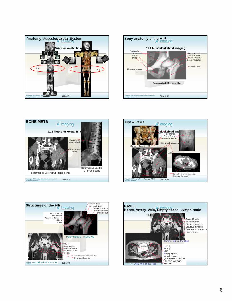
11.1 Musculoskeletal ImagingAcetabulim
IliumIshiumPubis
Bony anatomy of the HIP
Femoral HeadFemoral Neck
Greater TrocanterLesser trocanter
Femoral Shaft
Slide # 32
Obturator foramen
Reformatted CT Image Hip
Femoral Shaft
11.1 Musculoskeletal Imaging
BONE METS
Compressed vertebral body
Met to the pelvic
Slide # 33
Reformatted Coronal CT Image pelvis
Reformatted Sagittal CT Image Spine
bone
11.1 Musculoskeletal ImagingIlumIliac arteries
Psoas MusclesObturator foramen
Gleuteal MusclesCoronal reformatted CT
Hips & Pelvis
Slide # 34
Obturator Internus musclesObturator Externus
Coronal CT
Mass
Axial pelvis CT
11.1 Musculoskeletal Imaging
Femoral HeadFemoral Neck
Greater TrocanterLesser trocanter
Femoral Shaft
Structures of the HIP
(ASIS) Ilium Acetabulim
Obturator foramen Ishium
Pubis
Slide # 35
Reformatted CT Image Hip
Coronal MRI of the Hips
Ilium Acetabulim Glenoid LabrumFemoral Neck Femur
Obturator Internus musclesObturator Externus
11.1 Musculoskeletal Imaging
NAVELNerve, Artery, Vein, Empty space, Lymph node
Psoas MuscleIliacus MuscleGleuteus MaximusGleuteus minimusQuadracepts MuscleHamstrings
Slide # 36
Coronal MRI of the Hips
Axial MRI of the Hips
NerveArteryVeinEmpty spaceLymph nodesQuadracepts Muscle Gleuteus MaximusRectum
bladder
7
11.1 Musculoskeletal Imaging
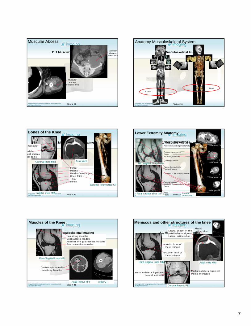
Muscular Abcess
Muscular abscess
Pelvic area
Slide # 37
Muscular abscess
Shoulder area
11.1 Musculoskeletal Imaging
Anatomy Musculoskeletal System
Slide # 38
KneeKnee
11.1 Musculoskeletal Imaging
Bones of the Knee
Coronal knee MRI Axial knee
i Condyle
ndylebeal plateaubial spine
Slide # 39
FemurPatellaPatello-femoral jointKnee JointTibiaFibula
Sagittal knee MRI
Coronal reformatted CT
11.1 Musculoskeletal ImagingAnterior cruciate ligament (ACL)
Posterior cruciate ligament (PCL)
Quadracepts musclesFemurHamstrings muscles
Quadcepts tendonMid sagittal slice (MRI)
Lower Extremity Anatomy
Axial pelvis CT
Axial femur CT
Slide # 40Coronal reformatted CT
Para sagittal slice (MRI)
Patello- Femoral JointPatellar ligament
Locations of the lateral collateral ligaments
Femoral coldylesMeniscus ([posterior horn, lateral meniscus)Patella
TibiaFibula
Mid sagittal slice (MRI) Axial femur CT
Axial femur CT
Axial knee CT
11.1 Musculoskeletal Imaging
Muscles of the Knee
Hamstring musclesQuadracepts TendonAttaches the quatracepts musclesGastrocnemius muscles
Slide # 41
Para Sagittal knee MRI
Quatracepts musclesHamstring Muscles
Axial Femur MRI Axial CT
11.1 Musculoskeletal Imaging
Meniscus and other structures of the knee
Anterior horn of the meniscus
Posterior horn of
Lateral aspect of the patello-femoral jointLateral retinaculum
Medial retinaculum
Slide # 42
Para Sagittal knee MRI
Coronal knee MRI
Posterior horn of the meniscus
Axial knee MRI
Medial collateral ligament Medial meniscusLateral collateral ligament
Lateral meniscus
8
11.1 Musculoskeletal Imaging
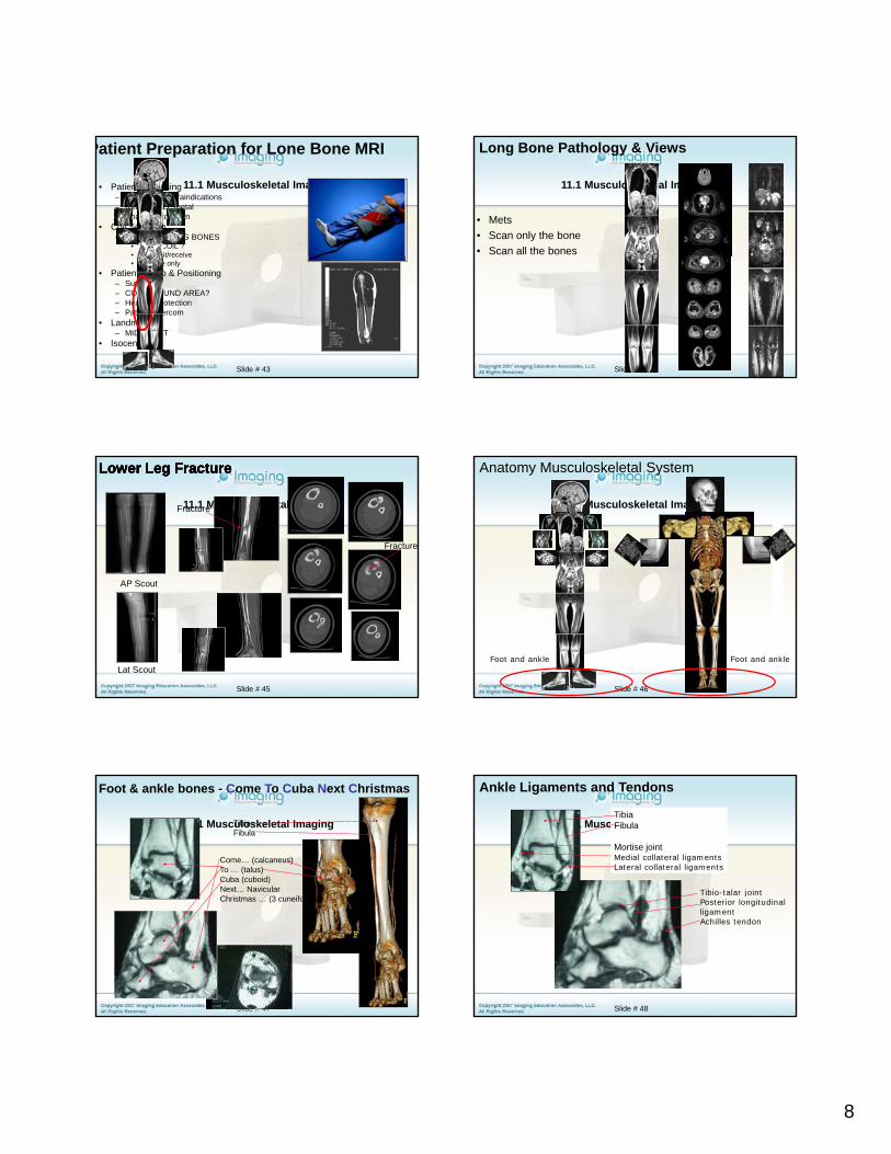
Patient Preparation for Lone Bone MRI
• Patient Screening– Check for contraindications– Remove ALL metal– Change into gown
• Coil Selection– For routine LONG BONES
• BODY COIL ?• Transmit/receive
Slide # 43
Transmit/receive• Receive only
• Patient set-up & Positioning– Supine– COIL AROUND AREA?– Hearing Protection– Patient Intercom
• Landmark– MID SHAFT
• Isocenter
11.1 Musculoskeletal Imaging
• Mets• Scan only the bone• Scan all the bones
Long Bone Pathology & Views
Slide # 44
11.1 Musculoskeletal Imaging
Lower Leg FractureLower Leg FractureLower Leg FractureLower Leg Fracture
Fracture
Fracture
Slide # 45
AP Scout
Lat Scout
11.1 Musculoskeletal Imaging
Anatomy Musculoskeletal System
Slide # 46
Foot and ankle Foot and ankle
11.1 Musculoskeletal Imaging
Come… (calcaneus)To … (talus)Cuba (cuboid)Next… NavicularCh i t (3 if !)
TibiaFibula
Foot & ankle bones - Come To Cuba Next Christmas
Slide # 47
Christmas … (3 cuneiforms!)
11.1 Musculoskeletal ImagingTibiaFibula
Mortise jointMedial collateral ligamentsLateral collateral ligaments
Ankle Ligaments and Tendons
Tibio-talar joint
Slide # 48
jPosterior longitudinal ligamentAchilles tendon
9
11.1 Musculoskeletal Imaging
Ankle Arthrogram
scar or hypertrophied synovium
Slide # 49
11.1 Musculoskeletal Imaging
Review, Peripheral Vascular Anatomy
Abdominal Aorta(AAA- abdominal aortic aneurysm)
Iliac Arteries
Femoral Arteries
Slide # 50Coronal reformatted CTACoronal MRA
Popliteal Arteries
Trifurcation…(anterior tibial)
(posterior Tibial)(Pernoeus Brevis)
Coronal reformatted CT
11.1 Musculoskeletal Imaging
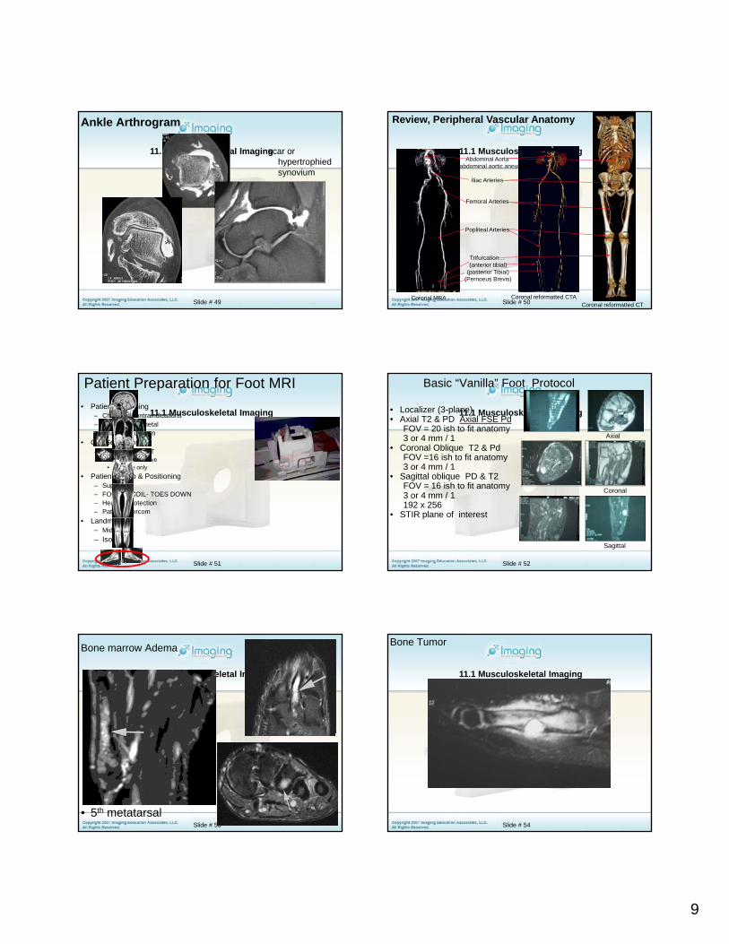
Patient Preparation for Foot MRI• Patient Screening
– Check for contraindications– Remove ALL metal– Change into gown
• Coil Selection– For routine FOOT
• Transmit/receive• Receive only
Slide # 51
• Patient set-up & Positioning– Supine– FOOT IN COIL- TOES DOWN– Hearing Protection– Patient Intercom
• Landmark– Mid tarsals– Isocenter
11.1 Musculoskeletal Imaging• Localizer (3-plane)• Axial T2 & PD Axial FSE Pd
FOV = 20 ish to fit anatomy3 or 4 mm / 1
• Coronal Oblique T2 & PdFOV =16 ish to fit anatomy3 or 4 mm / 1
Basic “Vanilla” Foot Protocol
Axial
Slide # 52
• Sagittal oblique PD & T2FOV = 16 ish to fit anatomy 3 or 4 mm / 1192 x 256
• STIR plane of interest
Coronal
Sagittal
11.1 Musculoskeletal Imaging
Bone marrow Adema
Slide # 53
• 5th metatarsal
11.1 Musculoskeletal Imaging
Bone Tumor
Slide # 54
10
11.1 Musculoskeletal Imaging
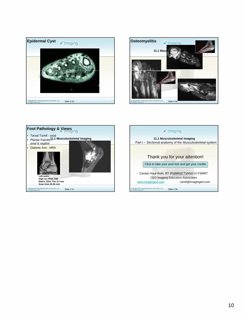
Epidermal Cyst
Slide # 55
11.1 Musculoskeletal Imaging
Osteomyelitis
Slide # 56
11.1 Musculoskeletal Imaging
Foot Pathology & Views• Tarsal Tunell - axial• Plantar Fascitis –
axial & sagittal• Diabetic foot - MRA
Slide # 57
Left ankleHigh res PDW TSEMatrix 1024, Thk 3.0 mmScan time 02:56 min
11.1 Musculoskeletal ImagingPart I – Sectional anatomy of the Musculoskeletal system
Thank you for your attention!
Slide # 58
Carolyn Kaut Roth, RT (R)(MR)(CT)(M)(CV) FSMRTCEO Imaging Education Associates
www.imaginged.com [email protected]
y yClick to take your post test and get your credits
Recommended